the formation and mineralization of mollusk shell
TRANSCRIPT

[Frontiers in Bioscience S4, 1099-1125 , January 1, 2012]
1099
The formation and mineralization of mollusk shell Frederic Marin1, Nathalie Le Roy1, Benjamin Marie1 1UMR CNRS 5561, Biogeosciences, Universite de Bourgogne, 6 Boulevard Gabriel, 21000 Dijon, France TABLE OF CONTENTS 1. Abstract 2. Mollusk shell
2.1. Introduction 2.2. Diversity of mollusk classes
3. Structural, microstructural and ultrastructural considerations on the shell 3.1. Structure of the shell and mineralogy 3.2. Different shell microstructures 3.3. Nacre: organization and ultrastructure
3.3.1. Why nacre is interesting to study 3.3.2. Brief definition of nacre 3.3.3. Complex organo-mineral interactions 3.3.4. Mechanism of nacre formation 4. Early stages of shell calcification: developmental aspects
4.1. Formation of the shell in relation with larval development 4.2. Genes and their products involved in the larval shell construction
5. Shell formation: physiological and cellular considerations 5.1. Calcification of the shell in normal conditions
5.1.1. Mantle and cells of the outer epithelium 5.1.2. Periostracum 5.1.3. Interface between the mantle and the shell 5.1.4. Transport of the precursor ions of mineralization 5.1.5. Synthetic cellular view of the shell formation process
5.2. Shell remodeling and shell repair process 5.3. A critical view of the standard model
6. Organic constituents of the shell 6.1. Biochemistry of the mollusk shell 6.2. Non protein organic shell components
6.2.1. Polysaccharides 6.2.2. Lipids, pigments, and other small organic molecules
6.3. Shell proteins 6.3.1. Biochemistry of shell proteins 6.3.2. Shell proteins: the “protein per protein” approach 6.3.3. Shell proteins: large screening of the “shellome” 7. Perspectives and future directions 8. Acknowledgements 9. References 1. ABSTRACT
In the last years, the field of mollusk biomineralization has known a tremendous mutation. The most recent advances deal with the nanostructure of shell biominerals, and with the identification of several shell matrix proteins: on one hand, the complex hierarchical organization of shell biominerals has been deciphered in few models, like nacre. On the other hand, although proteins represent a minor shell component, they are the major macromolecules that control biocrystal synthesis. Until recently, the paradigm was to consider that this control occurs by two antagonist mechanisms: crystal nucleation and growth inhibition. Emerging models try to translate a more complex reality, illustrated by the huge variety of shell proteins, characterized so far. The primary
structure of many of them is composed of different functional domains, some of which exhibit enzymatic activity, while others may be involved in cell signalling. Many of them have unknown functions. Today, the shell matrix appears as a whole system, which regulates protein-mineral, protein-protein, and epithelium-mineral interactions. These aspects should be taken in account for the future models of shell formation. 2. MOLLUSK SHELL
2.1. Introduction
Because mollusks are soft-bodied animals, many of them have invented a complex strategy for maintaining

Biomineralization of mollusk shell
1100
Figure 1. Phylogeny of the phylum Mollusca, according to Lecointre and Le Guyader (4). Only extant classes are indicated. For clarity, fossil groups, such as the Rostroconchia class, are not represented. For each class, the type of tegumentary mineralization is indicated as well as the polymorph used. We do not indicate the numerous types of minerals formed by mollusks by organs (like the radula) other than mantle tissues. * indicates particular cases where carbonated apatites (francolite, dahlite) are also present in the periostracal layer : such examples include the rock-burrowing ‘date mussel’ (Lithophaga), which uses these minerals for increasing the resistance of its shell against abrasion.
their soft tissues, for protecting themselves against predation and for precluding desiccation. This strategy relies on the elaboration of an external calcified rigid structure, the shell. The shell secreted by mollusks is the subject of the present review paper. In the living world, the shell constitutes without doubt one of the most studied biomineralizations, and by many aspects, one of the most fascinating.
The shell and the process by which it is secreted
enter the category of biologically-controlled mineralizations, by contrast with biologically-induced mineralizations, these latter being predominantly found in the bacterial world. The concept of biologically-controlled mineralization was popularized – not to say invented - by Stephen Mann (1). Schematically, it can be summarized as follows: a. The shell fabrication requires a specialized cellular machinery, both intracellular and extracellular, which, in other words, means that the shell formation is strictly under the control of cascades of genes; b. The formed minerals are far from equilibrium with the environment; this means that some minerals, that are thermodynamically unstable in natural conditions, can however be synthesized; c. The produced minerals differ in their shape and size from their inorganically formed counterpart, and their shapes are generally complex; in addition, contrarily to chemically-synthesized minerals, they assemble according to different levels of hierarchy; d. They are formed in a delimited space, not in direct contact
with the environment; e. The entire process of shell construction is modulated by an extracellular organic matrix, a part of which is occluded in the shell during calcification. These different aspects of shell formation are tackled in this paper. 2.2. Diversity of mollusk classes
The shell is often considered as a biomineralization typical of mollusks: its morphological characters and ornamentation are used indeed for determinations, at different taxonomic levels, from classes to species (2). However, this assertion is not fully exact: representatives of other phyla produce a similar external calcareous protection. One finds for example the brachiopods (also known as lamp shells), but also some annelids, which produce an external calcified tube (tube-forming polychaetes) (3). At last, within the arthropods, different groups of the Crustacea subphylum produce an external shell made of calcium carbonate: among them, cirripedians (barnacles), or ostracodes, a numerically important group of millimetric animals, which protect their body in a calcified bivalved shell. Furthermore, not all mollusks produce a shell. Figure 1 presents a consensual – and from now on ‘classical’ – phylogeny of the whole phylum. Living mollusks represent about 118 000 species (4), grouped in eight classes of unequal importance. In basal positions, one finds two classes of primitive worm-like mollusks, which do not secrete a shell, but tiny spicules or sclerites on the surface of their teguments; these are

Biomineralization of mollusk shell
1101
Figure 2. Shell structure of the freshwater mussel Unio pictorum (“painter’s mussel”; Palaeoheterodonta, Unionoida).
solenogastres and caudofoveates, which were earlier grouped in the polyphyletic ‘Aplacophora’ class. Note that the ‘basal’ status of these two classes is controversial: because they do not have any fossil record, some authors admit that they may represent extremely derived mollusks, which would have lost the ability to secrete a shell. Then, the next node of the tree individuates the group of polyplacophorans, better known as ‘chitons’. Chitons are grazing marine mollusks that do not produce a shell sensu stricto, but series of calcified plates that can slide with each other, when the animal cowers. The following node groups the true shell-forming mollusks, also known as the conchiferans. This huge subphylum comprises fives classes, which, are respectively the monoplacophorans, the bivalves, the scaphopods, the gastropods and the cephalopods. The monoplacophorans – which are generally considered as the oldest class of mollusks that appeared in the basal Cambrian times – correspond to a relict group: they comprises nowadays only 15 living deep-sea species, among which the well-known living-fossil Neopilina galathea, characterized by its thin univalve shell. The bivalves are among the most known mollusks, since many of them, like the mussel, the oyster, the scallop, the clam, the razor shell, the tellin or the cockle, are edible and commercially exploited forms. In addition, few genera, like Pinctada or Hyriopsis, are intensively harnessed for their ability to produce pearls. All bivalves are characterized by a shell with two valves – in most of the cases symmetrical - connected by a hinge. The morphology of the hinge, which comprises a leathery ligament and series of calcified teeth, is an important character for distinguishing the different bivalve orders. Living bivalves represent about 12000 species and they have colonized most of the aquatic environments, from deep marine (deep-sea mussels) to freshwater (unionid mussel) biotopes. This class appeared in the Lower Cambrian. The sister-group of bivalves is represented by the scaphopods. Also called tusk-shells, these univalve marine mollusks are characterized by a tooth-like shell, pierced at both ends. Although common on strands, scaphopods represent a small class, with only 400 living species. Scaphopods appeared presumably during the Ordovician, but are truly recognized as a class in the
Carboniferous (5). The two last classes are usually considered as the most evolved mollusks, because they possess a differentiated head and sensorial organs for vision. Gastropods, with more than 100000 living species, represent the biggest class of mollusks, and the most diverse one, comprising forms as dissimilar as keyhole limpets, abalones, cones, snails, slugs or queen conchs. In the course of evolution, they were able to colonize almost all environments, from deep-sea to terrestrial environments, even the most hostile ones, such as hot deserts, lightless caves, or alpine cold biotopes. Gastropods are characterized by univalve and coiled shells, but several derived forms - like the terrestrial slug or the sea hare – possess only a moderately-developed internal shell. Many of them are grazers or active predators (cones). Gastropods are considered as the sister-group of cephalopods, a class, which comprises about 900 living species, most of them living as active predators. During geological times, cephalopods knew different phases of radiations (followed by massive extinctions) in particular with the development of nautiloids in the Palaeozoic (Ordovician), and ammonoids in the Mesozoic (Jurassic-Cretaceous) (6). Similarly to gastropods, cephalopods were initially univalved mollusks. However, the macro-evolutionary trend of the class went to a reduction and internalization of the shell, observed in squids, ram’s horn squids or cuttlefishes, or to its complete disappearance, like in octopuses. Today, only two small phylogenetically-unrelated groups, the nautilids and the argonautids (‘paper nautilus’) possess an external calcified shell. While the shell of the nautilus can be considered as a true perennial shell, that of argonautid is a temporary brood chamber (eggcase), secreted by the dorsal tentacles of the females, before egg laying, and abandoned later. 3. STRUCTURAL, MICROSTRUCTURAL AND ULTRASTRUCTURAL CONSIDERATIONS ON THE SHELL 3.1. Structure of the shell and mineralogy
Mollusk shells, whatever their taxonomic origin, are always made of the superimposition of few calcified layers, generally two to five, and one organic layer. Figure 2 presents a section made in the shell of the freshwater mussel Unio pictorum, a common bivalve found in the rivers of European countries. From top to bottom (from outside to inside the shell), one finds a thin organic leathery layer called the periostracum, the role of which will be explained in section 5.1.2. In the present case, the periostracum, which remains non-eroded during all the life of the animal, gives the shell its external glazed olive-greenish colour. It has to be noted that for several species, the shells color does not come from the colour of the periostracum, but is due to pigments, which are disseminated within the mineralized layers, according to genetically-controlled patterns (see in particular the cone gastropods (7)). Subjacent to the periostracum is a mineralized layer, composed of elongated crystals developed perpendicularly to the shell surface. These crystals define the prismatic layer. For Unio pictorum, the prisms are made of aragonite, one of the six polymorphs of calcium carbonate that crystallizes in the orthorhombic

Biomineralization of mollusk shell
1102
system. Other conchiferan mollusks also exhibit an outer prismatic layer, but made of calcite, the second most employed polymorph of calcium carbonate, that crystallizes in the rhomboedric system. In the present example, beneath the prismatic layer, the main layer, that represents about 50% of the shell thickness, is composed of minute crystals that cannot be individually distinguished at low magnification. This layer is the nacreous layer, also called mother-of-pearl. It is the internal lustrous layer, observed in several mollusks, such as the mussel, the pearl oyster, the abalone or the nautilus. This layer is always aragonitic. Because of its intrinsically high mechanical properties, the nacreous layer is among the most studied mollusk shell microstructures. In section 3.3, we describe this layer more precisely.
The example of Unio pictorum illustrates the fact
that the association of two or more mineralized layers with very different stiffness, within a shell, results in a biomaterial with interesting mechanical properties. Actually, it seems that the strategy of superimposing calcified layers of different microstructures was invented soon after the invention of the shell, since well-preserved conchiferan mollusks of the Lower Cambrian of China already exhibited layered shells (8). Per se, the prismatic layer has a moderate resistance to fracture; however, it displays certain flexibility, which is even improved because of its association with the outer organic periostracal layer. On the other hand, the nacreous layer exhibits an extremely high resistance to fracture (9), but is more rigid, and has a tendency to crack when bending. One can assume that the association of both layers generates a biomaterial, which combines overall toughness and flexibility. Furthermore, the interest of depositing two layers of different textures together, which, in other words, allows introducing an interface in the biomaterial, means that a fracture cannot propagate directly throughout the whole thickness of the shell.
Another remark about the shell mineralogy is
that mollusks use mainly two polymorphs of calcium carbonate, calcite, the stable form, and aragonite, the metastable one, which tends to transform into calcite, under the influence of diagenetic processes. In the following sections (3.3 and 4), we see that mollusks also use transiently amorphous calcium carbonate. In exceptional circumstances (shell deformation), few of them use vaterite (10), an extremely unstable and rare polymorph that crystallizes in the hexagonal system. To our knowledge, none of the other polymorphs, protodolomite, monohydrocalcite, or ikaite (an hexa-hydrated calcium carbonate) are used by mollusks to fabricate their shell.
3.2. Different shell microstructures
Figure 2 shows the classical association of nacreous and prismatic textures. This example represents only one particular case, and it has to be clear in the reader’s mind that mollusks, and especially bivalves, use a wide variety of crystal habits to elaborate their shell layers. These different shell habits are collectively grouped under the ‘shell microstructure’ terminology. They have been the subjects of different monographs and treaties, among which
the most complete are those of Boggild (11), Kobayashi (12), Oberling (13), Taylor and co-workers (14, 15), Carter (16), Carter and Clark (17), Shimamoto (18), Carter (19), and Popov (20). For bivalves, palaeontologists currently utilize shell microstructures as a supplementary discriminating character for the classification of fossil forms (2).
Figure 3 gives a set of examples of
microstructures found in diverse mollusk shells, while Figure 4 gives a brief – although complete - overview of the typology used by Carter and Clark II (17), for attempting to describe accurately all the crystal morphologies and patterns, encountered when observing shell sections under scanning electron microscope. Without entering the details of the different microstructures, we give a brief description of the main types.
The prismatic microstructure terminology
describes elongated crystalline objects, rectilinear or curved, the opposite long sides of which are parallel. They are grouped together and their mutual boundaries do not strongly interdigitate (17). Prisms, which can be made of calcite or aragonite, includes very different objects, fine, medium or coarse, such as the big-sized regular simple calcitic type, oriented perpendicularly to the outer shell surface, encountered in the outer layer of the fan mussel, Pinna nobilis. They include also the tiny oblique and straight prisms of the edible mussel, Mytilus edulis, or the composite curved prisms of the Manila clam Venerupis philipinarum, that grow almost parallel to the shell surface an diverge in a fan-like manner towards the edge of the shell (20, 21). Such heterogeneity implies that all prisms are not produced in a single manner and that there must be very different mechanisms of crystal growth. Several parameters, such as the initiation of prisms at the internal surface of the periostracum, geometrical constraints, competition for space, position of the calcifying epithelium facing the mineralization front, are obviously crucial for explaining how prisms emerge (22).
The ‘spherulitic microstructure’ terminology
refers to spherical objects, made of tiny crystals, which radiate from a center. In several cases, spherulites represent the starting point of the growth of prisms (23), particularly in the case where the crystal growth is constrained in one direction. In the green ormer Haliotis tuberculata, we observed recently that spherulites were often produced in ‘emergency situations’, for filling a hole, in the case of shell repair (24).
The laminar microstructure designation is
applied to flat units, oriented parallel or nearly parallel to the general depositional surface (17). They comprise a broad range of rods, laths, blades and tablets, among which the most known are the nacreous and foliated microstructures. Nacreous microstructures, which are the subject of the next section, refer to flat tiny aragonitic crystals, densely packed together, and which form the inner iridescent layer of several mollusks (25). Foliated microstructures are thin calcitic laths, arranged in superimposed sheets (26). They are extremely developed in

Biomineralization of mollusk shell
1103
Figure 3. Few molluscan shell examples and their associated microstructures. A. Nucula sulcata (Bivalvia, Protobranchia, Nuculoida). B. Nacreous layer of Nucula sulcata. C. Mytilus edulis, the edible mussel (Bivalvia, Pteriomorphia, Mytiloida). D. Nacro-prismatic transition in Mytilus edulis: oblique prisms are on the top, nacre tablets, on bottom. E. Neotrigonia sp. (Bivalvia, Palaeoheterodonta, Trigonioida). F. The nacro-prismatic transition in Neotrigonia: prisms are on the top, nacre, on bottom. G. Nacre tablets in Neotrigonia. H. Juvenile Pinna nobilis, the noble fan mussel (Bivalvia, Pteriomorphia, Pterioida). I. Border of the prismatic layer. J. Growing nacre tablets. K. Haliotis tuberculata, the green ormer (Gastropoda, Vetigastropoda, Haliotidae). L. Columnar nacre of Haliotis tuberculata (polished section, etched with EDTA). M. Strombus gigas, the queen conch (Gastropoda, Caenogastropoda). N. Crossed-lamellar shell microstructure of Strombus gigas. O. Helix pomatia, the edible snail (Gastropoda, Stylommatophora). P. Crossed-lamellar shell microstructure of Helix pomatia. Q. Nautilus macromphalus, the bellybutton nautilus (Cephalopoda, Nautilida). R. Nacre tablets in Nautilus macromphalus shell.

Biomineralization of mollusk shell
1104
Figure 4. Classification of the shell microstructures according to Carter and Clark (17). On the right column, few bivalve and gastropod examples illustrate how diverse shell microstructures are.
edible oysters and scallops, for example. Although their mechanical properties are inferior to that of nacre, they represent presumably an efficient strategy developed by bivalves to rapidly mineralize and increase the thickness of their shell.
From a geometrical viewpoint, crossed structures
are the most complex ones and can be described as microstructures with ‘two or more non-horizontal dip directions of their elongate structural units relative to the depositional surface’ (17). They represent a diversified group comprising the crossed-lamellar, complex crossed-lamellar, crossed acicular microstructures, found in most of the heterodont bivalves and in several gastropods. Crossed-lamellar microstructures, the most common ones, consist of
a plywood-like arrangement of aragonite needles, according to different hierarchical levels (18). Although their resistance to fracture is lesser than that of nacre, crossed structures represent a remarkable strategy that mollusks have set up to combine a ‘cheap’ cost of calcification (due to the secretion of low amounts of shell organic matrix) and interesting mechanical properties, among which an aptitude to stop cracks (27; 28). The way crossed-lamellar microstructures emerge from the activity of the shell-forming organ, the mantle, is still a mystery, and would deserve extensive studies.
Homogeneous microstructures refer to
microstructures, which do not present an apparent organization of their crystallites, when observed with

Biomineralization of mollusk shell
1105
Figure 5. Different nacre microstructures found among mollusks. A, B. Cross-section of the sheet nacre of Pleiodon spekii, a freshwater bivalve (Palaeoheterodonta, Unionoida) of the African Great Lakes. C, D. Columnar nacre of the green ormer gastropod Haliotis tuberculata (Vetigastropoda, Haliotidae). C. Juvenile growing tablets, observed from above at the mineralization front. D. Cross-section. E, F. Row-stack nacre of the noble fan mussel Pinna nobilis (Periomorphia, Pterioida). E. Cross section. F. Nacre tablets observed from above.
optical or with electronic microscope. They can be fine grained, when the crystals units are minute (<5 µm) or coarse grained, for crystallites higher than 5 µm. In this case, they are named granular (17). Homogeneous microstructures are extremely frequent in heterodont bivalves (18). Interestingly, in a recent paper (21) where we studied the shell repair process of the edible Manila clam Venerupis (Ruditapes) philipinarum after a bacterial infestation, we observed that homogeneous microstructures in the repair zone can gradually self-organize into crossed acicular microstructures, which belong to the previous ‘crossed structures’ type.
At last, helical microstructures, isolated spicules
or spikes, and isolated crystal morphotypes are rare microstructures, the two last being found as sparsely distributed crystals (17). Isolated crystal morphotypes are found in shell repair zones, while isolated spicules are often associated to the periostracal layer.
In the following section, we detail mother-of-
pearl, or nacre, the most studied mollusk shell microstructure.
3.3. Nacre: organization and ultrastructure 3.3.1. Why nacre is interesting to study
Of all shell microstructures described here above, nacre, also known as mother-of-pearl, is among the most fascinating one. By far, this microstructure is the most solid one produced by mollusks and classical mechanical studies (29, 30) showed that its resistance to fracture was more than thousand times higher than that of its chemically precipitated counterpart, geological aragonite. Thus, nacre appears as an interesting natural composite, that serves as a model for the development of synthetic biomimetic materials (31, 32). In addition to these mechanical properties, nacre is characterized by a unique combination of optical properties that make it extremely attractive in jewelry. This attractiveness is the main reason of the circum-Pacific development of pearl culture, especially in Japan, China, Indonesia, Philippines, Cook Islands, Australia, Polynesia and Mexico (33). To give an example, pearl industry in French Polynesia represents about 5000 employments (on a total of 270 000 inhabitants), and an export value oscillating between 100 -120 million US dollars per year. This represents more than one half of the total income of the archipelago (34). Another aspect that renders nacre attractive is its potential use as a material for regenerating bone tissues, or as a source of bioactive organic molecules (35, 36): different studies aiming at measuring the effect of nacre particles or nacre organic extracts in vivo on vertebrates or in vitro on cell lines, strongly suggested that nacre possesses osteoinductive and osteogenic properties (37-39). The exact reason of such remarkable properties remains unknown, but may be related to the presence of diffusive bioactive factors in the nacre matrix.
3.3.2. Brief definition of nacre
As said before, nacre constitutes the inner lustrous shell layer of several mollusks. Contrarily to other microstructures such as prisms, which can be observed in several calcifying systems, nacre is almost exclusive to that phylum. It is widespread and can be found in the shells of bivalves, gastropods and cephalopods. In addition, nacre is found in monoplacophorans, although it seems that its distribution is restricted to one species (40) in that particular mollusk class. The origin of nacre has to be searched in the Lower Cambrian times (8, 41). Many researchers consider nacre as the reference microstructure to understand mollusk shell biomineralization, because of its apparent geometrical simplicity.
The “nacre” terminology refers to a well-defined
type of laminar microstructure, which ‘consists of polygonal to rounded tablets arranged in broad, regularly formed, parallel sheets’ (17). These tablets, which optically behave like monocrystals, but which are, in reality constituted of nano-elements, are always made of aragonite, and their thickness varies between half a micron and one micron, for a lateral extension of few microns. They are tightly packed together by a thin organic cement. They form superimposed layers of uniform thickness, the whole architecture being densely packed, without interstice. From a microstructural viewpoint, as shown in Figure 5, one finds few broad types of nacre, depending on the

Biomineralization of mollusk shell
1106
manner tablets are arranged (42, 43): the “brick wall” nacre, also called sheet nacre, is the most frequent, and almost exclusively observed among bivalves. In cross-section, crystals are positioned in staggered rows, just like bricks in a wall (Figure 5, A, B). Bivalve nacre tablets have their a, b and c axes co-oriented, with the c axis perpendicular to the nacre surface, and the b axis, parallel to the local growth direction of the shell margin (44). Another type of nacre is the ‘row-stack nacre’, described as a nacreous microstructure in which mutually parallel elongate tablets show vertical stacking in vertical sections perpendicular to their length axes, and brick wall and/or stair step stacking in vertical sections parallel to their length axes (17). This particular type is found in the bivalve Pinna nobilis, for example (Figure 5 E, F). At last, one finds the ‘columnar nacre’, common in gastropods (Figure 5 C, D), and also found in the cephalopod Nautilus. In this type, flat tablets grow above the subjacent tablets, forming piles (or towers) of crystals (45). In a single pile, the tablets are not completely aligned: there is a small lateral shift that allows interpenetration and tight association with the tablets of the neighbouring column. Tablets of the same pile are co-oriented, with their c axis along the axis of the pile, but from pile to pile, the a and b axes are not ordered. The structure of nacre suggests that the molecular process that guides nacre formation is the sum of repetitive sequences of elementary events.
3.3.3. Complex organo-mineral interactions
Nacre, whatever its fine structure, columnar or brickwall-type, exhibits an apparent simplicity, that contrasts to that of crossed-lamellar microstructure. However, this apparent simplicity masks an extremely complex organization of the organic matrix, associated to nacre tablets. Furthermore, this complexity looks even higher, by considering that most of the ultrastructural studies on nacre were performed on the “finished product”, i.e., on mature well-packed nacre. These studies, although extremely precise, make difficult to infer the 3D structure of the organic components at the precise moment of the formation of nacre tablets, when the different organic ingredients self assemble in a subtle architecture. Only recently, investigations aiming at understanding the dynamic process of tablet formation started to unveil the subtle topography of the organic matrix during nacre formation (46-48). In this section, we distinguish between mature nacre and forming nacre.
Schematically, mature nacre is characterized by
the presence of organic components around tablets - this is the intercrystalline matrix - but also of organic components within tablets: these latter components define the intracrystalline matrix. The intercrystalline matrix itself is not homogeneous. Indeed, nacre tablets lie on a thin layer (20-50 nm thick) of organic materials, deposited on a plan perpendicular to the c axis of the nacre tablets. This flat and continuous layer is usually defined as the interlamellar matrix. Earlier findings on abalone showed that this matrix is layered, i.e., composed of one core of electron-dense material, chitin, taken in sandwich between two layers of electron-less-dense proteinaceous material (43). The interlamellar matrix is the template for the nucleation and
growth of nacre tablets. The mechanism of tablet nucleation and growth has been assimilated to hetero-epitaxy (49). However, in different nacre models, in particular in columnar abalone nacre, the presence of pores in the interlamellar matrix has been clearly demonstrated (50), suggesting that nacre tablets grow by mineral bridges. It is possible that both mechanisms exist, depending on the type of nacre. The second type of intercrystalline matrix is found between adjacent tablets that belong to the same lamella. This matrix, called the intertabular matrix, is probably heterogeneous (43).
Similarly to the intercrystalline components, the
intracrystalline matrix of the mature nacre reveals a topographic complexity. Long ago, on mature nacre, Crenshaw and Ristedt (51), using histochemical techniques, were the first to map the distribution of organic components within a single nacre tablet of the nautilus. They evidenced that sulfated polysaccharides were localized in the central part of nacre tablets, where they were supposed to act as crystal nucleators. Mutvei (52), by etching nacre tablets with a glutaraldehyde-acetic acid solution, observed extremely complex structures, such as twinning patterns or concentric growth lamellae. More recently, histochemical observations on the nacre tablets of the nautilus (Nautilus pompilius) and of the rigid pen shell (Atrina rigida) by Nudelman and co-workers (53) confirmed the existence of a central nucleating zone. However, these authors clearly showed that the distribution of the organic components at the surface of nacre tablets was not identical in the two nacre types. In nautilus nacre, four concentric zones were mapped, which were, respectively: a central zone rich in carboxylates, presumably involved in nucleating aragonite, a thin ring rich in sulfates, an intermediate large zone rich in carboxylate, and finally, a tablet-surrounding matrix (intertabular) rich in carboxylates and sulfates. In the rigid pens shell, the concentric zonation was less marked, and consisted in a central nucleating zone, an intermediate zone where aragonite-nucleating proteins are less concentrated, and the intertabular matrix. A quite different picture of the intracrystalline matrix emerged from the work of Rousseau et al. (54), who, by using AFM on the nacre of the pearl oyster, showed that, in each tablet, this matrix is made of a continuous organic ‘lace-like’ or ‘foam-like’ network, that ‘breaks the mineral up into coherent nanograins’, all of which share the same crystallographic orientation. The size of the nanograins is about 45 nm. Oaki and Imai, by observing pearl oyster nacre by FESEM or FETEM, came to a similar conclusion, i.e., that each tablet is constituted of nano-tablets or nano-blocks. In other words, this means that each tablet is a mineral, which exhibits a hierarchical structure (55).
Beside ultrastructural observations on mature
nacre, the organo-mineral topography of the forming nacre has started to be partially elucidated. One decade ago, observations performed by Levi-Kalisman and coworkers (46), by using cryo-TEM contributed to better understand the organo-mineral interactions in the forming nacre of the rigid pen shell Atrina rigida. These authors developed a hypothetical topographic model, where the main

Biomineralization of mollusk shell
1107
macromolecular constituent of the interlamellar matrix is chitin. Chitin is the resistant and flexible polymer that gives the skeleton of the 3D framework. Between chitin sheets, nacre tablets grow in a hydrated gel of disordered hydrophobic proteins. This gel contains also polyanionic proteins that contribute to nucleate the mineral phase. The important contribution of this model was to claim that the mineral induction does not take place in aqueous solution, but in a much more viscous phase. Recent observations with NanoSIMS on the interface between newly formed nacre tablets and the secretory epithelium placed in vis-à-vis evidenced labile organic ring structures of the size of individual tablets. These ring structures seem to ‘mold’ – or at least constrain the growth of nacre tablets. Remarkably, they are observed both on top of the growing nacre layer and at the surface of the epithelium (48). The significance of these ring structures, which are obviously not observed in mature nacre, is still unclear.
3.3.4. Mechanism of nacre formation
From the descriptions given here above, it is clear that several questions are still addressed for understanding the dynamics of the formation of nacre tablets. Enumerating the sequence of elementary events that lead to solid nacre has to conciliate, somehow, different views at different scales. In the present section, we detail important features for the formation of nacre tablets, before enlisting the succession of putative molecular events that lead to a compact nacre.
First of all, for a set of nacre tablets that are
being synthesized, the formation of the organic framework precedes the crystallization of individual nacre tablets. The organic framework is ‘fed’ by the calcifying secretory epithelium. We will discuss more precisely the proteinaceous ingredients in section 6.
Although behaving as single crystals, nacre
tablets may be considered as mesocrystals. The concept of mesocrystals was invented by Cölfen and Antonietti (56). This innovative idea makes compatible the monocrystalline nature of biominerals and the fact that they contain intracrystalline organic components. Mesocrystals are defined as colloidal crystals that are built up from individual nanocrystals that are aligned in a common crystallographic register. The formation of mesocrystals typically follows a “non-classical”crystallization pathway. In short, the starting point is identical to that of classical chemical crystallization pathway, hydrated ions, which, by concentration, form nucleation clusters. In natural environments, these clusters can grow or disintegrate again ramdomly. When they grow, they can reach the critical size of crystal nucleus. In the case of non-classical crystallization pathways, the formed primary nanoparticles – transiently amorphous or already crystalline - are temporarily stabilized by organic polymers, which adsorb on their surfaces. The following stage implies that the nanoparticles, on which organic polymers are adsorbed, assemble and co-orient identically in a superstructure, a mesocrystal. By fusion of its oriented nanoparticles, the mesocrystal becomes a ‘single’ crystal. The polymers associated to this crystal remain entrapped after the welding
of the nanoparticles. The intratabular continuous organic framework revealed by the study of Rousseau and coworkers (54) is coherent with this scheme.
Following the view of Addadi and coworkers
(57) to which we subscribe, the formation of individual nacre tablets is tentatively described as follows:
- Matrix assembly: chitin self-organizes in
successive flat layers perpendicularly to the c axis of the future nacre tablets. Simultaneously, the space between each chitin layer is filled with a mixture of a gel of hydrophobic proteins and dispersed polyanionic proteins that form a tenuous continuous network. The gel is the medium where tablet will grow. In addition, it maintains the distance between two successive chitin layers.
- Formation of the primary minerals, which are
likely amorphous and nano-sized. The transient formation of amorphous calcium carbonate (ACC) seems to be a general principle in biomineralization (58). The way amorphous mineral precursors are delivered to the site of mineralization is still obscure: diffusion into the gel, transport via vesicles. In the peculiar gel-like environment, the transient ACC phase may be destabilized to become more amenable to crystallization (57).
- Nucleation of nacre tablets. It is assumed
that each nacre tablet nucleates and grows from one central spot containing specific reactive groups (polyanionic) such as carboxylates or sulfates. The nano-elements self-organize from the center, self-orient, forming the nacre tablet mesocrystal. The process is centrifugal. The welding of the nano-elements occludes a part of the matrix.
- Growth of the tablets. Each tablet grows
vertically, until reaching the upper chitin layer, then expands laterally (centrifugal growth). The lateral growth occurs at the expense of the gel, which is progressively pushed aside. The whole phenomenon may be driven by hydrophobic interactions. At the same time, some polyanionic proteins are entrapped in the growing tablets. When neighbouring tablets reach confluence, the gel is sealed, polymerized, and transformed into a deeply insoluble matrix. We do not exclude the possibility that the thin interface (5nm) between the tablet and the gel is kept amorphous (59), due to the increased concentration of impurities expelled during the lateral expansion of the tablet.
What we describe here is still purely
speculative, and new data can completely crumble this conceptual construction. Clearly, since a decade, the nacre model knows a complete revolution and requires the integration of different levels of observation, from micrometric to nanometric scales. The future topographic models will have to consider the fine architecture of the matrix, all the macromolecules involved in nacre formation, the sequence of the

Biomineralization of mollusk shell
1108
secretory events, as well as purely crystallographic and geometrical considerations, such as crystal competition.
4. EARLY STAGES OF SHELL CALCIFICATION: DEVELOPMENTAL ASPECTS
4.1. Formation of the shell in relation with larval development
From an embryologic viewpoint, the mollusk shell has an ectodermic origin. Its formation, which starts at early stages of development, depends on the modes of post-embryonic developmental processes found in mollusks. Indeed, two modes are observed: firstly, an indirect development, which is shared by most of mollusk classes, including monoplacophorans, bivalves, scaphopods and gastropods; it is characterized by a transition from a ciliated trochophore to a veliger larva and a metamorphosis from the veliger to a juvenile; the veliger larval stage, characterized by the acquisition of a velum used for swimming, is a developmental phase typical of mollusks. In gastropods, the veliger phase corresponds to the torsion stage where the head and foot twist by 180° relative to the shell, mantle and visceral mass. The metamorphosis occurs when the pelagic veliger larva settles down for a benthic existence. This transformation corresponds to the disappearance of the velum, the development of the foot, and the organization of the digestive gland and of the reproductive organs. The second mode of development is direct, without larval stages neither metamorphosis, which implies that juveniles look like adults in reduction. This most derived developmental mode is particular of cephalopods.
In bivalves, scaphopods and gastropods, the early
event, which is precursor of the shell formation process, takes place at the end of the gastrulation stage, when a group of epithelial cells thickens, defining the shell field (60). These cells invaginate transitorily, forming the shell gland during early trochophore stage. The peripheral cells of the shell gland, the cells, which are not internalized during invagination, produce an extracellular lamella – the future periostracum, the function of which is to provide the organic support where the first shell minerals can deposit. Following the secretion of the periostracal lamella, the shell gland evaginates and the shell field spreads by flattening of the cells and by mitotic divisions, thus becoming the calcifying mantle (60). The evagination is accompanied by the extension in size of the periostracum. Between the periostracum and the cells of the shell field, the primary mineralization takes place. In bivalves, the early shell, the prodissoconch I, exhibits a granular aspect and develops during the trochophore stage. It is followed by the prodissoconch II stage, formed during the veliger stage. The prodissoconch II shell is characterized by concentric growth lines, which mark a change in the calcifying regime. After metamorphosis, the juvenile specimen produces the dissoconch shell, which is often separated from the prodissoconch II by a sharp ridge on the shell outer surface (61). Among gastropods, similar shell growth stages are found, for which a slightly different terminology is employed: the protoconch I correspond to the first shell
developed in the late trochophore stage; the protoconch II is deposited during the veliger stage, and the teleoconch corresponds to the post-metamorphosis shell (61).
The mineralogy and microstructures of the larval
shell have been studied for few model organisms: the freshwater snail Biomphalaria glabrata (62, 63), the edible mussel Mytilus edulis (64), the edible oysters Ostrea edulis and Crassostrea gigas (65, 66), the American clam Mercenaria mercenaria (66), the pearl oyster Pinctada margaritifera (67) or the green ormer Haliotis tuberculata (68). Most of these studies underlined that amorphous calcium carbonate (ACC) is the first polymorph produced, in particular, in the prodissoconch I/ protoconch I stage. The study of Weiss et al. (66), and that, more recent, of Auzoux-Bordenave et al. (68) were probably the first ones to describe the microstructural changes of the larval shell at different growth stages. Interestingly, in spite of using three models with different microstructures in adult shells (the oyster, the clam and the abalone), they showed that the early thin mineralized layer below the periostracum was granular, and probably amorphous. At later stage, in the three cases, a thin inner prismatic layer was developing in contact with the granular layer.
4.2. Genes and their products involved in the larval shell construction
Contrarily to the sea urchin S. purpuratus, for which the complete gene regulatory network – including that involved with the formation of the larval skeleton - is known for the early developmental stages (69), the knowledge on this network is extremely lacunar for mollusks. A review enlists the few genes that have been discovered to be involved in the process of shell construction (70). The homeobox-containing regulatory gene engrailed has been shown to display a key-function in the shell genesis, by delimitating the cells involved in the shell secretion. Other genes, such as Hox1, Hox4, E32 are also involved, although their exact function is still uncharacterized.
Beside developmental genes, other genes are highly
expressed during the developmental process, in connection with the shell construction. Among these are the ones encoding enzymes (71-73) and endocrine peptides (73). Key-players are carbonic anhydrase, alkaline phosphatase, peroxidase, tyrosinase, chitin synthase and calcitonin gene-related peptide (CGRP). In the tropical abalone Haliotis asinina, a battery of ten genes has been characterized (74) in relation with the development of the shell, some of which being expressed in the mantle cells during the whole development (Has-ubfm, Has-ferrt, Has-calmbp1). Others, such as Has-tsfgr1 or Has-vm1, are expressed transiently in the trochophore/veliger stages, while Has-som or Has-lustA are expressed only at post-metamorphic (juvenile) stages. Similarly, a recent analysis of the expression of 6 shell proteins-encoding genes during the development of the pearl oyster Pinctada fucata has shown that these genes are not expressed simultaneously and equally, but that the expression of each of them varies according to the developmental phase (75), i.e., to the microstructure of the larval shell. To conclude on these aspects, it is clear that the study of the mollusk development in connection with the shell fabrication requires further investigations on different models.

Biomineralization of mollusk shell
1109
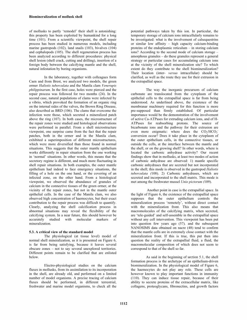
Figure 6. Physiology of the shell calcification of the arcoid bivalve Arca sp. (redrawn from Waller, 1980 (77)). The calcification occurs at the edge of the shell, at the interface between the mantle, the periostracum and the shell itself. Whether this interface corresponds to an extrapallial space or not is still debated.
5. SHELL FORMATION: PHYSIOLOGICAL AND CELLULAR CONSIDERATIONS 5.1. Calcification of the shell in normal conditions
The shell formation is typically a process that enters the category of epithelium-driven biomineralizations (76). The organ that secretes the shell is the mantle, the ciliated tegument that coats the inner surface of the shell. Although there may be some variations from group to group, the general principles that govern the physiology of shell formation are somehow valid for all conchiferan mollusks. The following diagram (Figure 6), redrawn from Waller (77), depicts a classical view of the shell formation, at the tissue level. The diagram focuses on the border of the growing shell of Arca, a pteriomorphid bivalve which exhibits a bi-layered shell made of a thick complex crossed-lamellar internal layer, and an outer crossed-lamellar one, both layers being aragonitic (14). For the normal calcification process, three separate elements have to be considered, the mantle and its outer epithelium, the periostracum, and the interface between the outer epithelium, the periostracum and the growing shell, respectively. Let us describe successively these elements. 5.1.1. Mantle and cells of the outer epithelium
The mantle is a polarized tissue, and comprises, from inside to outside, an inner epithelium, in contact with the ambient medium (for example, seawater), internal tissues, comprising pallial muscles, connective tissues, nerve fibers, and finally, the outer calcifying epithelium, the one that faces the shell and that secretes all the macromolecular and ionic ingredients for its synthesis. The outer epithelium – but the remark is also true for the inner one - is a monolayer of cells with typical microvilli,
interspersed by goblet cells (mucocytes), which produce mucus. When observing the cells along a radial axis starting from the hinge to the shell edge, the outer epithelium may appear rather homogeneous in term of cell typology, and does not exhibit a gradient for example. However, from recent works, there seems to be a subtle cell zonation, which cannot be simply distinguished by classical histology. This zonation seems to be strictly correlated to the shell microstructures, but its detection requires molecular markers, such as HIS probes or specific antibodies. Although nothing is known for crossed-lamellar bivalves such as Arca, this zonation has been illustrated by few examples of nacro-prismatic bivalves, in particular, the pearl oyster Pinctada. In the late nineties, the work of Sudo and co-workers (78) clearly showed by in situ hybridization that the transcript encoding one shell protein, MSI31, was localized in the distal zone (from the hinge) of the calcifying epithelium, which corresponds to the zone that produces the outer prismatic layer. More recent works based on immunohistology (79) or on HIS (80) with other markers confirmed the finding that distinct zones of the outer epithelium secrete prisms and nacre layers. Recently, in the frame of an initiative aiming at understanding the whole process of pearl fabrication (GDR ADEQUA) of the Polynesian pearl oyster (P. margaritifera), an unpublished work based on proteomic and transcriptomic investigations showed by in situ hybridization a clear limit between the nacre-secreting and the prisms-secreting cells (B. Marie, C. Montagnani, personal communication). This demonstrates without ambiguity that some proteins are shell layer-specific, which implies that the secretory regime of the cells that produce nacre on one side, and of the ones that produce prisms on the other side, are different. What has been demonstrated for nacro-prismatic bivalves could

Biomineralization of mollusk shell
1110
probably be transposed to other mollusks that possess a shell exhibiting combinations of shell textures different from the nacro-prismatic ones.
Another important point that should be
considered about the outer epithelium cells is the presence of membrane pumps and channels for extruding the inorganic precursors of calcium carbonate. It is obvious that, if calcium is released in the site of mineralization under an ionic form – and not as granules – this ion transport phenomenon is not passive, but requires the active role of transmembrane pumps, i.e., Ca-ATPases. However, these pumps are poorly documented at the molecular level. For the other precursor of calcium carbonate, bicarbonate ion, there might be the equivalent process, involving bicarbonate channels or bicarbonate pumps. For this ion, an alternative solution is the presence of transmembranar carbonic anhydrases that catalyze the hydration of carbon dioxide into bicarbonate. At last, as we will briefly discuss in section 5.1.5, H-ATPases may also participate to the process of shell formation, by actively pumping protons into the cytosol.
5.1.2. Periostracum
The edge of the mantle is characterized by a succession of folds, usually three in bivalves. The ridge between the outer and median folds defines a groove, known as the periostracal groove, in which specialized cells secrete the periostracum (Figure 6). The primary roles of the periostracum are multiple: firstly, as described in larval stages (section 4), it provides the primary template for receiving the extracellular mineralization. Secondly, it delimitates and seals a confined space between the mantle tissues and the shell itself, the extrapallial space. Actually, the invention of the periostracum represents a strategy that mollusks have set up for mineralizing extracellularly their shell in a minute space, separated from the environment, and which can be easily supersaturated with respect to calcium carbonate. The periostracum exerts also other minor functions, required in specific cases: it protects the shell against dissolution, in particular in acidic mangrove environments (81). It constitutes an efficient barrier against fouling by microorganisms (82). Finally, as it is often colored, it constitutes an efficient camouflage against predators, in particular in the case of sessile bivalves. The periostracum is secreted as a liquid film of tyrosine-rich instable soluble precursors, which become insoluble and sclerotized by a quinone-tanning process, as soon as they are released in the extracellular environment (83). Long ago, one of the soluble precursors was partly characterized and described as periostracin (84). In some cases, the periostracum may be not homogeneous, but stratified in two layers. It can persist during all the life of the animal, like in the edible mussel, or being more or less rapidly abraded, like in the American clam. The structure and chemical composition of the periostracum have been studied in a number of cases (83). However, due to its high insolubility, the classical biochemical approach failed to resolve its separate constituents, and more work is needed before we understand the subtle chemistry of this secretory product.
5.1.3. Interface between the mantle and the shell As said before, the third element of the system is
the interface between the outer mantle epithelium, the periostracum and the growing shell itself. Figure 6 shows that the epithelium is not in direct contact with the shell, but is separated from it by the extrapallial space, defined above. This space is supposed to be the confined medium where all the ingredients for calcification self-assemble. This space is filled with a fluid, the extrapallial fluid, which is supersaturated with respect to calcium carbonate. Only few analyses of this fluid were performed so far. This fluid seems to contain inorganic ions (85, 86), proteins (87, 88) and glycosaminoglycans (GAGs) (89, 90). One reason of the scarcity of molecular data on this fluid is that its sampling is tricky. On different occasions, having done ourselves these experiments with a small syringe and a tiny needle on different model organisms, we were never fully convinced that the fluid that we were sampling was the right one! Furthermore, in the light of what has been said about the radial zonation of the outer epithelium, it is likely that the composition of this fluid is not homogeneous, but varies from the central shell zone to the shell edge. Furthermore, it seems that the composition of this fluid also varies according to seasons (89). In addition to the precursors ions for mineralization - calcium and bicarbonate - this fluid contains several other inorganic ions, such as Na+, K+, Mg2+, Cl- and SO4
2-, and minor elements, such as Sr and Fe. Its pH is usually slightly basic, in the range of 7.4 – 8.3, for marine and freshwater mollusks. This fluid also contains organic molecules. As the fluid is supersaturated, these macromolecules – in particular acidic proteins and GAGs - are supposed to transiently maintain calcium in solution, by inhibiting the precipitation of calcium carbonate, and by allowing it to precipitate where needed. As far as we know, the protein content of the fluid does not necessarily reflect the protein content of the shell (87), and the protein diversity of this fluid appears to be singularly lower than that of the shell (unpublished observations). This addresses the puzzling questions of the correlation between the chemistry of the extrapallial fluid and of the shell, and the question of the incorporation of extrapallial fluid proteins in the shell during calcification.
5.1.4. Transport of the precursors ions of mineralization
Although the shell mineralization takes place in a restricted area, the border of the outer mantle epithelium, the molecular process is ‘prepared’ upstream elsewhere. This means that there must be somehow a complex pathway, for bringing the precursor ions of calcium carbonate, from the uptake site to the mineralization site. Curiously, this pathway is still poorly known in spite of electro-physiological experiments and measurements with radioactive calcium performed thirty years ago (91-93). In the case of marine or freshwater mollusks, calcium ions are supplied in the water or in the food. They are supposed to be absorbed in the inner mantle epithelium, in the gills, and in the digestive system. For terrestrial mollusks, only food contributes to supply the required calcium. The carbonate ions of the shell are supplied both from bicarbonate of the medium and from metabolism (76, 93).

Biomineralization of mollusk shell
1111
The manner the inorganic precursors of calcification are driven to the site of mineralization is still speculative, but we can reasonably argue that it follows two pathways: in soluble form, calcium and bicarbonate ions are then simply transported in the haemolymph, the interstitial fluid that circulates around the internal organs. In clustered forms, calcium can be stored and transported as amorphous granules (91, 94); these granules have been described for terrestrial and marine species. They can be intracellular, in specialized vesicles or extracellular (interstitial) (95). Amorphous granules offer several advantages. Under a compact volume, they constitute an important source of calcium, which is rapidly available by dissolution, in particular in emergency situations, such as shell repairs (24). Granules are also important for detoxification process, by trapping heavy metals, for example (96). Intracellular granules can be redissolved intracellularly and their constitutive ions can be massively extruded in the extrapallial space; alternatively, they can be released by exocytosis; interstitial granules can be dissolved again or can migrate through trans-epithelial channels (97).
5.1.5. Synthetic cellular view of the shell formation process
Although many pieces of the puzzle are still missing – or hypothetical - we can schematically summarize the different events that lead to the formation of the shell as follows: calcium ions are taken up from food, from water filtration activity, or from passive diffusion through all parts of the body. They are subsequently transported by the haemolymph, and may be transitorily stored in the connective tissues of the mantle or in the calcifying mantle epithelium. Two modes of storage can be inferred: in soluble form, calcium ions can be sequestered by low affinity – high capacity calcium-binding proteins of the ER; in insoluble form, they may be stored as granules. In the first case, the storage is limited and may account only for a small part of the required calcium. In the second case, granules are formed by the reaction between calcium ions with available intracellular bicarbonate. The granules are made of amorphous calcium carbonate, which can be subsequently dissolved again easily. These granules can be formed intracellularly, in the connective tissues of the mantle or in the cells of the calcifying epithelium. Alternately, the granules can be interstitial.
Bicarbonate ions can come from two sources: in
the ‘bicarbonate form’, it may be provided by the food, the water absorbed by filtration, or directly, by diffusion from the external medium through the body; then, it is transported by the haemolymph, similarly to calcium ions. Alternately, bicarbonate can result from the hydration of metabolic carbon dioxide. The reaction is catalyzed by carbonic anhydrase. The conversion reaction may take place far upstream the place where mineralization occurs. If so, the produced bicarbonate ions may be then transported by the haemolymph. Alternatively, the formation of bicarbonate may occur close to the site of mineralization, in the mantle cells of the underlying connective tissues or in the mantle epithelial cells.
Ultimately, the conversion of carbon dioxide to bicarbonate may occur in the extrapallial space.
If present in ionic forms in the epithelial cells,
calcium and bicarbonate ions are massively extruded in the extrapallial space. The role of transmembranar pumps, such as Ca-ATPases, or bicarbonate channels, is determinant for increasing locally the concentration of these ions, for reaching the supersaturation conditions. Beside these two ions, some minor ions (Na+, K+, Mg2+, Fe3+, Sr2+, Cl-, SO4
2-
) are also released in the extrapallial space, and are later incorporated in the shell. Simultaneously, the outer epithelial cells synthesize sets of proteins and glycoproteins and secrete them in the extrapallial space by exocytosis. The inorganic ions and the macromolecules interact in a controlled manner and self-assemble to form biominerals. Without taking in consideration the interaction with organic macromolecules, the production of calcium carbonate occurs according to the equation:
Ca2+ + HCO3
- CaCO3 + H+ As shown here, the precipitation of calcium
carbonate is accompanied by the production of one proton, which acidifies the medium. In order to subtract the proton from the reaction, different possibilities are offered: reabsorption of the proton by cells of the mantle epithelium, owing to transmembranar pumps such as H+-ATPases. The existence of such pumps in the mantle of mollusks is supported by a tenuous corpus of physiological data (98). However, one H+-ATPase (SwissProt accession number Q000T7, unpublished data) has been identified in the mantle of the pearl oyster Pinctada fucata. Alternately, as proposed by Wilbur and Saleuddin (99), the proton can be removed by reacting with bicarbonate to form CO2 and water, the reaction being catalyzed by carbonic anhydrase of the extrapallial fluid. Then, CO2 may diffuse out of the fluid into the medium, or may be uptaken by the mantle tissues. Another possibility, which was considered long time ago (100), would be the neutralization of protons by ammonia (NH3) to form ammonium ion (NH4
+). Ammonia would be a degradation product of urea by urease. However, this particular metabolism may be restricted to landsnails, and should be validated for different marine models.
This is how the process of shell calcification can
be described at the physiological and cellular levels. We will see in section 5.3 that this view leaves several questions unanswered.
5.2. Shell remodeling and shell repair process
Beside the normal calcification process, one aspect that made the mollusk shell a true evolutionary success lies in the ability of conchiferan mollusks to rapidly repair shell damages, an undeniable advantage for overcoming external aggressions of different sources: accidental physical shell cracks, active predation by fishes, shell boring by epibiont organisms, such as clionid sponges, or entrapment of foreign bodies between the mantle and the shell. Although the shell is a ‘dead’ non-cellular tissue, it exhibits certain plasticity, and the capacity
Biomineralization of mollusk shell
1112
of mollusks to partly ‘remodel’ their shell is astonishing: this property has been exploited by humankind for a long time (101). From a scientific viewpoint, the shell repair process has been studied in numerous models, including marine gastropods (102), land snails (103), bivalves (104) and cephalopods (105). The shell regeneration process has been analyzed according to different procedures: physical shell lesion (shell crack, cutting and drilling), insertion of a foreign body between the calcifying mantle and the shell, natural infestation by boring organisms.
In the laboratory, together with colleagues form
Caen and from Brest, we analyzed two models, the green ormer Haliotis tuberculata and the Manila clam Venerupis philippinarum. In the first case, holes were pierced and the repair process was followed for two months (24). In the second case, natural populations of clams were infected by a vibrio, which provoked the formation of an organic ring on the internal sides of the valves, the Brown Ring Disease, also described as BRD (106). The clams that overcome the infection were those, which secreted a mineralized patch above the ring (107). In both cases, the microstructure of the repair zones were studied and histological observations were performed in parallel. From a shell microstructure viewpoint, one surprise came from the fact that the repair patches, both in the ormer and in the Manila clam, exhibited a superimposition of different microstructures, which were more diversified than those found in normal situations. This suggests that the outer mantle epithelium works differently in repair situation from the one working in ‘normal’ situations. In other words, this means that the secretory regime is different, and much more fluctuating in shell repair situations. In both examples, the outer mantle epithelium had indeed to face emergency situations, the filling of a hole on the one hand, or the covering of an infected zone, on the other hand. From a histological viewpoint, we observed the abundance of granules of calcium in the connective tissues of the green ormer, at the vicinity of the repair zones, but not in the mantle outer epithelial cells. In the case of the Manila clam, we also observed high concentration of haemocytes, but their exact contribution to the repair process was difficult to quantify. Clearly, analyzing the shell calcification process in abnormal situations may reveal the flexibility of the calcifying system. In a near future, this should however be accurately studied with molecular markers of mineralization.
5.3. A critical view of the standard model
The physiological (at tissue level) model of normal shell mineralization, as it is presented on Figure 6, is far from being satisfying, because it leaves several obscure zones – not to say several unexplored territories. Different points remain to be clarified that are enlisted below.
Electro-physiological studies on the calcium
fluxes in mollusks, from its assimilation to its incorporation in the shell, are already old, and performed on a limited number of model organisms. A precise tracing of calcium fluxes should be performed, in different terrestrial, freshwater and marine model organisms, to check all the
potential pathways taken by this ion. In particular, the temporary storage of calcium ions intracellularly remains to be investigated: what is the involvement of calsequestrins or similar low affinity – high capacity calcium-binding proteins of the endoplasmic reticulum – in storing calcium ions? According to the second mode of calcium storage – amorphous granules – do these granules represent a general strategy or particular cases for accumulating calcium ions at the vicinity of the shell mineralization site? To which extent do they contribute to the shell biomineralization? Their location (inter- versus intracellular) should be clarified, as well as the route they use for their extrusion in the extrapallial space.
The way the inorganic precursors of calcium
carbonate are translocated from the cytoplasm of the epithelial cells to the extrapallial space is far from being understood. As underlined above, the existence of the membranar machinery required for this function is more pre-supposed than firmly established. Of particular importance would be the demonstration of the involvement of active Ca-ATPases for extruding calcium ions, and of H-ATPases for reabsorbing protons. The source of bicarbonate ions and the pathway for their extrusion are even more enigmatic: where does the CO2/HCO3
- conversion occur? Does it take place in the cytoplasm of the outer epithelium cells, in the connective tissues, or outside the cells, at the interface between the mantle and the shell, or on the growing shell? In other words, where is located the carbonic anhydrase activity? Our recent findings show that in mollusks, at least two modes of action of carbonic anhydrase are observed: 1) mantle specific carbonic anhydrases that are secreted but not incorporated in the shell; this mode is observed in the gastropod Haliotis tuberculata (108). 2) Carbonic anhydrases, which are secreted and incorporated to the shell matrix. This mode is met among the freshwater mussel Unio pictorum (109).
Another point in case is the extrapallial space. In
the light of Figure 6, the existence of the extrapallial space supposes that the outer epithelium controls the mineralization process ‘remotely’, without direct contact with the mineralization front. This also means that macromolecules of the calcifying matrix, when secreted, are ‘tele-guided’ and self-assemble in the extrapallial space without any cell intervention. This viewpoint has been put into question few years ago (57), and the subsequent NANOSIMS data obtained on nacre (48) tend to confirm that the mantle cells are in extremely close contact with the mineralization front. If this is true, this put then into question the reality of the extrapallial fluid, a fluid, the macromolecular composition of which does not seem to correspond to that of the shell so far.
As said in the beginning of section 5.1, the shell
formation process is the archetype of an epithelium-driven biomineralization. In the physiological model of Figure 6, the haemocytes do not play any role. These cells are however known to play important functions in immunity (110). They can induce tissue repair, because of their ability to secrete proteins of the extracellular matrix, like collagens, proteoglycans, fibronectins, and growth factors

Biomineralization of mollusk shell
1113
like IGF (111). Furthermore, they have been shown to be involved in shell repair (24, 112). In a provocative paper (104), Mount and coworkers suggested that haemocytes may be involved as well – not marginally but massively - in normal shell calcification. In particular, they observed that haemocytes of the oyster Crassostrea virginica were able to carry calcite crystals to the site of calcification, where they were remodeled and integrated to the growing biomineral. This suggests that haemocytes may play an underestimated role in shell mineralization, a role that should be urgently reevaluated.
6. ORGANIC CONSTITUENTS OF THE SHELL
6.1. Biochemistry of the mollusk shell
The fact that shells contain a small proportion of organic material is known for a long time: Frémy (113), one and half century ago, was the first in the world to characterize the organic fraction of a shell, a substance that he called conchiolin. This terminology was brought to encounter a certain success since it is still found in the today’s dictionaries. Frémy evidenced the high insolubility of conchiolin, and the fact that it was chemically different from bone ‘osseine’ (collagen) and from chitin. At that time, neither Frémy nor his most illustrious continuators of the 19th century (listed in (114)) associated the presence of organic substances in a shell to a putative function in mineralization. This concept was emphasized much later, after World War II, in particular with the development of biochemistry as a scientific discipline in the fifties. Roche et al. (115), then Grégoire and coworkers (116) were among the first ones to give amino acid compositions of different organic shell fractions. Following their work, profusion of data was published in the late fifties, during all the sixties, and in the early seventies on amino acid compositions of shell matrices (114). At that time, it became clear that shell organic constituents were defining a ‘matrix’, i.e., a mixture of extracellular macromolecular components that are secreted for ‘helping’ and guiding the mineralization, or, at least, for being used as a substrate for mineral deposition.
Numerous quantitative analyses indicate that the
organic moieties of the shell represent a minor fraction of the shell, between 0.01 and 5 wt-%. Although these proportions may appear to be low, they influence drastically the mechanical properties of the associated biominerals (27), increasing the fracture toughness of the shell by two or three orders of magnitude. The variations in the percentages are related to the shell microstructures to which organic constituents are associated (117). As a general rule, nacre and prisms microstructures are known to contain large amount of organic constituents, typically above 1wt-%. On the contrary, crossed-lamellar microstructures are characterized by low amounts of matrix. Different obvious reasons led the scientific community to focus on the protein moieties of shell matrix: firstly, they are indeed the dominant macromolecular constituents of the shell. Secondly, they give a direct access to the genomic information. Thus, following the classical view inherited from the sixties, obtaining their primary
structure gives an indication of their function in mineralization. However, proteins are not the single organic constituents of the shell, which is also composed of polysaccharides, lipids, pigments, free amino acids and peptides. For the clarity of the text, we distinguish the “non-protein organic molecules” (including also small peptides) from the proteins sensu stricto (polypeptides of molecular weight above 5 kDa) found in the shell.
6.2. Non-protein organic shell components 6.2.1. Polysaccharides
Quantitatively, polysaccharides represent the second class of important macromolecules, after proteins, in mollusk shells. They can be roughly divided in two groups: chitin and soluble acidic polymers. Chitin is a long-chain insoluble polymer made of a single monomer, N-acetyl glucosamine. In mollusks, chitin was identified by Frémy in cuttlefish bone and in squid feather (113). Goffinet and Jeuniaux (118) detected it in several shells. Although chitin is widely distributed in mollusk shells (119), it is not possible to ascertain that it is present in every kind of shell microstructure, or that it is associated only to specific ones. In nacre for example, its contribution to the 3D architecture of the shell matrix seems to be essential: in the topographic model described in sections 3.3.3 and 3.3.4, chitin plays a key-role by defining the interlamellar matrix between successive nacre tablets (46, 57). The synthesis of chitin is catalyzed by an enzyme, chitin synthase (120), and the inhibition of the activity of this enzyme has a drastic effect on the structure of nacre (121). Chitin, which, in mollusk shells is of the ß-type, forms with other macromolecules, in particular proteins, supramolecular complexes. Chitin can be directly detected by different manners (122). Its putative presence in shells can also be assessed indirectly, by characterizing proteins that exhibit typical chitin-binding motifs, called Rebers-Riddiford motifs (123), or by partially hydrolyzing the matrix and analyzing its monosaccharide composition: high levels of released glucosamine from the acid-insoluble matrix is a strong indication of the presence of chitin (124).
Beside chitin, soluble acidic polysaccharides
may also be present in the shell, but their characterization is still in its infancy. Many shell polysaccharides are covalently bound to protein core, forming then glycoproteins, or proteoglycans (125). It is likely that, in addition, some are free in the matrix, but this aspect is poorly investigated. Polysaccharides are constituted of neutral, amino, and acidic monosaccharides in variable proportions (124). In addition, they can be sulfated, i.e. negatively charged (125). In classical models of shell mineralization, sulfated polysaccharides play a cooperative role with proteins, by concentrating calcium ions at the vicinity of the nucleating factors (3). It is likely that they exert additional functions, such as tissue-to-cell communication, or sequestering of water molecules. Sulfated polysaccharides can be detected by specific staining, such as Alcian blue, or by FTIR.
Biomineralization of mollusk shell
1114
6.2.2. Lipids, pigments and other small organic molecules
In shells, lipids have been poorly investigated, since they represent an extremely minor fraction of the organic matrix. CoBabe and Pratt (126) identified fatty acids, cholesterol, phytadienes, and ketones in the shells of diverse fresh and fossil shells. More recently, lipids from the nacre of the pearl oyster were extracted and analyzed (127): they consist in a mixture of fatty acids, triglycerides, cholesterol and ceramides. These lipids seem to promote the repair of the stratum corneum, the upper layer of the skin. The role of lipids in the shell mineralization is unknown. We cannot exclude that a part of the lipid moieties combines covalently with polysaccharides or proteins, forming either lipopolysaccharides, or lipoproteins.
Although representing an ultraminor fraction of the
organic matrix, pigments are important components of the shell, since they form patterns on the shell surface, which, in numerous cases, are species specific (for example, in the cone snail family). Pigments are incorporated in the shells, where they seem to be bound to the shell matrix macromolecules, as they tend to co-elute with matrix macromolecule after size exclusion chromatography (F. Marin, personal observation). They mostly consist of unsubstituted chains of 8-13 conjugated double bonds polyenes (bound and unbound), and carotenoids comprising unmethylated polyacetylenic backbones (128, 129). The molecular mechanisms by which pigments are incorporated in the shell are unknown: however, few years ago, it was shown that the expression of one shell protein (ependymin-related protein; sometsuke gene) was discontinuous in the outer mantle fold of the tropical abalone Haliotis asinina, and strictly correlated with the shell pigmentation (74). This strongly suggests that pigments and some shell proteins form complexes.
At last, the organic matrix contains free amino
acids and small peptides, but their contribution as matrix constituents has passed largely unnoticed until recently. From our own experience, amino acid analysis of shell matrices without any hydrolysis always generates free amino acids in low proportions. Similarly, a recent proteomic analysis of the soluble pearl oyster nacre matrix filtrate – which had passed through a 1-kDa cutoff membrane - has generated about 110 different peptides (130) in the range 100-700 Da. So far, it is difficult to assess whether small peptides and free amino acids result from the cleavage of shell matrix proteins, or whether they are initially incorporated in the shell matrix during the synthesis of the shell biominerals. In the case of small peptides, their role in biomineralization is unknown, but, as they can diffuse readily at the shell interface, their function may be related to cell signaling. Alternately, some of them, in particular the most basic ones, may constitute bactericidal or anti-viral factors.
6.3. Shell proteins 6.3.1. Biochemistry of shell proteins
As we recall in section 6.1, the proteinaceous moieties of mollusk shell have been the main focus of hundreds - not to say more than one thousand - studies, since the beginning of the twentieth century (114). Basically,
proteins of the shell matrix are retrieved, by dissolving the shell powder in EDTA or in weak acid, and subsequent centrifugation of the resulting solution. These extraction steps allow identifying two protein fractions, according to their solubility: soluble proteins on the one hand, and insoluble proteins, on the other hand. Earlier studies, based on bulk amino acid analyses, tended to make a clear distinction between both fractions. The soluble one was enriched in acidic hydrophilic residues, in particular aspartic acid, while the insoluble fraction, characterized by the abundance of glycine and alanine, exhibited a marked hydrophobic character (3, 114). Furthermore, while the soluble matrix proteins were found to be mainly ‘within’ biominerals, i.e. intracrystalline, proteins of the insoluble matrix were localized around the crystal phase (‘intercrystalline’). From these data, a general model of shell mineralization emerged (3), where the insoluble hydrophobic matrix was supposed to act as a mold/template. When bound on the insoluble template, acidic polyanionic soluble proteins were supposed to promote crystal nucleation. When free in solution, these acidic proteins inhibited the deposition of calcium carbonate. The shape of shell biominerals was supposed to be largely controlled by a subtle equilibrium between nucleation and inhibition. Of course, this theoretical model, which was mostly based on nacre-type biocrystals, did not take in account the wide diversity of shell microstructures, as presented in section 3.2.
6.3.2. Shell proteins: the ‘protein per protein’ approach
The clear dichotomy ‘intracrystalline acidic soluble’ versus ‘intercrystalline hydrophobic insoluble’ was put into question, with the extensive use of molecular biology techniques that allowed obtaining partial or full-length sequences of transcripts encoding shell proteins. From the mid-nineties, several research groups around the world started to publish the primary structure of several mollusk shell proteins, one per one. The precise description of these proteins is far beyond the scope of this study: few review papers have described their respective biochemical characteristics (70; 131-137). Most of these proteins are mentioned in Figures 7 and 8. Let us make some remarks.
The “protein per protein” approach allowed
identifying between 40 and 50 mollusk shell proteins, in about a decade. So far, only few models are studied. For obvious economical reasons, the two models, which draw the attention, are the pearl oyster Pinctada among bivalves, and the edible abalone Haliotis, among gastropods. Both models, which exhibit nacro-prismatic textures, concentrate more than 90% of the molecular data. It is very unlikely that these two models represent a good sampling of the diversity of the mollusk phylum.
The proteins enlisted in Figures 7 and 8 exhibit a
huge diversity, which was totally unexpected according to what we said in section 6.3.1. In particular, the dichotomy “soluble acidic” versus “insoluble hydrophobic” is singularly blurred. This diversity encompasses the overall biochemical properties as well as sequence information. Concerning the overall biochemical properties, we plotted, in a previous paper, the theoretical isoelectric point of these shell proteins against their calculated molecular mass (70). Shell proteins distribute widely, from very acidic (below 3)

Biomineralization of mollusk shell
1115
Figure 7. Shell proteins in nacreous and non-nacreous bivalves. For each genus, the proteins are enlisted alphabetically. As shown here, most of the knowledge has been accumulated on a single model, the pearl oyster Pinctada sp. The molecular knowledge on the Manila clam (Venerupis), on the edible mussel (Mytilus) is mainly obtained from the combination of transcriptomics and proteomics. Proteins like calcine, N33, N44, N151, perline and perlucin-like proteins A-C from Venerupis are registered in UniProt protein database, but are not published yet, or are being published (see www.uniprot.org). CSP3 (Pinna) and upsalin (Unio) are neither published nor registered yet. Cl: class. Nacr: nacreous. Str: shell structure. * indicates proteins deduced from mantle transcriptomic data, without confirmation of their presence in the shell. Some proteins, like RP-1, caspartin, calprismin or most of the shell proteins from Unio (except upsalin), are only known by partial amino acid sequences.
to very basic (above 10) theoretical isoelectric points, and from low (10 kDa) to high (above 100 kDa) molecular weights. We observed that the proteins associated to calcite exhibit either a very acidic or a very basic theoretical isoelectric point, while proteins associated to aragonite possess a theoretical isoelectric point, which is slightly acidic to slightly basic. Of course, this representation is theoretical and applies to ‘naked’ amino acid sequences, i.e., sequences that do not exhibit post-translational modifications. The reality is more blurred since several proteins, which exhibit a moderately acidic to basic isoelectric point, can be rendered extremely acidic, due to post-translational modifications, such as phosphorylation or glycosylation by sulfated sugars. Concerning the sequence information, there is no simple grouping of these shell proteins in one or two families. Some proteins clearly belong to a same family (KRMP, shematrin, ‘nacrein-like’); some others are orphan (mucoperlin). The general
impression is that shell proteins form an extremely disparate group.
Many of these shell proteins exhibit a modular
organization of their sequence. Each module corresponds to a precise molecular function, and the modules are placed in tandem (one after each other) in the sequence. This feature is commonly encountered with proteins of the extracellular matrix among vertebrates. Consequently, the multidomain proteins are likely multifunctional. The best example is that of nacrein, which exerts an enzymatic function (carbonic anhydrase) and which interacts with calcium carbonate. We do not know yet whether these two functions are performed concomitantly, or one after the other. Among the remarkable features of shell proteins, one finds the abundance of repetitive low-complexity domains (RLCDs), which are characterized by the predominance of one, two or three amino acids that usually form short repeated motifs. The most represented residues are glycine, aspartic acid,

Biomineralization of mollusk shell
1116
Figure 8. Shell proteins in nacreous and non-nacreous gastropods and in the cephalopod nautilus. In gastropods, the nacreous abalone Haliotis represents the dominant model. Similarly to bivalves, most of the molecular knowledge results from the combination of proteomics on the shell matrix and of transcriptomics performed on the mantle tissues. For some proteins (perlbikunin, or proteins from the Lottia shell), the sequences are not available. CE: Cephalopoda. Cl: class. Nacr: nacreous. Str: shell structure. * indicates proteins deduced from mantle transcriptomic data, without confirmation of their presence in the shell. Some proteins, like ACLS40 (Strombus) or the shell proteins of Helix, are known only from partial sequences.
serine, asparagine, alanine, tyrosine, proline. RLCDs are intrinsically disordered, or unstructured (137). They seem to constitute key-domains in the interaction with the mineral phase.
One problem that all researchers involved in
characterizing mollusk shell proteins currently encounter is the difficulty to link the primary structure of a given protein to its function in biomineralization: with some exceptions, the sequences of several of these proteins do not match with that of known proteins of perfectly identified function, and homology searches are ineffective, or apply only on short domains. In that case, the knowledge of the primary structure of a given protein is not sufficient for guessing its true function. Two series of strategies may be employed for circumventing this problem: develop different in vitro tests, or work in vivo. Concerning the first aspect, the purified protein – or genetically produced – may be directly tested in classical in vitro assays (inhibition tests, CaCO3 interference test); Alternately, it can be tested together with other shell matrix macromolecules, to search for cooperative roles, such as chitin-binding (138). Synthetic peptides can be produced that mimic one shell protein domain (139); truncated forms of the protein can be genetically engineered and tested in vitro (140); at last, chimeric proteins can be produced and equally tested in vitro (141). Concerning the second aspect, gene-knock-down experiments (RNAi) can be performed (142), but they require a good control of embryo and larval development. Although technically demanding, the in vivo
approach represents probably the best option for understanding the function of each shell protein. 6.3.3. Shell proteins: large screening of the ‘shellome’
As it has become obvious that a “protein per protein” approach does not give a chance to understand the biomineralization process and to get the “full picture” of the shell content, new approaches have started, few years ago, to emerge, with the perspective to obtain, in one shot, all the proteinaceous constituents of a shell, the ‘shellome’. This has been performed, by working at the protein level, using proteomics, or by working at the transcriptional level, using transcriptomics, or by combining both approaches.
The first researchers to apply a transcriptomic
approach were Jackson and coworkers, on the tropical abalone Haliotis asinina (74, 143). These authors constructed EST libraries (Expressed Sequence Tag) from the calcifying mantle tissues of juvenile specimens. This robust approach allowed obtaining the secretome, i.e. the set of transcripts encoding secreted proteins of the mantle cells that are supposed to build the shell. Strikingly, most of the transcripts encoded unknown proteins, a fact which considerably broadened our view on the underlying machinery that constitutes the calcifying system of mollusks. Furthermore, subsequent comparisons between the abalone nacre secretome and that of the pearl oyster revealed huge differences, suggesting that both nacres do not have a shared origin, but are the products of convergent evolutions (144). This conclusion was remarkable, although

Biomineralization of mollusk shell
1117
puzzling. The advantage of the transcriptomic approach, as performed by Jackson and coworkers, was to obtain a complete set of genes, which are activated during shell formation. One drawback was that it was not possible to ascertain the contribution of unknown proteins to the construction of the shell, unless complementary experiments, such as in situ hybridization, were performed. Obviously, a second filter was needed to reduce the amount of data and to extract the most pertinent ones.
On our side, starting in 2007, we developed a
proteomic approach, which was applied on the shell nacre matrix of the freshwater mussel Unio pictorum, and on that of the cephalopod Nautilus macromphalus (109, 145). Our goal consisted in comparing different nacre matrices, to check whether they were constituted of similar proteins or not (146). This approach generated a large amount of peptides (9 to 21 residues), from the acetic acid-insoluble matrix, from the acetic acid-soluble matrix, from 1D-electrophoresis bands, and from spots obtained by 2D gel-electrophoresis. Our results were of three kinds: firstly, we observed that several identical peptides were found in the insoluble and soluble matrices. This finding contradicts the biochemical dichotomy observed in the old studies (insoluble = hydrophobic; soluble = acidic), but fits with the data obtained by the “protein per protein” approach. Secondly, within a nacre matrix, distinct proteins, characterized by clearly different molecular weights and different pIs, yielded identical peptides. This suggests that many nacre proteins exhibit “mosaic characters”. In other words, this means that they possess identical short functional motifs (for example, few amino acid-long), which do not necessarily occupy the same position along the sequence. Following this, one can infer a “genetic tinkering mechanism” (motif swapping), that allows the re-use of functional blocks in different proteins. At last, by comparing together the peptidic profiles of the freshwater mussel and of the nautilus, and by comparing them to known protein data obtained by the “protein per protein” approaches, we were surprised to register an extremely low sequence homology (146). This suggests that nacre matrices are probably far less evolutionary constrained than expected (high rate of punctual mutation), or that nacres evolved completely independently, from group to group. Our conclusions and the ones of Jackson et al. are clearly converging. One drawback of our approach is that our proteomic data gave only sequences fragments (and not full protein sequences) because no transcriptomic/genomic data were available: the nautilus and the freshwater mussel are not yet popular models in molecular genetics.
In this context, a substantial improvement has
recently occurred by using proteomics on the shell matrices of species for which mantle EST libraries are available. In that case, the proteomic approach performed on shell matrix acts as a secondary filter by discriminating true shell proteins from those which are secreted but which are not incorporated in the shell, or those which have little to do with calcification. This approach has been performed on the abalone Haliotis asinina (147), on the pearl oyster Pinctada margaritifera (148, 149), on the edible mussel Mytilus edulis (150), but also on non nacro-prismatic
mollusks such as the Manila clam Venerupis philipinarum (151), the edible oyster Crassostrea gigas (152), the giant owl limpet Lottia gigantea (manuscript in preparation) or the freshwater snail Lymnaea stagnalis (see Figures 7 and 8). This combined approach allows increasing drastically the quantity of information on shell proteins, in different models. So far, more than one hundred of proteins are available. This wealth of information allows “shellome-to-shellome” comparisons, from which macro-evolutionary trends can be sketched. Our preliminary conclusions are that, from matrix to matrix, one finds only few similar proteins that constitute the shared ’tools’ of the ‘molecular toolbox’ (150), and several proteins, which are different. Efforts are now made to classify these proteins according to their putative functional domains, and to try to decipher their exact synergistic role in shell construction.
7. PERSPECTIVES AND FUTURE DIRECTIONS
Drawing long-term perspectives in mollusk shell
biomineralization is risky: for a decade, the field knows remarkable advances that we have tried to summarize with the present review. We live an exciting period, but nobody knows how this field will evolve in the coming years. As the study of mollusk shells is typically interdisciplinary, we advocate combining different approaches, both physical and biological. From a physical viewpoint, one can bet on the development of destructive and non-destructive methods for analyzing in situ shell biominerals, and, in particular, for ‘observing’ the interactions between the organic macromolecules and the mineral phase. The recent data obtained with pulsed laser atom-probe tomography (194) on the chiton teeth are extremely promising. One can imagine that AFM investigations with protein-specific antibodies may also be a serious option to map the distribution of each macromolecule on a shell mineral surface.
The characterization of the shell matrix
macromolecules will continue, in particular, with the development of high throughput sequencing at the genomic, transcriptional (EST databases), or protein levels. Let us hope that mollusks, which are nowadays largely underrepresented in genomic sequencing programs (195), will occupy a more favorable position in the coming years. An important but tedious step will be the correct annotation of the genomes/transcriptomes. This step should be accompanied by functional in vitro and in vivo studies (in situ hybridization, gene knock-down, two-hybrid screening), in order to understand how the shell matrix works. The way shell proteins are post-translationally modified is another point in case, and we believe that the ‘glycomics of biomineralization’ should be tackled frontally. The development of these different levels of analysis will have a direct consequence on the knowledge of shell calcification at the cellular and tissue levels, these physiological aspects having been neglected for more than three decades.
Finally, mathematical concepts of self-assembly,
of emerging properties should continue to thrive, in order to push away the limits of a reductionist approach.

Biomineralization of mollusk shell
1118
Conciliating all these approaches require crossed dialogs between scientific disciplines, and the emergence of strong multidisciplinary consortiums. The future model of shell mineralization will have to integrate different levels of complexity.
8. ACKNOWLEDGEMENTS
This paper is a contribution to an ANR project (ACCRO-Earth, coordinator Gilles Ramstein) for the period 2007-2011 and to an INSU project (INTERRVIE) for the year 2010. Additional support includes the COST network on biomineralization (ref. TD0903, www.biomineralix.eu, coordinator D. Medakovic, Rovinj, Croatia) and the European ITN network BioMinTec (see www.biomintec.de), coordinated by the University of Mainz (Pr. H. C. Schröder). At last, F. Marin thanks Alain Godon for his kind help in redrawing 9. REFERENCES 1. S Mann: Mineralization in biological systems. Struct Bond 54, 125-174 (1983) 2. PW Skelton: Bivalvia - Rostroconchia. In: Atlas of Invertebrate macrofossils. Ed: JW Murray, Longman, The Palaeontological Association, London (1985) 3. HA Lowenstam, S. Weiner. On Biomineralization. Oxford University Press, New York (1989) 4. G Lecointre, H Le Guyader. Classification Phylogénétique du Vivant. 2ème édition, Belin, Paris (2001) 5. PD Reynolds, G Steiner. Scaphopoda. In: Phylogeny and Evolution of the Mollusca. Eds: WF Ponder, DR Lindberg, University of California Press, Berkeley (2008) 6. M Nishiguchi, RH Mapes. Cephalopoda. Phylogeny and Evolution of the Mollusca. Eds: WF Ponder, DR Lindberg, University of California Press, Berkeley (2008) 7. H Meinhardt. The Algorithmic Beauty of Sea Shells. 3rd Edition, Springer, Berlin (2003) 8. W Feng, W Sun: Phosphate replicated and replaced microstructure of molluscan shells from the earliest Cambrian of China. Acta Palaeontol Pol 48, 21-30 (2003) 9. AP Jackson, JFV Vincent, RM Turner: The mechanical design of nacre. Proc. R. Soc. Lond. B Biol. Sci. 234, 415-440 (1988) 10. N Spann, EM Harper, DC Aldridge: The unusual mineral vaterite in shells of the freshwater bivalve Corbicula fluminea from the UK. Naturwissenschaften 97, 743-751 (2010)
11. O Boggild: The shell structure of the mollusks. D Kgl Danske Videnskab Selsk Skrifter, Naturvidensk Og Mathem Afdel 9, 231-326 (1930) 12. I Kobayashi: Introduction to the shell structure of bivalved molluscs. Earth Sci. 73, 1-12 (1964) 13. JJ Oberling: Observations on some structural features of the pelecypod shell. Mittel Naturforschr Gesell Bern 20, 1-63 (1964) 14. JD Taylor, WJ Kennedy, A Hall: The shell structure and mineralogy of the Bivalvia. Introduction. Nuculacea-Trigonacea. Bull Brit Mus (Nat Hist) Zool Lond supplem 3, 1-125 (1969) 15. JD Taylor, WJ Kennedy, A Hall: The shell structure and mineralogy of the Bivalvia. II. Lucinacea-Clavagellacea. Conclusions. Bull Brit Mus (Nat Hist) Zool Lond 22, 253-294 (1973) 16. JG Carter. Guide to bivalve shell microstructures. In: Skeletal Growth of Aquatic Organisms. Eds: DC Rhoads, RA Lutz, Plenum Press, New York (1980) 17. JG Carter, GRII Clark. Classification and phylogenetic significance of mollusk shell microstructures. In: Mollusk, Note for a Short Course, Studies in Geology 13, Dpt. of Geological Sciences. Ed: TW Broadhead, University of Tennessee Press, Tennessee (1985) 18. M Shimamoto: Shell microstructure of the Veneridae (Bivalvia) and its phylogenetic implications. Sci Rep Tohoku Univ, Sendai 2nd Ser., 56, 1-39 (1986) 19. JG Carter. Skeletal Biomineralization: Patterns, Processes, and Evolutionary Trends. Van Nostrand Reinhold, New York (1990) 20. SV Popov: Mikrostruktura rakoviny nekotorykh grupp dvustvorchatykh molliuskov (Microstructure of shells of some groups of bivalve mollusks.) Rossiiskaia Akademiia Nauk, Paleontologicheskii Institut. Trudy 245, 1–46 (1992) 21. N Trinkler, N Guichard, M Labonne, L Plasseraud, C Paillard, F Marin: Variability of shell repair in the Manila clam Ruditapes philippinarum affected by the Brown Ring Disease : a microstructural and biochemical study. J Invert Pathol 106, 407-417 (2011) 22. AG Checa, AB Rodriguez-Navarro: Geometrical and crystallographic constraints determine the self-organization of shell microstructures in Unionidae (Bivalvia : Mollusca). Proc R Soc Lond B 268, 771-778 (2001) 23. JP Cuif, Y Dauphin, A Denis, D Gaspard, JP Keller: Etude des caractéristiques de la phase minérale dans les structures prismatiques du test de quelques mollusques. Bull Mus Natn Hist Nat Paris 4e sér 5, 679-717 (1983)

Biomineralization of mollusk shell
1119
24. C Fleury, F Marin, B Marie, G Luquet, J Thomas, C Josse, A Serpentini, JM Lebel: Shell repair process in the green ormer Haliotis tuberculata: a histological and microstructural study. Tissue Cell, 40, 207-218 (2008) 25. SW Wise: Microarchitecture and mode of formation of nacre (mother-of-pearl) in pelecypods, gastropods, and cephalopods. Eclogae geol Helv 63, 775-797 (1970) 26. B Runnegar: Crystallography of the foliated calcite shell layers of bivalve molluscs. Alcheringa 8, 273-290 (1984) 27. S Kamat, X Su, R Ballarini, AH Heuer: Structural basis for the fracture toughness of the shell of the conch Strombus gigas. Nature 405, 1036-1040 (2000) 28. B Pokroy, E Zolotoyabko: Microstructure and natural plywood-like ceramics: a study by high-resolution electron microscopy and energy-variable X-ray diffraction. J Mater Chem 13, 682-688 (2003) 29. JD Taylor, M Layman: The mechanical properties of bivalve (mollusca) shell structures. Palaeontology 15, 73-87 (1972) 30. AP Jackson, JFV Vincent, RM Turner: Comparison of nacre with other ceramic composites. J Mat Sci 25, 3173-3178 (1990) 31. M Fritz, DE Morse: The formation of highly organized biogenic polymer/ceramic composite materials: the high-performance microaluminate of molluscan nacre. Curr Op Colloid Interf Sci 3: 55-62 (1998) 32. QL Feng, FZ Cui, G Pu, RZ Wang, HD Li: Crystal orientation, toughening mechanisms and a mimic of nacre. Mat Sci Engineer C11, 19-25 (2000) 33. PC Southgate, JS Lucas. The Pearl Oyster. Elsevier, Amsterdam (2008) 34. C Tisdell, B Poirine. Economics of pearl farming. In: The Pearl Oyster. Eds: PC Southgate, JS Lucas, Elsevier, Amsterdam (2008) 35. E Lopez, B Vidal, S Berland, S Camprasse, G Camprasse, C Silve: Demonstration of the capacity of nacre to induce bone formation by human osteoblasts maintained in vitro. Tissue Cell 24, 667-679 (1992) 36. P Westbroek, F Marin: A marriage of bone and nacre. Nature 392, 861-862 (1998) 37. G Atlan, N Balmain, S Berland, B Vidal, E Lopez: Reconstruction of human maxillary defects with nacre powder: histological evidence for bone regeneration, C R Acad Sci Paris 320, 253-258 (1997) 38. MJ Almeida, L Pereira, C Milet, J Haigle, M Barbosa, E Lopez: Comparative effect of nacre water-soluble matrix and dexamethasone on the alkaline phosphatase activity of
MRC-5 fibroblasts. J Biomed Mater Res 57, 306-312 (2001) 39. M Rousseau, L Pereira-Mouriès, MJ Almeida, C Milet, E Lopez: The water-soluble matrix fraction from the nacre of Pinctada maxima produces earlier mineralization of MC3T3-E1 mouse pre-osteoblasts. Comp Biochem Physiol B135, 1-7 (2003) 40. AG Checa, J Ramirez-Rico, A Gonzales-Segura, A Sanchez-Navas: Nacre and false nacre (foliated aragonite) in extant monoplacophorans (=Tryblidiida : Mollusca). Naturwissenschaften 96, 111-122 (2009) 41. A Kouchinsky: Shell microstructures in Early Cambrian molluscs. Acta Palaeontol Pol 45, 119-150 (2000) 42. HK Erben: Uber die Bildung und das Wachstum von Perlmutt. Biomineralization 4, 15-46 (1972) 43. H Nakahara: Nacre formation in bivalve and gastropod mollusks. In: Mechanisms and Phylogeny of Mineralization in Biological Systems. Eds: S Suga, H Nakahara, Springer-Verlag, Tokyo (1991) 44. AG Checa, T Okamoto, J Ramirez: Organization pattern of nacre in Pteriidae (Bivalvia: Mollusca) explained by crystal competition. Proc R Soc B 273, 1329-1337 (2006) 45. A Lin, MA Meyers: Growth and structure in abalone shell. Mater Sci Eng 390, 27-41 (2005) 46. Y Levi-Kalisman, G Falini, L Addadi, S Weiner: Structure of the nacreous organic matrix of a bivalve mollusk shell examined in the hydrated state using cryo-TEM. J Struct Biol 135, 8-17 (2001) 47. F Nudelman, E Shimoni, E Klein, M Rousseau, X Bourrat, E Lopez, L Addadi, S Weiner: Forming nacreous layer of the shell of the bivalves Atrina rigida and Pinctada margaritifera: an environmental- and cryo-scanning electron microscopy study. J Struct Biol 162, 290-300 (2008) 48 M Rousseau, A Meibom, M Gèze, X Bourrat, M Angellier, E Lopez: Dynamics of sheet nacre formation in bivalves. J Struct Biol 165, 190-195 (2009) 49. S Weiner, W Traub: Macromolecules in mollusc shells and their function in biomineralization. Phil Trans R Soc Lond B304, 425-434 (1984) 50. TE Schäffer, C Ionescu-Zanetti, R Proksch, M Fritz, DA Walters, N Almqvist, CM Zaremba, AM Belcher, BL Smith, GD Stucky, DE Morse, PK Hansma: Does abalone nacre form by heteroepitaxial nucleation or by growth through mineral bridges? Chem Mater 9, 1731-1740 (1997) 51. MA Crenshaw, H Ridstedt: Histochemical and structural study of nautiloid septal nacre. Biomineralization 8, 1-15 (1975)

Biomineralization of mollusk shell
1120
52. H Mutvei: On the internal structures of the nacreous tablets in molluscan shells. Scann Electron Microsc 11, 457-462 (1979) 53. F Nudelman, BA Gotliv, L Addadi, S Weiner: Mollusk shell formation: mapping the distribution of organic matrix components underlying a single aragonitic tablet in nacre. J Struct Biol 153, 176-187 (2006) 54. M Rousseau, E Lopez, P Stempflé, M Brendlé, L Franke, A Guette, R Naslain, X Bourrat: Multiscale structure of sheet nacre. Biomaterials 26, 6254-6262 (2005) 55. Y Oaki, H Imai: The hierarchical architecture of nacre and its mimetic material. Angew Chem Int Ed Engl 44, 6571-6575 (2005) 56. H Cölfen, M Antonietti: Mesocrystals: inorganic superstructures made by highly parallel crystallization and controlled alignment. Angew Chem Int Ed 44, 5576-5591 (2005) 57. L Addadi, D Joester, F Nudelman, S Weiner: Mollusk shell formation: a source of new concepts for understanding biomineralization processes. Chem Eur J 12, 980-987 (2006) 58. S Weiner, Y Levi-Kalisman, S Raz, L Addadi: Biologically formed amorphous calcium carbonate. Connect Tissue Res 44, 214-218 (2003) 59. N Nassif, N Pinna, N Gehrke, M Antonietti, C Jäger, H Cölfen: Amorphous layer around aragonite platelets in nacre. Proc Natl Acad Sci USA 102, 12653-12655 (2005) 60. E Kniprath: Ontogeny of the molluscan shell field. Zoologica Scripta 10, 61-79 (1981) 61. D Jablonski, RA Lutz. Molluscan larval shell morphology – Ecological and paleontological applications. In: Skeletal Growth of Aquatic Organisms. Eds: DC Rhoads, RA Lutz, Plenum Press, New-York (1980) 62. B Hasse, H Ehrenberg, JC Marxen, W Becker, M Epple: Calcium carbonate modifications in the mineralised shell of the freshwater snail Biomphalaria glabrata. Chem Eur J 6, 3679-3685 (2000) 63. JC Marxen, W Becker, D Finke, B Hasse, M Epple: Early mineralization in Biomphalaria glabrata: microscopic and structural results. J Moll Stud 69 113-121 (2003) 64. D Medakovic: Carbonic anhydrase activity and biomineralization process in embryos, larvae and adult blue mussels Mytilus edulis L. Helgol Mar Res 54, 1-6 (2000) 65. D Medakovic, S Popovic, B Grzeta, M Plazonic, M Hrs-Brenko: X-ray diffraction study of calcification processes in embryos and larvae of the brooding oyster Ostrea edulis. Mar Biol 129, 615-623 (1997)
66. IM Weiss, N Tuross, L Addadi, S Weiner: Mollusc larval shell formation: amorphous calcium carbonate is a precursor phase for aragonite. J Exp Zool 293, 478-491 (2002) 67. L Mao Che, S Golubic, T Le Campion-Alsumard, C Payri: Developmental aspects of biomineralization in the polynesian pearl oyster Pinctada margaritifera var. cumingii. Oceanologica Acta 24, S37-S49 (2001) 68. S Auzoux-Bordenave, A Badou, B Gaume, S Berland, MN Helléouet, C Milet, S Huchette: Ultrastructure, chemistry and mineralogy of the growing shell of the European abalone Haliotis tuberculata. J Struct Biol 171, 277-290 (2010) 69. F Gao, EH Davidson: Transfer of a large gene regulatory apparatus to a new developmental address in echinoid evolution. Proc Natl Acad Sci USA 105, 6091-6096 (2008) 70. F Marin, G Luquet, B Marie, D Medakovic: Molluscan shell proteins : primary structure, origin, and evolution. Curr Topics Dev Biol 80, 209-276 (2008) 71. LPM Timmermans: Studies on shell formation in molluscs. Netherl J Zool 19, 417-523 (1969) 72. IM Weiss, V Schnönitzer, N Eichner, M Sumper: The chitin synthase involved in marine bivalve mollusk shell formation contains a myosin domain. FEBS Lett 580, 1846-1852 (2006) 73. B Gaume, M Fouchereau-Péron, A Badou, MN Helléouet, S Huchette, S Auzoux-Bordenave: Biomineralization markers during early shell formation in the European abalone Haliotis tuberculata, Linnaeus. Mar Biol 158, 341-353 (2011) 74 DJ Jackson, G Wörheide, BM Degnan: Dynamic expression of ancient and novel molluscan shell genes during ecological transitions. BMC Evol Biol 7, 160 (2007) 75. Y Miyazaki, T Nishida, H Aoki, T Samata: Expression of genes responsible for biomineralization of Pinctada fucata during development. Comp Biochem Physiol B 155, 241-248 (2010) 76. K Simkiss, KM Wilbur: Biomineralization, Cell biology and Mineral Deposition. Academic Press, Inc., New York (1989) 77. TR Waller: Scanning electron microscopy of shell and mantle in the order Arcoida (Mollusca: Bivalvia). Smithson Contrib Zool 313, 1-58 (1980) 78. S Sudo, T Fujikawa, T Nagakura, T Ohkubo, K Sakagushi, M Tanaka, K Nakashima: Structures of mollusc shell framework proteins. Nature 387, 563-564 (1997)

Biomineralization of mollusk shell
1121
79. C Jolly, S Berland, C Milet, S Borzeix, E Lopez, D Doumenc: Zonal localization of shell matrix proteins in mantle of Haliotis tuberculata (Mollusca, Gastropoda). Mar Biotechnol 6, 541-551 (2004) 80. T Takeuchi, K Endo: Biphasic and dually coordinated expression of the genes encoding major shell matrix proteins in the pearl oyster Pinctada fucata. Mar Biotech 8, 52-61 (2005) 81. SJ Isaji: Formation of organic sheets in the inner shell layer of Geloina (Bivalvia, Corbiculidae) – An adaptive response to shell dissolution. Veliger 36, 166-173 (1993) 82. AV Bers, F D’Souza, JW Klijnstra, PR Willemsen, M Wahl: Chemical defence in mussels : antifouling effect of crude extracts of the periostracum of the blue mussel Mytilus edulis. Biofouling 22, 251-259 (2006) 83. JH Waite. Quinone-tanned scleroproteins. In: The Mollusca, vol. 4: Physiology. Eds: ASM Saleuddin, KM Wilbur, Academic Press, New York (1983) 84. JH Waite, ASM Saleuddin, SO Andersen: Periostracin - A soluble precursor of sclerotized periostracum in Mytilus edulis L. J Comp Physiol 130, 301-307 (1979) 85. MJ Misogianes, ND Chasteen: A chemical and spectral characterization of the extrapallial fluid of Mytilus edulis. Anal Biochem 100, 324-334 (1979) 86. A Saha, SK Mukhopadhyay, TK Jana: Physico-chemical characterization of the extrapallial fluid of a common tellinid bivalve Macoma birmanica (Philippi) in the mudflats of Sundarbans mangrove, Bay of Bengal. Indian J Mar Sci 29, 158-164 (2000) 87. Y Yin, J Huang, ML Paine, VN Reinhold, ND Chasteen: Structural characterization of the major extrapallial fluid protein of the mollusc Mytilus edulis: implication for functions. Biochemistry 44, 10720-10731 (2005) 88. ZJ Ma, J Huang, J Sun, GN Wang, CZ Li, LP Xie, RQ Zhang: A novel extrapallial fluid protein controls the morphology of nacre lamellae in the pearl oyster, Pinctada fucata. J Biol Chem 282, 23253-23263 (2007) 89. G Moura, L Vilarinho, A Carvalho Santos, J Machado: Organic compounds in the extrapallial fluid and haemolymph of Anodonta cygnea (L.) with emphasis on the seasonal biomineralization process. Comp Biochem Physiol B 125, 293-306 (2000) 90. M Lopes-Lima, I Ribeiro, RA Pinto, J Machado: Isolation, purification and characterization of glycosaminoglycans in the fluids of the mollusc Anodonta cygnea. Comp Biochem Physiol A 141, 319-326 (2005) 91. M Istin: Rôle du manteau dans le métabolisme du calcium chez les lamellibranches. Bull Inform Sci Tech CEA 144, 53-80 (1970)
92. AP Wheeler, PL Blackwelder, KM Wilbur: Shell growth in scallop Argopecten irradians. 1. Isotope incorporation with reference to diurnal growth. Biol Bull 148, 472-482 (1975) 93. RM Dillaman, SE Ford: Measurement of calcium carbonate deposition in mollusks by controlled etching of radioactively labelled shells. Mar Biol 66, 133-143 (1982) 94. M Roinel, F Morel, M Istin: Etude des granules calcifiés du manteau des lamellibranches à l’aide de la microsonde électronique. Calcif Tissue Res 11, 163-170 (1973) 95. J Fournié, M Chétail: Accumulation calcique au niveau cellulaire chez les mollusques. Malacologia 22, 265-284 (1982) 96. K Simkiss: Amorphous minerals in biology. Bull Inst Oceanogr Monaco, n° spécial 14, 49-54 (1993) 97. D Sud, JM Poncet, A Saihi, JM Lebel, D Doumenc, E Boucaud-Camou: A cytological study of the mantle edge of Haliotis tuberculata L. (Mollusca, Gastropoda) in relation to shell structure. J Shellfish Res 21, 201-210 (2002) 98. J Coimbra, J Machado, PL Fernandes, HG Ferreira, KG Ferreira: Electrophysiology of the mantle of Anodonta cygnea. J Exp Biol 140, 65-88 (1988) 99. KM Wilbur, ASM Saleuddin. Shell formation. In: The Mollusca, vol 4: Physiology. Eds: ASM Saleuddin, KM Wilbur, Academic Press, New York (1983) 100. JW Campbell, KV Speege: Ammonia and the biological deposition of calcium carbonate. Nature 224, 725-726 (1969) 101. K Simkiss, K Wada: Cultured pearls – commercialized biomineralisation. Endeavour 4, 31-37 (1980) 102. EO Muzii, HC Skinner: Calcite deposition during shell repair by the aragonite gastropod Murex fulvescens. Science 151, 201-203 (1966) 103. W Chan, ASM Saleuddin: Evidence that Otala lactea (Müller) utilizes calcium from the shell. Proc Malacol Soc London 41, 195-200 (1974) 104. AS Mount, AP Wheeler, RP Paradkar, D Snider: Hemocyte-mediated shell mineralization in the eastern oyster. Science 304, 297-300 (2004) 105. VR Meenakshi, AW Martin, KM Wilbur: Shell repair in Nautilus macromphalus. Mar Biol 27, 27-35 (1974) 106. C Paillard, P Maes: Etiology of the Brown Ring Disease in Tapes philippinarum – Pathogenicity of a Vibrio sp. C R Acad Sci Sér III, 310, 15-20 (1990) 107. N Trinkler, M Labonne, F Marin, A Jolivet, M Bohn, C Poulain, JF Bardeau, C Paillard: Clam shell repair from

Biomineralization of mollusk shell
1122
the Brown Ring Disease: a study of the organic matrix using Confocal Raman micro-spectrometry and WDS microprobe. Anal Bioanal Chem 396, 555-567 (2010) 108. N Le Roy, B Marie, B Gaume, N Guichard, JY Sire, S Delgado, S Auzoux-Bordenave, F Marin: Identification of two carbonic anhydrases in the shell-forming mantle of the European abalone Haliotis tuberculata (Gastropoda, Haliotidae): phylogenetic implications. Submitted 109. B Marie, N Le Roy, G Luquet, I Zanella-Cléon, M Becchi, F Marin: Proteomic analysis of the acid-soluble nacre matrix of the bivalve Unio pictorum: detection of novel carbonic anhydrase and putative protease inhibitor proteins. ChemBioChem 11, 2138-2147 (2010) 110. Z Glinski, J Jarosz: Molluscan immune defenses. Arch Immunol Ther Exp 45, 149-155 (1997) 111. A Serpentini, C Ghayir, JM Poncet, V Hebert, P Galéra, JP Pujol, E Boucaud-Camou, JM Lebel: Collagen study and regulation of the de novo synthesis by IGF-1 in hemocytes from the gastropod mollusc, Haliotis tuberculata. J Exp Zool 287, 275-284 (2000) 112. N Watabe. Shell repair. In: The Mollusca, Vol. 4: Physiology. Eds: ASM Saleuddin, KM Wilbur, Academic Press, New York (1983) 113. ME Frémy: Recherches chimiques sur les os. Annales Chim Phys 43, 47-107 (1855) 114. C. Grégoire. Structure of the molluscan shell. In: Chemical Zoology, Vol. VII: Mollusca. Eds: M Florkin, BT Scheer, Academic Press, New York (1972) 115. J Roche, G Ranson, M Eysserie-Lafon: Sur la composition des scléroprotéines des coquilles des mollusques (conchiolines). Compt Rend Soc Biol 145, 1474 (1951) 116. C Grégoire, G Duchâteau, M Florkin: La trame protidique des nacres et des perles. Ann Inst Océanogr 31, 1-36 (1955) 117. S Weiner, L Hood: Soluble protein of the organic matrix of mollusk shells: a potential template for shell formation. Science 190, 987-989 (1975) 118. G Goffinet, C Jeuniaux: Distribution and quantitative importance of chitin in Mollusca shells. Cah Biol Mar 20, 341-349 (1979) 119. T Furuhashi, A Beran, M Blazso, Z Czegeny, C Schwarzinger, G Steiner: Pyrolysis GC/MS and IR spectroscopy in chitin analysis of molluscan shells. Biosci Biotechnol Biochem 73, 93-103 (2009) 120. IM Weiss, V Schnönitzer, N Eichner, M Sumper: The chitin synthase involved in marine bivalve mollusk shell formation contains a myosin domain. FEBS Lett 580, 1846-1852 (2006)
121. V Schönitzer, IM Weiss: The structure of mollusc larval shells formed in the presence of the chitin synthase inhibitor Nikkomycin Z. BMC Struct Biol 7 (2007) 122. IM Weiss, C Renner, MG Strigl, M Fritz: A simple and reliable method for the determination and localization of chitin in abalone nacre. Chem Mater 14, 3252-3259 (2002) 123. H Inoue, N Ozaki, H Nagasawa: Purification and structural determination of a phosphoryalted peptide with anti-calcification and chitin-binding activities in the exoskeleton of the crayfish, Procambarus clarkii. Biosci BioTechnol Biochem 65, 1840-1848 (2001) 124. Y Dauphin, F Marin: The compositional analysis of recent cephalopod shell carbohydrates by Fourier-Transform InfraRed spectrometry and high-performance anion-exchange-pulsed amperometric detection. Experientia 51, 278-283 (1995) 125. B Marie, G Luquet, L Bédouet, C Milet, N Guichard, D Medakovic, F Marin: Nacre calcification in the freshwater mussel Unio pictorum: carbonic anhydrase activity and purification of a 95-kDa calcium-binding glycoprotein. ChemBiochem 9, 2515-2523 (2008) 126. EA CoBabe, LM Pratt: Molecular and isotopic compositions of lipids in bivalve shells – A new perspective for molecular paleontology. Geochim Cosmochim Acta 59, 87-95 (1995) 127. M Rousseau, L Bédouet, E Lali, P Gasser, K Le NY, E Lopez: Restoration of stratum corneum with nacre lipids. Comp Biochem Physiol B 145, 1-9 (2006) 128. C Hedegaard, JF Bardeau, D Chateigner: Molluscan shell pigments: an in situ resonance Raman study. J mollus Stud 72, 157-162 (2006) 129. W Barnard, D de Wal: Raman investigation of pigmentary molecules in the molluscan biogenic matrix. J Raman Spectrosc 37, 342-352 (2006) 130. L Bédouet, F Rusconi, M Rousseau, D Duplat, A Marie, L Dubost, K Le Ny, S Berland, J Peduzzi, E Lopez: Identification of low molecular weight molecules as new components of the nacre organic matrix. Comp Biochem Physiol B, 144, 532-543 (2006) 131. FH Wilt, CE Killian, BT Livingston: Development of calcareous skeletal elements in invertebrates. Differentiation 71, 237-250 (2003) 132. F Marin, P Layrolle, K de Groot, P Westbroek. The origin of metazoan skeleton. In: Biomineralization: Formation, Diversity, Evolution, and Application. Eds: I Kobayashi, H Ozawa, Tokai University Press, Kanagawa, (2003) 133. F Marin, G Luquet: Molluscan shell proteins. C R Palevol 3, 469-492 (2004)

Biomineralization of mollusk shell
1123
134. A Matsushiro, T Miyashita: Evolution of hard-tissue mineralization: comparison of the inner skeletal system and the outer shell system. J Bone Mineral Metabol 22, 163-169 (2004) 135. C Zhang, R Zhang: Matrix proteins in the outer shells of molluscs. Mar Biotechnol 8, 572-586 (2006) 136. FH Wilt: Developmental biology meets material science: morphogenesis of biomineralized structures. Dev Biol 280, 15-25 (2005) 137. JS Evans: “Tuning in” to mollusk shell nacre- and prismatic-associated protein terminal sequences. Implications for biomineralization and the construction of high performance inorganic-organic composites. Chem Rev 108, 4455-4462 (2008) 138. A Matsushiro, T Miyashita, H Miyamoto, K Moromoto, B Tonomura, A Tanaka, K Sato: Presence of protein complex is prerequisite for aragonite crystallization in the nacreous layer. Mar Biotechnol 5, 37-44 (2003) 139. BA Wustman, DE Morse, JS Evans: Structural characterization of the N-terminal mineral modification domains from the molluscan crystal-modulating biomineralization proteins, AP7 and AP24. Biopolymers 74, 363-376 (2004) 140. H Miyamoto, F Miyoshi, J Kohno: The carbonic anhydrase domain protein nacrein is expressed in the epithelial cells of the matle and acts as a negative regulator in calcification in the mollusc Pinctada fucata. Zool Sci 22, 311-315 (2005) 141. T Asakura, M Hamada, SW Ha, DP Knight: Conformational study of silklike peptides modified by the addition of the calcium-binding sequence from the shell nacreous matrix protein MSI60 using C-13 CP/MAS NMR spectroscopy. Biomacromolecules 7, 1996-2002 (2006) 142. M Suzuki, K Saruwatari, T Kogure, Y Yamamoto, T Nishimura, T Kato, H Nagasawa: An acidic matrix protein, Pif, is a key macromolecule for nacre formation. Science 325, 1388-1390 (2009) 143. DJ Jackson, C McDougall, K Green, F Simpson, G Wörheide, BM Degnan: A rapidly evolving secretome builds and patterns a sea shell. BMC Biology 4, 40-49 (2006) 144. DJ Jackson, C McDougall, B Woodcroft, P Moase, RA Rose, M Kube, R Reinhardt, DS Rokhsar, C Montagnani, C Joubert, D Piquemal, BM Degnan: Parallel evolution of nacre building gene sets in molluscs. Mol Biol Evol 27, 591-608 (2010) 145. B Marie, F Marin, A Marie, L Bédouet, L Dubost, G Alcaraz, C Milet, G Luquet: Evolution of nacre: biochemistry and ‘shellomics’ of the shell organic matrix of the cephalopod Nautilus macromphalus. ChemBiochem 10, 1495-1510 (2009)
146. B Marie, N Le Roy, A Marie, L Dubost, C Milet, L Bédouet, M Becchi, I Zanella-Cléon, DJ Jackson, BM Degnan, G Luquet, F Marin: Nacre evolution: a proteomic approach. Mater Res Soc Symp Proc 1187, 3-8 (2009) 147. B Marie, A Marie, DJ Jackson, L Dubost, BM Degnan, C Milet, F Marin: Proteomic analysis of the organic matrix of the abalone Haliotis asinina calcified shell. Proteome Sci 8, 54 (2010) 148. C Joubert, D Piquemal, B Marie, L Manchon, F Pierrat, I Zanella-Cléon, N Cochennec-Loreau, Y Gueguen, C Montagnani: Transcriptome and proteome analysis of Pinctada margaritifera calcifying mantle and shell: focus on biomineralization. BMC Genomics 11, 613 (2010) 149. S Berland, A Marie, D Duplat, C Milet, JY Sire, L Bédouet: Coupling proteomics and transcriptomics for the identification of novel and variant forms of mollusk shell proteins: a study with P. margaritifera. ChemBioChem 12, 950-961 (2011) 150. B Marie, N Le Roy, I Zanella-Cléon, M Becchi, F Marin: Molecular evolution of mollusk shell proteins: insights from proteomic analysis of the edible mussel Mytilus. J Mol Evol, in press (2011) 151. B Marie, N Trinkler, I Zanella-Cléon, N Guichard, M Becchi, C Paillard, F Marin: Proteomic identification of novel proteins from the calcifying shell matrix of the Manila clam Venerupis philippinarum. Mar Biotechnol, in press (2011) 152. B Marie, I Zanella-Cléon, N Guichard, M Becchi, F Marin: Novel proteins from the calcifying shell matrix of the Pacific oyster Crassostrea gigas. Mar Biotechnol, in press (2011) 153. D Tsukamoto, I Sarashina, K Endo: Structure and expression of an unusually acidic matrix protein of pearl oyster shells. Biochem Biophys Res Co 320, 1175-1180 (2004) 154. C Zhang, LP Xie, J Huang, XL Liu, RQ Zhang: A novel matrix protein family participating in the prismatic layer formation of pearl oyster, Pinctada fucata. Biochem Biophys Res Co 344, 735-740 (2006) 155. T Masaoka, T Kobayashi: Analysis of nucleotide variation and inheritance of lysine-rich matrix protein (KRMP) genes participating in shell formation of pearl oyster. DNA polymorphism 17, 126-135 (2009) 156. C Zhang, LP Xie, Q Meng, T Jiang, R Pu, L Chen, RQ Zhang: A novel matrix protein participating in the nacre framework formation of pearl oyster, Pinctada fucata. Comp Biochem Physiol 133, 565-573 (2003) 157. B Marie, C Joubert, I Zanella-Cléon, F Marin, C Montagnani: MRNP34, a novel methionine-rich protein from the pearl oysters. FEBS Lett, in press (2011)

Biomineralization of mollusk shell
1124
158. H Miyamoto, T Miyashita, M Okushima, S Nakano, T Morita, A Matsushiro: A carbonic anhydrase from the nacreous layer in oyster pearls. Proc Natl Acad Sci USA 93, 9657-9660 (1996) 159. M Norizuki, T Samata: Distribution and function of the nacrein-related proteins inferred from structural analysis. Mar Biotechnol 10, 234-241 (2008) 160. T Samata, N Hayashi, M Kono, K Hasegawa, C Horita, S Akera: A new matrix protein family related to the nacreous layer formation of Pinctada fucata. FEBS Lett 462, 225-229 (1999) 161. M Kono, N Hayashi, T Samata: Molecular mechanism of the nacreous layer formation in Pinctada maxima. Biochem Bioph Res Co 269, 213-218 (2000) 162. T Miyashita, R Takagi, M Okushima, S Nakan, H Miyamoto, E Nishikawa, A Matsushiro: Complementary DNA cloning and characterization of pearlin, a new class of matrix protein in the nacreous layer of oyster pearls. Mar Biotechnol 2, 409-418 (2000) 163. M Yano, K Nagai, K Morimoto, H Miyamoto: A novel nacre protein N19 in the pearl oyster Pinctada fucata. Biochem Biophys Res Co 362, 158-163 (2007) 164. HL Liu, SF Liu, YJ Ge, J Liu, XY Wang, LP Xie, RQ Zhang, Z Wang: Identification and characterization of a biomineralization related gene PFMG1 highly expressed in the mantle of Pinctada fucata. Biochemistry 46, 844-851 (2007) 165. M Suzuki, E Murayama, H Inoue, N Ozaki, H Tohse, T Kogure, H Nagasawa: Characterization of Prismalin-14, a novel matrix protein from the prismatic layer of the Japanese pearl oyster (Pinctada fucata). Biochem J 382, 205-213 (2004) 166. R Takagi, T Miyashita: Prismin: a new matrix protein family in the Japanese pearl oyster (Pinctada fucata) involved in prismatic layer formation. Zool Sci 27, 416-426 (2010) 167. YW Kong, G Jing, ZG Yan, CZ Li, NP Gong, FJ Zhu, DX Li, YR Zhang, GL Zheng, HZ Wang, LP Xie, RQ Zhang: Cloning and characterization of prisilkin-39, a novel matrix protein serving a dual role in the prismatic layer formation from the oyster Pinctada fucata. J Biol Chem 284, 10841-10854 (2009) 168. M Yano, K Nagai, K Morimoto, H Miyamoto: Shematrin: a family of glycin-rich structural proteins in the shell of the pearl oyster. Comp Biochem Physiol 144, 254-262 (2006) 169. C Zhang, LP Xie, J Huang, L Chen, RQ Zhang: A novel putative tyrosinase involved in periostracum formation from the pearl oyster (Pinctada fucata). Biochem Biophy. Res Co 342, 632-639 (2006)
170. K Nagai, M Yano, K Morimoto, H Miyamoto: Tyrosinase localization in mollusc shells. Comp Biochem Physiol 146, 207-214 (2007) 171. F Marin, R Amons, N Guichard, M Stigter, A Hecker, G Luquet, P Layrolle, G Alcaraz, C Riondet, P Westbroek: Caspartin and calprismin, two proteins of the shell calcitic prisms of the Mediterranean fan mussel Pinna nobilis. J Biol Chem 280, 33895-33908 (2005) 172. F Marin, B Pokroy, G Luquet, P Layrolle, K De Groot: Protein mapping of calcium carbonate biominerals by immunogold. Biomaterials 28, 2368-2377 (2007) 173. F Marin, P Corstjens, B de Gaulejac, E de Vrind-de Jong, P Westbroek: Mucins and molluscan calcification: molecular characterization of mucoperlin, a novel mucin-like protein from the nacreous shell layer of the fan mussel Pinna nobilis (Bivalvia, Pteriomorphia). J Biol Chem 275, 20667-20675 (2000) 174. BA Gotliv, N Kessler, JL Sumerel, DE Morse, N Tuross, L Addadi, S Weiner: Asprich: a novel aspartic acid-rich protein family from the prismatic shell matrix of the bivalve Atrina rigida. ChemBiochem 6, 304-314 (2005) 175. SJ Hattan, TM Laue, ND Chasteen: Purification and characterization of a novel calcium-binding protein from the extrapallial fluid of the mollusc, Mytilus edulis. J Biol Chem 276, 4461-4468 (2001) 176. B Marie, G Luquet, L Bédouet, C Milet, N Guichard, D Medakovic, F Marin: Nacre calcification in the freshwater mussel Unio pictorum: carbonic anhydrase activity and purification of a 95-kDa calcium-binding glycoprotein. ChemBiochem 9, 2515-2523 (2008) 177. I Sarashina, K Endo: The complete primary structure of Molluscan Shell Protein 1 (MSP-1), an acidic glycoprotein in the shell matrix of the scallop Patinopecten yessoensis. Mar Biotechnol 3, 362-369 (2001) 178. Y Hasegawa, K Uchiyama: cDNA cloning of shell matrix proteins from scallop shell. Fish Sci 71, 1174-1178 (2005) 179. T Samata, D Ikeda, A Kajikawa, H Sato, C Nogawa, D Yamada, R Yamazaki, T Akiyama: A novel phosphorylated glycoprotein in the shell matrix of the oyster Crassostrea nippona. FEBS J 275, 2977-2989 (2008) 180. JE Donachy, B Drake, CS Sikes: Sequence and atomic-force microscopy analysis of a matrix protein from the shell of the oyster Crassostrea virginica. Mar Biol 114, 423-428 (1992) 181. BA Halloran, JE Donachy: Characterization of organic matrix macromolecules from the shell of the Antarctic scallop, Adamussium colbecki. Comp Biochem Physiol B 111, 221-231 (1995)

Biomineralization of mollusk shell
1125
182. M Michenfelder, G Fu, C Lawrence, JC Weaver, BA Wustman, L Taranto, JS Evans, DE Morse: Characterization of two molluscan crystal-modulating biomineralization proteins and identification of putative mineral binding domains. Biopolymers 70, 522-533 (2003) (Erratum in Biopolymers 73, 291 (2004)). 183. G Fu, S Valiyaveetil, B Wopenka, DE Morse: CaCO3 biomineralization: an acidic 8-kDa proteins isolated from aragonitic abalone shell nacre can specifically modify calcite crystal morphology. Biomacromolecules 6, 1289-1298 (2005) 184. X Shen, AM Belcher, PK Hansma, GD Stucky, DE Morse: Molecular cloning and characterization of lustrin A, a matrix protein from shell and pearl nacre of Haliotis rufescens. J Biol Chem 272, 32472-32481 (1997) 185. K Mann, F Siedler, L Treccani, F Heinemann, M Fritz: Perlinhibin, a cysteine-, histidine-, and arginine-rich miniprotein from abalone (Haliotis laevigata) nacre, inhibits in vitro calcium carbonate crystallization. Biophys J 93, 1246-1254 (2007) 186. K Mann, IM Weiss, S André, HJ Gabius, M Fritz: The amino acid sequence of the abalone (Haliotis laevigata) nacre protein perlucin. Eur J Biochem 267, 5257-5264 (2000) 187. IM Weiss, W Göhring, M Fritz, K Mann: Perlustrin, a Haliotis laevigata (abalone) nacre protein, is homologous to the insulin-like growth factor binding protein N-terminal module of vertebrates. Biochem Bioph Res Co 285, 244-249 (2001) 188. L Treccani, K Mann, F Heinemann, M Fritz: Perlwapin, an abalone nacre protein with three four-disulfide core (whey acidic protein) domains, inhibits the growth of calcium carbonate crystals. Biophys J 91, 2601-2608 (2006) 189. H Miyamoto, M Yano, T Miyashita: Similarities in the structure of nacrein, the shell-matrix protein, in a bivalve and a gastropod. J Mollus Stud 69, 87-89 (2003) 190. JC Marxen, M Nimtz, W Becker, K Mann: The major soluble 19.6 kDa protein of the organic shell matrix of the freswater snail Biomphalaria glabrata is an N-glycosylated dermatopontin. Biochim Biophys Acta 1650, 92-98 (2003) 191. C Pavat, N Guichard, D Medakovic, G Luquet, I Zanella-Cléon, M Becchi, JL Dommergues, A Serpentini, JM Lebel, F Marin: The shell matrix of the edible landsnail Helix aspersa maxima. Submitted. 192. B Pokroy, E Zolotoyabko, N Adir: Purification and functional analysis of a 40-kDa protein extracted from the Strombus decorus persicus mollusc shell. Biomacromolecules 7, 550-556 (2006) 193. B Marie, I Zanella-Cléon, M Corneillat, M Becchi, G Alcaraz, L Plasseraud, G Luquet, F Marin: Nautilin-63, a
novel acidic glycoprotein from the shell nacre of Nautilus macromphalus. FEBS J 278, 2117-2130 (2011) 194. D Joester, LM Gordon: Nanoscale chemical tomography of buried organic-inorganic interfaces in the chiton tooth. Nature 469, 194-197 (2011) 195. WB Simison, JL Boore. Molluscan evolutionary genomics. In: Phylogeny and Evolution of the Mollusca. Eds: WF Ponder, DR Lindberg, University of California Press, Berkeley, (2008) Key Words: mollusk, bivalves, gastropods, cephalopods, shell, amorphous calcium carbonate, aragonite, calcite, mantle, microstructure, nacre, prismatic, crossed-lamellar, periostracum, extracellular matrix, protein, repetitive low-complexity domains, proteomics, EST, post-translational modifications, Review Send correspondence to: Frederic Marin, UMR CNRS 5561, Biogeosciences, Universite de Bourgogne, 6 Boulevard Gabriel, 21000 Dijon, France, Tel: 33-380-3963- 72, Fax: 33-380-3963-87, E-mail: [email protected] http://www.bioscience.org/current/vol4S.htm