biomimetic mineralization on 3d printed pla scaffolds
TRANSCRIPT
polymers
Article
Biomimetic Mineralization on 3D Printed PLA Scaffolds:On the Response of Human Primary Osteoblasts Spheroids andIn Vivo Implantation
Marianna O. C. Maia-Pinto 1,2 , Ana Carolina B. Brochado 3,4, Bruna Nunes Teixeira 1 , Suelen C. Sartoretto 5 ,Marcelo J. Uzeda 6, Adriana T. N. N. Alves 7, Gutemberg G. Alves 3,4, Mônica D. Calasans-Maia 2,6 andRossana M. S. M. Thiré 1,*
�����������������
Citation: Maia-Pinto, M.O.C.;
Brochado, A.C.B.; Teixeira, B.N.;
Sartoretto, S.C.; Uzeda, M.J.; Alves,
A.T.N.N.; Alves, G.G.; Calasans-Maia,
M.D.; Thiré, R.M.S.M. Biomimetic
Mineralization on 3D Printed PLA
Scaffolds: On the Response of Human
Primary Osteoblasts Spheroids and In
Vivo Implantation. Polymers 2021, 13,
74. https://dx.doi.org/10.3390/
polym13010074
Received: 28 November 2020
Accepted: 23 December 2020
Published: 27 December 2020
Publisher’s Note: MDPI stays neu-
tral with regard to jurisdictional claims
in published maps and institutional
affiliations.
Copyright: © 2020 by the authors. Li-
censee MDPI, Basel, Switzerland. This
article is an open access article distributed
under the terms and conditions of the
Creative Commons Attribution (CC BY)
license (https://creativecommons.org/
licenses/by/4.0/).
1 COPPE/Program of Metallurgical and Materials Engineering, Universidade Federal do Rio de Janeiro—UFRJ,Rio de Janeiro, RJ 21941-599, Brazil; [email protected] (M.O.C.M.-P.);[email protected] (B.N.T.)
2 Clinical Research Laboratory in Dentistry, Universidade Federal Fluminense, Rua Mario Santos Braga,28/4◦ Andar, Niterói, RJ 24020-140, Brazil
3 Post-Graduation Program in Science & Biotechnology, Universidade Federal Fluminense,Rua Mario Santos Braga, 28/4◦ Andar, Niterói, RJ 24220-140, Brazil; [email protected] (A.C.B.B.);[email protected] (G.G.A.)
4 Clinical Research Unit, Antônio Pedro Hospital, Universidade Federal Fluminense, Rua Mario Santos Braga,28/4◦ Andar, Niterói, RJ 24220-140, Brazil
5 Oral Surgery Department, Dentistry School, Universidade Iguaçu, Avenida Abílio Augusto Távora, 2134,Nova Iguaçu, RJ 26260-045, Brazil; [email protected]
6 Oral Surgery Department, Dentistry School, Universidade Federal Fluminense, Rua Mario Santos Braga,28/4◦ Andar, Niterói, RJ 24020-140, Brazil; [email protected] (M.J.U.);[email protected] (M.D.C.-M.)
7 Department of Oral Diagnosis, Dentistry School, Universidade Federal Fluminense, Rua Mario Santos Braga,28/4◦ Andar, Niterói, RJ 24020-140, Brazil; [email protected]
* Correspondence: [email protected]; Tel.: +55-21-3938-8500
Abstract: This study aimed to assess the response of 3D printed polylactic acid (PLA) scaffoldsbiomimetically coated with apatite on human primary osteoblast (HOb) spheroids and evaluate thebiological response to its association with Bone Morphogenetic Protein 2 (rhBMP-2) in rat calvaria.PLA scaffolds were produced via 3D printing, soaked in simulated body fluid (SBF) solution topromote apatite deposition, and characterized by physical-chemical, morphological, and mechanicalproperties. PLA-CaP scaffolds with interconnected porous and mechanical properties suitable forbone repairing were produced with reproducibility. The in vitro biological response was assessedwith human primary osteoblast spheroids. Increased cell adhesion and the rise of in vitro releaseof growth factors (Platelet-Derived Growth Factor (PDGF), Basic Fibroblast Growth Factor (bFGF),Vascular Endothelial Growth Factor (VEGF) was observed for PLA-CaP scaffolds, when pre-treatedwith fetal bovine serum (FBS). This pre-treatment with FBS was done in a way to enhance theadsorption of serum proteins, increasing the number of bioactive sites on the surface of scaffolds, andto partially mimic in vivo interactions. The in vivo analysis was conducted through the implantationof 3D printed PLA scaffolds either alone, coated with apatite (PLA-CaP) or PLA-CaP loaded withrhBMP-2 on critical-sized defects (8 mm) of rat calvaria. PLA-CaP+rhBMP2 presented higher valuesof newly formed bone (NFB) than other groups at all in vivo experimental periods (p < 0.05), attaining44.85% of NFB after six months. These findings indicated two new potential candidates as alternativesto autogenous bone grafts for long-term treatment: (i) 3D-printed PLA-CaP scaffold associated withspheroids, since it can reduce the time of repair in situ by expression of biomolecules and growthfactors; and (ii) 3D-printed PLA-CaP functionalized rhBMP2 scaffold, a biocompatible, bioactivebiomaterial, with osteoconductivity and osteoinductivity.
Keywords: 3D printing; biomimetic; poly (lactic acid); spheroids; bone repair; 3D printed scaffold;bone morphogenetic protein 2; biomimetic apatite
Polymers 2021, 13, 74. https://dx.doi.org/10.3390/polym13010074 https://www.mdpi.com/journal/polymers
Polymers 2021, 13, 74 2 of 26
1. Introduction
Bone healing is a complex physiological process of reconstructing bone tissue thatdepends on the type and extent of the injury and patient’s age and gender [1,2]. The need fornew materials to act as a tissue scaffold has increased considerably. The body can follow tworoutes for repair: healing and regeneration. Healing corresponds to fibrous accumulationin the injured area, with scar formation and loss of tissue function. Regeneration producesa new tissue without loss of function. In cases where the body cannot repair the injury byitself, some known clinical strategies are available, including the use of biomaterials, eventhough significant limitations affect the choice for treatment [3]. Autografts, where bonetissue is taken from the patient, allografts (bone harvested from cadavers), and xenografts(bone taken from another species) present potential risk of contamination, limited availablequantities, and undesired immune response [4–7].
Tissue Engineering is conceptualized around a triad consisting of cells, regulatorysignals, and scaffolds, promoting the regeneration of lost or damaged tissue. Researchersin the field of Bone Tissue Engineering (BTE) have faced the challenge of creating newalloplastic biomaterials capable of actin as three-dimensional (3D) scaffolds to enablecell migration, angiogenesis, new extracellular matrix deposition, mineralization, andregeneration of bone tissue [8–10].
The manufacture of tissue substitutes requires specific biomechanical and structuralfeatures. The choice of a biomaterial’s chemical nature depends on the substituted tissue,the purpose of its applicability, and the expected properties to achieve it [11]. Therefore,during its development, there is a need to investigate the surface chemistry availablefor cell attachment, the biology of the tissue that will come into direct contact with theimplant, and the surface area, among other parameters. It must ensure that scaffoldswill be seeded with suitable cells in vitro and then surgically implanted into the place ofdamage. Thus, research in Bone Tissue Engineering has challenged the creation of newporous 3D scaffolds that can mimic bone tissue with suitable physicochemical and bio-logical features [12–14]. They must enable cell migration, angiogenesis, new extracellularmatrix deposition, mineralization, and bone tissue regeneration. These scaffolds can bemanufactured from synthetic or natural polymeric substrates, ceramics, or composites,using different fabrication approaches.
In this context, polymeric materials receive special attention in Tissue Engineeringapplications due to their controlled biodegradability and processability compared to othermaterials such as metals and ceramics. Biodegradable materials have excelled as scaffoldcandidates, allowing the grafted area’s gradual substitution with newly formed tissue. Inthis context, bioabsorbable polymers are great candidates for the fabrication of devicessuch as porous structures and temporary three-dimensional prostheses. It is expected thatpolymer implants will degrade by simple hydrolysis while providing support for cells toremodel the host tissue. Furthermore, its metabolites are incorporated into the biochemicalpathways or excreted by the kidneys [15,16].
Aliphatic polyesters, such as polylactic acid (PLA), have stood out due to their bio-compatibility, bioresorbability, and good thermal and physical properties in the productionof sutures, manufactured orthopedic parts, microspheres for controlled release of drugs,and support for tissue regeneration [17]. PLA is approved by the FDA—Food and DrugAdministration, for clinical trials and applications in contact with body fluids. PLA isobtained from lactic acid, an organic acid that originates from the fermentation of sugarsobtained from renewable resources, such as sugarcane [18–20]. Another advantage of PLAis that it can be degraded by simple hydrolysis of its ester bonds, without enzymes orcatalysts, and a second operation is not necessary for implant removal. PLA degradationresults in lactic acid, which is usually present in the body [21]. Furthermore, there is avariety of possible techniques for PLA processing, such as injection molding, extrusion,film casting and electrospinning [22].
Additive Manufacturing (AM) technologies, also known as 3D printing technologies,enable the layer-by-layer, controlled production of porous customized scaffolds for BTE.
Polymers 2021, 13, 74 3 of 26
Computer-aided design (CAD) files obtained from computer tomography, magnetic res-onance imaging or scanner data can serve as templates for the fabrication of scaffolds,generating personalized devices for each patient [23–29]. Among AM technologies, FusedDeposition Modeling (FDM) is an easy and scalable processing technology that uses poly-meric filament to create 3D geometries. The main advantages of FDM include: (i) noneed for organic solvents, which tend to introduce some toxicity to the material, (ii) repro-ducibility and accuracy of produced parts, and (iii) a low cost in comparison with other 3Dprinting techniques [30,31]. This technology also allows obtaining scaffolds with intercon-nectivity and good porosity, which is necessary for new tissue ingrowth and nutrient andoxygen changes.
The surface characteristics of biomaterials are essential for successful medical ap-plications, as they modulate the first contact with the biological medium and play anessential role in cell adhesion and proliferation [32,33]. It is known that calcium phosphate(CaP) coatings can increase synthetic scaffolds’ bioactivity [34]. The simulated body fluid(SBF) solution proposed by Kokubo in 1990 has been used as an effective method in theformation or precipitation of calcium phosphates (apatite) on the surfaces of different typesof biomaterials, predicting their bioactivity in the bone tissue in vivo [35,36]. The biomin-eralization of bone scaffolds in SBF medium has become one of the most critical surfacetechniques by the deposition of apatite minerals that mimic bone tissue, consequentlyincreasing their biocompatibility. One way to mineralize polymer’s surface is by directcontact with a solution rich in calcium and phosphate such as SBF, which has similar ionicconcentration to that of the human extracellular fluid [37,38]. This fluid can be used toevaluate the bioactivity of artificial materials in vitro, and favor the apatite precipitationunder biomimetic conditions, i.e., 37 ◦C and pH 7.4. The SBF treatment is independentof complex equipment or high temperatures and therefore represents a relatively simpletechnique to improve the biocompatibility and cell-surface interactions.
To assess the efficacy of surface functionalization in biocompatible biomaterials, themajority of in vitro assessments are performed through direct cell seeding, which creates amonolayer over the material surface [39,40]. However, new paradigms of in vitro studiesin the 21st century include the use of microtissues and 3D culture models [41]. Comparedto the 2D-monolayer model, the 3D cultured model can mimic the microenvironment innative tissue, with more precise tissue response simulation. Multicellular aggregates areformed by a process entitled self-assembly, when mono-dispersed cells attach throughadhesion molecules, forming 3D microtissues called spheroids [42–45]. Tridimensionalcell aggregates simulate gradients of oxygen, nutrients, metabolites, soluble signals, andcell-cell/cell-matrix interactions, which influence the physiological tissue responses [46,47].The idea of assembling tissue spheroids into more complex structures has been suggestedto produce improved scaffolds for tissue engineering [48] and as a predictive tool toinvestigate their biocompatibility with the target tissue [49].
CaP biomimetic coatings confer osteoconductivity to scaffolds, but not necessarilyosteoinductivity property. Thus, one of the challenges in bone tissue engineering is theinclusion of biological mediators that contribute to improving tissue regeneration. Growthfactors (GFs) can perform a range of regenerative functions and, among those, recombinanthuman bone morphogenetic protein-2 (rhBMP-2) is highlighted as one with the greatestosteoinductive potential. They are produced by osteoblasts and are associated with boneformation, recruiting osteoprogenitor cells [50]. Recombinant human BMP (rhBMP-2) issynthesized by genetic engineering and approved by the Food and Drug Administra-tion (FDA) for applications in dentistry and orthopedic areas, usually incorporated intoabsorbable collagen sponges (ACS) for treatment of small defects [51]. However, the com-bination of rhBMP-2 and biomaterials may also represent a promising strategy for use inlarge defects.
The implantation of biomaterials, prostheses, or medical devices results in a home-ostatic instability in the damaged region. Therefore, it is necessary to understand thematerial’s reactivity through in vitro assessments and animal models before clinical trials
Polymers 2021, 13, 74 4 of 26
and application. In this context, the present study aimed to assess the effect of a biomimeticcoating in 3D-printed PLA scaffolds through: (i) a tridimensional model of human primaryosteoblast aggregates, assessing cell-surface interaction and the release of mediators ofinterest to bone tissue engineering and (ii) an in vivo animal model to investigate theosteogenic potential of these PLA-CaP scaffolds associated with rhBMP-2 for bone tissueengineering.
2. Materials and Methods2.1. Scaffold Fabrication
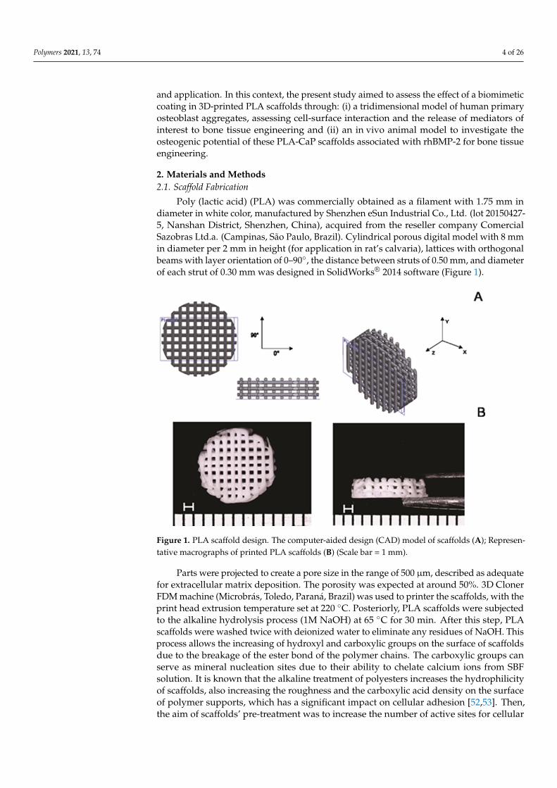
Poly (lactic acid) (PLA) was commercially obtained as a filament with 1.75 mm indiameter in white color, manufactured by Shenzhen eSun Industrial Co., Ltd. (lot 20150427-5, Nanshan District, Shenzhen, China), acquired from the reseller company ComercialSazobras Ltd.a. (Campinas, São Paulo, Brazil). Cylindrical porous digital model with 8 mmin diameter per 2 mm in height (for application in rat’s calvaria), lattices with orthogonalbeams with layer orientation of 0–90◦, the distance between struts of 0.50 mm, and diameterof each strut of 0.30 mm was designed in SolidWorks® 2014 software (Figure 1).
Polymers 2021, 13, x FOR PEER REVIEW 4 of 27
application. In this context, the present study aimed to assess the effect of a biomimetic coating in 3D-printed PLA scaffolds through: (i) a tridimensional model of human pri-mary osteoblast aggregates, assessing cell-surface interaction and the release of mediators of interest to bone tissue engineering and (ii) an in vivo animal model to investigate the osteogenic potential of these PLA-CaP scaffolds associated with rhBMP-2 for bone tissue engineering.
2. Materials and Methods 2.1. Scaffold Fabrication
Poly (lactic acid) (PLA) was commercially obtained as a filament with 1.75 mm in diameter in white color, manufactured by Shenzhen eSun Industrial Co., Ltd. (lot 20150427-5, Nanshan District, Shenzhen, China), acquired from the reseller company Comercial Sazobras Ltd.a. (Campinas, São Paulo, Brazil). Cylindrical porous digital model with 8 mm in diameter per 2 mm in height (for application in rat’s calvaria), lattices with orthogonal beams with layer orientation of 0–90°, the distance between struts of 0.50 mm, and diameter of each strut of 0.30 mm was designed in SolidWorks® 2014 software (Figure 1).
Figure 1. PLA scaffold design. The computer-aided design (CAD) model of scaffolds (A); Repre-sentative macrographs of printed PLA scaffolds (B) (Scale bar = 1 mm).
Parts were projected to create a pore size in the range of 500 µm, described as ade-quate for extracellular matrix deposition. The porosity was expected at around 50%. 3D Cloner FDM machine (Microbrás, Toledo, Paraná, Brazil) was used to printer the scaf-folds, with the print head extrusion temperature set at 220 °C. Posteriorly, PLA scaffolds were subjected to the alkaline hydrolysis process (1M NaOH) at 65 °C for 30 min. After this step, PLA scaffolds were washed twice with deionized water to eliminate any resi-dues of NaOH. This process allows the increasing of hydroxyl and carboxylic groups on the surface of scaffolds due to the breakage of the ester bond of the polymer chains. The carboxylic groups can serve as mineral nucleation sites due to their ability to chelate cal-cium ions from SBF solution. It is known that the alkaline treatment of polyesters increases the hydrophilicity of scaffolds, also increasing the roughness and the carboxylic acid den-sity on the surface of polymer supports, which has a significant impact on cellular adhe-sion [52,53]. Then, the aim of scaffolds’ pre-treatment was to increase the number of active
Figure 1. PLA scaffold design. The computer-aided design (CAD) model of scaffolds (A); Represen-tative macrographs of printed PLA scaffolds (B) (Scale bar = 1 mm).
Parts were projected to create a pore size in the range of 500 µm, described as adequatefor extracellular matrix deposition. The porosity was expected at around 50%. 3D ClonerFDM machine (Microbrás, Toledo, Paraná, Brazil) was used to printer the scaffolds, with theprint head extrusion temperature set at 220 ◦C. Posteriorly, PLA scaffolds were subjectedto the alkaline hydrolysis process (1M NaOH) at 65 ◦C for 30 min. After this step, PLAscaffolds were washed twice with deionized water to eliminate any residues of NaOH. Thisprocess allows the increasing of hydroxyl and carboxylic groups on the surface of scaffoldsdue to the breakage of the ester bond of the polymer chains. The carboxylic groups canserve as mineral nucleation sites due to their ability to chelate calcium ions from SBFsolution. It is known that the alkaline treatment of polyesters increases the hydrophilicityof scaffolds, also increasing the roughness and the carboxylic acid density on the surfaceof polymer supports, which has a significant impact on cellular adhesion [52,53]. Then,the aim of scaffolds’ pre-treatment was to increase the number of active sites for cellular
Polymers 2021, 13, 74 5 of 26
binding to materials, to favor apatite deposition by the biomimetic coating and to removepossible non-projected filaments present in the printed scaffolds.
2.2. Apatite Formation on the Surface of PLA Scaffolds (PLA-CaP)
According to the protocol established by Kokubo and Takadama (2006) [36], the SBFsolution was prepared in 1.0 and 1.5 concentrations, by dissolving the following reagents:NaCl, NaHCO3, KCl, K2HPO4 3H2O, CaCl2, Na2SO4 and MgCl2. 6H2O in distilled wa-ter. The pH was adjusted to 7.4 by adding 0.01 M Tris-hydroxymethyl aminomethane((CH2OH)3CNH2) (Tris) and 0.01 molL−1 HCl at 36.5 ◦C. The ionic concentration of 1.0 and1.5 SBF solutions in comparison with those of human blood plasma are show in Table 1.
Table 1. Ionic concentration of blood plasma, 1.0 and 1.5 SBF solutions.
SolutionConcentrations (mol/m3)
Na+ K+ Mg2+ Ca2+ Cl− HCO3− HPO43− SO42−
Blood plasma 142.0 5.0 1.5 2.5 103.0 27.0 1.0 0.5SBF 142.0 5.0 1.5 2.5 147.8 4.2 1.0 0.5
1.5 SBF 213.0 7.5 2.3 3.8 221.7 6.3 1.5 0.8
Apatite formation on PLA scaffolds’ surface was submitted to a procedure inspiredby Chim et al. [54]. A pilot was conducted to determine the scaffolds’ optimal exposuretime in SBF solution at room temperature (data not shown). It was established that a totalof 14 days of exposure were sufficient to form a homogeneous apatite layer since longerperiods (21 days) produced an irregular and thick apatite layer with flaking points.
Scaffolds were immersed for seven days in 15 mL of SBF (pH 7.4, 37 ◦C) with ionicconcentration similar to human blood plasma (1.0 SBF). In this period, it is expected initialmineral nucleation into carboxylic groups, promoting a good adhesion between the coatingand the substrate [55,56]. After this, scaffolds were re-immersed in 1.5 SBF solutions foran additional 7 days, with the solution refreshed every two days. This step was given tomaking calcium phosphates crystals grow. On day 14, after the CaP coating (PLA-CaP),scaffolds were washed with deionized water and dried at room temperature. All 3D printedscaffolds were sterilized by ethylene oxide (EtO), since it is approved by ISO 14937:2009standard [57]. Scaffolds were packed in self-seal sterilization pouches (Surgical Grade PaperThermoSealing + Pet film/PP Coex, CIPAMED, Brazil) and sent to the company ACECIL(Campinas, São Paulo, Brazil) to be sterilized by ethylene oxide. The operational ranges ofmain parameters were: temperature of 45–50 ◦C, gas concentration of 450–470 mg/L andexposure time of 24 h. After EtO sterilization, scaffolds were stored at room temperatureuntil in vitro and in vivo assays.
2.3. Characterization of PLA and PLA-CaP Scaffolds
The scaffolds’ morphology was assessed by Scanning Electron Microscopy (SEM, JEOLJSM 6460 LV, Peabody, MA, USA) operating at 15 KV, coupled with Energy DispersiveSpectroscopy (EDS-Thermo/System Six model 200, Peabody, MA, USA), which evaluatedelemental composition. The samples were placed on aluminum stubs with carbon tape,sputtered-coated with a thin gold layer (Emitech, K550, Abington, MA, USA) to avoidelectrical charging. Fourier transform infrared spectroscopy (FTIR, Spectrum 100, PerkinElmer spectrometer, Shelton, CT, USA) was performed in the range of 4000–550 cm–1,32 scans, and resolution 4 cm−1, using the ATR (attenuated total reflectance) mode. Thecrystalline phase of CaP on coated PLA scaffolds was assayed using X-ray diffraction (XRD).Data were collected with a Brüker D8 Discover diffractometer (Madison, WI, USA), usingCu Kα radiation, 2θ = 3◦ to 60◦ with a scan speed 1.23◦/min. For XDR analysis, PLA andPLA-CaP dense, square samples were produced. To evaluate the presence of the formationof a CaP coating onto the surface of the PLA-HA scaffolds, a thermogravimetric analysis(TGA) was performed with a Simultaneous Thermal Analyzer (STA) 6000 (Perkin Elmer,
Polymers 2021, 13, 74 6 of 26
Shelton, CT, USA), with a heating rate of 10.00 ◦C/min, from 22 ◦C to 700 ◦C, synthetic airatmosphere, a flow rate of 30 mL/min, on platinum support. The compressive propertiesof uncoated PLA and PLA-CaP scaffolds were measured in compression in an Instron 33R5567 tester (Instron, São José dos Pinhais, PR, Brazil), with a 5 kN load cell and crossheadspeed of 1 mm/min. For compressive analysis, PLA and PLA-HA, porous scaffolds with4 mm in axis z-axis, 6 × 6 mm in x and y-axes, respectively) were produced. Samples wereloaded until 50% strain.
2.4. Dimensional Deviation and Porosity
To evaluate if variations occurred in the scaffolds’ dimensions with the CAD modelused, scaffolds were measured (n = 5) with a digital caliper in all dimensions. The percent-age porosity was estimated as ratio between volume of pores within the scaffolds, VP, andthe volume of the scaffolds, VT, by following the ASTM F2450—18 Standard Guide [58](Equation (1)).
Porosity (%) = (VP/VT) × 100 (1)
Scaffolds volume, VT, was calculated by Equation (2) using the external dimensionsof printed parts: diameter (d) and height (h). While an estimative of the volume of pores,VP, was obtained using Equation (3).
VT =µ× d2
4× h (2)
VP = VT −mSρS
(3)
where ms is the mass of scaffolds and ρS is density of scaffolds.The Archimedes principle was employed to determine the density of scaffolds, ρS. A
hydrostatic balance was used to measure the dry mass, mdry, and the apparent mass ofscaffolds after immersion in ethanol, mwet. Density (ρS) was calculated by Equation (4), thedensity of ethanol, ρethanol , as 0.79 g/cm3 at the experiment’s temperature.
ρS =mS × ρethanol(mdry −mwet
) (4)
2.5. In Vitro Biological Evaluation2.5.1. Cytocompatibility Assay
Before assessing cell-surface interactions, a standardized cytocompatibility assay wasperformed according to ISO10993-5:2009 [59] in order to determine if the scaffolds couldindirectly affect cell viability. PLA and PLA-CaP scaffolds were immersed in DMEM(Dulbecco’s Modified Eagle Medium) at 200 mg/mL ratio and incubated for 24 h at37 ◦C/5% CO2 to produce conditioned media for indirect cell exposure.
Primary human Osteoblasts (HOb) were obtained from the collection of the ClinicalResearch Unit of Antônio Pedro University Hospital -UFF (UPC-HUAP-UFF, Niterói, RJ,Brazil) (Ethics Committee Approval 57080116.0.0000.5243). Cells maintained in DMEMcontaining 10% fetal bovine serum (FBS) and 1% penicillin/streptomycin at 37 ◦C/5% CO2were trypsinized and subcultured in 96-well plates at a density of 1.0 × 10³ cells for 24 huntil subconfluence. The culture media was replaced with the conditioned media of eachscaffold type in quintuplicates, and cells were exposed for 24 h at 37 ◦C/5% CO2. Mediaconditioned with biocompatible high-density polystyrene beads or fragments of latex, ata 200 mg/mL ratio, were employed as a negative and positive control for cytotoxicity,respectively. Cell viability was assessed with a commercial XTT assay kit (InCytotox,Xenometrix, Allschwil, Switzerland), performed according to the manufacturer, with cellviability related to the Optical Density (OD) measured with a Synergy II Microplate Reader(Biotek Inst., Winooski, VT, USA) at 405 nm.
Polymers 2021, 13, 74 7 of 26
2.5.2. Production of the 3D Osteoblasts Aggregates (Osteospheres)
The 3D model of human primary osteoblasts was produced through a protocol mod-ified from Restle et al. (2015) [49]. HOb cells were seeded on a density of 5 × 10³ cellsper well into a 96 well conical bottom plate covered by 1% sterile agar and maintainedin 150 uL of medium supplemented with 10% of FBS. Cells were incubated for 4 daysat 37 ◦C/5% CO2 on an orbital shaker set at 250 rpm, for the formation of aggregates.These had a mean diameter of 320 ± 38 micrometers and an aspect ratio of 1.05 ± 0.1(Height/Length), indicating a regular spheroid shape (osteospheres). After seven daysof culture, the number of viable cells within the spheroids was measured by trypan bluecounting after disaggregation with trypsin/collagenase always remained near 5 × 10³ cells(data not shown), indicating the stability of the model during the experimental times ofthis study.
2.5.3. Exposure of Osteospheres to 3D PLA and PLA-CaP Scaffolds
To simulate the interactions with protein-containing complex biological media, sam-ples of both groups of scaffolds (PLA and PLA-CaP) were pre-treated with inactivatedfetal bovine serum by immersion on 300 mL of pure FBS for 24 h. Therefore, four ex-perimental groups were investigated with in vitro assays: PLA, PLA-CaP, PLA+FBS, andPLA-CaP+FBS. Scaffolds from each group were placed in a 24 well plate (one scaffold perwell), and a total of eight aggregates were seeded on different points of the surface of eachscaffold. The plate was incubated for 7 days in 500 µL of DMEM supplemented with 10%of SFB at 37 ◦C/5% CO2. The culture medium was changed on the 4th day of culture.
2.5.4. Assessment of Cell-Surface Interactions
For the assessment of cell-surface interactions through fluorescence microscopy, thesamples were fixed in 4% paraformaldehyde, washed with PBS, and then exposed for10 min to an ammonium chloride solution. The samples were washed with PBS, and thencells had their membranes permeabilized with treatment with 1% Triton X-100 for 10 min.After washing with PBS, samples were immersed in 3% bovine serum albumin for 10 minand then stained with Phalloidin conjugated with Fluorescein isothiocyanate (FITC) (1:100)for cytoskeleton labeling for 30 min and 1:5000 DAPI (4′,6-diamidino-2-phenylindole),a nuclear marker, for 30 min. The samples were stored in a DABCO (1,4-diazabicyclo[2.2.2]octane) solution.
The cell density on the different scaffolds was compared in the different scaffolds fromtwo parameters: total protein content and the release of the cytoplasmic enzyme lactatedehydrogenase (LDH) after total cell permeabilization. At 7 days, scaffolds were washedwith PBS 1x, then exposed to 200 µm of serum-free DMEM containing 1% Triton X-100,and incubated for 24 h. The samples were diluted in PBS for decreasing the Triton X-100concentration from 1% to 0.01%.
A sample of 5 µL of each culture media was used to evaluate the total released proteinwith a Bradford assay kit (Protein Assay, Biorad, Hercules, CA, USA). Optical density wasmeasured at 595 nm using a Sinergy II Microplate Reader (Biotek Instruments, Winooski,VT, USA. The total amount of protein after permeabilization was subtracted from theprotein quantified before treatment with triton X100. A sample of 20 µL of each well wastransferred to another plate and added with 240 µL LDH II and LDH III solutions (InCytotox, Xenometrix, Allschwil, Switzerland). Optical density was measured at 540 nmusing a Sinergy II microplate reader (Biotek Instruments, Winooski, VT, USA) at kineticmode for 25 min at 37 ◦C.
2.5.5. Release of Biological Mediators
The release of some different biological mediators of interest to tissue engineeringby the exposed osteospheres was assessed of each scaffold/treatment culture media. Thequantification of the release of the basic Fibroblasts Growth Factor (FDF2), Platelet-DerivedGrowth Factor (PDGFbb) and the Vascular Endothelial Growth Factor (VEGF) was per-
Polymers 2021, 13, 74 8 of 26
formed using an Enzyme-Linked Immunosorbent Assay (ELISA) with standard ABTSELISA Development Kits (PeproTech, Rocky Hill, NJ, USA). The procedure followed therecommendations of the manufacturer. The reactions were stopped with a 5% sodiumdodecyl sulfate (SDS), and the OD was measured in a Synergy II microplate reader (Syn-ergy II, Biotek Instruments, Winooski, VT, USA) at 405 nm with wavelength correctionset at 650 nm. The activity of Alkaline Phosphatase (ALP), a marker of the early stages ofosteoblast maturation, was measured with a Kinetic Alkaline Phosphatase kit (Bioclin, Riode Janeiro, RJ, Brazil), in which the enzyme causes para nitrophenyl phosphate (PNPP)hydrolysis, and this process is measured colorimetrically. After incubation, the scaffoldswere washed with PBS 1x, then exposed to 200 µL of a medium solution without SFB with1% Triton X-100 and incubated for 24 h. 60 µL of the samples were transferred to a 96 wellmicroplate, and 200 µL of reagent was mixed per well. The plate was incubated for 3 h,and two readings were taken every 30 min at 37 ◦C at 405 nm.
2.6. In Vivo Study2.6.1. Ethical Aspects
The animal breeding and procedures followed the conventional guidelines of theNIH Guide for the Care and Use of Laboratory Animals, the Brazilian Directive for theCare and Use of Animals for Scientific and Didactic Purposes—DBCA, and the EuthanasiaPractice Guidelines of the CONCEA (Brazilian National Council for the Control of AnimalExperimentation). The Ethics Committee of Animal Use from Fluminense Federal Uni-versity (CEUA/UFF, Niterói, RJ, Brazil) approved this work’s research protocol (protocolnumber #934). The present study’s report considered the ARRIVE guidelines concerningthe relevant items [60] supplemented by PREPARE [61].
2.6.2. Welfare of Animals
Before and after surgical procedures, the animals were kept in individual cagesand under standard conditions (water ad libitum and regular rat pellets). The ambienttemperature was maintained between 16 and 20 ◦C, being ideal for the growth of theanimals and photoperiod control of 12 h light and 12 dark hours was established for thedevelopment of the complete metabolic cycle.
2.6.3. Experimental Groups
Forty-five Wistar rats, weighing between 300 and 400 g, were randomly divided intothree experimental groups as follows: PLA, PLA-CaP, and PLA-CaP plus 5 µh rhBMP2(Infuse® Bone Graft Small Kit, Medtronic Spinal and Biologics, Memphis, TN, USA) (PLA-CaP-BMP2) (n = 15). The scaffolds implantation was conducted following ISO standard10993-6 [62]. The animals were also randomly divided into three experimental periods of 1,3, and 6 months (n = 5).
2.6.4. Anesthesia and Surgical Procedures
The animals were deprived of the solid diet six hours before the surgery and submittedto general anesthesia, receiving as anesthetic medication a solution composed of 100 mg/kgof Ketamine (Francotar®—Virbac), 10 mg/kg of Xylazine (Sedazine®—Fort Dodge) and5 mg/Kg of Midazolam (Roche) intramuscularly.
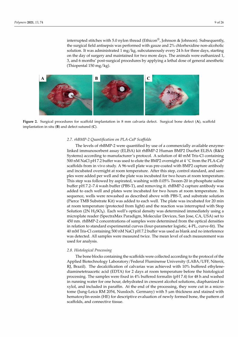
After general anesthesia and observing the absence of reflexes to the pain, the animalswere positioned in a ventral decubitus, trichotomized in calvaria, and submitted to theoperative field’s antisepsis with 2% non-alcoholic chlorhexidine. Subsequently, a semilunarincision was made on the calvaria using blade #15 C (Becton-Dickinson®, Curitiba, PR,Brazil). Then, with the aid of a periosteum detachment (Molt, Duflex, Barueri, SP, Brazil),the periosteum was removed. The bone defect was performed with a trephine bur with8 mm of internal diameter, mounted in contra-angle with reduction of 16:1, was coupled insurgical motor, at 1500 rpm, under constant irrigation with physiological solution. Finally,the scaffold was implanted on bone defect (Figure 2), the flap repositioned and sutured with
Polymers 2021, 13, 74 9 of 26
interrupted stitches with 5.0 nylon thread (Ethicon®, Johnson & Johnson). Subsequently,the surgical field antisepsis was performed with gauze and 2% chlorhexidine non-alcoholicsolution. It was administrated 1 mg/kg, subcutaneously every 24 h for three days, startingon the day of surgery and maintained for two more days. The animals were euthanized 1,3, and 6 months’ post-surgical procedures by applying a lethal dose of general anesthetic(Thiopental 150 mg/kg).
Polymers 2021, 13, x FOR PEER REVIEW 9 of 27
Finally, the scaffold was implanted on bone defect (Figure 2), the flap repositioned and sutured with interrupted stitches with 5.0 nylon thread (Ethicon®, Johnson & Johnson). Subsequently, the surgical field antisepsis was performed with gauze and 2% chlorhexi-dine non-alcoholic solution. It was administrated 1 mg/kg, subcutaneously every 24 h for three days, starting on the day of surgery and maintained for two more days. The animals were euthanized 1, 3, and 6 months’ post-surgical procedures by applying a lethal dose of general anesthetic (Thiopental 150 mg/kg).
Figure 2. Surgical procedures for scaffold implantation in 8 mm calvaria defect. Surgical bone defect (A), scaffold implan-tation in situ (B) and defect sutured (C).
2.7. rhBMP-2 Quantification on PLA-CaP Scaffolds The levels of rhBMP-2 were quantified by use of a commercially available enzyme-
linked immunosorbent assay (ELISA) kit rhBMP-2 Human BMP2 DuoSet ELISA (R&D Systems) according to manufacturer’s protocol. A solution of 40 mM Tris-Cl containing 500 nM NaCl pH 7.2 buffer was used to elute the BMP2 overnight at 4 °C from the PLA-CaP scaffolds from in vivo study. A 96-well plate was pre-coated with BMP2 capture an-tibody and incubated overnight at room temperature. After this step, control standard, and samples were added per well and the plate was incubated for two hours at room temperature. This step was followed by aspirated, washing with 0.05% Tween-20 in phos-phate saline buffer pH 7.2–7.4 wash buffer (PBS-T), and removing it. rhBMP-2 capture antibody was added to each well and plates were incubated for two hours at room tem-perature. In sequence, wells were rewashed as described above with PBS-T, and substrate solution (Pierce TMB Substrate Kit) was added to each well. The plate was incubated for 20 min at room temperature (protected from light) and the reaction was interrupted with Stop Solution (2N H2SO4). Each well’s optical density was determined immediately using a microplate reader (SpectraMax Paradigm, Molecular Devices, San Jose, CA, USA) set to 450 nm. rhBMP-2 concentrations of samples were determined from the optical densities in relation to standard experimental curves (four-parameter logistic, 4-PL, curve-fit). The 40 mM Tris-Cl containing 500 nM NaCl pH 7.2 buffer was used as blank and no interfer-ence was detected. All samples were measured twice. The mean level of each measure-ment was used for analysis.
2.8. Histological Processing The bone blocks containing the scaffolds were collected according to the protocol of
the Applied Biotechnology Laboratory/Federal Fluminense University (LABA/UFF, Nite-rói, RJ, Brazil). The decalcification of calvarias was achieved with 10% buffered ethylene-diaminetetraacetic acid (EDTA) for 2 days at room temperature before the histological processing. The samples were fixed in 4% buffered formalin (pH 7.4) for 48 h and washed in running water for one hour, dehydrated in crescent alcohol solutions, diaphanized in xylol, and included in paraffin. At the end of the processing, they were cut in a microtome (Jung-Leica RM 2054, Nussloch. Germany) with 5 µm thickness and stained with hema-toxylin-eosin (HE) for descriptive evaluation of newly formed bone, the pattern of scaf-folds, and connective tissue.
Figure 2. Surgical procedures for scaffold implantation in 8 mm calvaria defect. Surgical bone defect (A), scaffoldimplantation in situ (B) and defect sutured (C).
2.7. rhBMP-2 Quantification on PLA-CaP Scaffolds
The levels of rhBMP-2 were quantified by use of a commercially available enzyme-linked immunosorbent assay (ELISA) kit rhBMP-2 Human BMP2 DuoSet ELISA (R&DSystems) according to manufacturer’s protocol. A solution of 40 mM Tris-Cl containing500 nM NaCl pH 7.2 buffer was used to elute the BMP2 overnight at 4 ◦C from the PLA-CaPscaffolds from in vivo study. A 96-well plate was pre-coated with BMP2 capture antibodyand incubated overnight at room temperature. After this step, control standard, and sam-ples were added per well and the plate was incubated for two hours at room temperature.This step was followed by aspirated, washing with 0.05% Tween-20 in phosphate salinebuffer pH 7.2–7.4 wash buffer (PBS-T), and removing it. rhBMP-2 capture antibody wasadded to each well and plates were incubated for two hours at room temperature. Insequence, wells were rewashed as described above with PBS-T, and substrate solution(Pierce TMB Substrate Kit) was added to each well. The plate was incubated for 20 minat room temperature (protected from light) and the reaction was interrupted with StopSolution (2N H2SO4). Each well’s optical density was determined immediately using amicroplate reader (SpectraMax Paradigm, Molecular Devices, San Jose, CA, USA) set to450 nm. rhBMP-2 concentrations of samples were determined from the optical densitiesin relation to standard experimental curves (four-parameter logistic, 4-PL, curve-fit). The40 mM Tris-Cl containing 500 nM NaCl pH 7.2 buffer was used as blank and no interferencewas detected. All samples were measured twice. The mean level of each measurement wasused for analysis.
2.8. Histological Processing
The bone blocks containing the scaffolds were collected according to the protocol of theApplied Biotechnology Laboratory/Federal Fluminense University (LABA/UFF, Niterói,RJ, Brazil). The decalcification of calvarias was achieved with 10% buffered ethylene-diaminetetraacetic acid (EDTA) for 2 days at room temperature before the histologicalprocessing. The samples were fixed in 4% buffered formalin (pH 7.4) for 48 h and washedin running water for one hour, dehydrated in crescent alcohol solutions, diaphanized inxylol, and included in paraffin. At the end of the processing, they were cut in a micro-tome (Jung-Leica RM 2054, Nussloch. Germany) with 5 µm thickness and stained withhematoxylin-eosin (HE) for descriptive evaluation of newly formed bone, the pattern ofscaffolds, and connective tissue.
Polymers 2021, 13, 74 10 of 26
For the descriptive histological analysis, a Light Field Light microscope (OLYMPUS®
BX43, Tokyo, Japan) was used. Captures of the selected images were performed througha microscope-coupled camera (OLYMPUS® SC100, Tokyo, Japan), associated with high-resolution software (CELLSENS®1.9 DigitalImage, Tokyo, Japan).
2.9. Histomorphometric Analysis
Each histological calvaria slide was examined under a light microscope (OLYMPUSBX43, Tokyo, Japan). Five non-superimposing photomicrographs were captured at 40×magnification using a high-resolution digital camera (OLYMPUS BX43, Tokyo, Japan), tothe regions surrounding and into the implanted biomaterials. The histomorphometricevaluation was performed using Image-Pro Plus® 6.0 software (Media Cybernetics, SilverSpring, MD, USA) which generates a grid of 250 points that allowed for evaluating thevolume density of newly formed bone, biomaterial (scaffold), and connective tissue. Thevalues were stored in a database developed using Microsoft Excel® spreadsheet software.
2.10. Statistical Analysis
For the in vitro assessment, the results from three biological assays for each scaffoldtype and treatment (PLA, PLA-CaP, PLA+FBS, and PLA-CaP+FBS) were compared throughthe analysis of variance. A D’Agostino Pearsons normality test was performed, followedby Kruskal-Walis non-parametric tests with Dunn ad hoc post-test. Significance wasconsidered at 5% of alpha error. All statistical analyses were performed using GraphPadPrism 7.0 Software (GraphPad Inc., San Diego, CA, USA).
In the in vivo assessment, a quantitative description of the volume density of newlyformed bone, biomaterial (scaffold), and connective tissue was done by parametric descrip-tion with a p-value of < 0.05 considered significant. The obtained data were not normal(Shapiro-Wilk normality test); they were transformed in Y logarithm, and a p-value < 0.05was considered significant. ANOVA and Tukey’s post-tests were used to investigate thedifferences between groups at the same experimental period and differences between thesame group at different times. The calculations were performed using Prism Graph Pad7.0 software (Inc., La Jolla, CA, USA).
3. Results3.1. Characterization of PLA and PLA-CaP 3D Scaffolds
The uncoated PLA and PLA-CaP scaffolds were evaluated by optical microscopy, asshown in Figure 1. The printed scaffold presented a morphology compatible with theprojected CAD model (Figure 1 A,B). It was possible to design a scaffold suitable for bonetissue engineering, with interconnected pores. Also was seeing that the alkaline hydrolysiseffectively removed the possible non-projected filaments. The porosity of uncoated PLAand PLA-CaP scaffolds was found to be nearly 50%, with no difference between the groups.The pore volume for each was in the same range and indicated that CaP coating does notinterfere with the scaffold architecture (Table 1). Under compressive force, uncoated PLAand PLA-CaP scaffolds showed a plastic response with ~20 MPa of compressive strength,similar to trabecular bone (Table 2). Similarly, the elastic modulus, determined from theslope of the stress vs. strain curve, was ~0.5 GPa, also similar to trabecular bone.
Table 2. Mechanical properties and Archimedes calculus values (n= 5 ± SD). * Bose et al., 2013 [63].
Uncoated PLA PLA-CaP Trabecular Bone *
Compressive strength (MPa) 20.50 ± 1.95 18.22 ± 2.67 2–20Compressive Modulus (GPa) 0.512 ± 0.24 0.510 ± 0.11 0.1–2.0
Density (g/cm3) 1.23 ± 0.06 1.21 ± 0.02 –Pore volume (mm3) 42.64 ± 6.49 41.61 ± 5.17 –
Porosity (%) 49.93 ± 5.28 49.09 ± 3.22 30–90
Polymers 2021, 13, 74 11 of 26
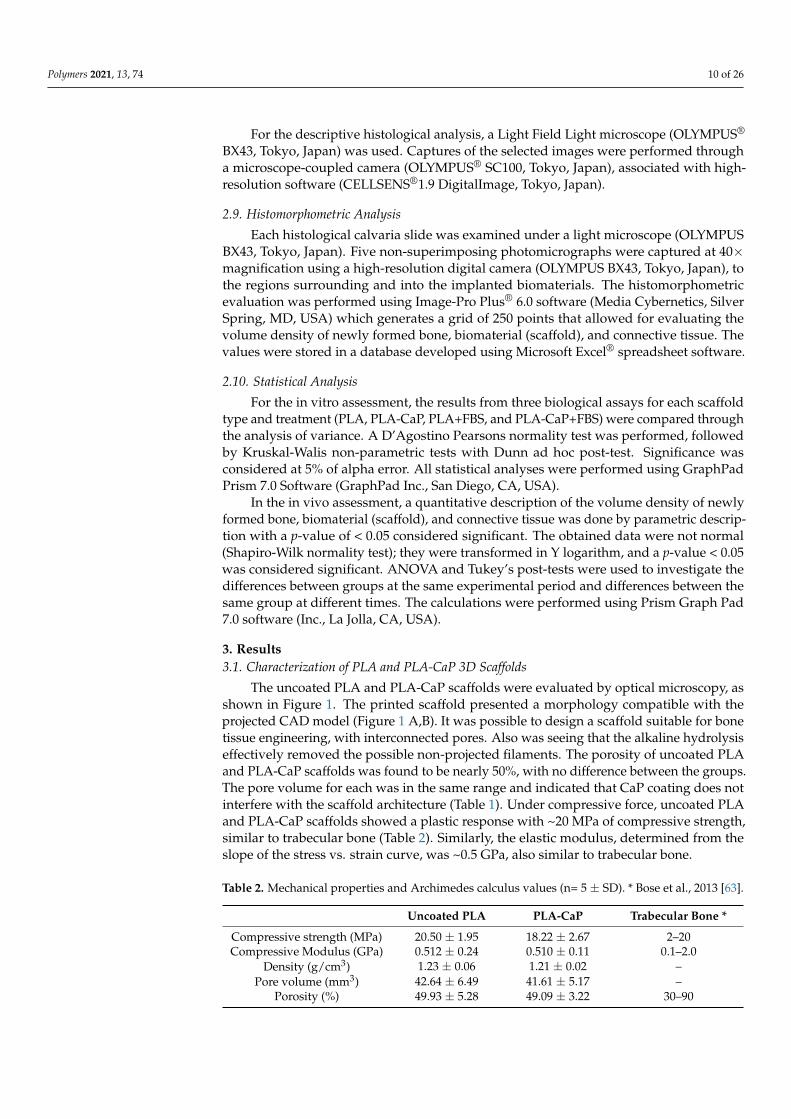
The SEM micrographs of the 3D scaffolds are shown in Figure 3. CaP biomimeticcoating was effective, forming a continuous and uniform bone-like apatite layer over theentire surface of PLA (Figure 3E,F). This behavior indicates that the number of nucleatingsites on the polymer’s surface during the first hours of immersion was high, favoring theformation of a dense and uniform CaP layer. The homogeneity of CaP layer is directlyrelated to the presence of nucleating sites during the first hours of SBF immersion. Thesebioactive sites may result from the alkaline hydrolysis, which incorporates OH-groups,tending to facilitate the Ca2+ deposition onto the surface [64].
Polymers 2021, 13, x FOR PEER REVIEW 11 of 27
Compressive Modu-lus (GPa)
0.512 ± 0.24 0.510 ± 0.11 0.1–2.0
Density (g/cm3) 1.23 ± 0.06 1.21 ± 0.02 –
Pore volume (mm3) 42.64 ± 6.49 41.61 ± 5.17 –
Porosity (%) 49.93 ± 5.28 49.09 ± 3.22 30–90
The SEM micrographs of the 3D scaffolds are shown in Figure 3. CaP biomimetic coating was effective, forming a continuous and uniform bone-like apatite layer over the entire surface of PLA (Figure 3E,F). This behavior indicates that the number of nucleating sites on the polymer’s surface during the first hours of immersion was high, favoring the formation of a dense and uniform CaP layer. The homogeneity of CaP layer is directly related to the presence of nucleating sites during the first hours of SBF immersion. These bioactive sites may result from the alkaline hydrolysis, which incorporates OH-groups, tending to facilitate the Ca2+ deposition onto the surface [64].
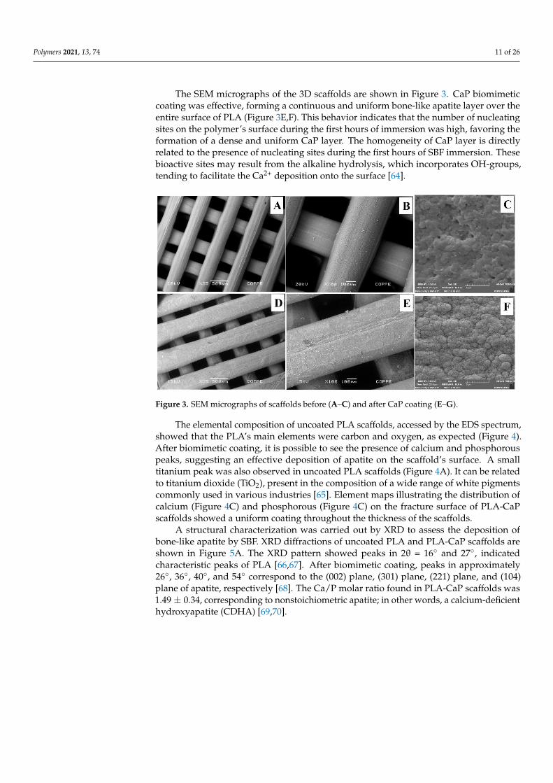
The elemental composition of uncoated PLA scaffolds, accessed by the EDS spec-trum, showed that the PLA’s main elements were carbon and oxygen, as expected (Figure 4). After biomimetic coating, it is possible to see the presence of calcium and phosphorous peaks, suggesting an effective deposition of apatite on the scaffold’s surface. A small tita-nium peak was also observed in uncoated PLA scaffolds (Figure 4A). It can be related to titanium dioxide (TiO2), present in the composition of a wide range of white pigments commonly used in various industries [65]. Element maps illustrating the distribution of calcium (Figure 4C) and phosphorous (Figure 4C) on the fracture surface of PLA-CaP scaf-folds showed a uniform coating throughout the thickness of the scaffolds.
Figure 3. SEM micrographs of scaffolds before (A–C) and after CaP coating (E–G). Figure 3. SEM micrographs of scaffolds before (A–C) and after CaP coating (E–G).
The elemental composition of uncoated PLA scaffolds, accessed by the EDS spectrum,showed that the PLA’s main elements were carbon and oxygen, as expected (Figure 4).After biomimetic coating, it is possible to see the presence of calcium and phosphorouspeaks, suggesting an effective deposition of apatite on the scaffold’s surface. A smalltitanium peak was also observed in uncoated PLA scaffolds (Figure 4A). It can be relatedto titanium dioxide (TiO2), present in the composition of a wide range of white pigmentscommonly used in various industries [65]. Element maps illustrating the distribution ofcalcium (Figure 4C) and phosphorous (Figure 4C) on the fracture surface of PLA-CaPscaffolds showed a uniform coating throughout the thickness of the scaffolds.
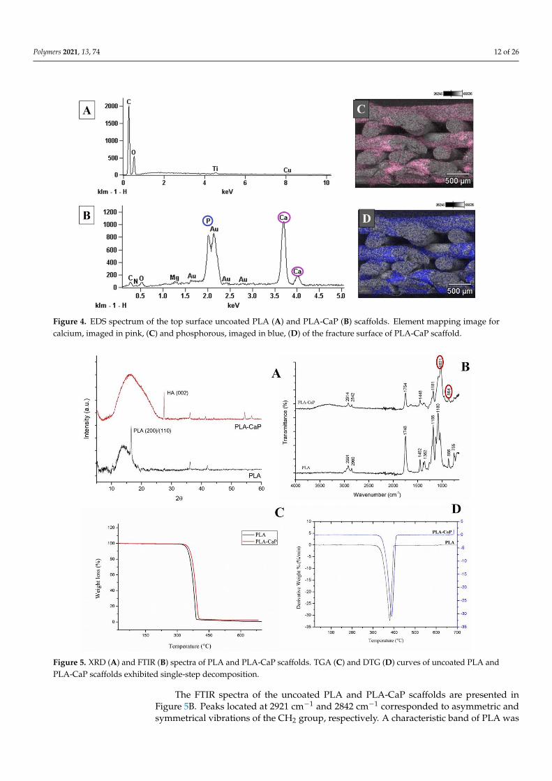
A structural characterization was carried out by XRD to assess the deposition ofbone-like apatite by SBF. XRD diffractions of uncoated PLA and PLA-CaP scaffolds areshown in Figure 5A. The XRD pattern showed peaks in 2θ = 16◦ and 27◦, indicatedcharacteristic peaks of PLA [66,67]. After biomimetic coating, peaks in approximately26◦, 36◦, 40◦, and 54◦ correspond to the (002) plane, (301) plane, (221) plane, and (104)plane of apatite, respectively [68]. The Ca/P molar ratio found in PLA-CaP scaffolds was1.49 ± 0.34, corresponding to nonstoichiometric apatite; in other words, a calcium-deficienthydroxyapatite (CDHA) [69,70].
Polymers 2021, 13, 74 12 of 26Polymers 2021, 13, x FOR PEER REVIEW 12 of 27
Figure 4. EDS spectrum of the top surface uncoated PLA (A) and PLA-CaP (B) scaffolds. Element mapping image for calcium, imaged in pink, (C) and phosphorous, imaged in blue, (D) of the fracture surface of PLA-CaP scaffold.
A structural characterization was carried out by XRD to assess the deposition of bone-like apatite by SBF. XRD diffractions of uncoated PLA and PLA-CaP scaffolds are shown in Figure 5A. The XRD pattern showed peaks in 2θ = 16° and 27°, indicated char-acteristic peaks of PLA [66,67]. After biomimetic coating, peaks in approximately 26°, 36°, 40°, and 54° correspond to the (002) plane, (301) plane, (221) plane, and (104) plane of apatite, respectively [68]. The Ca/P molar ratio found in PLA-CaP scaffolds was 1.49 ± 0.34, corresponding to nonstoichiometric apatite; in other words, a calcium-deficient hy-droxyapatite (CDHA) [69,70].
The FTIR spectra of the uncoated PLA and PLA-CaP scaffolds are presented in Figure 5B. Peaks located at 2921 cm−1 and 2842 cm−1 corresponded to asymmetric and symmetrical vibrations of the CH2 group, respectively. A characteristic band of PLA was found at 1746 cm−1, corresponding to the vibration of the carbonyl group [71,72]. PLA-CaP spectrum shows the presence of phosphate (1021 cm−1) and carbonate groups (867 cm−1), confirming the deposition of apatite coating [73,74]. A peak at 1448 cm−1 in PLA-CaP suggests that the apatite obtained is carbonate-containing hydroxyapatite (CO32− ion).
The thermal properties of uncoated PLA and PLA-CaP scaffolds were investigated by TGA (Figure 5C,D). The derivative curves of mass loss (DTG) are shown in Figure 5D. Considering the amount of the residues obtained in TGA analysis, it was possible to infer that approximately 2% wt of apatite was deposited onto the scaffolds. Both DTG curves exhibit a single-step of decomposition, where the main degradation temperature in-creased with the deposition of apatite (from 379.47 °C in uncoated PLA to 384.86 °C in PLA-CaP).
Figure 4. EDS spectrum of the top surface uncoated PLA (A) and PLA-CaP (B) scaffolds. Element mapping image forcalcium, imaged in pink, (C) and phosphorous, imaged in blue, (D) of the fracture surface of PLA-CaP scaffold.
Polymers 2021, 13, x FOR PEER REVIEW 13 of 27
Figure 5. XRD (A) and FTIR (B) spectra of PLA and PLA-CaP scaffolds. TGA (C) and DTG (D) curves of uncoated PLA and PLA-CaP scaffolds exhibited single-step decomposition.
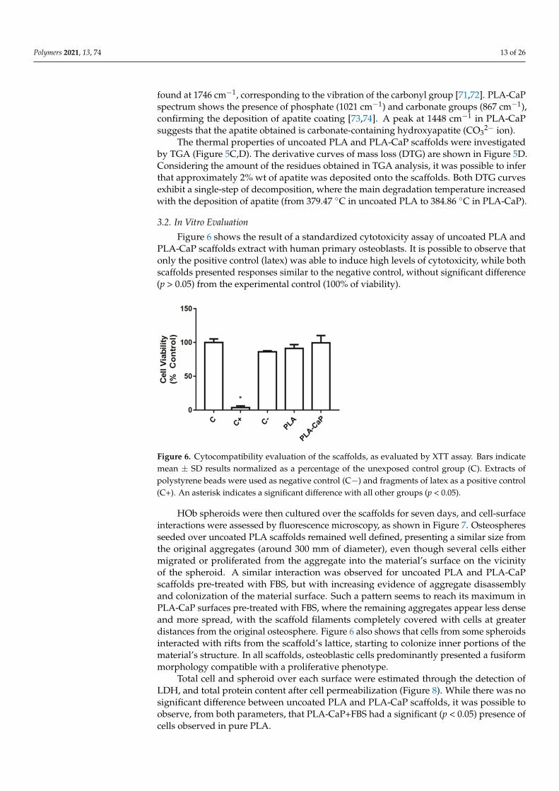
3.2. In Vitro Evaluation Figure 6 shows the result of a standardized cytotoxicity assay of uncoated PLA and
PLA-CaP scaffolds extract with human primary osteoblasts. It is possible to observe that only the positive control (latex) was able to induce high levels of cytotoxicity, while both scaffolds presented responses similar to the negative control, without significant differ-ence (p > 0.05) from the experimental control (100% of viability).
Figure 6. Cytocompatibility evaluation of the scaffolds, as evaluated by XTT assay. Bars indicate mean ± SD results normalized as a percentage of the unexposed control group (C). Extracts of pol-ystyrene beads were used as negative control (C−) and fragments of latex as a positive control (C+). An asterisk indicates a significant difference with all other groups (p < 0.05).
Figure 5. XRD (A) and FTIR (B) spectra of PLA and PLA-CaP scaffolds. TGA (C) and DTG (D) curves of uncoated PLA andPLA-CaP scaffolds exhibited single-step decomposition.
The FTIR spectra of the uncoated PLA and PLA-CaP scaffolds are presented inFigure 5B. Peaks located at 2921 cm−1 and 2842 cm−1 corresponded to asymmetric andsymmetrical vibrations of the CH2 group, respectively. A characteristic band of PLA was
Polymers 2021, 13, 74 13 of 26
found at 1746 cm−1, corresponding to the vibration of the carbonyl group [71,72]. PLA-CaPspectrum shows the presence of phosphate (1021 cm−1) and carbonate groups (867 cm−1),confirming the deposition of apatite coating [73,74]. A peak at 1448 cm−1 in PLA-CaPsuggests that the apatite obtained is carbonate-containing hydroxyapatite (CO3
2− ion).The thermal properties of uncoated PLA and PLA-CaP scaffolds were investigated
by TGA (Figure 5C,D). The derivative curves of mass loss (DTG) are shown in Figure 5D.Considering the amount of the residues obtained in TGA analysis, it was possible to inferthat approximately 2% wt of apatite was deposited onto the scaffolds. Both DTG curvesexhibit a single-step of decomposition, where the main degradation temperature increasedwith the deposition of apatite (from 379.47 ◦C in uncoated PLA to 384.86 ◦C in PLA-CaP).
3.2. In Vitro Evaluation
Figure 6 shows the result of a standardized cytotoxicity assay of uncoated PLA andPLA-CaP scaffolds extract with human primary osteoblasts. It is possible to observe thatonly the positive control (latex) was able to induce high levels of cytotoxicity, while bothscaffolds presented responses similar to the negative control, without significant difference(p > 0.05) from the experimental control (100% of viability).
Polymers 2021, 13, x FOR PEER REVIEW 13 of 27
Figure 5. XRD (A) and FTIR (B) spectra of PLA and PLA-CaP scaffolds. TGA (C) and DTG (D) curves of uncoated PLA and PLA-CaP scaffolds exhibited single-step decomposition.
3.2. In Vitro Evaluation Figure 6 shows the result of a standardized cytotoxicity assay of uncoated PLA and
PLA-CaP scaffolds extract with human primary osteoblasts. It is possible to observe that only the positive control (latex) was able to induce high levels of cytotoxicity, while both scaffolds presented responses similar to the negative control, without significant differ-ence (p > 0.05) from the experimental control (100% of viability).
Figure 6. Cytocompatibility evaluation of the scaffolds, as evaluated by XTT assay. Bars indicate mean ± SD results normalized as a percentage of the unexposed control group (C). Extracts of pol-ystyrene beads were used as negative control (C−) and fragments of latex as a positive control (C+). An asterisk indicates a significant difference with all other groups (p < 0.05).
Figure 6. Cytocompatibility evaluation of the scaffolds, as evaluated by XTT assay. Bars indicatemean ± SD results normalized as a percentage of the unexposed control group (C). Extracts ofpolystyrene beads were used as negative control (C−) and fragments of latex as a positive control(C+). An asterisk indicates a significant difference with all other groups (p < 0.05).
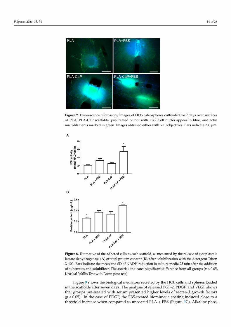
HOb spheroids were then cultured over the scaffolds for seven days, and cell-surfaceinteractions were assessed by fluorescence microscopy, as shown in Figure 7. Osteospheresseeded over uncoated PLA scaffolds remained well defined, presenting a similar size fromthe original aggregates (around 300 mm of diameter), even though several cells eithermigrated or proliferated from the aggregate into the material’s surface on the vicinityof the spheroid. A similar interaction was observed for uncoated PLA and PLA-CaPscaffolds pre-treated with FBS, but with increasing evidence of aggregate disassemblyand colonization of the material surface. Such a pattern seems to reach its maximum inPLA-CaP surfaces pre-treated with FBS, where the remaining aggregates appear less denseand more spread, with the scaffold filaments completely covered with cells at greaterdistances from the original osteosphere. Figure 6 also shows that cells from some spheroidsinteracted with rifts from the scaffold’s lattice, starting to colonize inner portions of thematerial’s structure. In all scaffolds, osteoblastic cells predominantly presented a fusiformmorphology compatible with a proliferative phenotype.
Total cell and spheroid over each surface were estimated through the detection ofLDH, and total protein content after cell permeabilization (Figure 8). While there was nosignificant difference between uncoated PLA and PLA-CaP scaffolds, it was possible toobserve, from both parameters, that PLA-CaP+FBS had a significant (p < 0.05) presence ofcells observed in pure PLA.
Polymers 2021, 13, 74 14 of 26
Polymers 2021, 13, x FOR PEER REVIEW 14 of 27
HOb spheroids were then cultured over the scaffolds for seven days, and cell-surface interactions were assessed by fluorescence microscopy, as shown in Figure 7. Osteo-spheres seeded over uncoated PLA scaffolds remained well defined, presenting a similar size from the original aggregates (around 300 mm of diameter), even though several cells either migrated or proliferated from the aggregate into the material’s surface on the vicin-ity of the spheroid. A similar interaction was observed for uncoated PLA and PLA-CaP scaffolds pre-treated with FBS, but with increasing evidence of aggregate disassembly and colonization of the material surface. Such a pattern seems to reach its maximum in PLA-CaP surfaces pre-treated with FBS, where the remaining aggregates appear less dense and more spread, with the scaffold filaments completely covered with cells at greater distances from the original osteosphere. Figure 6 also shows that cells from some spheroids inter-acted with rifts from the scaffold’s lattice, starting to colonize inner portions of the mate-rial’s structure. In all scaffolds, osteoblastic cells predominantly presented a fusiform mor-phology compatible with a proliferative phenotype.
Figure 7. Fluorescence microscopy images of HOb osteospheres cultivated for 7 days over surfaces of PLA, PLA-CaP scaffolds, pre-treated or not with FBS. Cell nuclei appear in blue, and actin mi-crofilaments marked in green. Images obtained either with X10 objectives. Bars indicate 200 µm.
Total cell and spheroid over each surface were estimated through the detection of LDH, and total protein content after cell permeabilization (Figure 8). While there was no significant difference between uncoated PLA and PLA-CaP scaffolds, it was possible to observe, from both parameters, that PLA-CaP+FBS had a significant (p < 0.05) presence of cells observed in pure PLA.
Figure 7. Fluorescence microscopy images of HOb osteospheres cultivated for 7 days over surfacesof PLA, PLA-CaP scaffolds, pre-treated or not with FBS. Cell nuclei appear in blue, and actinmicrofilaments marked in green. Images obtained either with ×10 objectives. Bars indicate 200 µm.
Polymers 2021, 13, x FOR PEER REVIEW 15 of 27
Figure 8. Estimative of the adhered cells to each scaffold, as measured by the release of cytoplas-mic lactate dehydrogenase (A) or total protein content (B), after solubilization with the detergent Triton X-100. Bars indicate the mean and SD of NADH reduction in culture media 25 min after the addition of substrates and solubilizer. The asterisk indicates significant difference from all groups (p < 0.05, Kruskal-Wallis Test with Dunn post-test).
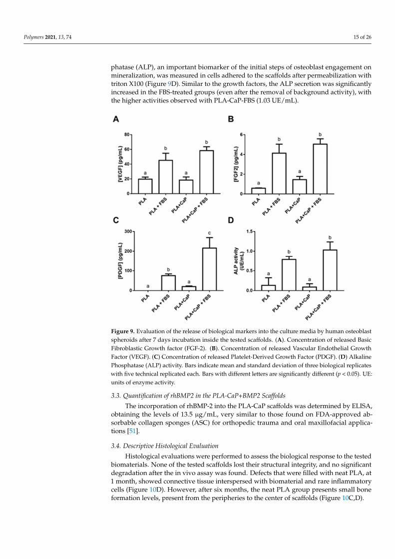
Figure 9 shows the biological mediators secreted by the HOb cells and spheres loaded in the scaffolds after seven days. The analysis of released FGF-2, PDGF, and VEGF shows that groups pre-treated with serum presented higher levels of secreted growth factors (p < 0.05). In the case of PDGF, the FBS-treated biomimetic coating induced close to a three-fold increase when compared to uncoated PLA + FBS (Figure 9C). Alkaline phosphatase (ALP), an important biomarker of the initial steps of osteoblast engagement on minerali-zation, was measured in cells adhered to the scaffolds after permeabilization with triton X100 (Figure 9D). Similar to the growth factors, the ALP secretion was significantly in-creased in the FBS-treated groups (even after the removal of background activity), with the higher activities observed with PLA-CaP-FBS (1.03 UE/mL).
Figure 8. Estimative of the adhered cells to each scaffold, as measured by the release of cytoplasmiclactate dehydrogenase (A) or total protein content (B), after solubilization with the detergent TritonX-100. Bars indicate the mean and SD of NADH reduction in culture media 25 min after the additionof substrates and solubilizer. The asterisk indicates significant difference from all groups (p < 0.05,Kruskal-Wallis Test with Dunn post-test).
Figure 9 shows the biological mediators secreted by the HOb cells and spheres loadedin the scaffolds after seven days. The analysis of released FGF-2, PDGF, and VEGF showsthat groups pre-treated with serum presented higher levels of secreted growth factors(p < 0.05). In the case of PDGF, the FBS-treated biomimetic coating induced close to athreefold increase when compared to uncoated PLA + FBS (Figure 9C). Alkaline phos-
Polymers 2021, 13, 74 15 of 26
phatase (ALP), an important biomarker of the initial steps of osteoblast engagement onmineralization, was measured in cells adhered to the scaffolds after permeabilization withtriton X100 (Figure 9D). Similar to the growth factors, the ALP secretion was significantlyincreased in the FBS-treated groups (even after the removal of background activity), withthe higher activities observed with PLA-CaP-FBS (1.03 UE/mL).
Polymers 2021, 13, x FOR PEER REVIEW 16 of 27
Figure 9. Evaluation of the release of biological markers into the culture media by human osteo-blast spheroids after 7 days incubation inside the tested scaffolds. (A). Concentration of released Basic Fibroblastic Growth factor (FGF-2). (B). Concentration of released Vascular Endothelial Growth Factor (VEGF). (C) Concentration of released Platelet-Derived Growth Factor (PDGF). (D) Alkaline Phosphatase (ALP) activity. Bars indicate mean and standard deviation of three biologi-cal replicates with five technical replicated each. Bars with different letters are significantly differ-ent (p < 0.05). UE: units of enzyme activity.
3.3. Quantification of rhBMP2 in the PLA-CaP+BMP2 Scaffolds The incorporation of rhBMP-2 into the PLA-CaP scaffolds was determined by ELISA,
obtaining the levels of 13.5 µg/mL, very similar to those found on FDA-approved absorb-able collagen sponges (ASC) for orthopedic trauma and oral maxillofacial applications [51].
3.4. Descriptive Histological Evaluation Histological evaluations were performed to assess the biological response to the
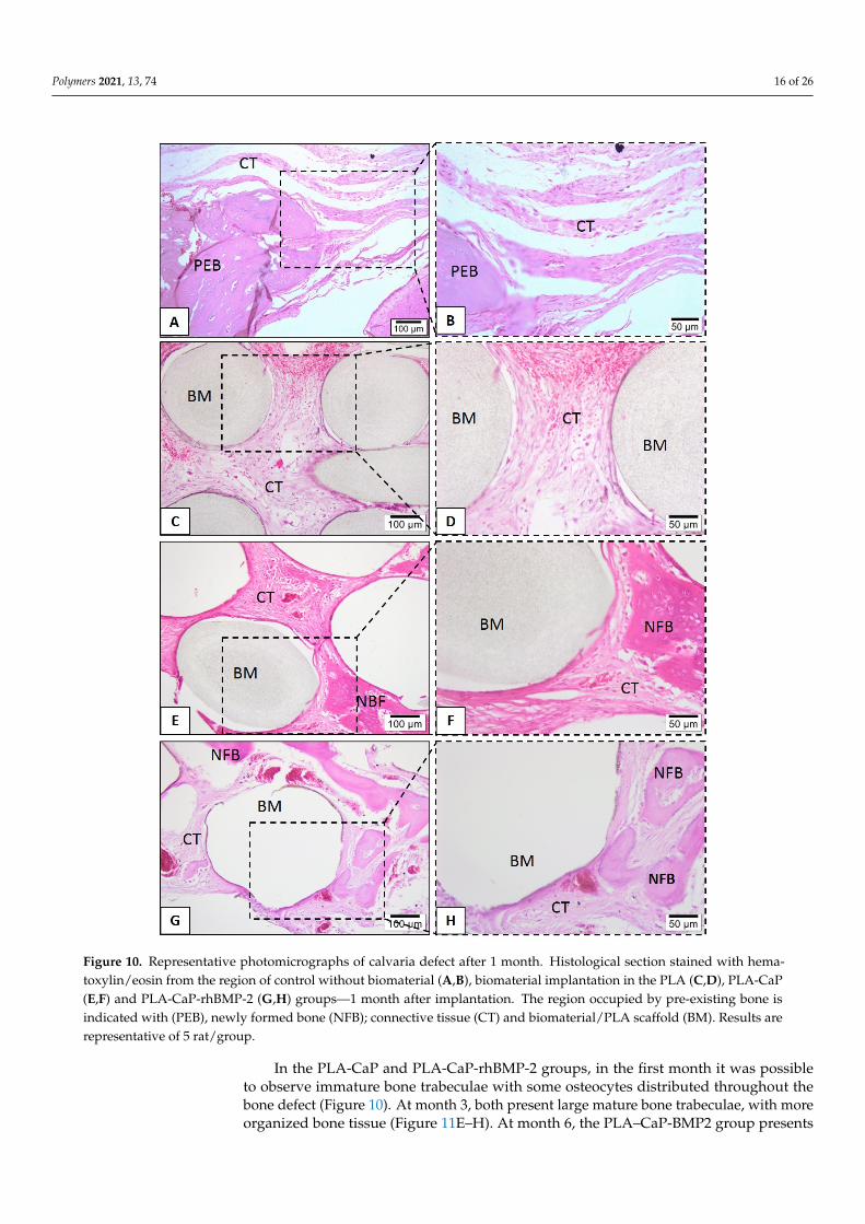
tested biomaterials. None of the tested scaffolds lost their structural integrity, and no sig-nificant degradation after the in vivo assay was found. Defects that were filled with neat PLA, at 1 month, showed connective tissue interspersed with biomaterial and rare inflam-matory cells (Figure 10D). However, after six months, the neat PLA group presents small bone formation levels, present from the peripheries to the center of scaffolds. (Figure 10C,D).
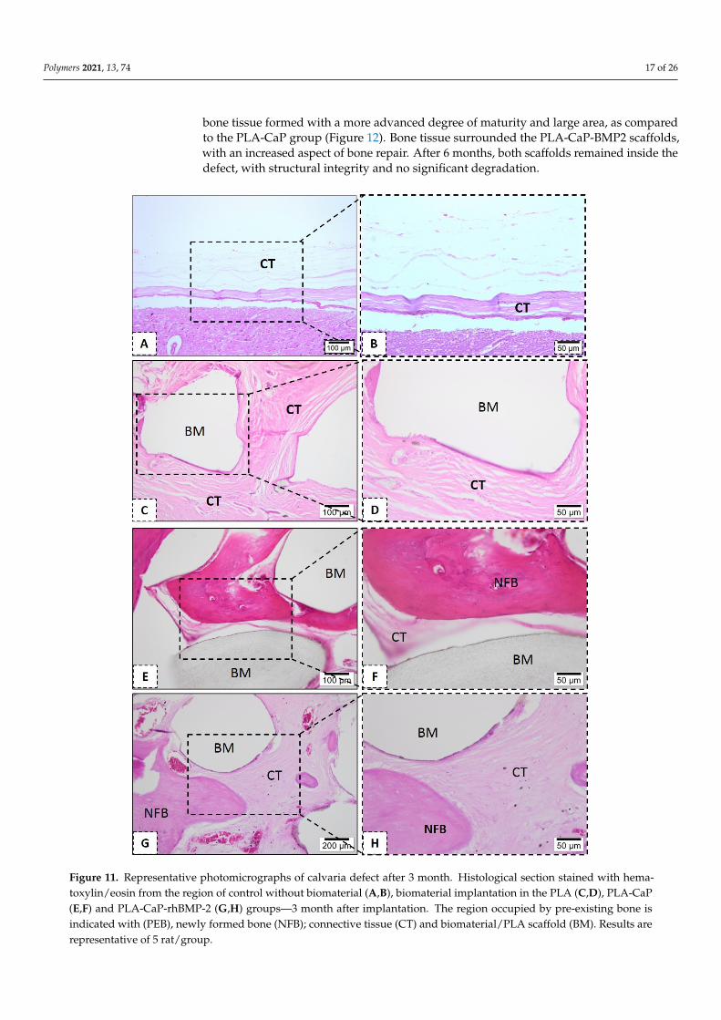
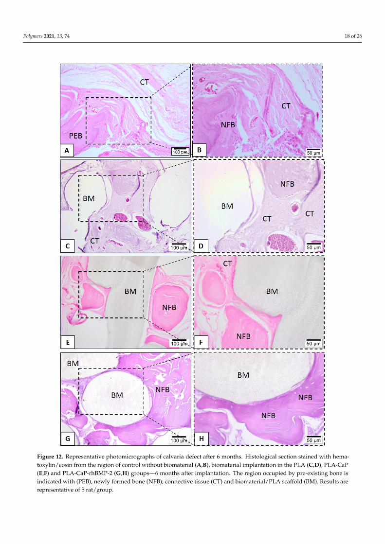
In the PLA-CaP and PLA-CaP-rhBMP-2 groups, in the first month it was possible to observe immature bone trabeculae with some osteocytes distributed throughout the bone defect (Figure 10). At month 3, both present large mature bone trabeculae, with more or-ganized bone tissue (Figure 11E–H). At month 6, the PLA–CaP-BMP2 group presents bone tissue formed with a more advanced degree of maturity and large area, as compared to the PLA-CaP group (Figure 12). Bone tissue surrounded the PLA-CaP-BMP2 scaffolds,
Figure 9. Evaluation of the release of biological markers into the culture media by human osteoblastspheroids after 7 days incubation inside the tested scaffolds. (A). Concentration of released BasicFibroblastic Growth factor (FGF-2). (B). Concentration of released Vascular Endothelial GrowthFactor (VEGF). (C) Concentration of released Platelet-Derived Growth Factor (PDGF). (D) AlkalinePhosphatase (ALP) activity. Bars indicate mean and standard deviation of three biological replicateswith five technical replicated each. Bars with different letters are significantly different (p < 0.05). UE:units of enzyme activity.
3.3. Quantification of rhBMP2 in the PLA-CaP+BMP2 Scaffolds
The incorporation of rhBMP-2 into the PLA-CaP scaffolds was determined by ELISA,obtaining the levels of 13.5 µg/mL, very similar to those found on FDA-approved ab-sorbable collagen sponges (ASC) for orthopedic trauma and oral maxillofacial applica-tions [51].
3.4. Descriptive Histological Evaluation
Histological evaluations were performed to assess the biological response to the testedbiomaterials. None of the tested scaffolds lost their structural integrity, and no significantdegradation after the in vivo assay was found. Defects that were filled with neat PLA, at1 month, showed connective tissue interspersed with biomaterial and rare inflammatorycells (Figure 10D). However, after six months, the neat PLA group presents small boneformation levels, present from the peripheries to the center of scaffolds (Figure 10C,D).
Polymers 2021, 13, 74 16 of 26
Polymers 2021, 13, x FOR PEER REVIEW 17 of 27
with an increased aspect of bone repair. After 6 months, both scaffolds remained inside the defect, with structural integrity and no significant degradation.
Figure 10. Representative photomicrographs of calvaria defect after 1 month. Histological section stained with hematox-ylin/eosin from the region of control without biomaterial (A,B), biomaterial implantation in the PLA (C,D), PLA-CaP (E,F) and PLA-CaP-rhBMP-2 (G,H) groups—1 month after implantation. The region occupied by pre-existing bone is indicated
Figure 10. Representative photomicrographs of calvaria defect after 1 month. Histological section stained with hema-toxylin/eosin from the region of control without biomaterial (A,B), biomaterial implantation in the PLA (C,D), PLA-CaP(E,F) and PLA-CaP-rhBMP-2 (G,H) groups—1 month after implantation. The region occupied by pre-existing bone isindicated with (PEB), newly formed bone (NFB); connective tissue (CT) and biomaterial/PLA scaffold (BM). Results arerepresentative of 5 rat/group.
In the PLA-CaP and PLA-CaP-rhBMP-2 groups, in the first month it was possibleto observe immature bone trabeculae with some osteocytes distributed throughout thebone defect (Figure 10). At month 3, both present large mature bone trabeculae, with moreorganized bone tissue (Figure 11E–H). At month 6, the PLA–CaP-BMP2 group presents
Polymers 2021, 13, 74 17 of 26
bone tissue formed with a more advanced degree of maturity and large area, as comparedto the PLA-CaP group (Figure 12). Bone tissue surrounded the PLA-CaP-BMP2 scaffolds,with an increased aspect of bone repair. After 6 months, both scaffolds remained inside thedefect, with structural integrity and no significant degradation.
Polymers 2021, 13, x FOR PEER REVIEW 18 of 27
with (PEB), newly formed bone (NFB); connective tissue (CT) and biomaterial/PLA scaffold (BM). Results are representa-tive of 5 rat/group.
Figure 11. Representative photomicrographs of calvaria defect after 3 month. Histological section stained with hema-toxylin/eosin from the region of control without biomaterial (A,B), biomaterial implantation in the PLA (C,D), PLA-CaP(E,F) and PLA-CaP-rhBMP-2 (G,H) groups—3 month after implantation. The region occupied by pre-existing bone isindicated with (PEB), newly formed bone (NFB); connective tissue (CT) and biomaterial/PLA scaffold (BM). Results arerepresentative of 5 rat/group.
Polymers 2021, 13, 74 18 of 26
Polymers 2021, 13, x FOR PEER REVIEW 19 of 27
Figure 11. Representative photomicrographs of calvaria defect after 3 month. Histological section stained with hematox-ylin/eosin from the region of control without biomaterial (A,B), biomaterial implantation in the PLA (C,D), PLA-CaP (E,F) and PLA-CaP-rhBMP-2 (G,H) groups—3 month after implantation. The region occupied by pre-existing bone is indicated with (PEB), newly formed bone (NFB); connective tissue (CT) and biomaterial/PLA scaffold (BM). Results are representa-tive of 5 rat/group.
Figure 12. Representative photomicrographs of calvaria defect after 6 months. Histological section stained with hematox-ylin/eosin from the region of control without biomaterial (A,B), biomaterial implantation in the PLA (C,D), PLA-CaP (E,F)
Figure 12. Representative photomicrographs of calvaria defect after 6 months. Histological section stained with hema-toxylin/eosin from the region of control without biomaterial (A,B), biomaterial implantation in the PLA (C,D), PLA-CaP(E,F) and PLA-CaP-rhBMP-2 (G,H) groups—6 months after implantation. The region occupied by pre-existing bone isindicated with (PEB), newly formed bone (NFB); connective tissue (CT) and biomaterial/PLA scaffold (BM). Results arerepresentative of 5 rat/group.
Polymers 2021, 13, 74 19 of 26
3.5. Histomorphometric Evaluation
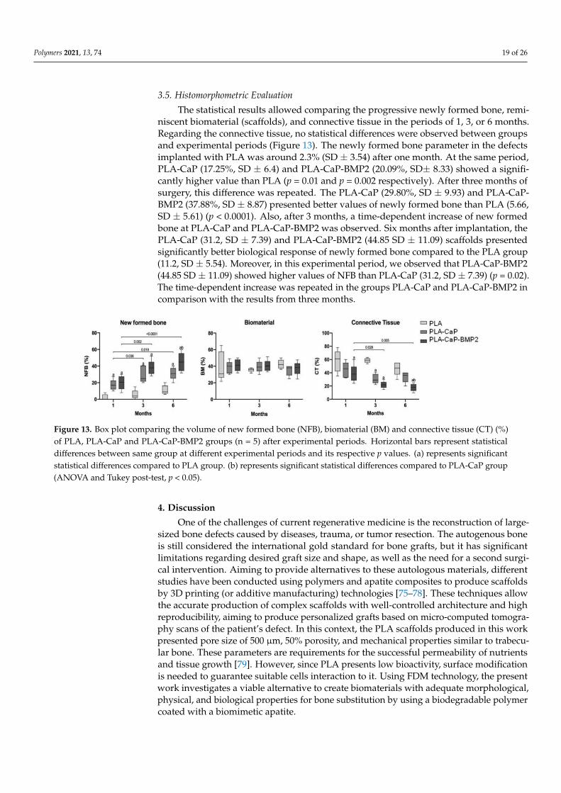
The statistical results allowed comparing the progressive newly formed bone, remi-niscent biomaterial (scaffolds), and connective tissue in the periods of 1, 3, or 6 months.Regarding the connective tissue, no statistical differences were observed between groupsand experimental periods (Figure 13). The newly formed bone parameter in the defectsimplanted with PLA was around 2.3% (SD ± 3.54) after one month. At the same period,PLA-CaP (17.25%, SD ± 6.4) and PLA-CaP-BMP2 (20.09%, SD± 8.33) showed a signifi-cantly higher value than PLA (p = 0.01 and p = 0.002 respectively). After three months ofsurgery, this difference was repeated. The PLA-CaP (29.80%, SD ± 9.93) and PLA-CaP-BMP2 (37.88%, SD ± 8.87) presented better values of newly formed bone than PLA (5.66,SD ± 5.61) (p < 0.0001). Also, after 3 months, a time-dependent increase of new formedbone at PLA-CaP and PLA-CaP-BMP2 was observed. Six months after implantation, thePLA-CaP (31.2, SD ± 7.39) and PLA-CaP-BMP2 (44.85 SD ± 11.09) scaffolds presentedsignificantly better biological response of newly formed bone compared to the PLA group(11.2, SD ± 5.54). Moreover, in this experimental period, we observed that PLA-CaP-BMP2(44.85 SD ± 11.09) showed higher values of NFB than PLA-CaP (31.2, SD ± 7.39) (p = 0.02).The time-dependent increase was repeated in the groups PLA-CaP and PLA-CaP-BMP2 incomparison with the results from three months.
Polymers 2021, 13, x FOR PEER REVIEW 20 of 27
and PLA-CaP-rhBMP-2 (G,H) groups—6 months after implantation. The region occupied by pre-existing bone is indicated with (PEB), newly formed bone (NFB); connective tissue (CT) and biomaterial/PLA scaffold (BM). Results are representa-tive of 5 rat/group.
3.5. Histomorphometric Evaluation The statistical results allowed comparing the progressive newly formed bone, remi-
niscent biomaterial (scaffolds), and connective tissue in the periods of 1, 3, or 6 months. Regarding the connective tissue, no statistical differences were observed between groups and experimental periods (Figure 13). The newly formed bone parameter in the defects implanted with PLA was around 2.3% (SD ± 3.54) after one month. At the same period, PLA-CaP (17.25%, SD ± 6.4) and PLA-CaP-BMP2 (20.09%, SD± 8.33) showed a significantly higher value than PLA (p = 0.01 and p = 0.002 respectively). After three months of surgery, this difference was repeated. The PLA-CaP (29.80%, SD ± 9.93) and PLA-CaP-BMP2 (37.88%, SD ± 8.87) presented better values of newly formed bone than PLA (5.66, SD ± 5.61) (p < 0.0001). Also, after 3 months, a time-dependent increase of new formed bone at PLA-CaP and PLA-CaP-BMP2 was observed. Six months after implantation, the PLA-CaP (31.2, SD ± 7.39) and PLA-CaP-BMP2 (44.85 SD ± 11.09) scaffolds presented significantly better biological response of newly formed bone compared to the PLA group (11.2, SD ± 5.54). Moreover, in this experimental period, we observed that PLA-CaP-BMP2 (44.85 SD ± 11.09) showed higher values of NFB than PLA-CaP (31.2, SD ± 7.39) (p = 0.02). The time-dependent increase was repeated in the groups PLA-CaP and PLA-CaP-BMP2 in compar-ison with the results from three months.
Figure 13. Box plot comparing the volume of new formed bone (NFB), biomaterial (BM) and connective tissue (CT) (%) of PLA, PLA-CaP and PLA-CaP-BMP2 groups (n = 5) after experimental periods. Horizontal bars represent statistical differ-ences between same group at different experimental periods and its respective p values. (a) represents significant statistical differences compared to PLA group. (b) represents significant statistical differences compared to PLA-CaP group (ANOVA and Tukey post-test, p < 0.05).
4. Discussion One of the challenges of current regenerative medicine is the reconstruction of large-
sized bone defects caused by diseases, trauma, or tumor resection. The autogenous bone is still considered the international gold standard for bone grafts, but it has significant limitations regarding desired graft size and shape, as well as the need for a second surgical intervention. Aiming to provide alternatives to these autologous materials, different stud-ies have been conducted using polymers and apatite composites to produce scaffolds by 3D printing (or additive manufacturing) technologies [75–78]. These techniques allow the accurate production of complex scaffolds with well-controlled architecture and high re-producibility, aiming to produce personalized grafts based on micro-computed tomogra-phy scans of the patient’s defect. In this context, the PLA scaffolds produced in this work presented pore size of 500 µm, 50% porosity, and mechanical properties similar to trabec-ular bone. These parameters are requirements for the successful permeability of nutrients and tissue growth [79]. However, since PLA presents low bioactivity, surface modification is needed to guarantee suitable cells interaction to it. Using FDM technology, the present work investigates a viable alternative to create biomaterials with adequate morphological,
Figure 13. Box plot comparing the volume of new formed bone (NFB), biomaterial (BM) and connective tissue (CT) (%)of PLA, PLA-CaP and PLA-CaP-BMP2 groups (n = 5) after experimental periods. Horizontal bars represent statisticaldifferences between same group at different experimental periods and its respective p values. (a) represents significantstatistical differences compared to PLA group. (b) represents significant statistical differences compared to PLA-CaP group(ANOVA and Tukey post-test, p < 0.05).
4. Discussion
One of the challenges of current regenerative medicine is the reconstruction of large-sized bone defects caused by diseases, trauma, or tumor resection. The autogenous boneis still considered the international gold standard for bone grafts, but it has significantlimitations regarding desired graft size and shape, as well as the need for a second surgi-cal intervention. Aiming to provide alternatives to these autologous materials, differentstudies have been conducted using polymers and apatite composites to produce scaffoldsby 3D printing (or additive manufacturing) technologies [75–78]. These techniques allowthe accurate production of complex scaffolds with well-controlled architecture and highreproducibility, aiming to produce personalized grafts based on micro-computed tomogra-phy scans of the patient’s defect. In this context, the PLA scaffolds produced in this workpresented pore size of 500 µm, 50% porosity, and mechanical properties similar to trabecu-lar bone. These parameters are requirements for the successful permeability of nutrientsand tissue growth [79]. However, since PLA presents low bioactivity, surface modificationis needed to guarantee suitable cells interaction to it. Using FDM technology, the presentwork investigates a viable alternative to create biomaterials with adequate morphological,physical, and biological properties for bone substitution by using a biodegradable polymercoated with a biomimetic apatite.
Polymers 2021, 13, 74 20 of 26
For resorbable bone substitutes, it is expected that the implanted biomaterial must beable to stimulate cell interaction and colonization of the implant surface, metabolizing thebiomaterial at the same time that they produce a new matrix, allowing osseointegration.Andu et al. [80] reported several advantages of 3D printed PLA scaffolds in comparisonto the particulate graft tested in a continuous defect in an animal model. Nevertheless,PLA was reported as with low bioactivity and, therefore, the treatment of its surface withapatite is an interesting and well-known alternative [81]. Biomimetic coating treatment wasperformed with the aim to induce a thin and homogeneous layer of hydroxycarbonated ap-atite, promoting a bioactive material. The use of SBF solutions presents a simple technique,which results in a bone-like apatite mineral coat on surface of polymer [82]. Indeed, theuse of SBF solution for 14 days promoted the deposition of a homogeneous layer of apatiteon the polymeric scaffold. Higher magnification micrographies show that the morphologyof the formed apatite was a flake-like assembly, while EDS analysis indicated that calciumand phosphorous were the main elements. Moreover, the biomimetic methodology usedwas efficient to uniformly coat all the scaffolds surface, even the internal struts. A recentstudy (Jaidev and Shatterjee, 2019) [83] showed that a biomimetic coating of 3D printedPLA scaffolds can be produced through a surface functionalization with polyethyleneimine(PEI) and citric acid, followed by immersion in SBF, achieving interesting biological re-sponses. Similarly, our results show that a biomimetic coating can also be achieved by theimmersion of non-functionalized, NaOH-treated PLA scaffolds. It is important to noticethat the alkaline treatment is a very important step in the production of polymeric scaffoldsbecause it is known that it increases their hydrophilicity, roughness, and carboxylic aciddensity on the surface of polymer matrices, which has a significant impact on cellularadhesion. This process allows the incorporation of carboxylic groups into the polymerchains by breaking the ester bond, increasing the number of active sites for cellular bindingto materials, favoring the deposition of apatite by the biomimetic coating, as well removingpossible non-projected filaments present in the scaffold.
Similar to in vivo mineralization, the deposition of biomimetic apatite layer on PLAsurface has two distinct stages. After surface hydrolysis, mineral nucleation occurs onthe polymer surface (primary nucleation). The carboxylic groups generated by alkalinehydrolysis serve as mineral nucleation sites. They can transfer to carboxylate anions atthe buffered solution (pH 7.4) and provide a negatively charged surface to adsorb positivecalcium ions in the surrounds. The locally excess calcium ions induce apatite nucleation bycombining phosphate ions in the vicinity. The next stage is characterized by the additionalmineral crystals grow on the nucleated mineral (crystal growth). Apatite continues to growby consuming calcium and phosphate ions of the SBF solution [55,56].
According to the number of residues identified as the inorganic portion in the TGAanalysis, nearly 2%wt of apatite was deposited onto the PLA scaffolds, suggesting a thinlayer of biomimetic coating. FTIR spectra have shown significant apatite peaks, and XDRanalysis detected that the crystalline phase deposited onto PLA scaffold was composedof hydroxyapatite. EDS graphic indicated a peak of titanium, which may be related totitanium dioxide (TiO2), commonly present in industrial white pigments. Nevertheless,the eventual presence of pigment residues did not affect the material’s biocompatibility, asshown by the performed cytotoxicity assay. The elemental composition of PLA-CaP scaffoldshowed that the Ca/P ratio was 1.49, corresponding to calcium-deficient hydroxyapatite(CDHA), which is easy to obtain with solutions with relatively high concentrations of Ca2+
and PO43− such as SBF. The chemical formula of CDHA presents great variability, resulting
in an unstable Ca/P ratio.For the initial assessment of such biological responses, the present study employed
an in vitro tridimensional model based on a spheroidal 3D culture of osteoblastic cells,as proposed previously [49]. The use of tridimensional cell aggregates can increase thesimilarity with the microenvironment present on in vivo tissues, with more informationthan 2D cells models [84,85], as cells can remain integrated into the spheroid or migrateand proliferate to colonize a material surface, depending on its osteoconductive properties.
Polymers 2021, 13, 74 21 of 26
The primary human osteoblast spheroids also allow the focal seeding of cells in a specificpoint of contact, providing insights on the migration and extent of occupation of a materialsurface over time. Recently, Baptista et al. (2018) [48] employed spheroids of adipose-derived stromal/stem cells to observe interactions with 3D-printed composites of PLA andcarbonated HA (CHA). The authors discuss the intimated interaction between spheroidsand PLA/CHA printed scaffolds, with cell spreading in the material surface. In the presentstudy, the 3D model enabled observing that NaOH-treated PLA scaffolds were already ableto interact with bone cells, showing some level of adhesion and migration over its surface.
Curiously, no striking differences in these parameters were observed from sampleswith biomimetic coating, even though a slight but significant (p < 0.05) increase in totalcell content (as measured by protein quantification) was observed in PLA-CaP. However,interesting results were observed in the presence of a pre-treatment of the surfaces withfetal bovine serum. The serum is a complex mixture of proteins and biomolecules, severalof which participate in cell adhesion, migration, and growth. The cell adhesion onto thesurface of materials occurs via integrin interaction with bioactive sites on the surface. Topartially emulate the in vivo complex interactions of serum proteins and scaffold surfaces,we included a pre-treatment with bovine fetal serum. Interestingly, PLA-CaP added withFBS components showed a more than twofold increase in cell content after 7 days ofincubation compared to untreated PLA, and was statistically higher than PLA + FBS, alsopresenting evidence of increased interactions with human osteoblast spheroids, as shownby fluorescence microscopy. The incorporation of serum proteins such as vitronectin andfibronectin may be a way to increase the number of bioactive sites that are recognized bycells on the surface of materials such as PLA [86].
Alkaline phosphatase activity, an important biomarker of early osteoblasts response,similarly increased in the FBS-treated groups (even after removal of background activity),with the higher levels observed with PLA-CaP+FBS. The increase in ALP activity mayindicate a greater response to cell differentiation capacity for PLA-CaP scaffold with FBSand is in accordance with recent results of studies with HA-coated PLA scaffolds in contactwith mesenchymal stem cells [84]. The present findings provide insight into the possiblerole of the biomimetic coating in PLA and the interface with such proteins, resulting inincreased interactions with cells. Further studies are required, though, to assess in-depththe participation of specific proteins in this process.
Another important observation is that the spheroids respond differently to the differentscaffolds regarding the release of growth factors of interest for regenerative medicine. VEGF,FGF-2, and PDGF are mediators that act either as recruiters of endothelial cells to initiateangiogenesis or as inductors of proliferation, migration, or differentiation of progenitorcells [87]. Osteospheres interacting with scaffold surfaces after pretreatment with serumproteins released higher levels of these GFs and, in the case of PDGF, a factor alreadyapproved by the FDA for uses in regenerative medicine [88], the presence of a biomimeticcoating increased such release. It can be suggested that apatite precipitation creates micro-environments capable of favoring cell-cell interaction that impacts on the production ofsome mediators by HOb spheroids. Considering that the local delivery of growth factorsin damaged tissue is an important component of the bioengineering triad, the presentresults point that modifications on the scaffold surface such as biomimetic coatings maycontribute to locally induce the production of at least some of these factors by cells fromthe material-tissue interface.
Nevertheless, other approach for the delivery of biological mediators of bone repairinvolves the direct use of the scaffolds as carriers. In the case of rhBMP-2, the developmentof novel biomimetic scaffolds becomes relevant as the FDA-approved collagen spongesare not suitable for the regeneration of larger defects. The idea of adding rhBMP-2 to thePLA-CaP scaffolds aims to increase the osteoinductive potential of the biomaterial, whilerhBMP-2 have more affinity to apatite than biodegradable polymers [89,90]. In this sense,the present results shown that the 3D PLA-CaP scaffolds can effectively load rhBMP-2,
Polymers 2021, 13, 74 22 of 26
according to the levels detected by ELISA, maintaining similar concentrations to thosefound on the absorbable collagen sponges (ACS) that comes in the INFUSE® kit [90].
The data from the in vivo assessment demonstrated that all versions of the PLA-CaP investigated were biocompatible with rat calvaria, with the absence of inflammatoryprocess. 3D PLA scaffolds promoted new bone formation after six months, even withoutpresence of rhBMP-2, in agreement with previous reports showing that PLA supportsbone formation [89]. However, the use of PLA-CaP-BMP2 scaffolds resulted in more boneformation than PLA-CaP and uncoated PLA scaffolds after six months of implantation.
In summary, this study demonstrated that PLA-CaP scaffolds could be produced as3D-printed structures by the FDM technique, followed by a simple protocol of alkalinetreatment and immersion in a biomimetic solution, with good reproducibility and accuracy,fundamental characteristics for graft manufacturing using personalized digital models(Computed tomography). Furthermore, after interaction with complex biological media,these scaffolds presented interesting biological responses, with increased cell migrationor proliferation, production of differentiation markers, and release of growth factors, asobserved in the assessment employing a novel 3D osteosphere in vitro model. Furthermore,the pre-clinical in vivo results showed considerable levels of newly formed bone in PLA-CaP scaffolds and even higher bone formation with the association of BMP2.
5. Conclusions
In this work, 3D-printed PLA scaffolds were successfully produced with pore sizeof 500 µm, 50% porosity, and mechanical properties similar to trabecular bone in a re-producible way. The biomimetic method was used to enhance scaffolds’ bioactivity bydeposition of homogeneous and uniform apatite (CaP) layer on external and inner sur-faces of 3D PLA scaffolds. The functionalization of 3D PLA scaffolds with a CaP coating(PLA-CaP) improved its properties in the presence of a complex biological media, inducingdesirable responses from human osteoblasts (HOb). The association of 3D PLA-CaP withspheroids could be a new strategy for tissue engineering, since it could reduce the timeof repair in situ by expression of biomolecules and growth factors. In vivo assays showedthat PLA-CaP scaffolds plus rhBMP-2 produced biocompatible and bioactive scaffolds,with enhanced osteogenesis. In summary, using a scalable 3D printing technique andfollowing simple protocols for surface biomimetic functionalization, it was possible toobtain scaffolds with good reproducibility and accuracy, fundamental characteristics for themanufacture of implantable biomaterials using customized digital models, besides present-ing suitable surface properties to improve bone defect treatment. Moreover, the potentialof these biomaterials as vehicles for local release of calcium and phosphate ions, growthfactors and proteins in damaged tissues can be highlighted. The clinical relevance of thesepre-clinical trials comes from the proposal of 3D printing technologies for manufacturingpersonalized scaffolds as alternatives for autologous grafts for long-term applications suchas craniofacial reconstruction involving larger damaged areas.
Author Contributions: Conceptialization, M.O.C.M.-P., R.M.S.M.T., G.G.A. and M.D.C.-M.; method-ology, M.O.C.M.-P., A.C.B.B., B.N.T., S.C.S., M.J.U., A.T.N.N.A.; analysis, M.O.C.M.-P., S.C.S., A.C.B.B.,G.G.A.; investigation, M.O.C.M.-P., S.C.S., M.J.U., A.T.N.N.A., G.G.A., R.M.S.M.T. and M.D.C.-M.;writing-original draft preparation, M.O.C.M.-P.; writing-review and editing, M.O.C.M.-P., B.N.T.,S.C.S., G.G.A., R.M.S.M.T. and M.D.C.-M.; supervision, R.M.S.M.T. and M.D.C.-M.; project adminis-tration, R.M.S.M.T. and M.D.C.-M.; funding acquisition, R.M.S.M.T. and M.D.C.-M. All authors haveread and agreed to the published version of the manuscript.
Funding: This paper was funded by the Brazilian Agencies: National Council for Scientific andTechnology Development (CNPq) and the Carlos Chagas Filho Foundation for Supporting Researchin the State of Rio de Janeiro (FAPERJ) and by the National Institute of Science and Technology forRegenerative Medicine (INCT-Regenera).
Acknowledgments: The authors thank the Mechanical Properties laboratory (PROPMEC)—PEMM/COPPE/UFRJ (Rio de Janeiro, RJ, Brazil) for compressive tests of scaffolds.
Polymers 2021, 13, 74 23 of 26
Conflicts of Interest: The authors declare no conflict of interest.
References1. Majidinia, M.; Sadeghpour, A.; Yousefi, B. The roles of signaling pathways in bone repair and regeneration. J. Cell Physiol. 2017,
233, 2937–2948. [CrossRef] [PubMed]2. Granjeiro, J.M.; Soares, G.D.A. Biomateriais em Odontologia: Princípios, Métodos Investigativos e Aplicações, 1st ed.; VM Cultural
Editora Ltd.: São Paulo, Brazil, 2011; pp. 111–128.3. García-Gareta, E.; Coathup, M.J.; Blunn, G.M. Osteoinduction of bone grafting materials for bone repair and regeneration. Bone
2015, 81, 112–212. [CrossRef] [PubMed]4. Roseti, L.; Parisi, V.; Petretta, M.; Cavallo, C.; Desando, G.; Bartolotti, I.; Grigolo, B. Scaffolds for Bone Tissue Engineering: State of
the art and new perspectives. Mater. Sci. Eng. C 2017, 78, 1246–1262. [CrossRef] [PubMed]5. Nair, L.S.; Laurencin, C.T. Biodegradable polymers as biomaterials. Prog. Polym. Sci. 2007, 32, 762–798. [CrossRef]6. Bose, S.; Vahabzadeh, S.; Bandyopadhyay, A. Bone tissue engineering using 3D printing. Mater. Today 2013, 16, 496–504. [CrossRef]7. Marx, R.E.; Tursun, R. Tissue Engineering. In Oral, Head and Neck Oncology and Reconstructive Surgery, 1st ed.; Bell, R.B., Andersen,
P.A., Fernandes, R., Eds.; Elsevier: St. Louis, MO, USA, 2018; Volume 1, pp. 208–220. [CrossRef]8. Sears, N.A.; Seshadri, D.R.; Dhavalikar, P.S.; Cosgriff-Hernandez, E. A Review of Three-Dimensional Printing in Tissue Engineer-
ing. Tissue Eng. Part B 2016, 22, 298–310. [CrossRef]9. Katari, R.S.; Peloso, A.; Orlando, G. Tissue Engineering. Adv. Surg. 2014, 48, 137–154. [CrossRef]10. Costa, P.F.; Martins, A.; Neves, N.; Gomes, E.M.; Reis, R.L. Automating the Processing Steps for Obtaining Bone Tissue-
Engineering Substitutes: From Imaging Tools to Bioreactors. Tissue Eng. Part B 2014, 20, 567–577. [CrossRef]11. Berthiaume, F.; Maguire, T.J.; Yarmush, M.L. Tissue Engineering and Regenerative Medicine: History, Progress, and Challenges.
Annu. Rev. Chem. Biomol. Eng. 2011, 2, 403–430. [CrossRef]12. Gao, C.; Peng, S.; Feng, P.; Shuai, C. Bone biomaterials and interactions with stem cells. Bone Res. 2017, 5. [CrossRef]13. Gregor, A.; Filová, E.; Novak, M.; Kronek, J.; Chlup, H.; Buzgo, M.; Blahnová, V.; Lukásová, V.; Bartos, M.; Necas, A.; et al.
Designing of PLA scaffolds for bone tissue replacement fabricated by ordinary commercial 3D printer. J. Biol. Eng. 2017, 11, 31.[CrossRef] [PubMed]
14. Breche, Q.; Chagnon, G.; Machado, G.; Girard, E.; Nottelet, B.; Garric, X.; Favier, D. Mechanical behaviour’s evolution of aPLA-b-PEG-b-PLA triblock copolymer during hydrolytic degradation. J. Mech. Behav. Biomed. Mater. 2016, 60, 288–300. [CrossRef][PubMed]
15. Savioli, L.M.; Jardini, A.L.; Maciel Filho, R. Poly (lactic acid) production for tissue engineering applications. Procedia Eng. 2012,42, 1402–1413. [CrossRef]
16. Narayanan, G.; Vernekar, V.N.; Kuyinu, E.L.; Laurencin, C.T. Poly (Lactic Acid)-Based Biomaterials for Orthopaedic RegenerativeEngineering. Adv. Drug Deliv. Rev. 2016, 107, 247–276. [CrossRef]
17. Dimitriou, R.; Jones, E.; McGonable, D.; Giannoudis, V. Bone regeneration: Current concepts and future directions. BMC Med.2011, 9, 1–10. [CrossRef]
18. Davachi, S.M.; Kaffashi, B. Polylactic Acid in Medicine. Polym.-Plastics Technol. Eng. 2015, 54, 944–967. [CrossRef]19. Gupta, B.; Revagade, N.; Hilborn, J. Poly (lactic acid) fiber: An overview. Prog. Polym. Sci. 2007, 32, 455–482. [CrossRef]20. Castro-Aguirre, E.; Franco, F.I.; Samsudin, H.; Fang, X.; Auras, R. Poly(lactic acid)—Mass production, processing, industrial
applications, and end of life. Adv. Drug Deliv. Rev. 2016, 107, 333–366. [CrossRef]21. Gartolla, D. A Literature Review of Poly (lactic acid). J. Polym. Environ. 2001, 9, 63–84. [CrossRef]22. Ferguson, B.S.; Rogatzki, M.J.; Goodwin, M.L.; Kane, D.A.; Rightmire, Z.; Gladden, L.B. Lactate metabolism: Historical context,
prior misinterpretations, and current understanding. Eur. J. Appl. Physiol. 2018. [CrossRef]23. Giannitelli, S.M.; Accoto, D.; Trombetta, M.; Rainer, A. Current trends in the design of scaffolds for computer-aided tissue
engineering. Acta Biomater. 2014, 10, 580–594. [CrossRef] [PubMed]24. Ramot, Y.; Haim-Zada, M.; Domb, A.J.; Nyska, A. Biocompatibility and safety of PLA and its copolymers. Adv. Drug Deliv. Rev.
2016, 107, 153–162. [CrossRef] [PubMed]25. O’Brien, F.J.; Harley, B.A.; Yannas, I.V.; Gibson, L.J. The effect of pore size on cell adhesion in collagen-GAG scaffolds. Biomaterials
2005, 26, 433–441. [CrossRef] [PubMed]26. Melchels, F.P.W.; Domingos, M.A.N.; Klein, T.J.; Malda, J.; Bartolo, P.J.; Hutmacher, D.W. Additive manufacturing of tissues and
organs. Prog. Polym. Sci. 2012, 37, 1079–1104. [CrossRef]27. Serra, T.; Timoneda, M.A.M.; Planell, J.A.; Navarro, M. 3D printed PLA-based scaffolds. A versatile tool in regenerative medicine.
Organogenesis 2013, 9, 239–244. [CrossRef] [PubMed]28. Valino, A.D.; Dizon, J.R.C.; Espera Jr, A.H.; Chen, Q.; Messman, J.; Advincula, R.C. Advances in 3D Printing of Thermoplastic
Polymer Composites and Nanocomposites. Prog. Polym. Sci. 2019, 98. [CrossRef]29. Nikolova, M.P.; Chavali, M.S. Recent advances in biomaterials for 3D scaffolds: A review. Bioact. Mater. 2019, 4, 271–292.
[CrossRef]30. Turnbull, G.; Clarke, J.; Picard, F.; Riches, P.; Jia, L.; Han, F.; Li, B.; Shu, W. 3D bioactive composite scaffolds for bone tissue
engineering. Bioact. Mater. 2018, 3, 278–314. [CrossRef]
Polymers 2021, 13, 74 24 of 26
31. Hoque, M.E.; Chuan, Y.L.; Plashby, I. Extrusion Based Rapid Prototyping Technique: Na Advanced Plataform for TissueEngineering Scaffold Fabrication. Biopolymers 2011, 97, 83–93. [CrossRef]
32. Wu, S.; Liu, X.; Yeung, K.W.K.; Liu, C.; Yang, X. Biomimetic porous scaffolds for bone tissue engineering. Mater. Sci. Eng. R Rep.2014, 80, 1–36. [CrossRef]
33. Kwak, K.-A.; Kim, Y.-H.; Kim, M.; Lee, B.-T.; Song, H.-Y. Bio-functionalization of polycaprolactone infiltrated B; CP scaffold withsilicon and fibronectin enhances osteoblast activity In Vitro. Appl. Surf. Sci. 2013, 279, 13–22. [CrossRef]
34. Nouri, A.; Castro, R.; Santos, J.L.; Fernandes, C.; Rodrigues, J.; Tomás, H. Calcium phosphate-mediated gene delivery usingsimulated body fluid (SBF). Int. J. Pharm. 2012, 434, 199–208. [CrossRef] [PubMed]
35. Bohner, M.; Lemaitre, J. Can bioactivity be tested in vitro with SBF solution? Biomaterials 2009, 30, 2175–2179. [CrossRef] [PubMed]36. Kokubo, T.; Takadama, H. How useful is SBF in predicting In Vivo bone bioactivity? Biomaterials 2006, 27, 2907–2915. [CrossRef]37. Kim, H.-M.; Himeno, T.; Kokubo, T.; Nakamura, T. Process and kinetics of bonelike apatite formation on sintered hydroxyapatite
in a simulated body fluid. Biomaterials 2005, 26, 4366–4373. [CrossRef]38. Tanahashi, M.; Yao, T.; Kokubo, T.; Minoda, M.; Miyamoto, T.; Nakamura, T.; Yamamuro, T. Apatite Coating on Organic Polymers
by a Biomimetic Process. J. Am. Ceram. Soc. 1994, 77, 2805–2808. [CrossRef]39. Farto-Vaamonde, X.; Auriemma, G.; Aquino, R.P.; Concheiro, A.; Alvarez-Lorenzo, C. Post-manufacture loading of filaments and
3D printed PLA scaffolds with prednisolone and dexamethasone for tissue regeneration applications. Eur. J. Pharm. Biopharm.2019, 141, 100–110. [CrossRef]
40. Danoux, C.B.; Barbieri, D.; Yuan, H.; de Brujn, J.D.; van Blitterswijk, C.A.; Habibovic, P. In Vitro and in vivo bioactivity assessmentof a polylactic acid/hydroxyapatite composite for bone regeneration. Biomatter 2014, 4, e27664. [CrossRef]
41. Pridgeon, C.S.; Schlott, C.; Wong, M.W.; Heringa, M.B.; Heckel, T.; Leedale, J.; Launay, L.; Gryshkova, V.; Przyborski, S.;Bearon, R.N.; et al. Innovative organotypic in vitro models for safety assessment: Aligning with regulatory requirements andunderstanding models of the heart, skin, and liver as paradigms. Arch. Toxicol. 2018, 92, 557–569. [CrossRef]
42. Baptista, S.L.; Kronemberger, G.S.; Silva, R.K.; Granjeiro, J.M. Spheroids of stem cells as endochondral templates for improvedbone engineering. Front. Biosci. 2018, 23, 1969. [CrossRef]
43. Fang, Y.; Eglen, R.M. Three-Dimensional Cell Cultures in Drug Discovery and Development. SLAS Doscov. 2017, 22, 456–472.44. Guven, S.; Chen, P.; Inci, F.; Tasoglu, S.; Erkmen, B.; Demirci, U. Multiscale assembly for tissue engineering and regenerative
medicine. Trends Biotechnol. 2015, 33, 269–279. [CrossRef] [PubMed]45. Elliott, N.; Yuan, F. A Review of Three-Dimensional in Vitro tissue Models for Drug Discovery and Transport Studies. J. Pharm.
Sci. 2011, 100, 59–74. [CrossRef] [PubMed]46. Mueller-Klieser, W. Multicellular spheroids. A review on cellular aggregates in cancer research. J. Cancer Res. Clin. Oncol. 1987,
113, 101–122. [CrossRef] [PubMed]47. Koledova, Z.; Zhang, X.; Streuli, C.; Clarke, R.B.; Klein, O.D.; Lu, P. SPRY1 regulates mammary epithelial morphogenesis
by modulating EGFR-dependent stromal paracrine signaling and ECM remodeling. Proc. Natl. Acad. Sci. USA 2016, 113,E5731–E5740. [CrossRef]
48. Baptista, L.S.; Kronemberger, G.S.; Côrtes, I.; Charelli, L.E.; Matsui RK, M.; Palhares, T.N.; Sohier, J.; Rossi, A.M.; Granjeiro, J.M.Adult Stem Cells Spheroids to Optimize Cell Colonization in Scaffolds for Cartilage and Bone Tissue Engineering. Int. J. Mol. Sci.2018, 19, 1285. [CrossRef]
49. Restle, L.; Costa-Silva, D.; Lourenço, E.S.; Bachinski, R.F.; Batista, A.C.; Linhares, A.B.; Alves, G.G. A 3D osteoblast in vitro modelfor the evaluation of biomedical materials. Adv. Mater. Sci. Eng. 2015, 2015, 268930. [CrossRef]
50. Grado, G.F.; Keller, L.; Gillet, I.Y.; Wagner, Q.; Musset, A.-M.; Jessel, N.B.; Bornert, F.; Offner, D. Bone substitutes: A review oftheir characteristics, clinical use, and perspectives for larfe bone defects management. J. Tissue Eng. 2018, 9. [CrossRef]
51. McKay, W.F.; Peckham, S.M.; Badura, J.M. A comprehensive clinical review of recombinant human bone morphogenetic protein-2(INFUSE® Bone Graft). Int. Orthop. 2007, 31, 729–734. [CrossRef]
52. Thire, R.M.; Meiga, T.O.; Dick, S.; Andrade, L.R. Functionalizations of biodegradable polyester for tissue engineering applications.In Macromolecular Symposia; Wiley: Hoboken, NJ, USA, 2007; Volume 258, pp. 38–44. [CrossRef]
53. Costa, P.F.; Hutmacher, D.W.; Theodoropoulos, C.; Gomes, M.E.; Reis, R.L.; Vaquette, C. Additively Manufactured Device forDynamic Culture of Large Arrays of 3D Tissue Engineered Constructs. Adv. Healthcare Mater. 2015. [CrossRef]
54. Chim, H.; Hutmacher, D.W.; Chou, A.M.; Oliveira, A.L.; Reis, R.L.; Lim, T.C.; Schantz, J.T. A comparative analysis of scaffoldmaterial modifications for load-bearing applications in bone tissue engineering. Int. J. Oral Maxillofac. Surg. 2006, 35, 928–934.[CrossRef] [PubMed]
55. Oliveira, A.L.; Malafaya, P.B.; Reis, R.L. Sodium silicate gel as a precursor for the in vitro nucleation and growth of a bone-likeapatite coating in compact and porous polymeric structures. Biomaterials 2003, 24, 2575–2584. [CrossRef]
56. Yilmaz, B.; Pazarceviren, A.E.; Tezcaner, A.; Evis, Z. Historical development of simulated body fluids used in biomedicalapplications: A review. Microchem. J. 2020, 155. [CrossRef]
57. International Organization for Standardization. ISO 14937: Sterilization of Health Care Products—General Requirements for Characteri-zation of a Sterilizing Agent and Development, Validation and Routine Control of a Sterilization Process for Medical Devices; InternationalOrganization for Standardization: Geneva, Switzerlnad, 2009.
58. ASTM F2450-18, Standard Guide for Assessing Microstructure of Polymeric Scaffolds for Use in Tissue-Engineered Medical Products;ASTM International: West Conshohocken, PA, USA, 2018.
Polymers 2021, 13, 74 25 of 26
59. International Organization for Standardization. ISO 10993-5: Biological Evaluation of Medical Devices—Part 5: Tests for In VitroCytotoxicity; International Organization for Standardization: Geneva, Switzerlnad, 2009.
60. Kilkenny, C.; Browne, W.J.; Cuthill, I.C.; Emerson, M.; Altman, D.G. Improving Bioscience Research Reporting: The ARRIVEGuidelines for Reporting Animal Research. PLoS Biol. 2010, 8, e1000412. [CrossRef] [PubMed]
61. Smith, A.J.; Clutton, R.E.; Lilley, E.; Hansen, K.A.A.; Brattelid, T. PREPARE: Guidelines for planning animal research and testing.Lab. Anim. 2018, 52, 135–141. [CrossRef] [PubMed]
62. International Organization for Standardization. ISO 10993-6: Biomedical Evaluation of Medical Devices—Part 6: Tests for Local Effectsafter Implantation; International Organization for Standardization: Geneva, Switzerlnad, 2016.
63. Bose, S.; Roy, M.; Bandyopadhyay, A. Recent advances in bone tissue engineering scaffolds. Trends Biotechnol. 2013, 30, 546–554.[CrossRef]
64. Danilevicius, P.; Georgiadi, L.; Pateman, C.J.; Claeyssens, F.; Chatzinikolaidou, M.; Farsari, M. The effect of porosity on cellingrowth into accurately defined, laser-made, polylactide-based 3D scaffolds. Appl. Surf. Sci. 2015, 336, 2–10. [CrossRef]
65. Maia-Pinto, M.O.C.; Maia, M.C.; Thiré, R.M.S.M. Estudo da biocompatibilidade in vivo de arcabouço de poli(ácido lático) (PLA)fabricados por impressão 3d para aplicações em engenharia tecidual. In A Produção do Conhecimento nas Ciências da Saúde 5, 1sted.; Silva Neto, B.R., Ed.; Atena Editora: Sao Paulo, Brazil, 2019; pp. 118–125. [CrossRef]
66. Zhang, R.; Ma, P.X. Porous poly (L-lactic acid)/apatite composites created by biomimetic process. J. Biomed. Mater. Res. 1999, 45,285–293. [CrossRef]
67. Brizzolara, D.; Cantow, H.-J.; Diederichs, K.; Keller, E.; Domb, A.J. Mechanism of the Stereocomplex Formation betweenEnantiomeric Poly(lactide)s. Macromolecules 1996, 29, 191–197. [CrossRef]
68. International Centre for Diffraction Data. Number 01-071-5048; International Centre for Diffraction Data: Newtown Square, PA,USA, 2019.
69. Dorozhkin, S.V. A history of calcium orthophosphates (CaPO4) and their biomedical applications. Morphologie 2017, 101, 143–153.[CrossRef]
70. Dorozhkin, S.V. Calcium orthophosphates (CaPO4): Occurrence and properties. Prog. Biomater. 2016, 5, 9–70. [CrossRef][PubMed]
71. San Román, M.S.; Holgado, M.J.; Salinas, B.; Rives, V. Drug release from layered double hydroxides and from their Poly (lacticacid) (PLA) nanocomposites. Appl. Clay Sci. 2013, 71, 1–7. [CrossRef]
72. Lasprilla, A.J.R.; Martinez, G.A.R.; Lunelli, B.H.; Figueroa, J.E.J.; Jardini, A.L.; Filho, R.M. Poly-lactic acid synthesis for applicationin biomedical devices—A review. Biotechnol. Adv. 2012, 30, 321–328. [CrossRef] [PubMed]
73. Arrieta, M.P.; López, J.; López, D.; Kenny, J.M.; Peponi, L. Development of flexible materials based on plasticized electrospunPLA–PHB blends: Structural, thermal, mechanical and disintegration properties. Eur. Polym. J. 2015, 73, 433–446. [CrossRef]
74. Barbeck, M.; Serra, T.; Booms, P.; Stojanovic Najman, S.; Engel, E.; Sader, R.; Kirkpatrick, C.J.; Navarro, M.; Chanaati, S. Analysisof the in vitro degradation and the In Vivo tissue response to bi-layered 3D-printed scaffolds combining PLA and biphasicPLA/bioglass components e Guidance of the inflammatory response as basis for osteochondral regeneration. Bioact. Mater. 2017,2, 208–223. [CrossRef] [PubMed]
75. Bruyas, A.; Lou, F.; Stahl, A.M.; Gardner, M.; Maloney, W.; Goodman, S.; Yang, Y.P. Systematic characterization of 3D-printedPCL/b-TCP scaffolds for biomedical devices and bone tissue engineering: Influence of composition and porosity. J. Mater. Res.2018, 33, 1948. [CrossRef] [PubMed]
76. Visscher, L.E.; Dang, H.D.; Knackstedt, M.A.; Hutmacher, D.W. 3D printed Polycaprolactone scaffolds with dual macro-microporosity for applications in local delivery of antibiotics. Mater. Sci. Eng. C 2018, 87, 78–89. [CrossRef]
77. Zhang, H.; Mao, X.; Du, Z.; Jiang, W.; Han, X.; Zhao, D.; Han, D.; Li, Q. Three dimensional printed macroporous polylacticacid/hydroxyapatite composite scaffolds for promoting bone formation in a critical-size rat calvarial defect model. Sci. Technol.Adv. Mater. 2016, 17, 136–148. [CrossRef]
78. Tanodekaew, S.; Channasanon, S.; Kaewkong, P.; Uppanan, P. PLA-HA scaffolds: Preparation and bioactivity. Procedia Eng. 2013,59, 144–149. [CrossRef]
79. Egan, P.F. Review: Integrated Design Approaches for 3D Printed Tissue Scaffolds: Review and Outlook. Materials 2019, 12, 2355.[CrossRef]
80. Andu, R.T.; Suresh, V.; Gounder, R.; Kannan, A. Comparison of the Efficacy of Three Different Bone Regeneration Materials: AnAnimal Study. Eur. J. Dent. 2019, 13, 22. [CrossRef]
81. Kepa, K.; Coleman, R.; Grondahl, L. In Vitro mineralization of functional polymers. Biosurf. Biotribol. 2015, 1, 214–227. [CrossRef]82. Abe, Y.; Kokubo, T. Apatite coating on ceramics, metals and polymers utilizing a biological process. J. Mater. Sci. Mater. Med.
1990, 1, 233–238. [CrossRef]83. Jaidev, L.R.; Chatterjee, K. Surface functionalization of 3D printed polymer scaffolds to augment stem cell response. Mater. Des.
2019, 161, 44–54. [CrossRef]84. Alves, G.G.; Costa-Silva, D. Microtissues and tridimensional cell models: A new paradigm in tissue engineering. Int. J. Growth
Factors Stem Cells Dent. 2019, 2, 1. [CrossRef]85. Weng, Q.; Zhou, L.; Xia, L.; Zheng, Y.; Zhang, X.; Li, F.; Li, Q. In Vitro evaluation of FL118 and 9-Q20 cytotoxicity and cellular
uptake in 2D and 3D different cell models. Cancer Chemother. Pharmacol. 2019, 84, 527–537. [CrossRef]

Polymers 2021, 13, 74 26 of 26
86. Imaninezhad, M.; Schober, J.; Griggs, D.; Ruminski, P.; Kuljanishvili, I.; Zustiak, S.P. Cell Attachment and Spreading on CarbonNanotubes Is Facilitated by Integrin Binding. Front. Bioeng. Biotechnol. 2018, 6, 129. [CrossRef]
87. Bai, Y.; Bai, L.; Zhou, J.; Chen, H.; Zhang, L. Sequential delivery of VEGF, FGF-2 and PDGF from the polymeric system enhanceHUVECs angiogenesis in vitro and CAM angiogenesis. Cell. Immunol. 2018, 323, 19–32. [CrossRef]
88. Kaigler, D.; Silva, E.A.; Mooney, D.J. Guided bone regeneration (GBR) utilizing injectable Vascular Endothelial Growth Factor(VEGF) delivery gel. J. Periodontol. 2013, 84, 230–238. [CrossRef]
89. Luvizuto, E.R.; Tangl, S.; Zanoni, G.; Okamoto, T.; Sonoda, C.K.; Gruber, R.; Okamoto, R. The effect of BMP-2 on the osteoconduc-tive properties ofb-tricalcium phosphate in rat calvaria defects. Biomaterials 2011, 32, 3855–3861. [CrossRef]
90. Xiao, W.; Fu, H.; Rahaman, M.N.; Liu, Y.; Sonny Bal, B. Hollow hydroxyapatite microspheres: A novel bioactive and osteocon-ductive carrier for controlled release of bone morphogenetic protein-2 in bone regeneration. Acta Biomaterialia 2013, 9, 8374.[CrossRef] [PubMed]