a new β-glucosidase gene from the zygomycete fungus rhizomucor miehei
TRANSCRIPT

ORIGINAL PAPER
A new b-glucosidase gene from the zygomycete fungusRhizomucor miehei
Miklos Tako • Adel Toth • Laszlo G. Nagy •
Judit Krisch • Csaba Vagvolgyi • Tamas Papp
Received: 18 June 2009 / Accepted: 23 September 2009 / Published online: 8 October 2009
� Springer Science+Business Media B.V. 2009
Abstract In this study, a b-glucosidase coding gene
(bgl) of the zygomycete fungus Rhizomucor miehei
has been cloned and characterized. The gene com-
prises a total of 2,826 bp including the coding
sequence of a 717 amino acids length putative protein
and 10 introns dispersed in the whole coding region.
The putative N-and C-terminal catalytic domains (aa
68 to aa 274 and aa 358–601, respectively) were
identified; the two domains are connected with a
84-amino-acids linker. The catalytic region showed an
extensive sequence homology with other fungal
b-glucosidases classified as family 3 glycoside hydro-
lases. The isolated Rhizomucor gene was expressed in
the related fungus Mucor circinelloides. Transformant
Mucor strains maintained the introduced plasmid in
an autoreplicative manner and showed significantly
higher cellobiase activity than the recipient strain.
Keywords Family 3 glycoside hydrolase �Cellobiase � Rhizomucor � Mucor �Heterologous expression
Introduction
b-Glucosidases (b-D-glucoside glucohydrolases;
3.2.1.21) constitute a group of well-characterized
and biologically important enzymes that catalyze the
transfer of glycosyl group between oxygen nucleo-
philes. Their activity is fundamental in many biolog-
ical pathways, such as degradation of structural and
storage polysaccharides, host–pathogen interactions,
cellular signalling and oncogenesis (Bhatia et al.
2002). These enzymes play a crucial role in large
scale saccharification of cellulose by removing cel-
lobiose that inhibits the exo- and endoglucanases.
Filamentous fungi are known to be good producers of
b-glucosidases and several fungal glucosidase genes
have already been cloned and analyzed. However,
Zygomycetes are rarely investigated from this aspect.
Although a few enzymes have been characterized in
this fungal group (Borgia and Mehnert 1982;
Petruccioli et al. 1999; Takii et al. 2005), cloning
and analysis of glucosidase genes have not been
reported until to date.
The genus Rhizomucor comprises two well-estab-
lished thermophilic fungal species, R. pusillus and
R. miehei (Schipper 1978; Vagvolgyi et al. 1999). In
particular, R. miehei is interesting from a biotechno-
logical aspect in consequence of its extracellular
enzyme production. Commercially produced R. mie-
hei aspartic protease is widely used in industrial
cheese making to substitute calf chymosin (Outtrup
and Boyce 1990; Rao et al. 1998) and its lipase is one
M. Tako � A. Toth � L. G. Nagy � C. Vagvolgyi �T. Papp (&)
Department of Microbiology, Faculty of Science and
Informatics, University of Szeged, 6726 Szeged,
Kozep fasor 52, Hungary
e-mail: [email protected]
J. Krisch
Department of Food Engineering, Faculty of Engineering,
University of Szeged, Szeged, Hungary
123
Antonie van Leeuwenhoek (2010) 97:1–10
DOI 10.1007/s10482-009-9382-z

of the most studied fungal lipases (Maheshwari et al.
2000). Despite their significance, members of the
genus Rhizomucor have remained genetically rela-
tively poorly characterized.
In a carbon source assimilation study (Vastag et al.
1998), R. miehei was able to utilize cellobiose and
other disaccharides as sole carbon sources. An early
publication reports on the cellulolytic activity of
R. pusillus, where it produced hydrolytic enzymes that
attacked native cellulose, acid-swollen cellulose, car-
boxymethylcellulose and cellobiose (Somkuti et al.
1969). The same study suggested the presence of a
multiple cellulolytic enzyme system in this fungus.
However, the literature lacks further information on
the hydrolytic breakdown of cellulose and its deriva-
tives by the members of the genus Rhizomucor.
The aim of the present study was to clone and
characterize the b-glucosidase gene and its adjacent
element from R. miehei. With this object, the isolated
gene was introduced into the related fungus Mucor
circinelloides and expression of the gene and induc-
tion of the promoter were examined in this heterol-
ogous system.
Materials and methods
Strains
The wild-type R. miehei NRRL 5282 was used for the
gene cloning and the M. circinelloides strain MS12
(Velayos et al. 1997) a leuA-, pyrG- mutant derived
from the wild-type CBS 277.49 served as a recipient
in the transformation experiments. Escherichia coli
DH5a strain was used in all cloning experiments and
plasmid amplifications cultivating on Luria–Bertani
medium at 37�C; if necessary, ampicillin was added
to a final concentration of 50 lg ml-1.
DNA manipulation techniques
R. miehei was grown in yeast extract—glucose liquid
medium (YEG; 5 g yeast extract, 20 g glucose per
litre) under continuous shaking (200 rpm) at 37�C for
3 days. Genomic DNA of R. miehei was prepared
from mycelia disrupted with pestle and mortar in
liquid nitrogen and purified in a 0.5 mg ml-1 bis-
benzimide-CsCl density gradient (Iturriaga et al.
1992). Plasmid DNA preparation, cloning and
transformation of E. coli, as well as electrophoresis
of nucleic acids, were performed following standard
procedures (Sambrook et al. 1989). Nucleic acids for
hybridization were labelled with the digoxigenin-
based ‘‘PCR DIG Probe Synthesis Kit’’ (Roche),
following the instructions of the manufacturer.
Immunological detection of nucleic acid blots was
performed using the DIG Nucleic Acid Detection Kit
(Roche), according to the conditions recommended
by the supplier.
Cloning of the R. miehei bgl gene
Analysing of known fungal b-glucosidase sequences,
two degenerated primers, BGL1 and BGL2, were
designed that correspond to the amino acid sequence
motifs GLDM and FPYLV, respectively. Using this
primer pair, a fragment of 742 bp was amplified by
PCR from the genomic DNA of R. miehei and its
sequence was determined. Table 1 presents the
nucleotide sequences of the primers used in the study.
The entire gene together with the upstream and
downstream flanking regions was determined by the
inverse PCR (IPCR) method (Ochman et al. 1988).
Figure 1 summarizes the PCR strategy used to clone
the Rhizomucor bgl gene. To obtain DNA templates
for the IPCR experiments, four sets of fragments
were prepared digesting 10 lg aliquots of the geno-
mic DNA with the enzymes XbaI, HindIII, PstI and
XhoI (Fermentas), each in a total volume of 120 ll.
The digested DNA was purified with phenol:chloro-
form:isoamyl alcohol (PCI; 25:24:1) followed by a
subsequent extraction with chloroform:isoamyl alco-
hol (CI; 24:1). After ethanol precipitation of the
aqueous phase, samples were air dried and resus-
pended in 17 ll sterile distilled water. Resulting
DNA fragments were self-ligated with T4 ligase
(Fermentas) for 18 h at 4�C and the circularised DNA
was extracted again with PCI and CI. After ethanol
precipitation and air drying, samples were resus-
pended in 10 ll distilled water, from which 50 ng
were used in the subsequent IPCR reactions.
IPCR primers were designed in opposite orienta-
tion to that of normal PCR (Table 1). PCR reactions
were carried out under the following parameters: a
denaturation was done at 94�C for 2 min, followed by
10 cycles of 94�C for 15 s, 55�C for 30 s and 68�C
for 3 min, followed by 20 cycles of 94�C for 15 s,
55�C for 30 s and 68�C for 3 min with a cycle
2 Antonie van Leeuwenhoek (2010) 97:1–10
123

elongation (5 s) for each successive cycle; the final
cycle was followed by an extension step of 68�C for
7 min. Each amplification product was cloned into
the plasmid pTZ57R/T using the InsT/Aclone PCR
Product Cloning Kit (Fermentas) according to the
instructions of the manufacturer and sequenced in
both directions.
Investigation of the bgl copy number
For Southern hybridization analysis, 5 lg R. miehei
genomic DNA was digested with the appropriate
restriction enzyme. After gel electrophoresis (0.8%
agarose/TAE), DNA was transferred onto nylon
membranes; prehybridization (1 h) and hybridization
(16 h) were carried out under low stringency (55�C)
and high stringency (65�C) conditions. For hybrid-
ization, the same digoxigenine labelled probe was
used as in the screening of the genomic library. After
hybridization, membranes were washed twice in 29
SSC and 0.1% SDS at room temperature for 5 min and
then twice with 0.59 SSC and 0.1% SDS at 55�C (for
low stringency) or with 0.19 SSC and 0.1% SDS at
65�C (for high stringency) for 15 min, respectively.
Sequence analysis
Blast searches (Altschul et al. 1997) in the EMBL and
GenBank databases were performed for prediction of
the coding sequence. The programs ProtParam
Table 1 Oligonucleotide
primers designed for this
study
Restriction sites designed in
the primers are underlined
Primer Sequence (50–30)
BGL1 GGCTCGAGGGYYTNGAYATG
BGL2 GGCTGCAGACNARRTANGGRAA
BGL3 GGCTCAGGCACTGTCGAC
BGL4 GCCTTCGTCTTGTCCCAACTTGTA
BGL5 GACGTCCTCTTTGGTGATGTCAAC
BGL6 AGCTGGTTGTAGCTACACATAATG
BGL7 GTCTTTCCTACACCACCTTTGAGT
BGL8 GTTACCAATGTAGTGTTTGGCAGT
BGL9 AGTACACTTTGCATATTGGTGCTA
BGL10 CGGCTACGCCTTGCATAGCTGAC
BGL17 CTCGCGGCCGCTTAGTAAAGATAGCTACGGCGCT
BGL19 GGATCGATATGTTTGCAAAGACTGCGTTG
Fig. 1 The R. miehei b-glucosidase gene and protein. aSchematic representation of the bgl gene and the main regions
of the encoded protein. Arrows indicate the positions of the
primers for the IPCR amplifications and cloning. b Comparison
of conserved sequence motifs of some family 3 glycoside
hydrolases. Bold characters indicate the putative H? donor and
the catalytic nucleophile
Antonie van Leeuwenhoek (2010) 97:1–10 3
123

(Gasteiger et al. 2005) and SignalP (Bendtsen et al.
2004) were used to characterize the proposed protein
and to predict the signal peptide, respectively.
Domain search and prediction were performed using
the Motif Scan (MyHits) program (Pagni et al. 2007).
Structure prediction was made by the Swiss-Modell
(Arnold et al. 2006) using the barley ExoI (NCBI
accession number: AAD23382) as template; for
3D-visualization and comparison of protein sequences
and structures, the Structure based Sequences Align-
ment Program (STRAP; http://www.charite.de/bioinf/
strap/) was used. All programs were accessed trough
the Swiss Expasy Server (www.expasy.ch) and used
with default parameters.
To perform phylogenetic analyses, amino acid
sequences of several b-glucosidases were downloaded
from the NCBI GenBank; raw sequence data of
Zygomycetes hypothetical proteins were obtained
from the genome sequence databases of M. circinello-
ides (DoE Joint Genome Institute; M. circinelloides
CBS277.49 v1.0; http://genome.jgi-psf.org/Mucci1/
Mucci1.home.html); Phycomyces blakesleeanus
(DoE Joint Genome Institute P. blakesleeanus v1.1;
http://genome.jgi-psf.org/Phybl1/Phybl1.home.html)
and Rhizopus oryzae (Broad Institute; R. oryzae
Database; http://www.broad.mit.edu/annotation/genome/
rhizopus_oryzae). Outgroups were selected so as to
represent the major groups within the glycoside
hydrolase family 3 recognized by Harvey et al. (2000).
Alignment of sequences utilized the following strat-
egy: first, larger groups were identified based on a NJ
tree computed from a rough alignment obtained with
ClustalW. Then, these groups were aligned separately
in ProbCons, a probabilistic multiple alignment tool
which outperforms ClustalW in many respects (Do
et al. 2005). A protein substitution matrix was com-
puted from the data by running the algorithm for ten
pre-training replicates. To refine the alignments, 25
iterative refinement steps were performed. Subse-
quently, the resulting multiple alignments were aligned
to each other by the profile option in Muscle (Edgar
2004). The resulting alignments were used to infer
trees using Bayesian MCMC algorithm implemented
in MrBayes 3.2.1 (Ronquist and Huelsenbeck 2003).
Best fit substitution models were estimated by the
Reversible-Jump MCMC approach, which does not
require a priori selection of substitution model, but
samples different models during the MCMC run in
proportion to their posterior probabilities. Markov
Chains were run for three million generations sampling
every 100th generations. The first 10,000 trees were
discarded as burn-in, and the remaining trees were used
to compute a 50% Majority Rule consensus tree.
Bayesian Posterior Probabilities (BPP) C 0.95 were
considered significant.
Plasmid construction and transformation
For heterologous expression in M. circinelloides, a
vector designated as pTM8 (Fig. 2) was constructed
as follows. The bgl gene of R. miehei was amplified
by PCR using NotI- and ClaI-modified specific
primers (BGL17 and BGL19; Table 1), and cloned
into pBluescript II SK? (Stratagene). Then, bgl gene
was cut out from this construct with the enzymes ClaI
and XbaI and it was transferred into the plasmid
pPT43 (Papp et al. 2006). The resulting plasmid was
digested with PvuII to obtain the gene flanked with
the promoter and terminator regions of the M.
circinelloides gpd1 gene (Wolff and Arnau 2002).
The fragment was then ligated into the plasmid pTM7
(Lukacs et al. 2009). This plasmid contain the
orotidine-5-phosphate-decarboxylase gene (pyrG) of
M. circinelloides (Benito et al. 1992) inserted into the
pSP73 (Fermentas).
For the transformation experiments, protoplasts of
M. circinelloides were prepared as described earlier
(Nagy et al. 1994). Polyethylene glycol mediated
transformations of protoplasts were performed
according to van Heeswijck and Roncero (1984).
Fig. 2 The plasmid pTM8 used for the expression of the R.miehei bgl gene in M. circinelloides
4 Antonie van Leeuwenhoek (2010) 97:1–10
123

Transformants were selected on the basis of the
complementation of uracil auxotrophy of the recipi-
ent strain. Selection and further culturing of the
transformants were performed on solid minimal
medium (YNB; 10 g glucose, 0.5 g yeast nitrogen
base without amino acids, 1.5 g (NH4)2SO4, 1.5 g
glutamic acid and 20 g agar per litre), supplemented
with leucine and/or uracil (200 lg ml-1, respec-
tively) as required.
Testing of the b-glucosidase activity
To test b-glucosidase activity of the transformants in
liquid culture, 5 9 105 spores were inoculated in
15 ml YNB containing 0.15 g wheat bran instead of
glucose and supplemented with 200 lg ml-1 leucine.
For culturing of the original MS12 strain, medium
was supplemented with 200 lg ml-1 uracil also.
Strains were cultivated under continuous shaking at
25�C for 6 days. Each day, 500 ll samples were
collected and filtered to remove insoluble particles.
The filtrates were centrifuged at 16,200g for 30 min
and the supernatant was used for enzyme activity
measurements (diluted 1:10 with distilled water if
necessary).
To test b-glucosidase activity in solid-state fer-
mentation, 5 g wheat bran and 5 ml distilled water
were mixed and supplemented with 200 lg ml-1
leucine (and with the same amount of uracil in the
case of the recipient strain). After inoculation this
medium with 106 spores, cultures were incubated at
25�C for 6 days. An Erlenmeyer flask was daily taken
and extracted with 30 ml distilled water at 4�C for
3 h. After filtration of the extract, 1 ml filtrate was
centrifuged at 16,200g for 10 min. The supernatant
was diluted to 1:1,000 with distilled water and
analyzed for total protein concentration and b-gluco-
sidase activity.
b-Glucosidase activity was determined by
p-nitrophenyl-b-D-glucopyranoside (PNPG, Sigma).
Twenty microliter of 7 mM PNPG was given to
180 ll diluted extract, and incubated for 30 min at
50�C. The reaction was stopped by 50 ll of 100 mM
sodium carbonate, and the p-nitrophenol release was
measured at 405 nm. One enzyme activity unit was
equal to 1 lmol p-nitrophenol released in 1 min.
Enzyme activities were measured in 96-well micro-
titer plates using an ASYS Jupiter HD (ASYS Hitech)
microplate reader.
Purification of the Rhizomucor BGL enzyme
R. miehei (106 spores) was inoculated in 130 g
wheat bran supplemented with 130 ml distilled
water and incubated at 40�C for 6 days. All steps
of the purification processes were carried out at 4�C.
The culture medium was extracted with 800 ml of
100 mM acetate buffer (pH 5.0) and left to stand for
12 h at 4�C. After filtration, the crude extract was
centrifuged at 5,040g for 15 min, and then, the
protein of the supernatant was precipitated by
ammonium sulphate. Precipitates from the fractions
of 50–85% saturation were collected by centrifuga-
tion at 5,040g for 15 min and re-dissolved in the
smallest possible volume of 100 mM acetate buffer
(pH 5.0). Precipitate of the fraction having satura-
tion between 75 and 85% showed the highest
PNPG-hydrolyzing activity. This concentrated
enzyme solution was loaded onto a Sephadex
G-100 (Sigma) column equilibrated with 50 mM
acetate buffer (pH 6.0); the exclusion range was
4–150 kDa (27 9 400 mm). Elution was performed
with the same buffer at a flow rate of 0.5 ml min-1.
Fractions containing PNPG-hydrolyzing activity
were collected and applied to a Macro-Prep High
Q anion exchange column (12.6 9 40 mm; Bio-
Rad) equilibrated with 50 mM acetate buffer (pH
6.0). The enzyme was eluted with a linear gradient
from 0 to 1 M NaCl at a flow rate of 1 ml min-1.
Final purification of b-glucosidase was performed
on a Sephacryl S-200 HR column (exclusion range
5–250 kDa; 16 9 60 mm; GE Healthcare) equili-
brated with 50 mM acetate buffer (pH 5.0) contain-
ing 150 mM NaCl, and eluted with the same buffer
at a flow rate of 0.5 ml min-1. The purity and the
molecular weight of the purified protein were
examined by sodium dodecyl sulphate–polyacryl-
amide gel electrophoresis (SDS–PAGE), which was
performed on NuPAGE Bis–Tris gel (Invitrogen)
using NuPAGE MES running buffer (Invitrogen)
according to the manufacturer’s instructions. Protein
bands were detected by staining the gel with
0.0025% Coomassie Brilliant Blue R-250. To iden-
tify the purified protein, tryptic peptides were
generated (Hellman et al. 1995) and analysed by
HPLC-ESI ion trap mass spectrometry. The LC–MS
analysis was carried out by the Proteomics Research
Group at the Biological Research Center of the
Hungarian Academy of Sciences.
Antonie van Leeuwenhoek (2010) 97:1–10 5
123
Results and discussion
The bgl gene of R. miehei
Using the IPCR method, a segment of 4,063 bp
containing the entire b-glucosidase gene of R. miehei
together with its flanking regions was isolated and
sequenced. The sequence was deposited to the EMBL
Nucleotide sequence database (accession no.:
AM922334). The coding sequence was predicted by
similarities to other known fungal b-glucosidase
genes found in sequence databases. The bgl gene
comprises a total of 2,826 bp including the coding
sequence of a putative 717 amino acids length protein
and 10 introns (70, 64, 68, 60, 76, 68, 77, 73, 82 and
34 bp in length) dispersed in the whole coding region
(Fig. 1).
The 578 nucleotides-long promoter region con-
tains a typical putative TATA box (TATAAAA)
locating at the expected -69 position from the start
codon and two pyrimidine-rich stretches at the
positions -49 and -385. The putative CAAT motif
was found 357 nucleotides upstream from the start
codon. Three possible motifs corresponding to the
consensus sequence (SYGGR) of the binding sites of
carbon catabolite repressor proteins (Suto and Tomita
2001) were observed at the positions -126 (CCG-
GAG), -135 (CCCCAC) and -248 (CCGGGG). In the
terminator region, two putative polyadenylation
sequence (AATAAA) were identified 20 and 37
nucleotides downstream from the stop codon.
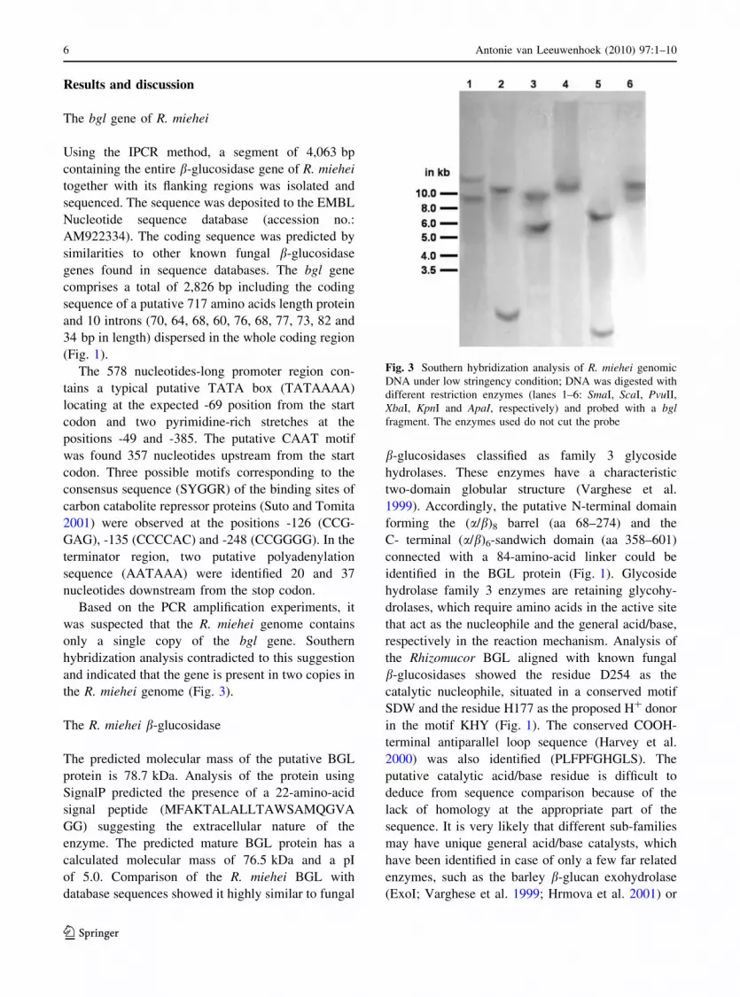
Based on the PCR amplification experiments, it
was suspected that the R. miehei genome contains
only a single copy of the bgl gene. Southern
hybridization analysis contradicted to this suggestion
and indicated that the gene is present in two copies in
the R. miehei genome (Fig. 3).
The R. miehei b-glucosidase
The predicted molecular mass of the putative BGL
protein is 78.7 kDa. Analysis of the protein using
SignalP predicted the presence of a 22-amino-acid
signal peptide (MFAKTALALLTAWSAMQGVA
GG) suggesting the extracellular nature of the
enzyme. The predicted mature BGL protein has a
calculated molecular mass of 76.5 kDa and a pI
of 5.0. Comparison of the R. miehei BGL with
database sequences showed it highly similar to fungal
b-glucosidases classified as family 3 glycoside
hydrolases. These enzymes have a characteristic
two-domain globular structure (Varghese et al.
1999). Accordingly, the putative N-terminal domain
forming the (a/b)8 barrel (aa 68–274) and the
C- terminal (a/b)6-sandwich domain (aa 358–601)
connected with a 84-amino-acid linker could be
identified in the BGL protein (Fig. 1). Glycoside
hydrolase family 3 enzymes are retaining glycohy-
drolases, which require amino acids in the active site
that act as the nucleophile and the general acid/base,
respectively in the reaction mechanism. Analysis of
the Rhizomucor BGL aligned with known fungal
b-glucosidases showed the residue D254 as the
catalytic nucleophile, situated in a conserved motif
SDW and the residue H177 as the proposed H? donor
in the motif KHY (Fig. 1). The conserved COOH-
terminal antiparallel loop sequence (Harvey et al.
2000) was also identified (PLFPFGHGLS). The
putative catalytic acid/base residue is difficult to
deduce from sequence comparison because of the
lack of homology at the appropriate part of the
sequence. It is very likely that different sub-families
may have unique general acid/base catalysts, which
have been identified in case of only a few far related
enzymes, such as the barley b-glucan exohydrolase
(ExoI; Varghese et al. 1999; Hrmova et al. 2001) or
Fig. 3 Southern hybridization analysis of R. miehei genomic
DNA under low stringency condition; DNA was digested with
different restriction enzymes (lanes 1–6: SmaI, ScaI, PvuII,
XbaI, KpnI and ApaI, respectively) and probed with a bglfragment. The enzymes used do not cut the probe
6 Antonie van Leeuwenhoek (2010) 97:1–10
123

the b-glucosidase of Flavobacterium meningosepti-
cum (Chir et al. 2002). Comparing the barley ExoI
and the R. miehei BGL, the motif PYTET (aa 488–
492) of ExoI containing the catalytic acid was aligned
with the motif IVVDG (aa 472–476), where D475
can be a potential acid/base catalyst. The structure
model predicted using the barley ExoI as a template
shows the putative nucleophile D254 and the residue
D475 being in appropriate positions (Fig. 4),
although this result must be interpreted with precau-
tion because of the relatively low similarity between
ExoI and BGL, and experimental identification of the
acid/base catalyst is needed to prove this suggestion.
To purify the extracellular b-glucosidase enzyme
in high amount, R. miehei was grown on wheat bran
medium for 6 days at 40�C. The enzyme was purified
from the crude extract to homogeneity by ammonium
sulphate precipitation and gel filtration followed by
anion exchange and size-exclusion chromatographies.
The protein was homogenous in terms of its behav-
iour on SDS–PAGE, as shown in Fig. 5. The
molecular weight of the purified enzyme estimated
by SDS–PAGE (about 75–80 kDa) corresponded
well to that predicted on the basis of the putative
amino acid sequence. Moreover, LC–MS analysis of
the peptides generated by trypsin digestion identified
the isolated protein as the predicted R. miehei BGL
and confirmed that the purified enzyme is encoded by
the bgl gene.
Evolution of fungal b-glucosidases
Blast searches with the amino acid sequence of the
R. miehei BGL revealed the existence of four, two
and five highly similar hypothetical proteins in the
databases of the M. circinelloides, P. blakesleeanus
and R. oryzae genomes, respectively. Within the
glycosyde hydrolase family 3, all these sequences
showed highest similarity to members of subfamily
4 containing fungal enzymes with b-glucosidase
activity as circumscribed by Harvey et al. (2000).
These Zygomycetes sequences were involved in
Fig. 4 Ribbon representation of the structure prediction for
the R. miehei b-glucosidase. The barley b-glucan exohydrolase
isoensyme ExoI was used as template for the prediction
Fig. 5 SDS–PAGE analysis of the purified R. miehei BGL
enzyme (a) and partially purified protein samples extracted
from the recipient M. circinelloides MS12 strain and one of the
transformants (b). In the latter case, culturing, extraction and
purification on Sephadex G100 were performed as described
for the R. miehei BGL in Sect. ‘‘Materials and methods’’. aLane 1, the purified b-glucosidase (4 lg); lane 2, SeeBlue Plus
2 Standard (Invitrogen) as molecular weight marker. b Lanes 1
and 2, partially purified protein samples from the transformant
MS12 ? pTM8/1 and the recipient MS12, respectively; lane 3,
SeeBlue Plus 2 Standard (Invitrogen)
Antonie van Leeuwenhoek (2010) 97:1–10 7
123

phylogenetic analyses carried out to address the
evolution of fungal b-glucosidases of the group 4; as
outgroups, some glucosidases representing the other
subfamilies were also involved into the analysis.
Members of the Reversible-Jump MCMC analysis
selected the WAG model implemented in MrBayes as
the best fit model to the data, with a posterior
probability of 1.00. The resulting consensus tree is
presented on Fig. 6. Almost all clades received high
BPPs (C0.95), and the topology is largely congruent
with that obtained by Harvey et al. (2000). Our
results support monophyly of zygomycetes b-gluco-
sidases. It is noteworthy that sequences labelled as
Phycomyces 17174, Mucor 74361 and Rhizopus
RO3G_09386.1 formed a separate, well-supported
clade (BPP = 1.00) in a sister position to all other
zygomycete sequences. Based on the inferred topol-
ogy it is most parsimonious to assume that the above
mentioned three sequences emerged by an early
duplication, preceding the speciation events giving
rise to Mucor, Rhizopus and Phycomyces taxa,
because two subclades of the zygomycete clade
correspond to the phylogeny of the species them-
selves (Voigt et al. 1999). Because the duplication
happened before divergence of Rhizomucor miehei, it
also suggests that R. miehei should have another
hitherto unrecognised copy of the bgl gene.
Heterologous expression of the R. miehei bgl
in M. circinelloides
The plasmid pTM8 contained the entire bgl gene of
R. miehei fused with the promoter and terminator
regions of the M. circinelloides gpd1 gene. This
plasmid was introduced into M. circinelloides by
PEG-mediated protoplast transformation. Transfor-
mation frequency was low: only three transformants
were gained from the experiment. Presence of the
plasmid in the transformants was proven by PCR
amplification and sequencing (results not shown).
Fig. 6 Phylogenetic
analysis of group 4 fungal
family 3 glycoside
hydrolases: Bayesian 50%
Majority Rule consensus
phylogram; numbers
indicate posterior
probabilities (BPP). Amino
acid sequences of
M. circinelloides,
P. blakesleeanus and R.oryzae were obtained by
similarity search in genome
databases (protein IDs and
designations of the
corresponding database are
indicated on the tree). Other
sequences were obtained
from the NCBI/EMBL
databases (accession
numbers are indicated on
the tree)
8 Antonie van Leeuwenhoek (2010) 97:1–10
123

Maintenance of the selective conditions was neces-
sary that the transformants retain the plasmid indi-
cating that the plasmid did not integrate into the
genome.
After culturing the recipient MS12 strain and the
transformants on wheat bran, their proteins were
extracted from the culture filtrates and partially
purified on a Sephadex G100 column; SDS–PAGE
analysis of these protein samples revealed the pres-
ence of an extra band in the protein pattern of the
transformants (Fig. 5). The size of this band corre-
sponded well to that of the Rhizomucor BGL
suggesting that the introduced gene had been
expressed in the transformants.
Extracellular b-glucosidase activity of the recipient
MS12 strain and some of the transformants was
determined after culturing the strains in both liquid
and solid-state cultures. Table 2 shows the volumetric
activities of the protein extracts purified from the
culturing media of the strains at the sixth culturing day.
R. miehei is a good glucosidase producer and
enzyme extracts of the NRRL 5282 strain also had
remarkable activity. In spite of the presence of four
copies of hypothetical b-glucosidase genes in the
M. circinelloides genome, this fungus showed rela-
tively low glucosidase activity under the applied
culturing conditions. Although transformants showed
much higher enzyme activities than the original
MS12 strain, their values did not reach that of the
R. miehei enzyme. Maybe the Mucor strain produces
and/or secretes the heterologous enzyme with some-
what lower efficiency than R. miehei or the latter
fungus produces another active b-glucosidase
enzyme. Anyway, introduction of the Rhizomucor
bgl gene significantly increased the enzyme activity
of the transformants in both liquid and solid-state
cultures indicating the functionality of the gene and
the effectiveness of the corresponding enzyme.
This paper reports on the isolation of a gene
encoding a b-glucosidase from R. miehei. As we
know, this is the first b-glucosidase gene from a
zygomycete fungus that has been characterized. The
corresponding protein has also been purified; detailed
characterization of the enzyme is in progress.
Acknowledgments The research was supported by the ETT
214/2006 grant of the Hungarian Medical Research Council.
We wish to thank the Proteomics Research Group at the
Biological Research Center of the Hungarian Academy of
Sciences for the LC–MS analysis of the BGL protein.
References
Altschul SF, Madden TL, Schaffer AA, Zhang J, Zhang Z,
Miller W, Lipman DJ (1997) Gapped BLAST and PSI-
BLAST: a new generation of protein database search
programs. Nucleic Acids Res 25:3389–3402
Arnold K, Bordoli L, Kopp J, Schwede T (2006) The SWISS-
MODEL workspace: a web-based environment for pro-
tein structure homology modelling. Bioinformatics 22:
195–201
Bendtsen JD, Nielsen H, von Heijne G, Brunak S (2004)
Improved prediction of signal peptides: SignalP 3.0. J Mol
Biol 340:783–795
Benito EP, Dıaz-Mınguez JM, Itturiaga EA, Campuzano EA,
Eslava AP (1992) Cloning and sequence analysis of the
Mucor circinelloides pyrG gene encoding orotidine-50-monophosphate decarboxylase: use of pyrG for homolo-
gous transformation. Gene 116:59–67
Bhatia Y, Mishra S, Bisaria VS (2002) Microbial b-glucosi-
dases: cloning, properties, and applications. Crit Rev
Biotechnol 22:375–407
Borgia P, Mehnert D (1982) Purification of a soluble and a
wall-bound form of b-glucosidase from Mucor racemo-sus. J Bacteriol 145:515–522
Chir J, Withers S, Wan C-F, Li Y-K (2002) Identification of the
two essential groups in the family 3 b-glucosidase from
Flavobacterium meningosepticum by labelling and tan-
dem mass spectrometric analysis. Biochem J 365:857–863
Do CB, Mahabhashyam MSP, Brudno M, Batzoglou S (2005)
PROBCONS: probabilistic consistency-based multiple
sequence alignment. Genome Res 15:330–340
Edgar RC (2004) MUSCLE: multiple sequence alignment with
high accuracy and high throughput. Nucleic Acids Res
32:1792–1797
Gasteiger E, Hoogland C, Gattiker A, Duvaud S, Wilkins MR,
Appel RD, Bairoch A (2005) Protein identification and
analysis tools on the ExPASy Server. In: Walker JM (ed)
The proteomics protocols handbook. Humana Press,
Clifton, pp 571–607
Table 2 b-Glucosidase activity of the original and the trans-
formed M. circinelloides, and the R. miehei strains
Strain Volumetric activity
(U ml-1)a
Liquid
culture
Solid-state
culture
M. circinelloides MS12 0.11 7.18
M. circinelloides MS12 ? pTM8/1 0.37 23.3
M. circinelloides MS12 ? pTM8/2 0.31 18.1
M. circinelloides MS12 ? pTM8/3 0.34 19.6
R. meiehei NRRL 5282 3.7 38.3
a Averages of three measurements
Antonie van Leeuwenhoek (2010) 97:1–10 9
123

Harvey AJ, Hrmova M, De Gori R, Varghese JN, Fincher GB
(2000) Comparative modelling of the three-dimensional
structures of family 3 glycoside hydrolases. Proteins 41:
257–269
Hellman U, Wernstedt C, Gonez J, Heldin CH (1995)
Improvement of an ‘‘In-Gel’’ digestion procedure for the
micropreparation of internal protein fragments for amino
acid sequencing. Anal Biochem 224:451–455
Hrmova M, Varghese JN, De Gori R, Smith BJ, Driguez H,
Fincher GB (2001) Catalytic mechanisms and reaction
intermediates along the hydrolytic pathway of a plant b-D-
glucan glucohydrolase. Structure 9:1005–1016
Iturriaga EA, Dıaz-Mınguez JM, Benito EP, Alvarez MI, Es-
lava AP (1992) Heterologous transformation of Mucorcircinelloides with the Phycomyces blakesleeanus leu1gene. Curr Genet 21:215–223
Lukacs Gy, Papp T, Somogyvari F, Csernetics A, Nyilasi I,
Vagvolgyi Cs (2009) Cloning of the Rhizomucor miehei3-hydroxy-3-methylglutaryl-coenzyme A reductase gene
and its heterologous expression in Mucor circinelloides.
Antonie Leeuwenhoek 95:55–64
Maheshwari R, Bharadwaj G, Mahalingeshwara K (2000)
Thermophilic fungi: their physiology and enzymes.
Microbiol Mol Biol Rev 64:461–488
Nagy A, Vagvolgyi Cs, Balla E, Ferenczy L (1994) Electro-
phoretic karyotype of Mucor circinelloides. Curr Genet
26: 45–48
Ochman H, Gerber AS, Hartl DL (1988) Genetic applications
of an inverse polymerase chain reaction. Genetics
120:621–623
Outtrup H, Boyce COL (1990) Microbial proteinases and
biotechnology. In: Fogarty WM, Kelly CT (eds) Microbial
enzymes and biotechnology. Elsevier, London, pp 227–
254
Pagni M, Ioannidis V, Cerutti L, Zahn-Zabal M, Jongeneel CV,
Hau J, Martin O, Kuznetsov D, Falquet L (2007) MyHits:
improvements to an interactive resource for analyzing
protein sequences. Nucleic Acids Res 35(Web Server
issue):W433–W437
Papp T, Velayos A, Bartok T, Eslava AP, Vagvolgyi Cs,
Iturriaga EA (2006) Heterologous expression of astaxan-
thin biosynthesis genes in Mucor circinelloides. Appl
Microbiol Biotechnol 69:526–531
Petruccioli M, Brimer L, Cicalini AR, Federici F (1999) The
linamarase of Mucor circinelloides LU M40 and its
detoxifying activity on cassava. J Appl Microbiol 86:
302–310
Rao MB, Tanksale AM, Ghatge MS, Deshpande VV (1998)
Molecular and biotechnological aspects of microbial
proteases. Microbiol Mol Biol Rev 62:597–635
Ronquist F, Huelsenbeck JP (2003) MRBAYES 3: Bayesian
phylogenetic inference under mixed models. Bioinfor-
matics 19:1572–1574
Sambrook J, Fritsch EF, Maniatis T (1989) Molecular cloning:
a laboratory manual, 2nd edn. Cold Spring Harbor Lab-
oratory, Cold Spring Harbor, New York
Schipper MAA (1978) On the genera Rhizomucor and Par-asitella. Stud Mycol 17:53–71
Somkuti GA, Babel FJ, Somkuti AC (1969) Cellulolysis by
Mucor pusillus. Appl Microbiol 17:888–892
Suto M, Tomita F (2001) Induction and catabolite repression
mechanism of cellulose in fungi. J Biosci Bioeng 92:
305–311
Takii Y, Ikeda K, Sato C, Yano M, Sato T, Konno H (2005)
Production and characterization of b-glucosidase from
Rhizopus oryzae MIBA348J. Biol Macromol 5:11–16
Vagvolgyi Cs, Vastag M, Acs K, Papp T (1999) Rhizomucortauricus: a questionable species of the genus. Mycol Res
103:1318–1322
van Heeswijck R, Roncero MIG (1984) High frequency
transformation of Mucor with recombinant plasmid DNA.
Carlsberg Res Commun 49:691–702
Varghese JN, Hrmova M, Fincher GB (1999) Three-dimen-
sional structure of a barley beta-D-glucan exohydrolase, a
family 3 glycosyl hydrolase. Structure 7:179–190
Vastag M, Papp T, Zs Kasza, Vagvolgyi Cs (1998) Differen-
tiation of Rhizomucor species by carbon source utilization
and isoenzyme analysis. J Clin Microbiol 36:2153–2156
Velayos A, Lopez-Matas MA, Ruiz-Hidalgo MJ, Eslava AP
(1997) Complementation analysis of carotenogenic
mutants of Mucor circinelloides. Fung Genet Biol 22:
19–27
Voigt K, Cigelnik E, O’Donnell K (1999) Phylogeny and PCR
identification of clinically important zygomycetes based
on nuclear ribosomal-DNA sequence data. J Clin Micro-
biol 37:3957–3964
Wolff AM, Arnau J (2002) Cloning of glyceraldehyde-3-
phosphate dehydrogenase-encoding genes in Mucor cir-cinelloides (syn. racemosus) and use of the gpd1 promoter
for recombinant protein production. Fung Genet Biol
35:21–29
10 Antonie van Leeuwenhoek (2010) 97:1–10
123