a baculovirus-mediated strategy for full-length plant virus coat protein expression and purification
TRANSCRIPT
Ardisson-Araújo et al. Virology Journal 2013, 10:262http://www.virologyj.com/content/10/1/262
RESEARCH Open Access
A baculovirus-mediated strategy for full-lengthplant virus coat protein expression andpurificationDaniel Mendes Pereira Ardisson-Araújo1, Juliana Ribeiro Rocha1, Márcio Hedil Oliveira da Costa1,Anamélia Lorenzetti Bocca1, André Nepomuceno Dusi2, Renato de Oliveira Resende1
and Bergmann Morais Ribeiro1*
Abstract
Background: Garlic production is severely affected by virus infection, causing a decrease in productivity andquality. There are no virus-free cultivars and garlic-infecting viruses are difficult to purify, which make specificantibody production very laborious. Since high quality antisera against plant viruses are important tools forserological detection, we have developed a method to express and purify full-length plant virus coat proteins usingbaculovirus expression system and insects as bioreactors.
Results: In this work, we have fused the full-length coat protein (cp) gene from the Garlic Mite-borne FilamentousVirus (GarMbFV) to the 3′-end of the Polyhedrin (polh) gene of the baculovirus Autographa californica multiplenucleopolyhedrovirus (AcMNPV). The recombinant baculovirus was amplified in insect cell culture and the virus wasused to infect Spodoptera frugiperda larvae. Thus, the recombinant fused protein was easily purified from insectcadavers using sucrose gradient centrifugation and analyzed by Western Blotting. Interestingly, amorphous crystalswere produced in the cytoplasm of cells infected with the recombinant virus containing the chimeric-protein genebut not in cells infected with the wild type and recombinant virus containing the hexa histidine tagged Polh.Moreover, the chimeric protein was used to immunize rats and generate antibodies against the target protein.The antiserum produced was able to detect plants infected with GarMbFV, which had been initially confirmed byRT-PCR.
Conclusions: The expression of a plant virus full-length coat protein fused to the baculovirus Polyhedrin inrecombinant baculovirus-infected insects was shown to produce high amounts of the recombinant protein whichwas easily purified and efficiently used to generate specific antibodies. Therefore, this strategy can potentially beused for the development of plant virus diagnostic kits for those viruses that are difficult to purify, are present inlow titers or are present in mix infection in their plant hosts.
Keywords: Baculovirus expression system, Polyhedrin, Garlic virus coat protein, Virus-indexing diagnostic kit
* Correspondence: [email protected] of Cell Biology, Laboratory of Electron Microscopy, Institute ofBiological Sciences, University of Brasília, Brasília, DF, BrazilFull list of author information is available at the end of the article
© 2013 Ardisson-Araújo et al.; licensee BioMed Central Ltd. This is an Open Access article distributed under the terms of theCreative Commons Attribution License (http://creativecommons.org/licenses/by/2.0), which permits unrestricted use,distribution, and reproduction in any medium, provided the original work is properly cited.

Ardisson-Araújo et al. Virology Journal 2013, 10:262 Page 2 of 9http://www.virologyj.com/content/10/1/262
BackgroundFor the establishment of a successful large-scale agricul-tural production, the use of healthy and pathogen-freeplants is an essential measure. Garlic, for instance, hasno virus-free cultivars, which represents a serious prob-lem due to economic losses and the difficulties ofcontrolling disease. Considering all agronomic param-eters important for garlic production, bulb growth isthe most severely affected by virus infections, causing a de-crease in productivity and quality, with a reduction of up to88% of the weight [1-3]. In fact, the introduction of the firstvirus-free cultivars produced in Brazil by thermotherapyand stem-tip culture has resulted in improved yields [4].However, for the efficient production of tissue culture
virus-free plants, some bottlenecks must be overcome.One of them is the detection of virus infections in garlicplants. Current detection is based on serological methods,using specific antiserum, symptomatology, transmissiontests in different host plants, and sequence data of the coatprotein gene [5,6]. In situations when a high numberof samples need to be tested, the use of moleculardiagnosis techniques, such as RT-PCR, is not an easytask, due to the requirement of expensive and fragilematerials (e.g. enzymes, dNTPs, termocycler). There-fore, serological methods are recommended for largescale evaluations, however, high quality antisera arenot available for all relevant viruses [7,8]. The dot-Enzyme-Linked Immunosorbent Assay (dot-ELISA) isa serological, less expensive and more practical alter-native method that could be used to detect plantvirus infections, despite of requiring the productionof specific antibodies.Many plant viruses of agricultural interest, including
the garlic-infecting viruses, are difficult to purify fromthe host because they accumulate in low titers and areoften present in mixed infections, which make specificantibody production very laborious [9-11].One way of circumventing these difficulties is to ex-
press the virus coat protein in prokaryotic or eukaryoticcell systems for further antisera production. The expres-sion of a soluble coat protein from a garlic virus in bac-teria (Resende RO, personal communication) and insectcells [12] for production of antiserum for the recombin-ant protein was previously carried out by our researchgroup but neither system worked. We were unable toproduce high titer antibody due to problems in proteinpurification, which makes it unsuitable for the large-scale species-specific diagnosis of the tested garlic vi-ruses. Despite baculovirus being a potent tool to expressdifferent proteins, the purification step is usually achallenge for further applications. To solve this prob-lem, one could attempt to tag the recombinant pro-tein with a carrier peptide or protein to facilitate theantigen purification.
Interestingly, the orally infective baculovirus virionsare surrounded by protein crystal matrix composedmainly of a single protein, the Polyhedrin, which ishighly expressed in the late stages of infection [13].Polyhedrin has been used as a carrier protein to facilitateantigen purification [14-18]. This strategy for purifica-tion of antigen has been patented (see http://otl.sinica.edu.tw/en/index.php?t=9&group_id=19&article_id=477).The system has advantages when compared to existingtechnologies, it is both easy and cheap and achieves apurity of over 95% without the need for tags or expen-sive column purification steps. In this work we haveexpressed the coat protein of the Allexivirus GarMbFV(Garlic mite-borne filamentous virus) in caterpillars asbioreactors using a Polyhedrin-based expression vectorin order to generate polyclonal antibodies for a potentialdevelopment of a large-scale dot-ELISA-mediated virus-indexing diagnostic kit.
ResultsFusion vectors and recombinant virus constructionIn order to express a chimeric protein containing theGarMbFV coat protein fused to the AcMNPV polhgene,a shuttle vector, pFB1-polh-6xHis with a modified polhgene was constructed (Figure 1). This modified ORFshows a unique NcoI restriction site for fusion in frameof any gene at the 3′-end and also six histidine codonsfor recombinant protein immunoblotting identification(Figure 1A-I and B). The GarMbFV coat protein genewas amplified (Figure 1A-II and C) and inserted into themodified polh gene to generate the plasmid pFB1-polh-GarMbFV-cp-6xHis (Figure 1A-III). The modified vectorpresented a new ORF containing the fusion proteinPolh-GarMbFV-CP-6xHis (Figure 1D). Both derived vec-tors were used to construct the recombinant viruses,vAc-polh-6xhis and vAc-polh-GarMbFV-cp-6xhis by theBac-to-bac system (Invitrogen). The recombinant viruseswere amplified in insect cells and confirmed by PCRanalysis (not shown). Furthermore, a donor vector wasconstructed for homologous recombination to generatean engineered virus expressing the non-fused GarMbFV-cp. This soluble protein was expressed under the controlof a late and a very late promoter in tandem (Wanget al., 1991) present in the recombinant vector pSyn-GarMbFV-cp.
Recombinant protein analysisSynthesis of recombinant proteins was analyzed byimmunoblotting. Virus-infected Tn5B extracts wereseparated by 12% SDS-PAGE (not shown) and theproteins transferred to a nitrocellulose membrane. Theproteins were detected using anti-hexa-histidine antibody(anti-6xHis) and anti-Polh antiserum (anti-Polh) (Figure 2).An immunoreactive band of 29.9 kDa was detected in

Figure 1 Gene and protein schemes with deduced amino acid sequence. (A) The polh-6xhis fragment was amplified and cloned into thecommercial vector (I), pFB1 to generate pFB1-polh-6xhis (not shown). We used BglII (primer added) and NotI (from the pGem-T® easy vector)restriction sites to clone the modified gene and NcoI (primer added) restriction site to use for virus coat protein fusion (II and III). These vectorswere used to construct recombinant viruses, vAc-polh-6xhis and vAc-GarMbFV-cp-polh-6xhis by site-specific transposition in E. coli (Bac-to-bac®system, Invitrogen). The virus expressing non-fused GarMbFV-CP was constructed by homologous recombination inside insect cells co-transfectedwith DNA from pSyn-GarMbFV-cp and vSynVI-gal (see Methods). Deduced amino acid sequence of the (B) non-fused coat protein, GarMbFV-CP(27.9 kDa), (C) Polh-6xHis (29.9 kDa), and (D) Polh-GarMbFV-CP-6xHis recombinant protein (50.0 kDa) are shown.
Figure 2 Expression analysis of wild type Polyhedrin and recombinant proteins. AcMNPV-, vAc-polh-6xhis- and vAc-polh-GarMbFV-cp-6xhis-infectedTn5B extracts were separated by 12% SDS-PAGE (not shown) and the proteins transferred to nitrocellulose membranes. The membranes were thentreated with specific antibodies anti-Polh (I – upper panels in A and B) and anti-6xHis (II – lower panels in A and B). The anti-Polh detected the wildtype Polyhedrin as well as the recombinant protein fused to Polyhedrin. On the other hand, the anti-6xHis detected only the recombinant proteins.
Ardisson-Araújo et al. Virology Journal 2013, 10:262 Page 3 of 9http://www.virologyj.com/content/10/1/262

Ardisson-Araújo et al. Virology Journal 2013, 10:262 Page 4 of 9http://www.virologyj.com/content/10/1/262
extracts of vAc-polh-6xHis-infected cells (72 h p.i.)when using anti-Polh antiserum (Figure 2A, I) andanti-6xHis (Figure 2A, II). No bands were detectedin non-infected cell extracts (not shown). Further,when extracts from AcMNPV-infected cells weretested with the same antibodies, just one bandreacted with the anti-Polh, showing a smaller massof 28.6 kDa (Figure 2A) as expected. Additionally,extracts containing the chimeric protein Polh-GarMbFVcp-6xHis were probed with anti-Polh and anti-6xHisshowing a 50 kDa immunoreactive band, thus reveal-ing correct recombinant protein fusion and expres-sion. No band was detected in mock-infected cellextracts (Figure 2B).
Light microscopy analysis of cells infected withrecombinant virusesvAc-polh-6xhis and vAc-polh-GarMbFV-cp-6xhis-infectedTn5B cells (72 h p.i.) were analyzed by light microscopy.Occlusion bodies resembling wild type polyhedra weredetected in the nucleus of vAc-polh-6xhis-infected cells(black arrows, Figure 3A). When cells were infectedwith vAc-polh-GarMbFV-cp-6xhis, it was possible tosee irregular shaped crystals mainly in the cells’ cyto-plasm (white arrows, Figure 3B).
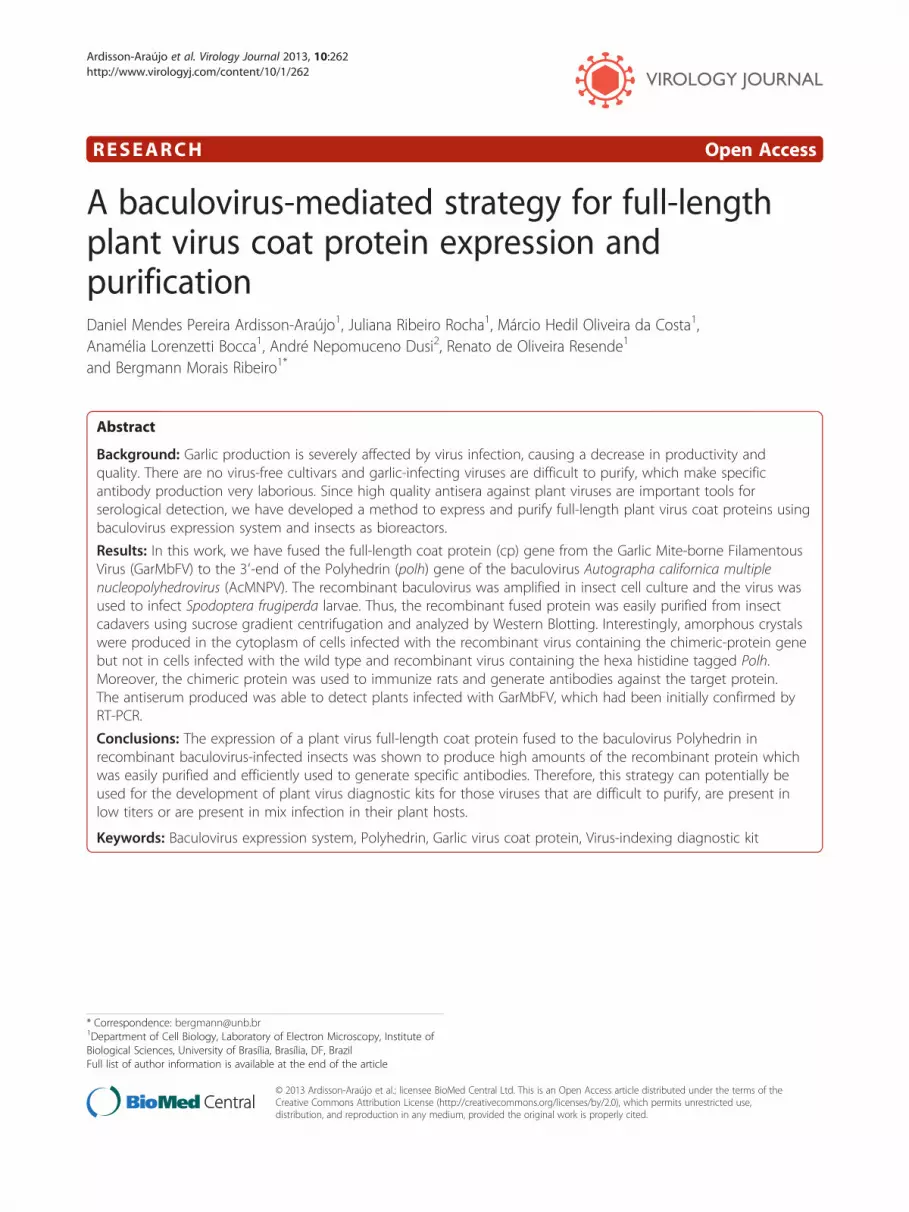
Analysis of purified recombinant crystalsPurified occlusion bodies from wild type and recombinantvirus-infected S. frugiperda larva were analyzed by scan-ning electron microscopy. All occlusion bodies formeda distinct band on the sucrose gradient (Figure 4A).AcMNPV occlusion bodies showed a regular cubicshape as expected (Figure 4B-I), on the other hand,the vAc-polh-6xhis showed mainly triangular shapedocclusion bodies (Figure 4B-II) and the vAc-polh-GarMbFV-cp-6xhis showed putative occlusion bodiesof amorphous shape (Figure 4B-III).
Figure 3 Structural analysis of both vAc-polh-6xhis- and vAc-polh-Garinfected cells showing the presence of numerous occlusion bodies inside tTn5B cells showing the occlusion bodies derived from the recombinant fusScale bar = 20 μm.
Antiserum production and identification ofGarMbFV-infected garlic plantsThe purified fusion protein Polh-GarMbFV-CP was sol-ubilized and used to immunize rats. The antiserum wastested in extracts derived from infected insect cells andgarlic plants with visible virus infection symptoms (mo-saic) by SDS-PAGE/immunoblotting and Dot-ELISA tech-nique, respectively. Virus-infected Tn5B extracts wereseparated by 12% SDS-PAGE (not shown) and transferredto a nitrocellulose membrane. Immunoreactive bands of29.9 and 50.0 kDa were detected in extracts of vAc-polh-6xhis- and vAc-polh-GarMbFV-cp-6xhis-infected cells(72 h p.i.) using anti-Polh-GarMbFV-CP-6xHis antiserum(Figure 5A, lane 1). Furthermore, the antiserum producedwas tested against vSyn-GarMbFV-cp- and mock-infectedextract cells. Two immunoreactive bands were found inthe first extract, one of 29.9 kDa, the molecular weight ofthe Polyhedrin and another one of 27.9 kDa, theGarMbFV-CP weight, as expected. The specificity of theantiserum produced against insect cell was checked andthere was no detection of any protein in mock-infectedcell extracts (Figure 5A, lane 2).For dot-ELISA, the purified fusion protein (A), both
GarMbFV-infected (C+) and uninfected (C-) garlic leafextracts were used as controls (Figure 6 square A, C+,and C-). Garlic leaf extracts from nine different plantsshowing symptons of virus infection reacted with the pro-duced antiserum (Figure 6, numbers 1 to 3 and 5 to 9).On the other hand, the extract number 4, even presentingsymptoms, did not react with the antiserum (Figure 6).RT-PCR was carried out to corroborate the dot-ELISAresults. The same positive and negative results wereobserved by the serological technique (not shown).
DiscussionBrazil produces less garlic (Allium sativum) than it con-sumes, even with a local production reaching 140,000 t
MbFV-cp-6xhis-infected Tn5B cells at 72 h p.i. (A) vAc-polh-6xhis-he cell nucleus (black arrows). (B) vAc-polh-GarMbFV-cp-6xhis-infecteded protein mainly in the cytoplasm of the cells (white arrows).
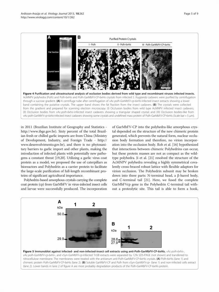
Figure 4 Purification and ultrastructural analysis of occlusion bodies derived from wild type and recombinant viruses infected insects.AcMNPV polyhedra (Polh) and Polh-6xHis and Polh-GarMbFV-CP-6xHis crystals from infected S. frugiperda cadavers were purified by centrifugationthrough a sucrose gradient. (A) A centrifuge tube after centrifugation of vAc-polh-GarMbFV-cp-6xHis-infected insect extracts showing a lowerband containing the putative crystals. The upper band shows the fat fraction from the insect cadavers. (B) The crystals were collectedfrom the gradient and prepared for scanning electron microscopy: (I) Occlusion bodies from wild type AcMNPV infected insect cadavers;(II) Occlusion bodies from vAc-polh-6xhis-infected insect cadavers showing a triangular shaped crystal; and (III) Occlusion bodies-like fromvAc-polh-GarMbFV-cp-6xhis-infected insect cadavers showing some crystals and undefined mass protein of Polh-GarMbFV-CP-6xHis (Scale bar = 5 μm).
Ardisson-Araújo et al. Virology Journal 2013, 10:262 Page 5 of 9http://www.virologyj.com/content/10/1/262
in 2011 (Brazilian Institute of Geography and Statistics -http://www.ibge.gov.br). Sixty percent of the total Brazil-ian fresh or chilled garlic imports are from China (Ministryof Development, Industry, and Foreign Trade - http://www.desenvolvimento.gov.br), and there is no phytosani-tary barriers to garlic import and other plants, making theintroduction of infected plants with potentially new patho-gens a constant threat [19,20]. Utilizing a garlic virus coatprotein as a model, we proposed the use of caterpillars asbioreactors and Polyhedrin as a carrier protein to facilitatethe large-scale purification of full-length recombinant pro-teins of significant agricultural importance.Polyhedrin-based amorphous crystals carrying the complete
coat protein (cp) from GarMbFV in virus-infected insect cellsand larvae were successfully produced. The incorporation
Figure 5 Immunoblot against infected- and non-infected-insect cell evAc-polh-GarMbFV-cp-6xhis-, and vSyn-GarMbFV-cp-infected Tn5B extracts wnitrocellulose membrane. The membranes were treated with the antiserumchimeric protein Polh-GarMbFV-CP-6xHis (lane 2). (B) Soluble GarMbFV-CP(lane 2). Lower bands in lane 2 of figure A are most probably degradation
of GarMbFV-CP into the polyhedra-like amorphous crys-tal depended on the structure of the new chimeric proteingenerated, which prevents the natural form, nuclear occlu-sion body formation and therefore, no virion incorpor-ation into the occlusion body. Roh et al. [16] hypothesizedthat interactions between chimeric Polyhedrins can occur,but these protein masses are not as compact as the wild-type polyhedra. Ji et al. [21] resolved the structure of theAcMNPV polyhedra revealing a highly symmetrical cova-lently cross-braced robust lattice with flexible adaptors forvirion occlusion. The Polyhedrin subunit may be brokendown into three parts: N-terminal head, a β-barrel body,and C-terminal tail [21]. Here, we fused the completeGarMbFVcp gene in the Polyhedrin C-terminal tail with-out a proteolytic site. This tail is able to form a hook,
xtracts using anti-Polh-GarMbFV-CP-6xHis. vAc-polh-6xhis-,ere separated by 12% SDS-PAGE (not shown) and transferred toanti-Polh-GarMbFV-CP-6xHis crystals. (A) Polh-6xHis (lane 1) and
and Polh from vSyn-GarMbFV-cp- (lane 1) and non-infected cells extractproducts of the Polh-GarMbFV-CP-6xHis protein.

Figure 6 Dot-ELISA of garlic leaf extracts. A shows the antigen (baculovirus-expressed GarMbFV-CP) used to obtain the antiserum, (C+) showsa positive control derived from a GarMbFV-infected leaf extract confirmed by RT-PCR (not shown), in (C-) a virus-free garlic leaf extract as negativecontrol. Nine extracts (1 to 9) from different plants showing virus infection symptoms were denaturated with modified Laemmili’s buffer,manually dotted, and tested with the antiserum obtained in this work. Only sample 4 did not react with the antiserum produced.
Ardisson-Araújo et al. Virology Journal 2013, 10:262 Page 6 of 9http://www.virologyj.com/content/10/1/262
jutting outwards which interacts with another Polyhedrinto form the crystal. Thus, the amorphous-shaped proteinmass observed by scanning microscopy compared to thewild-type and the 6x-His-tagged Polyhedrin (Figure 4B), isprobably due to both the size of the fused gene and the c-terminal fusion with the Polyhedrin protein. This featureavoided the polyhedral matrix formation and the nuclearlocalization by the chimeric protein. Previous research hasused, besides the polh gene fused with a gene of interest, asecond polh gene copy [14,17]. Although the presence of asecond copy of polh could improve the chimeric crystalformation and nuclear localization, we observed that thepresence of only one fused Polyhedrin-copy was sufficientto form a crystal structure. This allows recombinantprotein purification from insect cadavers and cells aspreviously observed in vitro [16]. The purification ofthe recombinant protein is carried out by using su-crose gradient centrifugation which is both easier andcheaper than column chromatography which is nor-mally used for recombinant protein purification.The expression of the full-length fusion protein pro-
duced a polyhedra-like crystal that presented alteredocclusion body morphology and localization, but this al-teration did not affect protein purification in our study.Purified crystals carrying the GarMbFVcp showed thatthe coat protein antigens elicited the production of anti-bodies in rats. The results indicated that immunizationwith a chimeric protein induced a significant serum anti-body reaction against Polyhedrin and the coat protein.This demonstrated that both were immunogenic andthat the baculovirus protein perhaps increases the
immune response as previously observed [17,22] foranother baculovirus carrier protein and the Polyhedrin.Similar methods have already been developed andemployed to purify target proteins [16-18]; however, thenovelty presented here is the expression of a full-lengthprotein in fusion with the Polyhedrin and the use ofcomplete cadavers of caterpillars as bioreactors.Unlike what was observed by Alves-Junior et al. [12],
the antiserum produced by this new approach using thePolyhedrin as a full-length protein carrier was able todetect infected plants in dot-ELISA assays and to distin-guish infected from non-infected plants at a high anti-serum dilution (1:1,000 from the crude antiserum) andthis detection was confirmed by RT-PCR using specificGarMbFV-cp primers (data not shown).A challenge for a virus-indexing diagnosis in the field
is the fact that this particular garlic disease is caused bya viral complex [20]. In Brazil alone, three virus generarelated to garlic mosaic symptoms [3] were found:Potyvirus [Onion yellow dwarf virus (OYDV) and Leekyellow stripe virus (LYSV)] [23], Carlavirus [Garlic com-mon latent virus (GCLV) and Shallot latent virus (SLV)][23,24], and Allexivirus, [Garlic miteborne filamentousvirus (GarMbFV), Garlic virus C (GarV-C) and Garlicvirus D (GarV-D)] [8,25]. Notably, a symptomatic plantwas found to be negative for GarMbFV, in both dot-ELISA and RT-PCR tests, suggesting that the producedantiserum did not cross-react to other viruses in the com-plex, although more experiments are necessary to confirmthis result. Moreover, indirect ELISA or sandwich ELISAkits based on our strategy can be also developed.

Ardisson-Araújo et al. Virology Journal 2013, 10:262 Page 7 of 9http://www.virologyj.com/content/10/1/262
ConclusionsThe expression of a plant virus full-length coat proteingene fused to the baculovirus Polyhedrin in recombinantbaculovirus-infected insects was shown to produce highamounts of the recombinant protein which was easilypurified and efficiently used to generate specific anti-bodies. Therefore, this strategy could be used for the de-velopment of plant virus diagnostic kits of those virusesthat are difficult to purify, are in low titers, or arepresent in mix infections in their plant hosts.
MethodsInsect cells, viruses and insectsTrichoplusia ni (cabbage looper) BTI-Tn5B1-4 (Tn5B)cells [26] were maintained in TC-100 medium (HIMEDIA)supplemented with 10% fetal bovine serum (Invitrogen),and an antibiotic-antimycotic mixture (Gibco) at 28°C. Wildtype Autrographa californica multiple nucleopolyhedrovirus(AcMNPV); vSynVI-gal [27] (an AcMNPV recombinantwhich contains the β-galactosidase (lac-Z) gene inplace of the polh gene); recombinant viruses vSyn-GarMbFV-cp, vAc-polh-GarMbFV-cp-6xhis and, vAc-polh-6xhis constructed in this work have been propagatedand their titers determined according to O’Reilly et al.[28]. Spodoptera frugiperda larvae, the fall armyworm, inearly five-instar was provided by EMBRAPA/CENARGEN– Genetic Resources and Biotechnology (Brasília, Brazil),maintained at 25°C, and fed on an artificial diet [29]. Theinfection was carried out by injection of 10 μl of mediumcontaining recombinant virus (106 viruses in BV pheno-type) into the hemocoel.
Coat protein and polh amplificationThe GarMbFV coat protein (GarMbFVcp) [25] (Genbankaccession number X98991) was amplified using the F-GarMbFVcpN (CCA TGG ACG ACC CTG TTG ACCCAA GC) and R-GarMbFVcpN (CCA TGG AGA ACGTAATCA TGG GAG G) oligonucleotides that modify thestop-codon and add NcoI restriction sites (in italics)flanking the gene for posterior fusion. The AcMNPV polhgene was modified by PCR in order to construct the fu-sion vector. Forward primer (Acpol-BglII F) adds a BglIIrestriction site (in italics) (CCG AGA TCT ATG CCAGAT TAT AGC TAT AGG CC) at the 5′-terminus andthe reverse primer (Ac-pol/hisc NcoI R) removes the genestop-codon and adds a NcoI restriction site (in italics), andsix histidines codons (underlined sequence) at the 3′-terminus (TTA GTG ATG ATG ATG ATG ATG TTCCAT GGA ATA ATA CGC GGG GCC GGT AAA CAGAGG TGC). The PCR program used for both reactionswas: 94°C/5 min, 35 cycles of 94°C/s, 50°C/20 s and 72°C/40 s and a final extension for 7 min at 72°C. Reactionscontained 10 ng of DNA-sample, 300 μM of dNTPmix (Fermentas), 0.4 μM of each set of primers
previously described, and the LongAmp enzyme (NewEngland Biolabs). The modified fragments obtained(GarMbFV-cp and polh-6xHis) were analyzed by elec-trophoresis in 0.8% agarose gels [30], eluted using the GFXPCR DNA and Gel Band Purification kit (GE Healthcare),cloned into the pGem®-T easy vector (Promega), and se-quenced (Macrogen, Korea) to certify the modifications.
Fusion vector and recombinant virus constructionThe polh-6xhis gene was removed from the cloning vec-tor by BglII and NotI restriction digestion following themanufacturer’s instructions (Promega) and analyzed byelectrophoresis in a 0.8% agarose gel [30]. The DNAfragment was then purified using the GFX™ PCR DNAand Gel Band Purification Kit (GE Healthcare) andcloned into the commercial donor vector pFastBac1®(pFB1 – Invitrogen) previously digested with BamHIand NotI (Promega) restriction enzymes in order to gen-erate the recombinant plasmid pFB1-polh-6xhis. TheNcoI-flanked GarMbFV-cp gene, was removed from thepGem-T easy vector by NcoI restriction digestion, thefragment was analyzed by electrophoresis and purifiedfrom the gel as described above. The purified fragmentwas then introduced into the unique NcoI site present inthe pFB1-polh-6xhis plasmid in order to construct thepFB1-polh-GarMbFV-cp-6xhis plasmid. Both vectors(pFB1-polh-6xhis and pFB1-polh-GarMbFV-cp-6xhis)were transformed into DH10-Bac cells (Invitrogen) byelectroporation [30] and recombinant bacmids were se-lected following the manufacturer’s instructions (Bac-to-Bac®, Baculovirus expression systems, Invitrogen). DNAfrom the bacmids were purified and the presence of therecombinant gene was checked by PCR using specificoligonucleotides as described by the manufacturer’sprotocol (Invitrogen). One microgram of each recombin-ant bacmid was transfected into Tn5B cells (106) usingliposomes (Cellfectin®) according to manufacturer’s in-structions (Invitrogen). Since naked baculovirus DNA isinfectious, the supernatant of 7 days post-transfectionTn5B cells containing the recombinant viruses were col-lected, tittered, and the virus amplified by infection of1,5 × 107 cells with a MOI of 1 in 75 cm2 flasks (TPP) asdescribed in O’Reilly et al. [28].For the construction of a recombinant virus containing
the coat protein gene, the previously described plas-mid, pGem-GarMbFV-cp [25], was digested with EcoRI(Promega) and analyzed by electrophoresis in a 0.8% agar-ose gel [30]. The DNA fragment was purified as previouslydescribed and cloned into the unique EcoRI restriction siteof the transfer vector pSynXIVVI + X3 [27], which enablesinsertion of the heterologous gene under the control oftwo strong promoters in tandem (pSyn and pXIV). Thevector pSynXIVVI + X3 had been previously digestedwith EcoRI (Promega) and dephosphorilated using SAP

Ardisson-Araújo et al. Virology Journal 2013, 10:262 Page 8 of 9http://www.virologyj.com/content/10/1/262
(Shrimp Alkaline Phosphatase) enzyme (Promega). Onemicrogram of the resulting recombinant plasmid was co-transfected with 0.5 μg of the viral DNA from vSynVI-gal[27] in Tn5B cells (106) using liposomes (as describedabove). Seven days after co-transfection, the cell super-natant was collected and used for recombinant virus isola-tion by serial dilution in 96-well plates [28]. Therecombinant virus was amplified by infection of Tn5B(1.5 × 10⁷ cells) with an MOI of 1 in 75 Cm2 flasks (TPP).
Putative recombinant protein crystals purificationWild type and recombinant viruses were used to infect200 S. frugiperda larvae each as described above. Afterthe death by infection, the cadavers were homogenizedwith the same volume of ddH2O (w/v), filtered throughgauzes and centrifuged at 7,000 x g for 10 min. The super-natant was discarded and the pellet was resuspended inthe same volume of 5% Triton X-100 and centrifuged at7,000 x g for 10 min. This procedure was repeated twice.The last pellet was resuspended in 0.5 M NaCl,centrifuged once more as above, and resuspended withddH2O. All solutions contained a Protease Inhibitor Cock-tail (Sigma – according to notes of use). The suspendedsolution was loaded on a discontinuous sucrose gradient(40-80% of sucrose in Phosphate Buffered Saline [PBS],137.0 mM NaCl, 2.7 mM KCl, 10.0 mM Na2HPO4,2.0 mM KH2PO4, pH 7.4) and centrifuged at 130,000 x gfor 3 h. The band containing the putative crystals were re-moved from the gradient, five-fold diluted with ddH2O,and centrifuged at 7,000 x g for 10 min. Cadavers of vAc-polh-6xhis-infected larva were purified according to stand-ard method for polyhedra purification [28]. The purifiedcrystals were subjected to SDS-PAGE and ultrastructuralanalysis.
Microscopy analysisA monolayer of Tn5B cells (5.0 × 10⁶) was infected at anMOI of 5 with vAc-polh-GarMbFV-cp-6xhis and vAc-polh-6xhis. The infected cells were observed andphotographed at 72 h p.i. in an Axiovert 100 invertedlight microscope (Zeiss). For scanning electron micros-copy (SEM), the purified putative crystals were dried ina critical point-dried (Balzers) and coated with gold in aSputter Coater (Balzers) before being observed in a SEMJeol JSM 840A at 10 kV.
Analysis of recombinant protein synthesisTn5B cells (5.0 × 10⁶) were infected (10 pfu/cell) withvSyn-GarMbFV-cp, vAc-polh-GarMbFV-cp-6xhis, vAc-polh-6xhis, and the wild-type AcMNPV. At 72 h p.i., theinfected cells were collected and centrifuged at 10,000 xg for 2 min. The resulting pellets were resuspended inPBS with an equal volume of 2x protein loading buffer(0.25 M Tris-Cl, pH 6.8, 4% SDS, 20% glycerol, 10% 2-
mercaptoethanol, and 0.02% bromophenol blue). Pro-teins were heated (100°C) for 5 min and analyzed by12% SDS-PAGE using the Mini Protean Tetra Cellaparatus (BioRad) following the manufacturer’s instruc-tions. For immunoblotting, samples were resolved by12% SDS-PAGE and transferred onto a nitrocellulosemembrane (Sigma) using the Trans-Blot® SD – Semi DryTransfer Cell (BioRad). The membrane was then blockedin 1× PBS Buffer (137 mM NaCl, 2.7 mM KCl, 10 mMNa2HPO4, 2 mM KH2PO4, pH 7.4) containing 3%skimmed milk powder for 16 h at 4°C, washed threetimes with PBS tween (0.05%) and probed with (i) mousemonoclonal anti-hexa-histidine (anti-6xHis) antibody(Sigma), (ii) rabbit anti-Polh antiserum [31], or (iii) anti-Polh-GarMbFV-CP-6xHis (produced in this work)followed by incubation with the alkaline phosphatase-conjugated anti-mouse/rat or anti-rabbit secondaryantibodies (Sigma). Blots were developed using theNBT/BCIP (Sigma) substrate dissolved in alkaline phos-phatase buffer (NaCl 100 mM, MgCl2 5 mM e Tri-HCl100 mM – pH 9.0).
Immunization and antiserum productionThe purified recombinant crystals of Polh-GarMbFV-CP-6xHis was dissolved for 1 h at 37°C in 0.1 M ofNa2CO3, neutralized with 0.1 M of Tris–HCl (pH 7.6)and centrifuged at 16,000 x g for 30 min. The proteinconcentration was estimated by SDS-PAGE, comparingdifferent BSA concentrations with the query protein(data not shown). The solution was filter sterilized andused for quadriceps intramuscular injection firstly withFreund’s complete adjuvant at 1:1 proportion, secondlywith Freund’s incomplete adjuvant at 1:1 proportion,and finally alone. Five Gnotobiotic Sprague–Dawleymale rats strain CD, 8 weeks old with food and water adlibidum, were used in this immunization experiment.The project was reviewed by the Ethics Committee onAnimal Use of the University of Brasília. The animalswere immunized with 15 days intervals for each injec-tion containing 500 μg of the recombinant protein.
Dot enzyme-linked immunosorbent assay (dot-ELISA)Garlic plants infected with Garlic Mite-borne FilamentousVirus (GarMbFV) showing typical symptoms on the leavessuch as yellow mosaic, stripes, and distortion and a gen-eral plant growth reduction were collected in the garlicgermplasm bank from Embrapa Vegetables (Brasilia-DF,Brazil). The garlic leafs were triturated in PBS using aproportion of 100 μl of PBS per each 100 mg of leaves.The samples were mixed with equal volumes of 2X pro-tein loading buffer without stain and heated (100°C) for5 min. Purified antigen (Polh-GarMbFV-CP-6xHis), virus-infected, and non-infected leaf samples were manuallydotted on a nitrocellulose membrane and probed against

Ardisson-Araújo et al. Virology Journal 2013, 10:262 Page 9 of 9http://www.virologyj.com/content/10/1/262
the crude rat anti-Polh-GarMbFV-CP-6xHis antiserumat 1:100, 1:500 and 1:1,000 (v/v) dilutions in 1X PBSplus 0.5% BSA (w/v) followed by incubation with thealkaline phosphatase-conjugated mouse secondary anti-bodies (Sigma) and NBT/BCIP substrate (Sigma) in alka-line phosphatase buffer. The positive and negative controlplant extracts were confirmed by RT-PCR accordingto Fayad-André et al. (2011).
Competing interestsThe authors declare that they have no competing interests.
Authors’ contributionsConceived and designed the experiments: DMPAA, BMR, ROR. Performed theexperiments: DMPAA, JRR, MHOC, Analyzed the data: DMPAA, BMR, ROR.Contributed reagents/materials/analysis tools: BMR, ROR, ALB, AND. Wrotethe paper: DMPAA, BMR, ROR. All author s read and approved the finalmanuscript.
AcknowledgementsWe thank Embrapa Recursos Genéticos e Biotecnologia-CENARGEM for kindlyproviding the Spodoptera frugiperda larva; Dr. Miguel Alves-Júnior forconstructing the vSyn-GarMbFV-cp recombinant virus; Dra. Fernanda RauschFernandes from Empraba Vegetables for providing garlic leaf extracts;Professor Dr. Fernando Lucas Melo for the critical reading of the manuscript;and Fernanda Araújo Ferreira for kindly reviewing the English.
Author details1Department of Cell Biology, Laboratory of Electron Microscopy, Institute ofBiological Sciences, University of Brasília, Brasília, DF, Brazil. 2EmbrapaVegetables, Embrapa, Brasília, Brazil.
Received: 19 June 2013 Accepted: 14 August 2013Published: 15 August 2013
References1. Lot H, Chovelon V, Souche S, Delecolle B: Effects of onion yellow dwarf
and leek yellow stripe viruses on symptomatology and yield loss ofthree French garlic cultivars. Plant Dis 1998, 82:5.
2. Conci VC, Canavelli A, Lunello P, Rienzo JD, Nome SF, Zumelzu G, Italia R:Yield losses associated with virus-infected garlic plants during fivesuccessive years. Plant Dis 2003, 87:5.
3. Lunello P, Rienzo JD, Conci VC: Yield Loss in Garlic Caused by Leek yellowstripe virus Argentinean Isolate. Plant Dis 2007, 15:6.
4. Torres AC, Fajardo TV, Dusi AN, Resende RO, Buso JA: Shoot tip cultureand thermotherapy for recovering virus-free plants of garlic.Horticultura Brasileira 2000, 18:3.
5. Tsuneyoshi T, Sumi S: Differentiation among garlic viruses in mixedinfections based on RT-PCR procedures and direct tissue blottingimmunoassays. Phytopathology 1996, 86:7.
6. Tsuneyoshi T, Matsumi T, Natsuaki KT, Sumi S: Nucleotide sequenceanalysis of virus isolates indicates the presence of three potyvirusspecies in Allium plants. Arch Virol 1998, 143:97–113.
7. Takaichi M, Nagakubo T, Oeda K: Mixed virus infections of garlicdetermined by a multivalent polyclonal antiserum and virus effects ondisease symptoms. Plant Dis 2001, 85:5.
8. Fayad-André MS, Dusi AN, Resende RO: Spread of viruses in garlic fieldscultivated under different agricultural production systems in Brazil.Trop Plant Pathol 2011, 36:3.
9. Huttinga H: Purification by molecular sievin of a leak virus related toonion yellow dwarf virus. Netherland J Plant Pathol 1975, 81:3.
10. Albrechtsen M, Heide M: Purification of plant viruses and virus coatproteins by high performance liquid chromatography. J Virol Methods1990, 28:245–256.
11. Filho FACR, Lima JA, Ramos NF, Gonçalves MFB, Carvalho KF: Produção deanti-soro para o vírus do mosaico da abóbora mediante imunização oralde coelhos. Revista Ciência Agronômica 2005, 36:4.
12. Alves-Junior M, Marraccini FM, Melo Filho PA, Dusi AN, Pio-Ribeiro G, RibeiroBM: Recombinant expression of Garlic virus C (GARV-C) capsid protein in
insect cells and its potential for the production of specific antibodies.Microbiol Res 2008, 163:354–361.
13. Jarvis DL, Bohlmeyer DA, Garcia A Jr: Enhancement of polyhedrin nuclearlocalization during baculovirus infection. J Virol 1992, 66:6903–6911.
14. Wei Q, Kim YS, Seo JH, Jang WS, Lee IH, Cha HJ: Facilitation of expressionand purification of an antimicrobial peptide by fusion with baculoviralpolyhedrin in Escherichia coli. Appl Environ Microbiol 2005, 71:5038–5043.
15. Seo JH, Yeo JS, Cha HJ: Baculoviral polyhedrin-Bacillus thuringiensis toxinfusion protein: a protein-based bio-insecticide expressed in Escherichiacoli. Biotechnol Bioeng 2005, 92:166–172.
16. Roh JY, Choi JY, Kang JN, Wang Y, Shim HJ, Liu Q, Tao X, Xu HG, Hyun J,Woo SD, et al: Simple purification of a foreign protein using polyhedrinfusion in a baculovirus expression system. Biosci Biotechnol Biochem 2010,74:5.
17. Lee KS, Sohn MR, Kim BY, Choo YM, Woo SD, Yoo SS, Je YH, Choi JY, Roh JY,Koo HN, Jin BR: Production of classical swine fever virus envelopeglycoprotein E2 as recombinant polyhedra in baculovirus-infectedsilkworm larvae. Mol Biotechnol 2012, 50:211–220.
18. Bae SM, Kim HJ, Lee JB, Choi JB, Shin TY, Koo HN, Choi JY, Lee KS, Je YH, JinBR, et al: Hyper-enhanced production of foreign recombinant protein byfusion with the partial polyhedrin of nucleopolyhedrovirus. PLoS One2013, 8:e60835.
19. Conci V, Nome SF, Milne RG: Filamentous viruses of garlic in Argentina.Plant Dis 1992, 76:3.
20. van Dijk P: Carlavirus isolates from cultivated Allium species representthree viruses. Netherland J Plant Pathol 1993, 99:25.
21. Ji X, Sutton G, Evans G, Axford D, Owen R, Stuart DI: How baculoviruspolyhedra fit square pegs into round holes to robustly package viruses.EMBO J 2010, 29:505–514.
22. Molinari P, Crespo MI, Gravisaco MJ, Taboga O, Moron G: Baculoviruscapsid display potentiates OVA cytotoxic and innate immune responses.PLoS One 2011, 6:e24108.
23. Fajardo TVM, Nishijima M, Buso JA, Torres AC, Ávila AC, Resende RO: GarlicViral Complex: Identification of Potyviruses and Carlavirus in CentralBrazil. Fitopatologia Brasileira 2001, 26:8.
24. Mituti T, Marubayashi JM, Moura MF, Krause-Sakate R, Pavan MA: FirstReport of Shallot latent virus in Garlic in Brazil. Plant Dis 2011, 95:1.
25. Melo Filho PA, Nagata T, Dusi NA, Buso JA, Torres AC, Eiras M, Resende RO:Detection of three Allexivirus species infecting garlic in Brazil.Pesquisa Agropecuária Brasileira 2004, 39:5.
26. Granados RR, Guoxun L, Derksen ACG, Mckenna KA: A new insect cell linefrom Trichoplusia ni (BTI-Tn-5B1-4) susceptible to Trichoplusia ni singleenveloped nuclearpolyhedrosis virus. J Invertebrate Pathol 1994, 64:6.
27. Wang XZ, Ooi BG, Miller LK: Baculovirus vectors for multiple geneexpression and for occluded virus production. Gene 1991, 100:131–137.
28. O’Reilly D, Miller LK, Luckow VA: Baculovirus Expression Vectors: a laboratorymanual. New York: Freeman and Company; 1992.
29. Greene GL, Leppla NC, Dickerson WA: Velvetbean caterpillar: a rearingprocedure and artificial medium. J Economical Entomol 1976, 69:2.
30. Sambrook J, Russel DW: Molecular Cloning: a laboratory manual. 3rd ed. edn.New York: Cold Spring Harbor; 2001.
31. Ribeiro BM, Generino AP, Acacio CN, Kalapothakis E, Bao SN:Characterization of a new Autographa californica multiplenucleopolyhedrovirus (AcMNPV) polyhedra mutant. Virus Res 2009,140:1–7.
doi:10.1186/1743-422X-10-262Cite this article as: Ardisson-Araújo et al.: A baculovirus-mediatedstrategy for full-length plant virus coat protein expression andpurification. Virology Journal 2013 10:262.