a bacterial source for mollusk pyrone polyketides
TRANSCRIPT

Chemistry & Biology
Article
A Bacterial Source for Mollusk Pyrone PolyketidesZhenjian Lin,1 Joshua P. Torres,6 Mary Anne Ammon,6 Lenny Marett,1,2 Russell W. Teichert,3 Christopher A. Reilly,4
Jason C. Kwan,1 RonaldW. Hughen,2 Malem Flores,6 Ma. Diarey Tianero,1 Olivier Peraud,1 James E. Cox,5 Alan R. Light,2
Aaron Joseph L. Villaraza,7 Margo G. Haygood,8 Gisela P. Concepcion,6 Baldomero M. Olivera,3 and Eric W. Schmidt1,3,*1Department of Medicinal Chemistry2Department of Anesthesiology3Department of Biology4Department of Pharmacology and Toxicology
University of Utah, Salt Lake City, UT 84112, USA5Metabolomics Core Research Facility and Department of Biochemistry, University of Utah Health Sciences, Salt Lake City, UT 84112, USA6Marine Science Institute7Institute of Chemistry
University of the Philippines, Diliman, Quezon City 1101, Philippines8Department of Environmental and Biomolecular Systems, OGI School of Science and Engineering, Oregon Health & Science University,
Beaverton, OR 97006, USA
*Correspondence: [email protected]://dx.doi.org/10.1016/j.chembiol.2012.10.019
SUMMARY
In the oceans, secondary metabolites often protectotherwise poorly defended invertebrates, such asshell-less mollusks, from predation. The origins ofthese metabolites are largely unknown, but many ofthem are thought to be made by symbiotic bacteria.In contrast, mollusks with thick shells and toxicvenoms are thought to lack these secondary metab-olites because of reduced defensive needs. Here, weshow that heavily defended cone snails also occa-sionally contain abundant secondary metabolites,g-pyrones known as nocapyrones, which aresynthesized by symbiotic bacteria. The bacteria,Nocardiopsis alba CR167, are related to widespreadactinomycetes that we propose to be casual symbi-onts of invertebrates on land and in the sea. Thenatural roles of nocapyrones are unknown, but theyare active in neurological assays, revealing thatmollusks with external shells are an overlookedsource of secondary metabolite diversity.
INTRODUCTION
Many marine animals are protected by arsenals of defensive
compounds, which have been exploited in the discovery of phar-
maceuticals (Putz and Proksch, 2010). These chemicals can be
divided into two groups: proteins and peptides (large molecules)
and secondary metabolites (small molecules). Protein-based
toxins include venoms, which are synthesized by the animals
themselves and are encoded in the animal genomes (Terlau
and Olivera, 2004). Small molecules are highly diverse in terms
of their chemical structures, and they are more commonly asso-
ciated with soft-bodied, ‘‘defenseless’’ animals, such as ascid-
ians, sponges, and nudibranch mollusks (Blunt et al., 2011). In
contrast to the casewith protein toxins, increasing evidence indi-
cates that symbiotic bacteria synthesize at least some of these
Chemistry & Biology 20,
small molecules, although only a few systems have been studied
in any detail (Piel, 2009; Kubanek et al., 1997).
Mollusks lacking shells are sometimes defended instead by
secondary metabolites (Benkendorff, 2010). Often, the mole-
cules seem to have a dietary origin (Fontana, 2006; Gavagnin
et al., 1994). However, a widely occurring group of mollusk
secondary metabolites, pyrones and structurally related polyke-
tides, are generated in the animals de novo, possibly even by the
mollusks themselves (Davies-Coleman and Garson, 1998; Marin
et al., 1999; Cimino et al., 1987; Ireland and Scheuer, 1979). The
close structural resemblance of these polyketides to bacterial
metabolites has led to the hypothesis that symbiotic bacteria,
and not the animals, produce the compounds in question
(Cutignano et al., 2009). This idea is complicated by the fact
that there are many convergent biosynthetic routes to pyrones
(Busch and Hertweck, 2009), so the ultimate source is a matter
of debate. Molecular or genetic evidence is still lacking to defin-
itively tie production of any gastropod metabolite to symbiotic
bacteria or to any other source.
Mollusks with shells, such as cone snails and turrids (Terlau
and Olivera, 2004; Lopez-Vera et al., 2004), synthesize animal
genome-derived peptide toxins, both for predation and for
defense (Olivera, 2002). In contrast to their shell-less relatives,
mollusks with external shells are usually believed to be deficient
in secondary metabolites (Benkendorff, 2010), leading re-
searchers to avoid the animals as sources of new natural prod-
ucts. For venom-containing mollusks, like cone snails, peptide
toxins should provide ample defense against predators.
Recently, we began to question whether cone snails might
also harbor symbiont-derived metabolites. If so, this might chal-
lenge current ideas about distribution and roles of marine natural
products and suggest that otherwise protected animals are
good sources for discovery. Because by far most mollusks are
defended by shells, this would open a new area for compound
discovery. We reported that at least three species of cone snails
contain abundant associated actinomycete bacteria, which are
well known to produce secondary metabolites in culture (Peraud
et al., 2009). There are now many reports of actinomycetes iso-
lated from marine animals. However, these actinomycetes have
73–81, January 24, 2013 ª2013 Elsevier Ltd All rights reserved 73
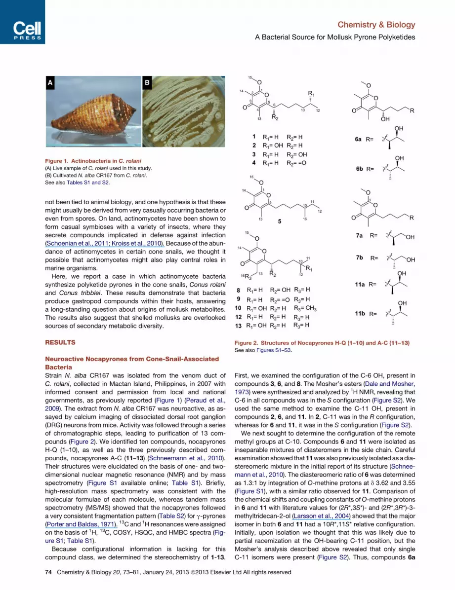
Figure 1. Actinobacteria in C. rolani
(A) Live sample of C. rolani used in this study.
(B) Cultivated N. alba CR167 from C. rolani.
See also Tables S1 and S2.
Figure 2. Structures of Nocapyrones H-Q (1–10) and A-C (11–13)See also Figures S1–S3.
Chemistry & Biology
A Bacterial Source for Mollusk Pyrone Polyketides
not been tied to animal biology, and one hypothesis is that these
might usually be derived from very casually occurring bacteria or
even from spores. On land, actinomycetes have been shown to
form casual symbioses with a variety of insects, where they
secrete compounds implicated in defense against infection
(Schoenian et al., 2011; Kroiss et al., 2010). Because of the abun-
dance of actinomycetes in certain cone snails, we thought it
possible that actinomycetes might also play central roles in
marine organisms.
Here, we report a case in which actinomycete bacteria
synthesize polyketide pyrones in the cone snails, Conus rolani
and Conus tribblei. These results demonstrate that bacteria
produce gastropod compounds within their hosts, answering
a long-standing question about origins of mollusk metabolites.
The results also suggest that shelled mollusks are overlooked
sources of secondary metabolic diversity.
RESULTS
Neuroactive Nocapyrones from Cone-Snail-AssociatedBacteriaStrain N. alba CR167 was isolated from the venom duct of
C. rolani, collected in Mactan Island, Philippines, in 2007 with
informed consent and permission from local and national
governments, as previously reported (Figure 1) (Peraud et al.,
2009). The extract from N. alba CR167 was neuroactive, as as-
sayed by calcium imaging of dissociated dorsal root ganglion
(DRG) neurons frommice. Activity was followed through a series
of chromatographic steps, leading to purification of 13 com-
pounds (Figure 2). We identified ten compounds, nocapyrones
H-Q (1–10), as well as the three previously described com-
pounds, nocapyrones A-C (11–13) (Schneemann et al., 2010).
Their structures were elucidated on the basis of one- and two-
dimensional nuclear magnetic resonance (NMR) and by mass
spectrometry (Figure S1 available online; Table S1). Briefly,
high-resolution mass spectrometry was consistent with the
molecular formulae of each molecule, whereas tandem mass
spectrometry (MS/MS) showed that the nocapyrones followed
a very consistent fragmentation pattern (Table S2) for g-pyrones
(Porter and Baldas, 1971). 13C and 1H resonanceswere assigned
on the basis of 1H, 13C, COSY, HSQC, and HMBC spectra (Fig-
ure S1; Table S1).
Because configurational information is lacking for this
compound class, we determined the stereochemistry of 1-13.
74 Chemistry & Biology 20, 73–81, January 24, 2013 ª2013 Elsevier
First, we examined the configuration of the C-6 OH, present in
compounds 3, 6, and 8. The Mosher’s esters (Dale and Mosher,
1973) were synthesized and analyzed by 1H NMR, revealing that
C-6 in all compounds was in the S configuration (Figure S2). We
used the same method to examine the C-11 OH, present in
compounds 2, 6, and 11. In 2, C-11 was in the R configuration,
whereas for 6 and 11, it was in the S configuration (Figure S2).
We next sought to determine the configuration of the remote
methyl groups at C-10. Compounds 6 and 11 were isolated as
inseparable mixtures of diasteromers in the side chain. Careful
examination showed that11wasalsopreviously isolatedasadia-
stereomeric mixture in the initial report of its structure (Schnee-
mann et al., 2010). The diastereomeric ratio of 6was determined
as 1.3:1 by integration of O-methine protons at d 3.62 and 3.55
(Figure S1), with a similar ratio observed for 11. Comparison of
the chemical shifts and coupling constants ofO-methine protons
in 6 and 11 with literature values for (2R*,3S*)- and (2R*,3R*)-3-
methyltridecan-2-ol (Larsson et al., 2004) showed that the major
isomer in both 6 and 11 had a 10R*,11S* relative configuration.
Initially, upon isolation we thought that this was likely due to
partial racemization at the OH-bearing C-11 position, but the
Mosher’s analysis described above revealed that only single
C-11 isomers were present (Figure S2). Thus, compounds 6a
Ltd All rights reserved

Table 1. Nocapyrones Detected in Methanolic Extracts of Conus rolani
Tissue Date Collected
Pyrones Detected
1 3 12
Mean ± SD
(mg/g)a d (ppm)bMean ± SD
(mg/g) d (ppm)
Mean ± SD
(mg/g) d (ppm)
1 Whole snail (n = 1)c Sept. 2009 12.4 ± 2.7 1.67 1,250 ± 154 �0.87 1.57 ± 0.26 1.22
2 Whole snail (n = 3)c Sept. 2010 7.84 ± 0.34 0.97 507 ± 31 �0.34 10.2 ± 1.5 0.87
3 Whole snail (n = 4)c Nov. 2011 nd 129 ± 37 �0.68 nd
4 Venom duct (n = 20)c July 2011 0.404 ± 0.012d 1.30 11.0 ± 2.2d 2.05 0.366 ± 0.095d 1.88
4a Other anatomical
regions (n = 2)
July 2011 nd N.D. nd
5 Mucus (n = 4)c Oct. 2011 44.7 ± 5.2e 0.86 105 ± 7.2e �1.77 31.1 ± 2.48e 1.44
6 Whole snail and
mucus (n = 20)cMar. 2012 nd nd nd
Mean over three independent measurements. Data analysis was done using Analyst QS software 2.0. nd, below detection limit. Statistical limit of
quantification: 0.29 ng/ml; practical detection limit: 0.10 ng/ml. See also Figures S4 and S5.aMicrogram compound per gram of snail tissue dry weight (each snail weighs �1 g).bParts per million (ppm) error of high-resolution mass spectrometric measurement.cn, number of samples averaged to determine quantity but with each individual sample measured separately. In these cases, each snail contained the
compounds, and averaging was done for ease of presentation.dNumbers reflect microgram per venom duct rather than per gram dry weight.eNumbers reflect microgram per snail rather than per gram dry weight.
Chemistry & Biology
A Bacterial Source for Mollusk Pyrone Polyketides
and 11a have the 10R, 11S configuration, whereas 6b and 11b
have the 10S, 11S configuration.
Using Mosher’s method, compound 7 was shown to be
present as an enatiomeric mixture, with a S:R ratio of 10:1 (Fig-
ure S3). Although in this case a primary alcohol was esterified,
whereas the Mosher’s method is used mainly for secondary
alcohols, an excellent literature precedent was available for
compounds with related functional groups: (2S)-2-methylhexyl
(2S)-3,3,3-trifluoro-2-methoxy-2-phenylpropanoate and (2R)-2-
methylhexyl (2S)-3,3,3-trifluoro-2-methoxy-2-phenylpropanoate
(Guintchin and Bienz, 2003). The Mosher’s ester of the major
isomer 7a exhibited chemical shifts of 4.24, 4.08 (AB of ABX,
JAB = 10.7 Hz, JAX = 6.6 Hz, JBX = 5.7 Hz, OCH2), which were
very similar to those reported in the literature precedent for the
S isomer (Figure S3). Likewise, 7b exhibited chemical shifts
consistent with the R isomer. Thus, the major 7a was present
as the S isomer.
Wewished to determine the remotemethyl center of 5 directly,
but no functional handle or standard compound was available. A
closely related fatty acid in the S configuration has a reported
optical rotation of [a]D = + 8.0 (Sonnet et al., 1990), whereas 5
has a measured rotation of [a]D = + 9.0. In addition, the major
isomers of related compounds 6 and 11 are in the S configura-
tion. Thus, we tentatively propose that 5 is likely also in the S
configuration.
Nocapyrones from Methanolic Extracts of C. rolaniWe noticed that nocapyrones were structurally related to previ-
ously reported polyketides from other gastropod mollusks.
Several pyrones are known to be made within mollusks based
upon labeling studies (Ireland and Scheuer, 1979; Di Marzo
et al., 1991), but whether by the animal, symbiotic bacteria, or
other organism was unknown. We hypothesized that N. alba
CR167 synthesizes pyroneswithin cone snail tissues.We already
Chemistry & Biology 20,
had demonstrated that actinobacteria live in the foot of snails,
indicating that cultivated strains, such asNocardiopsis sp.,might
be alive within the snails, rather than existing as inactive spores
(Peraud et al., 2009). However, this hypothesis still seemed
highly speculative because polyketides had never before been
reported from thickly shelled mollusks, such as cone snails.
Therefore, finding the Nocardiopsis pyrones within whole snail
tissue would provide strong support for the hypothesis.
To test this hypothesis, we extracted the whole animals with
ethanol and analyzed their organic content. Surprisingly, 1, 3,
and 12 were among the major organic components that could
be isolated from an individual snail collected in 2009. This
prompted us to perform a more detailed chemical analysis (Fig-
ure S4), collecting and analyzing snails from the same site near
Sogod, Cebu Island, during the period of 2009–2012 and deter-
mining the tissue localization and relative abundance of com-
pounds (Table 1). We synthesized a 13C-labeled pyrone as an
internal control to accurately calibrate the quantity of com-
pounds and performed control experiments to accurately deter-
mine concentrations in the snails (Figure S5). To ensure that
results would not be compromised by laboratory contamination,
work was divided between two geographical locations.
Compounds 1, 3, and 12 were variable in abundance in
C. rolani and C. tribblei samples and were sometimes not
present (Table 1; Figure S5). When present, the compounds
were relatively abundant, totaling between 0.01%–0.1% of snail
dry weight (not including the shell). Linearity was demonstrated
from calibration curves in the range 0.010 to 10.0 mg/ml with
correlation coefficients of at least 0.9996. Percent recovery of in-
jected standard (1 mg) from live snails was found to be 87.78% ±
3.71% (n = 3). This abundance is on par for what is commonly
found for other marine animal metabolites in better-studied
systems, such as sponges, ascidians, and soft-bodiedmollusks,
indicating that the compounds are present in a physiologically
73–81, January 24, 2013 ª2013 Elsevier Ltd All rights reserved 75

Chemistry & Biology
A Bacterial Source for Mollusk Pyrone Polyketides
relevant concentration range. Variability of secondary metabo-
lites between samples is frequently encountered in marine
natural products (Donia et al., 2011).
We investigated the tissue specificity of nocapyrones. Com-
pounds were only found in two locations and were absent else-
where (Table 1). Nocapyrones appeared to be largely localized
to the mucus, with a small amount (�1% of the compounds)
found in the venom duct. Pyrones and other polyketides have
previously been isolated from themucusof soft-bodiedmollusks,
where they are proposed to function in defense or communica-
tion (Di Marzo et al., 1991; Sleeper and Fenical, 1977).
Origin of Nocapyrones in Cone SnailThe data described above strongly supported the hypothesis
that bacteria produce cone snail pyrones, but there are many
potential caveats that necessitated further study (Schmidt,
2008). For example, convergent evolution or horizontal gene
transfer could lead to more complex scenarios (Schmidt,
2008). To tie the bacteria to pyrone production within the
animals, we identified the probable pyrone biosynthetic gene
cluster and showed that both this cluster and N. alba bacteria
were present in the snails themselves.
At least three convergent routes lead to the biosynthesis of py-
rones in nature (Busch and Hertweck, 2009). Therefore, to iden-
tify the pyrone biosynthetic gene cluster, we sequenced the
genome of N. alba CR167 to an average of 250 3 coverage.
The resulting genome was assembled into 353 contigs (N50 =
44.1 kbp); because of this fragmentation we analyzed both
assembled contigs and raw reads to find the pyrones gene
cluster. The central metabolic genes in the strain were similar to
those from the previously sequenced Nocardiopsis dassonvillei
(GenBank CP002040) (Sun et al., 2010), although both synteny
and sequence identity were relatively low between the genomes.
Very recently, the genome sequence of Nocardiopsis alba ATCC
BAA-2165, cultivated from the digestive tract of honeybees in
Ohio, was published in GenBank (CP003788). This genome
was >95% DNA sequence identical to that of N. alba CR167
and shared 16 out of 18 identified biosynthetic gene clusters,
usually at >99% DNA sequence identity.
Pyrones are known to be made by various types of polyketide
synthase (PKS) proteins. Using BLAST and antiSMASH (Me-
dema et al., 2011) analysis, only three PKS gene clusters were
present in N. alba CR167, including a type II PKS and two type
I PKS pathways. No type III PKSs were present. Nearly identical
pathways were also found in the well-assembled N. alba ATCC
BAA-2165 genome. Of the three PKS clusters, only one con-
tained a methyltransferase. That PKS cluster was also the only
one that appeared to have the correct domain specificity and
module architecture to produce pyrones. In addition, there is
a known pyrone g-O-methyltransferase from the jerangolid
gene cluster (Julien et al., 2006). BLAST searching for homologs
in the genome revealed only a single significant hit, also corre-
sponding to this PKS-clustered methyltransferase. Thus,
genomic data strongly implicated the identified ncp cluster as
being responsible for nocapyrone biosynthesis (Figure 3).
To provide initial chemical evidence in support of the bio-
informatics results, the jerangolid-like methyltransferase NcpB
was produced in Escherichia coli, purified (Figure S6), and
used in enzyme assays with various substrates (Nelson et al.,
76 Chemistry & Biology 20, 73–81, January 24, 2013 ª2013 Elsevier
2007). An authentic substrate was generated by chemical deme-
thylation of the natural products. Close structural homologs
were also available for assay. Although the natural substrates
were efficiently methylated with the anticipated regiochemistry,
chemically similar compounds (4-hydroxy-3,6-dimethyl-2-
pyrone, 4-hydroxy-6-methyl-2-pyrone, and germicidin A) were
not substrates (Figure 4). The narrow substrate selectivity of
this enzyme strongly supports the assignment of ncp as the
nocapyrones biosynthesis cluster. Genetic knockout data would
be desirable to reinforce this finding, but in initial experiments the
strain was resistant to transformation by standard methods
(Kieser et al., 2000).
On this basis, we proposed that ncp (deposited in GenBank,
JN792621) was responsible for pyrone synthesis (Figure 3).
ncp encodes a cluster of four genes, ncpA (oxidoreductase),
ncpB (methyltransferase), ncpC (PKS), and ncpD (putative free-
standing acyltransferase). The correct assembly of this gene
cluster was confirmed by PCR, as well as by a nearly identical
and syntenic cluster encoded in the chromosome of the recently
deposited N. alba ATCC BAA-2165. Based upon the domain
architecture of NcpC, and in analogy to other pyrone biosyn-
thetic pathways, a hypothetical biogenetic scheme can be
proposed (Figure 3). First, diverse fatty acyl CoA esters are
loaded onto NcpC. Subsequently, the PKS domains act itera-
tively, extending the fatty acid with two units of methylmalonyl
CoA. As is found with some other PKS proteins, in the absence
of a thioesterase cleavage of a diketoester proceeds in tandem
with pyrone formation (Frank et al., 2007). Finally, the resulting
products are substrates forO-methylation by NcpB. NcpAmight
catalyze C-oxidation. Existing data do not strongly define the
role for NcpD, although it may be involved in starter unit loading
or product offloading. It should be noted that other biosynthetic
routes are possible. We favor this scheme because the NcpC
acyltransferase is likely methylmalonate specific, and the side
chain variation in the pyrone series is strongly reminiscent of
the normal mixture found in actinomycete fatty acids. Finally,
a subtle point is that only the linear side-chain pyrones were iso-
lated from cone snails, which is expected in animals if the source
fatty acids originate from primary metabolism.
With the biosynthetic cluster in hand, PCR primers (Figure S7)
were designed to specifically detect the presence of the same
genes in whole snail tissues. Indeed, both ncpB and ncpC could
be amplified from snail tissue, as could the N. alba CR167 16S
rRNA gene sequence (Figure 5). The variable nature of
compound production and the relative difficulty of obtaining
samples (the snails live at �70 m) precluded more detailed
colocalization studies. Indeed, the characteristics of N. alba
CR167 are consistent with a lifestyle that is not host restricted.
N. alba CR167 was readily cultured and maintained in the labo-
ratory. Therefore, these bacteria are casual associates of cone
snails, rather than obligate symbionts. Finally, previously we
showed that actinobacteria specifically inhabit mucus-gener-
ating cells within C. rolani (Peraud et al., 2009). This location is
consistent with the chemical results found here.
ConeSnails as aSource of SmallMolecules for BioactiveCompound DiscoveryNocapyrones modulated nerve cell depolarization, with the most
active compounds achieving an IC50 of 2 mM. Nocapyrones
Ltd All rights reserved

Figure 3. Organization of the Nocapyrone Biosynthetic Gene Clusters and Model for Nocapyrone Biosynthesis
KS, b-keto acyl synthase; AT, acyl transferase; ACP, acyl carrier protein; mMCoA, methyl malonyl CoA;MT, methyltransferase; Ox, oxidoreductase. This scheme
shows the hypothetical biogenesis (see text). See also Figure S6.
Chemistry & Biology
A Bacterial Source for Mollusk Pyrone Polyketides
B (12) and H (1) were active against nearly all DRG neuronal cell
types at 50 mM in the calcium-imaging assay, whereas 3 was
inactive in the assay. We use the DRG assay because it provides
a rapid means to test response of diverse receptors in diverse
neuronal subtypes (Teichert et al., 2012a, 2012b; Lin et al.,
2011). DRG neurons are known to be heterogeneous, with
a variety of different cell types that respond to different stimuli.
Using the calcium-imaging assay, cell types can be identified
by their differential responses to discrete reagents (Teichert
et al., 2012a).
Depending on the DRG neuronal cell type, nocapyrones either
inhibited or amplified responses that were elicited by depolariz-
ing the cells with a brief application of high extracellular potas-
sium (KCl pulse). For example, both compounds 1 and 12
partially blocked depolarization-elicited (i.e., KCl-elicited)
increases in cytoplasmic calcium in large-diameter cells that
were capsaicin resistant, whereas they amplified the KCl-elicited
increases in cytoplasmic calcium of small-diameter, capsaicin-
sensitive cells (Figure 6). In additional tests, the effects of
compound 1 did not appear to correlate with acetylcholine-
sensitivity, whereas an amplification was observed consistently
in small, capsaicin- and histamine-sensitive cells. A near-
complete block was observed in a subset of menthol-sensitive
cells (Figure S8). The compounds did not affect sodium channels
Chemistry & Biology 20,
or a panel of �60 human channels and receptors (http://pdsp.
med.unc.edu/). We also tested compounds 1, 5, and 12 against
a panel of human cells overexpressing various transient receptor
potential (TRP) channels, and they were able to activate or inhibit
Ca2+ flux into those cells with IC50s between 2 and 70 mM, de-
pending upon the agent and channel subtype (Table S3).
Notably, the menthol-sensitive TRPM8 channel was inhibited,
whereas TRPA1 was activated. This is consistent with findings
using the DRG assay, where menthol-sensitive cells were
inhibited, whereas TRPA1 is abundantly expressed in capsa-
icin-sensitive neurons. However, the broad effects of the noca-
pyrones on TRP channels suggests that they may affect an
underlying channel-regulating element that leads to cell-type
specific effects, rather than acting directly on the channels or
cell-surface receptors tested.
Compounds 1 and 12 were also tested against the human
breast adenocarcinoma (MCF-7) and Chinese hamster ovary
(AA8) cell lines. Against MCF-7, 1 and 12 were cytotoxic with
IC50 values of 8.7 and 22.2 mM, respectively, whereas 3was inac-
tive against MCF-7 (>100 mM), and against AA8 only 1was cyto-
toxic with an IC50 of 10.2 mM. By contrast, the compounds were
not bacteriostatic or bactericidal. Schneemannet al. (2010) previ-
ously showed that nocapyrones A-D were inactive against an
even broader panel of bacteria and the yeast Candida glabrata.
73–81, January 24, 2013 ª2013 Elsevier Ltd All rights reserved 77

Figure 4. NcpB Is the Nocapyrone Ο-methyltransferase
High-performance liquid chromatography (HPLC) traces are shown with detection at 228 nm.
(A) Standards of pure 9 and 9a.
(B) Enzyme assay containing 9a, NcpB, and SAM.
(C) Control enzyme assay lacking SAM.
(D) Control enzyme assay with boiled NcpB.
(E–G) Other pyrone substrates were not accepted by the enzyme, using the same reaction conditions as shown in (B).
Chemistry & Biology
A Bacterial Source for Mollusk Pyrone Polyketides
DISCUSSION
Here, we show that bacteria produce pyrone polyketides in cone
snails. Pyrones and related polyketides have been isolated from
many different mollusks from many habitats, including diverse
sacoglossan, siphonarid, and cephalispidean mollusks (Gavag-
nin et al., 1994; Davies-Coleman and Garson, 1998; Ireland
and Scheuer, 1979; Di Marzo et al., 1991, 1993; Sleeper and
Fenical, 1977; Manker et al., 1988; Darias et al., 2006), where
they are produced de novo (Ireland and Scheuer, 1979; Sleeper
and Fenical, 1977; Manker et al., 1988). However, this informa-
tion does not prove the source. For example, sacoglossans eat
eukaryotic green algae and employ both the chloroplasts and
algal mRNA to survive on sunlight for extended periods (Rumpho
et al., 2000; Mujer et al., 1996). Feeding studies demonstrate that
sacoglossan pyrones are labeled by carbonate so that they obvi-
ously arise from intermediates of photosynthesis mediated by
the hosted chloroplasts (Ireland and Scheuer, 1979). The pyrone
C-methyl groups arise from propionate, which might indicate
a bacterial source (Di Marzo et al., 1991; Cutignano et al.,
2012). This is due to the fact that a-methylated polyketides often
arise from incorporation of the polyketide precursor methylmal-
onate (propionate) in bacteria, whereas in animals and other
78 Chemistry & Biology 20, 73–81, January 24, 2013 ª2013 Elsevier
eukaryotes a-methylated polyketides usually arise from later
methylation of polyketide intermediates.
Molecular genetics can provide the definitive answer to the
source question for marine natural products (Piel, 2009). Here,
we used multiple types of chemical and molecular biological
evidence to show that N. alba CR167 bacteria make in the
cone snail species, C. rolani and C. tribblei. These multiple
methods were required to demonstrate that the bacteria are
important to their hosts. This is not a given; indeed, bacteria culti-
vated from animals are most likely to be irrelevant to the core
biology of the animals (Donia et al., 2011; Klepzig et al., 2009).
As an example, actinomycetes have been previously isolated
from many different marine animals, where they have been
shown to be an excellent source of promising drug leads (Blunt
et al., 2011). However, in no prior case has an actinomycete
been directly tied to actual function in marine animals, to the
best of our knowledge. Prior knowledge (Donia et al., 2011)
suggests that the majority of such actinomycete isolates may
not actively grow with their hosts and so do not contribute abun-
dant chemicals to the whole animals. Tying individual cultivated
bacterial strains to function in vivo requires careful study.
Actinomycete symbionts are well known to produce
secondary metabolites used by terrestrial insects (Barke et al.,
Ltd All rights reserved

Figure 5. Genes for Nocapyrone Synthesis Are Found in Cone Snail
Tissue
TheN. albaCR167 ncpB, ncpC, and 16S rRNA genes were amplified from both
C. rolani and C. tribblei metagenome DNA by nested PCR experiments. The
PCR products were confirmed to be identical to the positive controls by
Sanger sequencing the gel-extracted PCR products.
(A) Primers noc16s_Fwd and noc16s_Rev.
(B) Primers MT_in_Fwd and MT_in_Rev.
(C) Primers PKS_in_Fwd and PKS_in_Rev. Template DNA: (1) C. tribblei; (2)
C. rolani; (3) N. alba CR167; (4) no DNA control; 0) DNA ladder.
See also Figure S7.
Figure 6. Activity of Compound 12 Observed by Calcium Imaging of
Dissociated DRG Neurons in Culture
Each trace is the response of a single neuron. Responses from �100 neurons
were monitored individually and simultaneously in a given experimental trial.
Selected traces are shown from a single experimental trial. The y axis is
a measure of relative intracellular (cytoplasmic) calcium concentration, [Ca2+]i,
obtained by standard ratiometric calcium-imaging techniques (i.e., ratio of
340 nm/380 nm excitation while monitoring fluorescence emission at 510 nm).
The x axis is time in minutes. Increases in [Ca2+]i were elicited by briefly de-
polarizing the neurons with 25 mM potassium (KCl pulse) at regular 7 min
intervals, as indicated by vertical arrows. Each KCl pulse elicited an increase in
[Ca2+]i (by activating voltage-gated calcium channels) that is observed as
a peak in each trace. Capsaicin (300 nM) was applied to the neurons for 1 min
at the end of the experiment, as indicated by the black circle. After the fourth
KCl pulse, the compound was applied to the cells for 6 min (horizontal bar). In
many small-diameter, capsaicin-sensitive DRG neurons, the compound
caused an amplification of the calcium-transient elicited by a KCl pulse
(bottom three traces) or directly elicited a calcium-transient (fourth trace from
the bottom). In contrast, in many large-diameter, capsaicin-resistant neurons,
the compound partially blocked the calcium-transient elicited by a KCl pulse
(top four traces). All effects of the compound were reversible, as shown.
See also Figure S8 and Table S3.
Chemistry & Biology
A Bacterial Source for Mollusk Pyrone Polyketides
2010), whereas in most marine symbioses so far studied
secondary metabolites are produced by proteobacteria or cya-
nobacteria. In ants, the actinomycete symbionts and their
produced chemicals vary over time and space, even in single
types of animals (Barke et al., 2010). Whether the symbionts
are variable or stable, the resulting compounds play important
ecological roles (Currie et al., 2003; Seipke et al., 2011). The
symbiosis reported here has many similarities to the casual
actinomycete symbioses reported on land but extends these
observations to the marine environment. It is noteworthy that
our data were acquired from repeated sampling of wild popula-
tions so that the results are relevant to the chemical ecology of
the organisms.
In 2010, Patil and colleagues isolatedN. alba ATCC BAA-2165
from the digestive tract of honeybees in Ohio (GenBank:
CP003788.1) (Patil et al., 2010). The bacteria could be reliably
cultivated as one of the major putative symbionts over all
seasons in a year. The strain showed modest antibiotic activity,
although for unknown reasons. While this manuscript was under
review, the genome of this strain was released, showing that
overall it is nearly identical to that of N. alba CR167. This
extremely close relationship shows that N. alba is a common
but casual symbiont of both marine and terrestrial invertebrates.
Moreover, the more distantly related N. dassonvillei is also an
animal associate, including very rarely acting as a human
pathogen (Penn and Jensen, 2012; Lejbkowicz et al., 2005).
Finally, nocapyrones 11–13 were previously reported from a
Nocardiopsis strain isolated from sponges. We propose that
N. alba, and possibly related Nocardiopsis strains, are widely
occurring, casual symbionts of diverse invertebrates and that
pyrones play a role in these symbioses. As the sponge and
honeybee cases have not been functionally studied, it is
unknown whether pyrones are produced in other invertebrates
or what the roles of these symbionts might be.
Our results show that mollusks that are heavily defended both
with a heavy shell and toxic venom can also harbor bioactive
small molecules in their mucus. These compounds were present
at concentrations relevant to chemical defense and that are
below those needed to directly affect mammalian cells and
neurons. Our system is challenging for ecology studies in that
Chemistry & Biology 20,
the snails live in relatively deep water (�70 m) and do not survive
well in aquaria. However, related molecules from shell-less
mollusks have been experimentally determined to serve roles
73–81, January 24, 2013 ª2013 Elsevier Ltd All rights reserved 79

Chemistry & Biology
A Bacterial Source for Mollusk Pyrone Polyketides
such as defense, regeneration, and communication (Gavagnin
et al., 1994; Davies-Coleman and Garson, 1998; Ireland and
Scheuer, 1979; Di Marzo et al., 1991, 1993; Sleeper and Fenical,
1977; Manker et al., 1988; Darias et al., 2006). Further study is
required to determine whether the pyrones play these or other
roles within cone snail mucus. It should be clear that, in this con-
text, communication hasmany possible meanings. For example,
the compounds speculatively could play a role in communication
between snails, between snails and bacteria, or even between
bacteria in roles such as quorum sensing.
SIGNIFICANCE
Otherwise defenseless, mollusks are known to be good
sources of small molecules for drug discovery. We show
that even thickly shelled gastropods are a good source of
secondary metabolites with medicinal potential. Dogma
suggested that these mollusks may be poor sources of
secondary metabolites, but mollusks with shells have not
been widely explored recently. Given the improvement in
analytical methods since the dawn of marine natural prod-
ucts research, gastropods with shells warrant a second
look as a source of new compounds.
Bacteria are often thought to be the ultimate source of
small molecules in animals, but this has only been closely
investigated in a relatively small number of studies. Although
many bacteria have been cultivated from marine animals, it
is hard to determine whether those bacteria play a role in
the biology of the animals and whether they produce
compounds within their hosts. Here, we provide evidence
that actinomycetes make secondary metabolites in marine
animals, using a series of methods to rigorously tie bacterial
biosynthesis to phenotype within animals. These relation-
ships are, surprisingly, closely related to interactions found
in very distant habitats on land, showing awidespread pene-
tration of casual actinomycete symbionts in invertebrates.
Finally, pyrones and related polyketides are a widespread
family of mollusk compounds, for which the ultimate biosyn-
thetic source was not known. Here, we show that these
compounds are produced by symbiotic bacteria within
cone snails. Although further study is required to determine
whether other mollusk polyketides are bacterially synthe-
sized, the methods we use are widely applicable.
EXPERIMENTAL PROCEDURES
See the Supplemental Experimental Procedures.
SUPPLEMENTAL INFORMATION
Supplemental Information includes eight figures, three tables, and Supple-
mental Experimental Procedures and can be found with this article online at
http://dx.doi.org/10.1016/j.chembiol.2012.10.019.
ACKNOWLEDGMENTS
This work was funded by an ICBG grant (U01TW008163) from Fogarty
(National Institutes of Health [NIH]). We thank the government of the
Philippines and the community of Mactan Island for permission to conduct
this study. We thank Brian Dalley and Brett Milash (University of Utah Geno-
mics Core Facility) for sequencing the genome of CR167 and aiding with
80 Chemistry & Biology 20, 73–81, January 24, 2013 ª2013 Elsevier
data transfer and Wes Tolman (University of Utah), Joshua Orvis, Kevin Ga-
lens, and Chris Hemmerich (all of the The Joint Genome Institute) for helping
us to install the Ergatis software and bioinformatics database in-house.
Received: July 18, 2012
Revised: October 11, 2012
Accepted: October 24, 2012
Published: January 24, 2013
REFERENCES
Barke, J., Seipke, R.F., Gruschow, S., Heavens, D., Drou, N., Bibb, M.J., Goss,
R.J.M., Yu, D.W., and Hutchings, M.I. (2010). Amixed community of actinomy-
cetes produce multiple antibiotics for the fungus farming ant Acromyrmex
octospinosus. BMC Biol. 8, 109.
Benkendorff, K. (2010). Molluscan biological and chemical diversity:
secondary metabolites and medicinal resources produced by marine
molluscs. Biol. Rev. Camb. Philos. Soc. 85, 757–775.
Blunt, J.W., Copp, B.R., Munro, M.H.G., Northcote, P.T., and Prinsep, M.R.
(2011). Marine natural products. Nat. Prod. Rep. 28, 196–268.
Busch, B., and Hertweck, C. (2009). Evolution of metabolic diversity in polyke-
tide-derived pyrones: using the non-colinear aureothin assembly line as
a model system. Phytochemistry 70, 1833–1840.
Cimino, G., Sodano, G., and Spinella, A. (1987). New propionate-derived
metabolites from Aglaja depicta and from its prey Bulla striata (opisthobranch
mollusks). J. Org. Chem. 52, 5326–5331.
Cutignano, A., Cimino, G., Villani, G., and Fontana, A. (2009). Shaping the
polypropionate biosynthesis in the solar-powered mollusc Elysia viridis.
ChemBioChem 10, 315–322.
Cutignano, A., Villani, G., and Fontana, A. (2012). One metabolite, two path-
ways: convergence of polypropionate biosynthesis in fungi and marine
molluscs. Org. Lett. 14, 992–995.
Currie, C.R., Wong, B., Stuart, A.E., Schultz, T.R., Rehner, S.A., Mueller, U.G.,
Sung, G.H., Spatafora, J.W., and Straus, N.A. (2003). Ancient tripartite coevo-
lution in the attine ant-microbe symbiosis. Science 299, 386–388.
Dale, J.A., and Mosher, H.S. (1973). Nuclear magnetic resonance enantiomer
regents. Configurational correlations via nuclear magnetic resonance
chemical shifts of diastereomeric mandelate, O-methylmandelate, and
a-methoxy-a-trifluoromethylphenylacetate (MTPA) esters. J. Am. Chem.
Soc. 95, 512–519.
Darias, J., Cueto, M., and Dıaz-Marrero, A.R. (2006). The chemistry of marine
pulmonate gastropods. Prog. Mol. Subcell. Biol. 43, 105–131.
Davies-Coleman, M.T., and Garson, M.J. (1998). Marine polypropionates. Nat.
Prod. Rep. 15, 477–493.
Di Marzo, V., Vardaro, R.R., De Petrocellis, L., Villani, G., Minei, R., and Cimino,
G. (1991). Cyercenes, novel pyrones from the ascoglossan mollusk Cyerce
cristallina. Tissue distribution, biosynthesis and possible involvement in
defense and regenerative processes. Cell. Mol. Life Sci. 47, 1221–1227.
DiMarzo, V., Marin, A., Vardaro, R.R., De Petrocellis, L., Villani, G., andCimino,
G. (1993). Histological and biochemical bases of defense mechanisms in four
species of Polybranchioidea ascoglossan mollusks. Mar. Biol. 117, 367–380.
Donia, M.S., Fricke, W.F., Ravel, J., and Schmidt, E.W. (2011). Variation in
tropical reef symbiont metagenomes defined by secondary metabolism.
PLoS ONE 6, e17897.
Fontana, A. (2006). Biogenetic proposals and biosynthetic studies on
secondary metabolites of opisthobranch molluscs. Prog. Mol. Subcell. Biol.
43, 303–332.
Frank, B., Wenzel, S.C., Bode, H.B., Scharfe, M., Blocker, H., and Muller, R.
(2007). From genetic diversity to metabolic unity: studies on the biosynthesis
of aurafurones and aurafuron-like structures in myxobacteria and streptomy-
cetes. J. Mol. Biol. 374, 24–38.
Gavagnin, M., Marin, A., Mollo, E., Crispino, A., Villani, G., and Cimino, G.
(1994). Secondary metabolites from the Mediterranean Elysioidea: origin and
biological role. Comp. Biochem. Physiol. B Biochem. Mol. Biol. 108, 107–115.
Ltd All rights reserved

Chemistry & Biology
A Bacterial Source for Mollusk Pyrone Polyketides
Guintchin, B., and Bienz, S. (2003). Stereoconvergent preparation of chiral
vinylsilanes by cuprate substitution of a-acetoxyallylsilanes. Application to
the synthesis of (S)-(+)-bishomomanicone. Tetrahedron 59, 7527–7533.
Ireland, C., and Scheuer, P.J. (1979). Photosynthetic marine mollusks: in vivo
14c incorporation into metabolites of the sacoglossan Placobranchus ocella-
tus. Science 205, 922–923.
Julien, B., Tian, Z.Q., Reid, R., and Reeves, C.D. (2006). Analysis of the ambru-
ticin and jerangolid gene clusters of Sorangium cellulosum reveals unusual
mechanisms of polyketide biosynthesis. Chem. Biol. 13, 1277–1286.
Kieser, T., Bibb, M.J., Buttner, M.J., Chater, K.F., and Hopwood, D.A. (2000).
Practical Streptomyces Genetics (Norwich: The John Innes Foundation).
Klepzig, K.D., Adams, A.S., Handelsman, J., and Raffa, K.F. (2009).
Symbioses: a key driver of insect physiological processes, ecological interac-
tions, evolutionary diversification, and impacts on humans. Environ. Entomol.
38, 67–77.
Kroiss, J., Kaltenpoth, M., Schneider, B., Schwinger, M.G., Hertweck, C.,
Maddula, R.K., Strohm, E., and Svatos, A. (2010). Symbiotic Streptomycetes
provide antibiotic combination prophylaxis for wasp offspring. Nat. Chem.
Biol. 6, 261–263.
Kubanek, J., Graziani, E.I., and Andersen, R.J. (1997). Investigations of terpe-
noid biosynthesis by the dorid nudibranch Cadlina luteomarginata. J. Org.
Chem. 62, 7239–7246.
Larsson, M., Galandrin, E., and Hogberg, H.-E. (2004). Diastereoselective
addition of organozinc reagents to 2-alkyl-3-(arylsulfanyl)propanals.
Tetrahedron 60, 10659–10669.
Lejbkowicz, F., Kudinsky, R., Samet, L., Belavsky, L., Barzilai, M., and
Predescu, S. (2005). Identification of Nocardiopsis dassonvillei in a blood
sample from a child. Am. J. Infect. Dis. 1, 1–4.
Lin, Z., Reilly, C.A., Antemano, R., Hughen, R.W.,Marett, L., Concepcion, G.P.,
Haygood, M.G., Olivera, B.M., Light, A., and Schmidt, E.W. (2011).
Nobilamides A-H, long-acting transient receptor potential vanilloid-1
(TRPV1) antagonists from mollusk-associated bacteria. J. Med. Chem. 54,
3746–3755.
Lopez-Vera, E., Heimer de la Cotera, E.P., Maillo, M., Riesgo-Escovar, J.R.,
Olivera, B.M., and Aguilar, M.B. (2004). A novel structural class of toxins: the
methionine-rich peptides from the venoms of turrid marine snails (Mollusca,
Conoidea). Toxicon 43, 365–374.
Marin, A., Alvarez, L.A., Cimino, G., and Spinella, A. (1999). Chemical defence
in cephalaspidean gastropods: origin, anatomical location and ecological role.
J. Molluscan Stud. 65, 121–131.
Manker, D.C., Garson,M.J., and Faulkner, D.J. (1988). De novo biosynthesis of
polypropionate metabolites in the marine pulmonate Siphonaria denticulata.
J. Chem. Soc. Chem. Commun. 16, 1061–1062.
Medema, M.H., Blin, K., Cimermancic, P., de Jager, V., Zakrzewski, P.,
Fischbach, M.A., Weber, T., Takano, E., and Breitling, R. (2011). antiSMASH:
rapid identification, annotation and analysis of secondary metabolite biosyn-
thesis gene clusters in bacterial and fungal genome sequences. Nucleic
Acids Res. 39 (Web Server issue), W339–W346.
Mujer, C.V., Andrews, D.L., Manhart, J.R., Pierce, S.K., and Rumpho, M.E.
(1996). Chloroplast genes are expressed during intracellular symbiotic associ-
ation of Vaucheria litorea plastids with the sea slug Elysia chlorotica. Proc. Natl.
Acad. Sci. USA 93, 12333–12338.
Chemistry & Biology 20,
Nelson, J.T., Lee, J., Sims, J.W., and Schmidt, E.W. (2007). Characterization of
SafC, a catechol 4-O-methyltransferase involved in saframycin biosynthesis.
Appl. Environ. Microbiol. 73, 3575–3580.
Olivera, B.M. (2002). Conus venom peptides: reflections from the biology of
clades and species. Annu. Rev. Ecol. Syst. 33, 25–47.
Patil, P.B., Zeng, Y., Coursey, T., Houston, P., Miller, I., and Chen, S. (2010).
Isolation and characterization of a Nocardiopsis sp. from honeybee guts.
FEMS Microbiol. Lett. 312, 110–118.
Penn, K., and Jensen, P.R. (2012). Comparative genomics reveals evidence of
marine adaptation in Salinispora species. BMC Genomics 13, 86.
Peraud, O., Biggs, J.S., Hughen, R.W., Light, A.R., Concepcion, G.P., Olivera,
B.M., and Schmidt, E.W. (2009). Microhabitats within venomous cone snails
contain diverse actinobacteria. Appl. Environ. Microbiol. 75, 6820–6826.
Putz, A., and Proksch, P. (2010). Chemical defence in marine ecosystems.
Annual Plant Rev. 39, 162–213.
Piel, J. (2009). Metabolites from symbiotic bacteria. Nat. Prod. Rep. 26,
338–362.
Porter, Q.N., and Baldas, J. (1971). Mass Spectrometry of Heterocyclic
Compounds (New York: Wiley-Interscience), 139–147.
Rumpho, M.E., Summer, E.J., and Manhart, J.R. (2000). Solar-powered sea
slugs. Mollusc/algal chloroplast symbiosis. Plant Physiol. 123, 29–38.
Schneemann, I., Ohlendorf, B., Zinecker, H., Nagel, K., Wiese, J., and Imhoff,
J.F. (2010). Nocapyrones A-D, g-pyrones from a Nocardiopsis strain isolated
from the marine sponge Halichondria panicea. J. Nat. Prod. 73, 1444–1447.
Schmidt, E.W. (2008). Trading molecules and tracking targets in symbiotic
interactions. Nat. Chem. Biol. 4, 466–473.
Schoenian, I., Spiteller, M., Ghaste, M., Wirth, R., Herz, H., and Spiteller, D.
(2011). Chemical basis of the synergism and antagonism in microbial
communities in the nests of leaf-cutting ants. Proc. Natl. Acad. Sci. USA
108, 1955–1960.
Seipke, R.F., Barke, J., Brearley, C., Hill, L., Yu, D.W., Goss, R.J.M., and
Hutchings, M.I. (2011). A single Streptomyces symbiont makes multiple
antifungals to support the fungus farming ant Acromyrmex octospinosus.
PLoS ONE 6, e22028.
Sleeper, H.L., and Fenical, W. (1977). Navenones A-C: trail-breaking alarm
pheromones from the marine opisthobranch Navanax inermis. J. Am. Chem.
Soc. 99, 2367–2368.
Sonnet, P.E., Gazzillo, J.A., Dudley, R.L., and Boswell, R.T. (1990). Synthesis
and characterization of enantiomers of 5- and 6-methyloctanoic acids. Chem.
Phys. Lipids 54, 205–214.
Sun, H., Lapidus, A., Nolan, M., Lucas, S., Del Rio, T.G., Tice, H., Cheng, J.F.,
Tapia, R., Han, C., Goodwin, L., et al. (2010). Complete genome sequence of
Nocardiopsis dassonvillei type strain (IMRU 509). Stand. Genomic Sci. 3,
325–336.
Teichert, R.W., Smith, N.J., Raghuraman, S., Yoshikami, D., Light, A.R., and
Olivera, B.M. (2012a). Functional profiling of neurons through cellular neuro-
pharmacology. Proc. Natl. Acad. Sci. USA 109, 1388–1395.
Teichert, R.W., Raghuraman, S., Memon, T., Cox, J.L., Foulkes, T., Rivier, J.E.,
and Olivera, B.M. (2012b). Characterization of two neuronal subclasses
through constellation pharmacology. Proc. Natl. Acad. Sci. USA 109,
12758–12763.
Terlau, H., and Olivera, B.M. (2004). Conus venoms: a rich source of novel ion
channel-targeted peptides. Physiol. Rev. 84, 41–68.
73–81, January 24, 2013 ª2013 Elsevier Ltd All rights reserved 81