991022289512203411.pdf - polyu electronic theses
TRANSCRIPT
Copyright Undertaking
This thesis is protected by copyright, with all rights reserved.
By reading and using the thesis, the reader understands and agrees to the following terms:
1. The reader will abide by the rules and legal ordinances governing copyright regarding the use of the thesis.
2. The reader will use the thesis for the purpose of research or private study only and not for distribution or further reproduction or any other purpose.
3. The reader agrees to indemnify and hold the University harmless from and against any loss, damage, cost, liability or expenses arising from copyright infringement or unauthorized usage.
IMPORTANT
If you have reasons to believe that any materials in this thesis are deemed not suitable to be distributed in this form, or a copyright owner having difficulty with the material being included in our database, please contact [email protected] providing details. The Library will look into your claim and consider taking remedial action upon receipt of the written requests.
Pao Yue-kong Library, The Hong Kong Polytechnic University, Hung Hom, Kowloon, Hong Kong
http://www.lib.polyu.edu.hk
ANALYSIS OF SAGITTAL PROFILE OF SPINE
OF ADOLESCENT IDIOPATHIC SCOLIOSIS
USING ULTRASOUND
LEE TIN YAN
PhD
The Hong Kong Polytechnic University
2019
The Hong Kong Polytechnic University
Department of Biomedical Engineering
Analysis of Sagittal Profile of Spine
of Adolescent Idiopathic Scoliosis Using Ultrasound
Lee Tin Yan
A thesis submitted in partial fulfillment of the requirements for the
degree of Doctor of Philosophy
September 2019

I
CERTIFICATE OF ORIGINALITY
I hereby declare that this thesis is my own work and that, to the best of my
knowledge and belief, it reproduces no material previously published or written, nor
material that has been accepted for the award of any other degree or diploma, except
where due acknowledgement has been made in the text.
____________________________ (Signed)
Timothy Lee Tin Yan (Name of Student)
II
ABSTRACT
Radiographic Cobb’s angle is the gold standard for evaluation of spinal curvature,
however, X-ray is ionizing and is not suggested for repeated scanning. In contrast,
ultrasound is non-ionizing and inexpensive, thus more accessible. Ultrasound has
been used to evaluate the coronal curvature and transverse vertebral rotation of the
spine of patients with adolescent idiopathic scoliosis (AIS). However, no study has
reported the reliability and accuracy of ultrasound on sagittal curvature analysis.
Since AIS is a three dimensional deformity, the pattern of deformity in the coronal
plane may be highly influenced by changes in the axial and sagittal planes due to the
effect of coupling in different planes. In addition, characterizing the differences in
sagittal profile between normal and scoliotic spines may also provide early detection
of vertebrae rotation, and quantifying spinal curvatures in different planes is useful
for preoperative planning, postoperative evaluation and monitoring curve progression,
thus there is a huge potential of using ultrasound for evaluating the sagittal spinal
curvature. The objective of this study was to investigate the feasibility of using
ultrasound to evaluate sagittal spinal profile, it was divided into three phases: 1)
Phantom study; 2) Human subjects study; and 3) Human subjects study for coronal-
sagittal coupling, including the exploratory stage and validation stage.
In the present study, sagittal ultrasound angles were demonstrated to be reliable and
valid for assessing sagittal curvature in both phantom and human subject studies. As
laminae were observed to have a better visualization in ultrasound images, and no
significant differences were revealed in the ultrasound sagittal measurements
obtained using spinous processes and laminae as demonstrated in the human subjects
study, ultrasound laminae angle was recommended to be used for sagittal
III
measurement in the future. The reliability of sagittal measurement using ultrasound
demonstrated in this study suggested that 3D ultrasound imaging could be a potential
non-ionizing tool for evaluating the sagittal profile of patients with AIS. The results
of the study also showed that there was a certain level of coupling between the
sagittal curvature and coronal curvature. Further studies are worthwhile to investigate
whether such coupling has an indication of curve progression. Due to the radiation-
free nature of ultrasound, it will also be very meaningful to conduct follow-up
investigation of patients with AIS for monitoring sagittal profile changes.
IV
PUBLICATIONS ARISING FROM THE THESIS
Journal Papers
1. Lee TTY, Jiang WW, Cheng CLK, Lai KKL, Begovic H, To MKT, Zheng YP,
Cheung JPY. A novel method to capture the sagittal profile in spinal deformities:
the reliability and feasibility of 3D ultrasound Imaging. Ultrasound Med Biol.
2019;45(10):2725-2735.
2. Lee TTY, Cheung JCW, Law SY, To MKT, Cheung JPY, Zheng YP. Analysis
of Sagittal Profile of Spine Using 3D Ultrasound Imaging: A Phantom Study and
Preliminary Subject Test. CMBBE: Imaging and Visualization 2018.
https://doi.org/10.1080/21681163.2019.1566025 (Published)
3. Zheng YP, Lee TTY, Lai KK, Yip BH, Zhou GQ, Jiang WW, Cheung JC, Wong
MS, Ng BK, Cheng JC, Lam TP. A reliability and validity study for Scolioscan: a
radiation-free scoliosis assessment system using 3D ultrasound imaging.
Scoliosis Spinal Disord 2016;11:13. (Published)
Conference proceedings
1. Lee TTY, Cheung JCW, Zheng YP. Investigation of sagittal profile of spine
using 3D ultrasound: A phantom study. 2016 Joint ICBMU & ISMA Conference.
2. Lee TTY, Cheung JCW, Zheng YP. Analysis of Sagittal Profile of Spine Using
3D Ultrasound. 2017 International Conference on Biomedical Ultrasound
(ICBMU).
3. Lee TTY, Cheung JCW, Law SY, Zheng YP. Sagittal spine analysis using 3D
ultrasound: a preliminary study using spine phantom. 2017 8th World Congress
on Bioengineering.
V
4. Lee TTY, Jiang WW, Cheng CLK, Lai KKL, Begovic H, Samartzis D, To MKT,
Cheung JPY, Zheng YP. Radiation-free 3D ultrasound can provide sagittal
profile of adolescent idiopathic scoliosis. 2018 SRS 53rd Annual Meeting &
Course.
5. Lee TTY, Lai KKL, Cheung, JPY, To MKT, YP Zheng. Analysis of sagittal
profile of spine using ultrasound imaging in adolescent idiopathic scoliosis with
the assistance of radiograph. 2018 SOSORT World Meeting 2018.
6. Lee TTY, Lai KKL, Cheung, JPY, To MKT, YP Zheng. A novel approach to
sagittal profiling of adolescent idiopathic scoliosis using 3D ultrasound. 2018
The International Research Society of Spinal Deformities (IRSSD).
VI
ACKNOWLEDGEMENTS
I am very grateful to all those people who have helped and supported me throughout
my Ph.D. study in PolyU. First and foremost, I would like to express my deepest
appreciation to my chief supervisor, Professor Zheng Yong-ping, for his continuous
guidance and support throughout my Ph.D. study. He is always very patient in
offering me insightful suggestions and comments to improve my research works and
enhance the quality of my written materials and presentation skills. It is my great
honor to have him as my research advisor and mentor.
I would also like to express my special thanks to my beloved parents for their
unconditional support and encouragement throughout my life especially during the
study period. This thesis would never have been completed without them. I also owe
many thanks to my dear girlfriend, Noel Ho, for giving me so many enjoyable
moments and providing me with continuous love and encouragement whenever I
encountered difficulties during these years of study.
I would also like to express many thanks to the research members of our group for
their precious assistance and comments. Especially, I would like to thank Kelly and
Heidi for delivering great quality scanning scans, Meng Qiang for providing the 3D
analysis software, Henry and William for technical support, Dr. Jason Cheung and
Dr. TP Lam for the valuable comments on my research directions, and Ms Sally
Ding for editing the thesis. Last but not least, I would like to express my gratitude to
Professor Rene Castelein, for allowing me so kindly during my 2-month visit at
University Medical Centre Utrecht.
VII
TABLE OF CONTENTS
CERTIFICATE OF ORIGINALITY………………………………………………I
ABSTRACT………………………………………………………………………...II
PUBLICATIONS ARISING FROM THE THESIS…………………….……....IV
ACKNOWLEDGEMENTS………………………………….………………..…..VI
TABLE OF CONTENTS………………………………..………………….……VII
LIST OF FIGURES…………………………..………………………….…………X
LIST OF TABLES…………………………………………………….…….……XV
LIST OF ABBREVIATIONS……………………………………...…………XVIII
CHAPTER 1. INTRODUCTION………………………………….………………..1
1.1. Background…………………………………………………………………...…1
1.2. Overall Objectives and Primary Contribution……………….…...……...………4
1.3. Outlines of the Thesis…………………………………………….………...……6
CHAPTER 2. LITERATURE REVIEW………………………………...…...……7
2.1. AIS Related Issues…………………………………………………….…………7
2.1.1. Etiology, Diagnosis of AIS…………………………………………………….7
2.1.2. Treatment for Patients with AIS………………………………………………12
2.1.3. Curve Progression of Patients with AIS……………………...………………14
2.2. Investigation of Sagittal Profile of Spine and Related Structures………...……20
2.2.1. Sagittal Profile of Normal Individuals and Its Importance…..………………20
2.2.2. Sagittal Compensatory Mechanism………………………..…………………23
2.2.3. Patients with Significantly Different Spinal Sagittal Curvature……………..27
2.2.4. Factors that Alter Sagittal Curvature of Spine………………...…..………….31
2.3. Different Approaches of Sagittal Assessments……………………...…………36
2.3.1. Assessment of Patient with AIS on Radiograph Using Cobb Angle...............36
VIII
2.3.2. Sagittal Evaluation Other than Cobb Angle on Radiograph…………..……40
2.3.3. Disadvantages of Current Radiographic Evaluation………..………………42
2.3.4. Sagittal Evaluation of Spine Using Other Methods…………………………44
2.3.5. Potential Application of Ultrasound on Sagittal Evaluation of Patients with
AIS………………………………………….………………………………..47
2.4. Summary…………………………………………………….…………….……51
CHAPTER 3. METHODS………………………………………………………....53
3.1. Spine Phantom Study for Sagittal Curvature of Spine…………………………53
3.1.1. Ultrasound System…………………………………………...............………53
3.1.2. Spine Phantom………………………………………….……………………53
3.1.3. Study Design and Data Acquisition…………………….……………………55
3.1.4. Statistical Analysis………………………………………………………..…60
3.2. Human Subject Study for Sagittal Curvature of Spine………………..………..63
3.2.1. Subjects………………………………………………………………………63
3.2.2. Scolioscan System……………………………………………………………64
3.2.3. Testing
Protocol……………………………………………………...…………...……65
3.2.4. Study Design and Data Acquisition………………………………….………68
3.2.5. Statistical Analysis…………………………………………..………………71
3.3. Human Subject Study for Coronal-Sagittal Coupling……………….……...….73
3.3.1. Subjects………………………………………………………………………73
3.3.2. Study Design and Data Acquisition……………………………………….…74
3.3.3. Statistical Analysis………………………………………………………..…77
CHAPTER 4. RESULTS…………………………………………………………..79
4.1. Results of Spine Phantom Study……………………………………….………79
IX
4.2. Results of Human Subject Study…………………………………..…………..84
4.3. Results of Human Subject Study for Coronal-Sagittal Coupling…….…….…..88
4.3.1. Exploratory Session…………………………………….……………………88
4.3.2. Validation Session…………………………………………...………………94
CHAPTER 5. DISCUSSION…………………………………………...………….96
5.1. Spine Phantom Study………………………………………………..…………96
5.2. Human Subjects Study…………………………………………………………99
5.3. Human Subjects Study for Coronal-Sagittal Coupling……………….………102
CHAPTER 6. CONCLUSION…………………………………………..……….108
APPENDICES ……………………………………………………………....…....111
A.1. Figures…………………………………………………………...…………111
A.2. Data Table……………………………………………………….…………115
A.3. Information Sheet……………………………………………….…….……138
A.4. Consent Form………………………..……………………………..………139
REFERENCES……………………………………………………………………140
X
LIST OF FIGURES
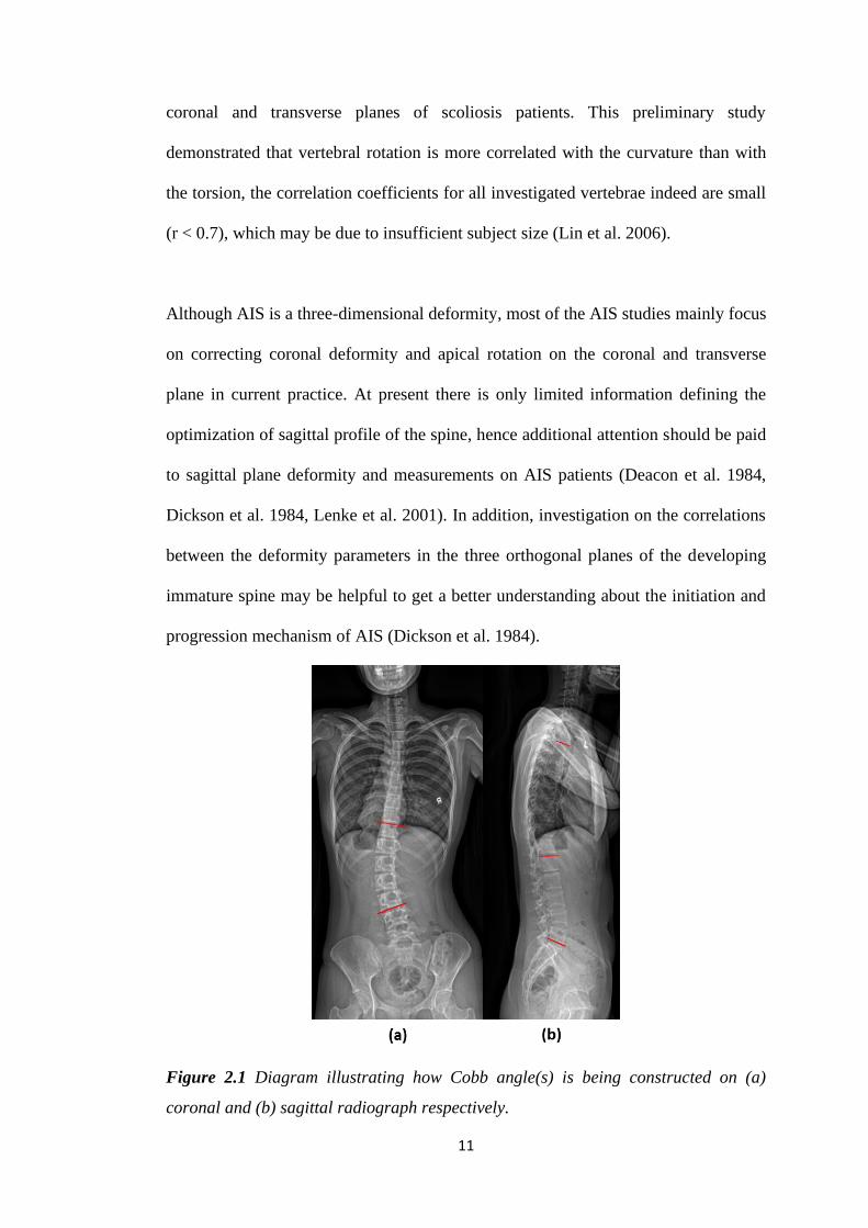
Figure 2.1 Diagram illustrating how Cobb angle(s) is being constructed on (a)
coronal and (b) sagittal radiograph respectively…………………………...………..11
Figure 2.2 The compensatory mechanism that contributes to keep the sagittal
balance of the spine………………………………………………………………….27
Figure 2.3 Different non-imaging assessment tools used to assess sagittal curvature
of human spine: (a) Stereo camera; (b) FaroArm; (c) reflective skin markers; (d)
radio-opaque markers; (e) inclinometers; (f) adapted arcometer; (g) Spinal Mouse; (h)
Flexicurve; and (i) Zebris motion analysis system…………………………………47
Figure 3.1 Experimental set-up and system block diagram for the phantom scanning.
The grey lines illustrate the connections between the devices.…………….…….…54
Figure 3.2 Four flexible spinal column phantoms with different simulated deformity
curvatures…………………………………………………………………………....54
Figure 3.3 (a) A typical B-mode image obtained from the phantom, and (b) 3D
ultrasound image series collected from the 3D ultrasound imaging system and
stacked according to the orientation and location of each image for further spinous
process angle measurement. The spinous process of each vertebra was manually
selected from the B-mode images………………………………………….………..57
Figure 3.4 Diagram illustrating spinous processes extracted from 3D ultrasound
images. (a) A stack of ultrasound images with spinous processes marked in 3D in
corresponding B-mode image, where the black region meaning there was an image
stacked and white region without B-mode images; (b) A typical B-mode image
containing a spinous process and marked accordingly; (c) Spinous process profile
XI
projected in sagittal plane; (d) Corresponding sagittal X-ray image and marked
spinous processes. Before data analysis, sagittal spinous process curvature obtained
from 3D ultrasound was compared with that from radiograph…………..………….58
Figure 3.5 The two measurement methods of curvature used on radiograph: (a)
Cobb’s method and (b) spinous process angle by selecting spinous process…….....59
Figure 3.6 Diagram showing the experiments conducted and the corresponding
statistical tests for reliability and repeatability in this study……………………..….61
Figure 3.7 The scolioscan system with its components labeled. The ultrasound
scanner, computer and the spatial sensor control box are installed inside the
device……………………………………………………………………………….64
Figure 3.8 The diagram shows the measurement of coronal ultrasound angle(s)…..67
Figure 3.9 Coronal and sagittal ultrasound images and the 3D ultrasound volume
obtained from the 3D ultrasound system illustrated by the customized 3D
software ………………………………………..………………………………...….68
Figure 3.10 Sagittal ultrasound image illustrating (a) spinous processes and (b)
laminae for sagittal measurement…………………………………………...……..70
Figure 3.11 Diagram showing the spinous processes and bilateral laminae of (a)
thoracic and (b) lumbar vertebrae…………………………………...…………...….70
Figure 3.12 Diagrams showing how (a) Measurement of ultrasound spinous process
angle (USSPA); (b) Measurement of ultrasound laminae angle (USLA); and (c)
Measurement of Cobb’s angle (XCA) were carried out respectively……………….72
XII
Figure 3.13 Diagrams showing how (a) Ultrasound laminae (USLA); (b) Cobb; (c)
Posterior Tangent (PT); and (d) Centre of Posterior Border (CPB) angles were
measured respectively…………………………………………………………...…..78
Figure 4.1 Scatter plot of thoracic and lumbar Cobb’s angle against X-ray spinous
process angle, with the associated trend line equation. (XSPA: X-ray spinous process
angle; XCA: X-ray Cobb’s angle)….…………………………………..…………...82
Figure 4.2 Scatter plot of the thoracic and lumbar ultrasound spinous process angle
against X-ray spinous process angle, with the associated trend line equation. (USSPA:
Ultrasound spinous process angle; XSPA: X-ray spinous process angle)……...…...83
Figure 4.3 Scatter plot of the thoracic and lumbar Cobb’s angle against ultrasound
spinous process angle, with the associated trend line equation. (USSPA: Ultrasound
spinous process angle; XCA: X-ray Cobb’s angle)… …..…..…..…..…..…...…..…83
Figure 4.4 Correlations (𝐑𝟐) and equations between the X-ray Cobb’s angles (XCA)
and the two sagittal ultrasound angles based on (a) spinous processes (USSPA) and
(b) laminae (USLA), are shown for the thoracic (grey) and lumbar (black)
curves………………………………..…..…..…..…..…..…..…..…..……………..86
Figure 4.5 Bland-Altman plots that show the differences between X-ray Cobb’s
angles (XCA) and the sagittal ultrasound angles corrected with the linear regression
equations. Ultrasound spinous process angle (USSPA) and ultrasound laminae angle
(USLA). SD: standard…………………..…..…..…..…..…..…..…..…..…...………87
Figure 4.6 Correlation and linear regression between coronal Cobb and SPA (SPA:
Spinous process angle)….………………………… …..…..…...…………………...88
XIII
Figure 4.7 Correlations (𝐑𝟐) and equations between the sagittal ultrasound angle
(USLA) and Cobb angles for subject with (a) Cobb ≤ 20º and (b) 20º < Cobb ≤ 40º
for the thoracic (black) and lumbar (red) curves………..…..…..…..…..…………..89
Figure 4.8 Correlations (𝐑𝟐) and equations between the sagittal ultrasound angle
(USLA) and Cobb angles for all subject for the thoracic (black) and lumbar (red)
curves…………………………………………………..…..…..…..…..………...….90
Figure 6.1 The coronal and sagittal images of a patient with double-curve AIS and
relatively larger value of thoracic kyphosis during their first (a and b) and second
visit (c and d) respectively. The coronal SPA angles were illustrated in black and the
thoracic kyphosis and lumbar lordosis were illustrated in
green. ……..…..…..…..…..…..…..…..…..…..…..…..…..…..…..…..…..…….…109
Figure 6.2 The coronal and sagittal images of a patient with double-curve AIS and
relatively smaller value of thoracic kyphosis during their first (a and b) and second
visit (c and d) respectively. The coronal SPA angles were illustrated in black and the
thoracic kyphosis and lumbar lordosis were illustrated in
green. ……………………..…..…..…..…..…..…..…..….…..…..…...…………..110
Figure A.1 The three images on the left were the coronal (a) and sagittal ultrasound
images illustrating the left (b) and right (c) laminae of an AIS patient with a single
thoracic Cobb less than 20º, whereas the three images on the right were the coronal
(d) and sagittal ultrasound images illustrating the left (e) and right (f) laminae of an
AIS patient with a single thoracic Cobb larger than 20º. The red line indicates the
T12 vertebrae level………………………..…..…..…..…..…..…..…..…..…..…...111
XIV
Figure A.2 The three images on the left were the coronal (a) and sagittal ultrasound
images illustrating the left (b) and right (c) laminae of an AIS patient with a single
thoracolumbar Cobb less than 20º, whereas the three images on the right were the
coronal (d) and sagittal ultrasound images illustrating the left (e) and right (f)
laminae of an AIS patient with a single thoracolumbar Cobb larger than 20º. The red
line indicates the T12 vertebrae level…………………..…………..……………...112
Figure A.3 The three images on the left were the coronal (a) and sagittal ultrasound
images illustrating the left (b) and right (c) laminae of an AIS patient with a single
lumbar Cobb less than 20º, whereas the three images on the right were the coronal (d)
and sagittal ultrasound images illustrating the left (e) and right (f) laminae of an AIS
patient with a single lumbar Cobb larger than 20º. The red line indicates the T12
vertebrae level…………..…………..…………..…………..…………..……….…113
Figure A.4 The three images on the left were the coronal (a) and sagittal ultrasound
images illustrating the left (b) and right (c) laminae of an AIS patient with both
thoracic and lumbar Cobb less than 20º, whereas the three images on the right were
the coronal (d) and sagittal ultrasound images illustrating the left (e) and right (f)
laminae of an AIS patient with both thoracic and lumbar Cobb larger than 20º. The
red line indicates the T12 vertebrae level…………………..…………………...…141
Figure A.5 Information sheet provided for the patients who participated in the
study……………..…………..…………..…………..…………..…………..……..138
Figure A.6 Consent form provided for the patients who participated in the
study……………………………………………………………….……………….139
XV
LIST OF TABLES
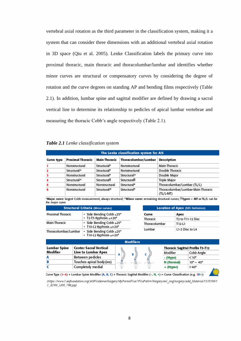
Table 2.1 Lenke classification system………………………………………………..8
Table 3.1 Summary of the data sets used in different reliability tests in the phantom
study……………………………………………………………………..62
Table 4.1 Intra-rater reliability of Rater B for sagittal curve measurement using the
three methods…………………………………………………...……….80
Table 4.2 Inter-rater reliability of the three angles by the two raters…………….…80
Table 4.3 Intra-operator reliability of Operator A for phantom scanning in terms of
spinous process angle obtained………………………………………….81
Table 4.4 Best-fit equations and absolute errors obtained from the three methods by
Rater B………………………………………………………………..…82
Table 4.5 Intra-rater and inter-rater reliability of the sagittal ultrasound angles by
Rater 1 and Rater 2………………………………………...……………84
Table 4.6 Analysis of sagittal parameters according to the main thoracic Cobb angle
for ultrasound and X-ray in the exploratory session………………….…91
Table 4.7 Analysis of sagittal parameters according to the thoraco(lumbar) Cobb
angle for ultrasound and X-ray in the exploratory session…………..….92
Table 4.8 Analysis of sagittal parameters according to the main thoracic SPA angle
for ultrasound in the exploratory session………………………..………92
Table 4.9 Analysis of sagittal parameters according to the thoraco(lumbar) SPA angle
for ultrasound in the exploratory session………………………………..93
Table 4.10 Sagittal parameters obtained from four different methods and the
significance tests for each parameter………………………………..…..93
XVI
Table 4.11 Overall analysis of thoracic kyphosis according to the main thoracic SPA
angle for ultrasound in the exploratory and verification
sessions………..………………………………………………….……..94
Table 4.12 Overall analysis of lumbar lordosis according to the (thoraco)lumbar
SPA angle for ultrasound in the exploratory and verification
sessions……………………………………………………….…………95
Table A.1 X-ray Cobb angle of the thoracic and lumbar region measured from the
four phantoms of four different curvatures by rater B and rater C…….115
Table A.2 X-ray spinous process angle of the thoracic and lumbar region measured
from the four phantoms of four different curvatures by rater B and rater
C………………………………………………………………………..116
Table A.3 Ultrasound spinous process angle of the thoracic and lumbar region
measured from the four phantoms of four different curvatures by rater B
and rater C…………………………………………………..…….……117
Table A.4 Ultrasound spinous process angle of the thoracic and lumbar region
measured from the patients of the human subject study by rater 1 and rater
2……………………………………………………………………..….118
Table A.5 The average ultrasound laminae angle (left and right laminae) of the
thoracic and lumbar region measured from the patients of the human
subject study by rater 1 and rater 2; and Cobb angle of the thoracic and
lumbar region measured from the patients of the pilot test by rater 3…120
Table A.6 The Cobb angle, posterior tangent angle and the centre of posterior border
angle of the thoracic and lumbar region measured in the sagittal plane,
together with the main thoracic and thoraco(lumbar)Cobb angle measured
XVII
in the coronal plane from the patients in the exploratory session. All
angles were measured on X-ray……………………………………..…122
Table A.7 The average ultrasound laminae angle (left and right laminae) of the
thoracic and lumbar region measured in the sagittal plane, together with
the main thoracic and thoraco(lumbar) spinous process angle measured in
the coronal plane from the patients in the exploratory session. All angles
were measured using ultrasound……………………………………….127
Table A.8 The average ultrasound laminae angle (left and right laminae) of the
thoracic and lumbar region measured in the sagittal plane, together with
the main thoracic and thoraco(lumbar) spinous process angle measured in
the coronal plane from the patients in the exploratory session. All angles
were measured using ultrasound……………………………………….132
XVIII
LIST OF ABBREVIATIONS
3D: Three Dimensional
AIS: Adolescent Idiopathic Scoliosis
CPB: Centre of Posterior Border
CR: Computed Radiography
CT: Computed Tomography
DICOM: Digital Imaging and Communications in Medicine
ICC: Intraclass Correlation Coefficient
LBP: Low Back Pain
LL: Lumbar lordosis
MAD: Mean Absolute Difference
MRI: Magnetic Resonance Imaging
NCJ: Neurocentral Junction
PT: Posterior Tangent
SD: Standard Deviation
SPA: Spinous Process Angle
TK: Thoracic Kyphosis
US: Ultrasound
USLA: Ultrasound Laminae Angle
USSPA: Ultrasound Spinous Process Angle
XCA: X-ray Cobb’s Angle
1
Chapter 1. Introduction
1.1. Background
Idiopathic scoliosis is a three-dimensional (3D) deformity characterized by lateral
deviation, sagittal misalignment and transverse axial rotation of the spine (Hattori et
al. 2011, Pope et al. 1984). Among all pediatric spine deformities, adolescent
idiopathic scoliosis (AIS) is most prevalent (Cheng et al. 2015, Fan et al. 2016, Fong
et al. 2015). Due to the effect of coupling in different planes, the pattern of deformity
in the coronal plane may be highly influenced by changes in the axial and sagittal
planes (Gum et al. 2007, Hayashi et al. 2009, Sullivan et al. 2017, Villemure et al.
2001). However, most of the AIS studies mainly focus on the coronal plane, indeed
additional attention should be paid on sagittal plane deformity and measurements on
AIS patients (Deacon et al. 1984, Dickson et al. 1984, Lenke et al. 2001).
Human spine composes of five regions: cervical, thoracic, lumbar, sacrum and
coccyx. Thoracic kyphosis and lumbar lordosis are two common sagittal parameters
when analyzing sagittal profile. For normal individuals, acceptable ranges for
kyphosis and lordosis are from 20 to 50 degrees and 31 to 79 degrees, respectively
(Boseker et al. 2000, Bridwell and Bernhardt 1989). Maintaining an optimal sagittal
spinal profile helps to maintain spine motor control with minimum energy
expenditure, enhance the load tolerance of the spine and increase spinal muscle
efficiency (Kim et al. 2006). The sagittal profile of patients with AIS had been
shown to be different from normal individuals (Carpineta et al. 2003, Cheung et al.
2018, Schlösser et al. 2014, Schmitz et al. 2011). For instance, reduction of lumbar
lordosis and sacrum inclination reduced the natural curvature of the lumbar spine
(Makhsous et al. 2013, Alexander et al. 2007, Drzał-Grabiec et al. 2015). In addition,
2
it was demonstrated that alternation of sagittal spinal curvature caused viscoelastic
deformation of spinal tissues (Solomonow et al. 2003), higher intra-discal pressure
(Wilke et al. 1999) and spine overloading and degeneration (Makhsous et al. 2013,
Alexander et al. 2007, Drzał-Grabiec et al. 2015, Beach et al. 2005). Moreover,
flattening of the thoracic kyphosis was found to be a risk factor for scoliosis
(Roussouly et al. 2013) and reportedly cause diminution of the lung function in
patients with scoliosis (Winter et al. 1975). Furthermore, shear loads experienced by
vertebrae were altered once the sagittal spinal profile was disturbed, hence facet
joints in the posterior portion of the posterior inclined vertebra were unlocked,
inducing rotational instability to the spinal column and causing further progression in
spinal deformity (Janssen et al. 2011, Schlösser et al. 2014, Castelein et al. 2005,
Kouwenhoven et al. 2007). Spinal sagittal imbalance also affects the quality of life of
an individual. Previous studies reported that alternation of the lumbar lordosis led to
the occurrence of lower back pain (Jackson et al. 2011, Bernard et al. al. 2008, de
Jonge et al. 2002), headaches, fatigue and cervical pain (Chow et al. 2007). In some
severe cases, social interaction of the patients was affected due to deficient forward
gaze (Roussouly and Nnadi 2010). Hence it is important to evaluate the spinal
sagittal profile of patients with AIS. Furthermore, characterizing the differences in
sagittal profile between normal and scoliotic spines may also provide early detection
of vertebral rotation (Schlösser et al. 2014), and quantifying spinal curvatures in
different planes is useful for surgical planning and monitoring curve progression
(Carlson et al. 2013, Cheung et al. 2013, Vrtovec et al. 2009).
X-ray and magnetic resonance imaging (MRI) are the two commonly used imaging
modalities for evaluating sagittal spinal curvature, where using Cobb’s method on
3
radiograph is the gold standard at present (Cobb 1948, Vrtovec et al. 2009, Harrison
et al. 2001). The major drawback of radiograph evaluation is that patients are
exposed to radiation. Ionizing radiation remains an issue for patients even using EOS,
a biplanar X-ray imaging system utilizing reduced dosage, which requires repetitive
scanning, on top of the high cost and installation complexity. MRI has been used for
spinal deformity evaluation because of its high resolution. However, it is costly and
less accessible (Diefenbach et al. 2013). Moreover, patients are required to be
imaged in supine position, hence the natural spinal curvature cannot be acquired
(Yazici et al. 2011). Furthermore, different topographic methods could only evaluate
the spinal curvature in an indirect way instead of measuring the actual curvature of
the spine itself.
Free-hand 3D ultrasound imaging, which combines a conventional B-mode imaging
system with a position sensor, has been developed over two decades and recently
become more popular due to its features of radiation-free, wider accessibility and
lower cost in comparison with other 3D imaging modalities (Huang et al. 2005,
Huang and Zeng 2017, Mozaffari et al. 2017). Ultrasound evaluation of coronal
curvature and vertebral rotation was reported by Suzuki et al. (1989) back to 1980’s.
Later, a number of 3D ultrasound imaging systems for the coronal plane assessment
of scoliosis have been reported by different groups (Cheung and Zheng 2010, Li et al.
2010, Prunama et al. 2010, Chen et al. 2013, Ungi et al. 2014). Cheung et al. (2013,
2015) reported preliminary tests on spinal column phantoms and human subjects
based on spinous process angle, and later the same system was used for testing a
larger number of subjects, demonstrating high intra- and inter-rater and operator
reliability, and good correlation with Cobb’s angle (Zheng et al. 2016, Brink et al.
4
2018). Spinous process angle was also used to investigate the effectiveness of
orthotic treatment for patients with AIS (Li et al. (2012). A study utilized tracked
ultrasound to localize vertebral transverse processes as landmarks along the spine to
measure curvature angles on spine phantoms (Ungi et al. 2014). Huang et al. (2018)
further developed this method by continuously monitoring image spatial information
to form a continuous curved plane for scoliosis assessment. Centre of laminae
methods has also been used for both coronal curvature and vertebral rotation
assessment (Chen et al. 2013, Young et al. 2015, Chen et al. 2016, Wang et al. 2016),
and all these studies demonstrated that the ultrasound angles obtained were reliable
and comparable to the angles obtained from conventional methods. However, no
study has been reported on the reliability of 3D ultrasound imaging for evaluating the
sagittal spinal curvature.
1.2. Overall Objective and Primary Contribution
The overall objective of this study is to investigate the reliability and validity of
using freehand 3D ultrasound system for evaluation of the sagittal curvature of the
spine, ultimately providing a radiation-free imaging modality for evaluating and
monitoring sagittal spinal profile for patients with adolescent idiopathic scoliosis. To
achieve this objective, scanning was performed first on spine phantoms and then on
human subjects with different extents of deformity. Intra- and inter-rater reliability of
the sagittal ultrasound spinous process and laminae angles, and the comparability of
these ultrasound angles with the X-ray Cobb angle were investigated. Preferred
ultrasound angles were then applied to a larger group of patients with AIS, in order
to investigate whether the coupling relationship observed from traditional radiograph
could also be demonstrated using ultrasound, either with or without the aid of X-ray.
5
An optimal value for sagittal thoracic profile for ultrasound would also be suggested
for reference in future sagittal ultrasound evaluation for spine.
The major achievements of this study were summarized as follow:
• Flexible spine phantoms with different degrees of simulated scoliosis were tested to
investigate the feasibility of ultrasound on evaluating spinal sagittal curvature under
different range of coronal deformation and the relationship between the Cobb’s angle
and the spinous process angles obtained from X-ray and ultrasound.
• Human pilot tests with different range of coronal deformities were conducted to
investigate the reliability of the ultrasound system, with the usage of the specially
customized 3D ultrasound software. Sagittal curvatures obtained from ultrasound,
using the spinous processes and laminae as reference landmarks, were found to have
good correlation with those obtained from traditional Cobb’s methods.
• Establishing laminae as a better method for assessing sagittal curvature using
ultrasound as the curvatures measured using such landmarks had no significant
differences with those obtained from spinous processes, at the same time with better
visualization in B-mode images.
• Demonstrating that relative hypokyphosis could be detected in patients with AIS
with larger coronal deformities, provided that the same phenomenon was reflected
from radiograph, by either with the aid of X-ray or using ultrasound alone.
• Providing a standard thoracic kyphosis value of patients with AIS using ultrasound
for future progression study.
1.3. Outlines of the Thesis
This thesis is composed of six chapters. Chapter 1 includes the background
information, motivation, objectives, primary contribution and the structure of this

6
thesis. Chapter 2 provides a comprehensive literature review of the related study,
including the related issues of AIS and sagittal profile of spine, and different
approaches used for evaluating sagittal curvature. In Chapter 3, the experimental
materials and methods of the phantom and human subjects tests were described. In
Chapter 4, the results obtained from the spine phantom and human subjects were
presented. In chapter 5, the results obtained in Chapter 4 and limitations of the
phantom and human subjects study were discussed. Finally, in Chapter 6, conclusion
from the study was drawn and recommendations on future work were given.
7
Chapter 2. Literature Review
2.1. AIS Related Issues
2.1.1. Etiology, Diagnosis of AIS
Idiopathic scoliosis is a three dimensional (3D) spine deformity problem (Stokes et al
1987). It is often associated with deviation in coronal plane, sagittal deformation and
axial rotational deformities (White et al. 1978, Pope et al. 1984, Hattori et al. 2011).
No single cause for idiopathic scoliosis (IS) has been identified at present (Arkin et
al. 1949) and it is found to be exclusive to humans (Castelein et al. 2005). Forms of
scoliosis reported in other vertebrates are induced using congenital, neuromuscular,
cicatricial or experimental methods (Pincott et al. 1982, MacEwen 1973, Beguiristain
et al. 1980, Ottander 1963). Generally Idiopathic scoliosis is divided into four stages:
1) Infantile; 2) Juvenile; 3) Adolescent; and 4) Adult idiopathic scoliosis. Adolescent
idiopathic scoliosis (AIS) is the most prevalent form of scoliosis which affects 2–3 %
of adolescents (Asher et al 2006). Approximately 20 million people are suffered from
scoliosis in the United States, and the prevalence of AIS is about 2% to 4% (Good et
al. 2009). The prevalence of AIS is about 3% in Hong Kong (Tang et al. 2003). AIS
is often diagnosed or detected during the pubertal growth spurt at ages 10–14 years
without an identifiable cause (Asher et al. 2006).
At present there are three classification systems. King classification is a two-
dimensional system because it only considers lateral deviation in the frontal plane
(King et al. 1983). Lenke classification is one of the most commonly used scoliosis
classification systems at present, which considers parameters in both coronal and
sagittal planes, thus is three dimensional (Lenke et al. 2003). A more recent
classification system made by Peking Union College Medicine (PUCM) added the
8
vertebral axial rotation as the third parameter in the classification system, making it a
system that can consider three dimensions with an additional vertebral axial rotation
in 3D space (Qiu et al. 2005). Lenke Classification labels the primary curve into
proximal thoracic, main thoracic and thoracolumbar/lumbar and identifies whether
minor curves are structural or compensatory curves by considering the degree of
rotation and the curve degrees on standing AP and bending films respectively (Table
2.1). In addition, lumbar spine and sagittal modifier are defined by drawing a sacral
vertical line to determine its relationship to pedicles of apical lumbar vertebrae and
measuring the thoracic Cobb’s angle respectively (Table 2.1).
Table 2.1 Lenke classification system
9
Prior to X-ray assessment, patients with scoliosis would receive a scoliometer
screening test to measure angle of trunk rotation with a scoliometer, since
scoliometer measurement has a good correlation (r = 0.7) with the Cobb angles
(Coelho et al. 2013, Vidal et al. 2013). Information such as gender, age, height,
weight, leg length, onset of menarche, family history, and diseases is collected for
determining a tentative prognosis. Physical and spinal examinations including
forward bending test, neurological examination, spine side-to-side symmetry,
shoulder height, iliac crest symmetry, and lateral examination are also conducted.
When the hump’s angle of trunk rotation is greater than seven degrees measured by
Bunnell Scoliometer under forward bending test, the patient is recommended for
undergoing the standard radiographic evaluation for suspected scoliosis (Bunnell
1984).
For current clinical practice, Cobb angle on standing postero-anterior X-ray
radiograph is the gold standard to evaluate the severity of scoliosis and sagittal
curvature of the spine (Cobb 1948) (Figure 2.1a and b). Coronal Cobb angle is
defined by the angle between the two straight lines that are drawn on the upper and
lower endplate of the most tilted vertebrae of a curve respectively on the coronal
radiograph. Patients with spinal curvature in the coronal plane more than 10 degrees
are treated as scoliosis (Cobb 1948). Different treatments are applied to different
types of AIS patients (Kim et al. 2010). For those with Cobb angle of 20 degrees or
less, clinical observation is recommended. Those with immature skeletal and Cobb’s
angle of between 20 to 40 degrees, brace treatment will be considered. For patients
with Cobb’s angle greater than 40 degrees and immature skeletal or Cobb’s angle
greater than 50 degrees and mature skeletal, surgical management may be necessary.
10
Vertebrae rotation is often observed in patients with AIS with severe scoliosis. At
present, the gold standard remains axial computed tomography because of its high
resolution (Gocen et al. 1998, Ho et al. 1992, Krismer et al. 1999). Accurate
measurement of vertebral rotation may assist in preoperative planning and screw
placement for patients with scoliosis.The apical vertebra, the vertebra which is not
tilted and most laterally deviated from the central sacral line, and usually presents
maximal axial rotation (Lenke 2000), can be visualized with computed tomography
(CT), which passes through the vertebral body, both pedicles, laminas, transverse
processes, and the spinous process. Yet it can also be assessed using radiographs
(Cobb 1948, Nash et al. 1969), ultrasound (Suzuki et al. 1989) and MRI (Birchall et
al. 1997). Several techniques have been developed for assessing vertebral rotation: 1)
Investigation of spinous process on coronal radiograph (Cobb 1948); 2) Investigating
the relative position of convex side pedicle (Nash and Moe 1969); and 3) Image
matching method by using multiple landmark methods (Mehta 1973) that estimates
vertebral rotation based on relative position of various elements on the vertebrae.
Coronal curvature has been observed to be positively correlated with the vertebrae
axial rotation in scoliotic spine, showing that these two components are related to
each other (White et al. 1978, Villemure et al. .2001, Gum et al. 2007). Carlson et al.
(2013) found out that angle of trunk inclination of the patients with either thoracic or
thoracolumbar AIS with mean Cobb’s angle of 63 and 48 degrees respectively is
positively correlated (r > 0.7) with Cobb’s angle and apical vertebral rotation angle
(with respect to the sagittal plane). Lin et al. (2006) investigated the correlation of
individual vertebra axial rotation angle with curvature and torsion from T2 to L4 by
using a simplified 3D spine model constructed by two radiographic images in the
11
coronal and transverse planes of scoliosis patients. This preliminary study
demonstrated that vertebral rotation is more correlated with the curvature than with
the torsion, the correlation coefficients for all investigated vertebrae indeed are small
(r < 0.7), which may be due to insufficient subject size (Lin et al. 2006).
Although AIS is a three-dimensional deformity, most of the AIS studies mainly focus
on correcting coronal deformity and apical rotation on the coronal and transverse
plane in current practice. At present there is only limited information defining the
optimization of sagittal profile of the spine, hence additional attention should be paid
to sagittal plane deformity and measurements on AIS patients (Deacon et al. 1984,
Dickson et al. 1984, Lenke et al. 2001). In addition, investigation on the correlations
between the deformity parameters in the three orthogonal planes of the developing
immature spine may be helpful to get a better understanding about the initiation and
progression mechanism of AIS (Dickson et al. 1984).
Figure 2.1 Diagram illustrating how Cobb angle(s) is being constructed on (a)
coronal and (b) sagittal radiograph respectively.
12
2.1.2. Treatment for Patients with AIS
When patients are assigned for brace treatment after diagnosis, they will generally
undergo a brace fitting process and follow-up assessment (Negrini et al 2009). The
conventional manual method of making a spinal brace is by firstly taking a negative
body cast from the patients with AIS (Wong et al. 2003), followed by filling the
negative body cast with plaster to prepare a positive cast. Then the positive cast is
rectified by removing and adding plaster to certain specific areas of the cast. Finally
a spinal orthosis is formed by molding a plastic sheet onto the rectified cast (Wong et
al. 2005). The goals for applying brace for patients with scoliosis are reducing the
magnitude of the deformity, maintaining spinal balance, and preventing progression
of the deformity, where preventing curve progression during adolescent growth spurt
is the major objective since it is the high-risk period (Havey et al. 2016, Weinstein et
al. 2008). Rigid spinal orthoses have been demonstrated to be effective for most of
the cases of moderate AIS, providing that the treatment has been carried out early
enough and the brace is worn for long enough every day and under properly applied
controlling forces (Wong et al. 2000, Wright 1977, Nachemson and Peterson 1995).
When fitting the brace, pads must be placed correctly and adjusted frequently
together with the brace for optimal patient outcomes. At present, taking standing X-
ray films is the traditional method to assess the effectiveness of bracing on scoliotic
curve correction. However, due to the radiation issue, repetitive imaging is not
recommended. At present, the existing biomechanical design of spinal orthoses
mainly applies external corrective forces by using the 3-point or 4-point pressure
systems to support and prevent further progression during the period of puberty of
the patients. In addition, according to Euler’s bucking model, length of the spine
decreases spine stability while diameter of the spine increases, hence bracing also
13
provides additional time for spine diameter to catch up with skeletal maturity and
effectively form a comparatively more stable spinal column (Havey et al. 2016).
Other than the effectiveness of the brace, the compliance and appearance of the brace
are also important to the patients. Generally, AIS patients are required to wear the
orthoses for up to twenty-three hours per day including bedtime (Chu et al. 2006).
History taking and physical examination to look for signs of wear are often used to
assess the compliance. Application of thermal and force sensors has also been used
to reflect the time spent on the orthosis (Lavelle et al. 1996) and force received by
the patients (Lou et al. 2002) to further study the compliance of the orthosis. Among
different types of scoliosis, patients with severe scoliosis would encounter physically
detrimental effects, hence aesthetics becomes another objective when dueling with
curve progression with a brace (Negrini et al. 2012). By designing and producing an
effective and good-looking brace, spinal pain syndromes and torso aesthetics of the
patients may be treated and improved by relieving the curve progression (Negrini et
al. 2012). Currently brace effectiveness is still an important area of study for the
International Scientific Society on Scoliosis Orthopaedic and Rehabilitation
Treatment (SOSORT) and the Scoliosis Research Society (SRS) and the five major
areas of study on bracing are: 1) Diagnostic and follow-up issue; 2) Optimization of
brace fitting; 3) Investigation of bracing compliance; 4) Monitoring in-brace forces;
and 5) Quality of life of the brace wearers.
Once the major curve of these patients reaches a Cobb angle greater than 40 degrees,
surgical management may be necessary to prevent further curve progression (Weiss
2008). The current objectives of the surgical treatment for AIS are to maximize the
correction of the spinal deformity, achieve balance in the coronal and sagittal plane
14
and axial derotation, and maintain spinal flexibility (Bridwell 1999, Majdouline et al.
2007). There is an increasing trend of the usage of pedicle screws for curve
correction (Kim et al. 2004, Suk et al. 2001, Kim et al. 2006), because of their
relatively superior major curve correction and biomechanical properties. However,
these posterior distraction devices further enhanced the coronal plane correction,
resulting in sacrificing the sagittal balance (Potter et al. 2004).
2.1.3. Curve Progression of Patients with AIS
It is also important to define whether scoliotic curves are progressive or not.
Progressive scoliosis is defined by a progression of major curve Cobb angle of more
than 6° between the first and the latest control (Pruijs et al. 1994) and a Cobb angle
between 25° and 50°. While stable scoliosis is defined by a progression of major
curve Cobb angle lower than 6° between the first and the latest control (Pruijs et al.
1994), a Cobb angle lower than 25° and a Risser stage ≥ 3 (Skalli et al. 2016).
Various factors have been found to cause curve progression in patients with AIS.
Axial and ventral shear forces were found to be one of the significant factors which
might cause progression. Vertebrates are normally experiencing predominant axial
and ventral shear loads in the vertebrae of the spine due to gravitational and muscle
force (Wilke et al. 1997). However, the spinal loading conditions are different for
humans. Humans walk in a fully upright posture most of the time during daily
activities, the center of mass of the upper body acts straight above the pelvis in
human (Hogervorst et al. 2009). For other vertebrates, the center of mass acts in front
of the pelvis instead (D’Aouˆt et al. 2002). In a mathematical model in Castelein et
al.’s (2005) study, the backward inclined segments of the spine were demonstrated to
15
be subjected to dorsal shear forces other than axial loading. In addition, facet joints
are essential to provide rotational stability for the spine, but these joints can only well
stabilize shear forces which act on the vertebrae in ventral direction instead of dorsal.
A biomechanical in vitro study shows that dorsally directed shear loads were
observed larger than ventrally directed shear loads (Kouwenhoven et al. 2007a). The
difference could be accounted to the gradual transition of shear force from ventrally
directed to dorsally directed in the adaptation of the fully upright posture during
walking (Castelein et al. 2005). Such transition of shear force would facilitate
rotatory instability (Kouwenhoven et al 2007b) due to the poor functioning of the
facet joints and the posterior location of the major spinal muscles and ligaments,
which may possibly contribute to axial vertebral rotation (Kouwenhoven et al.
2007a). Kouwenhoven et al. (2008) later on demonstrated that spine under dorsally
directed shear loads favored axial vertebral rotation more than ventrally directed
shear loads at the mid and lower thoracic in a biomechanical study. Thus the authors
hypothesized that dorsal shear loading is a possible enhancer of slight preexistent
vertebral rotation (Kouwenhoven 2006a, b, 2007) and progressive vertebral
deformation according to Hueter–Volkmann’s law (Fritz 2013), which would
ultimately lead to progressive scoliosis. Thus backward inclination of vertebrae in
the sagittal plane has a prognostic significance in the progression of AIS (Castelein et
al. 1992, Schlosser et al. 2014).
Asymmetric growth of the vertebrae could also be one of the factors that may lead to
the progression of AIS. In the study of Porter et al. (2000), vertebral canals of
skeletons with kyphotic spines were significantly longer than the vertebral length,
while such difference could not be observed in normal spine. Guo et al. (2003)
16
reported that the anterior part of all the thoracic vertebral bodies was found to be
longer than the posterior part in magnetic resonance images among girls with AIS,
and the ratio of differential growth was found significantly positively correlated to
scoliosis severity score. It is hypothesized that the difference is due to the differential
growth rate between the vertebral bodies and the posterior elements, which develops
thoracic hypokyphosis. The irregular growth can also be accounted for the uncoupled
neuro-osseous growth, which was common in AIS patients (Porter 2000, 2001a,b),
leading to a comparatively shorter spinal cord (Roth et al 1968). Chu et al. (2006)
also found that the ratio of the spinal cord to the length of the vertebral column at the
thoracic level smaller in AIS patients than normal individuals. The comparatively
faster growth of the vertebral bodies would tether the posterior vertebrae, which
eventually causes buckling and rotation of the spine. Such differential growth rate
between the vertebral bodies and the posterior elements would lead to hypokyphosis,
which is common among patients with AIS. Girls with AIS are found to be taller and
more slender than normal controls with the same sex and age (Cheung et al. 2003,
Nissinen et al. 1993, Nordwall et al. 1975, Willner 1975) and such differences can be
accounted for the flattening of the thoracic kyphosis (Archer et al 1985). Such
flattening process happens during adolescent growth spurt and gradually returns back
to normal (Cil et al. 2005, Poussa et al. 2005). Since girls mature earlier than boys,
they go through peak adolescent growth velocity when the thoracic kyphosis is at its
minimum, while boys go through the maximum growth period at a later stage
(Dickson et al. 1984). Hence girls have a higher potential from developing thoracic
lordosis. Developmental asymmetry was also observed in the intervertebral discs in
idiopathic scoliosis. During the development of scoliotic curvature, shifting of the
nucleus pulposus to the convex side of the curve could be observed (Bick et al. 1958).
17
Moreover, fibers of the annulus fibrosis were observed to be extended on the convex
side and compressed on the concave side of the curve (Michelsson et al. 1965). The
above evidence could explain why degenerative enzymes activity in the discs
increased (Zaleske et al. 1985) and elastic fiber network of the annulus fibrosus was
sparse and disrupted (Yu et al. 2005) in the discs in idiopathic scoliosis individuals.
Other than growth asymmetry, growth disturbance may also play an important role in
the development of AIS. Since curve deformities due to AIS are three dimensional,
the growth disturbance caused by curve deformities would also be in three
dimensions. A morphometric analysis study of the scoliotic spine stated that the apex
height of the scoliotic vertebral bodies was significantly smaller at the concave side
and the pedicles were smaller and shorter than those on the convex side (Parent et al.
2002). Growth kinetics between the convex and the concave side of scoliotic
vertebrae would become different once scoliosis is developed (Wang et al. 2007),
asymmetric loading would have resulted on the epiphyseal plates (Hueter 1862,
Volkmann 1882). Asymmetric loading has also been demonstrated to be causative of
AIS progression. By applying asymmetric loading on growing vertebrae in animals,
AIS-like deformities resulted (Aronsson et al. 1999, Mente et al. 1997,1999, Stokes
et al. 1996).
Bone quality also plays an important role in curve progression. Bone mass density
and peak bone mass in patients with AIS were found to be lower than those in
normal controls (Cook et al. 1987, Cheng et al. 1997, Velis et al. 1989), which could
be the combined result of abnormal bone mineralization and bone growth increment
during puberty (Lee et al. 2005, Cheung et al. 2006). Furthermore, inverse
18
relationship was observed between curve severity as measured with the Cobb’s angle
and BMD (Lee et al. 2005), concluding that osteopenia might be an important risk
factor of curve progression in AIS.
Preexistent vertebral rotation pattern was found to be existing even in a normal
nonscoliotic spine, where mid and lower thoracic vertebrae showed a predominant
rotation to right side in humans and quadrupeds (Kouwenhoven et al. 2006a,b). Situs
inversus totalis individuals had a reversed rotation trend of vertebrae compared to
common individuals (Kouwenhoven et al. 2007), stating that internal organs such as
heart play a role in the preexistent rotation pattern. It has later on been found out that
this preexistent rotation pattern would be significantly affected by body position,
where the rotation during quadrupedal-like position was significantly smaller than
that in bipedal and supine position (Janssen et al. 2010). Furthermore, it has been
shown that such rotation patterns exist in infantile and juvenile normal humans.
Patients with infantile idiopathic scoliosis generally presented left-sided thoracic
curvatures (Thompson et al. 1980). Those with juvenile idiopathic scoliosis had left
& right sided curves evenly divided (Figueiredo et al. 1981). For Adolescent
idiopathic scoliosis (AIS), patients most often had right-sided main thoracic curves in
the mid and/or lower thoracic region and left-sided main lumbar curves (Upper
thoracic/lumbar) (James et al. 1954, Moe et al. 1970, Ponseti et al. 1950). During
infantile stage, a significant leftward rotation was observed in the upper thoracic
spine, while in the juvenile stage, significant leftward rotation was only observed in
T4 of the upper thoracic spine (Janssen et al. 2011). But in the adolescent stage, the
mid and lower thoracic spine demonstrated a significant rightwards rotation (Janssen
et al. 2011). The above evidence showed that the mean rotation of the vertebra in the
19
thoracic spine shifts from left to right throughout the initial growth stage.
Furthermore, the rotation pattern was found more significantly leftwards in boys than
girls (Janssen et al. 2011). However, this phenomenon should be considered as a
physiologic process in the normal development of the spine, and an independent
process of the pathogenesis of scoliosis (Kouwenhoven et al. 2006b). In addition,
though Neurocentral junction (NCJ) activity was believed to be just a passive factor
on IS progression (Schlösser et al. 2013) and no conclusive statement has been made
on whether neurocentral junction activity would affect progression of IS
(Kouwenhoven et al. 2008). There might be a correlation between the NCJ closure
and the preexisting vertebral rotation. Neurocentral junction (NCJ) closure starts at
L1–L3 at 6 to 7 years old for both males and females. The closure spreads to higher
thoracic and L4–L5 and finishes in the mid- and low thoracic spine at 6-9 years old
for girls and 7-11 years old for boys respectively (Schlösser et al. 2013). It has been
found out that the mean NCJ area kept decreasing starting from the infantile to the
adolescent stage (Schlösser et al. 2013). Moreover, the mean NCJ area in the right-
hand side in the infantile stage was found larger than that in the left-hand side, and
the situation was totally reversed in the juvenile stage (Schlösser et al. 2013).
Other than NCJ closure, there are also factors whose roles in curve progression of
patients with AIS are unknown, yet attention is needed. For instance, it has been
known that paravertebral muscles are essential to hold the spine upright. Since
ligamentous spine alone cannot support vertical compressive forces and buckles at an
axial load of only 20 N (Lucas et al. 1961), paraspinal muscle diseases or imbalance
would destabilize the spine and likely cause the development of scoliosis (Aprin et al.
1982, Hsu et al. 1983, MacEwen 1974, Fidler et al. 1976, Ford et al. 1984, Riddle et
20
al. 1955). Previous studies showed increased paravertebral muscles EMG activities at
the convexity of the curve (Alexander et al. 1978, Cheung et al. 2005). Moreover, a
greater proportion of type I muscle fibers was observed in multifidus (Fidler et al.
1976) and in superficial muscles (Ford et al. 1984). In addition, ribs transmit muscle
forces from the sternum to the vertebral column through the transverse processes, the
costotransverse articulations and ligaments (Kouwenhoven et al. 2008).
Experimentally induced elongation of the ribs on growing rabbits asymmetrically
resulted in scoliotic curvature of the spine (Agadir et al. 1998) and curvature
correction could be done by lengthening the ribs in the shorter side. Incidence of
scoliosis in children who underwent thoracotomy was found higher than normal
(Kouwenhoven et al. 2008). Role of the spinal cord in the development of AIS
remains controversial (Burwell 2001). Neuromuscular disorder reportedly resulted in
the development of idiopathic or secondary scoliosis (Aprin et al. 1982, Hsu et al.
1983, MacEwen 1974, Madigan et al. 1981, Piggott 1980). Hence AIS patients were
found to have decreased sensory (proprioceptive) input (Pincott et al. 1982) and
abnormalities in EEG activity, postural control, and vestibular and somatosensory
function (Cheng et al. 1999, Guo et al. 2006, Petersen et al. 1979, Sahlstrand et al.
1979).
2.2. Investigation of Sagittal Profile of Spine and Related Structures
2.2.1. Sagittal Profile of Normal Individuals and Its Importance
At birth, sagittal profile of spine is C-shaped and globally kyphotic (El-Hawary et al.
2016). During growth, physiological curvatures are developed in the sagittal plane,
which are likely due to the changes resultants from posture and balance (Cil et al.
2005). Young children have been found to have positive sagittal balance, which
21
decreases throughout childhood and adolescence (Cil et al. 2005). It had been found
that thoracic kyphosis increases from 42° in children 3–9 years of age to 48° by age
of 10, while lumbar lordosis increases from 44° for children aged 3–9 years to 53° by
the age of 10 years (Mac-Thiong et al. 2011a). Generally for normal individuals, the
center of C7 vertebral body to the center of upper sacral endplate was found to be
very close to the vertical line (Kuntz et al. 2007, 2008; Roussouly et al. 2006). For
regional analysis, many parameters have been used in the spine and pelvis and the
average values of these values have been widely discussed in different studies. For
instance, thoracic kyphosis is between 20° and 50° (Boesker et al. 2000) and lumbar
lordosis ranges from 31° to 79° (Bridwell et al. 1989). Some studies suggested
reference values of 30° to 50° and 20° to 50° for lumbar mordosis (Bernhardt et al.
1989, Stagnara et al. 1982). Hardacker et al. (1997) stated that acceptable lordosis in
the cervical spine was 40° ± 9.7° for normal individuals. Spinal-sacral angle and
spinal tilt of normal adult individuals can be expected to be between 110° and 150°,
and between 85° and 100° respectively (Mac-Thiong et al. 2010). Pelvic incidence
angle measures approximately 52° with a range from 34°to 84° (Van Royen et al.
1998, 2000). This angle is fixed after skeletal maturity. The pelvic tilt angle
measures 12° with a range of 5°–30° (Van Royen et al. 1998). This angle changes
with compensatory posture and is therefore a postural angle. Sacral slope is
approximately 40°with a range of 20°–65° (Van Royen et al. 1998, 2000). Repetitive,
strenuous physical activity would cause structural abnormalities in the immature
vertebral body (Wojtys et al. 2000). Adolescent athletes with greater cumulative
training time demonstrated larger angles of thoracic kyphosis and lumbar lordosis
than those lack of sports (Wojtys et al. 2000). Sports with a predominance of
forward-bending postures was found to be associated with greater thoracic kyphosis
22
in standing (Rajabi et al. 2007), however no negative effect on sagittal spinal posture
was observed in young tennis players (Muyor et al. 2013). Hyperhyphosis was
observed in highly-trained young canoeists during standing position (López-Miñarro
et al. 2011), but with reduced thoracic and lumbar curvatures. Hyperhyphosis was
observed in elite cyclists during standing position but the thoracic spine was
straighter on the bicycle than in the standing posture (Muyor et al. 2011). Increased
thoracic kyphosis in adolescent elite skiers was observed after five years of intensive
training (Alricsson and Werner 2006). A progressive thoracic kyphosis and a
flattened lumbar lordosis in adolescent female volleyball players were also reported
(Grabara and Hadzik 2009).
Kyphosis and lordosis of the spine help to maintain spine motor control with
minimum energy expenditure, enhance load tolerance of the spinal column and
increase the efficiency of the spinal muscle (Kim et al. 2006). Modifications of spine
sagittal profile are closely related in musculoskeletal spinal issues development
(Betsch et al. 2014; Nam et al. 2014). Malalignment in the sagittal plane could lead
to significant pain and deformity to the patients. Social interaction would also be
affected due to deficient forward gaze (Roussouly and Nnadi, 2010). In addition,
alterations in physiological spinal curvatures would possibly lead to an increased risk
of injury due to an increase of intervertebral stress (Beach et al. 2005), viscoelastic
deformation of spinal tissues (Solomonow et al. 2003) or higher intradiscal pressure
(Wilke et al. 1999). Furthermore, flattening of thoracic kyphosis and lumbar lordosis
would cause diminution of the lung function (Winter et al. 1975) and decrease the
springing function of the spine (de Jonge et al. 2002) respectively in patients with
AIS, where the latter effect would cause early disc degeneration and low back pain.
23
In clinical point of view, the understanding of the regulatory principles of sagittal
balance is necessary for achieving good outcomes during treatments for different
spinal disorders in the orthopedic rehabilitation field and spine surgery (Mac-Thiong
et al. 2003; Cho et al. 2014), especially when treating major spinal deformities in
adults such as scoliosis. In addition, it also helps to minimize complications such as
adjacent segment disease, sagittal imbalance, pseudarthrosis, and progressive
deformity (Mac-Thiong et al. 2010). Quality of life was found to be correlated with
sagittal malalignment and loss of LL (Watanabe et al. 2015). Furthermore,
preservation of lumbar lordosis is important to avoid early progressive sagittal
positive imbalance (Bissolotti et al. 2015) as seen in the ageing process and in other
spinal diseases. Neverthelss, adequate restoration of sagittal plane alignment is
necessary to improve significantly clinical outcomes and avoid subsequent
pseudarthrosis (Farcy et al. 1997; Booth et al. 1999).
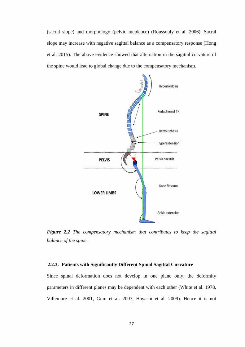
2.2.2. Sagittal Compensatory Mechanism
The spinal shape allows equal force distribution across spine column. Disruption of
such equilibrium would likely cause deformity (Roussouly and Nnadi, 2010).
Compensation mechanism would result in the pelvis and lower limbs. The possibility
of rotation of the pelvis around the femoral head’s axis is one of the best mechanisms
to regulate sagittal balance (Roussouly et al. 2011b). For global sagittal alignment, it
can be classified into three stages with respect to the severity of the imbalance: 1)
Balanced, compensated balance and unbalances (Barrey et al. 2013). Barrey et al.
(2013) also investigated the compensatory mechanism which contributes to keep the
sagittal balance of the spine as shown in Figure 2.2. Cervical hyperlordosis is typical
compensatory mechanism above a thoracic hyperkyphosis to maintain the
24
horizontality of the gaze. However, such an alternation would lead to an acceleration
of degenerative changes in the cervical spine, presence of axial neck pain, foraminal
stenosis and development of spondylotic myelopathy. Reduction of thoracic kyphosis
is also common during compensation. Decrease of thoracic kyphosis limits anterior
translation of the axis of gravity and is commonly observed in young patients with
flexible spine. For the elders, spine ageing will occur, the spine will become too rigid
and there is no possibility for the patient to reduce the magnitude of the thoracic
curve. Hyperextension of adjacent segments is a very common local compensatory
mechanism to limit the consequences of lumbar kyphosis on the shift of axis gravity.
It can be either global (multi-segment) or local (mono-segment). The advantage of
hypertension of these segments is that the upper spine will be placed more
posteriorly, however at the same time generating extra stresses on posterior
structures, increasing the risk of having retrolisthesis and possibly resulting in
accelerated facet joints arthritis. Posterior slippage of the upper vertebra in reference
to the lower vertebra, known as retrolisthesis, may also result. It is a 2–3 mm
slippage in the lumbar spine and commonly happens at lower or upper part of the
kyphotic spine: L5–S1 and upper lumbar spine (L1–L2 and L2–L3). It is generally
being underestimated on lying down radiological imaging techniques. The only
compensatory mechanism in the pelvis area is pelvis back tilt, which leads to
posterior positioning of the sacrum posterior to the coxo-femoral heads. Last but not
least, knee flessum correlates strongly with lack of lordosis (Obeid et al. 2011), while
ankle extension would possibly induce pelvis shift, which is a key component in
maintaining a fixed gravity line-heels offset and is a parameter as important as pelvis
tilting (Lafage et al. 2008).
25
Compensatory mechanism is effective to limit the sagittal unbalance, however at the
same time, it could possibly result in adverse effects such as mechanical pain and
compromise of neurological structures. To achieve the analysis of sagittal balance
and determine the presence of compensatory mechanism, the following three
procedures are suggested: 1) Investigate the value of the pelvis incidence, which
helps to predict the theoretical values of the spino-pelvic positional parameters; 2)
Determine whether the patient is globally balanced by analyzing the positioning of
C7 related to the sacrum, the angle between the sacral plate and the line connecting
the centroid of C7 vertebral body and the midpoint of the sacral plate, C7
plumbline/sacral femoral distance ratio; and 3) Investigate whether compensatory
mechanisms exist in (i) Spinal area such as cervical curvature, thoracic kyphosis and
lumbar lordosis and thoracic kyphosis; (ii) Pelvis area ; and (iii) Lower limbs area.
For instance, when an individual possesses in pathological kyphosis, corresponding
biomechanical adaptation of the compensatory balance may result. The possibility
for compensation to function depends on the location of the kyphosis & length of
lordosis and the flexibility of the spine (Roussouly et al. 2011b). If the kyphosis is
highly located, the lumbar lordosis is able to compensate the balance; but if the
kyphosis extends into the thoracolumbar area, the length of lordosis could be too
short to compensate. For a flexible spine, lumbar lordosis is largely curved and the
posterior arches are thinner as well as the spinous processes. This would promote a
better range of motion; however, it may induce spondylolysis. But when the kyphosis
occurs on a rigid spine, the only way for compensation is pelvic tilting, which would
result in a downward tilt of the head. In order to correct such posture, the patient
would need to tilt the pelvis backwards, extend the hips, flex the knees and dorsiflex
26
the ankles (Roussouly et al. 2010). However, for patients with small pelvic incidence,
they have a small capacity for sagittal imbalance compensation through pelvis
retroversion (Roussouly et al. 2010, 2011b), which inhibits the restoration of the
position of C7 plumb line behind the femoral head in case progressive kyphosis is
present in the patient.
Different relationships have been observed between sagittal parameters. Starting
from the top of the human spine, cervical angles were found significantly correlated
with cervico-thoracic angles and global sagittal alignments (Yu et al. 2013). In
addition, significant differences of cervical angles, cervico-thoracic angles and
thoracic kyphosis were observed among individuals with no kyphosis, cervical
kyphosis, cervical-middle-thoracic kyphosis and cervical-lower-thoracic kyphosis. A
hypokyphotic thoracic spine was found to coexist with a kyphosis in cervical spine in
idiopathic scoliosis (Canavese et al. 2011). Occipital-C2 angle was found to be
significantly negatively correlated with C2-C7 angle, while T1 slope was positively
correlated with cervical lordosis (Kaplan et al. (2015). Significant correlation
between loss of thoracic kyphosis and cervical kyphosis development was also
reported (Hilibrand et al. 1995). The top of thoracic curve on C7 was found to be
very stable over the sacrum (Roussouly and Pinheiro-Franco, 2011a). Positive
correlation was found between thoracic kyphosis and lumbar lordosis (Van Royen et
al. 1998). Lumbar Lordosis mainly depends on sacral slope orientation (Roussouly
and Pinheiro-Franco, 2011a). Pelvic incidence was closely related to lumbar lordosis
in normal adolescents and adults (Mac-Thiong et al. 2007) and in scoliotic adults
(Legaye et al. 1998). Spinal alignment could be affected by pelvic posture (Van
Royen et al. 1998). Spino-sacral angle was closely related to sacropelvic balance
27
(sacral slope) and morphology (pelvic incidence) (Roussouly et al. 2006). Sacral
slope may increase with negative sagittal balance as a compensatory response (Hong
et al. 2015). The above evidence showed that alternation in the sagittal curvature of
the spine would lead to global change due to the compensatory mechanism.
Figure 2.2 The compensatory mechanism that contributes to keep the sagittal
balance of the spine.
2.2.3. Patients with Significantly Different Spinal Sagittal Curvature
Since spinal deformation does not develop in one plane only, the deformity
parameters in different planes may be dependent with each other (White et al. 1978,
Villemure et al. 2001, Gum et al. 2007, Hayashi et al. 2009). Hence it is not
28
surprising that different sagittal profiles could be observed in patients with AIS
compared to normal individuals.
Scoliosis is also one of the relevant clinical expressions of patients with Parkinson’s
disease (Doherty et al. 2011, Baik et al. 2009; Schwab et al. 2012). Thoracic
kyphosis was found to be positively correlated with lumbar lordosis in groups with or
without scoliosis in patients with Parkinson’s disesase (Bissolotti et al. 2015).
Though lower spinosacral angle was observed in scoliosis group (Bissolotti et al.
2015), no significant difference was found between Cobb angles and any of the
spinopelvic and sagittal balance parameters (Bissolotti et al. 2015). Sagittal profile of
patients with low back pain (LBP) was also found to be significantly different from
normal individuals. Patients with LBP were characterized by a more vertical sacrum
and more proximal lumbar lordosis (Jackson et al. 1994). In addition, thoracic
kyphosis, thoracic tilt, lumbar tilt, lumbosacral angle, sacral slope, pelvic incidence
of adult LBP group were found to be significantly different from normal individuals,
with the sacral slope, lumbar lordosis and pelvic incidence being generally smaller in
patients with LBP (Chaléat-Valayer et al. 2011). However, sagittal parameters were
similar between French men and women in the LBP group for the thoracic, lumbar
and pelvis regions (Chaléat-Valayer et al. 2011). Adam et al. (1999) stated that the
loss of LL was a risk factor for the first time occurrence of serious low back pain
(Adams et al. 1999) and degenerative changes for IVD and vertebral body (Schlegel
et al. 1996). Hence it was common that spine degeneration was commonly observed
in patients with LBP. C7 plumbline was observed to be stabilized or slightly moved
forward in patients with degenerated spine (Cil et al. 2005). Moreover, significant
reduction of both lumbar lordosis and thoracic kyphosis was observed in patients
29
with disc disease and disc herniation (Barrey et al. 2007). Furthermore, El-Hawary et
al. (2016) mentioned that various etiologies could affect sagittal plane alignment
such as postural kyphosis, Scheuermann’s Condition, congenital kyphosis,
myelodysplasia, spinal tuberculosis, posttraumatic kyphosis, syndromic kyphosis,
achondroplasia, postsurgical kyphosis and ostlaminectomy.
Patients with AIS were the main focus group of this study. In previous practice, most
of the AIS studies mainly focused on the coronal plane, but indeed additional
attention should be paid to sagittal plane deformity and measurements on AIS
patients (Deacon et al. 1984, Dickson et al. 1984, Lenke et al. 2001). Studies had
found that patients with primary thoracic scoliosis were generally hypokyphotic (De
jonge et al. 2002, Ilharreborde et al. 2018). Dickson et al. (1984) found that 75% of
the apical lateral profiles were found to be lordotic and a mean lordosis of 3° was
reported at the apex of the kyphotic thoracic spine of scoliosis patients. The anterior
part of the apical vertebrae was higher than the posterior part, with endplate
irregularities most often found in the posterior part. A negative correlation was also
observed between thoracic kyphosis and the lateral thoracic curves of patients with
idiopathic thoracic and double major scoliosis, and the average thoracic kyphosis of
these patients was 10 degrees smaller than those without scoliosis in supine position
(Schmitz et al. 2001). However, no significant difference was observed in lumbar
lordosis between patients with lumbar and double major curves and non-scoliotic
subjects. Mac-Thiong et al. (2013) conducted a study on young patients with average
43 degrees Cobb angle and discovered that thoracic kyphosis was significantly lower
for patients who possessed thoracic curves than those with lumbar curves, but no
significant difference was observed in lumbar lordosis between different curve types.
30
In addition, no significant change between the groups was observed for the sacral
slope, pelvic tilt, or pelvic incidence. Furthermore, the pelvic incidence was
significantly correlated with the lumbar lordosis, sacral slope, and pelvic tilt for all
the groups. The lumbar lordosis was strongly related to the sacral slope in all cases,
but not with the thoracic kyphosis, except in the case of thoracolumbar curves. In
addition, there is increasing evidence suggesting that hypokyphosis in the thoracic
region and non-uniform growth of the posteroanterior vertebrae mismatch
contributes to the development of AIS. Previous study has shown that posteriorly
inclined human spine segments would likely lead to a decrement of rotational
stiffness, inducing rotational instability to the spine column (Kouwenhoven et al.
2007). Schlösser et al. (2014) and Janssen et al. (2011) demonstrated that patients
with mild thoracic scoliosis had a significantly less kyphotic spine and longer and
more posteriorly inclined segment from C7-T8 than the normal and those with mild
lumbar scoliosis. However, patients with lumbar scoliosis were found to have
significantly greater thoracic kyphosis than the normal and those with thoracic
scoliosis. In addition, steeper posterior inclination from T12-L2 was observed from
patients with lumbar scoliosis. Such hypokypotic phenomenon has further been
confirmed in thoracic AIS patients using EOS, a biplanar radiograph modality
(Sullivan et al. 2017). Furthermore, elucidation of the timing of the onset of
hypokyphosis and scoliosis could provide valuable insight into the curve
development of the AIS patients. Nault et al. (2014) observed that patients with
progressive AIS were found to have significantly larger angle of the plane of
maximal curvature, hypokyphosis, larger apical intervertebral rotation and torsion
than non-progressive AIS, with the angle of the plane of maximal curvature
demonstrating the most significant difference, by evaluating 3D spinal morphology
31
of progressive and non-progressive patients with similar spinal curve of Cobb angles
less than 40 degrees at the first visit. Moreover, the height/width ratio of the
vertebrae from T6 to L4 using local and regional measurement was also found higher
for non-progressive AIS (Nault et al. 2014). From a study of Ni et al. (2010),
thoracolumbar kyphosis was observed in more than 65% of patients with
thoracolumbar/lumbar AIS within T11-L2 region. In addition, significant
correlations were also observed between apical vertebrae rotation and coronal
thoracolumbar/lumbar curvature; and sagittal Cobb angle of T11-L2 thoracolumbar
sagittal curve. Moreover, the apex vertebrae in patients with thoracolumbar scoliosis
were usually more dorsally inclined.
2.2.4. Factors that Alter Sagittal Curvature of Spine
For young individuals, such as schoolchildren, one of the potential threats that could
alter their sagittal profile is carriage of overloaded backpack, which is common
among schoolchildren in different regions (Goodgold et al. 2002, Cottalorda et al.
2004). Overloaded backpack has been found to cause back pain and spinal
deformities (Moore et al. 2007, Hong et al. 2011, Talbott et al. 2009). Other factors
have also been reported to cause deformity in spinal sagittal profile, such as improper
practice of elongation exercises (Drzał-Grabiec et al. 2014), hereditary factors
(Janssen et al. 2013), patients with idiopathic scoliosis (Roussouly et al. 2013, Yong
et al. 2012, Janssen et al. 2011, Hu et al. 2016, Hong et al. 2016), intervertebral disc
degeneration (Barrey et al. 2007, Cil et al. 2005), Parkinson’s disease (Baik et al.
2009, Schwab et al. 2012), gender effect (Chaléat-Valayer et al. 2011, Abelin-
Genevois et al. 2014, Takács et al. 2015), and age (Hammerberg and Wood 2003,
Gelb et al. 1995, Tang et al. 2012, Li and Hong 2004).
32
The posture adopted by an individual is a key factor that affects the spinal sagittal
curvature. Forward-bending posture has become one of the most standard positions
to detect scoliosis and has been used as a basis for the screening test in schools
recommended by Scoliosis Research Society (SRS) (Stokes et al. 1987). The spinal
column changes its profile completely into a long kyphosis during forward bending
(Hackenberg et al. 2006). Forward bending in standing position would also result in
an increase in the maximum rotation of the surface of the back for the thoracic region
and lumbar region with a larger extent. A significant positive correlation in the
lumbar region was found between the change in rotation of the surface of the back
and the Cobb angle, but no significant relationship was observed between the change
of the back surface rotation and the degree of forward bending (Stokes et al. 1987).
Minor changes of curvature will be induced in the sagittal plane in the thoracic
region regardless of the degree of bending, however, lumbar lordosis is reversed to a
kyphosis. This suggested that the thoracic region is less flexible due to the presence
of rib cage and stiffer thoracic spine, while lumbar is more flexible due to the
increase in rotation of the surface of the back in the lumbar region. Spinal shape also
varies during different body postures other than forward bending. An average of 19%
correction of the scoliosis angle was induced during supine position (Zetterberg et al.
1983). Mean Cobb angle measured at standing position from Girls with AIS was
found approximately 9 degrees larger than that in the supine position, and the
difference was 45 degrees in the maximum (Troell et al. 1985). The mean Cobb
angle and the rotational angle on a standing radiograph were approximately 16
degrees and 6 degrees larger than those in the supine position respectively (Yazici et
al. 2001), suggesting that supine position may spontaneously modify the curves in
both the coronal and transverse planes; and error would be introduced when
33
performing analysis on the true extent of rotational deformity if only radiograph in
supine position is being measured. Janssen et al. (2010) found that the spine is more
kyphotic in the upper thoracic region, equally curved in the mid-thoracic and
thoracolumbar region and more lordotic in the lower lumbar region during standing
(Janssen et al. 2010). In addition, Salem et al. (2015) found that kyphosis and
lordosis angle of patients significantly decreased from standing to prone lying for
about 13.4 degrees. Pelvic parameters that relate with hip angle also significantly
altered the lumbar lordosis during supine posture (Driscoll et al. 2012). Furthermore,
lumbar lordosis was smaller in supine position than in standing position but only for
older subjects (Lee et al. 2014). Hence it has been suggested that supine position may
spontaneously modify the curves in both the coronal and transverse planes; and error
will be introduced when performing analysis on the true extent of rotational
deformity if only radiograph in supine position is being measured (Yazici et al. 2001).
Moreover, anterior/posterior translations of thoracic cage could cause significant
changes in thoracic kyphosis (26 degrees), lumbar curve, and pelvic tilt (Harrison et
al. 2002).
Age was also considered as a significant factor which would cause the change of
sagittal profile of the spine. An increased risk of the development of spinal
deformities had been noticed during periods when extremely rapid growth is
assumed, which is around months 6–24, years 5–8 and years 11–14. Cervical spine
alignment in patients aged 10 years old shows significantly more lordosis of 6°
versus 1° in those older than 10 years, which was possibly caused by the strong
influence of craniocervical orientation and thoracic shape (Abelin-Genevois et al.
2014). Asymptomatic children with mean age 3-7 years also had a lower rate of
34
cervical hypolordosis and kyphosis than older patients (Lee et al. 2012). In addition,
T1-T2, T10-L2 and L4-S1 are the main different areas in terms of sagittal plane
alignment with children younger than 10 years compared to those older (Cil et al.
2005). C7 plumbline was found to have a tendency to move backwards from child
until adulthood (Cil et al. 2005), but there was no significant forward displacement of
C7 plumbline with respect to sacrum with aging (Mac-Thiong et al. 2010). Cervical
sagittal alignment was significantly different between French pediatric patients aged
below 11 and those 11 or above (Abelin-Genevois et al. 2014). Pelvic incidence and
lumbar lordosis were found to be significantly increased with age in pediatric
asymptomatic subjects (Abelin-Genevois et al. 2014). Though hypokyphosis was
observed in patients with AIS, thoracic kyphosis and sacral slope were significantly
higher in adult patients with more severe scoliosis (Hong et al. 2017). The severity of
scoliosis in adults was found to increase with higher pelvis incidence (Roussouly et
al. 2013). It had also been demonstrated that spinal sagittal alignment worsens with
age in adults (Hammerberg and Wood 2003, Gelb et al. 1995, Tang et al 2012). Early
progressive sagittal positive imbalance could be observed in the ageing process
(Hammerberg and Wood 2003) and in other spinal diseases such as Parkison’s
disease (Bissolotti et al. 2014) and degenerative spondylolisthesis (Schuller et al.
2011).
There were also controversial findings about the effect of gender on the patterns of
spinal sagittal profile. A study performed three-dimensional reconstruction of
radiographs from asymptomatic young adults and found that female’s spine was
more backwardly inclined than the male spine (Janssen et al. 2009). Abelin-Genevois
et al. (2014) found that pediatric asymptomatic female subjects had larger lumbar
35
lordosis than males. Young female tennis players demonstrated significantly larger
lumbar lordosis and lower thoracic kyphosis than male players (Muyor et al. 2013).
Significant differences of thoracic kyphosis and lumbar lordosis were observed in
patients with AIS of different ages and genders (Takács et al. 2015). The cervical
sagittal angles and the lumbar lordosis among 150 French pediatric patients were
significantly different between boys and girls below 18 years old (Abelin-Genevois
et al. 2014). Pelvic tilt was significantly greater and pelvic retroversion began earlier
among females for 60-89 age groups, while average T1 slope was significantly larger
in males than females from a study investigating standing radiographs of subjects
aged above 50 with Cobb’s angle less than 25 degrees in coronal plane (Oe et al.
2015). However, Hu et al. (2015) and Mac-Thiong et al. (2010) demonstrated that
gender had no effect on pelvis incidence, pelvic tilt, sacral slope, pelvic tilt in their
study.
Recently, the influence of a spinal fusion on thoracic kyphosis and overall sagittal
alignment has been evaluated. Depending on instrumentation and correction
techniques used, postoperative thoracic kyphosis was found to either increase or
decrease (de Jonge et al. 2002, Lowenstein et al. 2007, Kim et al. 2006, Vora et al.
2007, Kim et al. 2004). Such alternation in thoracic kyphosis was believed to have an
impact in lumbar lordosis because correction of thoracic scoliosis would result in a
spontaneous lumbar curve correction, depending on the surgical approach used (Patel
et al. 2008, Lenke et al. 1999, Betz et al. 1999, Potter et al. 2005). Flat back
syndrome, which is rectification of both thoracic kyphosis and lumbar lordosis, had
been increasingly observed in patients after posterior pedicle screws surgery during
long term follow up (Thompson et al. 1990, Newton et al. 2010). This syndrome
36
generally includes back pain, degenerative disc disease, and functional disability (La
Grone 1999). Newton et al. 2010 had found that anterior and posterior thoracic
fusion increased and decreased both thoracic kyphosis and lumbar lordosis
respectively at 2-year follow-up. Roussouly et al. (2013) reported that a slight but
significant increase of pelvic tilt with a decrease of sacral slope and lumbar lordosis
was observed in AIS patients. Kim et al. (2006) suggested that the risk factors which
significantly associated with suboptimal sagittal balance 2 years after lumbar spine
fusion surgery were: 1) Sagittal imbalance preoperative and 8 weeks postoperatively
≥5 cm; 2) Bigger thoracic kyphosis and pelvic incidence compared with lumbar
lordosis ( ≥ 45°) before surgery; 3) Lower uppermost instrumented vertebra; 4)
Smaller lumbar lordosis compared with thoracic kyphosis ( ≤ 20°) at 8 weeks
postoperatively; 5) Patients whose age at surgery older than 55 years. In addition,
they found out that there was a trend of getting suboptimal sagittal balance after
posterior spinal instrumentation and fusion from the thoracolumbar spine to the L5–
S1 was observed if the total angle of lumbar lordosis, thoracic kyphosis, and pelvic
incidence was ≥45° (Kim et al. 2006). Significant curve correction of the primary
scoliosis curve was achieved when the patients underwent posterior only surgical
correction and fusion, but significant increase of pelvis tilting had also resulted after
surgery (La Maida et al 2013).
2.3. Different Approaches of Sagittal Assessments
2.3.1. Assessment of Patient with AIS on Radiograph Using Cobb Angle
Other than the routine evaluation of the coronal deformity of patients with AIS,
evaluation of the sagittal plane was sometimes involved by measuring the thoracic
kyphosis and lumbar lordosis using constrained (Boseker et al. 2000, Lee et al. 2011,
37
Schlösser et al. 2014) and non-constrained (Stagnara et al. 1982; Voutsinas et al.
1986) Cobb techniques. For constrained limit vertebrae method, which is commonly
used nowadays, thoracic and lumbar vertebrae are defined by the authors. For
kyphosis, lower endplate of T12 was always used as the standard in medical
literature, but the end of the kyphosis cranial varied from T2 (Boseker et al. 2000),
T3 (Lee et al. 2011) and T4 (Schlösser et al. 2014). For lordosis, the angle is
commonly defined between two drawn lines to the upper endplate of L1 vertebra and
to the lower endplate of the L5 vertebra. But there were also studies using different
levels for lumbar lordosis evaluation (Polly et al. 1996). For non-constrained method,
the most tilted vertebra needs to be identified to classify thoracic kyphosis and
lumbar lordosis (Mac-Thiong et al. 2007). Different from the coronal curvature, at
present there is no screening categorization for the sagittal curvature. Rotation was
generally evaluated based on the relative position of the pedicles in relation to the
underlying vertebral body (Nash et al. 1969) and approximate range of degrees (5-
degree interval) represented by each gradation of rotation is determined. However,
this method is not a direct measurement of the degree of rotation or the clinical
deformity. For cervical measurement, the most typical evaluation methods were
occiputo-C2 angle, C2-7 sagittal vertical axis, segmental angle and T1 slope (Oe et al.
2015, Kaplan et al. 2015). Sacropelvic region was also commonly considered when
evaluating sagittal balance, in terms of pelvic incidence (PI), pelvic tilt (PT) and
sacral slope (SS). There was a geometrical relation between the PI (morphological
parameter) and PT/SS (functional parameters), according to the equation: PI = PT +
SS (Roussouly et al. 2003).
38
Other than local measurements, various angular methods had been used to evaluate
sagittal global balance, such as 1) Spino-sacral angle: formed by a line from the
centre of C7 to the centre of the sacral endplate and the surface of the sacral endplate
(Mac-Thiong et al. 2011); 2) Spinal tilt: formed by a line from the centre of C7 to the
centre of the sacral endplate and the horizontal surface (Mac-Thiong et al. 2011); 3)
spino pelvic tilt: formed by a line from the centre of C7 to the centre of the sacral
endplate and a line from the centre of the sacral endplate to the centre of the femoral
head (Roussouly and Nnadi, 2010; Mac-Thiong et al. 2011). These angles were
typically used to quantify global kyphosis, for instance, severe kyphosis generally
implied decreased spino-sacral angle (Mac-Thiong et al. 2011). Chin-brow vertical
angle was measured by drawing a line from the brow to the chin and the vertical axis,
while the patient stood with hips and knees extended and the neck in a neutral or
fixed posture (Bridwell et al. 1989). A classification system has also been established
for sagittal global balance evaluation, where no calculation or measurement is
involved (Mac-Thiong et al. 2010).
A biplanar X-ray system, EOS 2D/3D imaging system, was recently developed for
spine evaluation. It used slot-scanning technology that could reduce the radiation
dosage at the same time enhancing image quality (Deschênes et al. 2010). SterEOS
3D, a special 3D construction software, has been developed to accompany the EOS
2D/3D imaging system. This system enabled the generation of realistic 3D models
based on a combination of geometric and statistic modelling by a parametric surface
3D reconstruction, and its precision was similar to CT based spinal 3D reconstruction
procedure (Mitulescu et al. 2002; Le Bras et al. 2003). Somoskeöy et al. (2012)
investigated the accuracy and reliability of the commercial EOS 2D/3D system and
39
sterEOS 3D software under routine clinical circumstances. By predefining end
vertebrae before measurements (Dimar et al. 2008; Mok et al. 2008), intra-observer
and inter-rater reproducibilities for either 2D or sterEOS 3D measurements were
excellent and the results were strongly and significantly correlated with traditional
2D measurement. Other than spine evaluation, the EOS system could be used to
investigate other body parts such as pelvic tilt (Lazennec et al. 2011), hip and knee
geometrical parameters (Than et al. 2012), acetabular anteversion and inclination
(Journé et al. 2012) and musculoskeletal imaging for assessment and follow-up of
balance disorder of the spine and of the lower limbs (Wybier and Bossard 2013).
However, there were several limitations for the EOS system. Relatively large
difference in accuracy was found between the anterior and the posterior vertebral
regions, since several anatomical landmarks on the posterior arch such as the
transverse and/or spinous processes may be barely visible on the X-ray images,
which caused reconstruction error leading to results discrepancies (Mitulescu et al.
2002). In addition, the time needed to reconstruct a detailed 3D spine model was an
average of 20 to 30 minutes, and cases with a severe scoliotic curve would definitely
require a significantly longer time (Somoskeöy et al. 2012), which was not feasible
under routine clinical circumstances.
Moreover, specific devices were needed to maintain the superior limbs and the head
of the most unstable patients since movement artefacts due to the long-lasting time of
acquisition may lead to the repetition of the examination, resulting in radiation dose
increase (Wybier and Bossard 2013). Furthermore, a mild sagittal gap between feet
was necessary to avoid superimposition of the lower limbs in the lateral examination,
which may induce artificial anterior knee flessum and posterior knee recurvatum
40
(Wybier and Bossard 2013). EOS also had a limitation for assessing posture. It at
present cannot provide study in the supine position, which may be requested in some
scoliosis pre-operative assessment (Wybier and Bossard 2013). In terms of cost
effectiveness, EOS can be shown to be cost-effective when compared with computed
radiography (CR) only if the utilization for EOS is about twice the utilization of CR.
However, as CR is widely accepted as the golden standard for spine evaluation
method, such estimation would not be possible (McKenna et al. 2012). Also, though
EOS applies less radiation dosage during scanning, no evidence shows whether or
not these image-related health benefits exist, let alone whether or not they reach the
magnitude necessary for EOS to be cost-effective. Furthermore, these extra health
gains would be possible only if a sufficient proportion of patients experienced a
change in therapeutic management, with a consequent improvement in outcomes,
following the use of EOS rather than CR (McKenna et al. 2012).
2.3.2. Sagittal Evaluation Other than Cobb Angle on Radiograph
Other than Cobb angle, different kinds of methods have been used for spinal sagittal
angle measurement. Various tangential methods were also introduced for sagittal
curvature evaluation. Posterior tangents and Anterior tangents were proposed by
Harrison et al. (1996) and Schular et al. (2004), where the curvature was defined by
the angle between the two straight lines drawn tangentially to the posterior / anterior
vertebral body wall of the end vertebrae. Chernukha et al. (1998) introduced the
tangential radiologic assessment of lumbar lordosis (TRALL) method, where the
posterosuperior corner of the superior end vertebra, the posteroinferior corner of the
inferior end vertebra and the point with the maximal orthogonal distance from the
spine were identified for the calculation of the sagittal angle. Another tangent method,
41
namely the Tangent circle method, valuable for evaluating global sagittal geometry
was later introduced by Vaz et al. (2002). This method was modified by using two
circular arcs and the curvature angle was defined between the straight lines that
connected the centers of the circular arcs with the corners of vertebral bodies at both
ends of the spine curve and the reference horizontal line. Centroid method was later
suggested by Chen (1999), where the angle was defined by two straight lines passing
through two vertebral centroids. Followed by computerized centroid method by
Briggs et al. (2007), where vertebral body corners from T1 to T12 were manually
identified. These methods had been proved to have greater consistency than using
Cobb method. Harrison et al. (2001) introduced the Best fit ellipses approach, where
an elliptical arc was aligned to the manually identified posterior vertebral bodies’
corners using least square technique.
One of the earliest studies which used mathematical models for sagittal curvature
measurement was presented by Singer et al. (1990), in which sagittal curvatures were
measured by first and second derivative of the sixth-degree polynomial that passed
through manually identified anterior and posterior vertebral body contour (Singer et
al. 1994). A similar method was introduced in later study, which also used the same
landmarks from the vertebral body to evaluate the mean radius of curvature.
However, these methods require manual identification of a large number of vertebral
landmarks.
Various indexes have also been used to evaluate the thoracic profile of the spine. A
spinal line was drawn by connecting the posteroinferior corners of the bodies of the
end vertebrae and constructing and additional lines were constructed orthogonally to
42
the spinal line to the posteroinferior corners of the remaining vertebral bodies in the
spine curve. The sum of the lengths of these additional lines was considered as
Ishihara index (Ishihara et al. 1968). A similar index, Index of kyphosis (Voutsinas
and MacEwen 1986), was proposed later, but the sum of additional lines was
replaced by the maximal orthogonal distance, which was mainly used for predicting
vertebral deformities from kyphosis measurement. The area under curve, which was
used as a descriptor of sagittal curvature, was further introduced by Yang et al.
(2007), by first manually identifying all postero-superior vertebral body corners,
which were then all passed through by spine curve interpolation.
2.3.3. Disadvantages of Current Radiographic Evaluation
Since skeletally immature patients are at risk for curve progression, follow-up
posterior-anterior radiographs are necessary for every half a year to one year to
monitor the progression (Thomsen et al. 2006). In clinical practice, measuring
Cobb’s angle from the standing posterio-anterior X-ray radiograph is still the gold
standard method to evaluate the severity of scoliosis (Cobb 1948). Routine
evaluation and classification of spinal morphology remained predominantly 2D
(Lenke et al. 2005; Clements et al. 2011). Sagittal X-ray may somehow be avoided
unless necessary to avoid patients from overexposing to radiation. Measurement
errors of the intra- and inter-observer reliabilities of Cobb’s angle measurement had
been reported to be in the range of 3 to 8 degrees using measurement tools (pencils,
protractors etc) (Shea et al. 1998, Srinivasalu et al. 2008). These errors were likely
due to subjective factors such as end vertebrae selection and observers’ skill level
(Chockalingam et al. 2002, Stokers et al. 2006). Moreover, routine 2D measurements
of thoracic kyphosis erroneously underestimated the preoperative loss of kyphosis in
43
AIS because of errors associated with axial plane rotation, an inherent component of
thoracic scoliosis (Newton et al. 2015). Furthermore, it has been found that there is
still a risk of progression for just evaluating and monitoring the Cobb’s angle of the
patients (Helenius et al. 2003, Tan et al. 2009).
Mass screening or frequent therapy outcome measurements is not recommended due
to radiation applied to the patient. In fact, only 10% of patients with curve
progression warranted intervention, which means that the rest would expose to
unnecessary radiation. Moreover, two radiographs are required and stitched together
to produce whole spine view for assessment (Malfair et al. 2010), increasing the risk
of radiation exposure. Such radiation accumulated by AIS patients from long-term
follow-up may increase the risk of breast cancer in girls with scoliosis (Hoffman et al.
1989, Doody et al. 2000). In addition, radiographic diagnostics in childhood
contributed significantly to leukemia and prostate cancer (Schmitz-Feuerhake et al.
2011). Other than radiation issue, another disadvantage of X-ray assessment is that
vertebral rotation information in scoliotic spine cannot be directly acquired, since
these radiographs do not demonstrate the true magnitude of the 3D spinal deformity
present in these patients (Yazici et al. 2001). Though improvements have been made
in radiographical technology and techniques to minimize radiation exposure, such as
the invention of the EOS system that could provide lower radiation dosage imaging,
it was still claimed that long-term health complications remained an inherent risk
from limited doses of radiation exposure (Lee et al. 2013). In addition, female
patients with scoliosis still had a 4.2% increased risk of breast cancer (Bone and
Hsieh 2000). It should be noted that in some countries physiotherapists were not
authorized to request X-ray examination (de Oliveira et al. 2012). Hence an
44
alternative system that can accurately measure spine deformity for patients with AIS
in all three planes mass screening and longitudinal follow-up during treatments
without any hazard of radiation is required in the field.
2.3.4. Sagittal Evaluation of Spine Using Other Methods
For imaging modalities, magnetic resonance imaging (MRI) and computed
tomography (CT) are also common for investigating scoliotic spine in the research
fields these days due to their high resolutions. However, both MRI and CT are pricey
and less accessible (Diefenbach et al. 2013). In addition, MRI requires expertise to
operate with long acquisition time, whereas CT requires a higher amount of radiation
dosage than traditional radiograph. Moreover, direct measurement of the axial
rotation of the spine with a CT scan is inaccurate unless the section is at a right angle
to the spinal column, so it is impossible to obtain information on the whole spinal
column and the measurement of rotation is disputable. Most importantly, patients are
required to be assessed in supine position for both modalities, which could lead to
errors in interpretation of the spinal curvature. It had been observed that Cobb’s
angle derived from the supine posture was significantly and spontaneously corrected
from the standing posture (Yazici et al. 2001). Though the results obtained from
supine MRI were significantly linearly correlated with the Cobb angles measured on
standing x-ray in AIS patients, Cobb angles measured by supine MRI were
significantly smaller in both coronal and sagittal planes (Shi et al. 2015, Brink et al.
2017), stating that the parameters from supine and prone scans could not directly be
compared to the those from upright radiographs.
45
Various topographic methods have been used for measuring sagittal curvatures, but
they have their own disadvantages. Stereo camera (Figure 2.3a) was used to estimate
spine curvature but it was found to be not precise enough and was not able to assess
vertebral rotation and visualize the bone architecture (Goldberg et al. 2001). A study
using such method even reported differences up to 9 degrees when compared with X-
ray Cobb angle measurement (Frerich et al. 2012). A continuous acquisition 3D
digitizer called FaroArm (Figure 2.3b) had been used to measure the sagittal spine
profile along the spinous processes through sliding on the skin (Salem et al. 2015). It
worked by first acquiring 2D sagittal spine models with coordinates, followed by
interpolating the data into a five-degree polynomial to obtain thoracic and lumbar
angles from the first and second derivative. However, the results were possibly
prone to errors since relative positions of the spinous processes were just estimated
from the skin surface. A noninvasive anthropometric technique using reflective skin
markers (Figure 2.3c) had been used to calculate thoracic kyphosis and lumbar
lordosis through obtaining the spatial locations of these markers (Leroux et al. 2000).
Schmid et al. (2015) further conducted a study to validate the use of reflective
markers, by using radio-opaque markers on patients during radiograph (Figure 2.3d).
Repeatable and accurate results would be obtained if palpation was precisely
performed on patients. However, it would take a long time for locating the landmarks
and attaching the markers to the predefined locations. Lewis et al. (2010) employed
inclinometers to determine thoracic kyphosis by placing them over T1/T2 and
T12/L1 processes (Figure 2.3e) and demonstrated excellent intra-rater reliability with
high ICC and small error using this method. However, there was no certainty that
this method was stable over time due to natural variation in postures and accurate
palpation was required for every measurement. Chaise et al. (2011) used an adapted
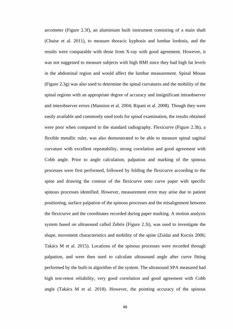
46
arcometer (Figure 2.3f), an aluminium built instrument consisting of a main shaft
(Chaise et al. 2011), to measure thoracic kyphosis and lumbar lordosis, and the
results were comparable with those from X-ray with good agreement. However, it
was not suggested to measure subjects with high BMI since they had high fat levels
in the abdominal region and would affect the lumbar measurement. Spinal Mouse
(Figure 2.3g) was also used to determine the spinal curvatures and the mobility of the
spinal regions with an appropriate degree of accuracy and insignificant intraobserver
and interobserver errors (Mannion et al. 2004; Ripani et al. 2008). Though they were
easily available and commonly used tools for spinal examination, the results obtained
were poor when compared to the standard radiography. Flexicurve (Figure 2.3h), a
flexible metallic ruler, was also demonstrated to be able to measure spinal sagittal
curvature with excellent repeatability, strong correlation and good agreement with
Cobb angle. Prior to angle calculation, palpation and marking of the spinous
processes were first performed, followed by folding the flexicurve according to the
spine and drawing the contour of the flexicurve onto curve paper with specific
spinous processes identified. However, measurement error may arise due to patient
positioning, surface palpation of the spinous processes and the misalignment between
the flexicurve and the coordinates recorded during paper marking. A motion analysis
system based on ultrasound called Zebris (Figure 2.3i), was used to investigate the
shape, movement characteristics and mobility of the spine (Zsidai and Kocsis 2006;
Takács M et al. 2015). Locations of the spinous processes were recorded through
palpation, and were then used to calculate ultrasound angle after curve fitting
performed by the built-in algorithm of the system. The ultrasound SPA measured had
high test-retest reliability, very good correlation and good agreement with Cobb
angle (Takács M et al. 2018). However, the pointing accuracy of the spinous
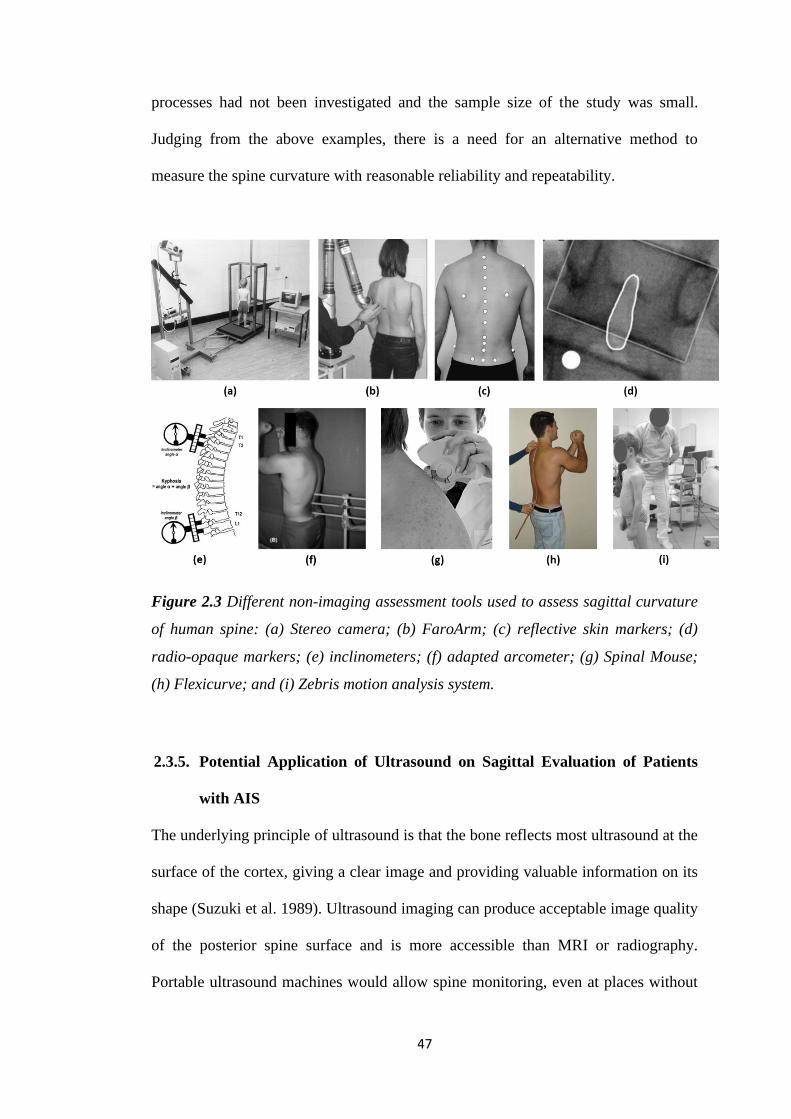
47
processes had not been investigated and the sample size of the study was small.
Judging from the above examples, there is a need for an alternative method to
measure the spine curvature with reasonable reliability and repeatability.
Figure 2.3 Different non-imaging assessment tools used to assess sagittal curvature
of human spine: (a) Stereo camera; (b) FaroArm; (c) reflective skin markers; (d)
radio-opaque markers; (e) inclinometers; (f) adapted arcometer; (g) Spinal Mouse;
(h) Flexicurve; and (i) Zebris motion analysis system.
2.3.5. Potential Application of Ultrasound on Sagittal Evaluation of Patients
with AIS
The underlying principle of ultrasound is that the bone reflects most ultrasound at the
surface of the cortex, giving a clear image and providing valuable information on its
shape (Suzuki et al. 1989). Ultrasound imaging can produce acceptable image quality
of the posterior spine surface and is more accessible than MRI or radiography.
Portable ultrasound machines would allow spine monitoring, even at places without
48
permanent medical imaging devices (Unqi et al. 2014). In addition, cost of
installation and the footprint of tracked ultrasound systems are only a fraction of that
for radiography systems, which may facilitate wider accessibility and reduce the cost
of patient follow-up (Unqi et al. 2014). Hence there is an emerging trend of using
freehand 3D ultrasound system for spinal curvature measurement (Nelson et al. 1998,
Huang et al. 2005, Fenster et al. 2001; Gee et al. 2003; Cheung CWJ et al. 2010;
Purnama et al. 2010).
3D ultrasound was first explored on AIS patients by Suzuki et al. (1989). Spinous
processes of the patients were first marked and drawn parallel to the inclination of
the vertebrae as seen on an anteroposterior radiograph of the spine. Then the
transducer with an attached inclinometer was placed on the spinous process in this
line until the image of the laminae became horizontal on the screen. A correlation
was found between the Cobb’s angle and the vertebra rotation in both thoracic and
lumbar curve. Li et al. (2012) investigated the effectiveness of orthotic treatment for
patients with AIS using 3D ultrasound in order to enhance the effectiveness of
orthotic treatment. SPA was measured from the ultrasound and used as a clinical
parameter to estimate the Cobb’s angle in order to determine the location of the
pressure pad. The results showed that the ultrasound-assisted fitting method of spinal
orthosis was effective and beneficial to 62 % of the patients. The intra- and inter-
rater reliability of the coronal curvature asymmetry were also investigated on cadaver
spinal column phantom (Chen et al. 2013) and patients with AIS (Young et al. 2015).
Center of lamina method was used for ultrasound and compared with laser scan using
Cobb’s method in the study. Coronal curvature measurements were compared to the
Cobb angle recorded on the same day. The center of lamina method showed high
49
intra- and inter-rater reliability and moderate correlation with X-ray Cobb. Wang et
al. (2015) further investigated the reliability and validity of the centre of lamina
methods in clinical setting by comparing to the corresponding MRI measurement on
16 patients with AIS, and similarly, the centre of laminae methods showed high
intra- and inter-rater reliability, no significant difference with MRI Cobb and good
agreement for coronal angle measurement (Wang et al. 2015). Ungi et al. (2014)
utilized tracked ultrasound to localize vertebral transverse processes as landmarks
along the spine to measure curvature angles on spine phantoms. Close correlation
was found between the tracked ultrasound transverse process angle and the
radiographic Cobb measurements (Ungi et al. 2014). In addition, the inter-operator
difference of the sonographic transverse process angle was significantly lower than
that with radiographic Cobb measurement, though practical usability of the system in
this study was yet to be proved. Koo et al. (2014) developed a customized ultrasound
system which could identify spinous process from dry bone specimen, by digitizing
the spatial position of the spinous process. Different polynomial regression was
applied to generate spinal curves, and LOESS (0.4)–posterior deformity angle
method was found to best correlate with Cobb’s angle.
Cheung et al. (2013) developed a 3D ultrasound imaging method for the radiation-
free assessment of scoliosis. Preliminary tests were conducted on flexible spinal
column phantoms (Cheung et al. 2013) and 27 human subjects (Cheung et al. 2015).
3D ultrasound volume was obtained through freehand scanning and a virtual 3D
model of the spine was formed using the spinous, transverse and/or superior articular
processes which were manually chosen. The 3D image was then projected to a 2D
plane for X-ray comparison. Ultrasound angles were found to have good linear
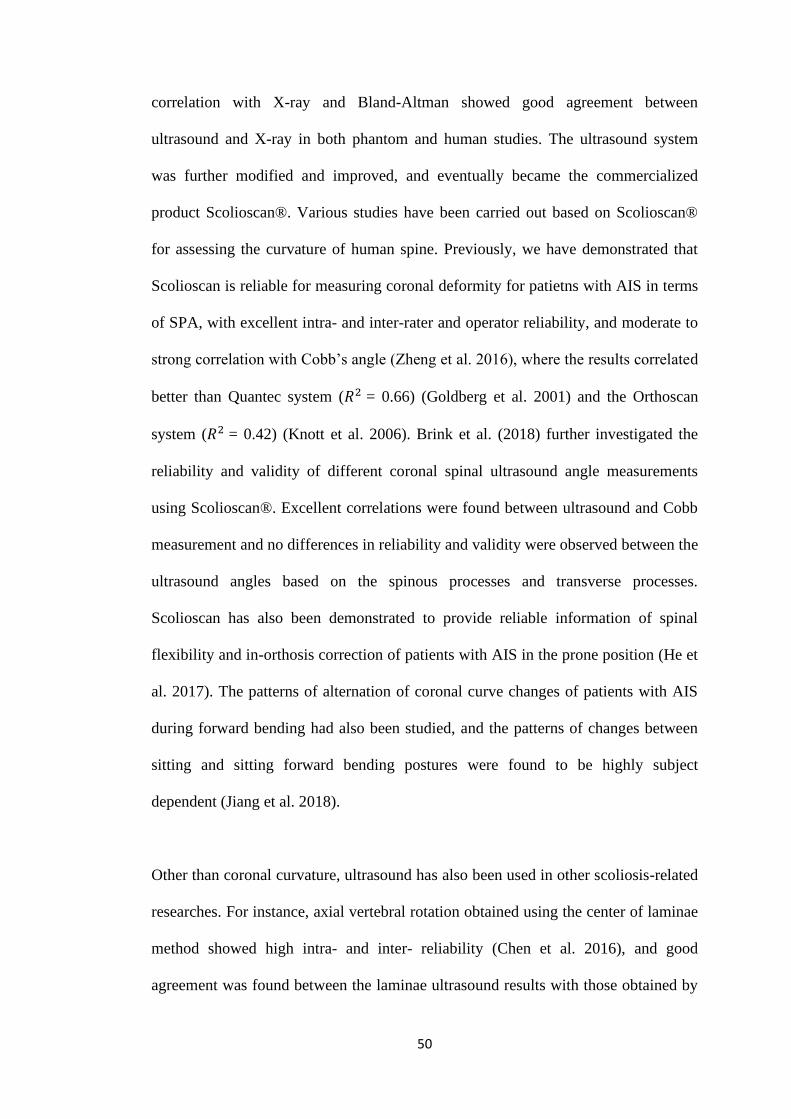
50
correlation with X-ray and Bland-Altman showed good agreement between
ultrasound and X-ray in both phantom and human studies. The ultrasound system
was further modified and improved, and eventually became the commercialized
product Scolioscan®. Various studies have been carried out based on Scolioscan®
for assessing the curvature of human spine. Previously, we have demonstrated that
Scolioscan is reliable for measuring coronal deformity for patietns with AIS in terms
of SPA, with excellent intra- and inter-rater and operator reliability, and moderate to
strong correlation with Cobb’s angle (Zheng et al. 2016), where the results correlated
better than Quantec system (𝑅2 = 0.66) (Goldberg et al. 2001) and the Orthoscan
system (𝑅2 = 0.42) (Knott et al. 2006). Brink et al. (2018) further investigated the
reliability and validity of different coronal spinal ultrasound angle measurements
using Scolioscan®. Excellent correlations were found between ultrasound and Cobb
measurement and no differences in reliability and validity were observed between the
ultrasound angles based on the spinous processes and transverse processes.
Scolioscan has also been demonstrated to provide reliable information of spinal
flexibility and in-orthosis correction of patients with AIS in the prone position (He et
al. 2017). The patterns of alternation of coronal curve changes of patients with AIS
during forward bending had also been studied, and the patterns of changes between
sitting and sitting forward bending postures were found to be highly subject
dependent (Jiang et al. 2018).
Other than coronal curvature, ultrasound has also been used in other scoliosis-related
researches. For instance, axial vertebral rotation obtained using the center of laminae
method showed high intra- and inter- reliability (Chen et al. 2016), and good
agreement was found between the laminae ultrasound results with those obtained by
51
the Aaro-Dahlborn method in the magnetic resonance images (Wang et al. 2016).
Ultrasonographic evaluation of the Risser sign in patients with AIS was investigated
and found to be a reliable method (Thaler et al. 2008). Torlak et al. (2012) further
demonstrated that ultrasound could provide Risser sign evaluation with good intra-
and inter-rater agreement. Lam et al. evaluated the use of quantitative ultrasound for
predicting curve progression (Lam et al. 2011) and determining bone quality (Lam et
al. 2013) in patients with AIS. Bone mass density at the femoral head and bone
quality in terms of broadband ultrasound attenuation, velocity of sound and stiffness
index were found to be significantly lower (P < 0.05) in patients with AIS than
normal subjects (Lam et al. 2011). Stiffness index was further demonstrated as an
independent and significant prognostic factor for AIS and could be considered in
addition to other prognostic factors when estimating the risk for curve progression
and planning treatment for patients with AIS (Lam et al. 2013).
2.4. Summary
This chapter gives a review of the etiology, diagnosis and treatment for patients with
AIS. The human spine is a relatively complex and articulated anatomical structure,
with an infinite range of natural biological variability. Though AIS is a 3D spinal
deformity, most studies mainly focused on the correction of coronal plane and
vertebrae rotation, and the criteria to define optimal sagittal profile were
insufficiently investigated. Sagittal Profiles between normal individuals and patients
with AIS and other diseases were discussed. In addition, the importance of obtaining
optimal sagittal profile was indicated. Potential factors which would cause the
disturbance of the sagittal profile of the spine were also discussed and evaluated in
detail. Among various sagittal parameters, hypokyphosis has been commonly
52
observed in patients with AIS who possess thoracic scoliosis. Cobb’s method and
different evaluation approaches on sagittal profile of spine had been discussed for
giving an overall picture of the effects of different measurements on the spine.
Existing problems of radiograph and the advanced biplanar radiographic EOS have
been extensively reviewed. Apart from using radiograph, non-radiation approaches
using other imaging modalities, optical systems and other topographic methods have
also been reviewed for mapping torso surface and locating vertebrae features to
indirectly calculate the spinal sagittal curvature. This leads to the overall objective of
this study, which is to investigate the reliability and validity of using 3D ultrasound
to accurately measure spinal sagittal curvature for patients with AIS, and to provide
optimal criteria for the sagittal profile of these patients by using ultrasound in order
to provide mass screening and longitudinal follow-up during treatments without any
hazard of radiation.
53
Chapter 3. Methods
3.1. Spine Phantom Study for Sagittal Curvature of Spine
3.1.1. Ultrasound System
The 3D ultrasound imaging of spine was achieved using an ultrasound scanner
(EUB-8500, Hitachi Medical Corporation, Tokyo, Japan) with a linear probe
(L53L/10-5) with frequency of 5-10 MHz, an electromagnetic spatial sensing system
(MiniBird Ascension Technology Corporation, Burlington, VT, USA) with its sensor
mounted on the probe surface, a desktop PC installed with a video capture card
(NIIMAQ PCI/PXI-1411, National Instruments Corporation, Austin, TX, USA) and
a PC program written using Microsoft Visual Studio 6 with Visual C++ data
collection, image processing, visualization and analysis (Figure 3.1) (Cheung, et al.
2015a, Cheung, et al. 2015b). According to the manufacturer, the positional accuracy,
position resolution, the angular accuracy and angular resolution of the
electromagnetic spatial sensor in terms of root-mean-square were 1.8 mm, 0.5 mm,
0.5 degree and 0.1 degree, respectively.
3.1.2. Spine Phantoms
Four flexible spinal column phantoms featured with soft intervertebral discs allowing
deformation (VB84, 3B Scientific, Germany) were used in this study (Figure 3.2).
Each spine phantom was scanned using a water-tank scanning approach (Figure 3.1).
Plastic frames made of acrylic plates and nylon screws were fabricated for the four
phantoms and spatial sensor transmitter to avoid any induced motion by the operator
during the scanning process and transportation for X-ray imaging. These spine
phantoms were 105cm in height without any deformity. Each of the phantoms was
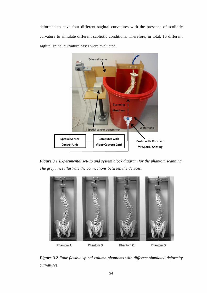
54
deformed to have four different sagittal curvatures with the presence of scoliotic
curvature to simulate different scoliotic conditions. Therefore, in total, 16 different
sagittal spinal curvature cases were evaluated.
Figure 3.1 Experimental set-up and system block diagram for the phantom scanning.
The grey lines illustrate the connections between the devices.
Figure 3.2 Four flexible spinal column phantoms with different simulated deformity
curvatures.
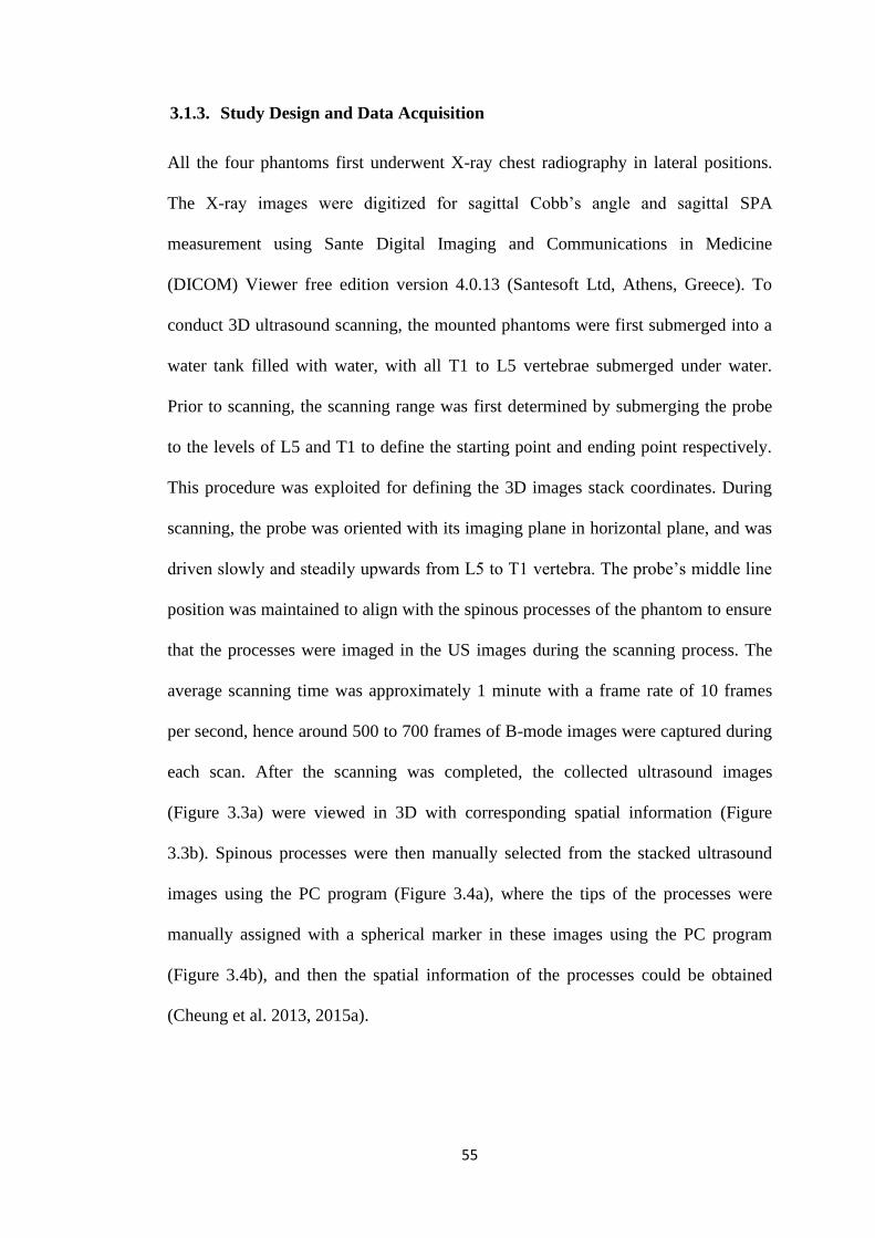
55
3.1.3. Study Design and Data Acquisition
All the four phantoms first underwent X-ray chest radiography in lateral positions.
The X-ray images were digitized for sagittal Cobb’s angle and sagittal SPA
measurement using Sante Digital Imaging and Communications in Medicine
(DICOM) Viewer free edition version 4.0.13 (Santesoft Ltd, Athens, Greece). To
conduct 3D ultrasound scanning, the mounted phantoms were first submerged into a
water tank filled with water, with all T1 to L5 vertebrae submerged under water.
Prior to scanning, the scanning range was first determined by submerging the probe
to the levels of L5 and T1 to define the starting point and ending point respectively.
This procedure was exploited for defining the 3D images stack coordinates. During
scanning, the probe was oriented with its imaging plane in horizontal plane, and was
driven slowly and steadily upwards from L5 to T1 vertebra. The probe’s middle line
position was maintained to align with the spinous processes of the phantom to ensure
that the processes were imaged in the US images during the scanning process. The
average scanning time was approximately 1 minute with a frame rate of 10 frames
per second, hence around 500 to 700 frames of B-mode images were captured during
each scan. After the scanning was completed, the collected ultrasound images
(Figure 3.3a) were viewed in 3D with corresponding spatial information (Figure
3.3b). Spinous processes were then manually selected from the stacked ultrasound
images using the PC program (Figure 3.4a), where the tips of the processes were
manually assigned with a spherical marker in these images using the PC program
(Figure 3.4b), and then the spatial information of the processes could be obtained
(Cheung et al. 2013, 2015a).
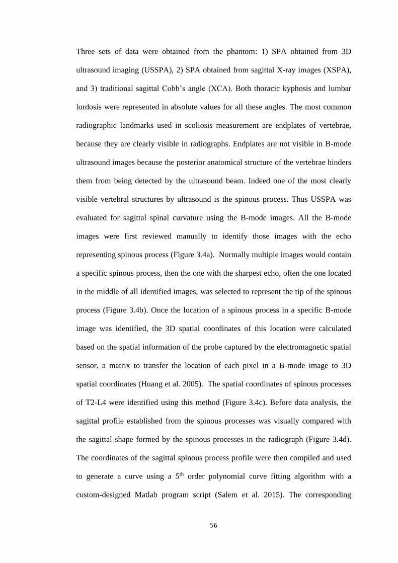
56
Three sets of data were obtained from the phantom: 1) SPA obtained from 3D
ultrasound imaging (USSPA), 2) SPA obtained from sagittal X-ray images (XSPA),
and 3) traditional sagittal Cobb’s angle (XCA). Both thoracic kyphosis and lumbar
lordosis were represented in absolute values for all these angles. The most common
radiographic landmarks used in scoliosis measurement are endplates of vertebrae,
because they are clearly visible in radiographs. Endplates are not visible in B-mode
ultrasound images because the posterior anatomical structure of the vertebrae hinders
them from being detected by the ultrasound beam. Indeed one of the most clearly
visible vertebral structures by ultrasound is the spinous process. Thus USSPA was
evaluated for sagittal spinal curvature using the B-mode images. All the B-mode
images were first reviewed manually to identify those images with the echo
representing spinous process (Figure 3.4a). Normally multiple images would contain
a specific spinous process, then the one with the sharpest echo, often the one located
in the middle of all identified images, was selected to represent the tip of the spinous
process (Figure 3.4b). Once the location of a spinous process in a specific B-mode
image was identified, the 3D spatial coordinates of this location were calculated
based on the spatial information of the probe captured by the electromagnetic spatial
sensor, a matrix to transfer the location of each pixel in a B-mode image to 3D
spatial coordinates (Huang et al. 2005). The spatial coordinates of spinous processes
of T2-L4 were identified using this method (Figure 3.4c). Before data analysis, the
sagittal profile established from the spinous processes was visually compared with
the sagittal shape formed by the spinous processes in the radiograph (Figure 3.4d).
The coordinates of the sagittal spinous process profile were then compiled and used
to generate a curve using a 5th order polynomial curve fitting algorithm with a
custom-designed Matlab program script (Salem et al. 2015). The corresponding
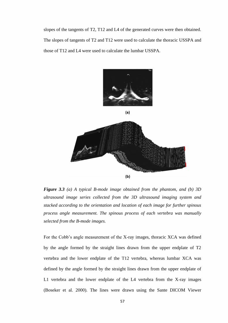
57
slopes of the tangents of T2, T12 and L4 of the generated curves were then obtained.
The slopes of tangents of T2 and T12 were used to calculate the thoracic USSPA and
those of T12 and L4 were used to calculate the lumbar USSPA.
Figure 3.3 (a) A typical B-mode image obtained from the phantom, and (b) 3D
ultrasound image series collected from the 3D ultrasound imaging system and
stacked according to the orientation and location of each image for further spinous
process angle measurement. The spinous process of each vertebra was manually
selected from the B-mode images.
For the Cobb’s angle measurement of the X-ray images, thoracic XCA was defined
by the angle formed by the straight lines drawn from the upper endplate of T2
vertebra and the lower endplate of the T12 vertebra, whereas lumbar XCA was
defined by the angle formed by the straight lines drawn from the upper endplate of
L1 vertebra and the lower endplate of the L4 vertebra from the X-ray images
(Boseker et al. 2000). The lines were drawn using the Sante DICOM Viewer
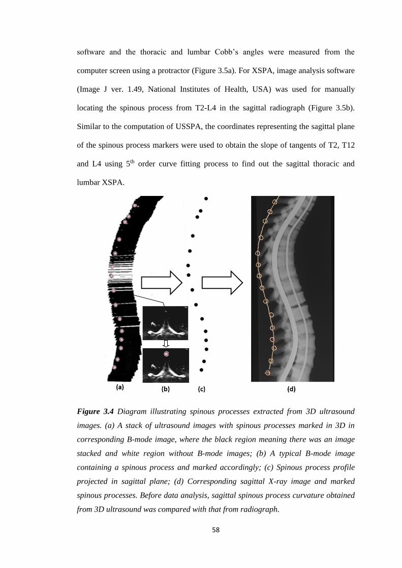
58
software and the thoracic and lumbar Cobb’s angles were measured from the
computer screen using a protractor (Figure 3.5a). For XSPA, image analysis software
(Image J ver. 1.49, National Institutes of Health, USA) was used for manually
locating the spinous process from T2-L4 in the sagittal radiograph (Figure 3.5b).
Similar to the computation of USSPA, the coordinates representing the sagittal plane
of the spinous process markers were used to obtain the slope of tangents of T2, T12
and L4 using 5th order curve fitting process to find out the sagittal thoracic and
lumbar XSPA.
Figure 3.4 Diagram illustrating spinous processes extracted from 3D ultrasound
images. (a) A stack of ultrasound images with spinous processes marked in 3D in
corresponding B-mode image, where the black region meaning there was an image
stacked and white region without B-mode images; (b) A typical B-mode image
containing a spinous process and marked accordingly; (c) Spinous process profile
projected in sagittal plane; (d) Corresponding sagittal X-ray image and marked
spinous processes. Before data analysis, sagittal spinous process curvature obtained
from 3D ultrasound was compared with that from radiograph.
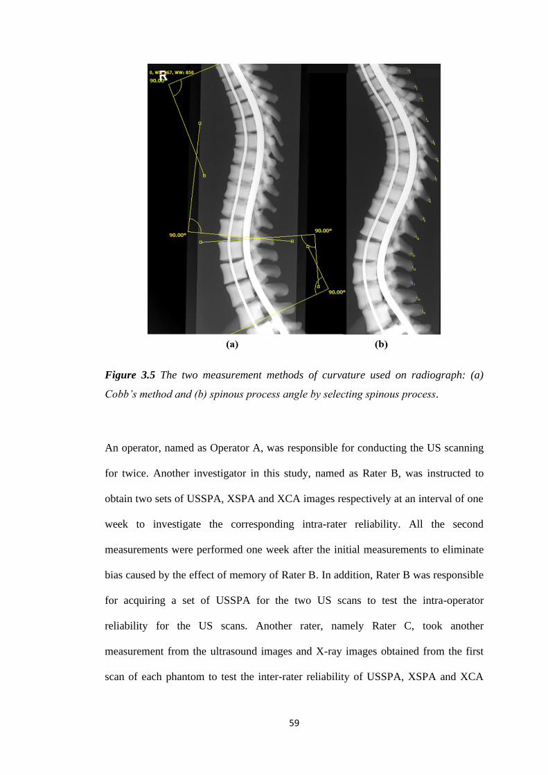
59
Figure 3.5 The two measurement methods of curvature used on radiograph: (a)
Cobb’s method and (b) spinous process angle by selecting spinous process.
An operator, named as Operator A, was responsible for conducting the US scanning
for twice. Another investigator in this study, named as Rater B, was instructed to
obtain two sets of USSPA, XSPA and XCA images respectively at an interval of one
week to investigate the corresponding intra-rater reliability. All the second
measurements were performed one week after the initial measurements to eliminate
bias caused by the effect of memory of Rater B. In addition, Rater B was responsible
for acquiring a set of USSPA for the two US scans to test the intra-operator
reliability for the US scans. Another rater, namely Rater C, took another
measurement from the ultrasound images and X-ray images obtained from the first
scan of each phantom to test the inter-rater reliability of USSPA, XSPA and XCA
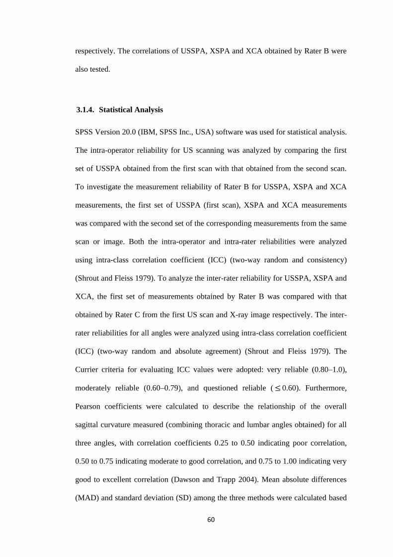
60
respectively. The correlations of USSPA, XSPA and XCA obtained by Rater B were
also tested.
3.1.4. Statistical Analysis
SPSS Version 20.0 (IBM, SPSS Inc., USA) software was used for statistical analysis.
The intra-operator reliability for US scanning was analyzed by comparing the first
set of USSPA obtained from the first scan with that obtained from the second scan.
To investigate the measurement reliability of Rater B for USSPA, XSPA and XCA
measurements, the first set of USSPA (first scan), XSPA and XCA measurements
was compared with the second set of the corresponding measurements from the same
scan or image. Both the intra-operator and intra-rater reliabilities were analyzed
using intra-class correlation coefficient (ICC) (two-way random and consistency)
(Shrout and Fleiss 1979). To analyze the inter-rater reliability for USSPA, XSPA and
XCA, the first set of measurements obtained by Rater B was compared with that
obtained by Rater C from the first US scan and X-ray image respectively. The inter-
rater reliabilities for all angles were analyzed using intra-class correlation coefficient
(ICC) (two-way random and absolute agreement) (Shrout and Fleiss 1979). The
Currier criteria for evaluating ICC values were adopted: very reliable (0.80–1.0),
moderately reliable (0.60–0.79), and questioned reliable ( ≤ 0.60). Furthermore,
Pearson coefficients were calculated to describe the relationship of the overall
sagittal curvature measured (combining thoracic and lumbar angles obtained) for all
three angles, with correlation coefficients 0.25 to 0.50 indicating poor correlation,
0.50 to 0.75 indicating moderate to good correlation, and 0.75 to 1.00 indicating very
good to excellent correlation (Dawson and Trapp 2004). Mean absolute differences
(MAD) and standard deviation (SD) among the three methods were calculated based
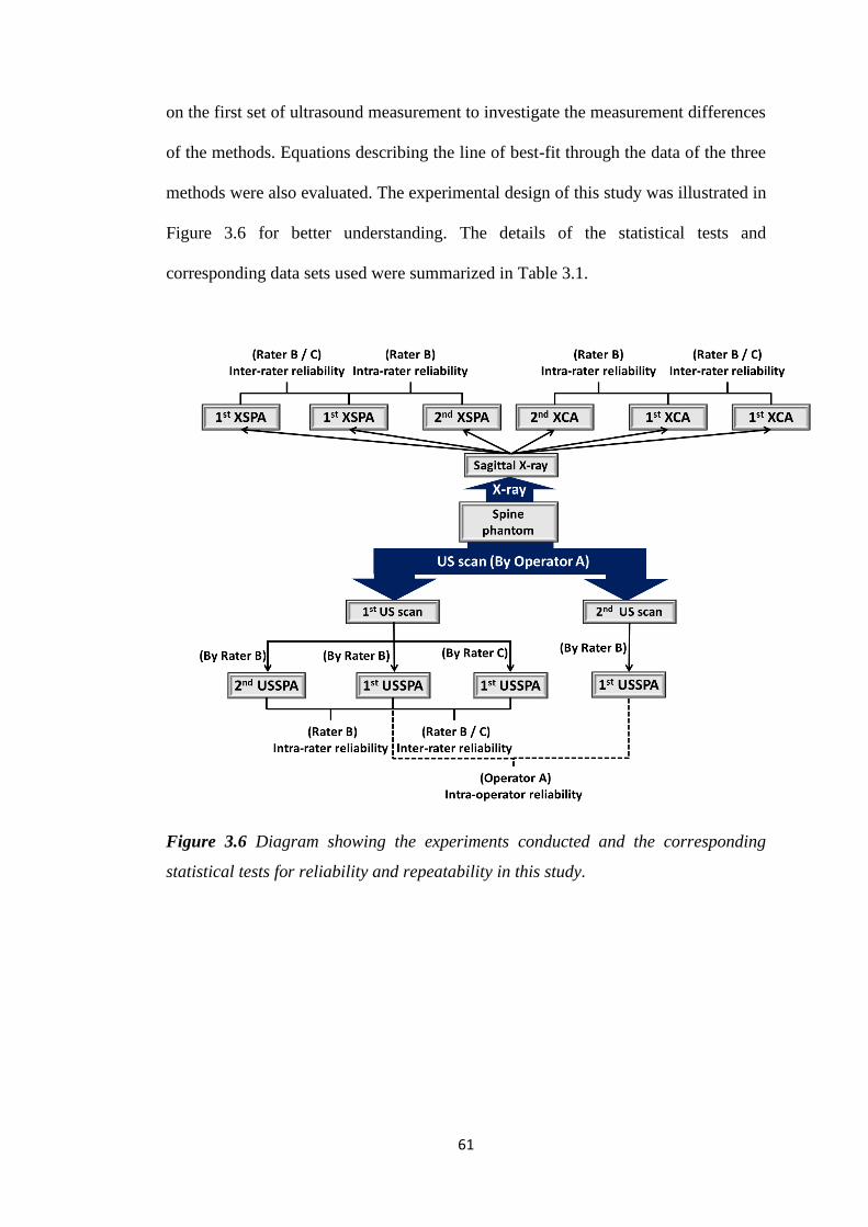
61
on the first set of ultrasound measurement to investigate the measurement differences
of the methods. Equations describing the line of best-fit through the data of the three
methods were also evaluated. The experimental design of this study was illustrated in
Figure 3.6 for better understanding. The details of the statistical tests and
corresponding data sets used were summarized in Table 3.1.
Figure 3.6 Diagram showing the experiments conducted and the corresponding
statistical tests for reliability and repeatability in this study.
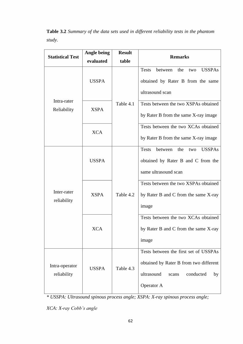
62
Table 3.2 Summary of the data sets used in different reliability tests in the phantom
study.
Statistical Test Angle being
evaluated
Result
table Remarks
Intra-rater
Reliability
USSPA
Table 4.1
Tests between the two USSPAs
obtained by Rater B from the same
ultrasound scan
XSPA
Tests between the two XSPAs obtained
by Rater B from the same X-ray image
XCA
Tests between the two XCAs obtained
by Rater B from the same X-ray image
Inter-rater
reliability
USSPA
Table 4.2
Tests between the two USSPAs
obtained by Rater B and C from the
same ultrasound scan
XSPA
Tests between the two XSPAs obtained
by Rater B and C from the same X-ray
image
XCA
Tests between the two XCAs obtained
by Rater B and C from the same X-ray
image
Intra-operator
reliability
USSPA Table 4.3
Tests between the first set of USSPAs
obtained by Rater B from two different
ultrasound scans conducted by
Operator A
* USSPA: Ultrasound spinous process angle; XSPA: X-ray spinous process angle;
XCA: X-ray Cobb’s angle
63
3.2. Human Subject Study for Sagittal Curvature of Spine
3.2.1. Subjects
Patients diagnosed with AIS were recruited to participate in this test, and all
the patients were recruited from a tertiary scoliosis referral center. Patients were
requested to receive both ultrasound scanning and X-ray imaging on the same day.
This study was approved by the local institutional review board. Signed informed
consents were obtained from all the subjects and guardians of the patients aged
below 18 prior to the start of the study. Patients with metallic implants and BMI
higher than 25.0 kg/m2 were excluded, as metallic implants would affect the spatial
sensing accuracy of the ultrasound probe and high BMI would likely lead to poor
image quality in the lumbar region. Other exclusion criteria included: 1) Patients who
underwent previous spine surgery; 2) Patients who were wearing a brace during x-
ray, and 3) Patients who were allergic to ultrasound gel. In this study we aimed to
investigate the reliability and validity of the ultrasound methods for sagittal
deformity measurement, thus both USSPA and USLA for all the 21 AIS patients
included in this study were obtained from their corresponding sagittal ultrasound
images. This number of patients fulfilled the simple approximation that allows the
calculation of required sample size of subjects K when the number of replicates n
(which was two in this study) is fixed, by setting 𝛼 set to 0.05 and 𝛽 (type II error)
set to 0.2, while the acceptable and preferred reliability was set as 0.8 and 0.9
respectively (Walter et al.1998).
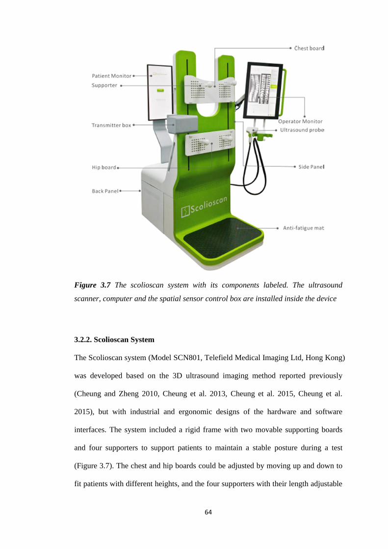
64
Figure 3.7 The scolioscan system with its components labeled. The ultrasound
scanner, computer and the spatial sensor control box are installed inside the device
3.2.2. Scolioscan System
The Scolioscan system (Model SCN801, Telefield Medical Imaging Ltd, Hong Kong)
was developed based on the 3D ultrasound imaging method reported previously
(Cheung and Zheng 2010, Cheung et al. 2013, Cheung et al. 2015, Cheung et al.
2015), but with industrial and ergonomic designs of the hardware and software
interfaces. The system included a rigid frame with two movable supporting boards
and four supporters to support patients to maintain a stable posture during a test
(Figure 3.7). The chest and hip boards could be adjusted by moving up and down to
fit patients with different heights, and the four supporters with their length adjustable
65
can be fixed on the boards by inserting to the fixation holes and locked by rotating
the supporter by 90°. The locations of boards in vertical direction, the positions of
supporters along vertical and horizontal directions, as well as the lengths of
supporters could be recorded, and the information could be used in follow-up
assessments for the same patient. The 3D ultrasound imaging of the spine was
achieved through freehand scanning of the ultrasound probe (frequency of 7.5 MHz,
width of 7.5 cm), inside which an electromagnetic spatial sensor was installed to
detect the position and orientation of the probe. The electromagnetic transmitter was
located inside the transmitter box as indicated in Figure 3.7. The other screen on the
back was used to provide information for patients, including a green eye-spot with
location set according to the height of the patient to facilitate him/her to keep a stable
head and neck posture during scanning. This screen also showed additional
information including different steps of evaluation procedures, so as to keep the
patient informed of the process, and thus more cooperative.
3.2.3. Testing Protocol
The patient was requested to undress upper garments and shoes before the scanning
session and was provided a back-opening dressing gown for ease of scanning. All
metallic objects, electronics goods, magnets, and other possible ferromagnetic
materials were removed. The patient was asked to stand on the Scolioscan platform
for supporter adjustment and the chest and hip boards were repositioned at his/her
reasonable height. Two supporters on the chest board and hip board were relocated to
align with clavicle anterior concavities and bilateral anterior superior iliac spines
respectively. The lengths of supporter’s shafts on both boards were adjusted until
they came in contact with the patient. The patient was instructed to maintain a natural
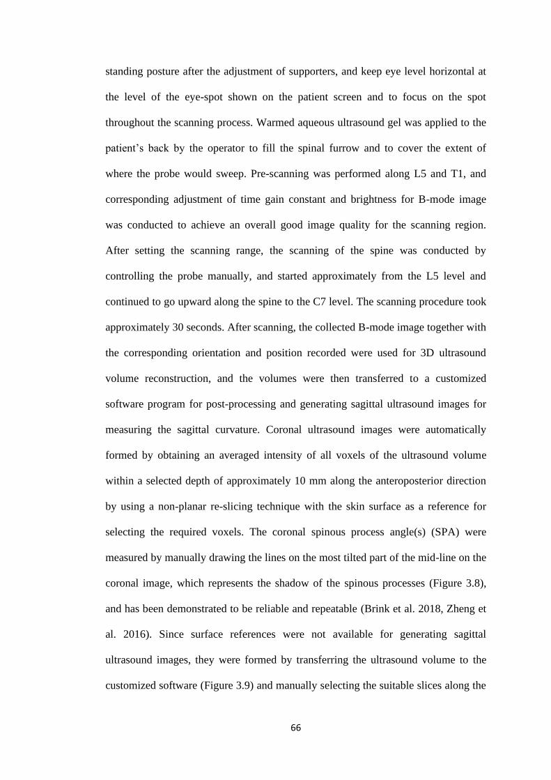
66
standing posture after the adjustment of supporters, and keep eye level horizontal at
the level of the eye-spot shown on the patient screen and to focus on the spot
throughout the scanning process. Warmed aqueous ultrasound gel was applied to the
patient’s back by the operator to fill the spinal furrow and to cover the extent of
where the probe would sweep. Pre-scanning was performed along L5 and T1, and
corresponding adjustment of time gain constant and brightness for B-mode image
was conducted to achieve an overall good image quality for the scanning region.
After setting the scanning range, the scanning of the spine was conducted by
controlling the probe manually, and started approximately from the L5 level and
continued to go upward along the spine to the C7 level. The scanning procedure took
approximately 30 seconds. After scanning, the collected B-mode image together with
the corresponding orientation and position recorded were used for 3D ultrasound
volume reconstruction, and the volumes were then transferred to a customized
software program for post-processing and generating sagittal ultrasound images for
measuring the sagittal curvature. Coronal ultrasound images were automatically
formed by obtaining an averaged intensity of all voxels of the ultrasound volume
within a selected depth of approximately 10 mm along the anteroposterior direction
by using a non-planar re-slicing technique with the skin surface as a reference for
selecting the required voxels. The coronal spinous process angle(s) (SPA) were
measured by manually drawing the lines on the most tilted part of the mid-line on the
coronal image, which represents the shadow of the spinous processes (Figure 3.8),
and has been demonstrated to be reliable and repeatable (Brink et al. 2018, Zheng et
al. 2016). Since surface references were not available for generating sagittal
ultrasound images, they were formed by transferring the ultrasound volume to the
customized software (Figure 3.9) and manually selecting the suitable slices along the
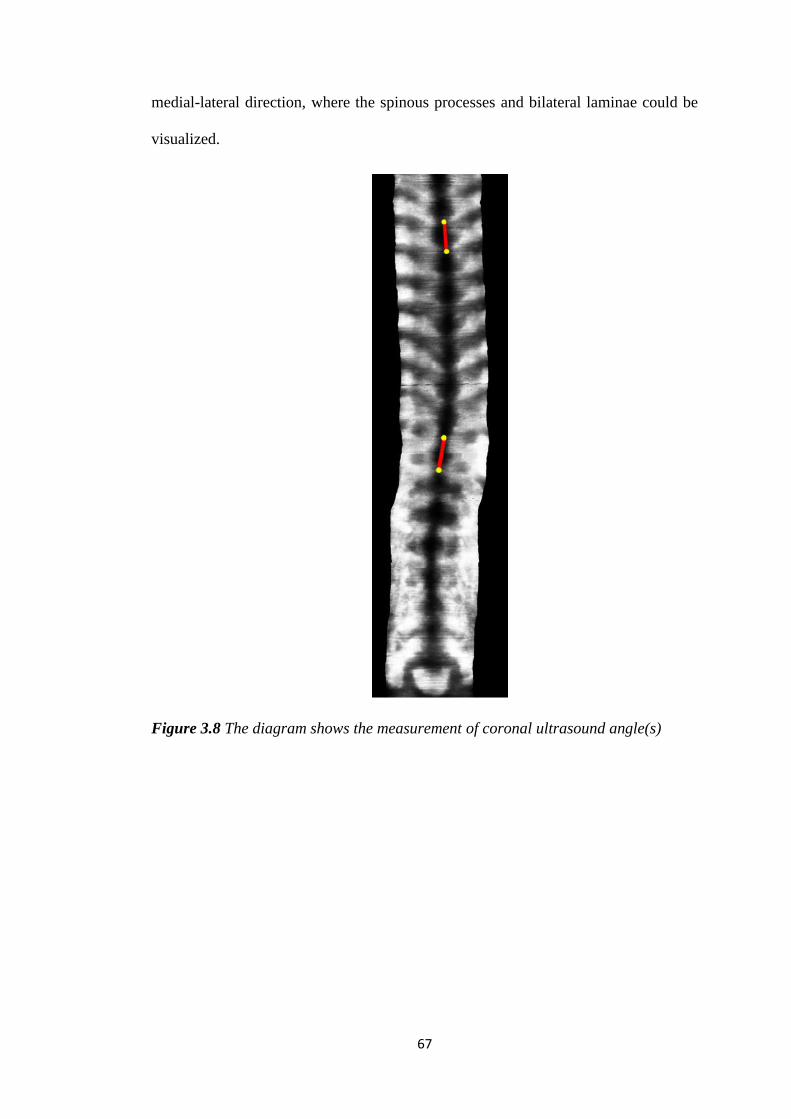
67
medial-lateral direction, where the spinous processes and bilateral laminae could be
visualized.
Figure 3.8 The diagram shows the measurement of coronal ultrasound angle(s)
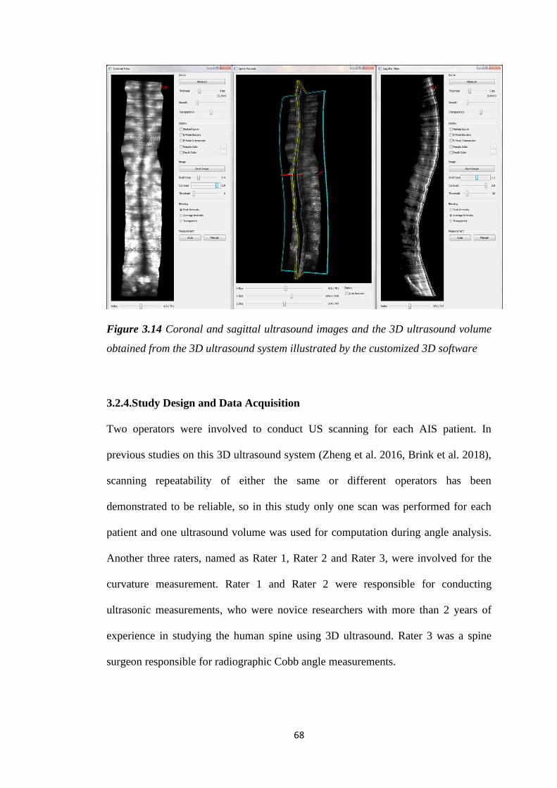
68
Figure 3.14 Coronal and sagittal ultrasound images and the 3D ultrasound volume
obtained from the 3D ultrasound system illustrated by the customized 3D software
3.2.4.Study Design and Data Acquisition
Two operators were involved to conduct US scanning for each AIS patient. In
previous studies on this 3D ultrasound system (Zheng et al. 2016, Brink et al. 2018),
scanning repeatability of either the same or different operators has been
demonstrated to be reliable, so in this study only one scan was performed for each
patient and one ultrasound volume was used for computation during angle analysis.
Another three raters, named as Rater 1, Rater 2 and Rater 3, were involved for the
curvature measurement. Rater 1 and Rater 2 were responsible for conducting
ultrasonic measurements, who were novice researchers with more than 2 years of
experience in studying the human spine using 3D ultrasound. Rater 3 was a spine
surgeon responsible for radiographic Cobb angle measurements.
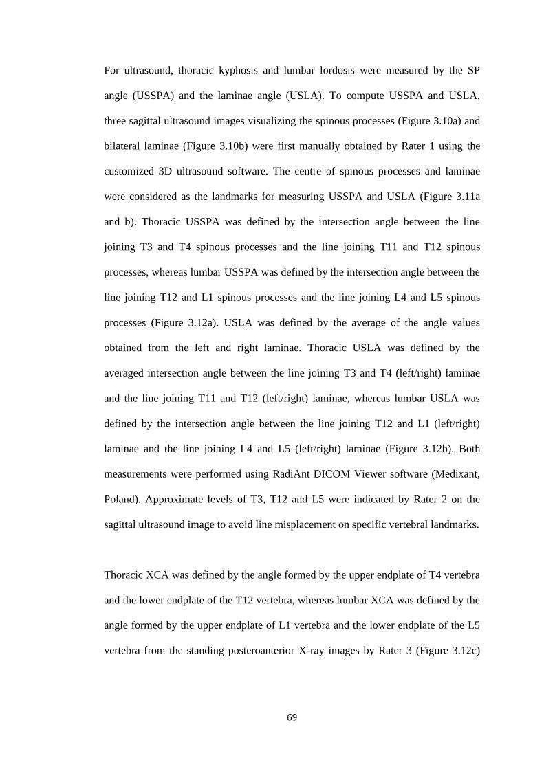
69
For ultrasound, thoracic kyphosis and lumbar lordosis were measured by the SP
angle (USSPA) and the laminae angle (USLA). To compute USSPA and USLA,
three sagittal ultrasound images visualizing the spinous processes (Figure 3.10a) and
bilateral laminae (Figure 3.10b) were first manually obtained by Rater 1 using the
customized 3D ultrasound software. The centre of spinous processes and laminae
were considered as the landmarks for measuring USSPA and USLA (Figure 3.11a
and b). Thoracic USSPA was defined by the intersection angle between the line
joining T3 and T4 spinous processes and the line joining T11 and T12 spinous
processes, whereas lumbar USSPA was defined by the intersection angle between the
line joining T12 and L1 spinous processes and the line joining L4 and L5 spinous
processes (Figure 3.12a). USLA was defined by the average of the angle values
obtained from the left and right laminae. Thoracic USLA was defined by the
averaged intersection angle between the line joining T3 and T4 (left/right) laminae
and the line joining T11 and T12 (left/right) laminae, whereas lumbar USLA was
defined by the intersection angle between the line joining T12 and L1 (left/right)
laminae and the line joining L4 and L5 (left/right) laminae (Figure 3.12b). Both
measurements were performed using RadiAnt DICOM Viewer software (Medixant,
Poland). Approximate levels of T3, T12 and L5 were indicated by Rater 2 on the
sagittal ultrasound image to avoid line misplacement on specific vertebral landmarks.
Thoracic XCA was defined by the angle formed by the upper endplate of T4 vertebra
and the lower endplate of the T12 vertebra, whereas lumbar XCA was defined by the
angle formed by the upper endplate of L1 vertebra and the lower endplate of the L5
vertebra from the standing posteroanterior X-ray images by Rater 3 (Figure 3.12c)
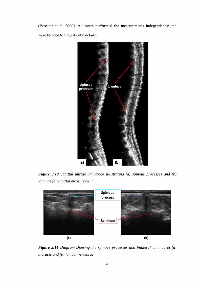
70
(Boseker et al. 2000). All raters performed the measurements independently and
were blinded to the patients’ details.
Figure 3.10 Sagittal ultrasound image illustrating (a) spinous processes and (b)
laminae for sagittal measurement.
Figure 3.11 Diagram showing the spinous processes and bilateral laminae of (a)
thoracic and (b) lumbar vertebrae.
71
3.2.5.Statistical Analysis
Data was reported as mean ± standard deviation (SD). Statistical analysis was
conducted using SPSS Version 20.0 (IBM, SPSS Inc., USA) software. Intra-class
coefficient (ICC) (two-way random and consistency model) was calculated to
evaluate the intra-rater reliability of USSPA and USLA of the two raters (Shrout and
Fleiss 1979), by comparing their two measurements made on the same customized
sagittal US image individually. The first measurement results obtained by Rater 1
and Rater 2 were compared using ICC (two-wayrandom and absolute agreement
model) to determine inter-rater reliability (Shrout and Fleiss 1979). The Currier
criteria for evaluating ICC values were adopted: very reliable (0.80–1.0), moderately
reliable (0.60–0.79), and questioned reliable (≤0.60) (Currier 1984). Ultrasound
measurements were compared with XCA respectively using linear correlation for
thoracic curves and thoracic curves. Linear regression equations with intersections
were analyzed with correlation coefficients 0.25 to 0.50 indicating poor correlation,
0.50 to 0.75 indicating moderate to good correlation, and 0.75 to 1.00 indicating very
good to excellent correlation (Dawson and Trapp 2004). Bland-Altman method was
used to test the agreement between XCA and the adjusted ultrasound angles. To
measure the differences in agreement for USSPA and USLA, the mean absolute
differences (MAD) between XCA and the adjusted ultrasound angles were calculated
and compared using paired t-tests. The significance level was set at 0.05.
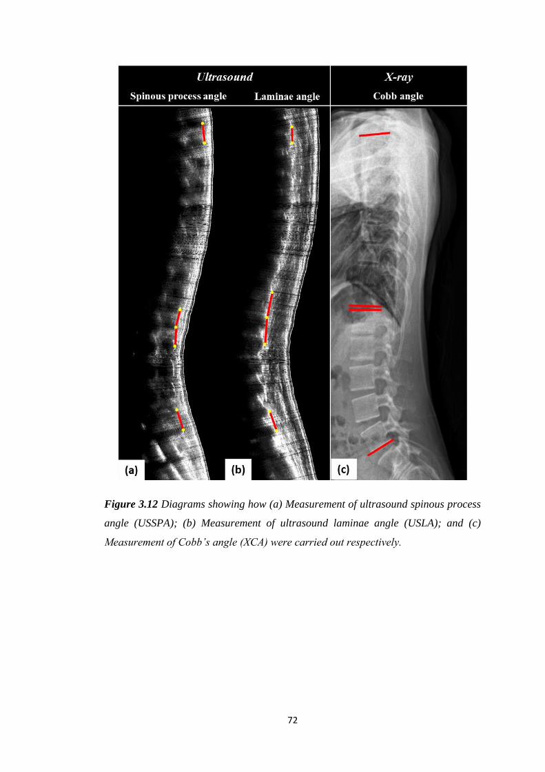
72
Figure 3.12 Diagrams showing how (a) Measurement of ultrasound spinous process
angle (USSPA); (b) Measurement of ultrasound laminae angle (USLA); and (c)
Measurement of Cobb’s angle (XCA) were carried out respectively.
73
3.3. Human Subject Study for Coronal-Sagittal Coupling
3.3.1.Subjects
Total 300 AIS patients diagnosed with scoliosis and scanned by radiographs were
invited for this study. 150 subjects were allocated into the first stage of the study,
whereas the second 150 subjects were allocated into the second stage (See details in
Section 3.3.2). All these patients were recruited consecutively in the Department of
Orthopaedics and Traumatology of The Chinese University of Hong Kong. The
study got human subject ethical approvals from both The Hong Kong Polytechnic
University (No. 20070321001) and The Chinese University of Hong Kong (No.
2009.622). Informed consent was obtained from all patients (or their parents for
those under 18 years of age). The patients received standing plain radiographs within
three months before the Scolioscan assessment, and their Cobb angles were
measured. Patients with metallic implants and BMI higher than 25.0 kg/m2 were
excluded, as metallic implants may potentially affect the accuracy of ultrasound
probe spatial sensing and high BMI may lead to poor image quality in the lumbar
region using the current probe.
To study the coupling effect between the coronal and sagittal curves, 115 AIS
patients with Cobb < 40° participated in the exploratory stage using the following
exclusion criteria: 1) Cobb angle > 40° (N = 25); 2) Special cases: Appeared to be
kyphotic in lumbar region in US (N = 3); 3) T3 level was not captured in the
ultrasound scans (N = 3); 4) Poor scanning quality in the thoracic region due to the
hindrance of the scapula (N = 3); 5) Sagittal X-ray was not available (N = 1). For the
validation session, exclusions of patients were made due to: 1) Poor quality due to
large rotation (N = 10), possibly caused by large Cobb angle; 2) Special cases:
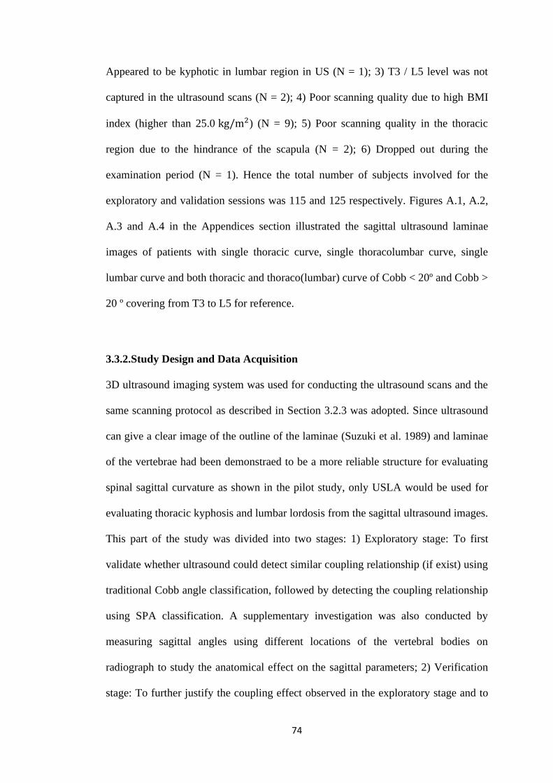
74
Appeared to be kyphotic in lumbar region in US (N = 1); 3) T3 / L5 level was not
captured in the ultrasound scans (N = 2); 4) Poor scanning quality due to high BMI
index (higher than 25.0 kg/m2) (N = 9); 5) Poor scanning quality in the thoracic
region due to the hindrance of the scapula (N = 2); 6) Dropped out during the
examination period (N = 1). Hence the total number of subjects involved for the
exploratory and validation sessions was 115 and 125 respectively. Figures A.1, A.2,
A.3 and A.4 in the Appendices section illustrated the sagittal ultrasound laminae
images of patients with single thoracic curve, single thoracolumbar curve, single
lumbar curve and both thoracic and thoraco(lumbar) curve of Cobb < 20º and Cobb >
20 º covering from T3 to L5 for reference.
3.3.2.Study Design and Data Acquisition
3D ultrasound imaging system was used for conducting the ultrasound scans and the
same scanning protocol as described in Section 3.2.3 was adopted. Since ultrasound
can give a clear image of the outline of the laminae (Suzuki et al. 1989) and laminae
of the vertebrae had been demonstraed to be a more reliable structure for evaluating
spinal sagittal curvature as shown in the pilot study, only USLA would be used for
evaluating thoracic kyphosis and lumbar lordosis from the sagittal ultrasound images.
This part of the study was divided into two stages: 1) Exploratory stage: To first
validate whether ultrasound could detect similar coupling relationship (if exist) using
traditional Cobb angle classification, followed by detecting the coupling relationship
using SPA classification. A supplementary investigation was also conducted by
measuring sagittal angles using different locations of the vertebral bodies on
radiograph to study the anatomical effect on the sagittal parameters; 2) Verification
stage: To further justify the coupling effect observed in the exploratory stage and to
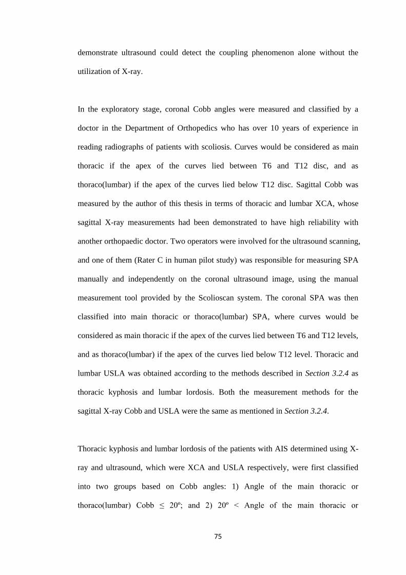
75
demonstrate ultrasound could detect the coupling phenomenon alone without the
utilization of X-ray.
In the exploratory stage, coronal Cobb angles were measured and classified by a
doctor in the Department of Orthopedics who has over 10 years of experience in
reading radiographs of patients with scoliosis. Curves would be considered as main
thoracic if the apex of the curves lied between T6 and T12 disc, and as
thoraco(lumbar) if the apex of the curves lied below T12 disc. Sagittal Cobb was
measured by the author of this thesis in terms of thoracic and lumbar XCA, whose
sagittal X-ray measurements had been demonstrated to have high reliability with
another orthopaedic doctor. Two operators were involved for the ultrasound scanning,
and one of them (Rater C in human pilot study) was responsible for measuring SPA
manually and independently on the coronal ultrasound image, using the manual
measurement tool provided by the Scolioscan system. The coronal SPA was then
classified into main thoracic or thoraco(lumbar) SPA, where curves would be
considered as main thoracic if the apex of the curves lied between T6 and T12 levels,
and as thoraco(lumbar) if the apex of the curves lied below T12 level. Thoracic and
lumbar USLA was obtained according to the methods described in Section 3.2.4 as
thoracic kyphosis and lumbar lordosis. Both the measurement methods for the
sagittal X-ray Cobb and USLA were the same as mentioned in Section 3.2.4.
Thoracic kyphosis and lumbar lordosis of the patients with AIS determined using X-
ray and ultrasound, which were XCA and USLA respectively, were first classified
into two groups based on Cobb angles: 1) Angle of the main thoracic or
thoraco(lumbar) Cobb ≤ 20º; and 2) 20º < Angle of the main thoracic or
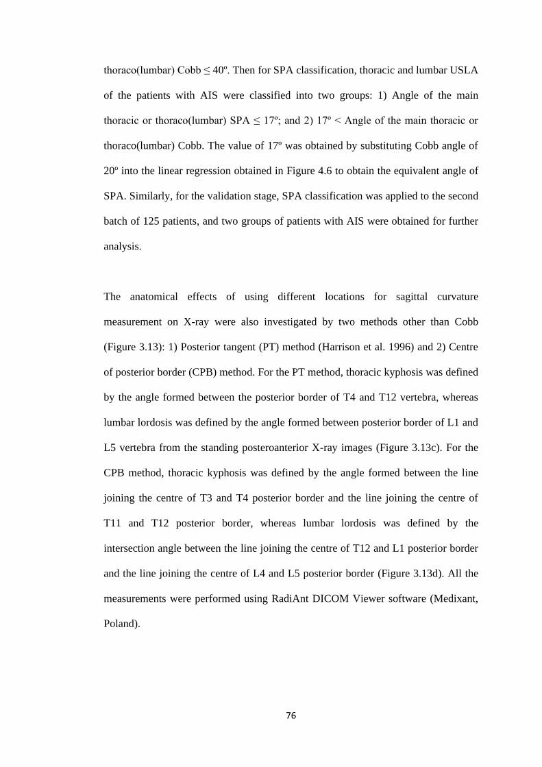
76
thoraco(lumbar) Cobb ≤ 40º. Then for SPA classification, thoracic and lumbar USLA
of the patients with AIS were classified into two groups: 1) Angle of the main
thoracic or thoraco(lumbar) SPA ≤ 17º; and 2) 17º < Angle of the main thoracic or
thoraco(lumbar) Cobb. The value of 17º was obtained by substituting Cobb angle of
20º into the linear regression obtained in Figure 4.6 to obtain the equivalent angle of
SPA. Similarly, for the validation stage, SPA classification was applied to the second
batch of 125 patients, and two groups of patients with AIS were obtained for further
analysis.
The anatomical effects of using different locations for sagittal curvature
measurement on X-ray were also investigated by two methods other than Cobb
(Figure 3.13): 1) Posterior tangent (PT) method (Harrison et al. 1996) and 2) Centre
of posterior border (CPB) method. For the PT method, thoracic kyphosis was defined
by the angle formed between the posterior border of T4 and T12 vertebra, whereas
lumbar lordosis was defined by the angle formed between posterior border of L1 and
L5 vertebra from the standing posteroanterior X-ray images (Figure 3.13c). For the
CPB method, thoracic kyphosis was defined by the angle formed between the line
joining the centre of T3 and T4 posterior border and the line joining the centre of
T11 and T12 posterior border, whereas lumbar lordosis was defined by the
intersection angle between the line joining the centre of T12 and L1 posterior border
and the line joining the centre of L4 and L5 posterior border (Figure 3.13d). All the
measurements were performed using RadiAnt DICOM Viewer software (Medixant,
Poland).
77
3.3.3.Statistical Analysis
Sagittal data was reported as mean ± standard deviation (SD). Statistical analysis was
conducted using SPSS Version 20.0 (IBM, SPSS Inc., USA). In the exploratory stage,
sagittal ultrasound angles were compared with those obtained from X-ray
respectively using linear correlation for corresponding Cobb ≤ 20º, 20º < Cobb ≤ 40º
and all cases combined. Independent t-tests were used: 1) To compare thoracic
kyphosis and lumbar lordosis of X-ray and ultrasound of the patients based on
coronal Cobb values (Cobb ≤ 20º and 20º < Cobb ≤ 40º); and 2) To compare thoracic
kyphosis and lumbar lordosis of ultrasound based on SPA values (SPA ≤ 17º and 17º
< SPA). Pearson correlations between the corresponding coronal and sagittal
measurements of the two imaging modalities were also evaluated to describe the
relationship between the spinal parameters in the two planes. Similarly in the
validation stage, independent t-tests were used to compare thoracic kyphosis and
lumbar lordosis based on SPA values (SPA ≤ 17º and 17º < SPA) on the second
batch of patients, and finally on all the patients involved in both exploratory and
validation sessions. Paired t-tests were used to evaluate the difference in spinal
sagittal measurements between ultrasound and the three X-ray methods (Figure 3.13).
The significance level was set at 0.05.
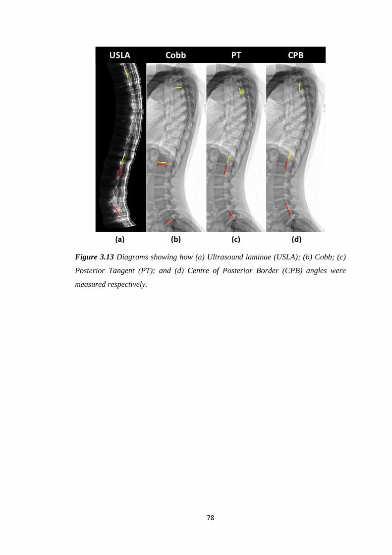
78
Figure 3.13 Diagrams showing how (a) Ultrasound laminae (USLA); (b) Cobb; (c)
Posterior Tangent (PT); and (d) Centre of Posterior Border (CPB) angles were
measured respectively.
79
Chapter 4. Results
4.1. Results of Spine Phantom Study
Though 3D coordinates of the spinous processes and coronal plane of the X-ray
images were acquired, only the sagittal curvatures of the spine phantoms were
analyzed and compared for USSPA, XSPA and XCA since validation of our
proposed ultrasound method on sagittal spinal analysis was the focus of this study.
The average sagittal curvatures and ranges of the phantoms measured for the three
angles by Rater B were: USSPA: 25.6 ± 12.3 degrees (5.5 to 36.9 degrees), 26.5 ±
9.9 degrees (7.6 to 41.3 degrees); XSPA: 23.9 ± 9.7 degrees (4.0 to 36.9 degrees),
25.7 ± 8.6 degrees (11.1 to 39.5 degrees); and XCA: 30.5 ± 8.9 degrees (19.0 to 46.0
degrees), 28.9 ± 5.0 degrees (21.0 to 36.0 degrees) for the thoracic region and lumbar
region, respectively.
Raters demonstrated excellent intra-rater and inter-rater reproducibility for USSPA,
XSPA and XCA. For intra-rater reliability, the ICC ranged from 0.97 to 0.99 for the
angle measured in the thoracic region and from 0.91 to 0.99 for the angle measured
in the lumbar region among the three angles (Table 4.1). For inter-rater reliability,
the ICC ranged from 0.93 to 0.99 and from 0.86 to 0.98 for the thoracic and lumbar
regions, respectively (Table 4.2). In addition, scanning skill for Operator A was
found to be very reliable since the ICC values were greater than 0.9 for the results
obtained for both the regions (Table 4.3).
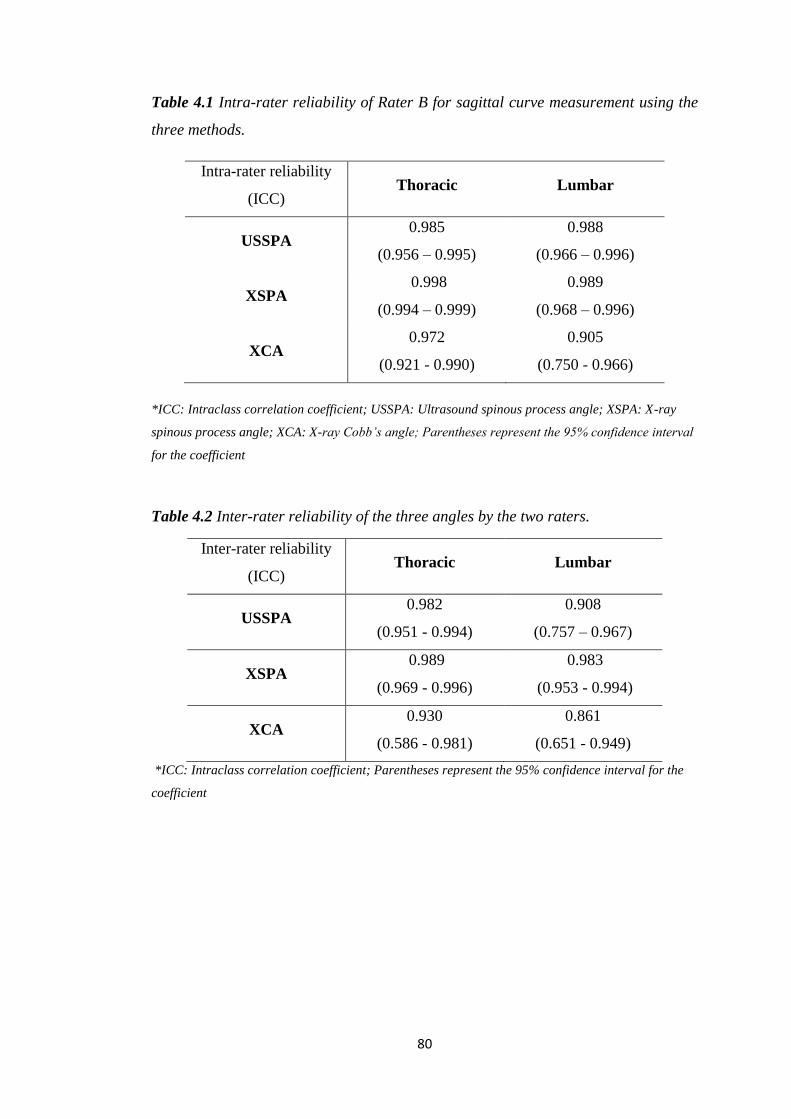
80
Table 4.1 Intra-rater reliability of Rater B for sagittal curve measurement using the
three methods.
Intra-rater reliability
(ICC) Thoracic Lumbar
USSPA 0.985
(0.956 – 0.995)
0.988
(0.966 – 0.996)
XSPA 0.998
(0.994 – 0.999)
0.989
(0.968 – 0.996)
XCA 0.972
(0.921 - 0.990)
0.905
(0.750 - 0.966)
*ICC: Intraclass correlation coefficient; USSPA: Ultrasound spinous process angle; XSPA: X-ray
spinous process angle; XCA: X-ray Cobb’s angle; Parentheses represent the 95% confidence interval
for the coefficient
Table 4.2 Inter-rater reliability of the three angles by the two raters.
Inter-rater reliability
(ICC) Thoracic Lumbar
USSPA 0.982
(0.951 - 0.994)
0.908
(0.757 – 0.967)
XSPA 0.989
(0.969 - 0.996)
0.983
(0.953 - 0.994)
XCA 0.930
(0.586 - 0.981)
0.861
(0.651 - 0.949)
*ICC: Intraclass correlation coefficient; Parentheses represent the 95% confidence interval for the
coefficient
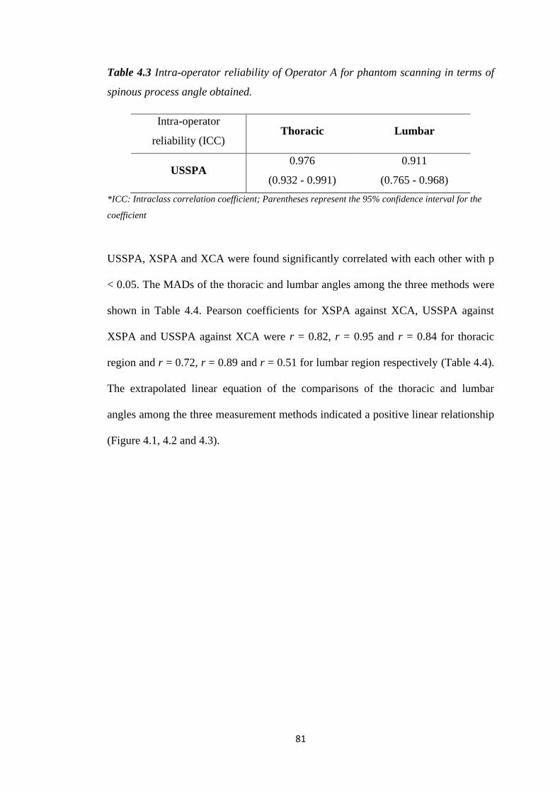
81
Table 4.3 Intra-operator reliability of Operator A for phantom scanning in terms of
spinous process angle obtained.
Intra-operator
reliability (ICC) Thoracic Lumbar
USSPA 0.976
(0.932 - 0.991)
0.911
(0.765 - 0.968)
*ICC: Intraclass correlation coefficient; Parentheses represent the 95% confidence interval for the
coefficient
USSPA, XSPA and XCA were found significantly correlated with each other with p
< 0.05. The MADs of the thoracic and lumbar angles among the three methods were
shown in Table 4.4. Pearson coefficients for XSPA against XCA, USSPA against
XSPA and USSPA against XCA were r = 0.82, r = 0.95 and r = 0.84 for thoracic
region and r = 0.72, r = 0.89 and r = 0.51 for lumbar region respectively (Table 4.4).
The extrapolated linear equation of the comparisons of the thoracic and lumbar
angles among the three measurement methods indicated a positive linear relationship
(Figure 4.1, 4.2 and 4.3).
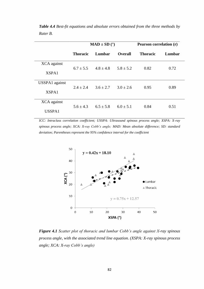
82
Table 4.4 Best-fit equations and absolute errors obtained from the three methods by
Rater B.
MAD ± SD (°) Pearson correlation (r)
Thoracic Lumbar Overall Thoracic Lumbar
XCA against
XSPA1
6.7 ± 5.5 4.8 ± 4.8 5.8 ± 5.2 0.82 0.72
USSPA1 against
XSPA1
2.4 ± 2.4 3.6 ± 2.7 3.0 ± 2.6 0.95 0.89
XCA against
USSPA1
5.6 ± 4.3 6.5 ± 5.8 6.0 ± 5.1 0.84 0.51
ICC: Intraclass correlation coefficient; USSPA: Ultrasound spinous process angle; XSPA: X-ray
spinous process angle; XCA: X-ray Cobb’s angle; MAD: Mean absolute difference; SD: standard
deviation; Parentheses represent the 95% confidence interval for the coefficient
Figure 4.1 Scatter plot of thoracic and lumbar Cobb’s angle against X-ray spinous
process angle, with the associated trend line equation. (XSPA: X-ray spinous process
angle; XCA: X-ray Cobb’s angle)
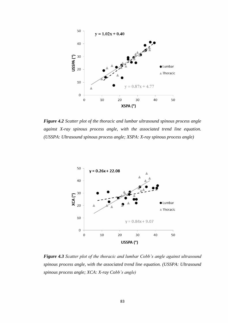
83
Figure 4.2 Scatter plot of the thoracic and lumbar ultrasound spinous process angle
against X-ray spinous process angle, with the associated trend line equation.
(USSPA: Ultrasound spinous process angle; XSPA: X-ray spinous process angle)
Figure 4.3 Scatter plot of the thoracic and lumbar Cobb’s angle against ultrasound
spinous process angle, with the associated trend line equation. (USSPA: Ultrasound
spinous process angle; XCA: X-ray Cobb’s angle)
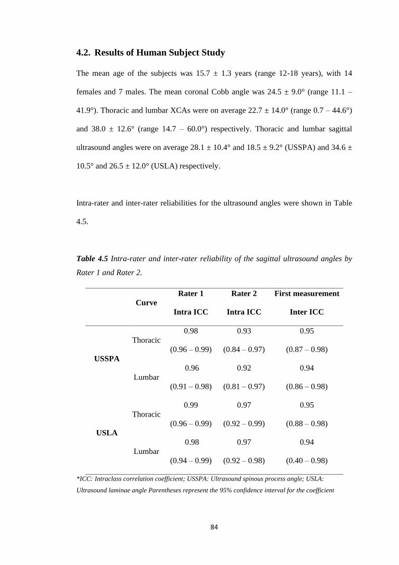
84
4.2. Results of Human Subject Study
The mean age of the subjects was 15.7 ± 1.3 years (range 12-18 years), with 14
females and 7 males. The mean coronal Cobb angle was 24.5 ± 9.0° (range 11.1 –
41.9°). Thoracic and lumbar XCAs were on average 22.7 ± 14.0° (range 0.7 – 44.6°)
and 38.0 ± 12.6° (range 14.7 – 60.0°) respectively. Thoracic and lumbar sagittal
ultrasound angles were on average 28.1 ± 10.4° and 18.5 ± 9.2° (USSPA) and 34.6 ±
10.5° and 26.5 ± 12.0° (USLA) respectively.
Intra-rater and inter-rater reliabilities for the ultrasound angles were shown in Table
4.5.
Table 4.5 Intra-rater and inter-rater reliability of the sagittal ultrasound angles by
Rater 1 and Rater 2.
Curve
Rater 1
Intra ICC
Rater 2
Intra ICC
First measurement
Inter ICC
USSPA
Thoracic
0.98
(0.96 – 0.99)
0.93
(0.84 – 0.97)
0.95
(0.87 – 0.98)
Lumbar
0.96
(0.91 – 0.98)
0.92
(0.81 – 0.97)
0.94
(0.86 – 0.98)
USLA
Thoracic
0.99
(0.96 – 0.99)
0.97
(0.92 – 0.99)
0.95
(0.88 – 0.98)
Lumbar
0.98
(0.94 – 0.99)
0.97
(0.92 – 0.98)
0.94
(0.40 – 0.98)
*ICC: Intraclass correlation coefficient; USSPA: Ultrasound spinous process angle; USLA:
Ultrasound laminae angle Parentheses represent the 95% confidence interval for the coefficient
85
Both USSPA and USLA showed moderate to good linear correlations with XCA
(Figure 4.4a and b). Thoracic USLA was found to have the lowest R2 value (0.574),
while lumbar USLA was found to have the highest R2 value (0.701). The Bland-
Altman plot showed a good agreement between the ultrasound angles adjusted with
the linear equations and the XCA (Figure 4.5a and b). No significant difference was
found between both adjusted ultrasound angles (MAD: USSPA 6.4 ± 4.8° / 6.1 ±
4.4°; USLA 7.5 ± 4.9° / 5.3 ± 4.2°; p ≥ 0.326 for thoracic / lumbar curves
respectively).
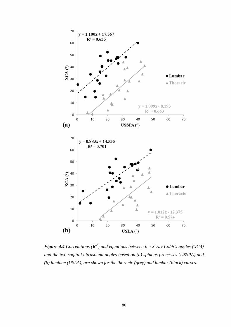
86
Figure 4.4 Correlations (𝑹𝟐) and equations between the X-ray Cobb’s angles (XCA)
and the two sagittal ultrasound angles based on (a) spinous processes (USSPA) and
(b) laminae (USLA), are shown for the thoracic (grey) and lumbar (black) curves.
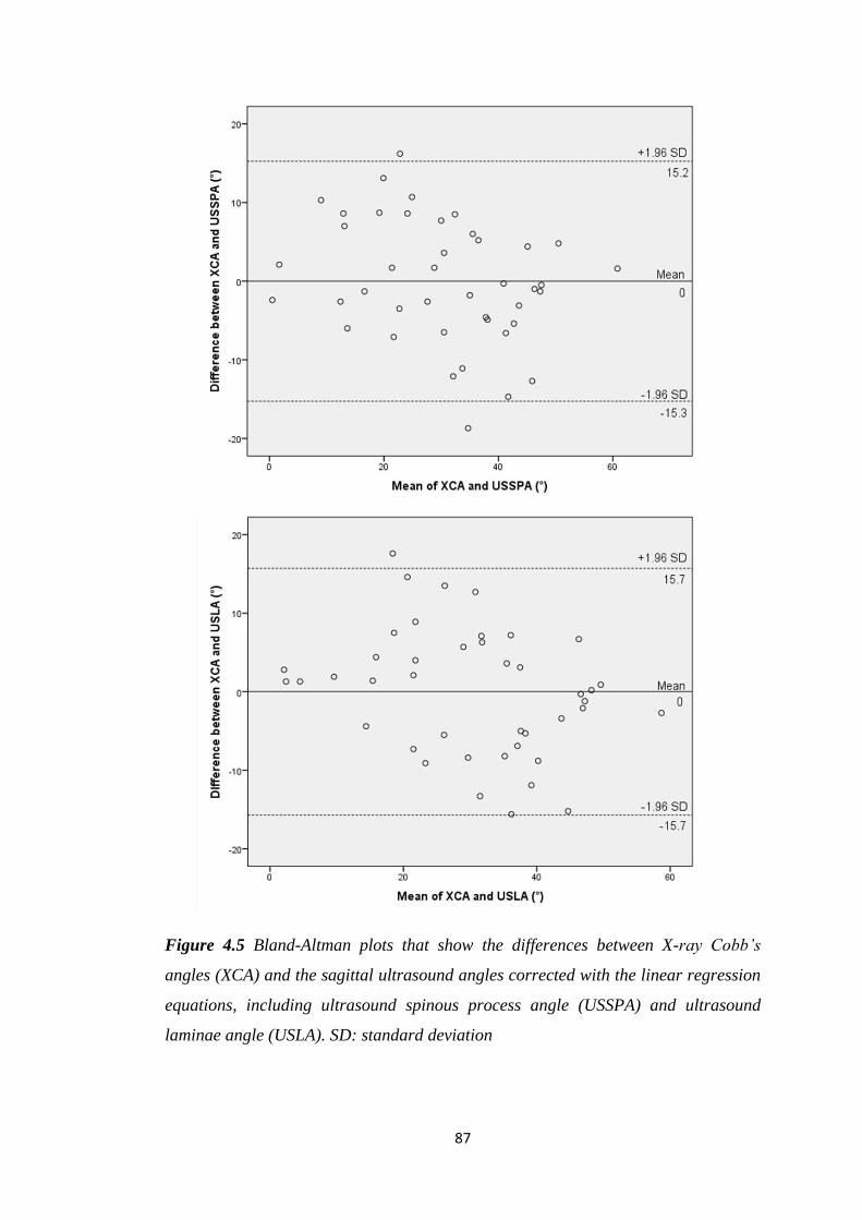
87
Figure 4.5 Bland-Altman plots that show the differences between X-ray Cobb’s
angles (XCA) and the sagittal ultrasound angles corrected with the linear regression
equations, including ultrasound spinous process angle (USSPA) and ultrasound
laminae angle (USLA). SD: standard deviation
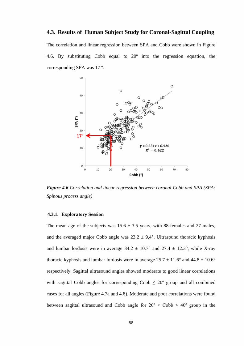
88
4.3. Results of Human Subject Study for Coronal-Sagittal Coupling
The correlation and linear regression between SPA and Cobb were shown in Figure
4.6. By substituting Cobb equal to 20º into the regression equation, the
corresponding SPA was 17 º.
Figure 4.6 Correlation and linear regression between coronal Cobb and SPA (SPA:
Spinous process angle)
4.3.1. Exploratory Session
The mean age of the subjects was 15.6 ± 3.5 years, with 88 females and 27 males,
and the averaged major Cobb angle was 23.2 ± 9.4°. Ultrasound thoracic kyphosis
and lumbar lordosis were in average 34.2 ± 10.7° and 27.4 ± 12.3°, while X-ray
thoracic kyphosis and lumbar lordosis were in average 25.7 ± 11.6° and 44.8 ± 10.6°
respectively. Sagittal ultrasound angles showed moderate to good linear correlations
with sagittal Cobb angles for corresponding Cobb ≤ 20º group and all combined
cases for all angles (Figure 4.7a and 4.8). Moderate and poor correlations were found
between sagittal ultrasound and Cobb angle for 20º < Cobb ≤ 40º group in the
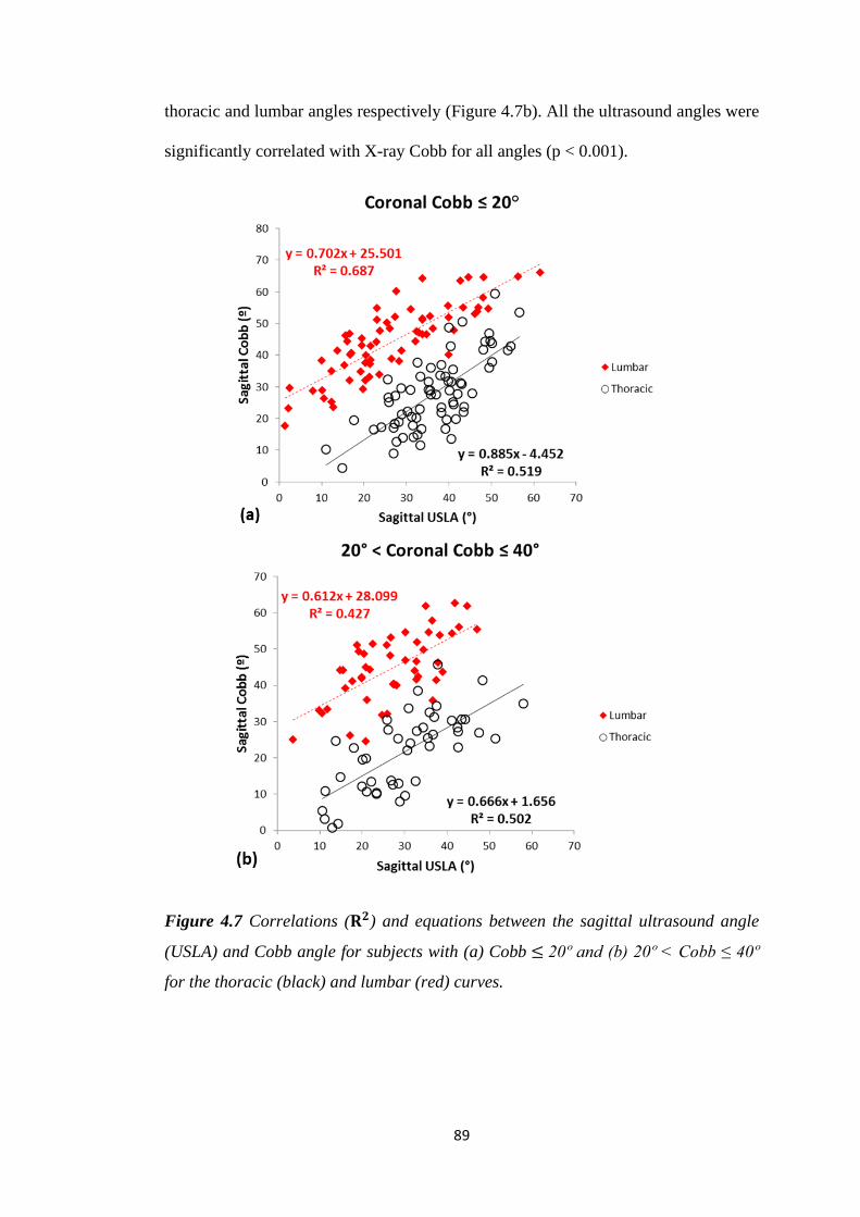
89
thoracic and lumbar angles respectively (Figure 4.7b). All the ultrasound angles were
significantly correlated with X-ray Cobb for all angles (p < 0.001).
Figure 4.7 Correlations (𝐑𝟐) and equations between the sagittal ultrasound angle
(USLA) and Cobb angle for subjects with (a) Cobb ≤ 20º and (b) 20º < Cobb ≤ 40º
for the thoracic (black) and lumbar (red) curves.
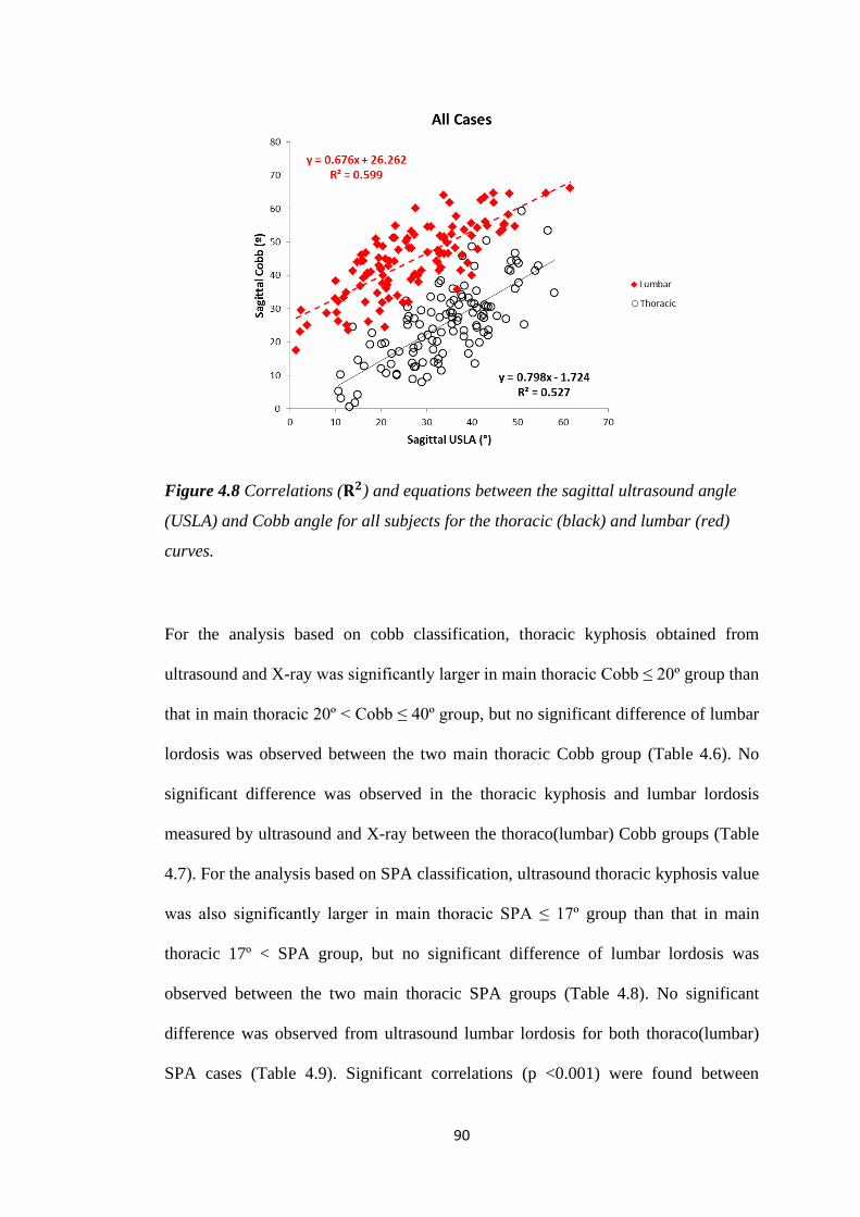
90
Figure 4.8 Correlations (𝐑𝟐) and equations between the sagittal ultrasound angle
(USLA) and Cobb angle for all subjects for the thoracic (black) and lumbar (red)
curves.
For the analysis based on cobb classification, thoracic kyphosis obtained from
ultrasound and X-ray was significantly larger in main thoracic Cobb ≤ 20º group than
that in main thoracic 20º < Cobb ≤ 40º group, but no significant difference of lumbar
lordosis was observed between the two main thoracic Cobb group (Table 4.6). No
significant difference was observed in the thoracic kyphosis and lumbar lordosis
measured by ultrasound and X-ray between the thoraco(lumbar) Cobb groups (Table
4.7). For the analysis based on SPA classification, ultrasound thoracic kyphosis value
was also significantly larger in main thoracic SPA ≤ 17º group than that in main
thoracic 17º < SPA group, but no significant difference of lumbar lordosis was
observed between the two main thoracic SPA groups (Table 4.8). No significant
difference was observed from ultrasound lumbar lordosis for both thoraco(lumbar)
SPA cases (Table 4.9). Significant correlations (p <0.001) were found between
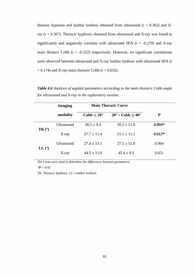
91
thoracic kypsosis and lumbar lordosis obtained from ultrasound (r = 0.362) and X-
ray (r = 0.367). Thoracic kyphosis obtained from ultrasound and X-ray was found to
significantly and negatively correlate with ultrasound SPA (r = -0.279) and X-ray
main thoracic Cobb (r = -0.322) respectively. However, no significant correlations
were observed between ultrasound and X-ray lumbar lordosis with ultrasound SPA (r
= 0.174) and X-ray main thoracic Cobb (r = 0.032).
Table 4.6 Analysis of sagittal parameters according to the main thoracic Cobb angle
for ultrasound and X-ray in the exploratory session.
Imaging
modality
Main Thoracic Curve
Cobb ≤ 20º 20º < Cobb ≤ 40º P
TK (º)
Ultrasound 36.5 ± 9.3 30.2 ± 11.8 0.004*
X-ray 27.7 ± 11.4 21.1 ± 11.2 0.012*
LL (º)
Ultrasound 27.4 ± 13.1 27.5 ± 11.0 0.964
X-ray 44.5 ± 11.0 45.4 ± 9.9 0.651
The t tests were used to determine the differences between parameters.
*P < 0.05
TK: Thoracic kyphosis; LL: Lumbar lordosis
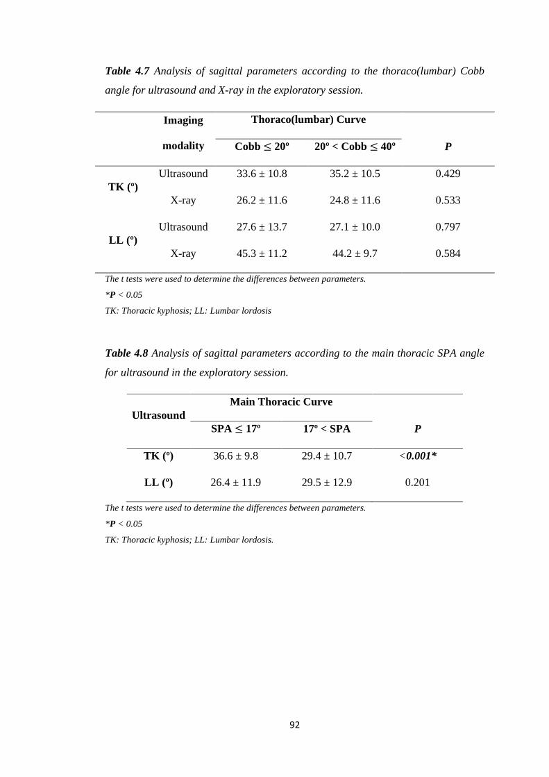
92
Table 4.7 Analysis of sagittal parameters according to the thoraco(lumbar) Cobb
angle for ultrasound and X-ray in the exploratory session.
Imaging
modality
Thoraco(lumbar) Curve
Cobb ≤ 20º 20º < Cobb ≤ 40º P
TK (º)
Ultrasound 33.6 ± 10.8 35.2 ± 10.5 0.429
X-ray 26.2 ± 11.6 24.8 ± 11.6 0.533
LL (º)
Ultrasound 27.6 ± 13.7 27.1 ± 10.0 0.797
X-ray 45.3 ± 11.2 44.2 ± 9.7 0.584
The t tests were used to determine the differences between parameters.
*P < 0.05
TK: Thoracic kyphosis; LL: Lumbar lordosis
Table 4.8 Analysis of sagittal parameters according to the main thoracic SPA angle
for ultrasound in the exploratory session.
Ultrasound
Main Thoracic Curve
SPA ≤ 17º 17º < SPA P
TK (º) 36.6 ± 9.8 29.4 ± 10.7 <0.001*
LL (º) 26.4 ± 11.9 29.5 ± 12.9 0.201
The t tests were used to determine the differences between parameters.
*P < 0.05
TK: Thoracic kyphosis; LL: Lumbar lordosis.
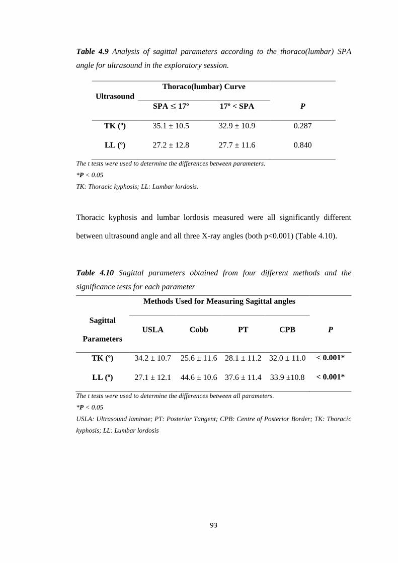
93
Table 4.9 Analysis of sagittal parameters according to the thoraco(lumbar) SPA
angle for ultrasound in the exploratory session.
Ultrasound
Thoraco(lumbar) Curve
SPA ≤ 17º 17º < SPA P
TK (º) 35.1 ± 10.5 32.9 ± 10.9 0.287
LL (º) 27.2 ± 12.8 27.7 ± 11.6 0.840
The t tests were used to determine the differences between parameters.
*P < 0.05
TK: Thoracic kyphosis; LL: Lumbar lordosis.
Thoracic kyphosis and lumbar lordosis measured were all significantly different
between ultrasound angle and all three X-ray angles (both p<0.001) (Table 4.10).
Table 4.10 Sagittal parameters obtained from four different methods and the
significance tests for each parameter
Methods Used for Measuring Sagittal angles
Sagittal
Parameters
USLA Cobb PT CPB P
TK (º) 34.2 ± 10.7 25.6 ± 11.6 28.1 ± 11.2 32.0 ± 11.0 < 0.001*
LL (º) 27.1 ± 12.1 44.6 ± 10.6 37.6 ± 11.4 33.9 ±10.8 < 0.001*
The t tests were used to determine the differences between all parameters.
*P < 0.05
USLA: Ultrasound laminae; PT: Posterior Tangent; CPB: Centre of Posterior Border; TK: Thoracic
kyphosis; LL: Lumbar lordosis
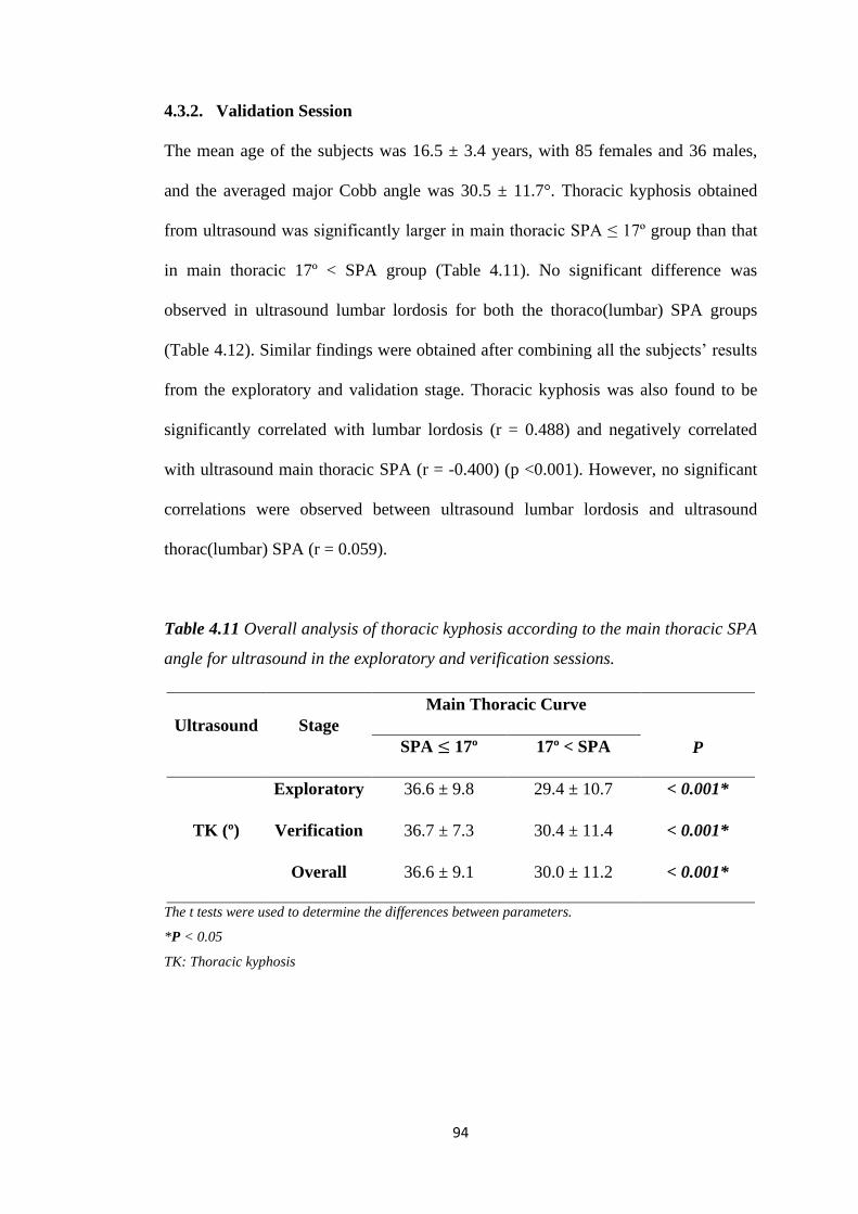
94
4.3.2. Validation Session
The mean age of the subjects was 16.5 ± 3.4 years, with 85 females and 36 males,
and the averaged major Cobb angle was 30.5 ± 11.7°. Thoracic kyphosis obtained
from ultrasound was significantly larger in main thoracic SPA ≤ 17º group than that
in main thoracic 17º < SPA group (Table 4.11). No significant difference was
observed in ultrasound lumbar lordosis for both the thoraco(lumbar) SPA groups
(Table 4.12). Similar findings were obtained after combining all the subjects’ results
from the exploratory and validation stage. Thoracic kyphosis was also found to be
significantly correlated with lumbar lordosis (r = 0.488) and negatively correlated
with ultrasound main thoracic SPA (r = -0.400) (p <0.001). However, no significant
correlations were observed between ultrasound lumbar lordosis and ultrasound
thorac(lumbar) SPA (r = 0.059).
Table 4.11 Overall analysis of thoracic kyphosis according to the main thoracic SPA
angle for ultrasound in the exploratory and verification sessions.
Ultrasound Stage
Main Thoracic Curve
P SPA ≤ 17º 17º < SPA
TK (º)
Exploratory 36.6 ± 9.8 29.4 ± 10.7 < 0.001*
Verification 36.7 ± 7.3 30.4 ± 11.4 < 0.001*
Overall 36.6 ± 9.1 30.0 ± 11.2 < 0.001*
The t tests were used to determine the differences between parameters.
*P < 0.05
TK: Thoracic kyphosis
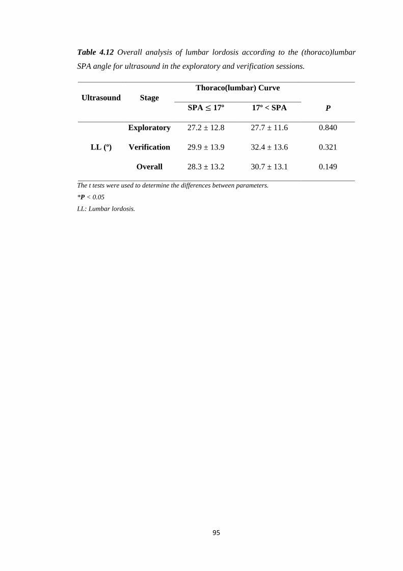
95
Table 4.12 Overall analysis of lumbar lordosis according to the (thoraco)lumbar
SPA angle for ultrasound in the exploratory and verification sessions.
Ultrasound Stage
Thoraco(lumbar) Curve
P SPA ≤ 17º 17º < SPA
LL (º)
Exploratory 27.2 ± 12.8 27.7 ± 11.6 0.840
Verification 29.9 ± 13.9 32.4 ± 13.6 0.321
Overall 28.3 ± 13.2 30.7 ± 13.1 0.149
The t tests were used to determine the differences between parameters.
*P < 0.05
LL: Lumbar lordosis.
96
Chapter 5. Discussion
5.1. Spine Phantom Study
The reliability of using 3D ultrasound imaging system for the measurement of
sagittal spinal curvature was tested and comparisons of the US results with those
obtained from traditional X-ray images were made in this study. All the parameters
obtained from either X-ray or 3D ultrasound were demonstrated to have excellent
reliability. Both USSPA and XSPA were obtained using the spinous process angle.
Though the imaging modality was different, the MAD between them was the
smallest and the Pearson correlation was the greatest among the three comparisons of
the three angles. The difference could be possibly due to the nature of the selection
processes of the lateral radiograph and US stack image. Selection of spinous
processes was performed from the 2D X-ray image of the spine phantom in the
sagittal plane and from the B-mode images of 3D ultrasound volume stack
respectively. Thus the perspective difference was one of the major reasons that
explained the discrepancies of the results (Vrtovec et al. 2009, Gstoettner et al. 2007).
Indeed, a nearly one-to-one relationship was observed between these two parameters,
suggesting that they were very much comparable.
Since USSPA was measured using spinous processes and XCA was measured using
superior and inferior endplates of the vertebral bodies, lumbar curvatures formed
from the spinous processes were likely to be smaller than those measured from
vertebrae because the bulky shape of the processes prohibits the lumbar region of the
spine phantom for further progression during deformation, while the soft
intervertebral structures between the vertebral plates allow larger degrees of
deformation. Hence, lumbar USSPA tended to be underestimated compared with
97
XCA. A study used biplanar radiographs to evaluate the apical thoracic sagittal
profile (Hayashi et al. 2009). By comparing the results obtained from the standard
lateral projection with those from the “true lateral” view, it was found that the
sagittal curvature was significantly greater (p < 0.001) in the traditional sagittal view
by 10 degrees in average than the “true lateral” view (Hayashi et al. 2009). This
suggested that XCA obtained in the study might not be reflecting the ‘real’ sagittal
curvature, but indeed a slightly larger curvature. In addition, the study suggested that
the larger the thoracic Cobb’s angle in the coronal plane measured, the more
kyphotic the thoracic apical profile on the standard lateral radiograph would appear,
which would eventually lead to a greater difference in the thoracic apical alignment
between the two views (Hayashi et al. 2009). Hence, it is necessary to measure the
sagittal spinal curvature using an alternative method instead of using the traditional
2D X-ray projection, and ultrasound could be a potential method for sagittal spinal
curvature evaluation.
The Pearson correlations obtained from the phantom study suggested that ultrasound
angles in the thoracic regions were more representative than those in the lumbar
regions. The differences in the results between the thoracic and lumbar regions might
be accounted for the level differences involved for these two regions, where the
thoracic vertebrae levels involved to compute the thoracic angle were much more
than those for the lumbar angle. Comparison of the mean Cobb values of normal
lumbar lordosis in previous studies found out that lesser the vertebral levels involved
would likely result in smaller lordosis angle (Stagnara et al. 1982, Fernand and Fox.
1985, Saraste et al. 1985). This effect might be emphasized when using spinous
processes for sagittal measurement.
98
A previous study investigated 39 adolescent girls with double-curved idiopathic
scoliosis and reported that the linear relationship between XSPA and XCA was XCA
= 0.84*XSPA + 9.63 and XCA = 0.66*XSPA + 33.96 for thoracic and lumbar
regions (Delorme et al. 1999), whereas the best-fit equations obtained in our study
for XCA against XSPA were XCA = 0.75*XSPA + 12.57 and XCA = 0.42*XSPA +
18.10 for the two regions respectively. Since Pearson correlations obtained in the two
studies for both thoracic and lumbar regions were similar and the intra-reliabilities of
the measurement in our study were excellent, one of the possible reasons for the
discrepancies of the results could be the difference in the calculation of XSPA used
in the two studies. However, this could not be confirmed as the calculation details
were not described in the previous study (Delorme et al. 1999). In addition, the levels
of vertebrae involved for lumbar curvature were different for the parameters, where
L1-L4 levels were used for XCA while T12-L4 levels were used for XSPA and
USSPA. The reason for such a selection was that we noted the spinous process of
T12 (instead of L1) is more aligned with the upper plate of L1, and that of L4 is more
aligned with the lower plate of L4. Furthermore, only a single point was selected to
represent the spinous process of all vertebrae from T2 to L4 in ultrasound images in
this study. However, the spinous process is not a single point, and selection of
different locations of the same spinous process may induce variations in curvature
measurement.
3D ultrasound imaging, supported by the phantom data, is a potential imaging
modality for screening and monitoring the development of individual’s sagittal spine
profile. It should be always noted that ultrasound and X-ray measurements were
99
based on different structures of the vertebrae, thus it was reasonable that the results
of these two modalities did not represent each other. The excellent intra-operator
reliability for ultrasound scanning on phantoms as well as excellent intra- and inter-
rater reliabilities for angle measurement obtained in this study demonstrated that 3D
ultrasound could be used for evaluating sagittal profile on spine phantom. However,
water was used as the tissue mimicking background, hence the image quality of
ultrasound would be another story when human subjects were being scanned. In
addition, for the phantom tests reported in this study, the phantom was fixed thus had
no motion, whereas in real subject tests, the subject would likely move forward and
backward to change the spinal sagittal profile during the ultrasound scanning. Hence
in the following human subjects study, the subject would be stabilized during
scanning as introduced by Zheng et al. (2016) for their coronal curvature study.
Furthermore, the reliability and validity of ultrasound on sagittal spinal curvature
measurement with different deformity curvatures and patterns were conducted in the
human subjects test for evaluation of the ultrasound system.
5.2. Human Subjects Study
Traditionally sagittal curvature of spine is evaluated by radiographic Cobb angle,
which necessitates ionizing radiation to form the images. Alternative imaging
modalities have been suggested to minimize or avoid the radiation issue. Bi-planar
stereoradiography utilizes lower dosage of radiation, but it is expensive and not
readily available to most medical practitioners. Ultrasound imaging is non-ionizing
and relatively cheap. During ultrasound scanning, subjects are maintained in the
upright posture, the same as that adopted during traditional radiographic examination
thereby providing a real alternative to erect x-ray images. Previous studies have
100
investigated the possibility of ultrasound for assessing the coronal spine curvature,
and to the best of our knowledge, the present paper is the first to report the reliability
and validity of different sagittal ultrasound angle measurements. Excellent intra- and
inter-rater reliabilities were demonstrated for ultrasound sagittal angle measurement,
and good to moderate linear correlations were obtained between the ultrasound
angles and Cobb angles. The average MAD of the ultrasound measurements was
about 6.3°, without significant difference in the different ultrasound measurements.
The MADs were higher in the thoracic region for both the ultrasound methods. This
is not surprising since spinous processes and laminae in the lower regions of the
spine can be identified more easily than the upper regions, which leads to smaller
errors during measurement.
The sagittal ultrasound angles obtained were larger in the thoracic curves and smaller
in the lumbar curves as compared with their corresponding sagittal radiographic
Cobb angles. There were several possible reasons for the discrepancies: 1) The
ultrasound measurements were based on SP and bilateral laminae, which are
structures located more posteriorly than the vertebral body and where Cobb angle
was measured (Brink et al. 2018). Differences in structures used for measurements
thus will possibly lead to a different projection of the 3D deformity (Herzenberg et
al. 1990). In addition, it has been found that measurements based on posterior
vertebral structures would cause the angular value differences (Chernukha et al.
1998); 2) Different positions of arms were adopted for different imaging modalities,
where patients were in a relaxed standing posture with arms relaxed at the sides and
fists overlying ipsilateral clavicles were adopted for ultrasound and X-ray scanning
respectively (Pasha et al. 2016). A decrease in kyphosis and an increase in lordosis

101
were observed when patients adopted the fists overlying ipsilateral clavicles position
compared to relaxed standing (Marks et al. 2009); 3) Different levels of vertebrae
were involved for X-ray and ultrasound assessments due to different measurement
techniques. T3 was involved for the ultrasound measurement, which may possibly
lead to the larger value in the thoracic ultrasound angle. We should bear in mind that
using traditional Cobb angles alone would not be sufficient to comprehensively study
the complex 3D deformity. In addition, sagittal ultrasound images formed in this
study were based on the projection of the SP or bilateral laminae selected in the 3D
ultrasound volume. This reflected a real sagittal profile of the scoliotic spine rather
than the projection on lateral radiographs which was potentially influenced by
vertebral rotation and magnitude of deformity. Traditional sagittal Cobb
measurements of the thoracic spine in lateral radiographs were found larger than
those obtained from 3D view on patients with scoliosis due to axial vertebral rotation
(Newton et al. 2015), but in this study the finding was the opposite.
We observed that sagittal curvature analysis using ultrasound required a higher
demand on scanning and image quality control in comparison with coronal curvature
analysis. Hence additional attention should be paid in the future during scanning,
such as using lower frequencies ultrasound for patients with a lumbar curve in order
to capture the vertebral structures since they are deeper from the skin surface.
Patients with Cobb angles larger than 50 degrees were excluded in this study since
spinal sagittal measurement might be prone to measurement error due to the presence
of severe rotation (Bao et al. 2018). Spinous processes in the ultrasound images of
some patients were deformed for very thin patients due to the protruded scanning
trajectories when their backs were being scanned, thus the spinous processes
102
acquired in the ultrasound images were not suitable to compile the USSPA. However,
the laminae would not be affected by the above issue, thus the USLA could be used
for these types of patients. And since the MADs of the two ultrasound angles were
not significantly different, USLA was used in the human subjects study for coronal-
sagittal coupling.
5.3. Human Subjects Study for Coronal-Sagittal Coupling
Only the laminae angle was used for evaluating the ultrasound angle in this session.
One of the reasons was that spinous processes might not be a good parameter for
evaluating scoliotic spine. Since vertebral rotation is likely to exist in deformed
spines, it would cause spinous process deviations. The spinous processes
increasingly deviate to the concave side during the rotation progression and the ribs
also follow the rotation of the vertebrae (Middleditch et al. 2005). Such deviation
could lead to developmental asymmetries of the neural arch, rotation of entire
vertebra, and isolated deviation (Van Schaik et al. 1989), causing confusion when
interpreting the PA radiographs of spine and imitating vertebral body malalignment
(Mellado et al.2011a; 2011b). Moreover, since bracing and surgical treatment will be
suggested for skeletally immature AIS patients once their major Cobb angles reach
20° and 40° respectively, and thoracic kyphosis of patients with Cobb smaller than
20° was found to be larger than those having Cobb angles from 20° to 40° (Hong et
al. 2017), 20° and 40° were set as the border criteria for the sagittal analysis in this
study. In addition, for patients with Cobb larger than 40°, the evaluation of the
sagittal curvature would be highly affected by the coronal deformity and vertebral
rotation, especially for radiograph, hence these patients were not included in this
study. Furthermore, T3 was set to be the upper boundary as features of T1 and T2
103
vertebrae could hardly be observed in the US images, similar to the findings of
Young et al. (2015).
Correlation between the thoracic kyphosis measurements by ultrasound and X-ray
was higher in the smaller Cobb group than the larger Cobb group, while the
correlation between the lumbar lordosis measurements by the two methods was
similar for both groups. This suggested that the magnitude of the thoracic curves had
a larger effect on causing discrepancies between ultrasound and X-ray than the
lumbar curves. The discrepancies could also be accounted for the larger vertebrae
rotation of patients with AIS with larger Cobb (White et al. 1978, Villemure et al.
2001, Gum et al. 2007), as rotation in the laminae was found to be greater than that
in the vertebral body using CT scans (Suzuki et al 1989). Hayashi et al. (2009) found
that the mean apical thoracic sagittal curvature was significantly larger in the
standard lateral view than that in the “true lateral” view by an average of 10 degrees,
and the magnitude of the thoracic Cobb was also found to be significantly correlated
with the thoracic apical sagittal kyphosis in standard lateral view but not in the “true
lateral” view. This may suggest that the thoracic apical profile may appear to be
more kyphotic on the 2D imaging for patients with larger coronal deformity. Hence
in this study, the spinal sagittal profile of patients with large coronal deformation was
not investigated as the clinical implications for sagittal profile of severely deformed
spine were not significant. In future study, 3D ultrasound analysis would be needed
for severe scoliosis cases in order to attain the realistic spinal deformation
information (Hayashi et al. 2009).
104
Patients with larger curve deformity in the main thoracic region were found to be
significantly hypokyphotic, but no significant differences were observed in terms of
lumbar lordosis among subjects with different lumbar curve deformity, based on the
data analysis from either ultrasound or X-ray. This suggested that most of the cases
being analyzed had similar behaviour to the case illustrated in Figure A.1, where
patients with smaller thoracic deformation had a larger thoracic kyphosis. These
findings were similar as those reported in previous studies (Schmitz et al. 2001, De
jonge et al. 2002, Thiong et al. 2013, Hong et al. 2017, Ilharreborde et al. 2018).
Schlösser et al. (2014) and Janssen et al. (2011) demonstrated that patients with mild
thoracic scoliosis had a significantly less kyphotic spine, but those with mild lumbar
scoliosis were found to have significantly larger lumbar lordosis compared with the
thoracic kyphosis and control subjects. The discrepancies of the findings may be due
to the differences of the curve natures of the subjects with AIS involved. Patients
with only either thoracic or lumbar curve were invited in their studies, whereas
patients with mixed curves were involved in our study. In addition, a negative
correlation was revealed between thoracic kyphosis and the thoracic coronal
deformation, which was also demonstrated in Schmitz et al.’s study (2001). However,
such significant correlation was not shown in the study of Hong et al. (2017). It
should be noted that Cobb angle measurement was not categorized when evaluating
correlation with thoracic kyphosis and lumbar lordosis.
Patients with larger thoracic curve appeared to be relatively hypokyphotic in
radiographic evaluation, and ultrasound was demonstrated to be effective in detecting
similar findings in this study. However, it should be noted that sagittal angles
measured by ultrasound were larger than those measured by X-ray. The absolute
105
differences between the ultrasound and X-ray measurements decreased from the
Cobb method to PT method, and from PT method to CPB method. The mean and SD
of the absolute difference between ultrasound laminae angle and central of posterior
border angle was 5.8 ± 4.3º and 7.3 ± 5.0º, respectively. The result showed that the
discrepancies between ultrasound laminae angle and Cobb angle were caused by the
differences of the measurement techniques and the anatomical landmarks chosen for
the two modalities. In addition, average inclination between T3 and T4 on radiograph
of chronic LBP patients was found to be 4.8º (Harrison et al. 2001), which may
potentially lead to the thoracic lyphosis value differences obtained between
ultrasound and X-ray since T3 level was involved for ultrasound measurement. A
study reported a method similar to the posterior tangent method, the tangential
radiologic assessment, for the measurement of lumbar lordosis. The angle measured
was found to be 8º to 16º smaller than that by Cobb method (Chernukha et al. 1998).
However, it was claimed to be a more reliable method than Cobb for scoliosis
measurement since it was not subject to variation in vertebral body contour.
Furthermore, the distance between T12 and L5 was too small for lumbar curvature
measurement using posterior elements of the vertebrae, hence the curvature
measurement would not be representative to radiologic lordosis (Leroux et al. 2000).
In future study, S1 might be considered for the evaluation of lumbar lordosis using
ultrasound and thus the ultrasound scanning could be started in a more inferior
position in order to capture the laminae of S1.
Patients with progressive AIS diagnosed were found to possess significant
hypokyphosis by a mean of 4.2º compared to the non-progressive AIS patients (Nault
et al. 2014). In addition, increasing severity of major thoracic curve in AIS patients
106
was found to be associated with a progressive loss of thoracic kyphosis (Sullivan et
al. 2017). However, such phenomenon could not be proved in this study as
randomized controlled cases. A longitudinal study should be carried out on patients
with AIS using ultrasound during the first and follow-up visits to investigate whether
there is a change between sagittal profiles of the patients and curve progression. The
average thoracic kyphosis of AIS patients with thoracic SPA larger than 17 degrees
was 36.6 degrees, in both exploratory and validation studies, hence such value could
be used as reference in future progression study to investigate whether the initial
value of thoracic kyphosis would affect the curve progression in the thoracic region.
Spinal ligaments and paravertebral muscles also played an important role in
maintaining spinal stability (Kouwenhoven et al. 2008), hence in future study, the
correlation between morphologic differences of the spinal muscles and different
curve severity could be studied in patients with AIS using ultrasound. Quantifying
either spinal curvatures different planes or vertebrae rotation is useful for planning
surgery, predicting prognosis and monitoring curve progression (Vrtovec et al.2009,
Carlson et al. 2013, Cheung et al. 2013).
It should be noted that ultrasound examination cannot replace the X-ray examination
for the evaluation of sagittal spinal curvatures during the diagnosis and annual
evaluation of progression. The reliability of sagittal measurement using ultrasound as
shown in this study suggested that 3D ultrasound imaging could be a non-ionizing
tool to evaluate the sagittal profile of patients with AIS and to give an ultrasound
standard of optimal sagittal curvature. 3D ultrasound imaging could also be used to
provide a more frequent ultrasound spinal examination for follow-ups, such as
monitoring the sagittal thoracic and lumbar curvature, together with coronal curves,

107
to assess the effectiveness of various therapies, which generally can not be done in
clinical routine as sagittal X-ray images are not commonly evaluated and are not
available in clinics with EOS.
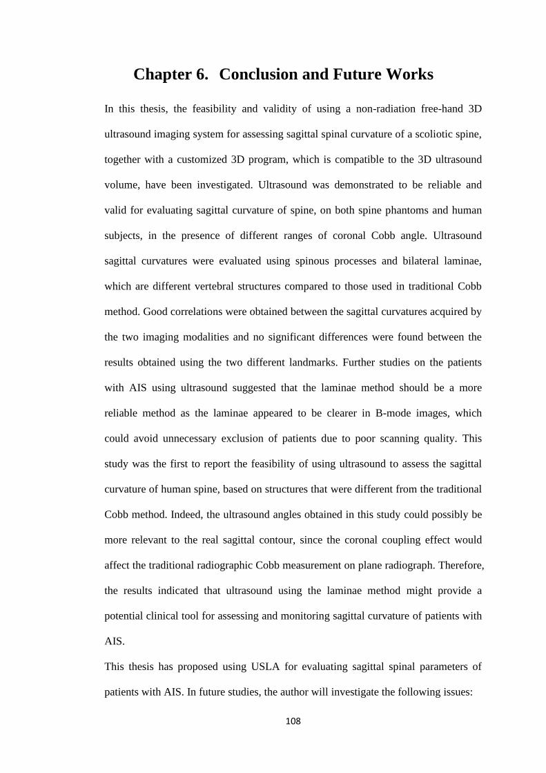
108
Chapter 6. Conclusion and Future Works
In this thesis, the feasibility and validity of using a non-radiation free-hand 3D
ultrasound imaging system for assessing sagittal spinal curvature of a scoliotic spine,
together with a customized 3D program, which is compatible to the 3D ultrasound
volume, have been investigated. Ultrasound was demonstrated to be reliable and
valid for evaluating sagittal curvature of spine, on both spine phantoms and human
subjects, in the presence of different ranges of coronal Cobb angle. Ultrasound
sagittal curvatures were evaluated using spinous processes and bilateral laminae,
which are different vertebral structures compared to those used in traditional Cobb
method. Good correlations were obtained between the sagittal curvatures acquired by
the two imaging modalities and no significant differences were found between the
results obtained using the two different landmarks. Further studies on the patients
with AIS using ultrasound suggested that the laminae method should be a more
reliable method as the laminae appeared to be clearer in B-mode images, which
could avoid unnecessary exclusion of patients due to poor scanning quality. This
study was the first to report the feasibility of using ultrasound to assess the sagittal
curvature of human spine, based on structures that were different from the traditional
Cobb method. Indeed, the ultrasound angles obtained in this study could possibly be
more relevant to the real sagittal contour, since the coronal coupling effect would
affect the traditional radiographic Cobb measurement on plane radiograph. Therefore,
the results indicated that ultrasound using the laminae method might provide a
potential clinical tool for assessing and monitoring sagittal curvature of patients with
AIS.
This thesis has proposed using USLA for evaluating sagittal spinal parameters of
patients with AIS. In future studies, the author will investigate the following issues:
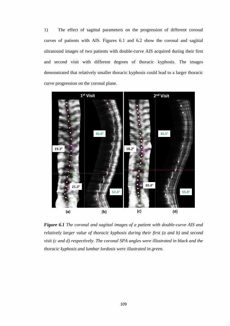
109
1) The effect of sagittal parameters on the progression of different coronal
curves of patients with AIS. Figures 6.1 and 6.2 show the coronal and sagittal
ultrasound images of two patients with double-curve AIS acquired during their first
and second visit with different degrees of thoracic kyphosis. The images
demonstrated that relatively smaller thoracic kyphosis could lead to a larger thoracic
curve progression on the coronal plane.
Figure 6.1 The coronal and sagittal images of a patient with double-curve AIS and
relatively larger value of thoracic kyphosis during their first (a and b) and second
visit (c and d) respectively. The coronal SPA angles were illustrated in black and the
thoracic kyphosis and lumbar lordosis were illustrated in green.
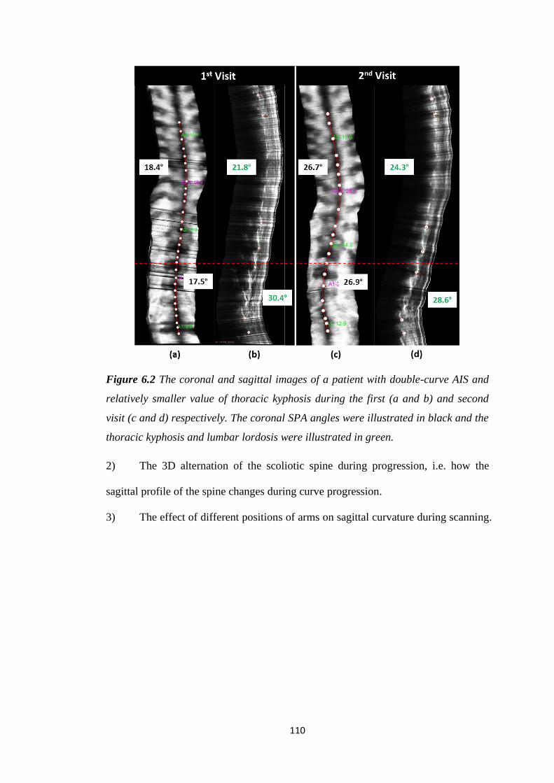
110
Figure 6.2 The coronal and sagittal images of a patient with double-curve AIS and
relatively smaller value of thoracic kyphosis during the first (a and b) and second
visit (c and d) respectively. The coronal SPA angles were illustrated in black and the
thoracic kyphosis and lumbar lordosis were illustrated in green.
2) The 3D alternation of the scoliotic spine during progression, i.e. how the
sagittal profile of the spine changes during curve progression.
3) The effect of different positions of arms on sagittal curvature during scanning.
111
Appendices
A.1. Figures
Figure A.1 The three images on the left were the coronal (a) and sagittal ultrasound
images illustrating the left (b) and right (c) laminae of an AIS patient with a single
thoracic Cobb less than 20º, whereas the three images on the right were the coronal
(d) and sagittal ultrasound images illustrating the left (e) and right (f) laminae of an
AIS patient with a single thoracic Cobb larger than 20º. The red line indicates the
T12 vertebrae level.
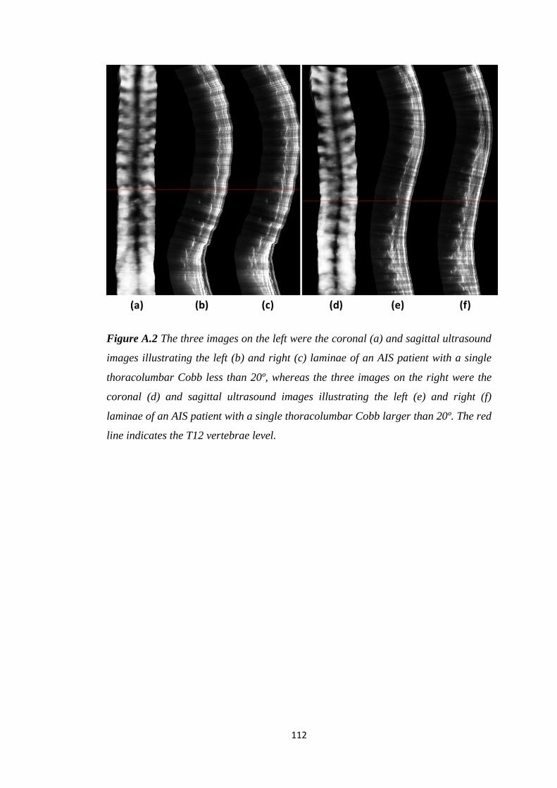
112
Figure A.2 The three images on the left were the coronal (a) and sagittal ultrasound
images illustrating the left (b) and right (c) laminae of an AIS patient with a single
thoracolumbar Cobb less than 20º, whereas the three images on the right were the
coronal (d) and sagittal ultrasound images illustrating the left (e) and right (f)
laminae of an AIS patient with a single thoracolumbar Cobb larger than 20º. The red
line indicates the T12 vertebrae level.
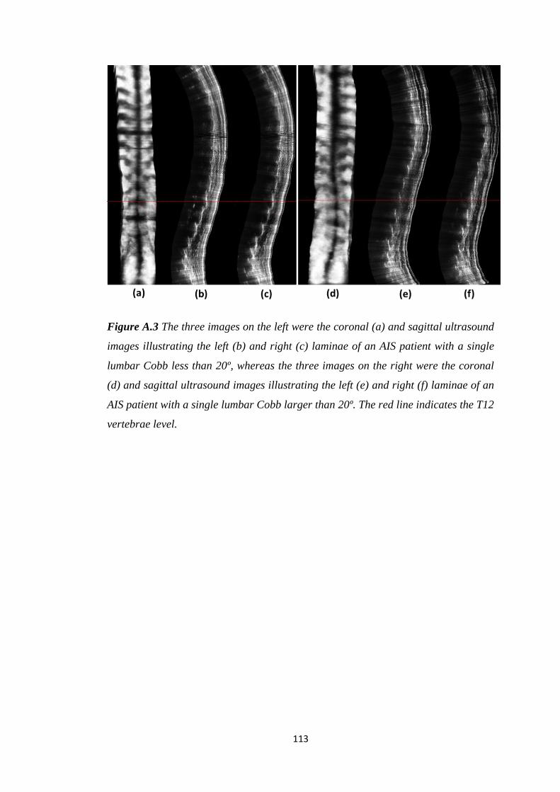
113
Figure A.3 The three images on the left were the coronal (a) and sagittal ultrasound
images illustrating the left (b) and right (c) laminae of an AIS patient with a single
lumbar Cobb less than 20º, whereas the three images on the right were the coronal
(d) and sagittal ultrasound images illustrating the left (e) and right (f) laminae of an
AIS patient with a single lumbar Cobb larger than 20º. The red line indicates the T12
vertebrae level.
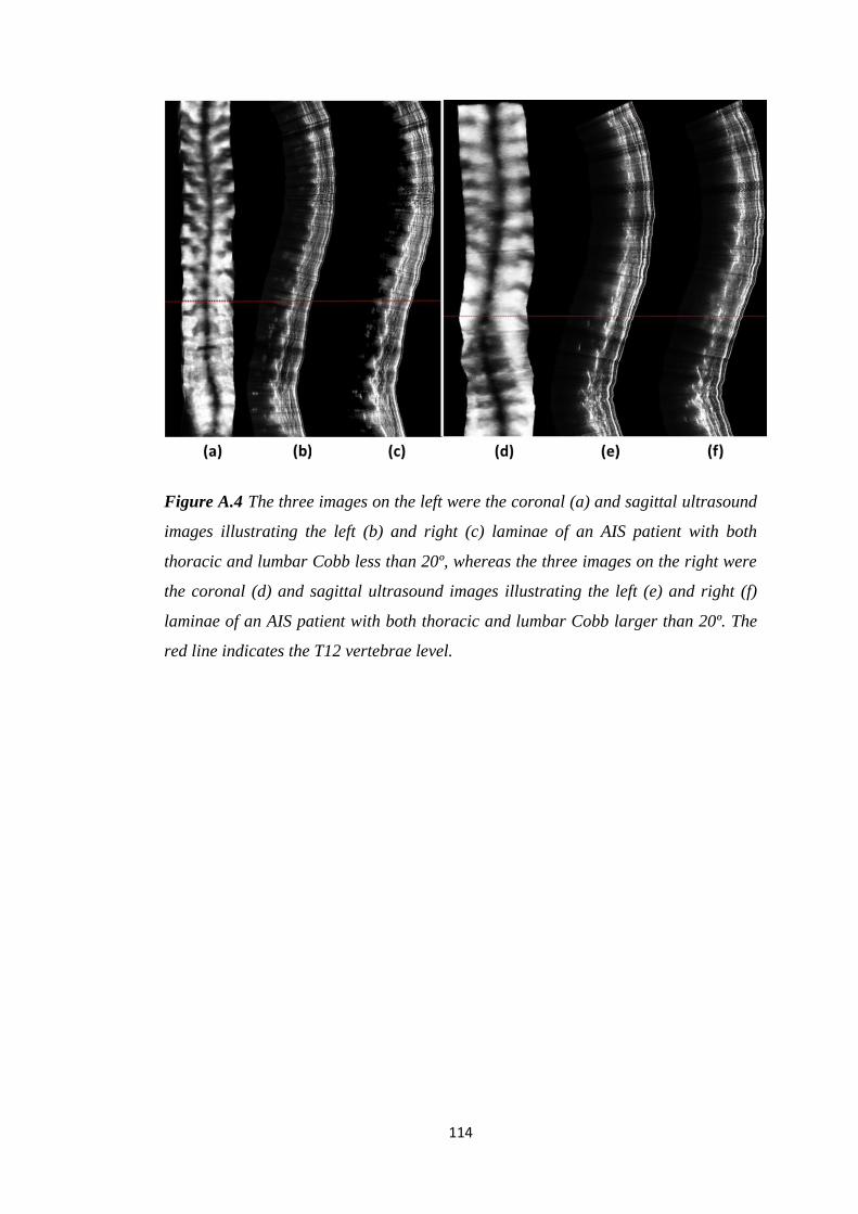
114
Figure A.4 The three images on the left were the coronal (a) and sagittal ultrasound
images illustrating the left (b) and right (c) laminae of an AIS patient with both
thoracic and lumbar Cobb less than 20º, whereas the three images on the right were
the coronal (d) and sagittal ultrasound images illustrating the left (e) and right (f)
laminae of an AIS patient with both thoracic and lumbar Cobb larger than 20º. The
red line indicates the T12 vertebrae level.
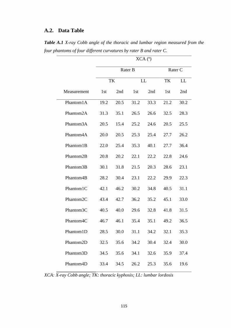
115
A.2. Data Table
Table A.1 X-ray Cobb angle of the thoracic and lumbar region measured from the
four phantoms of four different curvatures by rater B and rater C.
XCA (º)
Rater B Rater C
TK LL TK LL
Measurement 1st 2nd 1st 2nd 1st 2nd
Phantom1A 19.2 20.5 31.2 33.3 21.2 30.2
Phantom2A 31.3 35.1 26.5 26.6 32.5 28.3
Phantom3A 20.5 15.4 25.2 24.6 20.5 25.5
Phantom4A 20.0 20.5 25.3 25.4 27.7 26.2
Phantom1B 22.0 25.4 35.3 40.1 27.7 36.4
Phantom2B 20.8 20.2 22.1 22.2 22.8 24.6
Phantom3B 30.1 31.8 21.5 20.3 28.6 23.1
Phantom4B 28.2 30.4 23.1 22.2 29.9 22.3
Phantom1C 42.1 46.2 30.2 34.8 40.5 31.1
Phantom2C 43.4 42.7 36.2 35.2 45.1 33.0
Phantom3C 40.5 40.0 29.6 32.8 41.8 31.5
Phantom4C 46.7 46.1 35.4 35.1 49.2 36.5
Phantom1D 28.5 30.0 31.1 34.2 32.1 35.3
Phantom2D 32.5 35.6 34.2 30.4 32.4 30.0
Phantom3D 34.5 35.6 34.1 32.6 35.9 37.4
Phantom4D 33.4 34.5 26.2 25.3 35.6 19.6
XCA: X-ray Cobb angle; TK: thoracic kyphosis; LL: lumbar lordosis
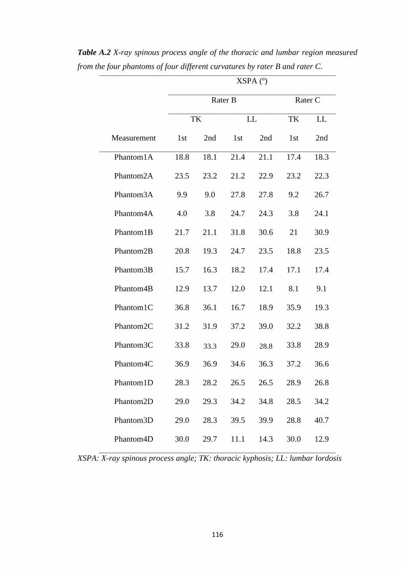
116
Table A.2 X-ray spinous process angle of the thoracic and lumbar region measured
from the four phantoms of four different curvatures by rater B and rater C.
XSPA (º)
Rater B Rater C
TK LL TK LL
Measurement 1st 2nd 1st 2nd 1st 2nd
Phantom1A 18.8 18.1 21.4 21.1 17.4 18.3
Phantom2A 23.5 23.2 21.2 22.9 23.2 22.3
Phantom3A 9.9 9.0 27.8 27.8 9.2 26.7
Phantom4A 4.0 3.8 24.7 24.3 3.8 24.1
Phantom1B 21.7 21.1 31.8 30.6 21 30.9
Phantom2B 20.8 19.3 24.7 23.5 18.8 23.5
Phantom3B 15.7 16.3 18.2 17.4 17.1 17.4
Phantom4B 12.9 13.7 12.0 12.1 8.1 9.1
Phantom1C 36.8 36.1 16.7 18.9 35.9 19.3
Phantom2C 31.2 31.9 37.2 39.0 32.2 38.8
Phantom3C 33.8 33.3 29.0 28.8 33.8 28.9
Phantom4C 36.9 36.9 34.6 36.3 37.2 36.6
Phantom1D 28.3 28.2 26.5 26.5 28.9 26.8
Phantom2D 29.0 29.3 34.2 34.8 28.5 34.2
Phantom3D 29.0 28.3 39.5 39.9 28.8 40.7
Phantom4D 30.0 29.7 11.1 14.3 30.0 12.9
XSPA: X-ray spinous process angle; TK: thoracic kyphosis; LL: lumbar lordosis
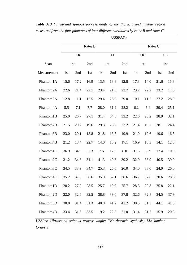
117
Table A.3 Ultrasound spinous process angle of the thoracic and lumbar region
measured from the four phantoms of four different curvatures by rater B and rater C.
USSPA(º)
Rater B Rater C
TK LL TK LL
Scan 1st 2nd 1st 2nd 1st 1st
Measurement 1st 2nd 1st 1st 2nd 1st 1st 2nd 1st 2nd
Phantom1A 15.6 17.2 16.9 13.5 13.8 12.8 17.3 14.0 21.6 11.3
Phantom2A 22.6 21.4 22.1 23.4 21.0 22.7 23.2 22.2 23.2 17.5
Phantom3A 12.8 11.1 12.5 29.4 26.9 29.0 10.1 11.2 27.2 28.9
Phantom4A 5.5 7.1 7.7 28.0 31.9 28.2 6.2 6.4 29.4 25.1
Phantom1B 25.0 26.7 27.1 31.4 34.5 33.2 22.6 23.2 28.9 32.1
Phantom2B 21.5 20.2 19.6 29.3 28.2 27.2 21.4 19.7 28.1 24.4
Phantom3B 23.0 20.1 18.8 21.8 13.5 19.9 21.0 19.6 19.6 16.5
Phantom4B 21.2 18.4 22.7 14.0 15.2 17.1 16.9 18.3 14.1 12.5
Phantom1C 36.9 34.3 37.3 7.6 17.3 8.0 37.5 35.9 17.4 10.9
Phantom2C 31.2 34.8 31.1 41.3 40.3 39.2 32.0 33.9 40.5 39.9
Phantom3C 34.5 33.9 34.7 25.3 26.0 26.0 34.0 33.0 24.0 26.0
Phantom4C 35.2 37.3 36.6 35.0 37.1 36.6 36.7 37.6 30.6 28.8
Phantom1D 28.2 27.0 28.5 25.7 19.9 25.7 28.3 29.3 25.8 22.1
Phantom2D 32.0 32.6 32.5 38.8 39.0 37.8 32.6 32.8 34.5 37.9
Phantom3D 30.8 31.4 31.3 40.8 41.2 41.2 30.5 31.3 44.1 41.3
Phantom4D 33.4 31.6 33.5 19.2 22.8 21.0 31.4 31.7 15.9 20.3
USSPA: Ultrasound spinous process angle; TK: thoracic kyphosis; LL: lumbar
lordosis
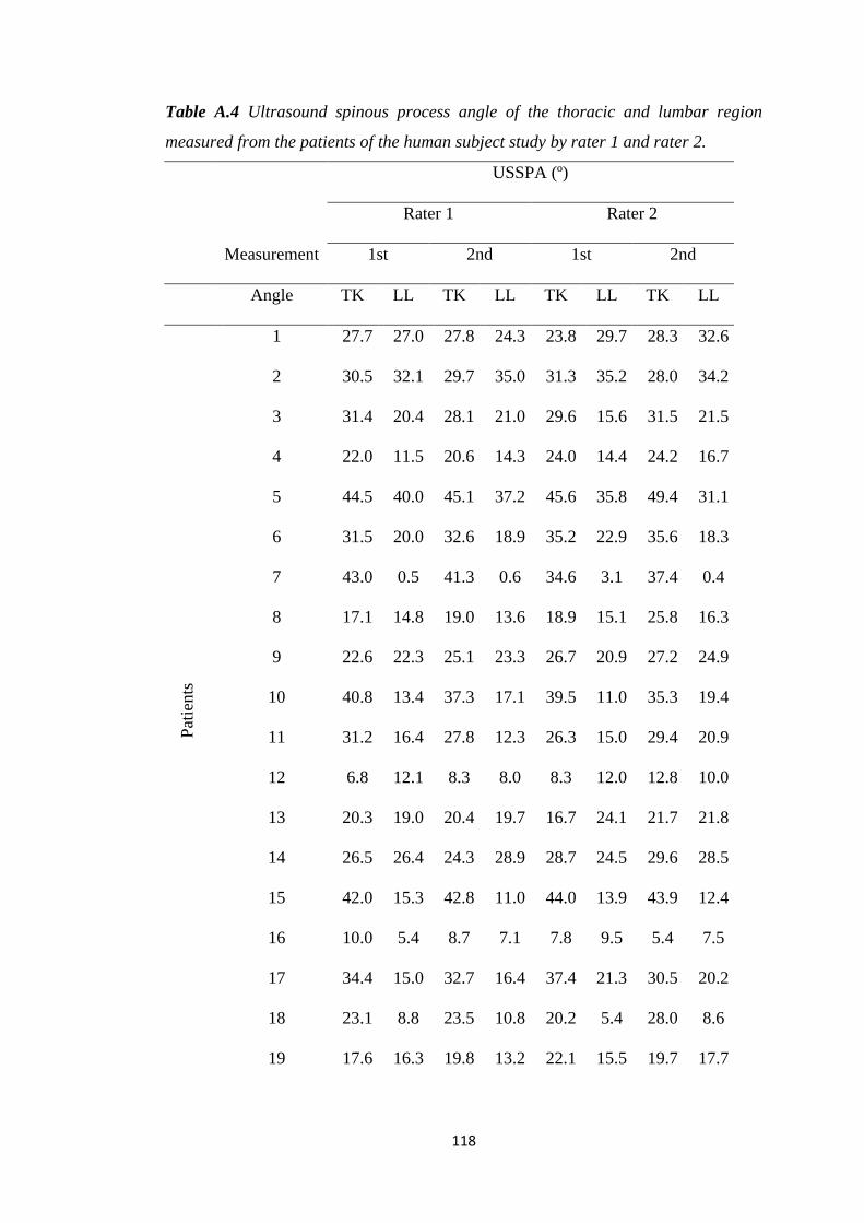
118
Table A.4 Ultrasound spinous process angle of the thoracic and lumbar region
measured from the patients of the human subject study by rater 1 and rater 2.
USSPA (º)
Rater 1 Rater 2
Measurement 1st 2nd 1st 2nd
Angle TK LL TK LL TK LL TK LL
Pat
ients
1 27.7 27.0 27.8 24.3 23.8 29.7 28.3 32.6
2 30.5 32.1 29.7 35.0 31.3 35.2 28.0 34.2
3 31.4 20.4 28.1 21.0 29.6 15.6 31.5 21.5
4 22.0 11.5 20.6 14.3 24.0 14.4 24.2 16.7
5 44.5 40.0 45.1 37.2 45.6 35.8 49.4 31.1
6 31.5 20.0 32.6 18.9 35.2 22.9 35.6 18.3
7 43.0 0.5 41.3 0.6 34.6 3.1 37.4 0.4
8 17.1 14.8 19.0 13.6 18.9 15.1 25.8 16.3
9 22.6 22.3 25.1 23.3 26.7 20.9 27.2 24.9
10 40.8 13.4 37.3 17.1 39.5 11.0 35.3 19.4
11 31.2 16.4 27.8 12.3 26.3 15.0 29.4 20.9
12 6.8 12.1 8.3 8.0 8.3 12.0 12.8 10.0
13 20.3 19.0 20.4 19.7 16.7 24.1 21.7 21.8
14 26.5 26.4 24.3 28.9 28.7 24.5 29.6 28.5
15 42.0 15.3 42.8 11.0 44.0 13.9 43.9 12.4
16 10.0 5.4 8.7 7.1 7.8 9.5 5.4 7.5
17 34.4 15.0 32.7 16.4 37.4 21.3 30.5 20.2
18 23.1 8.8 23.5 10.8 20.2 5.4 28.0 8.6
19 17.6 16.3 19.8 13.2 22.1 15.5 19.7 17.7
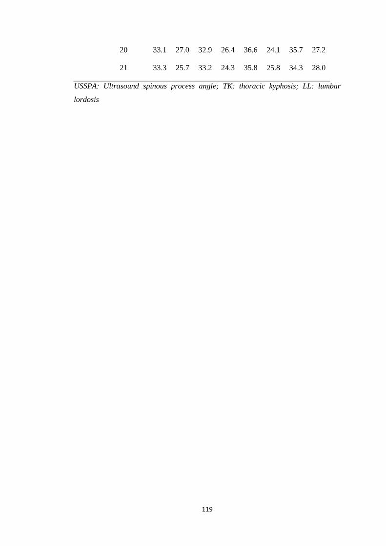
119
20 33.1 27.0 32.9 26.4 36.6 24.1 35.7 27.2
21 33.3 25.7 33.2 24.3 35.8 25.8 34.3 28.0
USSPA: Ultrasound spinous process angle; TK: thoracic kyphosis; LL: lumbar
lordosis
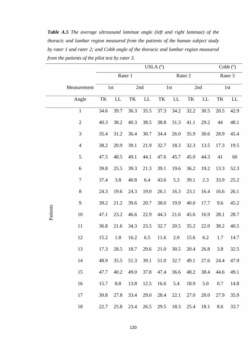
120
Table A.5 The average ultrasound laminae angle (left and right laminae) of the
thoracic and lumbar region measured from the patients of the human subject study
by rater 1 and rater 2; and Cobb angle of the thoracic and lumbar region measured
from the patients of the pilot test by rater 3.
USLA (º) Cobb (º)
Rater 1 Rater 2 Rater 3
Measurement 1st 2nd 1st 2nd 1st
Angle TK LL TK LL TK LL TK LL TK LL
Pat
ients
1 34.6 39.7 36.3 35.5 37.3 34.2 32.2 30.5 20.5 42.9
2 40.3 38.2 40.3 38.5 38.8 31.3 41.1 29.2 44 48.1
3 35.4 31.2 36.4 30.7 34.4 26.0 35.9 30.6 28.9 45.4
4 38.2 20.9 39.1 21.9 32.7 18.3 32.3 13.5 17.3 19.5
5 47.5 48.5 49.1 44.1 47.6 45.7 45.0 44.3 41 60
6 39.8 25.5 39.3 21.3 39.1 19.6 36.2 19.2 13.3 52.3
7 37.4 3.8 40.8 6.4 43.6 5.3 39.1 2.3 33.9 25.2
8 24.3 19.6 24.3 19.0 26.1 16.3 23.1 16.4 16.6 26.1
9 39.2 21.2 39.6 20.7 38.0 19.9 40.0 17.7 9.6 45.2
10 47.1 23.2 46.6 22.9 44.3 21.6 45.6 16.9 28.1 28.7
11 36.8 21.6 34.3 23.5 32.7 20.5 35.2 22.0 38.2 40.5
12 15.2 1.8 16.2 6.5 11.6 2.0 15.6 6.2 1.7 14.7
13 17.3 28.5 18.7 29.6 21.0 30.5 20.4 26.8 3.8 32.5
14 48.9 35.5 51.3 39.1 51.0 32.7 49.1 27.6 24.4 47.9
15 47.7 40.2 49.0 37.8 47.4 36.6 48.2 38.4 44.6 49.1
16 15.7 8.8 13.8 12.5 16.6 5.4 18.9 5.0 0.7 14.8
17 30.8 27.8 33.4 29.0 28.4 22.1 27.0 20.0 27.9 35.9
18 22.7 25.8 23.4 26.5 29.5 18.3 25.4 18.1 8.6 33.7
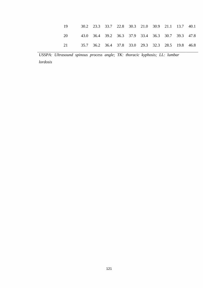
121
19 30.2 23.3 33.7 22.8 30.3 21.0 30.9 21.1 13.7 40.1
20 43.0 36.4 39.2 36.3 37.9 33.4 36.3 30.7 39.3 47.8
21 35.7 36.2 36.4 37.8 33.0 29.3 32.3 28.5 19.8 46.8
USSPA: Ultrasound spinous process angle; TK: thoracic kyphosis; LL: lumbar
lordosis
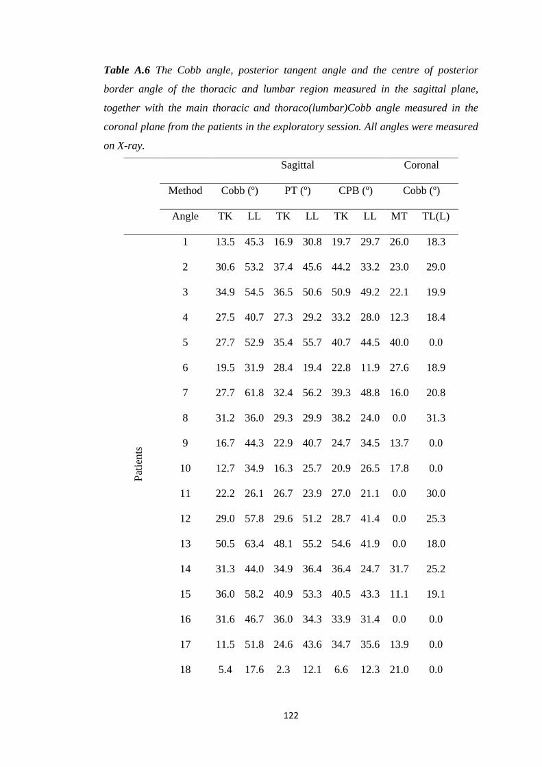
122
Table A.6 The Cobb angle, posterior tangent angle and the centre of posterior
border angle of the thoracic and lumbar region measured in the sagittal plane,
together with the main thoracic and thoraco(lumbar)Cobb angle measured in the
coronal plane from the patients in the exploratory session. All angles were measured
on X-ray.
Sagittal Coronal
Method Cobb (º) PT (º) CPB (º) Cobb (º)
Angle TK LL TK LL TK LL MT TL(L)
Pat
ients
1 13.5 45.3 16.9 30.8 19.7 29.7 26.0 18.3
2 30.6 53.2 37.4 45.6 44.2 33.2 23.0 29.0
3 34.9 54.5 36.5 50.6 50.9 49.2 22.1 19.9
4 27.5 40.7 27.3 29.2 33.2 28.0 12.3 18.4
5 27.7 52.9 35.4 55.7 40.7 44.5 40.0 0.0
6 19.5 31.9 28.4 19.4 22.8 11.9 27.6 18.9
7 27.7 61.8 32.4 56.2 39.3 48.8 16.0 20.8
8 31.2 36.0 29.3 29.9 38.2 24.0 0.0 31.3
9 16.7 44.3 22.9 40.7 24.7 34.5 13.7 0.0
10 12.7 34.9 16.3 25.7 20.9 26.5 17.8 0.0
11 22.2 26.1 26.7 23.9 27.0 21.1 0.0 30.0
12 29.0 57.8 29.6 51.2 28.7 41.4 0.0 25.3
13 50.5 63.4 48.1 55.2 54.6 41.9 0.0 18.0
14 31.3 44.0 34.9 36.4 36.4 24.7 31.7 25.2
15 36.0 58.2 40.9 53.3 40.5 43.3 11.1 19.1
16 31.6 46.7 36.0 34.3 33.9 31.4 0.0 0.0
17 11.5 51.8 24.6 43.6 34.7 35.6 13.9 0.0
18 5.4 17.6 2.3 12.1 6.6 12.3 21.0 0.0
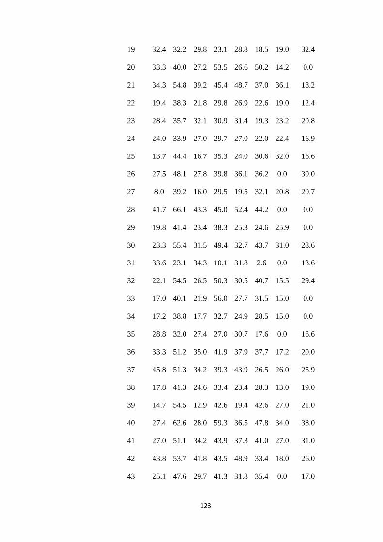
123
19 32.4 32.2 29.8 23.1 28.8 18.5 19.0 32.4
20 33.3 40.0 27.2 53.5 26.6 50.2 14.2 0.0
21 34.3 54.8 39.2 45.4 48.7 37.0 36.1 18.2
22 19.4 38.3 21.8 29.8 26.9 22.6 19.0 12.4
23 28.4 35.7 32.1 30.9 31.4 19.3 23.2 20.8
24 24.0 33.9 27.0 29.7 27.0 22.0 22.4 16.9
25 13.7 44.4 16.7 35.3 24.0 30.6 32.0 16.6
26 27.5 48.1 27.8 39.8 36.1 36.2 0.0 30.0
27 8.0 39.2 16.0 29.5 19.5 32.1 20.8 20.7
28 41.7 66.1 43.3 45.0 52.4 44.2 0.0 0.0
29 19.8 41.4 23.4 38.3 25.3 24.6 25.9 0.0
30 23.3 55.4 31.5 49.4 32.7 43.7 31.0 28.6
31 33.6 23.1 34.3 10.1 31.8 2.6 0.0 13.6
32 22.1 54.5 26.5 50.3 30.5 40.7 15.5 29.4
33 17.0 40.1 21.9 56.0 27.7 31.5 15.0 0.0
34 17.2 38.8 17.7 32.7 24.9 28.5 15.0 0.0
35 28.8 32.0 27.4 27.0 30.7 17.6 0.0 16.6
36 33.3 51.2 35.0 41.9 37.9 37.7 17.2 20.0
37 45.8 51.3 34.2 39.3 43.9 26.5 26.0 25.9
38 17.8 41.3 24.6 33.4 23.4 28.3 13.0 19.0
39 14.7 54.5 12.9 42.6 19.4 42.6 27.0 21.0
40 27.4 62.6 28.0 59.3 36.5 47.8 34.0 38.0
41 27.0 51.1 34.2 43.9 37.3 41.0 27.0 31.0
42 43.8 53.7 41.8 43.5 48.9 33.4 18.0 26.0
43 25.1 47.6 29.7 41.3 31.8 35.4 0.0 17.0
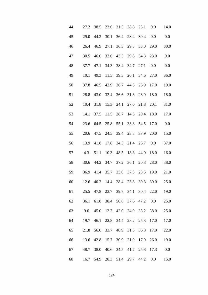
124
44 27.2 38.5 23.6 31.5 28.8 25.1 0.0 14.0
45 29.0 44.2 30.1 36.4 28.4 30.4 0.0 0.0
46 26.4 46.9 27.1 36.3 29.8 33.0 29.0 30.0
47 30.5 46.6 32.6 43.5 29.8 34.3 23.0 0.0
48 37.7 47.1 34.3 38.4 34.7 27.1 0.0 0.0
49 10.1 49.3 11.5 39.3 20.1 34.6 27.0 36.0
50 37.8 46.5 42.9 36.7 44.5 26.9 17.0 19.0
51 28.8 43.0 32.4 36.6 31.8 28.0 18.0 18.0
52 10.4 31.8 15.3 24.1 27.0 21.8 20.1 31.0
53 14.1 37.5 11.5 28.7 14.3 20.4 18.0 17.0
54 23.6 64.5 25.8 55.1 33.8 54.5 17.0 0.0
55 20.6 47.5 24.5 39.4 23.8 37.9 20.0 15.0
56 13.9 41.8 17.8 34.3 21.4 26.7 0.0 37.0
57 4.3 51.1 10.3 48.5 18.3 44.0 18.0 16.0
58 30.6 44.2 34.7 37.2 36.1 20.8 28.0 38.0
59 36.9 41.4 35.7 35.0 37.3 23.5 19.0 21.0
60 12.6 40.2 14.4 28.4 23.8 30.3 39.0 25.0
61 25.5 47.8 23.7 39.7 34.1 30.4 22.0 19.0
62 36.1 61.8 38.4 50.6 37.6 47.2 0.0 25.0
63 9.6 45.0 12.2 42.0 24.0 38.2 38.0 25.0
64 19.7 46.1 22.8 34.4 28.2 25.3 17.0 17.0
65 21.8 56.0 33.7 48.9 31.5 36.8 17.0 22.0
66 13.6 42.8 15.7 30.9 21.0 17.9 26.0 19.0
67 48.7 38.0 40.6 34.5 41.7 25.8 17.3 0.0
68 16.7 54.9 28.3 51.4 29.7 44.2 0.0 15.0
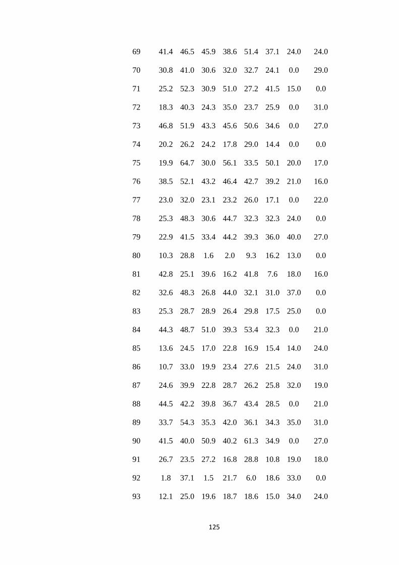
125
69 41.4 46.5 45.9 38.6 51.4 37.1 24.0 24.0
70 30.8 41.0 30.6 32.0 32.7 24.1 0.0 29.0
71 25.2 52.3 30.9 51.0 27.2 41.5 15.0 0.0
72 18.3 40.3 24.3 35.0 23.7 25.9 0.0 31.0
73 46.8 51.9 43.3 45.6 50.6 34.6 0.0 27.0
74 20.2 26.2 24.2 17.8 29.0 14.4 0.0 0.0
75 19.9 64.7 30.0 56.1 33.5 50.1 20.0 17.0
76 38.5 52.1 43.2 46.4 42.7 39.2 21.0 16.0
77 23.0 32.0 23.1 23.2 26.0 17.1 0.0 22.0
78 25.3 48.3 30.6 44.7 32.3 32.3 24.0 0.0
79 22.9 41.5 33.4 44.2 39.3 36.0 40.0 27.0
80 10.3 28.8 1.6 2.0 9.3 16.2 13.0 0.0
81 42.8 25.1 39.6 16.2 41.8 7.6 18.0 16.0
82 32.6 48.3 26.8 44.0 32.1 31.0 37.0 0.0
83 25.3 28.7 28.9 26.4 29.8 17.5 25.0 0.0
84 44.3 48.7 51.0 39.3 53.4 32.3 0.0 21.0
85 13.6 24.5 17.0 22.8 16.9 15.4 14.0 24.0
86 10.7 33.0 19.9 23.4 27.6 21.5 24.0 31.0
87 24.6 39.9 22.8 28.7 26.2 25.8 32.0 19.0
88 44.5 42.2 39.8 36.7 43.4 28.5 0.0 21.0
89 33.7 54.3 35.3 42.0 36.1 34.3 35.0 31.0
90 41.5 40.0 50.9 40.2 61.3 34.9 0.0 27.0
91 26.7 23.5 27.2 16.8 28.8 10.8 19.0 18.0
92 1.8 37.1 1.5 21.7 6.0 18.6 33.0 0.0
93 12.1 25.0 19.6 18.7 18.6 15.0 34.0 24.0
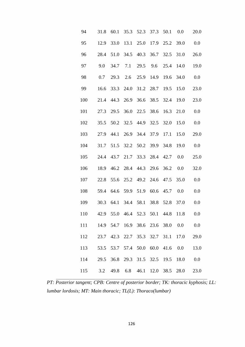
126
94 31.8 60.1 35.3 52.3 37.3 50.1 0.0 20.0
95 12.9 33.0 13.1 25.0 17.9 25.2 39.0 0.0
96 28.4 51.0 34.5 40.3 36.7 32.5 31.0 26.0
97 9.0 34.7 7.1 29.5 9.6 25.4 14.0 19.0
98 0.7 29.3 2.6 25.9 14.9 19.6 34.0 0.0
99 16.6 33.3 24.0 31.2 28.7 19.5 15.0 23.0
100 21.4 44.3 26.9 36.6 38.5 32.4 19.0 23.0
101 27.3 29.5 36.0 22.5 38.6 16.3 21.0 0.0
102 35.5 50.2 32.5 44.9 32.5 32.0 15.0 0.0
103 27.9 44.1 26.9 34.4 37.9 17.1 15.0 29.0
104 31.7 51.5 32.2 50.2 39.9 34.8 19.0 0.0
105 24.4 43.7 21.7 33.3 28.4 42.7 0.0 25.0
106 18.9 46.2 28.4 44.3 29.6 36.2 0.0 32.0
107 22.8 55.6 25.2 49.2 24.6 47.5 35.0 0.0
108 59.4 64.6 59.9 51.9 60.6 45.7 0.0 0.0
109 30.3 64.1 34.4 58.1 38.8 52.8 37.0 0.0
110 42.9 55.0 46.4 52.3 50.1 44.8 11.8 0.0
111 14.9 54.7 16.9 38.6 23.6 38.0 0.0 0.0
112 23.7 42.3 22.7 35.3 32.7 31.1 17.0 29.0
113 53.5 53.7 57.4 50.0 60.0 41.6 0.0 13.0
114 29.5 36.8 29.3 31.5 32.5 19.5 18.0 0.0
115 3.2 49.8 6.8 46.1 12.0 38.5 28.0 23.0
PT: Posterior tangent; CPB: Centre of posterior border; TK: thoracic kyphosis; LL:
lumbar lordosis; MT: Main thoracic; TL(L): Thoraco(lumbar)
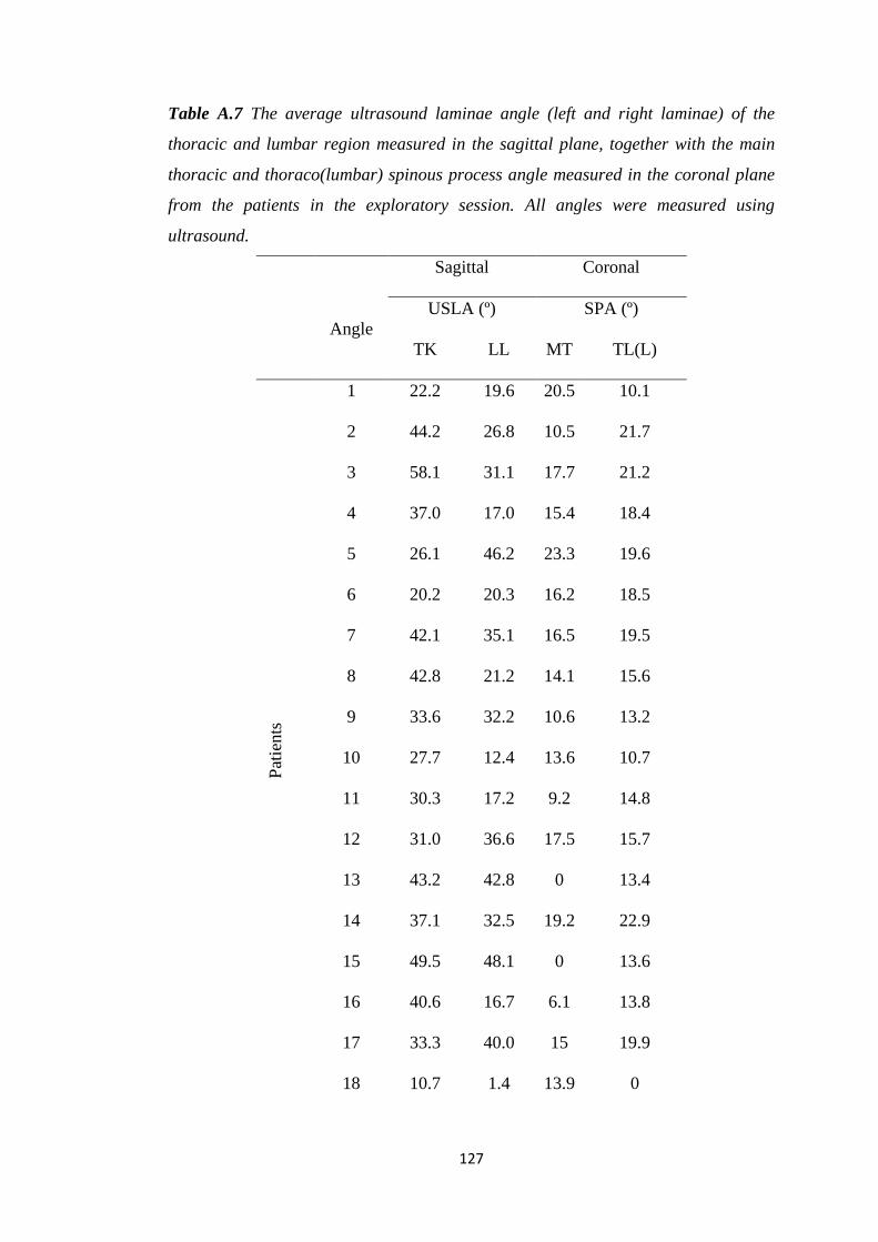
127
Table A.7 The average ultrasound laminae angle (left and right laminae) of the
thoracic and lumbar region measured in the sagittal plane, together with the main
thoracic and thoraco(lumbar) spinous process angle measured in the coronal plane
from the patients in the exploratory session. All angles were measured using
ultrasound.
Sagittal Coronal
Angle
USLA (º) SPA (º)
TK LL MT TL(L)
Pat
ients
1 22.2 19.6 20.5 10.1
2 44.2 26.8 10.5 21.7
3 58.1 31.1 17.7 21.2
4 37.0 17.0 15.4 18.4
5 26.1 46.2 23.3 19.6
6 20.2 20.3 16.2 18.5
7 42.1 35.1 16.5 19.5
8 42.8 21.2 14.1 15.6
9 33.6 32.2 10.6 13.2
10 27.7 12.4 13.6 10.7
11 30.3 17.2 9.2 14.8
12 31.0 36.6 17.5 15.7
13 43.2 42.8 0 13.4
14 37.1 32.5 19.2 22.9
15 49.5 48.1 0 13.6
16 40.6 16.7 6.1 13.8
17 33.3 40.0 15 19.9
18 10.7 1.4 13.9 0
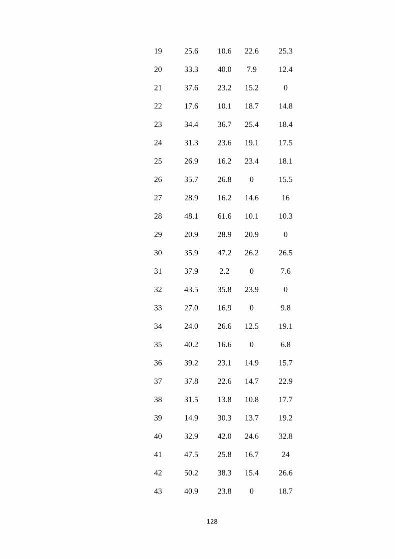
128
19 25.6 10.6 22.6 25.3
20 33.3 40.0 7.9 12.4
21 37.6 23.2 15.2 0
22 17.6 10.1 18.7 14.8
23 34.4 36.7 25.4 18.4
24 31.3 23.6 19.1 17.5
25 26.9 16.2 23.4 18.1
26 35.7 26.8 0 15.5
27 28.9 16.2 14.6 16
28 48.1 61.6 10.1 10.3
29 20.9 28.9 20.9 0
30 35.9 47.2 26.2 26.5
31 37.9 2.2 0 7.6
32 43.5 35.8 23.9 0
33 27.0 16.9 0 9.8
34 24.0 26.6 12.5 19.1
35 40.2 16.6 0 6.8
36 39.2 23.1 14.9 15.7
37 37.8 22.6 14.7 22.9
38 31.5 13.8 10.8 17.7
39 14.9 30.3 13.7 19.2
40 32.9 42.0 24.6 32.8
41 47.5 25.8 16.7 24
42 50.2 38.3 15.4 26.6
43 40.9 23.8 0 18.7
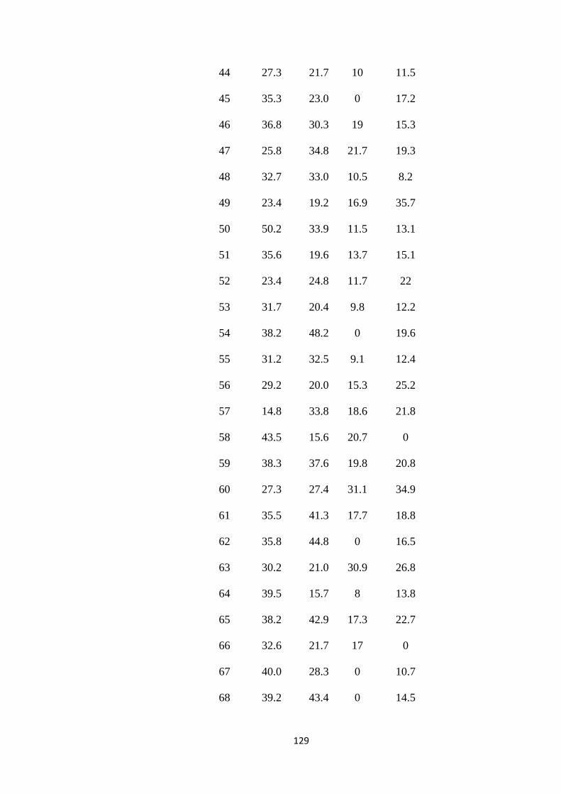
129
44 27.3 21.7 10 11.5
45 35.3 23.0 0 17.2
46 36.8 30.3 19 15.3
47 25.8 34.8 21.7 19.3
48 32.7 33.0 10.5 8.2
49 23.4 19.2 16.9 35.7
50 50.2 33.9 11.5 13.1
51 35.6 19.6 13.7 15.1
52 23.4 24.8 11.7 22
53 31.7 20.4 9.8 12.2
54 38.2 48.2 0 19.6
55 31.2 32.5 9.1 12.4
56 29.2 20.0 15.3 25.2
57 14.8 33.8 18.6 21.8
58 43.5 15.6 20.7 0
59 38.3 37.6 19.8 20.8
60 27.3 27.4 31.1 34.9
61 35.5 41.3 17.7 18.8
62 35.8 44.8 0 16.5
63 30.2 21.0 30.9 26.8
64 39.5 15.7 8 13.8
65 38.2 42.9 17.3 22.7
66 32.6 21.7 17 0
67 40.0 28.3 0 10.7
68 39.2 43.4 0 14.5
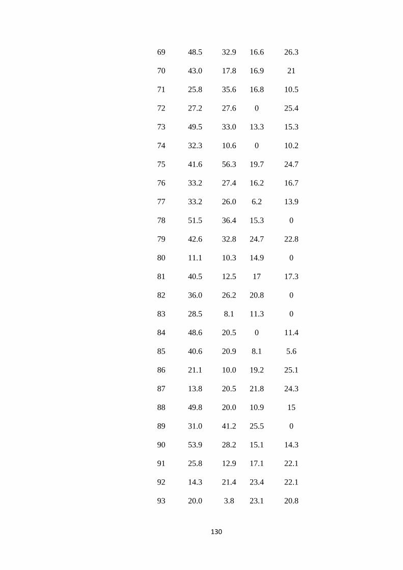
130
69 48.5 32.9 16.6 26.3
70 43.0 17.8 16.9 21
71 25.8 35.6 16.8 10.5
72 27.2 27.6 0 25.4
73 49.5 33.0 13.3 15.3
74 32.3 10.6 0 10.2
75 41.6 56.3 19.7 24.7
76 33.2 27.4 16.2 16.7
77 33.2 26.0 6.2 13.9
78 51.5 36.4 15.3 0
79 42.6 32.8 24.7 22.8
80 11.1 10.3 14.9 0
81 40.5 12.5 17 17.3
82 36.0 26.2 20.8 0
83 28.5 8.1 11.3 0
84 48.6 20.5 0 11.4
85 40.6 20.9 8.1 5.6
86 21.1 10.0 19.2 25.1
87 13.8 20.5 21.8 24.3
88 49.8 20.0 10.9 15
89 31.0 41.2 25.5 0
90 53.9 28.2 15.1 14.3
91 25.8 12.9 17.1 22.1
92 14.3 21.4 23.4 22.1
93 20.0 3.8 23.1 20.8
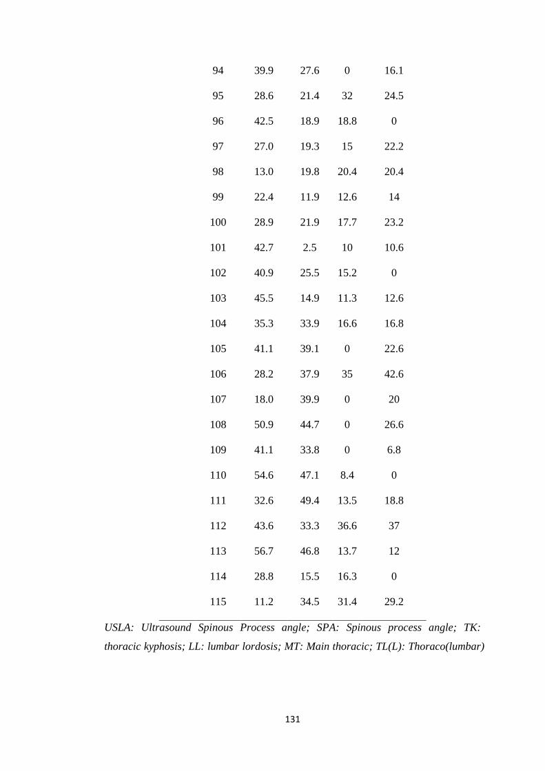
131
94 39.9 27.6 0 16.1
95 28.6 21.4 32 24.5
96 42.5 18.9 18.8 0
97 27.0 19.3 15 22.2
98 13.0 19.8 20.4 20.4
99 22.4 11.9 12.6 14
100 28.9 21.9 17.7 23.2
101 42.7 2.5 10 10.6
102 40.9 25.5 15.2 0
103 45.5 14.9 11.3 12.6
104 35.3 33.9 16.6 16.8
105 41.1 39.1 0 22.6
106 28.2 37.9 35 42.6
107 18.0 39.9 0 20
108 50.9 44.7 0 26.6
109 41.1 33.8 0 6.8
110 54.6 47.1 8.4 0
111 32.6 49.4 13.5 18.8
112 43.6 33.3 36.6 37
113 56.7 46.8 13.7 12
114 28.8 15.5 16.3 0
115 11.2 34.5 31.4 29.2
USLA: Ultrasound Spinous Process angle; SPA: Spinous process angle; TK:
thoracic kyphosis; LL: lumbar lordosis; MT: Main thoracic; TL(L): Thoraco(lumbar)
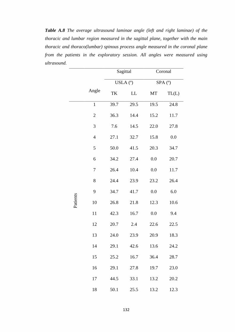
132
Table A.8 The average ultrasound laminae angle (left and right laminae) of the
thoracic and lumbar region measured in the sagittal plane, together with the main
thoracic and thoraco(lumbar) spinous process angle measured in the coronal plane
from the patients in the exploratory session. All angles were measured using
ultrasound.
Sagittal Coronal
Angle
USLA (º) SPA (º)
TK LL MT TL(L)
Pat
ients
1 39.7 29.5 19.5 24.8
2 36.3 14.4 15.2 11.7
3 7.6 14.5 22.0 27.8
4 27.1 32.7 15.8 0.0
5 50.0 41.5 20.3 34.7
6 34.2 27.4 0.0 20.7
7 26.4 10.4 0.0 11.7
8 24.4 23.9 23.2 26.4
9 34.7 41.7 0.0 6.0
10 26.8 21.8 12.3 10.6
11 42.3 16.7 0.0 9.4
12 20.7 2.4 22.6 22.5
13 24.0 23.9 20.9 18.3
14 29.1 42.6 13.6 24.2
15 25.2 16.7 36.4 28.7
16 29.1 27.8 19.7 23.0
17 44.5 33.1 13.2 20.2
18 50.1 25.5 13.2 12.3
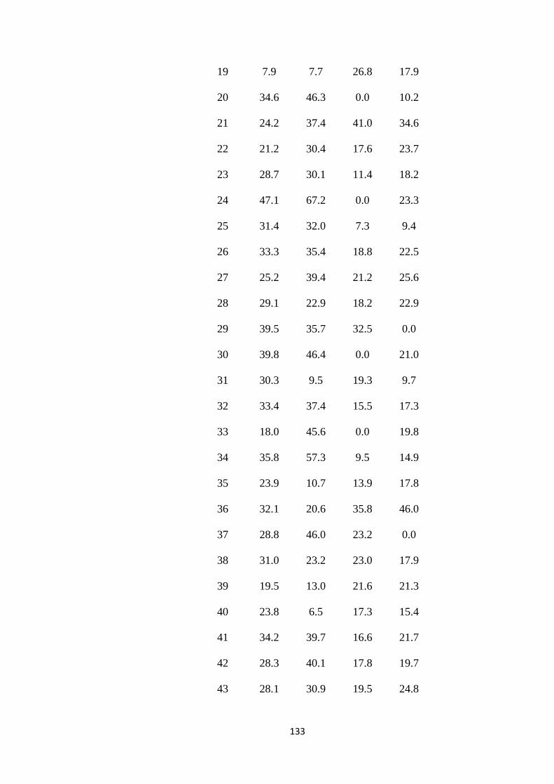
133
19 7.9 7.7 26.8 17.9
20 34.6 46.3 0.0 10.2
21 24.2 37.4 41.0 34.6
22 21.2 30.4 17.6 23.7
23 28.7 30.1 11.4 18.2
24 47.1 67.2 0.0 23.3
25 31.4 32.0 7.3 9.4
26 33.3 35.4 18.8 22.5
27 25.2 39.4 21.2 25.6
28 29.1 22.9 18.2 22.9
29 39.5 35.7 32.5 0.0
30 39.8 46.4 0.0 21.0
31 30.3 9.5 19.3 9.7
32 33.4 37.4 15.5 17.3
33 18.0 45.6 0.0 19.8
34 35.8 57.3 9.5 14.9
35 23.9 10.7 13.9 17.8
36 32.1 20.6 35.8 46.0
37 28.8 46.0 23.2 0.0
38 31.0 23.2 23.0 17.9
39 19.5 13.0 21.6 21.3
40 23.8 6.5 17.3 15.4
41 34.2 39.7 16.6 21.7
42 28.3 40.1 17.8 19.7
43 28.1 30.9 19.5 24.8
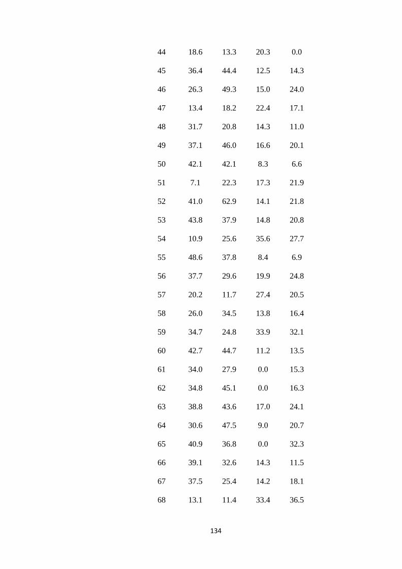
134
44 18.6 13.3 20.3 0.0
45 36.4 44.4 12.5 14.3
46 26.3 49.3 15.0 24.0
47 13.4 18.2 22.4 17.1
48 31.7 20.8 14.3 11.0
49 37.1 46.0 16.6 20.1
50 42.1 42.1 8.3 6.6
51 7.1 22.3 17.3 21.9
52 41.0 62.9 14.1 21.8
53 43.8 37.9 14.8 20.8
54 10.9 25.6 35.6 27.7
55 48.6 37.8 8.4 6.9
56 37.7 29.6 19.9 24.8
57 20.2 11.7 27.4 20.5
58 26.0 34.5 13.8 16.4
59 34.7 24.8 33.9 32.1
60 42.7 44.7 11.2 13.5
61 34.0 27.9 0.0 15.3
62 34.8 45.1 0.0 16.3
63 38.8 43.6 17.0 24.1
64 30.6 47.5 9.0 20.7
65 40.9 36.8 0.0 32.3
66 39.1 32.6 14.3 11.5
67 37.5 25.4 14.2 18.1
68 13.1 11.4 33.4 36.5
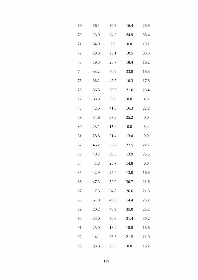
135
69 36.1 30.6 16.4 20.9
70 13.9 24.2 24.9 38.4
71 34.0 2.9 0.0 19.7
72 29.3 23.1 18.5 36.3
73 19.8 28.7 18.4 16.2
74 33.2 40.9 33.8 18.3
75 38.2 47.7 10.3 17.8
76 56.5 30.0 21.6 26.4
77 33.9 3.9 0.0 4.3
78 42.6 41.8 16.3 22.2
79 34.6 37.3 25.2 0.0
80 23.1 15.4 0.0 3.4
81 28.8 21.4 13.0 0.0
82 45.2 52.8 27.5 22.7
83 40.2 39.5 13.9 25.5
84 41.8 25.7 14.8 0.0
85 42.9 25.4 13.0 16.8
86 47.3 52.9 30.7 21.6
87 17.5 34.8 26.6 21.3
88 51.0 45.0 14.4 23.2
89 20.2 40.9 35.8 25.2
90 33.0 30.6 31.4 30.2
91 25.0 18.4 18.8 18.6
92 14.1 26.5 21.5 11.0
93 33.8 23.3 0.0 16.2
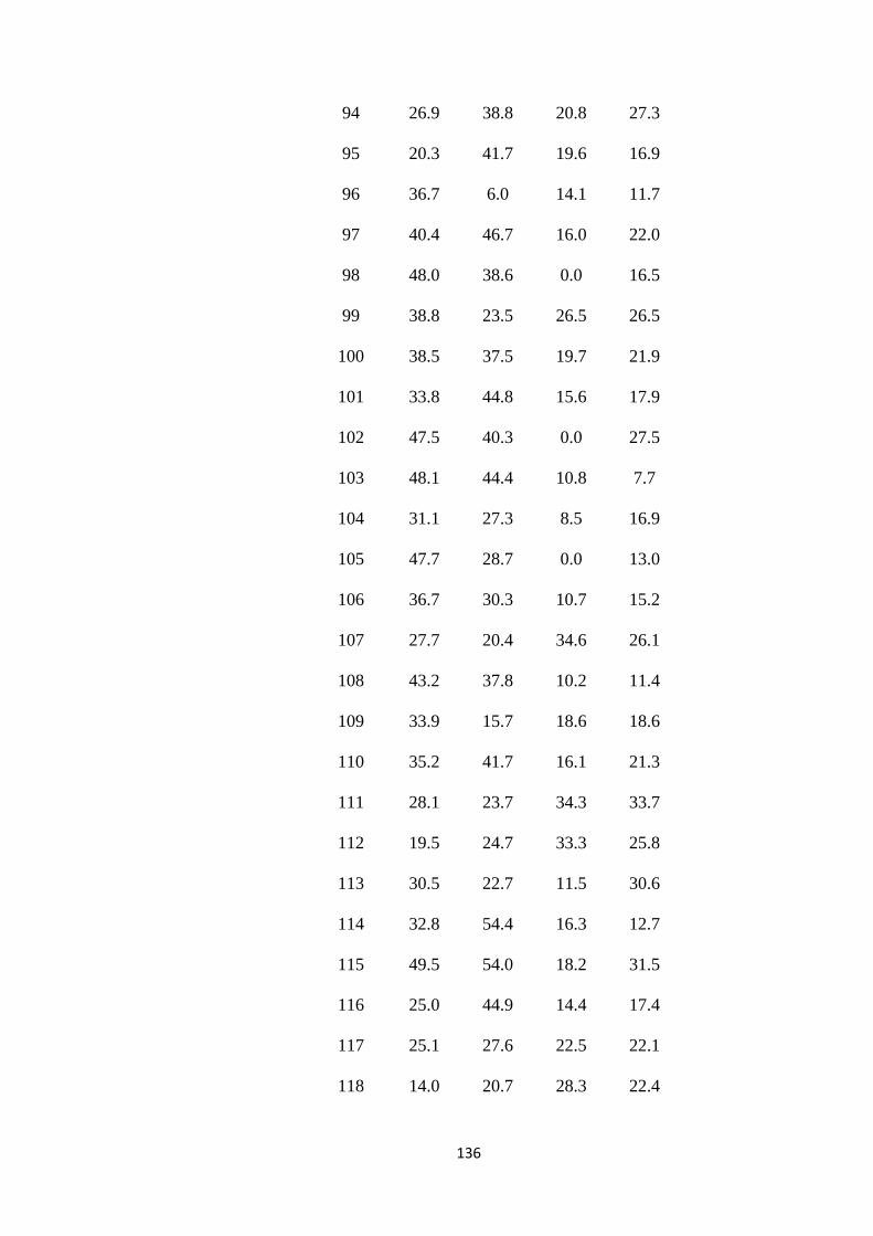
136
94 26.9 38.8 20.8 27.3
95 20.3 41.7 19.6 16.9
96 36.7 6.0 14.1 11.7
97 40.4 46.7 16.0 22.0
98 48.0 38.6 0.0 16.5
99 38.8 23.5 26.5 26.5
100 38.5 37.5 19.7 21.9
101 33.8 44.8 15.6 17.9
102 47.5 40.3 0.0 27.5
103 48.1 44.4 10.8 7.7
104 31.1 27.3 8.5 16.9
105 47.7 28.7 0.0 13.0
106 36.7 30.3 10.7 15.2
107 27.7 20.4 34.6 26.1
108 43.2 37.8 10.2 11.4
109 33.9 15.7 18.6 18.6
110 35.2 41.7 16.1 21.3
111 28.1 23.7 34.3 33.7
112 19.5 24.7 33.3 25.8
113 30.5 22.7 11.5 30.6
114 32.8 54.4 16.3 12.7
115 49.5 54.0 18.2 31.5
116 25.0 44.9 14.4 17.4
117 25.1 27.6 22.5 22.1
118 14.0 20.7 28.3 22.4
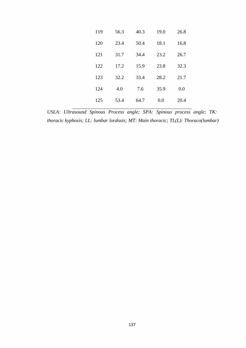
137
119 56.3 40.3 19.0 26.8
120 23.4 50.4 18.1 16.8
121 31.7 34.4 23.2 26.7
122 17.2 15.9 23.8 32.3
123 32.2 33.4 28.2 21.7
124 4.0 7.6 35.9 0.0
125 53.4 64.7 0.0 20.4
USLA: Ultrasound Spinous Process angle; SPA: Spinous process angle; TK:
thoracic kyphosis; LL: lumbar lordosis; MT: Main thoracic; TL(L): Thoraco(lumbar)
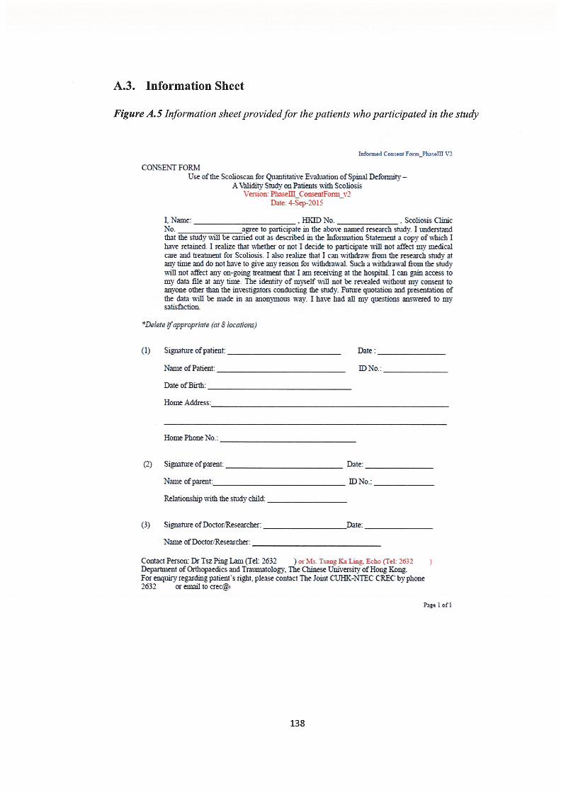

140
References
Abelin-Genevois K, Idjerouidene A, Roussouly P, Vital JM, Garin C. Cervical spine
alignment in the pediatric population: a radiographic normative study of 150
asymptomatic patients. Eur Spine J 2014;23(7):1442-8
Adams MA, Mannion AF, Dolan P. Personal risk factors for first-time low back pain.
Spine 1999;24(23):2497-50
Agadir M, Sevastik B, Sevastik JA, et al. Induction of scoliosis in the growing rabbit
by unilateral rib-growth stimulation. Spine 1988;13:1065-9
Dimeglio A, Canavese F and Bonnel F. Normal Growth of the Spine and Thorax.
The growing spine. Chapter 4. P.48-80
Alexander MA, Season EH. Idiopathic scoliosis: an electromyographic study. Arch
Phys Med Rehabil 1978;59:314-5
Alricsson M, Werner S. Young elite cross-country skiers and low back pain-A 5-year
study. Phys Ther Sport 2006;7(4):181-4
Ames CP, Blondel B, Scheer JK, Schwab FJ, Le Huec JC, Massicotte EM, Patel AA,
Traynelis VC, Kim HJ, Shaffrey CI, Smith JS, Lafage V. Cervical
radiographical alignment: comprehensive assessment techniques and potential
importance in cervical myelopathy. Spine 2013;38(22):S149-60
Aprin H, Bowen JR, MacEwen GD, Hall JE. Spine fusion in patients with spinal
muscular atrophy. J Bone Joint Surg Am. 1982;64:1179-87
Archer IA, Dickson RA. Stature and idiopathic scoliosis. A prospective study. J
Bone Joint Surg Br 1985;67:185-8
Arkin AM. The mechanism of the structural changes in scoliosis. J Bone Joint Surg
Am 1949;31:519-28
141
Aronsson DD, Stokes IA, Rosovsky J, Spence H. Mechanical modulation of calf tail
vertebral growth: implications for scoliosis progression. J Spinal Disord
1999;12:141-6
Asher MA, Burton DC. Adolescent idiopathic scoliosis: natural history and long term
treatment effects. Scoliosis 2006;1(1):2
Aylott CE, Puna R, Robertson PA, Walker C. Spinous process morphology: the
effect of ageing through adulthood on spinous process size and relationship to
sagittal alignment. Eur Spine J 2012;21(5):1007-12
Baik JS, Kim JY, Park JH, Han SW, Park JH, Lee MS. Scoliosis in patients with
Parkinson’s disease. J Clin Neurol 2009;5(2):91-94
Bao H, Liu Z, Bao M, Zhu Z, Yan P, Liu S, Feng Z, Qian B, Qiu Y. Predicted final
spinal height in patients with adolescent idiopathic scoliosis can be achieved by
surgery regardless of maturity status. Bone Joint J. 2018;100-B(10):1372-1376.
Barrey C, Jund J, Noseda O, Roussouly P. Sagittal balance of the pelvis-spine
complex and lumbar degenerative diseases: a comparative study about 85 cases.
Eur Spine J 2007;16:1459-67
Barrey C, Roussouly P, Le Huec JC, D'Acunzi G, Perrin G. Compensatory
mechanisms contributing to keep the sagittal balance of the spine. Eur Spine J
2013;22 Suppl 6:S834-41
Beach TA, Parkinson RJ, Stothart JP, Callaghan JP. Effects of prolonged sitting on
the passive flexion stiffness of the in vivo lumbar spine. Spine J 2005;5(2):145-
54
Beguiristain JL, De Salis J, Oriaifo A, Cañadell J. Experimental scoliosis by
epiphysiodesis in pigs. Int Orthop 1980;3:317-21
142
Bernhardt M, Bridwell KH. Segmental analysis of the sagittal plane alignment of the
normal thoracic and lumbar spines and thoracolumbar junction. Spine
1989;14:717-21
Betsch M, Wehrle R, Dor L, Rapp W, Jungbluth P, Hakimi M, Wild M. Spinal
posture and pelvic position during pregnancy:a prospective rasterstereographic
pilot study. Eur Spine J 2015;24(6):1282-8
Betz RR, Harms J, Clements DH III, Lenke LG, Lowe TG, Shufflebarger HL,
Jeszenszky D, Beele B. Comparison of anterior and posterior instrumentation for
correction of adolescent thoracic idiopathic scoliosis. Spine 1999;24:225-39
Bick EM, Barash ES, Lehman L. Congenital scoliosis without vertebral anomaly; the
vertebral histology of two cases. Clin Orthop 1958;12:285-90
Birchall D, Hughes DG, Hindle J, Robinson L, Williamson JB. Measurement of
vertebral rotation in adolescent idiopathic scoliosis using three-dimensional
magnetic resonance imaging. Spine 1997;22:2403-7
Bissolotti L, Donzelli S, Gobbo M, Zaina F, Villafañe JH, Negrini S. Association
between sagittal and scoliosis in patients with Parkinson Disease: A Cross-
sectional study. Am J Phys Med Rehabil 2016;95(1):39-46
Bissolotti L, Gobbo M, Villafane JH, Negrini S. Spinopelvic balance: New
biomechanical insights with clinical implications for Parkinson’s disease. Eur
Spine J 2014;23(3):576-83
Boesker EH, Moe JH, Winter RB, Koop SE. A determination of the normal thoracic
kyphosis: a roentgenographic study of 121 normal children. J Pediatr Orthop
2000;20:796-8
Bone CM, Hsieh GH. The risk of carcinogenesis from radiographs to pediatric
orthopaedic patients. J Pediatr Orthop 2000;20:251-4
143
Booth KC, Bridwell KH, Lenke LG, Baldus CR, Blanke KM. Complications and
predictive factors for the successful treatment of flatback deformity (fixed
sagittal imbalance). Spine 1999;24(16):1712-20
Boseker EH, Moe JH, Winter RB, Koop SE. Determination of "normal" thoracic
kyphosis:a roentgenographic study of 121 "normal" children. J Pediatr Orthop.
2000;20(6):796-8
Bridwell KH, Bernhardt M. Segmental Analysis of the sagittal plane alignment of the
normal thoracic and lumbar spines and thoracolumbar junction. Spine
1989;14:717-21
Bridwell KH, Betz RR, Capelli AM, Huss G, Harvey C. Sagittal plane analysis in
idiopathic scoliosis patients treated with Cotrel–Dubousset instrumentation.
Spine 1990;15:921-6
Briggs AM, Wrigley TV, Tully EA, Adams PE, Greig AM, Bennell KL.
Radiographic measures of thoracic kyphosis in osteoporosis: Cobb and vertebral
centroid angles. Skeletal Radiol 2007;36(8):761-7
Brink RC, Colo D, Schlösser TPC, Vincken KL, van Stralen M, Hui SCN, Shi L,
Chu WCW, Cheng JCY, Castelein RM. Upright, prone, and supine spinal
morphology and alignment in adolescent idiopathic scoliosis. Scoliosis Spinal
Disord. 2017;12:6
Brink RC, Wijdicks SPJ, Tromp IN, Schlösser TPC, Kruyt MC, Beek FJA, Castelein
RM. A reliability and validity study for different coronal angles using ultrasound
imaging in adolescent idiopathic scoliosis. Spine J. 2018;18(6):979-85
Bunnell WP. An objective criterion for scoliosis screening. J Bone Joint Surg Am
1984;66(9):1381-7
144
Burwell RG. Comment to “The pathogenesis of idiopathic scoliosis: uncoupled
neuro-osseous growth?” by R. W. Porter. Eur Spine J 2001;10:482-7
Canavese F, Turcot K, De Rosa V, de Coulon G, Kaelin A. Cervical spine sagittal
alignment variations following posterior spinal fusion and instrumentation
foradolescent idiopathic scoliosis. Eur Spine J 2011;20(7):1141-8.
Carlson BB, Burton DC, Asher MA. Comparison of trunk and spine deformity in
adolescent idiopathic scoliosis. Scoliosis 2013;8(1):2
Castelein RM, van Dieen JH, Smit TH. The role of dorsal shear forces in the
pathogenesis of adolescent idiopathic scoliosis: a hypothesis. Med Hypotheses
2005;65:501-8
Castelein RM, Veraart B. Idiopathic scoliosis: prognostic value of the profile. Eur
Spine J 1992;1:167-9.
Chaise FO, Candotti CT, Torre ML, Furlanetto TS, Pelinson PP, Loss JF. Validation,
repeatability and reproducibility of a noninvasive instrument for measuring
thoracic and lumbar curvature of the spine in the sagittal plane. Rev Bras
Fisioter 2011;15(6):511-7
Chaléat-Valayer E, Mac-Thiong JM, Paquet J, Berthonnaud E, Siani F, Roussouly P.
Sagittal spino-pelvic alignment in chronic low back pain. Eur Spine J 2011;20
Suppl 5:634-40
Chen W, Le LH, Lou EH. 2016. Reliability of the axial vertebral rotation
measurements of adolescent idiopathic scoliosis using the center of lamina
method on ultrasound images: in vitro and in vivo study. Eur Spine J
25(10):3265-73
145
Chen W, Lou EH, Le LH. Using ultrasound imaging to identify landmarks in
vertebra models to assess spinal deformity. Conf Proc IEEE Eng Med Biol Soc
2011;2011:8495-8
Chen W, Lou EH, Zhang PQ, Le LH, Hill D. Reliability of assessing the coronal
curvature of children with scoliosis by using ultrasound images. J Child Orthop
2013;7(6):521-9
Chen YL. Vertebral centroid measurement of lumbar lordosis compared with the
Cobb technique. Spine 1999;24(17):1786-90
Cheng JC1, Guo X, Sher AH, Chan YL, Metreweli C. Correlation between curve
severity, somatosensory evoked potentials, and magnetic resonance imaging in
adolescent idiopathic scoliosis. Spine 1999;24:1679-84
Cheng JC, Guo X. Osteopenia in adolescent idiopathic scoliosis. A primaryproblem
or secondary to the spinal deformity? Spine 1997;22:1716-21
Chernukha KV, Daffner RH, Reigel DH. Lumbar lordosis measurement. A new
method versus Cobb technique. Spine 1998;23(1):74-9; discussion 79-80
Cheung CJ, Zhou GQ, Law SY, Lai KL, Jiang WW, Zheng YP. Freehand three-
dimensional ultrasound system for assessment of scoliosis. J Orthop Translat
2015;3(3):123-33
Cheung CS, Lee WT, Tse YK, Lee KM, Guo X, Qin L, Cheng JC. Generalized
osteopenia in adolescent idiopathic scoliosis–association with abnormal pubertal
growth, bone turnover, and calcium intake? Spine 2006;31:330-8
Cheung CW, Law SY, Zheng YP. Development of 3-D ultrasound system for
assessment of adolescent idiopathic scoliosis (AIS): and system validation. Conf
Proc IEEE Eng Med Biol Soc 2013;2013:6474-7
146
Cheung CWJ, Zheng YP. Development of 3-D Ultrasound System for Assessment of
Adolescent Idiopathic Scoliosis. In: Lim CT, Goh JCH, ed. WCB 2010, IFMBE
Proceedings 31, 2010:584-7
Cheung J, Halbertsma JP, Veldhuizen AG, Sluiter WJ, Maurits NM, Cool JC, van
Horn JR. A preliminary study on electromyographic analysis of the paraspinal
musculature in idiopathic scoliosis. Eur Spine J 2005;14:130-7
Cho KJ, Kim YT, Shin SH, Suk SI. Surgical treatment of adult degenerative scoliosis.
Asian Spine J 2014;8(3):371-81
Chockalingam N, Dangerfield PH, Giakas G, Cochrane T, Dorgan JC. Computer-
assisted Cobb measurement of scoliosis. Eur Spine J 2002;11(4):353-7
Chu WC, Lam WW, Chan YL, Ng BK, Lam TP, Lee KM, Guo X, Cheng JC.
Relative shortening and functional tethering of spinal cord in adolescent
idiopathic scoliosis? Study with multiplanar reformat magnetic resonance
imaging and somatosensory evoked potential. Spine 2006;31(1):E19-25
Cil A, Yazici M, Uzumcugil A, Kandemir U, Alanay A, Alanay Y, Acaroglu RE,
Surat A. The evolution of sagittal segmental alignment of the spine during
childhood. Spine 2005;30:93-100
Clements DH, Marks M, Newton PO, Betz RR, Lenke L, Shufflebarger H; Harms
Study Group. Did the Lenke classification change scoliosis treatment? Spine
2011;36:1142-5
Cobb JR. Outline for the study of scoliosis. In American Academy of Orthopaedic
Surgeons, Instructional Course Lectures. St Louis, CV Mosby, 1948, pp. 261-75
Coelho DM, Bonagamba GH, Oliveira AS. Scoliometer measurements of patients
with idiopathic scoliosis. Braz J Phys Ther 2013;17(2):179-84
147
Colonna PC, Vom Saal F. A study of paralytic scoliosis based on five hundred cases
of poliomyelitis. J Bone Joint Surg Am 1941;335-53
Cook SD, Harding AF, Morgan EL, Nicholson RJ, Thomas KA, Whitecloud TS,
Ratner ES. Trabecular bone mineral density in idiopathic scoliosis. J Pediatr
Orthop 1987;7:168-74
Currier DP. Elements of research in physical therapy. 3rd edn Williams & Wilkins
1984, Baltimore, pp 160-71
D’Aouˆt K, Aerts P, De Clercq D, De Meester K, Van Elsacker L. Segment and joint
angles of hind limb during bipedal and quadrupedal walking of the bonobo (Pan
paniscus). Am J Phys Anthropol 2002;119:37-51
Dawson B, Trapp RG. Basic and Clinical Biostatistics. 4th Ed. New York: Lange
Medical Books/McGraw-Hill; 2004
de Jonge T, Dubousset JF, Illés T. Sagittal plane correction in idiopathic scoliosis.
Spine 2002;27(7):754-60
de Oliveira TS, Candotti CT, La Torre M, Pelinson PP, Furlanetto TS, Kutchak FM,
Loss JF. Validity and reproducibility of the measurements obtained using the
flexicurve instrument to evaluate the anglesof thoracic and lumbar curvatures of
the spine in the sagittal plane. Rehabil Res Pract 2012;2012:186156.
Deacon P, Flood BM, Dickson RA. Idiopathic scoliosis in three dimensions. A
radiographic and morphometric analysis. J Bone Joint Surg Br 1984;66(4):509-
12
Delorme S, Labelle H, Aubin CE, de Guise JA, Rivard CH, Poitras B, Coillard C,
Dansereau J. Intraoperative comparison of two instrumentation techniques for
the correction of adolescent idiopathic scoliosis. Rod rotation and translation.
Spine 1999;24(19):2011-7
148
Denis F. The three column spine and its significance in the classification of acute
thoracolumbar spinal injuries. Spine,1983;8(8):817-31
Deschênes S, Charron G, Beaudoin G, Labelle H, Dubois J, Miron MC, Parent S.
Diagnostic imaging of spinal deformities: reducing patients radiation dose with a
new slot-scanning X-ray imager. Spine 2010;35(9):989-94
Dickson RA, Lawton JO, Archer IA, Butt WP. The pathogenesis of idiopathic
scoliosis. Biplanar spinal asymmetry. J Bone Joint Surg Br 1984;66:8-15
Dickson RA. The etiology and pathogenesis of idiopathic scoliosis. Acta Orthop
Belg 1992;58:21-5
Diefenbach C, Lonner BS, Auerbach JD, Bharucha N, Dean LE. Is radiation-free
diagnostic monitoring of adolescent idiopathic scoliosis feasible using upright
positional magnetic resonance imaging? Spine 2013;38:576-80
Dimar JR, Carreon LY, Labelle H, Djurasovic M, Weidenbaum M, Brown C,
Roussouly P. Intra- and inter-observer reliability of determining radiographic
sagittal parameters of the spine and pelvis using a manual and a computer-
assisted method. Spine 2008;17:1373-9
Doody MM, Lonstein JE, Stovall M, Hacker DG, Luckyanov N, Land CE. Breast
cancer mortality after diagnostic radiography: findings from the U.S. Scoliosis
Cohort Study. Spine 2000;25(16):2052-63
D'Osualdo F, Schierano S, Iannis M. Validation of clinical measurement of kyphosis
with a simple instrument, the arcometer. Spine 1997;22(4):408-13
Driscoll C, Aubin CE, Canet F, Labelle H, Horton W, Dansereau J. Biomechanical
study of patient positioning: influence of lower limb positioning on spinal
geometry. J Spinal Disord Tech 2012;25(2):69-76
149
Dubousset J. Three-dimensional analysis of the scoliosis deformity. In: Weinstein S
(ed) The pediatric spine: principles and practice. Raven 1994, New York, pp
479-96
El-Hawary R, Gauthier L, Ohlin A. Sagittal Plane alignment and deformities in
growing children. The growing spine. 2016, Chapter 23. P.396-410
Eom KS, Kim TY, Moon SK. Extreme elongation of the transverse process of the
fifth limber vertebra: An unusual variant. Turk Neurosurg, 2011;21(4):648-650.
Farcy JP, Schwab FJ. Management of flatback and related kyphotic decomposition
syndromes. Spine 1997;22(20):2452-7
Farkas A. Physiological Scoliosis. J Bone Joint Surg 1941;23:607-27
Farley CW, Curt BA, Pettigrew DB, Holtz JR, Dollin N, Kuntz C 4th. Spinal cord
intramedullary pressure in thoracic kyphotic deformity: a cadaveric study. Spine
2012, 37(4):E224-30
Fehlings MG, Tetreault LA, Wilson JR, Skelly AC. Cervical spondylotic myelopathy:
current state of the art and future directions. Spine 2013;38:S1-8
Fenster A, Downey DB, Cardinal HN. Three-dimensional ultrasound imaging.
PhysMed Biol,2001;46:R67-99
Fidler MW, Jowett RL. Muscle imbalance in the aetiology of scoliosis. J Bone Joint
Surg Br 1976;58:200-1
Ford DM, Bagnall KM, McFadden KD, Greenhill BJ, Raso VJ. Paraspinal muscle
imbalance in adolescent idiopathic scoliosis. Spine 1984;9:373-6
Frerich JM, Hertzler K, Knott P, Mardjetko S. Comparison of radiographic and
surface topography measurements in adolescents with idiopathic scoliosis. Open
Orthop J 2012;6:261-5
150
Gee AH, Prager RW, Treece GM, Berman L. Engineering a freehand 3D ultrasound
system. Pattern Recognition Lett, 2003;24(12):757-7
Gelb DE, Lenke LG, Bridwell KH, Blanke K, McEnery KW. An analysis of sagittal
spinal alignment in 100 asymptomatic middle and older aged volunteers. Spine
1995;20:1351-8
Glassman SD, Bridwell K, Dimar JR, Horton W, Berven S, Schwab F. The impact of
positive sagittal balance in adult spinal deformity. Spine 2005;30(18):2024-9
Gocen S, Aksu MG, Baktirolu L, Ozcan O, Gocen S, Aksu MG, Baktirolu L, Ozcan
O. Evaluation of computed tomographic methods to measure vertebral rotation
in adolescent idiopathic scoliosis: an intraobserver and interobserver analysis. J
Spinal Disord 1998;11:210-4
Goldberg CJ, Kaliszer M, Moore DP, Fogarty EE, Dowling FE. Surface topography,
Cobb angles, and cosmetic change in scoliosis. Spine 2001; 26(4):E55-63
Good CR. The genetic basis of adolescent. J Spinal Res Found 2009;4 (1):15-64
Grabara M, Hadzik A. Postural variables in girls practicing volleyball. Biomedical
Human Kinetics 2009;1:67-71
Grivas TB, Wade MH, Negrini S, O'Brien JP, Maruyama T, Hawes MC, Rigo M,
Weiss HR, Kotwicki T, Vasiliadis ES, Sulam LN, Neuhous T. SOSORT
consensus paper: school screening for scoliosis. Where are we today? Scoliosis
2007;2:17
Gum JL, Asher MA, Burton DC, Lai SM, Lambart LM. Transverse plane pelvic
rotation in adolescent idiopathic scoliosis: primary or compensatory? Eur Spine
J 2007;16(10):1579-86
151
Guo X, Chau WW, Chan YL, Cheng JC. Relative anterior spinal overgrowth in
adolescent idiopathic scoliosis. Results of disproportionate endochondral-
membranous bone growth. J Bone Joint Surg Br 2003;85(7):1026-31
Guo X, Chau WW, Hui-Chan CW, Cheung CS, Tsang WW, Cheng JC. Balance
control in adolescents with idiopathic scoliosis and disturbed somatosensory
function. Spine 2006;31:E437-40.
Hackenberg L, Hierholzer E, Bullmann V, Liljenqvist U, Götze C.
Rasterstereographic analysis of axial back surface rotation in standing versus
forward bending posture in idiopathic scoliosis. Eur Spine J 2006;15(7):1144-9
Hammerberg EM, Wood KB. Sagittal profile of the elderly. J Spinal Disord Tech
2003;16(1):44-50
Hardacker JW, Shuford RF, Capicotto RN, Pryor PW. Radiographic standing
cervical segmental alignment in adult volunteers without neck symptoms. Spine
1997;22:1472-80
Harrison DD, Cailliet R, Janik TJ, Troyanovich SJ, Harrison DE, Holland B.
Elliptical modeling of the sagittal lumbar lordosis and segmental rotation angles
as a method to discriminatebetween normal and low back pain subjects. J Spinal
Disord 1998;11(5):430-9
Harrison DD, Janik TJ, Troyanovich SJ, Holland B. Comparisons of lordotic cervical
spine curvatures to a theoretical ideal model of the static sagittal cervical spine.
Spine 1996;21(6):667-75
Harrison DE, Cailliet R, Harrison DD, Janik TJ, Holland B. Changes in sagittal
lumbar configuration with a new method of extension traction: nonrandomized
clinical controlled trial. Arch Phys Med Rehabil 2002;83(11):1585-91
152
Harrison DE, Cailliet R, Harrison DD, Janik TJ, Holland B. Reliability of centroid,
Cobb, and Harrison posterior tangent methods: which to choose for analysis of
thoracic kyphosis. Spine 2001;26(11):E227-34
Harrison DE, Cailliet R, Harrison DD, Janik TJ. How do anterior/posterior
translations of the thoracic cage affect the sagittal lumbar spine, pelvic tilt,
andthoracic kyphosis? Eur Spine J 2002;11(3):287-93
Hattori T, Sakaura H, Iwasaki M, Nagamoto Y, Yoshikawa H, Sugamoto K. In vivo
three-dimensional segmental analysis of adolescent idiopathic scoliosis. Eur
Spine J 2011;20:1745-50
Havey RM, Gavin TM, Patwardhan AG. Stability of the Scoliotic Spine: Effect of
Scoliosis Braces. Spine 2016;41 Suppl 7:S18-9
Hayashi K, Upasani VV, Pawelek JB, Aubin CE, Labelle H, Lenke LG, Jackson R,
Newton PO. Three-dimensional analysis of thoracic apical sagittal alignment in
adolescent idiopathic scoliosis. Spine 2009;34(8):792-7
He C, To MK, Cheung JP, Cheung KM, Chan CK, Jiang WW, Zhou GQ, Lai KK,
Zheng YP, Wong MS. An effective assessment method of spinal flexibility to
predict the initial in-orthosis correction on the patients with adolescent
idiopathic scoliosis (AIS). PLoS One. 2017;12(12):e0190141.
Hefti F. Pathogenesis and biomechanics of adolescent idiopathic scoliosis (AIS). J
Child Orthop 2013;7(1):17-24
Helenius I, Remes V, Yrjonen T, Ylikoski M, Schlenzka D, Helenius M, Poussa M.
Harrington and Cotrel-Dubousset instrumentation in adolescent idiopathic
scoliosis. Long-term functional and radiographic outcomes. J Bone Joint Surg
Am 2003;85-A(12):2303-9
153
Hilibrand AS, Tannenbaum DA, Graziano GP, Loder RT, Hensinger RN. The
sagittal alignment of the cervical spine in adolescent idiopathic scoliosis. J
Pediatr Orthop 1995;15:627-32
Ho EK, Upadhyay SS, Ferris L, Chan FL, Bacon-Shone J, Hsu LC, Leong JC. A
comparative study of computed tomographic and plain radiographic methods to
measure vertebral rotation in adolescent idiopathic scoliosis. Spine 1992;17:771-
4.
Hoffman DA, Lonstein JE, Morin MM, Visscher W, Harris BS 3rd, Boice JD Jr.
Breast cancer in women with scoliosis exposed to multiple diagnostic X-rays. J
Natl Cancer Inst 1989;81(17):1307-12
Hogervorst T, Bouma HW, De Vos J. Evolution of the hip and pelvis. Acta Orthop
2009;80:1-39
Hong JY, Kim KW, Suh SW, Park SY, Yang JH. Effect of Coronal Scoliotic
Curvature on Sagittal Spinal Shape: Analysis of Parameters in Mature
Adolescent Scoliosis Patients. Clin Spine Surg. 2017;30(4):E418-22
Hsu JD. The natural history of spine curvature progression in the nonambulatory
Duchenne muscular dystrophy patient. Spine 1983;8:771-5.
Hu P, Yu M, Liu X, Zhu B, Liu X, Liu Z. Analysis of the relationship between
coronal and sagittal deformities in adolescent idiopathic scoliosis. Eur Spine J.
2016;25(2):409-16
Hu PP, Yu M, Liu XG, Chen ZQ, Liu ZJ. Correltation analysis between the sagittal
and coronal parameters of spino-pelvic in Lenke type 1 adolescent idiopathic
scoliosis. Beijing Da Xue Xue Bao 2015;47(2):248-52
Huang QH, Zheng YP, Lu MH, Chi ZR. Development of a portable 3D ultrasound
imaging system for musculoskeletal tissues. Ultrasonics 2005;43(3):153-63
154
Hueter C. Anatomische Studien an den Extremitaetengelenken Neugeborener und
Erwachsener. Virchows Archiv Path Anat Physiol 1862;25:572-99
Hung VW, Qin L, Cheung CS. Osteopenia: a new prognostic factor of curve
progression in adolescent idiopathic scoliosis. J Bone Joint Surg Am 2005;87:
2709-16
Hwang SW, Samdani AF, Cahill PJ: The impact of segmental and en bloc derotation
maneuvers on scoliosis correction and rib prominence in adolescent idiopathic
scoliosis. J Neurosurg Spine 2012;16:345-50
Ilharreborde B, Pesenti S, Ferrero E, Accadbled F, Jouve JL, De Gauzy JS, Mazda K.
Correction of hypokyphosis in thoracic adolescent idiopathic scoliosis using
sublaminar bands: a 3D multicenter study. Eur Spine J 2018;27(2):350-7
Ishihara A. Roentgenographic studies on the normal pattern of the cervical curvature.
Nihon Seikeigeka Gakkai Zasshi 1968;42(11):1033-44
Jackson RP, MacManus AC. Radiographic analysis of sagittal plane alignment and
balance in standing volunteers and patients with low back pain matched for age,
sex and size. Spine 1994;19:1611-8
James JI. Paralytic scoliosis. J Bone Joint Surg Br. 1956;38-B:660–85.
Janssen MM, Drevelle X, Humbert L, Skalli W, Castelein RM. Differences in male
and female spino-pelvic alignment in asymptomatic young adults: a three-
dimensional analysis using upright low-dose digital biplanar X-rays. Spine
2009;34(23):E826-32
Janssen MM, Kouwenhoven JW, Schlösser TP, Viergever MA, Bartels LW,
Castelein RM, Vincken KL. Analysis of preexistent vertebral rotation in the
normal infantile, juvenile, and adolescent spine. Spine 2011;36(7):E486-91
155
Janssen MM, Vincken KL, Kemp B, Obradov M, de Kleuver M, Viergever MA,
Castelein RM, Bartels LW. Pre-existent vertebral rotation in the human spine is
influenced by body position. Eur Spine J 2010;19(10):1728-34
Janssen MM, Vincken KL, van Raak SM, Vrtovec T, Kemp B, Viergever MA,
Bartels LW, Castelein RM. Sagittal spinal profile and spinopelvic balance in
parents of scoliotic children. Spine J 2013;13(12):1789-800
Journé A, Sadaka J, Belicourt C, Sautet A. New method for measuring acetabular
component positioning with EOS imaging: feasibility study on dry bone. Int
Orthop 2012; 36(11):2205-9
Kaplan SC, Eksi MS. Cervical Sagittal Alignment Parameters of Patients Admitted
to Neurosurgery and Emergency Clinics in a State Hospital at Eastern Part of
Turkey. Korean J Spine 2015;12(2):75-8
Kim H, Kim HS, Moon ES, Yoon CS, Chung TS, Song HT, Suh JS, Lee YH, Kim S.
Scoliosis imaging: what radiologists should know. Radiographics.
2010;30(7):1823-42
Kim YJ, Bridwell KH, Lenke LG, Rhim S, Cheh G. An analysis of sagittal spinal
alignment following long adult lumbar instrumentation and fusion to L5 or S1:
can we predict ideal lumbar lordosis? Spine 2006 Sep 15;31(20):2343-52
Kim YJ, Lenke LG, Cho SK, Bridwell KH, Sides B, Blanke K. Comparative analysis
of pedicle screw versus hook instrumentation in posterior spinal fusion of
adolescent idiopathic scoliosis. Spine 2004;29:2040-8
King HA, Moe JH, Bradford DS, Winter RB. The selection of fusion levels in
thoracic idiopathic scoliosis. J Bone Joint Surg Am 1983;65(9):1302-13
156
Knott P, Mardjetko S, Nance D, Dunn M. Electromagnetic topographical technique
of curve evaluation for adolescent idiopathic scoliosis. Spine 2006;31(24):E911-
5
Knott PT, Mardjetko SM, Techy F. The use of the T1 sagittal angle in predicting
overall sagittal balance of the spine. Spine J 2010;10:994-8.
Koo TK, Guo JY, Ippolito C, Bedle JC. Assessment of scoliotic deformity using
spinous processes: comparison of different analysis methods of an
ultrasonographic system. J Manipulative Physiol Ther 2014;37(9):667-77
Kotwicki T, Napiontek M. Intravertebral deformation in idiopathic scoliosis: a
transverse plane computer tomographic study. J Pediatr Orthop. 2008;28(2):225-
9
Kouwenhoven JW, Bartels LW, Vincken KL, Viergever MA, Verbout AJ, Delhaas T,
Castelein RM. The relation between organ anatomy and pre-existent vertebral
rotation in the normal spine: magnetic resonance imaging study in humans with
situs inversus totalis. Spine 2007b;32:1123-8.
Kouwenhoven JW, Castelein RM. The pathogenesis of adolescent idiopathic
scoliosis: review of the literature. Spine 2008 Dec 15;33(26):2898-908
Kouwenhoven JW, Smit TH, van der Veen AJ, Kingma I, van Diee¨n JH, Castelein
RM. Effects of dorsal versus ventral shear loads on the rotational stability of the
thoracic spine: a biomechanical porcine and human cadaveric study. Spine
2007b;32:2545-50
Kouwenhoven JW, Vincken KL, Bartels LW, Castelein RM. Analysis of preexistent
vertebral rotation in the normal spine. Spine 2006a;31:1467-72.
157
Krismer M, Chen AM, Steinlechner M, Haid C, Lener M, Wimmer C, Krismer.
Measurement of vertebral rotation: a comparison of two methods based on CT
scans. J Spinal Disord 1999;12:126-30
Kuntz C 4th1, Levin LS, Ondra SL, Shaffrey CI, Morgan CJ. Neutral upright sagittal
spinal alignment from the occiput to the pelvis in asymptomatic adults: a review
and re-synthesis of the literature. J Neurosurg Spine 2007;6(2):104-12.
Kuntz C IV, Shaffrey CI, Ondra SL, Durrani AA, Mummaneni PV, Levin LS,
Pettigrew DB. Spinal deformity: a new classification derived from neutral
upright spinal alignment measurements in asymptomatic juvenile, adolescent,
adult, and geriatric individuals. Neurosurgery 2008;63(3 Suppl):25-39
La Grone MO. Loss of lumbar lordosis. A complication of spinal fusion for scoliosis.
Orthop Clin North Am. 1988;19(2):383-93
La Maida GA, Zottarelli L, Mineo GV, Misaggi B. Sagittal balance in adolescent
idiopathic scoliosis: radiographic study of spino-pelvic compensation after
surgery. Eur Spine J 2013;22 Suppl 6:S859-67
Lafage V, Schwab F, Skalli W, Hawkinson N, Gagey PM, Ondra S, Farcy JP.
Standing balance and sagittal plane spinal deformity: analysis of spinopelvic and
gravity line parameters. Spine 32008;3:1572-8
Lam TP, Hung VW, Yeung HY, Chu WC, Ng BK, Lee KM, Qin L, Cheng JC.
Quantitative ultrasound for predicting curve progression in adolescent idiopathic
scoliosis: a prospective cohort study of 294 cases followed-up beyond skeletal
maturity. Ultrasound Med Biol. 2013;39(3):381-7
Lam TP, Hung VW, Yeung HY, Tse YK, Chu WC, Ng BK, Lee KM, Qin L, Cheng
JC. Abnormal bone quality in adolescent idiopathic scoliosis: a case-control
158
study on 635 subjects and 269 normal controls with bone densitometry and
quantitative ultrasound. Spine 2011;36(15):1211-7
Lazennec JY, Brusson A, Rousseau MA. Hip-spine relations and sagittal balance
clinical consequences. Eur Spine J 2011;20 Suppl 5:686-98
Le Bras A, Laporte S, Mitton D, de Guise JA, Skalli W. Three-dimensional (3D)
detailed reconstruction of human vertebrae with low-dose digital
stereoradiography. Eur J Orthop Surg Traumatol 2003;91:286-90
Lee CS, Chung SS, Kang KC, Park SJ, Shin SK. Normal patterns of sagittal
alignment of the spine in young adults radiological analysis in a Korean
population. Spine 2011;36(25):E1648-54
Lee CS, Noh H, Lee DH, Hwang CJ, Kim H, Cho SK. Analysis of sagittal spinal
alignment in 181 asymptomatic children. J Spinal Disord Tech
2012;25(8):E259-63
Lee ES, Ko CW, Suh SW, Kumar S, Kang IK, Yang JH. The effect of age on sagittal
plane profile of the lumbar spine according to standing, supine, and
varioussitting positions. J Orthop Surg Res 2014;9(1):11
Lee MC, Solomito M, Patel A. Supine magnetic resonance imaging Cobb
measurements for idiopathic scoliosis are linearly related tomeasurements from
standing plain radiographs. Spine 2013;38(11):E656-61
Lee WT, Cheung CS, Tse YK, Guo X, Qin L, Lam TP, Ng BK, Cheng JC.
Association of osteopenia with curve severity in adolescent idiopathic scoliosis:
a study of 919 girls. Osteoporos Int 2005;16:1924-32
Lee WT, Cheung CS, Tse YK, Guo X, Qin L, Ho SC, Lau J, Cheng JC. Generalized
low bone mass of girls with adolescent idiopathic scoliosis is related to
159
inadequate calcium intake and weight bearing physical activity in peripubertal
period. Osteoporos Int 2005;16:1024-35
Legaye J, Duval-Beaupe`re G, Hecquet J, Marty C. Pelvic incidence: a fundamental
pelvic parameter for three-dimensional regulation of spinal sagittal curves. Eur
Spine J 1998;7:99-103
Lenke G. Lenke classification system of adolescent idiopathic scoliosis: treatment
recommendations. Instr Course Lect 2005;54:537-42
Lenke L, SRS Working Group. 3-D Classification and the Terminology Committee.
Revised Glossary of Terms [SRS Web site]. March 2000. Available at:
http://srs.org/professionals/glossary. Accessed June 30, 2007
Lenke LG, Betz RR, Bridwell KH, Harms J, Clements DH, Lowe TG. Spontaneous
lumbar curve coronal correction after selective anterior or posterior thoracic
fusion in adolescent idiopathic scoliosis. Spine 1999;24:1663–71; discussion 72.
Lenke LG, Edwards CC 2nd, Bridwell KH. The Lenke classification of adolescent
idiopathic scoliosis: how it organizes curve patterns as a template to perform
selective fusions of the spine. Spine 2003;28(20):S199-207
Leroux MA, Zabjek K, Simard G, Badeaux J, Coillard C, Rivard CH. A noninvasive
anthropometric technique for measuring kyphosis and lordosis: an application
for idiopathic scoliosis. Spine 2000;25(13):1689-94
Lewis JS, Valentine RE. Clinical measurement of the thoracic kyphosis. A study of
the intra-rater reliability in subjects with and without shoulder pain. BMC
Musculoskelet Disord 2010;11:39
Li M, Cheng J, Ying M, Ng B, Zheng Y, Lam T, Wong W, Wong M. Could clinical
ultrasound improve the fitting of spinal orthosis for the patients with AIS? Eur
Spine J 2012;21(10):1926-35
160
Liljenqvist UR, Link TM, Halm HF. Morphometric analysis of thoracic and lumbar
vertebrae in idiopathic scoliosis. Spine 2000;25(10):1247-53
Lin H. The correlation comparison of vertebral axial rotation relative to curvature
and torsion in scoliosis by simplified 3D spine model. Conf Proc IEEE Eng Med
Biol Soc. 2006;1:1517-20.
López-Miñarro PA, Muyor JM, Alacid F. Sagittal spinal curvatures and pelvic tilt in
elite young kayakers. Medicine dello Sport 2010;63:509-19
López-Miñarro PA, Muyor JM, Alacid F. Sagittal spinal and pelvic postures of
highly-trained young canoeists. J Hum Kinet 2011;29:41-8
Lowenstein JE, Matsumoto H, Vitale MG, Weidenbaum M, Gomez JA, Lee FY,
Hyman JE, Roye DP Jr. Coronal and sagittal plane correction in adolescent
idiopathic scoliosis: a comparison between all pedicle screw versus hybrid
thoracic hook lumbar screw constructs. Spine 2007; 32:448-52
Lucas DB, Bresler B. Stability of the ligamentous spine. Vol 40. San Francisco, CA:
University of California, Biomechanics Laboratory; 1961.
MacEwen GD. Cerebral palsy and scoliosis. In: Spinal Deformity in Neurological
and Muscular Disorders. New York, NY: Mosby; 1974:191-9.
MacEwen GD. Experimental scoliosis. IsrJ Med Sci 1973;9:714-8.
Mac-Thiong JM, Labelle H, Berthonnaud E, Betz RR, Roussouly P. Sagittal
spinopelvic balance in normal children and adolescents. Eur Spine J
2007;16(2):227-34
Mac-Thiong JM, Labelle H, Charlebois M, Huot MP, de Guise JA. Sagittal plane
analysis of the spine and pelvis in adolescent idiopathic scoliosis according to
the coronal curve type. Spine 2003;28:1404-9
161
Mac-Thiong JM, Labelle H, Roussouly P. Pediatric sagittal alignment. Eur Spine J
2011a;20 Suppl 5:586-90
Mac-Thiong JM, Roussouly P, Berthonnaud E, Guigui P. Age- and sex-related
variations in sagittal sacropelvic morphology and balance in asymptomatic
adults. Eur Spine J 2011;20 Suppl 5:572-7
Mac-Thiong JM, Roussouly P, Berthonnaud E, Guigui P. Sagittal parameters of
global spinal balance: normative values from a prospective cohort of seven
hundred nine Caucasian asymptomatic adults. Spine 2010;35(22):E1193-8
Madigan RR, Wallace SL. Scoliosis in the institutionalized cerebral palsy population.
Spine 1981;6:583-90
Majdouline Y, Aubin CE, Robitaille M, Sarwark JF, Labelle H. Scoliosis correction
objectives in adolescent idiopathic scoliosis. J Pediatr Orthop 2007;27(7):775-81
Malfair D, Flemming AK, Dvorak MF, Munk PL, Vertinsky AT, Heran MK, Graeb
DA. Radiograph evaluation of scoliosis: Review. Am J Roentgenol
2010;194(3):S8-S22
Mannion AF, Knecht K, Balaban G, Dvorak J, Grob D. A new skin-surface device
for measuring the curvature and global and segmental range of motion of the
spine: reliability of measurements and comparison with data reviewed from the
literature. Eur Spine J 2004;13(2):122-36
McKenna C, Wade R, Faria R, Yang H, Stirk L, Gummerson N, Sculpher M,
Woolacott N. EOS 2D/3D X-ray imaging system: a systematic review and
economic evaluation. Health Technol Assess 2012;16(14):1-188
McMaster MJ, Singh H. Natural history of congenital kyphosis and kyphoscoliosis.
A study of one hundred and twelve patients. J Bone Joint Surg Am 1999;
81(10):1367-83
162
Mehta MH. Radiographic estimation of vertebral rotation in scoliosis. J Bone Joint
Surg Br 1973;55:513-20
Mellado JM, Larrosa R, Martin J, Yanguas N, Solanas S, Cozcolluela R. MDCT of
variations and anomalies of the neural arch and its processes: part 1— pedicles,
pars interarticularis, laminae, and spinous process. AJR, 2011a;197:W104-13
Mente PL, Aronsson DD, Stokes IA, Iatridis JC. Mechanical modulation of growth
for the correction of vertebral wedge deformities. J Orthop Res 1999;17:518-24
Mente PL, Stokes IA, Spence H, Aronsson DD. Progression of vertebral wedging in
an asymmetrically loaded rat tail model. Spine 1997;22:1292-6
Michelsson JE. The development of spinal deformity in experimental scoliosis. Acta
Orthop Scand 1965;(suppl 81):1-91
Middleditch A, Oliver J. Functional anatomy of the spine. 2e. Elsevier,2005;1-62
Mitulescu A, Skalli W, Mitton D, De Guise JA. Three-dimensional surface rendering
reconstruction of scoliotic vertebrae using a non stereo-corresponding points
technique. Eur Spine J 2002;11:344-52
Mok JM, Berven SH, Diab M, Hackbarth M, Hu SS, Deviren V. Comparison of
observer variation in conventional and three digital radiographic methods used
in the evaluation of patients with adolescent idiopathic scoliosis. Spine
2008;33:681-6
Muyor JM, López-Miñarro PA, Alacid F. A comparison of the thoracic spine in the
sagittal plane between elite cyclists and non-athlete subjects. J Back
Musculoskelet Rehabil 2011;24(3):12935
Muyor JM, Sánchez-Sánchez E, Sanz-Rivas D, López-Miñarro PA. Sagittal spinal
morphology in highly trained adolescent tennis players. J Sports Sci Med
2013;12(3):588-93
163
Nachemson AL, Peterson LE. Effectiveness of treatment with a brace in girls who
have adolescent idiopathic scoliosis: a prospective controlled study based on
data from the brace study of the scoliosis research society. J Bone Joint Surg
1995;77A:815-21
Nam WD, Chang BS, Lee CK, Cho JH. Clinical and radiological predictive factors to
be related with the degree of lumbar back muscle degeneration: difference by
gender. Clin Orthop Surg 2014;6(3):318-23
Nash C, Moe JH. A study of vertebral rotation. J Bone Joint Surg Am 1969;51:223-9
Nault ML, Mac-Thiong JM, Roy-Beaudry M, Turgeon I, de Guise J, Labelle H,
Parent S. Three-dimensional spinal morphology can differentiate between
progressive and nonprogressive patients with adolescent idiopathic scoliosis at
the initial presentation: a prospective study. Spine 2014;39(10):E601-6
Negrini S, Aulisa AG, Aulisa L, Circo AB, de Mauroy JC, Durmala J, Grivas TB,
Knott P, Kotwicki T, Maruyama T, Minozzi S, O’Brien JP, Papadopoulos D,
Rigo M, Rivard CH, Romano M, Wynne JH, Villagrasa M, Weiss HR, Zaina F.
2011 SOSORT guidelines: Orthopaedic and Rehabilitation treatment of
idiopathic scoliosis during growth. Scoliosis 2012;7(1):3
Negrini S, Grivas TB, Kotwicki T, Rigo M, Zaina F; International Society on
Scoliosis Orthopaedic and Rehabilitation Treatment (SOSORT). Guidelines on
‘‘Standards of management of idiopathic scoliosis with corrective braces in
everyday clinics and in clinical research’’: SOSORT Consensus 2008. Scoliosis
2009;4:2
Nelson TR, Pretorius DH. Three-dimensional ultrasound imaging. Ultrasound Med
Biol 1998;24(9):1243-70
164
Newton PO, Fujimori T, Doan J, Reighard FG, Bastrom TP, Misaghi A. Defining the
"Three-Dimensional Sagittal Plane" in Thoracic Adolescent Idiopathic Scoliosis.
J Bone Joint Surg Am 2015;97(20):1694-701
Newton PO, Yaszay B, Upasani VV, Pawelek JB, Bastrom TP, Lenke LG, Lowe T,
Crawford A, Betz R, Lonner B; Harms Study Group. Preservation of thoracic
kyphosis is critical to maintain lumbar lordosis in the surgical treatment of
adolescent idiopathic scoliosis. Spine 2010;35(14):1365-70
Ni H, Zhu X, He S, Yang C, Wang C, Zhao Y, Wu D, Xu J, Li M. An increased
kyphosis of the thoracolumbar junction is correlated to more axial vertebral
rotation in thoracolumbar/lumbar adolescent idiopathic scoliosis. Spine
2010;35(23):E1334-8
Nissinen M, Heliövaara M, Seitsamo J, Poussa M. Trunk asymmetry, posture,
growth, and risk of scoliosis. A three-year follow-up of Finnish prepubertal
school children. Spine 1993;18(1):8-13
Nordwall A, Willner S. A study of skeletal age and height in girls with idiopathic
scoliosis. Clin Orthop Relat Res 1975;6-10
Obeid I, Hauger O, Aunoble S, Bourghli A, Pellet N, Vital JM. Global analysis of
sagittal spinal alignment in major deformities: correlation between lack of
lumbar lordosis and flexion of the knee. Eur Spine J 2011;20(Suppl 5):681-5
Oe S, Togawa D, Nakai K, Yamada T, Arima H, Banno T, Yasuda T, Kobayasi S,
Yamato Y, Hasegawa T, Yoshida G, Matsuyama Y. The influence of age and
gender on cervical spinal alignment among volunteers aged over 50. Spine 2015;
40(19):1487-94
Öhlén G, Spangfort E, Tingvall C. Measurement of spinal sagittal configuration and
mobility with Debrunner's kyphometer. Spine 1989;14(6):580-3
165
Ottander HG. Experimental progressive scoliosis in a pig. Acta Orthop Scand
1963;91-7
Parent S, Labelle H, Skalli W, Latimer B, de Guise J. Morphometric analysis of
anatomic scoliotic specimens. Spine 2002;27(21):2305-11
Parvaresh KC, Osborn EJ, Reighard FG, Doan J, Bastrom TP, Newton PO.
Predicting 3D Thoracic Kyphosis Using Traditional 2D Radiographic
Measurements in Adolescent Idiopathic Scoliosis. Spine Deform.
2017;5(3):159-65
Patel PN, Upasani VV, Bastrom TP, Marks MC, Pawelek JB, Betz RR, Lenke LG,
Newton PO. Spontaneous lumbar curve correction in selective thoracic fusions
of idiopathic scoliosis: a comparison of anterior and posterior approaches. Spine
2008;33:1068-73
Petersen I, Sahlstrand T, Sellden U. Electroencephalographic investigation of
patients with adolescent idiopathic scoliosis. Acta Orthop Scand 1979;50:283-93
Phillips FM, Reuben J, Wetzel FT. Intervertebral disc degeneration adjacent to a
lumbar fusion. An experimental rabbit model. J Bone Joint Surg Br
2002;84:289-94
Piggott H. The natural history of scoliosis in myelodysplasia. J Bone Joint Surg Br
1980;62-B:54-8
Pincott JR, Taffs LF. Experimental scoliosis in primates: a neurological cause. J
Bone Joint Surg Br 1982;64:503-7
Polly DW Jr, Kilkelly FX, McHale KA, Asplund LM, Mulligan M, Chang AS.
Measurement of lumbar lordosis. Evaluation of intraobserver, interobserver, and
technique variability. Spine 1996;21(13):1530-5
166
Ponrartana S, Fisher CL, Aggabao PC, Chavez TA, Broom AM, Wren TA, Skaggs
DL, Gilsanz V. Small vertebral cross-sectional area and tall intervertebral disc in
adolescent idiopathic scoliosis. Pediatr Radiol 2016;46(10):1424-9.
Pope MH, Stokes IAF, Moreland M. The biomechanics of scoliosis. CRC Crit Rev
Biomed Eng 1984;11:157-88
Porter R. Idiopathic scoliosis: the relation between the vertebral canal and the
vertebral bodies. Spine 2000;25:1360-6
Porter RW. Can a short spinal cord produce scoliosis? Eur Spine J 2001b; 10:2–9.
Porter RW. The pathogenesis of idiopathic scoliosis: uncoupled neuroosseous growth?
Eur Spine J 2001a;10:473-81
Potter BK, Kuklo TR, Lenke LG. Radiographic outcomes of anterior spinal fusion
versus posterior spinal fusion with thoracic pedicle screws for treatment of
Lenke Type I adolescent idiopathic scoliosis curves. Spine 2005;30: 1859-66
Poussa MS, Heliovaara MM, Seitsamo JT, Könönen MH, Hurmerinta KA, Nissinen
MJ. Development of spinal posture in a cohort of children from the age of 11 to
22 years. Eur Spine J 2005;14:738-42
Prince RL, Devine A, Dick IM. The clinical utility of measured kyphosis as a
predictor of the presence of vertebral deformities. Osteoporos Int
2007;18(5):621-7
Pruijs JE, Hageman MA, Keessen W, van der Meer R, van Wieringen JC. Variation
in Cobb angle measurements in scoliosis. Skeletal Radiol 1994;23(7):517-20
Purnama KE, Wilkinson MH, Veldhuizen AG, van Ooijen PM, Lubbers J, Burgerhof
JG, Sardjono TA, Verkerke GJ. A framework for human spine imaging using a
freehand 3D ultrasound system. Technol Health Care 2010;18(1):1-17
167
Qiu G, Zhang J, Wang Y, Xu H, Zhang J, Weng X, Lin J, Zhao Y, Shen J, Yang X,
Luk KD, Lu D, Lu WW. A new operative classification of idiopathic scoliosis: a
peking union medical college method. Spine 2005;30(12):1419-26
Rajabi R, Doherty P, Goodarzi M and Hemayattalab R. Comparison of thoracic
kyphosis in two groups of elite Greco-Roman and free style wrestlers and a
group of non-athletic subjects. British Journal of Sports Medicine 2007; 42, 229-
32
Riddle HF, Roaf R. Muscle imbalance in the causation of scoliosis. Lancet
1955;268:1245-7
Ripani, M, Di Cesare A, Giombini A, Agnello L, Fagnani F, Pigozzi F. Spinal
curvature: comparison of frontal measurements with the Spinal Mouse and
radiographic assessment. J Sports Med Phys Fitness 2008;48(4):488-94
Roaf R. Vertebral growth and its mechanical control. J Bone Joint Surg Br
1960;42:40-59
Robin GC. Scoliosis of spina bifida and infantile paraplegia. Isr J Med Sci
1972;8:1823-9
Roth M. Idiopathic scoliosis caused by a short spinal cord. Acta Radiol Diagn
(Stockh) 1968;7:257-71
Roussouly P, Berthonnaud E, Dimnet J. Geometrical and mechanical analysis of
lumbar lordosis in an asymptomatic population: proposed classification. Rev
Chir Orthop Reparatrice Appar Mot 2003;89(7):632-9
Roussouly P, Gollogly S, Noseda O, Berthonnaud E, Dimnet J. The vertical
projection of the sum of the ground reactive forces of a standing patient is not
the same as the C7 plumb line: a radiographic study of the sagittal alignment of
153 asymptomatic volunteers. Spine 2006 15;31(11):E320-5
168
Roussouly P, Labelle H, Rouissi J, Bodin A. Pre- and post-operative sagittal balance
in idiopathic scoliosis: a comparison over the ages of two cohorts of 132
adolescents and 52 adults. Eur Spine J 2013;22 Suppl 2:S203-15
Roussouly P, Nnadi C. Sagittal plane deformity: an overview of interpretation and
management. Eur Spine J 2010;19(11):1824-36
Roussouly P, Pinheiro-Franco JL. Biomechanical analysis of the spino-pelvic
organization and adaptation in pathology. Eur Spine J 2011; 20(Suppl 5): 609-18
Roussouly P, Pinheiro-Franco JL. Biomechanical analysis of the spino-pelvic
organization and adaptation in pathology. Eur Spine J 2011b;20 Suppl 5:609-18
Roussouly P, Pinheiro-Franco JL. Sagittal parameters of the spine: biomechanical
approach. Eur Spine J 2011a;20 Suppl 5:578-85
Sahlstrand T, Petruson B, Ortengren R. Vestibulospinal reflex activity in patients
with adolescent idiopathic scoliosis. Postural effects during caloric labyrinthine
stimulation recorded by stabilometry. Acta Orthop Scand 1979;50:275-81
Salem W, Coomans Y, Brismée JM, Klein P, Sobczak S, Dugailly PM. Sagittal
Thoracic and Lumbar Spine Profiles in upright standing and Lying Prone
Positions among Healthy Subjects: Influence of various Biometric features.
Spine 2015; 40(15):E900-8
Schlegel JD, Smith JA, Schleusener RL. Lumbar motion segment pathology adjacent
to thoracolumbar, lumbar, and lumbosacral fusions. Spine 1996;21:970-81
Schlösser TP, Shah SA, Reichard SJ, Rogers K, Vincken KL, Castelein RM.
Differences in early sagittal plane alignment between thoracic and lumbar
adolescent idiopathic scoliosis. Spine J 2014;14(2):282-90
Schlösser TP, Vincken KL, Attrach H, Kuijf HJ, Viergever MA, Janssen MM,
Castelein RM. Quantitative analysis of the closure pattern of the neurocentral
169
junction as related to preexistent rotation in the normal immature spine. Spine J
2013;13(7):756-63.
Schmid S, Studer D, Hasler CC, Romkes J, Taylor WR, Brunner R, Lorenzetti S.
Using Skin Markers for Spinal Curvature Quantification in Main Thoracic
Adolescent Idiopathic Scoliosis: An Explorative Radiographic Study. PLoS One
2015;10(8):e0135689
Schmitz A, Ja¨ger U, Ko¨nig R, Kandyba J, Gieske J, Schmitt O. Sagittale Cobb–
Winkel Messungen bei Skoliose mittels MR-Ganzwirbelsa¨ulenaufnahme. Z
Orthop Ihre Grenzgeb 2001;139:304-7
Schmitz-Feuerhake I, Pflugbeil S. ‘Lifestyle’ and cancer rates in former East and
West Germany: the possible contribution of diagnostic radiation exposures.
Radiat Prot Dosimetry 2011;147(1-2):310-3
Schuler TC, Subach BR, Branch CL, Foley KT, Burkus JK; Lumbar Spine Study
Group. Segmental lumbar lordosis: manual versus computer-assisted
measurement using seven different techniques. J Spinal Disord Tech
2004;17(5):372-9
Schuller S, Charles YP, Steib JP. Sagittal spinopelvic alignment and body mass
index in patients with degenerative spondylolisthesis. Eur Spine J 2011;20:713-
719
Schwab F, Ungar B, Blondel B, Buchowski J, Coe J, Deinlein D, DeWald C,
Mehdian H, Shaffrey C, Tribus C, Lafage V. Scoliosis Research Society-
Schwab adult spinal deformity classification: a validation study. Spine
2012;37(12):1077-82
170
Sevastikoglou JA, Linderholm H, Lindgren U. Effect of the Milwaukee brace on
vital and ventilatory capacity of scoliotic patients. Acta Orthop Scand
1976;47:540-5
Shamji MF, Ames CP, Smith JS, Rhee JM, Chapman JR, Fehlings MG. Myelopathy
and spinal deformity. Spine 2013;38:S147-8
Shea KG, Stevens PM, Nelson M, Smith JT, Masters KS, Yandow S. A comparison
of manual versus computer-assisted radiographic measurement. Intra-observer
measurement variability for Cobb angles. Spine 1998;23(5):551-5
Shrout PE, Fleiss JL. Intraclass correlations: uses in assessing rater reliability.
Psychol Bull 1979;86(2):420-8
Singer KP, Edmondston SJ, Day RE, Breidahl WH. Computer-assisted curvature
assessment and Cobb angle determination of the thoracic kyphosis. An in vivo
and in vitro comparison. Spine 1994;19(12):1381-4
Singer KP, Jones TJ, Breidahl PD. A comparison of radiographic and computer-
assisted measurements of thoracic and thoracolumbar sagittalcurvature. Skeletal
Radiol 1990;19(1):21-6
Skalli W, Vergari C, Ebermeyer E, Courtois I, Drevelle X, Kohler R, Abelin-
Genevois K, Dubousset J. Early Detection of Progressive Adolescent Idiopathic
Scoliosis: A Severity Index. Spine 2017;42(11):823-30
Smit TH. The use of a quadruped as an in vivo model for the study of the spine–
biomechanical considerations. Eur Spine J 2002;11:137-44
Smith JS, Lafage V, Ryan DV, Shaffrey CI, Schwab FJ, Patel AA, Brodke DS,
Arnold PM, Riew KD, Traynelis VC, Radcliff K, Vaccaro AR, Fehlings MG,
Ames CP. Association of myelopathy scores with cervical sagittal balance and
normalized spinal cord volume. Spine 2013;38:S161-70
171
Solomonow M, Zhou BH, Baratta RV, Burger E. Biomechanics and
electromyography of a cumulative lumbar disorder: response to static flexion.
Clin Biomech 2003;18(10):890-8
Somoskeöy S, Tunyogi-Csapó M, Bogyó C, Illés T. Accuracy and reliability of
coronal and sagittal spinal curvature data based on patient-specific three-
dimensional models created by the EOS 2D/3D imaging system. Spine J
2012a;12(11):1052-9
Srinivasalu S, Modi HN, Smehta S, Suh SW, Chen T, Murun T. Cobb angle
measurement of scoliosis using computer measurement of digitally acquired
radiographs. Intra-observer and inter-observer variability. Asian Spine J
2008;2(2):90-3
Stagnara P, De Mauroy JC, Dran G, Gonon GP, Costanzo G, Dimnet J, Pasquet A.
Reciprocal angulation of vertebral bodies in a sagittal plane: approach to
references for the evaluation of kyphosis and lordosis. Spine 1982;7:335-42
Stokes IA, Aronsson DD. Computer-assisted algorithms improve reliability of King
Classification and Cobb angle measurement of scoliosis. Spine 2006;31(6):665-
70
Stokes IA, Bigalow LC, Moreland MS. Three-dimensional spinal curvature in
idiopathic scoliosis. J Orthop Res 1987;5(1):102-13
Stokes IA, Moreland MS. Measurement of the shape of the surface of the back in
patients with scoliosis. The standing and forward-bending positions. J Bone
Joint Surg Am 1987;69(2):203-11
Stokes IA, Spence H, Aronsson DD, Kilmer N. Mechanical modulation of vertebral
body growth. Implications for scoliosis progression. Spine 1996;21: 1162-7
172
Stokes IAF, Gardner-Morse M. Analysis of the interaction between vertebral lateral
deviation and axial rotation in rotation in scoliosis. J Biomech 1991;24(8):753-9
Sullivan TB, Bastrom T, Reighard F, Jeffords M, Newton PO. A Novel Method for
Estimating Three-Dimensional Apical Vertebral Rotation Using Two-
Dimensional Coronal Cobb Angle and Thoracic Kyphosis. Spine Deform
2017;5(4):244-9
Sullivan TB, Reighard FG, Osborn EJ, Parvaresh KC, Newton PO. Thoracic
Idiopathic Scoliosis Severity Is Highly Correlated with 3D Measures of
Thoracic Kyphosis. J Bone Joint Surg Am 2017;99(11):e54
Suzuki S, Yamamuro T, Shikata J, Shimizu K, Iida H. Ultrasound measurement of
vertebral rotation in idiopathic scoliosis. J Bone Joint Surg Br 1989;71(2):252-5
Swank ML. Adjacent segment failure above lumbosacral fusions instrumented to L1
or L2. Paper presented at: 37th Annual Scoliosis Research Society Meeting 2002;
Seattle, WA.
Takács M, Orlovits Z, Jáger B, Kiss RM. Comparison of spinal curvature parameters
as determined by the ZEBRIS spine examination method and the Cobb method
in children with scoliosis. PLoS One 2018;13(7):e0200245
Takács M, Rudner E, Kovács A, Orlovits Z, Kiss RM. The assessment of the spinal
curvatures in the sagittal plane of children using an ultrasound-based motion
analysing system. Ann Biomed Eng 2015;43(2):348-62
Tan KJ, Moe MM, Vaithinathan R, Wong HK. Curve progression in idiopathic
scoliosis: follow-up study to skeletal maturity. Spine 2009;34(7):697-700
Tang JA, Scheer JK, Smith JS, Deviren V, Bess S, Hart RA, Lafage V, Shaffrey CI,
Schwab F, Ames CP; ISSG. The impact of standing regional cervical sagittal
173
alignment on outcomes in posterior cervical fusion surgery. Neurosurgery
2012;71:662-9
Tang SP, Cheng JCY, Ng BKW, Lam TP. Adolescent idiopathic scoliosis (AOS): An
overview of the etiology and basic management principles. HK J Paediatr
2003;8:299-306
Thaler M, Kaufmann G, Steingruber I, Mayr E, Liebensteiner M, Bach C.
Radiographic versus ultrasound evaluation of the Risser Grade in adolescent
idiopathic scoliosis: a prospective study of 46 patients. Eur Spine J
2008;17(9):1251-5
Than P, Szuper K, Somoskeoy S, Warta V, Illes T. Geometrical values of the normal
and arthritic hip and knee detected with the EOS imaging system. Int Orthop
2012;36(6):1291-7
Thawait GK, Chhabra A, Carrino JA. Spine segmentation and enumeration and
normal variants. Radiol Clin North Am 2012;50(4):587-98
Thompson JP, Transfeldt EE, Bradford DS, Ogilvie JW, Boachie- Adjei O.
Decompensation after Cotrel-Dubousset instrumentation of idiopathic scoliosis.
Spine 1990;15(9):927-31
Thomsen M, Abel R. Imaging in scoliosis from the orthopaedic surgeon's point of
view. Eur J Radiol 2006;58(1):41-7
Torell G, Nachemson A, Haderspeck-Grib K, Schultz A. Standing and supine Cobb
measures in girls with idiopathic scoliosis. Spine 1985;10:425-7
Torlak G, Kiter E, Oto M, Akman A. Ultrasonographic evaluation of the Risser sign.
Is it a reliable and reproducible method? Spine 2012;37(4):316-20
174
Ungi T, Abolmaesumi P, Jalal R, Welch M, Ayukawa I, Nagpal S, Lasso A, Jaeger
M, Borschneck DP, Fichtinger G, Mousavi P. Spinal needle navigation by
tracked ultrasound snapshots. IEEE Trans Biomed Eng 2012;59:2766-72
Ungi T, King F, Kempston M, Keri Z, Lasso A, Mousavi P, Rudan J, Borschneck DP,
Fichtinger G. Spinal curvature measurement by tracked ultrasound snapshots.
Ultrasound Med Biol 2014;40(2):447-54
Van Royen BJ, De Gast A, Smit TH. Deformity planning for sagittal plane corrective
osteotomies of the spine in ankylosing spondylitis. Eur Spine J 2000;9(6):492-8
Van Royen BJ, Toussaint HM, Kingma I, Bot SD, Caspers M, Harlaar J, Wuisman
PI. Accuracy of the sagittal vertical axis in a standing lateral radiograph as a
measurement of balance in spinal deformities. Eur Spine J 1998;7(5):408-12
Van Schaik JP, Verbiest H, Van Schaik FD, Isolated spinous process deviation: a
pitfall in the interpretation of AP radiographs of the lumbar spine. Spine
1989;14:970-6
Vaz G, Roussouly P, Berthonnaud E, Dimnet J. Sagittal morphology and equilibrium
of pelvis and spine. Eur Spine J 2002;11(1):80-7
Velis KP, Healey JH, Schneider R. Peak skeletal mass assessment in young adults
with idiopathic scoliosis. Spine 1989;14:706-11
Venn G, Mehta MH, Mason RM. Solubility of spinal ligament collagen in idiopathic
and secondary scoliosis. Clin Orthop Relat Res 1983;294-301
Vidal C, Ilharreborde B, Azoulay R, Sebag G, Mazda K. Reliability of cervical
lordosis and global sagittal spinal balance measurements in adolescent
idiopathic scoliosis. Eur Spine J 2013;22(6):1362-7
175
Villemure I, Aubin CE, Grimard G, Dansereau J, Labelle H. Progression of vertebral
and spinal three-dimensional deformities in adolescent idiopathic scoliosis: a
longitudinal study. Spine 2001;26(20):2244-50
Volkmann R. Verletzungen und Krankenheiten der Bewegungsorgane. Pitha-Billroth,
Handbuch der allgemeinen und speciellen Chirurgie Bd II Teil II. Stuttgart:
Ferdinand Enke; 1882.
Vora V, Crawford A, Babekhir N, Boachie-Adjei O, Lenke L, Peskin M, Charles G,
Kim Y. A pedicle screw construct gives an enhanced posterior correction of
adolescent idiopathic scoliosis when compared with other constructs: myth or
reality. Spine 2007;32:1869-74
Voutsinas S, MacEwen G. Sagittal profiles of the spine. Clin Orthop Relat Res
1986;210:235–242
Vrtovec T, Pernus F, Likar B. A review of methods for quantitative evaluation of
spinal curvature. Eur Spine J 2009;18(5):593-607
Walter SD, Eliasziw M, Donner A. Sample size and optimal designs for reliability
studies. Stat Med 1998;17(1):101-10
Wang Q, Li M, Lou EH, Chu WC, Lam TP, Cheng JC, Wong MS. 2016. Validity
Study of Vertebral Rotation Measurement Using 3-D Ultrasound in Adolescent
Idiopathic Scoliosis. Ultrasound Med Biol 42(7):1473-81
Wang Q, Li M, Lou EH, Wong MS. Reliability and Validity Study of
Clinical Ultrasound Imaging on Lateral Curvature of Adolescent Idiopathic
Scoliosis. PLoS One 2015;10(8):e0135264
Wang S, Qiu Y, Zhu Z, Ma Z, Xia C, Zhu F. Histomorphological study of the spinal
growth plates from the convex side and the concave side in adolescent idiopathic
scoliosis. J Orthop Surg 2007;2:19
176
Wang ZW, Wang WJ, Sun MH, Liu Z, Zhu ZZ, Zhu F, Qiu XS, Qian BP, Wang SF,
Qiu Y. Characteristics of the pelvic axial rotation in adolescent idiopathic
scoliosis: a comparison between major thoracic curve and major
thoracolumbar/lumbar curve. Spine J 2014;14(9):1873-8
Waters RL, Morris JM. An in vitro study of normal and scoliotic interspinous
ligaments. J Biomech 1973;6:343-8
Weinstein SL, Dolan LA, Cheng JC, Danielsson A, Morcuende JA. Adolescent
idiopathic scoliosis. Lancet 2008;371:1527-37
Wessberg P, Danielson BI, Willen J. Comparison of Cobb angles in idiopathic
scoliosis on standing radiographs and supine axially loaded MRI. Spine
2006;31:3039-44
Wilke HJ, Kettler A, Wenger KH, Claes LE. Anatomy of the sheep spine and its
comparison to the human spine. Anat Rec 1997;247:542-55
Wilke HJ, Neef P, Caimi M, Hoogland T, Claes LE. New in vivo measurements of
pressures in the intervertebral disc in daily life. Spine 1999;24(8):755-62
Willner S. Spinal pantograph – a non-invasive technique for describing kyphosis and
lordosis in the thoraco-lumbar spine. Acta Orthop Scand 1981;52(5):525-9
Willner S. The proportion of legs to trunk in girls with idiopathic structural scoliosis.
Acta Orthop Scand 1975;46:84-9
Winter R, Hall JE (1978) Kyphosis in childhood and adolescence. Spine 3:285-308
Winter R, Lovell WW, Moe J. Excessive thoracic lordosis and loss of pulmonary
function in patients with idiopathic scoliosis. J Bone Joint Surg 1975;57: 972-7
Wojtys EM, Ashton-Miller JA, Huston LJ, Moga PJ. The association between
athletic training time and the sagittal curvature of the immature spine. Am J
Sports Med 2000;28(4):490-8
177
Wong MS, Cheng JC, Lo KH. A comparison of treatment effectiveness between the
CAD/CAM method and the manual method for managing adolescent idiopathic
scoliosis. Prosthet Orthot Int 2005;29(1):105-11
Wong MS, Lee JT, Luk KD, Chan LC. Effect of different casting methods on
adolescent idiopathic scoliosis. Prosthet Orthot Int 2003;27(2):121-31
Wong MS, Mak AF, Luk KD, Evans JH, Brown B. Effectiveness and biomechanics
of spinal orthoses in the treatment of adolescent idiopathic scoliosis (AIS).
Prosthet Orthot Int 2000;24(2):148-62
Wren TA, Aggabao PC, Poorghasamians E, Chavez TA, Ponrartana S, Gilsanz V.
Association between vertebral cross-sectional area and lumbar lordosis angle in
adolescents. PLoS One 2017a;12(2):e0172844
Wright A. The conservative management of adolescent idiopathic scoliosis. Phys
Ther Rev 1977;2:153-63
Wybier M, Bossard P. Musculoskeletal imaging in progress: the EOS imaging
system. Joint Bone Spine 2013;80(3):238-43
Xiong B, Sevastik J, Hedlund R, Sevastik B. Sagittal configuration of the spine and
growth of the posterior elements in early scoliosis. J Orthop Res 1994;12:113-8
Yagi M, Takeda K, Machida M, Asazuma T. Discordance of gravity line and C7PL
in patient with adult spinal deformity-factors affecting the occiput-trunk sagittal
discordance. Spine J 2015;15:213-21
Yang B, Yang C, Ondra S. A novel mathematical model of the sagittal spine. Spine
2007;32:466-70
Yazici M, Acaroglu ER, Alanay A, Deviren V, Cila A, Surat A. Measurement of
vertebral rotation in standing versus supine position in adolescent idiopathic
scoliosis. J Pediatr Orthop 2001;21(2):252-6
178
Yong Q, Zhen L, Zezhang Z, Bangping Q, Feng Z, Tao W, Jun J, Xu S, Xusheng Q,
Weiwei M, Weijun W. Comparison of sagittal spinopelvic alignment in Chinese
adolescents with and without idiopathic thoracic scoliosis. Spine
2012;37(12):E714-20
Young M, Hill DL, Zheng R, Lou E. Reliability and accuracy of ultrasound
measurements with and without the aid of previous radiographs in adolescent
idiopathic scoliosis (AIS). Eur Spine J 2015;24(7):1427-33
Yu J, Fairbank JC, Roberts S, et al. The elastic fiber network of the anulus fibrosus
of the normal and scoliotic human intervertebral disc. Spine 2005;30:1815-20
Yu M, Silvestre C, Mouton T, Rachkidi R, Zeng L, Roussouly P. Analysis of the
cervical spine sagittal alignment in young idiopathic scoliosis: a morphological
classification of 120 cases. Eur Spine J 2013;22(11):2372-81
Zaleske DJ, Ehrlich MG, Hall JE. Association of glycosaminoglycan depletion and
degradative enzyme activity in scoliosis. Clin Orthop Relat Res 1980;177-81
Zetterberg C, Hansson T, Lindström J, Irstam L, Andersson GB. Postural and time-
dependent effects on body height and scoliosis angle in adolescent idiopathic
scoliosis. Acta Orthop Scand 1983;54(6):836-40
Zheng R, Young M, Hill D, Le LH, Hedden D, Moreau M, Mahood J, Southon
S, Lou E. Improvement on the Accuracy and Reliability of Ultrasound Coronal
Curvature Measurement on Adolescent Idiopathic Scoliosis With the Aid of
Previous Radiographs. Spine 2016;41(5):404-11
Zsidai, A, Kocsis L. Ultrasound based measuring diagnostic and muscle activity
measuring system for spinal analysis. Technol Health Care 2006;14(4-5):243-50