3d high resolution photoacoustic imaging based on pure optical photoacoustic microscopy with...
TRANSCRIPT
3D high resolution photoacoustic imaging based on pure optical photoacoustic microscopy with microring resonator
Zhixing Xie*a, Chao Tiana, Sung-Liang Chena, Tao Lingb, Cheng Zhangb, L. Jay Guo*b, Paul L.
Carsona, and Xueding Wang*a
aDepartment of Radiology, University of Michigan, Ann Arbor, MI, USA 48109; bDepartment of Electrical Engineering and Computer Science, University of Michigan, Ann Arbor,
MI, USA 48109;
ABSTRACT
For three-dimensional imaging of optical absorbance, the existing technology of photoacoustic microscopy (PAM) has quite poor axial resolution, the tens of microns to hundreds of microns. This is despite the fact that PAM has recently achieved lateral resolutions on the order of a micron or submicron, comparable to that of optical microscopy. In this paper, a pure optical photoacoustic microscopy (POPAM) with optical rastering of a focused excitation beam and optically sensing of the photoacoustic signal using a microring resonator was developed with the super broad bandwidth of the system more than 350MHz. With unprecedented broad bandwidth of POPAM, 3.8µm axial resolution was achieved without deconvolution processing. Sectioning imaging ability along axial direction presenting 3D morphologic features was shown based on imaging printed phantom. The impact of this approach will be similar to how confocal optical microscopy revolutionized the conventional optical microscopy by enabling the axial sectioning capability. Tissue imaging comparing POPAM and conventional PAM based on needle hydrophone demonstrated that though such broad bandwidth compromised the sensitivity of POPAM 4.35 times than that of conventional PAM, the noise equivalent detectable pressure (NEDP) was estimated as 74Pa, still able to get the tissue imaging.
Keywords: Photoacoustic microscopy, Pure optical photoacoustic microscopy, Sectioning imaging
1. INTRODUCTION For imaging by optical scattering and fluorescence, leading edge technologies for modern optical microscopy have entered the realm of super resolution[1-4], breaking the diffraction limit. Unfortunately, for three-dimensional imaging of optical absorbance, the existing technology of photoacoustic microscopy (PAM) [5] has quite poor axial resolution, tens to hundreds of microns often seen in ultrasound imaging. This is despite the fact that PAM has recently achieved lateral resolutions on the order of a micron or submicron [6, 7], comparable to that of optical microscopy. With the hope that PAM can achieve high optical resolution in both lateral and axial dimensions, the concept of pure optical photoacoustic microscopy (POPAM) was proposed in our previous work [8]. The POPAM was based on optical rastering of a focused excitation beam and optically sensing the photoacoustic signal using a microring resonator fabricated by a nanoimprinting technique. The inherent superbroad bandwidth of the optical microring resonator combined with an optically focused scanning beam provided POPAM with high resolution in the axial as well as both lateral directions while the axial resolution of conventional photoacoustic microscopy (PAM) suffers from the limited bandwidth of PZT detectors. Our previous work [8] achieved POPAM with a whole detecting bandwidth of 125MHz providing the axial resolution of 8um with the sensitivity of NEDP value of 29Pa. The potential of approximately GHz bandwidth of the microring resonator should allow much higher resolution in microscopy of optical absorption and acoustic propagation properties at depths in unfrozen tissue specimens or thicker tissue section. In this paper, we take the first step to exploit and explore such extremity of the properties of POPAM. Our goal is to clarify three issues through this work: If the bandwidth of the POPAM can be promoted to more than 300MHz; What is the axial resolution achievable and can it bring the POPAM sectioning imaging ability along axial direction?; How will the sensitivity compromise with such broad bandwidth? Is that still enough for tissue imaging? *[email protected]; [email protected]; [email protected]
Photons Plus Ultrasound: Imaging and Sensing 2014, edited by Alexander A. Oraevsky, Lihong V. Wang, Proc. of SPIE Vol. 8943, 894314 · © 2014 SPIE · CCC code: 1605-7422/14/$18 · doi: 10.1117/12.2041010
Proc. of SPIE Vol. 8943 894314-1
Downloaded From: http://www.spiedl.org/ on 12/06/2014 Terms of Use: http://spiedl.org/terms
2. METHODS AND RESULTS
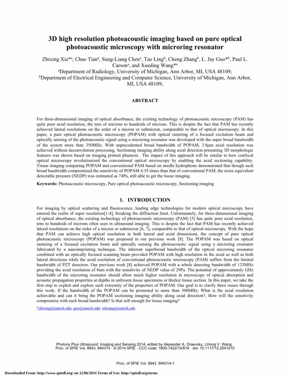
Fig. 1 (a) presents the schematic of the POPAM based on the microring resonator. An Nd:YAG laser (Spot-10-200-532, Elforlight Ltd, UK) served as the irradiation source. The Nd:YAG laser working at a 532 nm wavelength has a pulse duration of 2 ns and a repetition rate (PRR) of 1 KHz. The laser light was spatially filtered by an iris and then expanded into a parallel beam, which was rastered by 2D Galvanometers. The intensity and the stability of the laser beam was monitored and calibrated by a photodiode (DET10A, Thorlabs, NJ). An achromatic lens with a focal length of 40 mm was used as the objective lens. The focused laser spot was scanned over the tissue sample to excite the photoacoustic signal. The induced photoacoustic signals were detected by a microring resonator detector. A custom-built needle hydrophone with a center frequency of 35MHz and a -6dB bandwidth of 100% was also used to detect photoacoustic signals in a way of conventional PAM as control. The detected photoacoustic signals, after a low noise amplifier, were digitized by an A/D card (Razor CS22G8, GaGe, IL). The lateral resolution of the system was measured by imaging an USAF resolution template (T-20-P-TM, Applied Image Inc, NY). The lateral resolution was mainly determined by the size of optical focal spot. Because both POPAM and conventional PAM shared the same optical focusing and scanning architecture, the same lateral resolutions of them were calibrated as 4 µm.
0 10 20 30 40 50 60 70 80 90 100400
600
800
1000
1200
1400
1600
1800
Sense Wavelength (nm)
Am
plitu
de (m
V)
(a) (b)
Figure 1. (a) Schematic of pure optical photoacoustic microscopy (POPAM) and (b) transmission spectrum of the microring resonator measured. The microring resonator has a ring-shaped form coupled with a straight waveguide as optical input and output. It is fabricated by nanoprinting the polystyrene (PS) on a 4um thick SiO2 on a Si substrate. Instead of adding a Mylar protective layer and ultrasound coupling pad layer on it in our previous work [8], the current one is covered by a CYTOP layer with matched refractive index of the water to seal and protect the microring waveguide while sparing high frequency components of the photoacoustic signals during the propagation through it. The microring has a size of 60um in diameter and the waveguide has a cross section of 1.0X1.4um2. A tunable laser (TLB-6712, New Focus, CA) provides the light source for the microring resonator at a wavelength tuned to the maximal slope of the resonance peak of the microring’s transmission spectrum. Acoustic pressure modulates the resonance condition, leading to a shift of the resonance wavelength. When the microring is probed at a fixed wavelength with a high slope in the transmission spectrum, the input photoacoustic wave translates into the output optical intensity, which is then recorded by a high-speed photodetector. Fig.1b shows the transmission spectrum of the microring measured. The whole range of the wavelength is from 773.65 to 773.85nm. The extreme sharp and narrow dipping resonance peak demonstrates the high quality factor (Q) of 2.4E5 and 70% sensing dynamic range based on the difference of the dipping value and base value
Proc. of SPIE Vol. 8943 894314-2
Downloaded From: http://www.spiedl.org/ on 12/06/2014 Terms of Use: http://spiedl.org/terms
of the spectrum. Such high Q factor provides the microring resonator much higher detector sensitivity than other resonant optical structures such as etalons, fiber gratings and dielectric multilayer interference filters. From the optical point of view, the detector’s response time will be limited by the cavity’s photon lifetime which is the time required for energy to decay to its original value and is given by
ωτ Q= [9]. Inverse of the response time will give the cut-off
frequency. So the current device’s frequency response can be up to 8.4GHz. From the acoustic point of view, a maximum modulation frequency of 570 MHz can be estimated considering the 1.4-µm-thick PS waveguide and the acoustic impedance of the cladding, polymer, and the substrate [10]. By designing the PS thickness less than 0.8 µm, microrings with a bandwidth limit of more than 1 GHz can be realized. If assuming strong acoustic reflections from the rigid substrate and negligible reflections from the cladding-polymer boundary, an approximated formula gives a quick estimation on the bandwidth limit:
,|)sin(|2|)(|kl
klkPl = (1)
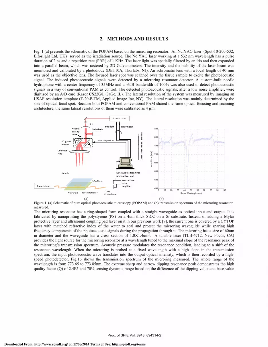
where P is the mean distribution of stress across the thickness l of the sensing film due to an normally incident plane acoustic wave with wave number k. A new photodetector based on a 1GHz avalanche photodiode (APD 210, Thorlabs, NJ) and a preamplifier with bandwidth of 500MHz and low noise base was developed to record the output of the microring resonator instead of previous 125MHz photodetecor [8]. Fig.2 (a) demonstrated the Hilbert transform of measured point spread function (PSF) along axis direction of the whole POPAM system based on microring detector including the effects of excitation laser pulse shape, microring bandwidth, 1GHz avalanche photodiode, 500MHz preamplifer. It indicates that Up to 3.8um resolution can be achieved without any deconvolution and compensation processing, which is best axial resolution of the PAM systems reported so far. Laser pulse shape measured directly with same 1GHz avalanche photodiode using no preamplifier is shown in Fig.2 (b). The pulse duration is 2.5ns. Given the speed of sound of the delivering medium is about 1.497m/s, it can be concluded that the most limitation of the bandwidth and axial resolution 3.8um of the whole POPAM system comes from the pulse width of the laser beam. The deconvolution processing of the laser pulse profile should be able to improve the axial resolution of the system further. Fig.2 (c) shows the frequency spectrum of the PSF along axis direction of the whole POPAM system, manifesting that -6db bandwidth of the whole system is more than 350MHz without deconvolution or compensation processing.
0.26 0.27 0.28 0.29 0.3 0.31 0.32
0
0.1
0.2
0.3
0.4
0.5
0.6
Depth [mm]
PA
sig
nal
PA signal versus the depth
(a) (b)
Proc. of SPIE Vol. 8943 894314-3
Downloaded From: http://www.spiedl.org/ on 12/06/2014 Terms of Use: http://spiedl.org/terms
0 50 100 150 200 250 300 350 400 450 500
-15
-10
-5
0
5
10Spectrum
Frequency (MHz)
ampl
itude
(dB
)
(c)
Fig. 2. (a) Hilbert transform of PSF along axis direction of the whole POPAM system based on microring detector , (b) laser pulse temporal profile measured, and (c) frequency spectrum of the PSF along axis direction of the whole POPAM system
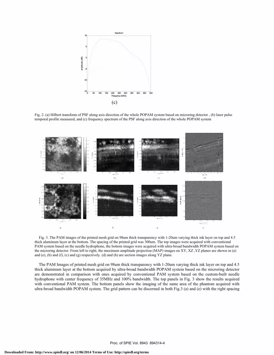
Fig. 3. The PAM images of the printed mesh grid on 98um thick transparency with 1-20um varying thick ink layer on top and 4.5 thick aluminum layer at the bottom. The spacing of the printed grid was 300um. The top images were acquired with conventional PAM system based on the needle hydrophone, the bottom images were acquired with ultra-broad bandwidth POPAM system based on the microring detector. From left to right, the maximum amplitude projection (MAP) images on XY, XZ ,YZ planes are shown in (a) and (e), (b) and (f), (c) and (g) respectively. (d) and (h) are section images along YZ plane. The PAM Images of printed mesh grid on 98um thick transparency with 1-20um varying thick ink layer on top and 4.5 thick aluminum layer at the bottom acquired by ultra-broad bandwidth POPAM system based on the microring detector are demonstrated in comparison with ones acquired by conventional PAM system based on the custom-built needle hydrophone with center frequency of 35MHz and 100% bandwidth. The top panels in Fig. 3 show the results acquired with conventional PAM system. The bottom panels show the imaging of the same area of the phantom acquired with ultra-broad bandwidth POPAM system. The grid pattern can be discerned in both Fig.3 (a) and (e) with the right spacing
Proc. of SPIE Vol. 8943 894314-4
Downloaded From: http://www.spiedl.org/ on 12/06/2014 Terms of Use: http://spiedl.org/terms
of 300um. However Fig. 3(e) presents the grid pattern with much clearer edges and more accurately depicting of the roughness of the surface than the convolved image shown in Fig.3 (a). This is due to the ultra-broad bandwidth providing the POPAM super axial resolution, which brings the sectional imaging ability promoting the imaging quality like the confocal fluorescence microscopy over the normal fluorescence microscopy. It is further confirmed in comparing the images projected on axial planes. In Fig.3 (b), (c) and (f), (g), though both PAM systems can differentiate the printed layer and aluminum layer with the right spacing of 98um, the ultra-broad bandwidth POPAM delineates the two layers with clear profiles while the conventional PAM with the needle hydrophone indicates some weak but long afterpulses along axial direction, some of them can be involved with the pattern of the different layer. The sectional images in Fig.3 (d) and (h) verifies the much better axial resolution of the ultra-broad bandwidth POPAM with a-lines indicating much shorter responses of photoacoustic signals to the printed layer and aluminum layer.
Y axis [mm]
Dep
th [
mm
]
0.2 0.4 0.6 0.8 1 1.2 1.4 1.6 1.8 2
0.2
0.4
0.6
0.8
1
1.2
1.4
Y axis [mm]
Dep
th [
mm
]
0.2 0.4 0.6 0.8 1 1.2 1.4 1.6 1.8 2
0.2
0.4
0.6
0.8
1
1.2
1.4
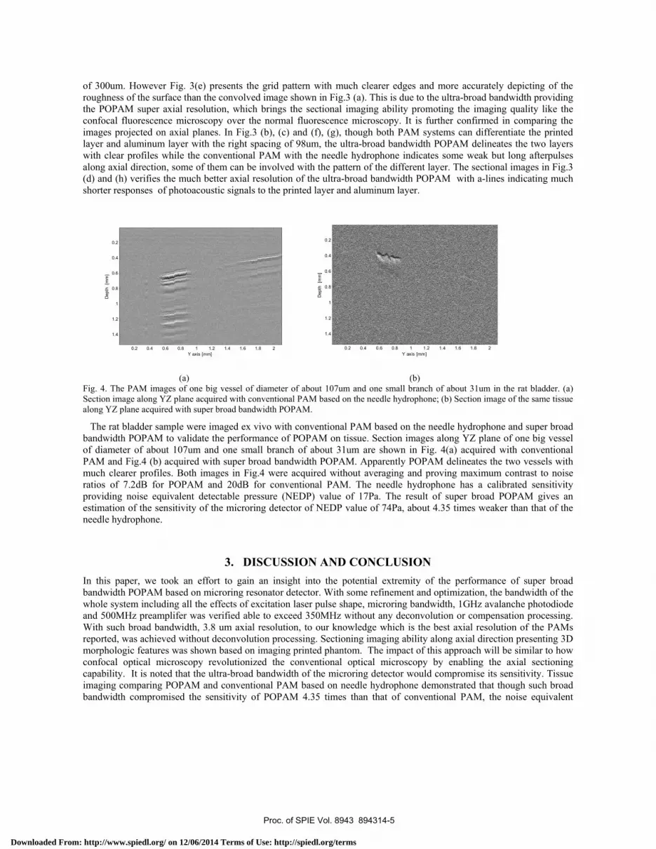
(a) (b) Fig. 4. The PAM images of one big vessel of diameter of about 107um and one small branch of about 31um in the rat bladder. (a) Section image along YZ plane acquired with conventional PAM based on the needle hydrophone; (b) Section image of the same tissue along YZ plane acquired with super broad bandwidth POPAM.
The rat bladder sample were imaged ex vivo with conventional PAM based on the needle hydrophone and super broad bandwidth POPAM to validate the performance of POPAM on tissue. Section images along YZ plane of one big vessel of diameter of about 107um and one small branch of about 31um are shown in Fig. 4(a) acquired with conventional PAM and Fig.4 (b) acquired with super broad bandwidth POPAM. Apparently POPAM delineates the two vessels with much clearer profiles. Both images in Fig.4 were acquired without averaging and proving maximum contrast to noise ratios of 7.2dB for POPAM and 20dB for conventional PAM. The needle hydrophone has a calibrated sensitivity providing noise equivalent detectable pressure (NEDP) value of 17Pa. The result of super broad POPAM gives an estimation of the sensitivity of the microring detector of NEDP value of 74Pa, about 4.35 times weaker than that of the needle hydrophone.
3. DISCUSSION AND CONCLUSION In this paper, we took an effort to gain an insight into the potential extremity of the performance of super broad bandwidth POPAM based on microring resonator detector. With some refinement and optimization, the bandwidth of the whole system including all the effects of excitation laser pulse shape, microring bandwidth, 1GHz avalanche photodiode and 500MHz preamplifer was verified able to exceed 350MHz without any deconvolution or compensation processing. With such broad bandwidth, 3.8 um axial resolution, to our knowledge which is the best axial resolution of the PAMs reported, was achieved without deconvolution processing. Sectioning imaging ability along axial direction presenting 3D morphologic features was shown based on imaging printed phantom. The impact of this approach will be similar to how confocal optical microscopy revolutionized the conventional optical microscopy by enabling the axial sectioning capability. It is noted that the ultra-broad bandwidth of the microring detector would compromise its sensitivity. Tissue imaging comparing POPAM and conventional PAM based on needle hydrophone demonstrated that though such broad bandwidth compromised the sensitivity of POPAM 4.35 times than that of conventional PAM, the noise equivalent
Proc. of SPIE Vol. 8943 894314-5
Downloaded From: http://www.spiedl.org/ on 12/06/2014 Terms of Use: http://spiedl.org/terms
detectable pressure (NEDP) was estimated as 74Pa, still able to get the tissue imaging. It is not a gap unbridgeable to be improved back to the 29Pa achieved from 125MHz bandwidth POPAM system in our previous work [8].
In future there are several ways to improve the sensitivity of the microring detector further, including optimizing the fabrication of the microring to obtain higher Q factor and reduce the loss of the energy of the light propagating in the microring waveguides, increasing the energy intput into the microring by choosing better tunable laser, adopting more sensitive and lower noise-level photodetector to record the output of the microring. Comparing the PSF along axis direction of the whole POPAM system in Fig. 2(a) with the excitation laser pulse temporal profile in Fig. 2 (b), the most limitation of the bandwidth 350MHz and axial resolution 3.8um of the whole POPAM system comes from the pulse width of the laser beam. The deconvolution processing of the laser pulse profile should be able to improve the axial resolution of the system significantly. The axial resolution can also be promoted further by optimizing the fabrication parameters of the microring such as less thickness of the waveguide, as well as increasing the bandwidths of the photodetector and the amplifer. The potential of approximately GHz bandwidth of the microring resonator with enough sensitivity for tissue imaging should bring POPAM much higher resolution in microscopy of optical absorption and acoustic propagation properties at depths in unfrozen tissue specimens or thicker tissue sections not now imageable with current optical or acoustic microscopes of comparable resolution.
ACKNOWLEGEMENTS Support from Samsung GRO 2012 Program, NIH Grant No. R01 CA91713, CA91713-S1, R01 AR055179, R01AR060350, NSF Grant No. DBI-1256001 and UM-SJTU joint program are gratefully acknowledged.
REFERENCES
[1] Fernandes-Suares M, and Ting AY, “Fluorescent probes for super-resolution imaging in living cells,” Nature Rev. Mol. Cell Biol. 9, 929–943 (2008).
[2] Betzig E, Patterson GH, Sougrat R, Lindwasser OW, Olenych S, Bonifacino JS, Davidson MW, Lippincott-Schwartz J, and Hess HF, “Imaging Intracellular Fluorescent Proteins at Nanometer Resolution,” Science 313, 1642-1645 (2006).
[3] Gustafsson MGL, “Nonlinear structured-illumination microscopy: Wide-field fluorescence imaging with theoretically unlimited resolution,” Proc. Natl. Acad. Sci. USA 102, 13081–13086 (2005).
[4] Zhuang X, “Nano-imaging with STORM,” Nature Photon. 3, 365–367 (2009). [5] Zhang HF, Maslov K, Stoica G, and Wang LV, “Functional photoacoustic microscopy for high-resolution and
noninvasive in vivo imaging,” Nature Biotechnol. 24, 848–851 (2006). [6] Xie Z, Jiao S, Zhang HF, and Puliafito CA, “Laser-scanning optical-resolution photoacoustic microscopy”, Opt.
Lett. 34, 1771-1773 (2009). [7] Maslov K, Zhang HF, Hu S, and Wang LV, “Optical-resolution photoacoustic microscopy for in vivo imaging
of single capillaries,” Opt. Lett. 33, 929–931 (2008). [8] Xie Z, Chen S, Ling T, Guo LJ, Carson PL, and Wang X, “ Pure optical photoacoustic microscopy”, Opt.
Express 19(10), 9027-9034 (2011). [9] Armani DK, Kippenberg TJ, Spillane SM, and Vahala KJ, “Ultra-high-Q toroid microcavity on chip,” Nature
421, 925-928 (2003). [10] Beard P, Perennes F, and Mills TN, “Transduction mechanisms of the Fabry Perot polymer film sensing
concept for wideband ultrasound detection,” IEEE Trans. Ultrason. Ferroelectr. Freq. Control 46, 1575-1582 (1999).
Proc. of SPIE Vol. 8943 894314-6
Downloaded From: http://www.spiedl.org/ on 12/06/2014 Terms of Use: http://spiedl.org/terms