16.1 | regulation of gene expression - the expert ta
TRANSCRIPT
16.1 | Regulation of Gene Expression
By the end of this section, you will be able to do the following:
• Discuss why every cell does not express all of its genes all of the time
• Describe how prokaryotic gene regulation occurs at the transcriptional level
• Discuss how eukaryotic gene regulation occurs at the epigenetic, transcriptional, post-transcriptional,translational, and post-translational levels
For a cell to function properly, necessary proteins must be synthesized at the proper time and place. All cellscontrol or regulate the synthesis of proteins from information encoded in their DNA. The process of turning ona gene to produce RNA and protein is called gene expression. Whether in a simple unicellular organism or acomplex multi-cellular organism, each cell controls when and how its genes are expressed. For this to occur,there must be internal chemical mechanisms that control when a gene is expressed to make RNA and protein,how much of the protein is made, and when it is time to stop making that protein because it is no longer needed.
The regulation of gene expression conserves energy and space. It would require a significant amount of energyfor an organism to express every gene at all times, so it is more energy efficient to turn on the genes only whenthey are required. In addition, only expressing a subset of genes in each cell saves space because DNA must beunwound from its tightly coiled structure to transcribe and translate the DNA. Cells would have to be enormousif every protein were expressed in every cell all the time.
The control of gene expression is extremely complex. Malfunctions in this process are detrimental to the cell andcan lead to the development of many diseases, including cancer.
Prokaryotic versus Eukaryotic Gene Expression
To understand how gene expression is regulated, we must first understand how a gene codes for a functionalprotein in a cell. The process occurs in both prokaryotic and eukaryotic cells, just in slightly different manners.
Prokaryotic organisms are single-celled organisms that lack a cell nucleus, and their DNA therefore floatsfreely in the cell cytoplasm. To synthesize a protein, the processes of transcription and translation occur almostsimultaneously. When the resulting protein is no longer needed, transcription stops. As a result, the primarymethod to control what type of protein and how much of each protein is expressed in a prokaryotic cell isthe regulation of DNA transcription. All of the subsequent steps occur automatically. When more protein isrequired, more transcription occurs. Therefore, in prokaryotic cells, the control of gene expression is mostly atthe transcriptional level.
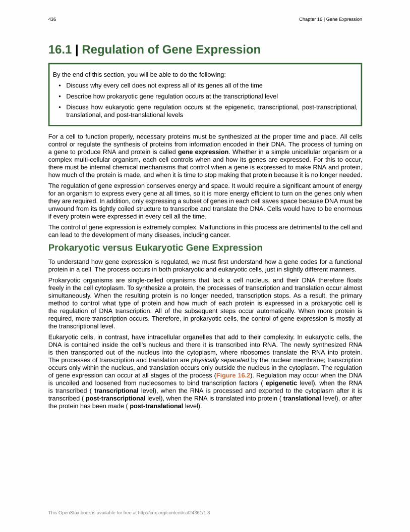
Eukaryotic cells, in contrast, have intracellular organelles that add to their complexity. In eukaryotic cells, theDNA is contained inside the cell’s nucleus and there it is transcribed into RNA. The newly synthesized RNAis then transported out of the nucleus into the cytoplasm, where ribosomes translate the RNA into protein.The processes of transcription and translation are physically separated by the nuclear membrane; transcriptionoccurs only within the nucleus, and translation occurs only outside the nucleus in the cytoplasm. The regulationof gene expression can occur at all stages of the process (Figure 16.2). Regulation may occur when the DNAis uncoiled and loosened from nucleosomes to bind transcription factors ( epigenetic level), when the RNAis transcribed ( transcriptional level), when the RNA is processed and exported to the cytoplasm after it istranscribed ( post-transcriptional level), when the RNA is translated into protein ( translational level), or afterthe protein has been made ( post-translational level).
436 Chapter 16 | Gene Expression
This OpenStax book is available for free at http://cnx.org/content/col24361/1.8
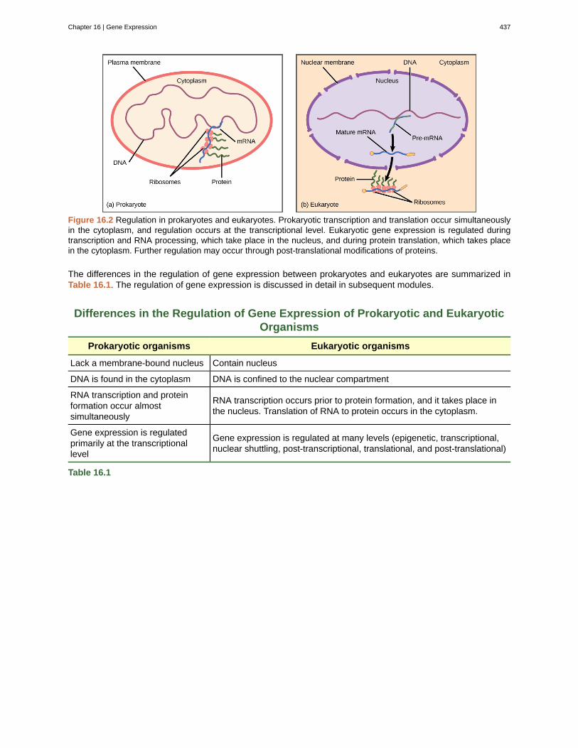
Figure 16.2 Regulation in prokaryotes and eukaryotes. Prokaryotic transcription and translation occur simultaneouslyin the cytoplasm, and regulation occurs at the transcriptional level. Eukaryotic gene expression is regulated duringtranscription and RNA processing, which take place in the nucleus, and during protein translation, which takes placein the cytoplasm. Further regulation may occur through post-translational modifications of proteins.
The differences in the regulation of gene expression between prokaryotes and eukaryotes are summarized inTable 16.1. The regulation of gene expression is discussed in detail in subsequent modules.
Differences in the Regulation of Gene Expression of Prokaryotic and EukaryoticOrganisms
Prokaryotic organisms Eukaryotic organisms
Lack a membrane-bound nucleus Contain nucleus
DNA is found in the cytoplasm DNA is confined to the nuclear compartment
RNA transcription and proteinformation occur almostsimultaneously
RNA transcription occurs prior to protein formation, and it takes place inthe nucleus. Translation of RNA to protein occurs in the cytoplasm.
Gene expression is regulatedprimarily at the transcriptionallevel
Gene expression is regulated at many levels (epigenetic, transcriptional,nuclear shuttling, post-transcriptional, translational, and post-translational)
Table 16.1
Chapter 16 | Gene Expression 437
Evolution of Gene RegulationProkaryotic cells can only regulate gene expression by controlling the amount of transcription. As eukaryoticcells evolved, the complexity of the control of gene expression increased. For example, with the evolutionof eukaryotic cells came compartmentalization of important cellular components and cellular processes. Anuclear region that contains the DNA was formed. Transcription and translation were physically separatedinto two different cellular compartments. It therefore became possible to control gene expression byregulating transcription in the nucleus, and also by controlling the RNA levels and protein translation presentoutside the nucleus.
Most gene regulation is done to conserve cell resources. However, other regulatory processes may bedefensive. Cellular processes such as developed to protect the cell from viral or parasitic infections. If thecell could quickly shut off gene expression for a short period of time, it would be able to survive an infectionwhen other organisms could not. Therefore, the organism evolved a new process that helped it survive, andit was able to pass this new development to offspring.
16.2 | Prokaryotic Gene Regulation
By the end of this section, you will be able to do the following:
• Describe the steps involved in prokaryotic gene regulation
• Explain the roles of activators, inducers, and repressors in gene regulation
The DNA of prokaryotes is organized into a circular chromosome, supercoiled within the nucleoid region of thecell cytoplasm. Proteins that are needed for a specific function, or that are involved in the same biochemicalpathway, are encoded together in blocks called operons. For example, all of the genes needed to use lactoseas an energy source are coded next to each other in the lactose (or lac) operon, and transcribed into a singlemRNA.
In prokaryotic cells, there are three types of regulatory molecules that can affect the expression of operons:repressors, activators, and inducers. Repressors and activators are proteins produced in the cell. Bothrepressors and activators regulate gene expression by binding to specific DNA sites adjacent to the genes theycontrol. In general, activators bind to the promoter site, while repressors bind to operator regions. Repressorsprevent transcription of a gene in response to an external stimulus, whereas activators increase thetranscription of a gene in response to an external stimulus. Inducers are small molecules that may be producedby the cell or that are in the cell’s environment. Inducers either activate or repress transcription depending onthe needs of the cell and the availability of substrate.
The trp Operon: A Repressible Operon
Bacteria such as Escherichia coli need amino acids to survive, and are able to synthesize many of them.Tryptophan is one such amino acid that E. coli can either ingest from the environment or synthesize usingenzymes that are encoded by five genes. These five genes are next to each other in what is called thetryptophan (trp) operon (Figure 16.3). The genes are transcribed into a single mRNA, which is then translatedto produce all five enzymes. If tryptophan is present in the environment, then E. coli does not need to synthesizeit and the trp operon is switched off. However, when tryptophan availability is low, the switch controllingthe operon is turned on, the mRNA is transcribed, the enzyme proteins are translated, and tryptophan issynthesized.
438 Chapter 16 | Gene Expression
This OpenStax book is available for free at http://cnx.org/content/col24361/1.8
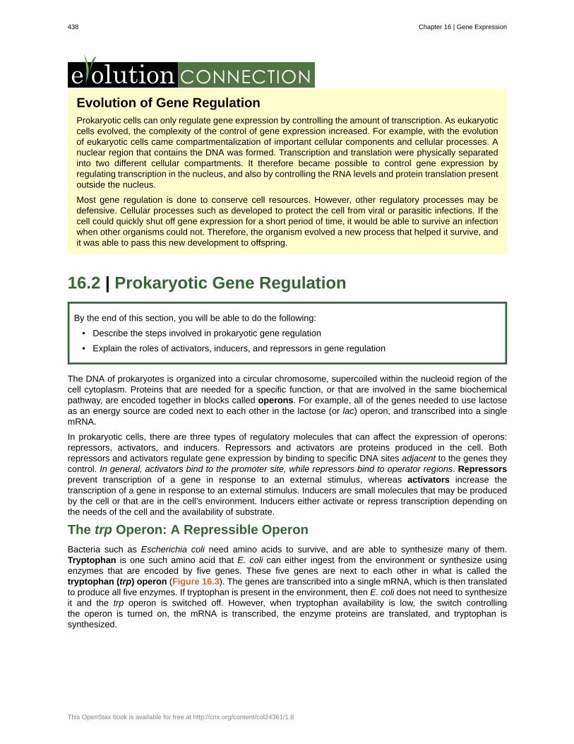
Figure 16.3 The tryptophan operon. The five genes that are needed to synthesize tryptophan in E. coli are locatednext to each other in the trp operon. When tryptophan is plentiful, two tryptophan molecules bind the repressor proteinat the operator sequence. This physically blocks the RNA polymerase from transcribing the tryptophan genes. Whentryptophan is absent, the repressor protein does not bind to the operator and the genes are transcribed.
The trp operon includes three important regions: the coding region, the trp operator and the trp promoter. Thecoding region includes the genes for the five tryptophan biosynthesis enzymes. Just before the coding region isthe transcriptional start site. The promoter sequence, to which RNA polymerase binds to initiate transcription,is before or “upstream” of the transcriptional start site. Between the promoter and the transcriptional start site isthe operator region.
The trp operator contains the DNA code to which the trp repressor protein can bind. However, the repressoralone cannot bind to the operator. When tryptophan is present in the cell, two tryptophan molecules bind tothe trp repressor, which changes the shape of the repressor protein to a form that can bind to the trp operator.Binding of the tryptophan–repressor complex at the operator physically prevents the RNA polymerase frombinding to the promoter and transcribing the downstream genes.
When tryptophan is not present in the cell, the repressor by itself does not bind to the operator, the polymerasecan transcribe the enzyme genes, and tryptophan is synthesized. Because the repressor protein actively bindsto the operator to keep the genes turned off, the trp operon is said to be negatively regulated and the proteinsthat bind to the operator to silence trp expression are negative regulators.
Watch this video to learn more about the trp operon. (This multimedia resource will open in a browser.) (http://cnx.org/content/m66504/1.3/#eip-id1169842033659)
Catabolite Activator Protein (CAP): A Transcriptional Activator
Just as the trp operon is negatively regulated by tryptophan molecules, there are proteins that bind to thepromoter sequences that act as positive regulators to turn genes on and activate them. For example, whenglucose is scarce, E. coli bacteria can turn to other sugar sources for fuel. To do this, new genes to processthese alternate sugars must be transcribed. When glucose levels drop, cyclic AMP (cAMP) begins to accumulatein the cell. The cAMP molecule is a signaling molecule that is involved in glucose and energy metabolism inE. coli. Accumulating cAMP binds to the positive regulator catabolite activator protein (CAP), a protein that
Chapter 16 | Gene Expression 439
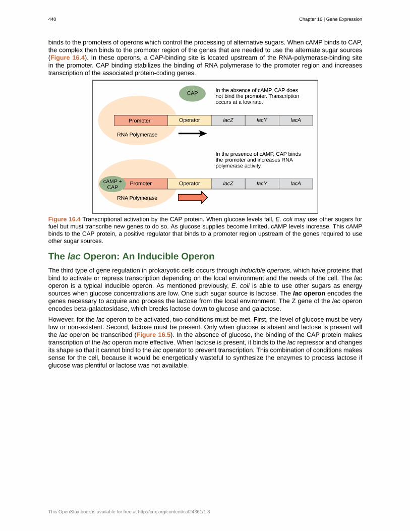
binds to the promoters of operons which control the processing of alternative sugars. When cAMP binds to CAP,the complex then binds to the promoter region of the genes that are needed to use the alternate sugar sources(Figure 16.4). In these operons, a CAP-binding site is located upstream of the RNA-polymerase-binding sitein the promoter. CAP binding stabilizes the binding of RNA polymerase to the promoter region and increasestranscription of the associated protein-coding genes.
Figure 16.4 Transcriptional activation by the CAP protein. When glucose levels fall, E. coli may use other sugars forfuel but must transcribe new genes to do so. As glucose supplies become limited, cAMP levels increase. This cAMPbinds to the CAP protein, a positive regulator that binds to a promoter region upstream of the genes required to useother sugar sources.
The lac Operon: An Inducible Operon
The third type of gene regulation in prokaryotic cells occurs through inducible operons, which have proteins thatbind to activate or repress transcription depending on the local environment and the needs of the cell. The lacoperon is a typical inducible operon. As mentioned previously, E. coli is able to use other sugars as energysources when glucose concentrations are low. One such sugar source is lactose. The lac operon encodes thegenes necessary to acquire and process the lactose from the local environment. The Z gene of the lac operonencodes beta-galactosidase, which breaks lactose down to glucose and galactose.
However, for the lac operon to be activated, two conditions must be met. First, the level of glucose must be verylow or non-existent. Second, lactose must be present. Only when glucose is absent and lactose is present willthe lac operon be transcribed (Figure 16.5). In the absence of glucose, the binding of the CAP protein makestranscription of the lac operon more effective. When lactose is present, it binds to the lac repressor and changesits shape so that it cannot bind to the lac operator to prevent transcription. This combination of conditions makessense for the cell, because it would be energetically wasteful to synthesize the enzymes to process lactose ifglucose was plentiful or lactose was not available.
440 Chapter 16 | Gene Expression
This OpenStax book is available for free at http://cnx.org/content/col24361/1.8
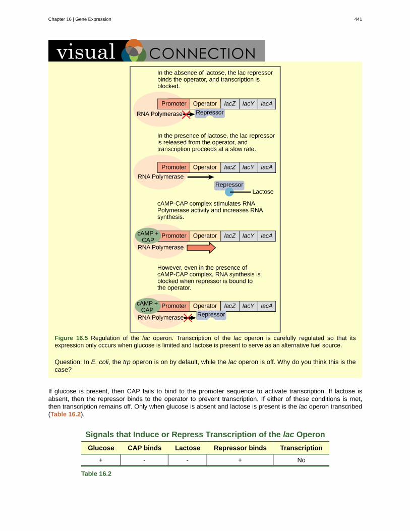
Figure 16.5 Regulation of the lac operon. Transcription of the lac operon is carefully regulated so that itsexpression only occurs when glucose is limited and lactose is present to serve as an alternative fuel source.
Question: In E. coli, the trp operon is on by default, while the lac operon is off. Why do you think this is thecase?
If glucose is present, then CAP fails to bind to the promoter sequence to activate transcription. If lactose isabsent, then the repressor binds to the operator to prevent transcription. If either of these conditions is met,then transcription remains off. Only when glucose is absent and lactose is present is the lac operon transcribed(Table 16.2).
Signals that Induce or Repress Transcription of the lac Operon
Glucose CAP binds Lactose Repressor binds Transcription
+ - - + No
Table 16.2
Chapter 16 | Gene Expression 441
Signals that Induce or Repress Transcription of the lac Operon
Glucose CAP binds Lactose Repressor binds Transcription
+ - + - Some
- + - + No
- + + - Yes
Table 16.2
Watch an animated tutorial about the workings of lac operon here. (This multimedia resource will open in a browser.) (http://cnx.org/content/m66504/1.3/#eip-id1165239273914)
16.3 | Eukaryotic Epigenetic Gene Regulation
By the end of this section, you will be able to do the following:
• Explain how chromatin remodeling controls transcriptional access
• Describe how access to DNA is controlled by histone modification
• Describe how DNA methylation is related to epigenetic gene changes
Eukaryotic gene expression is more complex than prokaryotic gene expression because the processes oftranscription and translation are physically separated. Unlike prokaryotic cells, eukaryotic cells can regulate geneexpression at many different levels. Epigenetic changes are inheritable changes in gene expression that do notresult from changes in the DNA sequence. Eukaryotic gene expression begins with control of access to theDNA. Transcriptional access to the DNA can be controlled in two general ways: chromatin remodeling and DNAmethylation. Chromatin remodeling changes the way that DNA is associated with chromosomal histones. DNAmethylation is associated with developmental changes and gene silencing.
Epigenetic Control: Regulating Access to Genes within theChromosome
The human genome encodes over 20,000 genes, with hundreds to thousands of genes on each of the 23 humanchromosomes. The DNA in the nucleus is precisely wound, folded, and compacted into chromosomes so that itwill fit into the nucleus. It is also organized so that specific segments can be accessed as needed by a specificcell type.
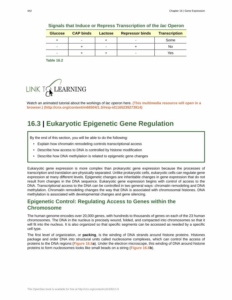
The first level of organization, or packing, is the winding of DNA strands around histone proteins. Histonespackage and order DNA into structural units called nucleosome complexes, which can control the access ofproteins to the DNA regions (Figure 16.6a). Under the electron microscope, this winding of DNA around histoneproteins to form nucleosomes looks like small beads on a string (Figure 16.6b).
442 Chapter 16 | Gene Expression
This OpenStax book is available for free at http://cnx.org/content/col24361/1.8
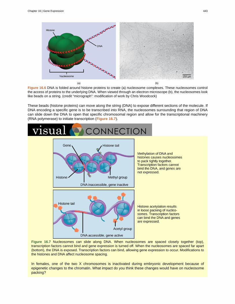
Figure 16.6 DNA is folded around histone proteins to create (a) nucleosome complexes. These nucleosomes controlthe access of proteins to the underlying DNA. When viewed through an electron microscope (b), the nucleosomes looklike beads on a string. (credit “micrograph”: modification of work by Chris Woodcock)
These beads (histone proteins) can move along the string (DNA) to expose different sections of the molecule. IfDNA encoding a specific gene is to be transcribed into RNA, the nucleosomes surrounding that region of DNAcan slide down the DNA to open that specific chromosomal region and allow for the transcriptional machinery(RNA polymerase) to initiate transcription (Figure 16.7).
Figure 16.7 Nucleosomes can slide along DNA. When nucleosomes are spaced closely together (top),transcription factors cannot bind and gene expression is turned off. When the nucleosomes are spaced far apart(bottom), the DNA is exposed. Transcription factors can bind, allowing gene expression to occur. Modifications tothe histones and DNA affect nucleosome spacing.
In females, one of the two X chromosomes is inactivated during embryonic development because ofepigenetic changes to the chromatin. What impact do you think these changes would have on nucleosomepacking?
Chapter 16 | Gene Expression 443
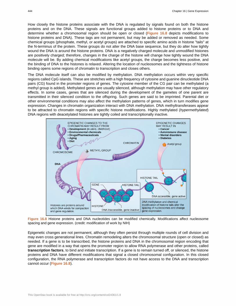
How closely the histone proteins associate with the DNA is regulated by signals found on both the histoneproteins and on the DNA. These signals are functional groups added to histone proteins or to DNA anddetermine whether a chromosomal region should be open or closed (Figure 16.8 depicts modifications tohistone proteins and DNA). These tags are not permanent, but may be added or removed as needed. Somechemical groups (phosphate, methyl, or acetyl groups) are attached to specific amino acids in histone "tails" atthe N-terminus of the protein. These groups do not alter the DNA base sequence, but they do alter how tightlywound the DNA is around the histone proteins. DNA is a negatively charged molecule and unmodified histonesare positively charged; therefore, changes in the charge of the histone will change how tightly wound the DNAmolecule will be. By adding chemical modifications like acetyl groups, the charge becomes less positive, andthe binding of DNA to the histones is relaxed. Altering the location of nucleosomes and the tightness of histonebinding opens some regions of chromatin to transcription and closes others.
The DNA molecule itself can also be modified by methylation. DNA methylation occurs within very specificregions called CpG islands. These are stretches with a high frequency of cytosine and guanine dinucleotide DNApairs (CG) found in the promoter regions of genes. The cytosine member of the CG pair can be methylated (amethyl group is added). Methylated genes are usually silenced, although methylation may have other regulatoryeffects. In some cases, genes that are silenced during the development of the gametes of one parent aretransmitted in their silenced condition to the offspring. Such genes are said to be imprinted. Parental diet orother environmental conditions may also affect the methylation patterns of genes, which in turn modifies geneexpression. Changes in chromatin organization interact with DNA methylation. DNA methyltransferases appearto be attracted to chromatin regions with specific histone modifications. Highly methylated (hypermethylated)DNA regions with deacetylated histones are tightly coiled and transcriptionally inactive.
Figure 16.8 Histone proteins and DNA nucleotides can be modified chemically. Modifications affect nucleosomespacing and gene expression. (credit: modification of work by NIH)
Epigenetic changes are not permanent, although they often persist through multiple rounds of cell division andmay even cross generational lines. Chromatin remodeling alters the chromosomal structure (open or closed) asneeded. If a gene is to be transcribed, the histone proteins and DNA in the chromosomal region encoding thatgene are modified in a way that opens the promoter region to allow RNA polymerase and other proteins, calledtranscription factors, to bind and initiate transcription. If a gene is to remain turned off, or silenced, the histoneproteins and DNA have different modifications that signal a closed chromosomal configuration. In this closedconfiguration, the RNA polymerase and transcription factors do not have access to the DNA and transcriptioncannot occur (Figure 16.8).
444 Chapter 16 | Gene Expression
This OpenStax book is available for free at http://cnx.org/content/col24361/1.8
View this video that describes how epigenetic regulation controls gene expression. (This multimedia resource will open in a browser.) (http://cnx.org/content/m66505/1.3/#eip-id1169842033590)
16.4 | Eukaryotic Transcription Gene Regulation
By the end of this section, you will be able to do the following:
• Discuss the role of transcription factors in gene regulation
• Explain how enhancers and repressors regulate gene expression
Like prokaryotic cells, the transcription of genes in eukaryotes requires the action of an RNA polymerase to bindto a DNA sequence upstream of a gene in order to initiate transcription. However, unlike prokaryotic cells, theeukaryotic RNA polymerase requires other proteins, or transcription factors, to facilitate transcription initiation.RNA polymerase by itself cannot initiate transcription in eukaryotic cells. There are two types of transcriptionfactors that regulate eukaryotic transcription: General (or basal) transcription factors bind to the core promoterregion to assist with the binding of RNA polymerase. Specific transcription factors bind to various regions outsideof the core promoter region and interact with the proteins at the core promoter to enhance or repress the activityof the polymerase.
View the process of transcription—the making of RNA from a DNA template. (This multimedia resource will open in a browser.) (http://cnx.org/content/m66506/1.3/#eip-id1168020166468)
The Promoter and the Transcription Machinery
Genes are organized to make the control of gene expression easier. The promoter region is immediatelyupstream of the coding sequence. This region can be short (only a few nucleotides in length) or quite long(hundreds of nucleotides long). The longer the promoter, the more available space for proteins to bind. Thisalso adds more control to the transcription process. The length of the promoter is gene-specific and candiffer dramatically between genes. Consequently, the level of control of gene expression can also differ quitedramatically between genes. The purpose of the promoter is to bind transcription factors that control theinitiation of transcription.
Within the core promoter region, 25 to 35 bases upstream of the transcriptional start site, resides the TATAbox. The TATA box has the consensus sequence of 5’-TATAAA-3’. The TATA box is the binding site for aprotein complex called TFIID, which contains a TATA-binding protein. Binding of TFIID recruits other transcriptionfactors, including TFIIB, TFIIE, TFIIF, and TFIIH. Some of these transcription factors help to bind the RNApolymerase to the promoter, and others help to activate the transcription initiation complex.
In addition to the TATA box, other binding sites are found in some promoters. Some biologists prefer to restrictthe range of the eukaryotic promoter to the core promoter, or polymerase binding site, and refer to these
Chapter 16 | Gene Expression 445
additional sites as promoter-proximal elements, because they are usually found within a few hundred base pairsupstream of the transcriptional start site. Examples of these elements are the CAAT box, with the consensussequence 5’-CCAAT-3’ and the GC box, with the consensus sequence 5’-GGGCGG-3’. Specific transcriptionfactors can bind to these promoter-proximal elements to regulate gene transcription. A given gene may have itsown combination of these specific transcription-factor binding sites. There are hundreds of transcription factorsin a cell, each of which binds specifically to a particular DNA sequence motif. When transcription factors bind tothe promoter just upstream of the encoded gene, it is referred to as a cis-acting element, because it is on thesame chromosome just next to the gene. Transcription factors respond to environmental stimuli that cause theproteins to find their binding sites and initiate transcription of the gene that is needed.
Enhancers and Transcription
In some eukaryotic genes, there are additional regions that help increase or enhance transcription. Theseregions, called enhancers, are not necessarily close to the genes they enhance. They can be located upstreamof a gene, within the coding region of the gene, downstream of a gene, or may be thousands of nucleotidesaway.
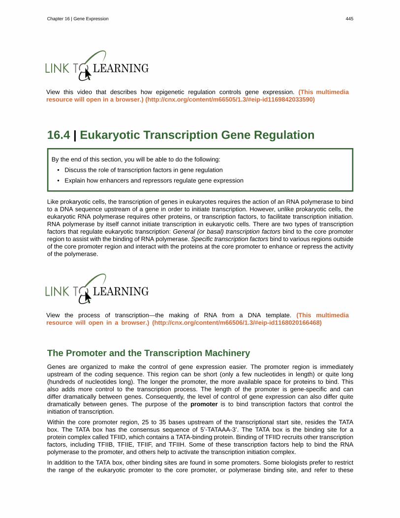
Enhancer regions are binding sequences, or sites, for specific transcription factors. When a protein transcriptionfactor binds to its enhancer sequence, the shape of the protein changes, allowing it to interact with proteins atthe promotor site. However, since the enhancer region may be distant from the promoter, the DNA must bend toallow the proteins at the two sites to come into contact. DNA bending proteins help to bend the DNA and bringthe enhancer and promoter regions together (Figure 16.9). This shape change allows for the interaction of thespecific activator proteins bound to the enhancers with the general transcription factors bound to the promoterregion and the RNA polymerase.
Figure 16.9 Interaction between proteins at the promoter and enhancer sites. An enhancer is a DNA sequence thatpromotes transcription. Each enhancer is made up of short DNA sequences called distal control elements. Activatorsbound to the distal control elements interact with mediator proteins and transcription factors. Two different genes mayhave the same promoter but different distal control elements, enabling differential gene expression.
Turning Genes Off: Transcriptional Repressors
Like prokaryotic cells, eukaryotic cells also have mechanisms to prevent transcription. Transcriptional repressorscan bind to promoter or enhancer regions and block transcription. Like the transcriptional activators, repressorsrespond to external stimuli to prevent the binding of activating transcription factors.
446 Chapter 16 | Gene Expression
This OpenStax book is available for free at http://cnx.org/content/col24361/1.8
16.5 | Eukaryotic Post-transcriptional Gene Regulation
By the end of this section, you will be able to do the following:
• Understand RNA splicing and explain its role in regulating gene expression
• Describe the importance of RNA stability in gene regulation
RNA is transcribed, but must be processed into a mature form before translation can begin. This processingthat takes place after an RNA molecule has been transcribed, but before it is translated into a protein, iscalled post-transcriptional modification. As with the epigenetic and transcriptional stages of processing, this post-transcriptional step can also be regulated to control gene expression in the cell. If the RNA is not processed,shuttled, or translated, then no protein will be synthesized.
RNA Splicing, the First Stage of Post-transcriptional Control
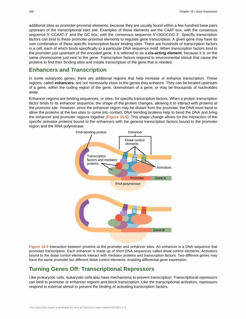
In eukaryotic cells, the RNA transcript often contains regions, called introns, that are removed prior to translation.The regions of RNA that code for protein are called exons. (Figure 16.10). After an RNA molecule has beentranscribed, but prior to its departure from the nucleus to be translated, the RNA is processed and the intronsare removed by splicing. Splicing is done by spliceosomes, ribonucleoprotein complexes that can recognize thetwo ends of the intron, cut the transcript at those two points, and bring the exons together for ligation.
Figure 16.10 Pre-mRNA can be alternatively spliced to create different proteins.
Chapter 16 | Gene Expression 447
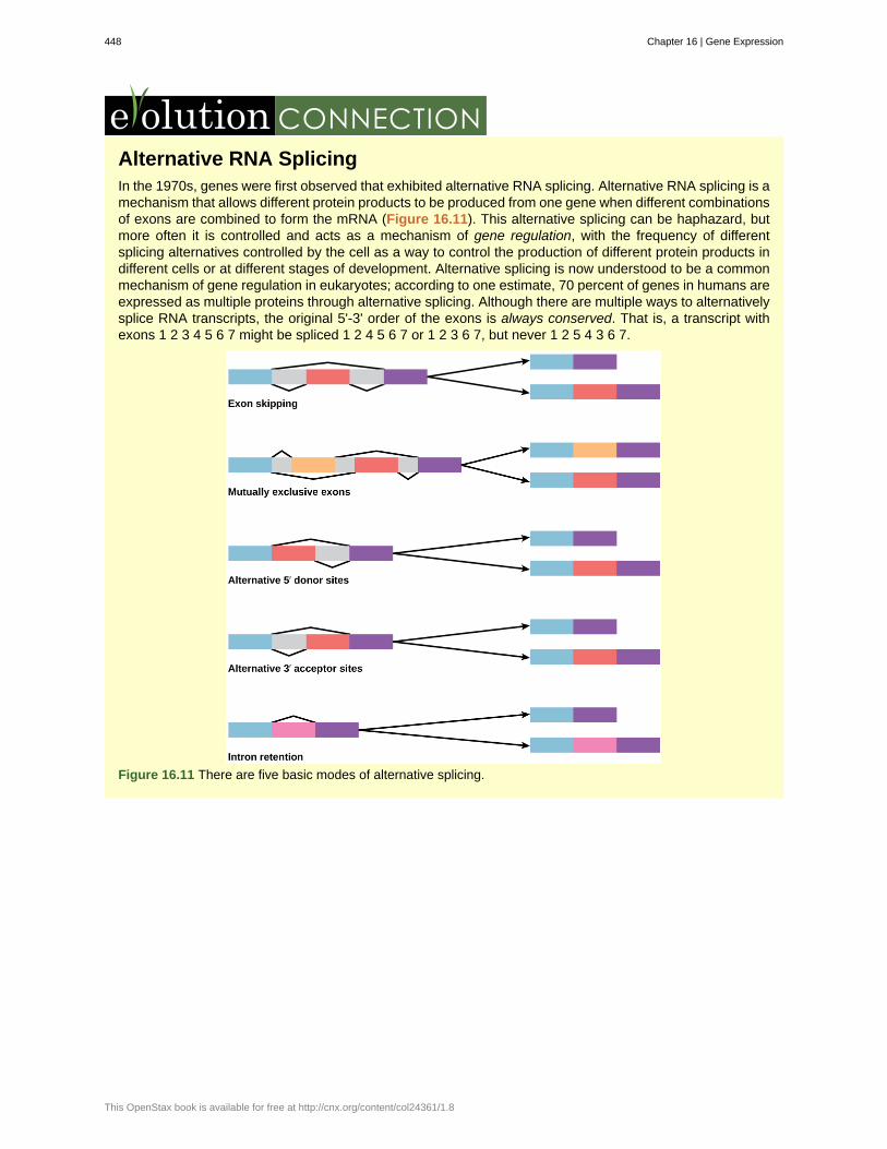
Alternative RNA SplicingIn the 1970s, genes were first observed that exhibited alternative RNA splicing. Alternative RNA splicing is amechanism that allows different protein products to be produced from one gene when different combinationsof exons are combined to form the mRNA (Figure 16.11). This alternative splicing can be haphazard, butmore often it is controlled and acts as a mechanism of gene regulation, with the frequency of differentsplicing alternatives controlled by the cell as a way to control the production of different protein products indifferent cells or at different stages of development. Alternative splicing is now understood to be a commonmechanism of gene regulation in eukaryotes; according to one estimate, 70 percent of genes in humans areexpressed as multiple proteins through alternative splicing. Although there are multiple ways to alternativelysplice RNA transcripts, the original 5'-3' order of the exons is always conserved. That is, a transcript withexons 1 2 3 4 5 6 7 might be spliced 1 2 4 5 6 7 or 1 2 3 6 7, but never 1 2 5 4 3 6 7.
Figure 16.11 There are five basic modes of alternative splicing.
448 Chapter 16 | Gene Expression
This OpenStax book is available for free at http://cnx.org/content/col24361/1.8
How could alternative splicing evolve? Introns have a beginning- and ending-recognition sequence; it iseasy to imagine the failure of the splicing mechanism to identify the end of an intron and instead find theend of the next intron, thus removing two introns and the intervening exon. In fact, there are mechanisms inplace to prevent such intron skipping, but mutations are likely to lead to their failure. Such “mistakes” wouldmore than likely produce a nonfunctional protein. Indeed, the cause of many genetic diseases is abnormalsplicing rather than mutations in a coding sequence. However, alternative splicing could possibly create aprotein variant without the loss of the original protein, opening up possibilities for adaptation of the newvariant to new functions. Gene duplication has played an important role in the evolution of new functions ina similar way by providing genes that may evolve without eliminating the original, functional protein.
Question: In the corn snake Pantherophis guttatus, there are several different color variants, includingamelanistic snakes whose skin patterns display only red and yellow pigments. The cause of amelanism inthese snakes was recently identified as the insertion of a transposable element into an intron in the OCA2(oculocutaneous albinism) gene. How might the insertion of extra genetic material into an intron lead to anonfunctional protein?
Visualize how mRNA splicing happens by watching the process in action in this video. (This multimedia resource will open in a browser.) (http://cnx.org/content/m66507/1.5/#eip-id1171119155972)
Control of RNA Stability
Before the mRNA leaves the nucleus, it is given two protective "caps" that prevent the ends of the strandfrom degrading during its journey. 5' and 3' exonucleases can degrade unprotected RNAs. The 5' cap, whichis placed on the 5' end of the mRNA, is usually composed of a methylated guanosine triphosphate molecule(GTP). The GTP is placed "backward" on the 5' end of the mRNA, so that the 5' carbons of the GTP and theterminal nucleotide are linked through three phosphates. The poly-A tail, which is attached to the 3' end, isusually composed of a long chain of adenine nucleotides. These changes protect the two ends of the RNA fromexonuclease attack.
Once the RNA is transported to the cytoplasm, the length of time that the RNA resides there can be controlled.Each RNA molecule has a defined lifespan and decays at a specific rate. This rate of decay can influencehow much protein is in the cell. If the decay rate is increased, the RNA will not exist in the cytoplasm as long,shortening the time available for translation of the mRNA to occur. Conversely, if the rate of decay is decreased,the mRNA molecule will reside in the cytoplasm longer and more protein can be translated. This rate of decay isreferred to as the RNA stability. If the RNA is stable, it will be detected for longer periods of time in the cytoplasm.
Binding of proteins to the RNA can also influence its stability. Proteins called RNA-binding proteins, or RBPs,can bind to the regions of the mRNA just upstream or downstream of the protein-coding region. These regions inthe RNA that are not translated into protein are called the untranslated regions, or UTRs. They are not introns(those have been removed in the nucleus). Rather, these are regions that regulate mRNA localization, stability,and protein translation. The region just before the protein-coding region is called the 5' UTR, whereas the regionafter the coding region is called the 3' UTR (Figure 16.12). The binding of RBPs to these regions can increaseor decrease the stability of an RNA molecule, depending on the specific RBP that binds.
Chapter 16 | Gene Expression 449
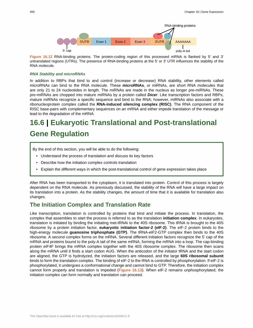
Figure 16.12 RNA-binding proteins. The protein-coding region of this processed mRNA is flanked by 5' and 3'untranslated regions (UTRs). The presence of RNA-binding proteins at the 5' or 3' UTR influences the stability of theRNA molecule.
RNA Stability and microRNAs
In addition to RBPs that bind to and control (increase or decrease) RNA stability, other elements calledmicroRNAs can bind to the RNA molecule. These microRNAs, or miRNAs, are short RNA molecules thatare only 21 to 24 nucleotides in length. The miRNAs are made in the nucleus as longer pre-miRNAs. Thesepre-miRNAs are chopped into mature miRNAs by a protein called Dicer. Like transcription factors and RBPs,mature miRNAs recognize a specific sequence and bind to the RNA; however, miRNAs also associate with aribonucleoprotein complex called the RNA-induced silencing complex (RISC). The RNA component of theRISC base-pairs with complementary sequences on an mRNA and either impede translation of the message orlead to the degradation of the mRNA.
16.6 | Eukaryotic Translational and Post-translational
Gene Regulation
By the end of this section, you will be able to do the following:
• Understand the process of translation and discuss its key factors
• Describe how the initiation complex controls translation
• Explain the different ways in which the post-translational control of gene expression takes place
After RNA has been transported to the cytoplasm, it is translated into protein. Control of this process is largelydependent on the RNA molecule. As previously discussed, the stability of the RNA will have a large impact onits translation into a protein. As the stability changes, the amount of time that it is available for translation alsochanges.
The Initiation Complex and Translation Rate
Like transcription, translation is controlled by proteins that bind and initiate the process. In translation, thecomplex that assembles to start the process is referred to as the translation initiation complex. In eukaryotes,translation is initiated by binding the initiating met-tRNAi to the 40S ribosome. This tRNA is brought to the 40Sribosome by a protein initiation factor, eukaryotic initiation factor-2 (eIF-2). The eIF-2 protein binds to thehigh-energy molecule guanosine triphosphate (GTP). The tRNA-eIF2-GTP complex then binds to the 40Sribosome. A second complex forms on the mRNA. Several different initiation factors recognize the 5' cap of themRNA and proteins bound to the poly-A tail of the same mRNA, forming the mRNA into a loop. The cap-bindingprotein eIF4F brings the mRNA complex together with the 40S ribosome complex. The ribosome then scansalong the mRNA until it finds a start codon AUG. When the anticodon of the initiator tRNA and the start codonare aligned, the GTP is hydrolyzed, the initiation factors are released, and the large 60S ribosomal subunitbinds to form the translation complex. The binding of eIF-2 to the RNA is controlled by phosphorylation. If eIF-2 isphosphorylated, it undergoes a conformational change and cannot bind to GTP. Therefore, the initiation complexcannot form properly and translation is impeded (Figure 16.13). When eIF-2 remains unphosphorylated, theinitiation complex can form normally and translation can proceed.
450 Chapter 16 | Gene Expression
This OpenStax book is available for free at http://cnx.org/content/col24361/1.8
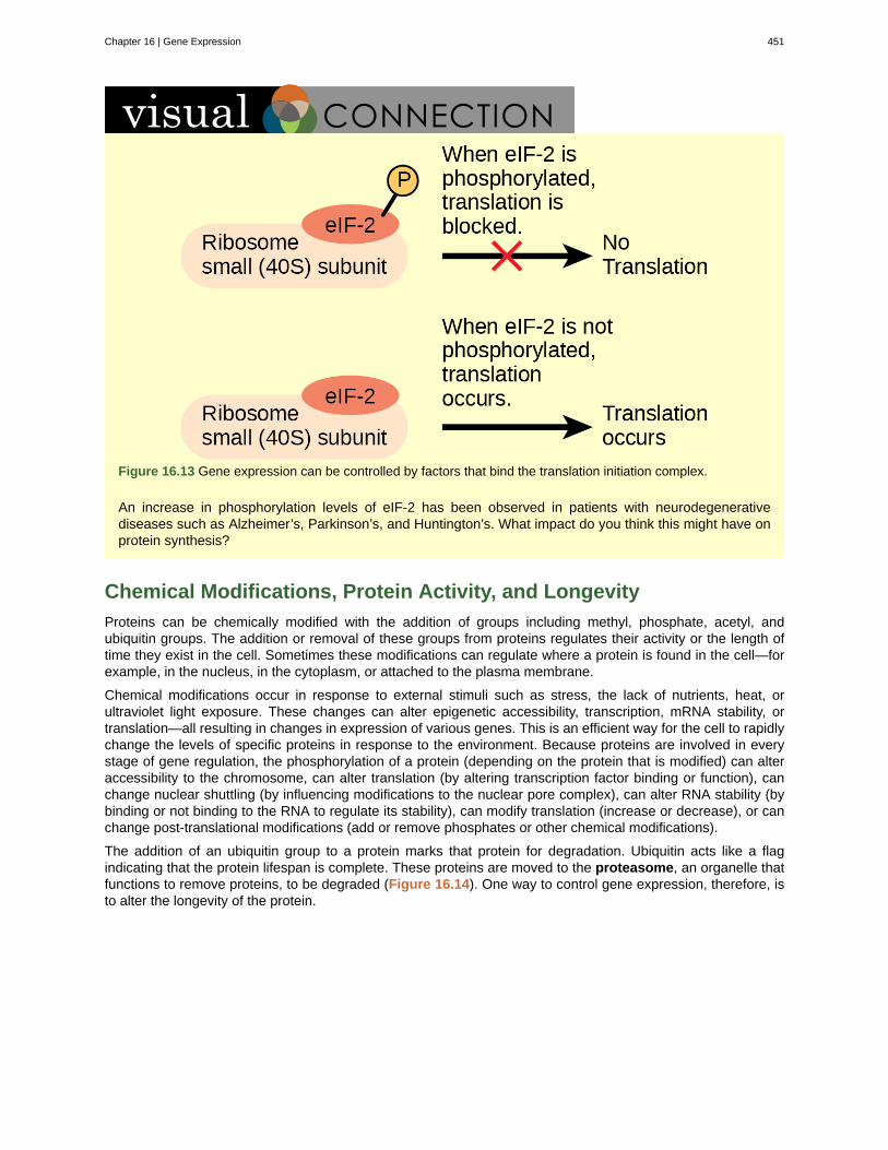
Figure 16.13 Gene expression can be controlled by factors that bind the translation initiation complex.
An increase in phosphorylation levels of eIF-2 has been observed in patients with neurodegenerativediseases such as Alzheimer’s, Parkinson’s, and Huntington’s. What impact do you think this might have onprotein synthesis?
Chemical Modifications, Protein Activity, and Longevity
Proteins can be chemically modified with the addition of groups including methyl, phosphate, acetyl, andubiquitin groups. The addition or removal of these groups from proteins regulates their activity or the length oftime they exist in the cell. Sometimes these modifications can regulate where a protein is found in the cell—forexample, in the nucleus, in the cytoplasm, or attached to the plasma membrane.
Chemical modifications occur in response to external stimuli such as stress, the lack of nutrients, heat, orultraviolet light exposure. These changes can alter epigenetic accessibility, transcription, mRNA stability, ortranslation—all resulting in changes in expression of various genes. This is an efficient way for the cell to rapidlychange the levels of specific proteins in response to the environment. Because proteins are involved in everystage of gene regulation, the phosphorylation of a protein (depending on the protein that is modified) can alteraccessibility to the chromosome, can alter translation (by altering transcription factor binding or function), canchange nuclear shuttling (by influencing modifications to the nuclear pore complex), can alter RNA stability (bybinding or not binding to the RNA to regulate its stability), can modify translation (increase or decrease), or canchange post-translational modifications (add or remove phosphates or other chemical modifications).
The addition of an ubiquitin group to a protein marks that protein for degradation. Ubiquitin acts like a flagindicating that the protein lifespan is complete. These proteins are moved to the proteasome, an organelle thatfunctions to remove proteins, to be degraded (Figure 16.14). One way to control gene expression, therefore, isto alter the longevity of the protein.
Chapter 16 | Gene Expression 451
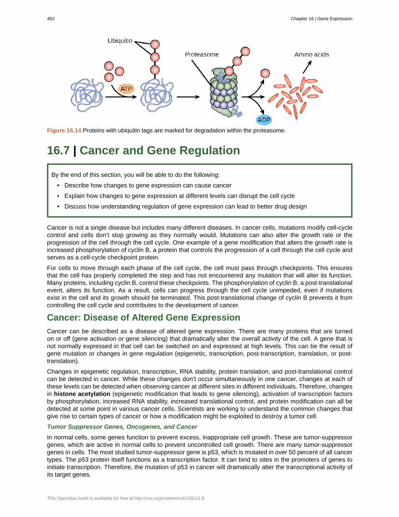
Figure 16.14 Proteins with ubiquitin tags are marked for degradation within the proteasome.
16.7 | Cancer and Gene Regulation
By the end of this section, you will be able to do the following:
• Describe how changes to gene expression can cause cancer
• Explain how changes to gene expression at different levels can disrupt the cell cycle
• Discuss how understanding regulation of gene expression can lead to better drug design
Cancer is not a single disease but includes many different diseases. In cancer cells, mutations modify cell-cyclecontrol and cells don’t stop growing as they normally would. Mutations can also alter the growth rate or theprogression of the cell through the cell cycle. One example of a gene modification that alters the growth rate isincreased phosphorylation of cyclin B, a protein that controls the progression of a cell through the cell cycle andserves as a cell-cycle checkpoint protein.
For cells to move through each phase of the cell cycle, the cell must pass through checkpoints. This ensuresthat the cell has properly completed the step and has not encountered any mutation that will alter its function.Many proteins, including cyclin B, control these checkpoints. The phosphorylation of cyclin B, a post-translationalevent, alters its function. As a result, cells can progress through the cell cycle unimpeded, even if mutationsexist in the cell and its growth should be terminated. This post-translational change of cyclin B prevents it fromcontrolling the cell cycle and contributes to the development of cancer.
Cancer: Disease of Altered Gene Expression
Cancer can be described as a disease of altered gene expression. There are many proteins that are turnedon or off (gene activation or gene silencing) that dramatically alter the overall activity of the cell. A gene that isnot normally expressed in that cell can be switched on and expressed at high levels. This can be the result ofgene mutation or changes in gene regulation (epigenetic, transcription, post-transcription, translation, or post-translation).
Changes in epigenetic regulation, transcription, RNA stability, protein translation, and post-translational controlcan be detected in cancer. While these changes don’t occur simultaneously in one cancer, changes at each ofthese levels can be detected when observing cancer at different sites in different individuals. Therefore, changesin histone acetylation (epigenetic modification that leads to gene silencing), activation of transcription factorsby phosphorylation, increased RNA stability, increased translational control, and protein modification can all bedetected at some point in various cancer cells. Scientists are working to understand the common changes thatgive rise to certain types of cancer or how a modification might be exploited to destroy a tumor cell.
Tumor Suppressor Genes, Oncogenes, and Cancer
In normal cells, some genes function to prevent excess, inappropriate cell growth. These are tumor-suppressorgenes, which are active in normal cells to prevent uncontrolled cell growth. There are many tumor-suppressorgenes in cells. The most studied tumor-suppressor gene is p53, which is mutated in over 50 percent of all cancertypes. The p53 protein itself functions as a transcription factor. It can bind to sites in the promoters of genes toinitiate transcription. Therefore, the mutation of p53 in cancer will dramatically alter the transcriptional activity ofits target genes.
452 Chapter 16 | Gene Expression
This OpenStax book is available for free at http://cnx.org/content/col24361/1.8
Watch this animation (http://openstaxcollege.org/l/p53_cancer) to learn more about the use of p53 infighting cancer.
Proto-oncogenes are positive cell-cycle regulators. When mutated, proto-oncogenes can become oncogenesand cause cancer. Overexpression of the oncogene can lead to uncontrolled cell growth. This is becauseoncogenes can alter transcriptional activity, stability, or protein translation of another gene that directly orindirectly controls cell growth. An example of an oncogene involved in cancer is a protein called myc. Mycis a transcription factor that is aberrantly activated in Burkett’s Lymphoma, a cancer of the lymph system.Overexpression of myc transforms normal B cells into cancerous cells that continue to grow uncontrollably.High B-cell numbers can result in tumors that can interfere with normal bodily function. Patients with Burkett’slymphoma can develop tumors on their jaw or in their mouth that interfere with the ability to eat.
Cancer and Epigenetic Alterations
Silencing genes through epigenetic mechanisms is also very common in cancer cells. There are characteristicmodifications to histone proteins and DNA that are associated with silenced genes. In cancer cells, the DNAin the promoter region of silenced genes is methylated on cytosine DNA residues in CpG islands. Histoneproteins that surround that region lack the acetylation modification that is present when the genes are expressedin normal cells. This combination of DNA methylation and histone deacetylation (epigenetic modifications thatlead to gene silencing) is commonly found in cancer. When these modifications occur, the gene present inthat chromosomal region is silenced. Increasingly, scientists understand how epigenetic changes are altered incancer. Because these changes are temporary and can be reversed—for example, by preventing the action ofthe histone deacetylase protein that removes acetyl groups, or by DNA methyl transferase enzymes that addmethyl groups to cytosines in DNA—it is possible to design new drugs and new therapies to take advantageof the reversible nature of these processes. Indeed, many researchers are testing how a silenced gene can beswitched back on in a cancer cell to help re-establish normal growth patterns.
Genes involved in the development of many other illnesses, ranging from allergies to inflammation to autism,are thought to be regulated by epigenetic mechanisms. As our knowledge of how genes are controlled deepens,new ways to treat diseases like cancer will emerge.
Cancer and Transcriptional Control
Alterations in cells that give rise to cancer can affect the transcriptional control of gene expression. Mutationsthat activate transcription factors, such as increased phosphorylation, can increase the binding of a transcriptionfactor to its binding site in a promoter. This could lead to increased transcriptional activation of that gene thatresults in modified cell growth. Alternatively, a mutation in the DNA of a promoter or enhancer region canincrease the binding ability of a transcription factor. This could also lead to the increased transcription andaberrant gene expression that is seen in cancer cells.
Researchers have been investigating how to control the transcriptional activation of gene expression in cancer.Identifying how a transcription factor binds, or a pathway that activates where a gene can be turned off, has led tonew drugs and new ways to treat cancer. In breast cancer, for example, many proteins are overexpressed. Thiscan lead to increased phosphorylation of key transcription factors that increase transcription. One such exampleis the overexpression of the epidermal growth-factor receptor (EGFR) in a subset of breast cancers. The EGFRpathway activates many protein kinases that, in turn, activate many transcription factors which control genesinvolved in cell growth. New drugs that prevent the activation of EGFR have been developed and are used totreat these cancers.
Cancer and Post-transcriptional Control
Changes in the post-transcriptional control of a gene can also result in cancer. Recently, several groups ofresearchers have shown that specific cancers have altered expression of miRNAs. Because miRNAs bind to the3' UTR of RNA molecules to degrade them, overexpression of these miRNAs could be detrimental to normal
Chapter 16 | Gene Expression 453
cellular activity. Too many miRNAs could dramatically decrease the RNA population, leading to a decrease inprotein expression. Several studies have demonstrated a change in the miRNA population in specific cancertypes. It appears that the subset of miRNAs expressed in breast cancer cells is quite different from the subsetexpressed in lung cancer cells or even from normal breast cells. This suggests that alterations in miRNA activitycan contribute to the growth of breast cancer cells. These types of studies also suggest that if some miRNAsare specifically expressed only in cancer cells, they could be potential drug targets. It would, therefore, beconceivable that new drugs that turn off miRNA expression in cancer could be an effective method to treatcancer.
Cancer and Translational/Post-translational Control
There are many examples of how translational or post-translational modifications of proteins arise in cancer.Modifications are found in cancer cells from the increased translation of a protein to changes in proteinphosphorylation to alternative splice variants of a protein. An example of how the expression of an alternativeform of a protein can have dramatically different outcomes is seen in colon cancer cells. The c-Flip protein, aprotein involved in mediating the cell-death pathway, comes in two forms: long (c-FLIPL) and short (c-FLIPS).Both forms appear to be involved in initiating controlled cell-death mechanisms in normal cells. However, incolon cancer cells, expression of the long form results in increased cell growth instead of cell death. Clearly, theexpression of the wrong protein dramatically alters cell function and contributes to the development of cancer.
New Drugs to Combat Cancer: Targeted Therapies
Scientists are using what is known about the regulation of gene expression in disease states, including cancer, todevelop new ways to treat and prevent disease development. Many scientists are designing drugs on the basisof the gene expression patterns within individual tumors. This idea, that therapy and medicines can be tailoredto an individual, has given rise to the field of personalized medicine. With an increased understanding of generegulation and gene function, medicines can be designed to specifically target diseased cells without harminghealthy cells. Some new medicines, called targeted therapies, have exploited the overexpression of a specificprotein or the mutation of a gene to develop a new medication to treat disease. One such example is the useof anti-EGF receptor medications to treat the subset of breast cancer tumors that have very high levels of theEGF protein. Undoubtedly, more targeted therapies will be developed as scientists learn more about how geneexpression changes can cause cancer.
Clinical Trial CoordinatorA clinical trial coordinator is the person managing the proceedings of the clinical trial. This job includescoordinating patient schedules and appointments, maintaining detailed notes, building the database to trackpatients (especially for long-term follow-up studies), ensuring proper documentation has been acquired andaccepted, and working with the nurses and doctors to facilitate the trial and publication of the results. Aclinical trial coordinator may have a science background, like a nursing degree, or other certification. Peoplewho have worked in science labs or in clinical offices are also qualified to become a clinical trial coordinator.These jobs are generally in hospitals; however, some clinics and doctor’s offices also conduct clinical trialsand may hire a coordinator.
454 Chapter 16 | Gene Expression
This OpenStax book is available for free at http://cnx.org/content/col24361/1.8
3' UTR
5' cap
5' UTR
activator
catabolite activator protein (CAP)
cis-acting element
Dicer
DNA methylation
enhancer
epigenetic
eukaryotic initiation factor-2 (eIF-2)
gene expression
guanine diphosphate (GDP)
guanine triphosphate (GTP)
histone acetylation
inducible operon
initiation complex
lac operon
large 60S ribosomal subunit
microRNA (miRNA)
myc
negative regulator
operator
operon
poly-A tail
KEY TERMS
3' untranslated region; region just downstream of the protein-coding region in an RNA molecule that isnot translated
a methylated guanosine triphosphate (GTP) molecule that is attached to the 5' end of a messenger RNAto protect the end from degradation
5' untranslated region; region just upstream of the protein-coding region in an RNA molecule that is nottranslated
protein that binds to prokaryotic operators to increase transcription
protein that complexes with cAMP to bind to the promoter sequences ofoperons which control sugar processing when glucose is not available
transcription factor binding sites within the promoter that regulate the transcription of a geneadjacent to it
enzyme that chops the pre-miRNA into the mature form of the miRNA
epigenetic modification that leads to gene silencing; a process involving adding a methylgroup to the DNA molecule
segment of DNA that is upstream, downstream, perhaps thousands of nucleotides away, or onanother chromosome that influence the transcription of a specific gene
heritable changes that do not involve changes in the DNA sequence
protein that binds first to an mRNA to initiate translation
processes that control the turning on or turning off of a gene
molecule that is left after the energy is used to start translation
energy-providing molecule that binds to eIF-2 and is needed for translation
epigenetic modification that leads to gene silencing; a process involving adding orremoving an acetyl functional group
operon that can be activated or repressed depending on cellular needs and the surroundingenvironment
protein complex containing eIF-2 that starts translation
operon in prokaryotic cells that encodes genes required for processing and intake of lactose
second, larger ribosomal subunit that binds to the RNA to translate it into protein
small RNA molecules (approximately 21 nucleotides in length) that bind to RNA moleculesto degrade them
oncogene that causes cancer in many cancer cells
protein that prevents transcription
region of DNA outside of the promoter region that binds activators or repressors that control geneexpression in prokaryotic cells
collection of genes involved in a pathway that are transcribed together as a single mRNA in prokaryoticcells
a series of adenine nucleotides that are attached to the 3' end of an mRNA to protect the end from
Chapter 16 | Gene Expression 455