11.1 the process of meiosis - the expert ta
TRANSCRIPT
11.1 | The Process of Meiosis
By the end of this section, you will be able to do the following:
• Describe the behavior of chromosomes during meiosis, and the differences between the first and secondmeiotic divisions
• Describe the cellular events that take place during meiosis
• Explain the differences between meiosis and mitosis
• Explain the mechanisms within the meiotic process that produce genetic variation among the haploidgametes
Sexual reproduction requires the union of two specialized cells, called gametes, each of which contains oneset of chromosomes. When gametes unite, they form a zygote, or fertilized egg that contains two sets ofchromosomes. (Note: Cells that contain one set of chromosomes are called haploid; cells containing twosets of chromosomes are called diploid.) If the reproductive cycle is to continue for any sexually reproducingspecies, then the diploid cell must somehow reduce its number of chromosome sets to produce haploid gametes;otherwise, the number of chromosome sets will double with every future round of fertilization. Therefore, sexualreproduction requires a nuclear division that reduces the number of chromosome sets by half.
Most animals and plants and many unicellular organisms are diploid and therefore have two sets ofchromosomes. In each somatic cell of the organism (all cells of a multicellular organism except the gametes orreproductive cells), the nucleus contains two copies of each chromosome, called homologous chromosomes.Homologous chromosomes are matched pairs containing the same genes in identical locations along theirlengths. Diploid organisms inherit one copy of each homologous chromosome from each parent.
Meiosis is the nuclear division that forms haploid cells from diploid cells, and it employs many of the samecellular mechanisms as mitosis. However, as you have learned, mitosis produces daughter cells whose nucleiare genetically identical to the original parent nucleus. In mitosis, both the parent and the daughter nuclei areat the same “ploidy level”—diploid in the case of most multicellular most animals. Plants use mitosis to grow assporophytes, and to grow and produce eggs and sperm as gametophytes; so they use mitosis for both haploidand diploid cells (as well as for all other ploidies). In meiosis, the starting nucleus is always diploid and thedaughter nuclei that result are haploid. To achieve this reduction in chromosome number, meiosis consists ofone round of chromosome replication followed by two rounds of nuclear division. Because the events that occurduring each of the division stages are analogous to the events of mitosis, the same stage names are assigned.However, because there are two rounds of division, the major process and the stages are designated with a “I”or a “II.” Thus, meiosis I is the first round of meiotic division and consists of prophase I, prometaphase I, andso on. Likewise, Meiosis II (during which the second round of meiotic division takes place) includes prophase II,prometaphase II, and so on.
Meiosis I
Meiosis is preceded by an interphase consisting of G1, S, and G2 phases, which are nearly identical to thephases preceding mitosis. The G1 phase (the “first gap phase”) is focused on cell growth. During the Sphase—the second phase of interphase—the cell copies or replicates the DNA of the chromosomes. Finally, inthe G2 phase (the “second gap phase”) the cell undergoes the final preparations for meiosis.
During DNA duplication in the S phase, each chromosome is replicated to produce two identical copies—sisterchromatids that are held together at the centromere by cohesin proteins, which hold the chromatids togetheruntil anaphase II.
Prophase I
Early in prophase I, before the chromosomes can be seen clearly with a microscope, the homologouschromosomes are attached at their tips to the nuclear envelope by proteins. As the nuclear envelope begins tobreak down, the proteins associated with homologous chromosomes bring the pair closer together. Recall thatin mitosis, homologous chromosomes do not pair together. The synaptonemal complex, a lattice of proteinsbetween the homologous chromosomes, first forms at specific locations and then spreads outward to coverthe entire length of the chromosomes. The tight pairing of the homologous chromosomes is called synapsis. In
308 Chapter 11 | Meiosis and Sexual Reproduction
This OpenStax book is available for free at http://cnx.org/content/col24361/1.8
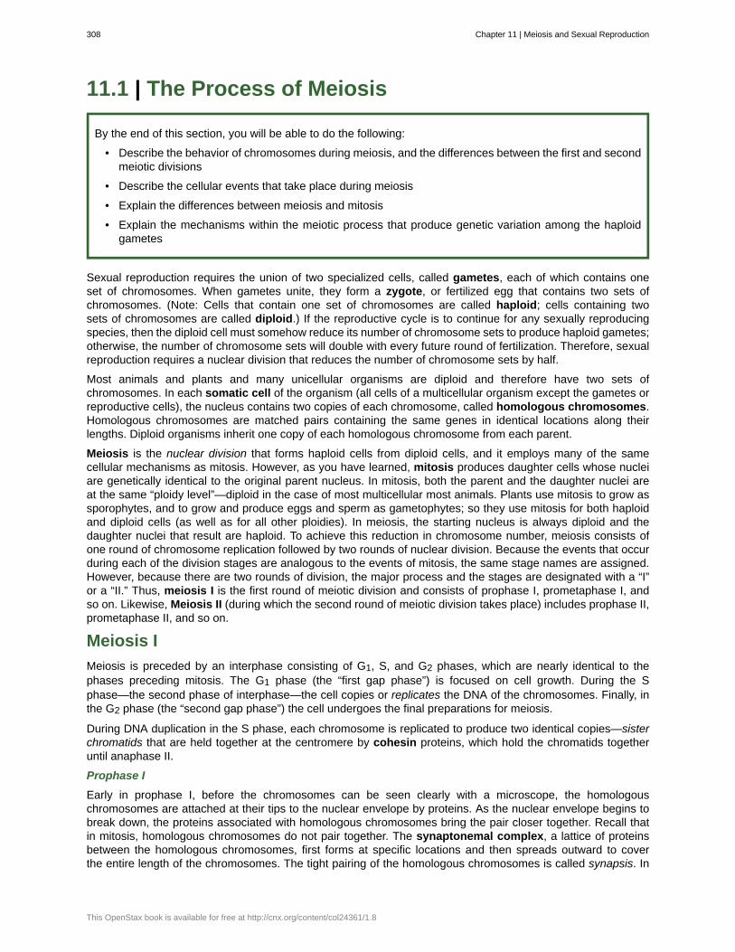
synapsis, the genes on the chromatids of the homologous chromosomes are aligned precisely with each other.The synaptonemal complex supports the exchange of chromosomal segments between homologous nonsisterchromatids—a process called crossing over. Crossing over can be observed visually after the exchange aschiasmata (singular = chiasma) (Figure 11.2).
In humans, even though the X and Y sex chromosomes are not completely homologous (that is, most of theirgenes differ), there is a small region of homology that allows the X and Y chromosomes to pair up duringprophase I. A partial synaptonemal complex develops only between the regions of homology.
Figure 11.2 Early in prophase I, homologous chromosomes come together to form a synapse. The chromosomes arebound tightly together and in perfect alignment by a protein lattice called a synaptonemal complex and by cohesinproteins at the centromere.
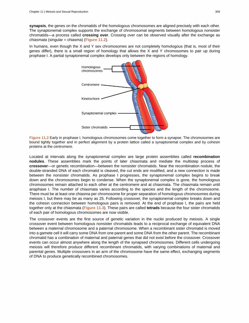
Located at intervals along the synaptonemal complex are large protein assemblies called recombinationnodules. These assemblies mark the points of later chiasmata and mediate the multistep process ofcrossover—or genetic recombination—between the nonsister chromatids. Near the recombination nodule, thedouble-stranded DNA of each chromatid is cleaved, the cut ends are modified, and a new connection is madebetween the nonsister chromatids. As prophase I progresses, the synaptonemal complex begins to breakdown and the chromosomes begin to condense. When the synaptonemal complex is gone, the homologouschromosomes remain attached to each other at the centromere and at chiasmata. The chiasmata remain untilanaphase I. The number of chiasmata varies according to the species and the length of the chromosome.There must be at least one chiasma per chromosome for proper separation of homologous chromosomes duringmeiosis I, but there may be as many as 25. Following crossover, the synaptonemal complex breaks down andthe cohesin connection between homologous pairs is removed. At the end of prophase I, the pairs are heldtogether only at the chiasmata (Figure 11.3). These pairs are called tetrads because the four sister chromatidsof each pair of homologous chromosomes are now visible.
The crossover events are the first source of genetic variation in the nuclei produced by meiosis. A singlecrossover event between homologous nonsister chromatids leads to a reciprocal exchange of equivalent DNAbetween a maternal chromosome and a paternal chromosome. When a recombinant sister chromatid is movedinto a gamete cell it will carry some DNA from one parent and some DNA from the other parent. The recombinantchromatid has a combination of maternal and paternal genes that did not exist before the crossover. Crossoverevents can occur almost anywhere along the length of the synapsed chromosomes. Different cells undergoingmeiosis will therefore produce different recombinant chromatids, with varying combinations of maternal andparental genes. Multiple crossovers in an arm of the chromosome have the same effect, exchanging segmentsof DNA to produce genetically recombined chromosomes.
Chapter 11 | Meiosis and Sexual Reproduction 309
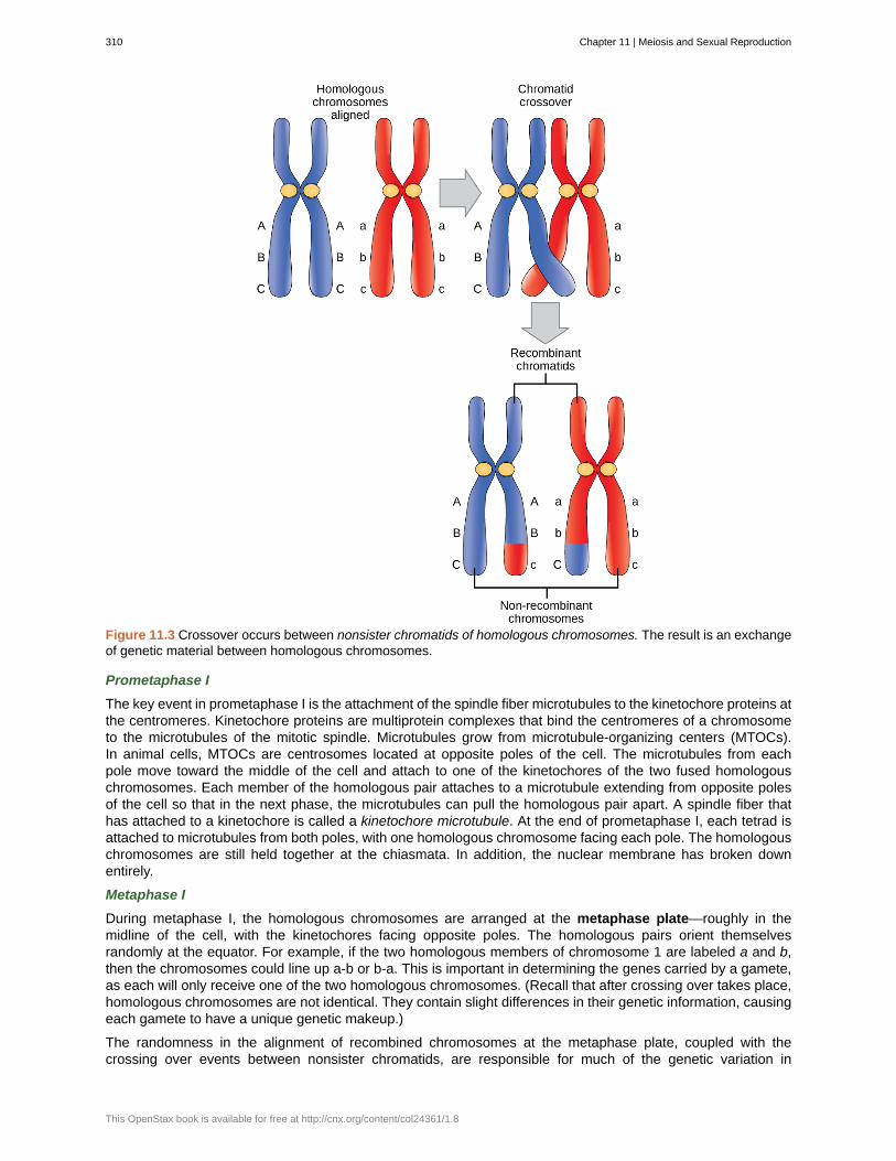
Figure 11.3 Crossover occurs between nonsister chromatids of homologous chromosomes. The result is an exchangeof genetic material between homologous chromosomes.
Prometaphase I
The key event in prometaphase I is the attachment of the spindle fiber microtubules to the kinetochore proteins atthe centromeres. Kinetochore proteins are multiprotein complexes that bind the centromeres of a chromosometo the microtubules of the mitotic spindle. Microtubules grow from microtubule-organizing centers (MTOCs).In animal cells, MTOCs are centrosomes located at opposite poles of the cell. The microtubules from eachpole move toward the middle of the cell and attach to one of the kinetochores of the two fused homologouschromosomes. Each member of the homologous pair attaches to a microtubule extending from opposite polesof the cell so that in the next phase, the microtubules can pull the homologous pair apart. A spindle fiber thathas attached to a kinetochore is called a kinetochore microtubule. At the end of prometaphase I, each tetrad isattached to microtubules from both poles, with one homologous chromosome facing each pole. The homologouschromosomes are still held together at the chiasmata. In addition, the nuclear membrane has broken downentirely.
Metaphase I
During metaphase I, the homologous chromosomes are arranged at the metaphase plate—roughly in themidline of the cell, with the kinetochores facing opposite poles. The homologous pairs orient themselvesrandomly at the equator. For example, if the two homologous members of chromosome 1 are labeled a and b,then the chromosomes could line up a-b or b-a. This is important in determining the genes carried by a gamete,as each will only receive one of the two homologous chromosomes. (Recall that after crossing over takes place,homologous chromosomes are not identical. They contain slight differences in their genetic information, causingeach gamete to have a unique genetic makeup.)
The randomness in the alignment of recombined chromosomes at the metaphase plate, coupled with thecrossing over events between nonsister chromatids, are responsible for much of the genetic variation in
310 Chapter 11 | Meiosis and Sexual Reproduction
This OpenStax book is available for free at http://cnx.org/content/col24361/1.8
the offspring. To clarify this further, remember that the homologous chromosomes of a sexually reproducingorganism are originally inherited as two separate sets, one from each parent. Using humans as an example,one set of 23 chromosomes is present in the egg donated by the mother. The father provides the other set of23 chromosomes in the sperm that fertilizes the egg. Every cell of the multicellular offspring has copies of theoriginal two sets of homologous chromosomes. In prophase I of meiosis, the homologous chromosomes formthe tetrads. In metaphase I, these pairs line up at the midway point between the two poles of the cell to formthe metaphase plate. Because there is an equal chance that a microtubule fiber will encounter a maternally orpaternally inherited chromosome, the arrangement of the tetrads at the metaphase plate is random. Thus, anymaternally inherited chromosome may face either pole. Likewise, any paternally inherited chromosome may alsoface either pole. The orientation of each tetrad is independent of the orientation of the other 22 tetrads.
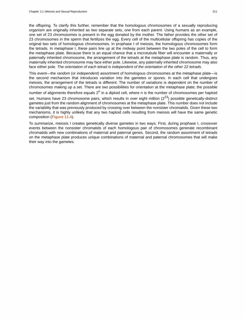
This event—the random (or independent) assortment of homologous chromosomes at the metaphase plate—isthe second mechanism that introduces variation into the gametes or spores. In each cell that undergoesmeiosis, the arrangement of the tetrads is different. The number of variations is dependent on the number ofchromosomes making up a set. There are two possibilities for orientation at the metaphase plate; the possible
number of alignments therefore equals 2n in a diploid cell, where n is the number of chromosomes per haploid
set. Humans have 23 chromosome pairs, which results in over eight million (223) possible genetically-distinctgametes just from the random alignment of chromosomes at the metaphase plate. This number does not includethe variability that was previously produced by crossing over between the nonsister chromatids. Given these twomechanisms, it is highly unlikely that any two haploid cells resulting from meiosis will have the same geneticcomposition (Figure 11.4).
To summarize, meiosis I creates genetically diverse gametes in two ways. First, during prophase I, crossoverevents between the nonsister chromatids of each homologous pair of chromosomes generate recombinantchromatids with new combinations of maternal and paternal genes. Second, the random assortment of tetradson the metaphase plate produces unique combinations of maternal and paternal chromosomes that will maketheir way into the gametes.
Chapter 11 | Meiosis and Sexual Reproduction 311
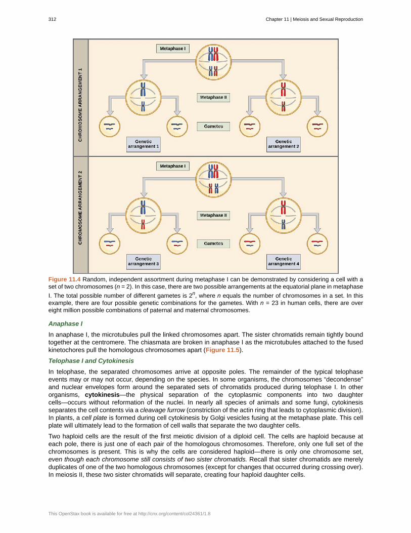
Figure 11.4 Random, independent assortment during metaphase I can be demonstrated by considering a cell with aset of two chromosomes (n = 2). In this case, there are two possible arrangements at the equatorial plane in metaphase
I. The total possible number of different gametes is 2n, where n equals the number of chromosomes in a set. In thisexample, there are four possible genetic combinations for the gametes. With n = 23 in human cells, there are overeight million possible combinations of paternal and maternal chromosomes.
Anaphase I
In anaphase I, the microtubules pull the linked chromosomes apart. The sister chromatids remain tightly boundtogether at the centromere. The chiasmata are broken in anaphase I as the microtubules attached to the fusedkinetochores pull the homologous chromosomes apart (Figure 11.5).
Telophase I and Cytokinesis
In telophase, the separated chromosomes arrive at opposite poles. The remainder of the typical telophaseevents may or may not occur, depending on the species. In some organisms, the chromosomes “decondense”and nuclear envelopes form around the separated sets of chromatids produced during telophase I. In otherorganisms, cytokinesis—the physical separation of the cytoplasmic components into two daughtercells—occurs without reformation of the nuclei. In nearly all species of animals and some fungi, cytokinesisseparates the cell contents via a cleavage furrow (constriction of the actin ring that leads to cytoplasmic division).In plants, a cell plate is formed during cell cytokinesis by Golgi vesicles fusing at the metaphase plate. This cellplate will ultimately lead to the formation of cell walls that separate the two daughter cells.
Two haploid cells are the result of the first meiotic division of a diploid cell. The cells are haploid because ateach pole, there is just one of each pair of the homologous chromosomes. Therefore, only one full set of thechromosomes is present. This is why the cells are considered haploid—there is only one chromosome set,even though each chromosome still consists of two sister chromatids. Recall that sister chromatids are merelyduplicates of one of the two homologous chromosomes (except for changes that occurred during crossing over).In meiosis II, these two sister chromatids will separate, creating four haploid daughter cells.
312 Chapter 11 | Meiosis and Sexual Reproduction
This OpenStax book is available for free at http://cnx.org/content/col24361/1.8
Review the process of meiosis, observing how chromosomes align and migrate, at Meiosis: An InteractiveAnimation (http://openstaxcollege.org/l/animal_meiosis) .
Meiosis II
In some species, cells enter a brief interphase, or interkinesis, before entering meiosis II. Interkinesis lacks anS phase, so chromosomes are not duplicated. The two cells produced in meiosis I go through the events ofmeiosis II in synchrony. During meiosis II, the sister chromatids within the two daughter cells separate, formingfour new haploid gametes. The mechanics of meiosis II are similar to mitosis, except that each dividing cell hasonly one set of homologous chromosomes, each with two chromatids. Therefore, each cell has half the numberof sister chromatids to separate out as a diploid cell undergoing mitosis. In terms of chromosomal content, cellsat the start of meiosis II are similar to haploid cells in G2, preparing to undergo mitosis.
Prophase II
If the chromosomes decondensed in telophase I, they condense again. If nuclear envelopes were formed, theyfragment into vesicles. The MTOCs that were duplicated during interkinesis move away from each other towardopposite poles, and new spindles are formed.
Prometaphase II
The nuclear envelopes are completely broken down, and the spindle is fully formed. Each sister chromatid formsan individual kinetochore that attaches to microtubules from opposite poles.
Metaphase II
The sister chromatids are maximally condensed and aligned at the equator of the cell.
Anaphase II
The sister chromatids are pulled apart by the kinetochore microtubules and move toward opposite poles.Nonkinetochore microtubules elongate the cell.
Chapter 11 | Meiosis and Sexual Reproduction 313
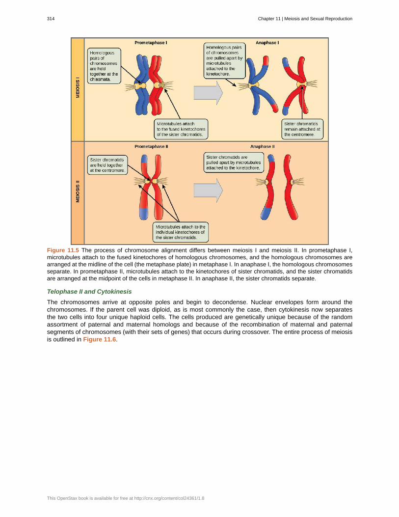
Figure 11.5 The process of chromosome alignment differs between meiosis I and meiosis II. In prometaphase I,microtubules attach to the fused kinetochores of homologous chromosomes, and the homologous chromosomes arearranged at the midline of the cell (the metaphase plate) in metaphase I. In anaphase I, the homologous chromosomesseparate. In prometaphase II, microtubules attach to the kinetochores of sister chromatids, and the sister chromatidsare arranged at the midpoint of the cells in metaphase II. In anaphase II, the sister chromatids separate.
Telophase II and Cytokinesis
The chromosomes arrive at opposite poles and begin to decondense. Nuclear envelopes form around thechromosomes. If the parent cell was diploid, as is most commonly the case, then cytokinesis now separatesthe two cells into four unique haploid cells. The cells produced are genetically unique because of the randomassortment of paternal and maternal homologs and because of the recombination of maternal and paternalsegments of chromosomes (with their sets of genes) that occurs during crossover. The entire process of meiosisis outlined in Figure 11.6.
314 Chapter 11 | Meiosis and Sexual Reproduction
This OpenStax book is available for free at http://cnx.org/content/col24361/1.8
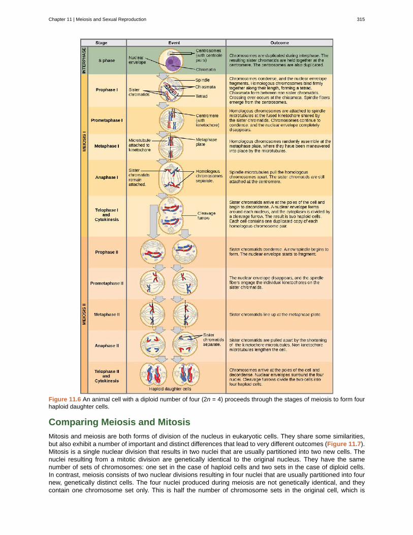
Figure 11.6 An animal cell with a diploid number of four (2n = 4) proceeds through the stages of meiosis to form fourhaploid daughter cells.
Comparing Meiosis and Mitosis
Mitosis and meiosis are both forms of division of the nucleus in eukaryotic cells. They share some similarities,but also exhibit a number of important and distinct differences that lead to very different outcomes (Figure 11.7).Mitosis is a single nuclear division that results in two nuclei that are usually partitioned into two new cells. Thenuclei resulting from a mitotic division are genetically identical to the original nucleus. They have the samenumber of sets of chromosomes: one set in the case of haploid cells and two sets in the case of diploid cells.In contrast, meiosis consists of two nuclear divisions resulting in four nuclei that are usually partitioned into fournew, genetically distinct cells. The four nuclei produced during meiosis are not genetically identical, and theycontain one chromosome set only. This is half the number of chromosome sets in the original cell, which is
Chapter 11 | Meiosis and Sexual Reproduction 315
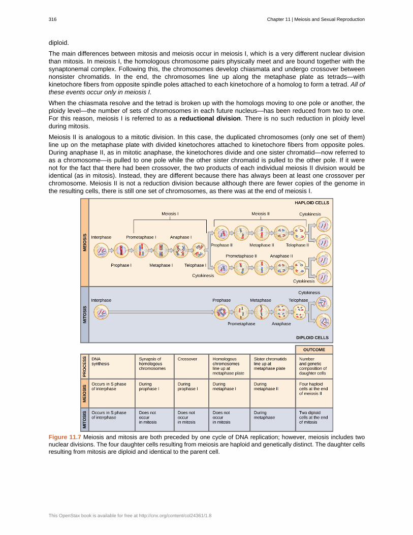
diploid.
The main differences between mitosis and meiosis occur in meiosis I, which is a very different nuclear divisionthan mitosis. In meiosis I, the homologous chromosome pairs physically meet and are bound together with thesynaptonemal complex. Following this, the chromosomes develop chiasmata and undergo crossover betweennonsister chromatids. In the end, the chromosomes line up along the metaphase plate as tetrads—withkinetochore fibers from opposite spindle poles attached to each kinetochore of a homolog to form a tetrad. All ofthese events occur only in meiosis I.
When the chiasmata resolve and the tetrad is broken up with the homologs moving to one pole or another, theploidy level—the number of sets of chromosomes in each future nucleus—has been reduced from two to one.For this reason, meiosis I is referred to as a reductional division. There is no such reduction in ploidy levelduring mitosis.
Meiosis II is analogous to a mitotic division. In this case, the duplicated chromosomes (only one set of them)line up on the metaphase plate with divided kinetochores attached to kinetochore fibers from opposite poles.During anaphase II, as in mitotic anaphase, the kinetochores divide and one sister chromatid—now referred toas a chromosome—is pulled to one pole while the other sister chromatid is pulled to the other pole. If it werenot for the fact that there had been crossover, the two products of each individual meiosis II division would beidentical (as in mitosis). Instead, they are different because there has always been at least one crossover perchromosome. Meiosis II is not a reduction division because although there are fewer copies of the genome inthe resulting cells, there is still one set of chromosomes, as there was at the end of meiosis I.
Figure 11.7 Meiosis and mitosis are both preceded by one cycle of DNA replication; however, meiosis includes twonuclear divisions. The four daughter cells resulting from meiosis are haploid and genetically distinct. The daughter cellsresulting from mitosis are diploid and identical to the parent cell.
316 Chapter 11 | Meiosis and Sexual Reproduction
This OpenStax book is available for free at http://cnx.org/content/col24361/1.8
The Mystery of the Evolution of MeiosisSome characteristics of organisms are so widespread and fundamental that it is sometimes difficult toremember that they evolved like other simple traits. Meiosis is such an extraordinarily complex series ofcellular events that biologists have had trouble testing hypotheses concerning how it may have evolved.Although meiosis is inextricably entwined with sexual reproduction and its advantages and disadvantages,it is important to separate the questions of the evolution of meiosis and the evolution of sex, because earlymeiosis may have been advantageous for different reasons than it is now. Thinking outside the box andimagining what the early benefits from meiosis might have been is one approach to uncovering how it mayhave evolved.
Meiosis and mitosis share obvious cellular processes, and it makes sense that meiosis evolved frommitosis. The difficulty lies in the clear differences between meiosis I and mitosis. Adam Wilkins and Robin
Holliday[1]
summarized the unique events that needed to occur for the evolution of meiosis from mitosis.These steps are homologous chromosome pairing and synapsis, crossover exchanges, sister chromatidsremaining attached during anaphase, and suppression of DNA replication in interphase. They argue thatthe first step is the hardest and most important and that understanding how it evolved would make theevolutionary process clearer. They suggest genetic experiments that might shed light on the evolution ofsynapsis.
There are other approaches to understanding the evolution of meiosis in progress. Different forms of meiosisexist in single-celled protists. Some appear to be simpler or more “primitive” forms of meiosis. Comparingthe meiotic divisions of different protists may shed light on the evolution of meiosis. Marilee Ramesh and
colleagues[2]
compared the genes involved in meiosis in protists to understand when and where meiosismight have evolved. Although research is still ongoing, recent scholarship into meiosis in protists suggeststhat some aspects of meiosis may have evolved later than others. This kind of genetic comparison can tellus what aspects of meiosis are the oldest and what cellular processes they may have borrowed from inearlier cells.
Click through the steps of this interactive animation to compare the meiotic process of cell division to that ofmitosis at How Cells Divide (http://openstaxcollege.org/l/how_cells_dvide) .
11.2 | Sexual Reproduction
By the end of this section, you will be able to do the following:
• Explain that meiosis and sexual reproduction are highly evolved traits
• Identify variation among offspring as a potential evolutionary advantage of sexual reproduction
• Describe the three different life-cycle types among sexually reproducing multicellular organisms.
1.Adam S. Wilkins and Robin Holliday, “The Evolution of Meiosis from Mitosis,” Genetics 181 (2009): 3–12.2. Marilee A. Ramesh, Shehre-Banoo Malik and John M. Logsdon, Jr, “A Phylogenetic Inventory of Meiotic Genes: Evidence for Sex inGiardia and an Early Eukaryotic Origin of Meiosis,” Current Biology 15 (2005):185–91.
Chapter 11 | Meiosis and Sexual Reproduction 317