1 developing a commercial bumble bee clean stock
TRANSCRIPT

1
Developing a Commercial Bumble Bee Clean Stock Certification Program: A white paper
of the North American Pollinator Protection Campaign Bombus Task Force
James P. Strange1*, Sheila R. Colla2, Michelle Duennes3, Elaine Evans4, Laura L.
Figueroa5,6, David W. Inouye7, David M. Lehmann8, Heather Moylett9, Leif Richardson10,
Ben M. Sadd11, James W. Smith12, Amber D. Tripodi,13 Laurie Davies Adams 14
1Department of Entomology, The Ohio State University, Columbus, OH 43214, USA
2SRC: Faculty of Environmental and Urban Change, York University, Toronto, ON, Canada
3Department of Biology, Saint Vincent College, Latrobe, PA 15650, USA
4Department of Entomology, University of Minnesota, Saint Paul, MN 55108 USA
5Department of Environmental Conservation, University of Massachusetts, Amherst, Amherst,
MA, 01003, USA
6Department of Entomology, Cornell University, Ithaca, NY, 14850, USA
7Department of Biology, University of Maryland, College Park, MD 20742, and Rocky Mountain
Biological Laboratory, PO Box 510, Crested Butte, CO 81224, USA
8Center for Public Health and Environmental Assessment (CPHEA), Health and Environmental
Effects Assessment Division, Integrated Health Assessment Branch, US - Environmental
Protection Agency, Research Triangle Park, NC, 27711, USA
9Unaffiliated, Garner, North Carolina, 27529, USA
10The Xerces Society for Invertebrate Conservation, 628 NE Broadway, Suite 20, Portland, OR
97232-1324, USA
11School of Biological Sciences, Illinois State University, Normal, IL 61790, USA
12Retired USDA-Animal and Plant Health Inspection Service, Fuquay-Varina, NC 27526, USA
13Unaffiliated, Raleigh, North Carolina, 27604, USA
14Pollinator Partnership, Transamerica Pyramid, 600 Montgomery Street, Suite 440, San
Francisco, CA 94111 USA
*To whom correspondence should be addressed [email protected]
Disclaimer: The views expressed in this article are those of the author(s) and do not
necessarily represent the views or policies of the U.S. Environmental Protection Agency.

2
Abstract
The commercial production and subsequent movement of bumble bees for pollination of
agricultural field and greenhouse crops is a growing industry in North America and globally.
Concerns have been raised about the impacts of pathogen spillover from managed bees to wild
pollinators, including from commercial bumble bees. This white paper discusses the need to
develop a program to mitigate disease risk in commercial bumble bee production, which will in
turn reduce disease stressors on wild pollinators and other insects. We give an overview of what
is known about bumble bee pathogens, parasites and other symbionts and methods for their
detection, quantification, and control. We also provide information on assessment of risk for
select bumble bee symbionts and highlight key knowledge gaps. Finally, we provide
recommendations for the components of a clean stock program with specific best management
practices for rearing commercial bumble bees including related products such as pollen, wax
and other nesting material.
Introduction
Bumble bees (Bombus spp.) are important pollinators of commercially grown crops, a
variety of garden vegetables, and native flowering plants. Approximately 40 bumble bee species
are native to the United States and Canada (Williams et al., 2014) and three of them are
commercially available in those countries. By far the most economically important managed
bumble bee species in the United States and Canada is Bombus impatiens, a native to the
eastern United States and Canada (Velthuis & van Doorn, 2006). However, Bombus huntii is
available for use in western Canada and Bombus vosnesenskii was recently approved for use in
California and is now being sold commercially, and it is expected that use of these new
3
commercial species will grow in the western United States and Canada in the future. Currently,
these species are produced in facilities in Michigan (United States) and Ontario (Canada) and
shipped throughout North America for crop pollination, most notably, greenhouse grown
tomatoes (Strange, 2015; Velthuis & van Doorn, 2006)
While the commercial producers of bumble bees make efforts to maintain clean stock in
production facilities (Huang et al., 2015), and provide guidelines to end-users for containment
when bees are sold outside of their native range, commercial bumble bee hives are not isolated
from wild bumble bee communities because they often forage outside of greenhouses via vents
(Whittington et al., 2004). Bumble bees are deployed frequently in open-field situations to
augment pollination of field tomatoes, tree fruit, and berry crops. The use of these bees where
they can come into contact with wild bees poses a clear risk for the movement of pathogens and
parasites within and beyond the bumble bee community (Colla et al., 2006; Fürst et al., 2014;
Murray et al., 2013). Managed bumble bees have the potential to amplify existing pathogens
and parasites in the wild bumble bee community, through pathogen spillback (Pereira et al.,
2021), but the introduction of pathogens and parasites with managed colonies represents a
greater concern. High pathogen incidence has been correlated to facilities that deploy
commercial bumble bees, leading to concerns of pathogen spillover (Colla et al., 2006; Murray
et al., 2013).
Notably, declining bumble bee populations in the United States (Cameron et al., 2011)
and Canada (Colla & Packer, 2008) have been linked to higher levels of pathogens (Cordes et
al., 2012; Kent et al., 2018). However, a clear causative link between population status and
infection remains elusive, due to a lack of baseline data on differential susceptibilities. Declines
in some species have raised concerns about extinction risk and over 20% of North American
species have been identified through the International Union for the Conservation of Nature Red
List as ‘at risk’ (updated at https://www.iucnredlist.org/). In addition, several species are legally
recognized in the United States and Canada as endangered, including Bombus affinis, the
4
Rusty Patched Bumble Bee, which is federally protected in both countries. The impacts of
commercial bumble bees on these declining species are poorly understood, but previous
disease outbreaks in rearing facilities have been implicated in declines (Flanders et al., 2003).
Commercial bumble bee production begins in captivity when lab-raised queens are
provisioned with honey bee-collected pollen and sugar syrup and confined to a nest box where
they commence nesting (Huang et al., 2015; Velthuis & van Doorn, 2006). Within a few days of
confinement, the queen bumble bee will oviposit on the pollen mass and begin brooding her
developing offspring. More pollen is provided as needed as the developing nest remains in
isolation in the facility. As worker bees reach adulthood and the nest grows, the nest is moved
to a shipping box and is ready for sale about 60 days after nest initiation. Once colonies reach a
desired size (e.g., 50-100 workers), the nests are shipped from the production facilities to
growers, and do not return (Huang et al., 2015), nor are the nesting materials from sold colonies
returned to the facility; however a percentage of the colonies reared in facilities must be retained
to supply future reproductive individuals for the operation (Huang et al., 2015; Velthuis & van
Doorn, 2006). Growers dispose of the colonies once their crop has completed flowering or the
colony starts producing reproductive individuals instead of workers.
Although the bumble bee production environment is closed, the rearing system does
have external inputs. Notably, sugar and pollen must be supplied to developing colonies, and
nesting material is also essential (Huang et al., 2015). Nesting boxes from major bumble bee
producers are currently composed of plastic boxes that are manufactured for the purpose and
arrive as clean, sterile plastic into the system. Similarly, sugar syrup is provided, generally in a
proprietary nutrient and preservative mixture, and this is sterilized before delivery (Velthuis &
van Doorn, 2006). Pollen must be obtained in large quantities for commercial production and
this necessitates purchasing bulk pollen that has been collected by beekeepers from honey bee
hives (Velthuis & van Doorn, 2006). The collection of pollen from honey bee hives is done using
standard pollen traps deployed on the entrance of a honey bee colony. The traps remove pollen
5
from the corbiculae of returning honey bee foragers and collect this pollen in trays that the
beekeeper can empty. Because the pollen is retrieved from a biological system and has had
contact with honey bees in a hive, it is frequently contaminated with pathogens (Chen et al.,
2006; Gilliam et al., 1988; Graystock, Yates, Darvill, et al., 2013; Higes et al., 2008) and detritus,
and may be contaminated with pesticides or other environmental contaminants (Mullin et al.,
2010). Pollen sourcing thus represents a significant risk to bumble bee production. It is not
known to what extent new queens or males for mating are brought into rearing facilities to
increase genetic diversity and avoid inbreeding of captive stock, but that is another possible
external input.
A large number of pathogens and parasites are known to attack and infect bumble bees
(Goulson, 2010); however, not all of the pathogens pose risks on an economically important
scale. Likewise, some parasites that are already abundant in the wild would seem to pose little
threat of being spread by captive reared bees, due to their complex life cycles. Furthermore,
some pests such as wax moths or Indian meal moths can become a problem in rearing facilities,
but probably pose little threat to bees in native communities. Yet, some pathogens such as
Varimorpha bombi, Apicystis bombi, Crithidia bombi, a variety of viral diseases, and potentially
emergent pathogens can infect commercial colonies and be moved quickly through shipments
across the continent. Bee movement regulation and clean stock guidelines are needed to
ensure tolerable levels of pathogens are not exceeded and so that outbreaks are quickly
detected and contained. Implementation of a clean stock program would align with needs
identified in the National Strategy on Pollinators in the Pollinator Research Action Plan from the
Pollinator Health Task Force (2015) and the National Strategy for Biosurveillance (2012) both
of which highlight the need for detection and monitoring of diseases with potential to impact
agricultural production. A clean stock certification program would help reduce the threat and
impacts that managed bumble bees have on wild bee populations, and help commercial
companies avoid economic costs associated with outbreaks.
6
In this document we adopt the term “potentially deleterious symbiont” to mean
organisms (including viruses) that have a known or suspected deleterious association with
bumble bees in captivity or the wild. Not all symbionts are thought to have ecologically or
economically relevant impacts. We define “clean stock program” as a documented system 1) to
detect pathogens of concern in commercial rearing facilities that pose a threat to wild bees, 2) to
prevent the spread of infections both within and outside of facilities, and 3) to produce
actionable information for federal, state, and provincial regulators and conservation
professionals if a suspected disease outbreak occurs. The clean stock program can be applied
equally to laboratories rearing bumble bees for research or conservation purposes.
The goals of this report are to:
1) summarize known bumble bee potentially deleterious symbionts
2) produce a summary symbiont list identifying potentially deleterious symbionts of
concern for clean stock and commercial bumble bee rearing (Appendix A)
3) provide recommended methodology for detection and quantification of bumble bee
symbionts of concern
4) summarize treatment for symbionts of concern, control methods, and management
strategies, if they exist
5) point out knowledge gaps and the risks they pose
6) provide recommendations for a clean stock program for commercial bumble bees
including related products such as wax and pollen
The development of a clean stock program would enable producers, regulators,
conservation groups, and end users of bumble bees to ensure that all reasonable measures are
being taken to maintain healthy bumble bee communities in both production and wild systems.
7
1) Identifying known bumble bee potentially deleterious symbionts
In this section, we address some of the most important, most commonly encountered,
and most discussed potentially deleterious symbionts of bumble bees, particularly those that are
of interest in captive rearing environments. This is far from a complete list (See Appendix A,
Supplementary Symbiont List) but interested readers who wish to read about some of the more
obscure organisms associated with bumble bees are encouraged to seek the works on
parasitism (Beaurepaire et al., 2020; de Miranda et al., 2013; MacFarlane et al., 1995; Schmid-
Hempel, 1998) and bumble bee natural history (Alford, 1975; Goulson, 2010). Additionally, we
use the term “parasite” broadly to refer to organisms of all taxa, including viruses that sustain
themselves at the expense of their hosts and have the potential to cause harm to their hosts, a
definition which, for our purposes, also encompasses the term “pathogen”.
Viruses
To date, all of the named viruses detected in bumble bees were previously known only
from honey bees. There are approximately 60 honey bee viruses currently known, although
next-generation sequencing technologies are allowing for the exploratory discovery of additional
viruses of managed honey bees and wild bees (Beaurepaire et al., 2020; de Miranda et al.,
2013; Remnant et al., 2017; Schoonvaere et al., 2016). A single virus, perhaps specific to
bumble bees, was noted in three North American species in the 1980s (present in B.
pensylvanicus, B. impatiens, and B. fervidus; absent in B. bimaculatus and B. vagans), although
nothing is known about these “entomopoxvirus-like particles”, aside from their original
description (Clark, 1982). Most honey bee-associated viruses found in bumble bees are single-
stranded, positive-strand RNA (ss-RNA) viruses. The structure of these ss-RNA viruses allows
for the diagnosis of active replication through detection of the negative (replicating) strand.
Although negative-strand detection has indicated that the so-called honey bee viruses do

8
replicate within bumble bees (Fürst et al., 2014; Li et al., 2011; Radzevičiūtė et al., 2017), the
effects of infection on individuals and colonies are largely unknown, and it is not clear whether
presence of these viruses is maintained largely through spillover or if substantial transmission
occurs within the wild bee community (Manley et al., 2015). Many honey bee viruses persist
within honey bee colonies as non-apparent, chronic infections that exhibit symptoms only when
the colony is exposed to additional stressors or intracuticular exposure, such as seen with the
strains transmitted by Varroa mites (McMenamin et al., 2016). Although these viruses are
considered honey bee viruses, there is little known of their true host ranges or their ability to
cause disease in non-Apis hosts (Tehel et al., 2016).
Deformed Wing Virus (DWV) is one of the most commonly detected honey bee viruses
in both Europe and North America (Dolezal et al., 2016; McMahon et al., 2015). DWV is known
to affect colonies negatively and can be transferred by feeding on infected pollen. Although
infected individuals often eclose as adults with crippled wings, cryptic, asymptomatic infections
are known, and other factors can deform the wings of bees during pupation, including infections
of Vairimorpha bombi (Rutrecht & Brown, 2009). The first detection of the virus in bumble bees
was based on visual inspection of overt pathology. In a commercial rearing facility in Europe,
about 10% of new B. terrestris queens exhibited characteristic crumpled wings upon eclosion,
and these, as well as asymptomatic honey bees in a co-located apiary, were shown to be
harboring DWV (Genersch et al., 2006). The host range of DWV might be quite broad with
replicating DWV found in a number of insect orders, including Blattodea and Dermaptera, and
even in Varroa destructor, a member of the class Arachnida and an ectoparasite of honey bees
(Gisder et al., 2009; Manley et al., 2015).
Using molecular means, DWV has been detected across a broad spectrum of wild bee
hosts in many families. In the United Kingdom, asymptomatic cases of DWV have been
detected in wild, flying individuals of B. terrestris and B. pascuorum, as well as in the wasp
Vespula vulgaris (Evison et al., 2012). Prevalence of DWV is often quite high in some of the

9
insect populations surveyed, (e.g., Apis mellifera (100%); B. terrestris (29%), and V. vulgaris
(30%)), although other species of bumble bees surveyed at these same sites were free of DWV
(Evison et al., 2012). DWV has also been detected in North American bumble bee species,
including field-collected B. ternarius and B. vagans, wild and lab-reared B. huntii, and
commercially sourced B. impatiens (Levitt et al., 2013; Li et al., 2011; Sachman-Ruiz et al.,
2015; Singh et al., 2010). The virus has also been observed in bumble bees from commercially
sourced colonies in Europe (Graystock, Yates, Evison, et al., 2013). In the US, active replication
of DWV has been observed in B. huntii, B. impatiens, and B. vagans (Levitt et al., 2013; Li et al.,
2011). There were no measurable differences between quantified levels of virus in wild bees
and wild-caught honey bees in a study in the US, although wild-caught honey bees had much
higher levels in a quantification study in the United Kingdom (Dolezal et al., 2016; McMahon et
al., 2015). Few experiments have addressed the incidence of disease in DWV-infected bumble
bees, but DWV has been shown to increase mortality in experimentally infected individuals both
alone and on co-infection with the protozoan Apicystis bombi (Fürst et al., 2014; Graystock,
Meeus, et al., 2016). However, a laboratory study considering the efficacy of proposed natural
transmission routes suggested that transmission in the wild may be limited (Gusachenko et al.,
2020). The closely related Varroa destructor viruses (VDVs) and kakugo virus (KV) are
considered by some to be variants of a DWV species complex (McMahon et al., 2015). Alger et
al., (2019) examined spillover of honey bee viruses to wild bumble bees and found DWV and
Black Queen Cell Virus (BQCV) to be higher in bumble bees foraging in areas where apiaries
were found. Additionally, they confirmed the presence of these viruses on flowers near apiaries,
which indicates the potential of bee viruses to be spread due to shared flower use in agricultural
landscapes where managed bees are most commonly used.
Acute Bee Paralysis Virus (ABPV), Kashmir Bee Virus (KBV), and Israeli Acute Paralysis
Virus (IAPV) are closely related and considered strains of the same virus complex (AKI-
complex) (Gisder et al., 2009; McMahon et al., 2015). ABPV was the first honey bee virus to be
10
detected in bumble bee hosts, and all bumble bee species tested are susceptible to
experimental infection and show classic symptoms, although its occurrence in natural
populations and effect on bumble bee health through natural infection routes are unknown
(Bailey & Gibbs, 1964). In honey bees, ABPV causes trembling, loss of motor control, and
eventual death within a few days of infection (Bailey & Gibbs, 1964). ABPV is systemic but it
can be found in high concentrations within the salivary glands of honey bees and can be
transmitted through pollen, honey, and trophallaxis (Bailey & Gibbs, 1964; Benjeddou et al.,
2001). The virus is shed in large quantities in feces and remains infectious for months (Bailey &
Gibbs, 1964). A recent survey in the United Kingdom found ABPV to be the most common virus
detected in bumble bees, and that ABPV was more common in bumble bees than in honey bees
collected from the same sites (McMahon et al., 2015). Commercial colonies of B. impatiens in
Mexico also tested positive for ABPV (Sachman-Ruiz et al., 2015). Although KBV has been
reported from bumble bees in North America and New Zealand, these records are vague and do
not include which species were infected (Singh et al., 2010; Ward et al., 2007). However, one
colony of commercial B. impatiens tested positive for KBV in Mexico (Sachman-Ruiz et al.,
2015). KBV is detectable in feces, suggesting this may be a relevant infection route for foraging
bees sharing floral resources (Hung, 2000).
In addition to detection within Bombus spp., there is some information on the
transmission and virulence of viruses in the AKI-complex for Bombus. IAPV causes shivering,
paralysis, and death in infected honey bees, with increased mortality in the presence of Varroa
(Gisder et al., 2009; Palacios et al., 2008). IAPV has been detected in commercially reared B.
impatiens, and cross-infectivity studies suggest that transmission between honey bees and
bumble bees can occur through shared food sources (Sachman-Ruiz et al., 2015; Singh et al.,
2010). The route of infection may be very important to the virulence of this virus complex. Orally
administered IAPV and KBV did not induce mortality in infected B. terrestris individuals, but KBV
infected microcolonies suffered slower colony establishment and lower offspring production,

11
with the latter also seen for IAPV (Meeus et al., 2014). A subsequent study has shown that oral
administration can result in acute infections with associated virulence, but at much higher doses
(Wang et al., 2018). Another study showed that injections of as few as 20 particles of IAPV into
B. terrestris caused rapid mortality, with all experimental bees dead after only eight days; in
contrast, bees injected with as many as 20,000 particles of another, unrelated virus, Slow Bee
Paralysis Virus (SBPV), showed no increase in mortality over control bees (Niu et al., 2016).
Yet, SBPV virulence can be condition-dependent, with even orally administered SBPV
increasing B. terrestris mortality under nutritional limitation (Manley et al., 2015). SBPV has also
been detected in bumble bees from the UK, at a slightly, but non-significantly, higher prevalence
than honey bees, whereas IAPV was not detected in either host (McMahon et al., 2015).
In honey bees, Chronic Bee Paralysis Virus (CBPV) is recognizable by the presence of
congregations of trembling bees at the hive entrance, yet infections rarely impact colonies
unless other stressors, such as overcrowding or nutritional stress, are also present (Allen & Ball,
1996). Replicating CBPV has been detected in non-Apis organisms, including the mite Varroa
destructor, which is a parasite of honey bees, and the ant Camponotus vagus, which
opportunistically feeds on dead honey bees, suggesting a wider host range for this virus than is
currently documented (Celle et al., 2008). CBPV and ABPV were the most common viruses
detected in commercial colonies of B. impatiens in Mexico (Sachman-Ruiz et al., 2015), and
CBPV has also been detected in native bumble bees in Argentina (Fernandez de Landa et al.,
2020). Cloudy Wing Virus (CWV, initially described as CW Particle) is a similar, but likely
unrelated virus (Bailey et al., 1980). There are few data about the pathology of this virus, even
in honey bees. It appears to exist primarily as an asymptomatic infection in honey bees,
although under some circumstances, it may cause rapid mortality (Bailey et al., 1980; Carreck et
al., 2010). In Korea, the virus has been detected in captive, field-deployed colonies of B.
terrestris and B. ignitus, and may have been an agent of mortality when present in combination
with other viruses, such as KBV and Sacbrood virus (SBV) (Choi et al., 2010).

12
Black Queen Cell Virus (BQCV) is one of the most common honey bee viruses and has
been detected in multiple hymenopteran hosts, including ants, wasps, and bees including
mining bees, sweat bees, carpenter bees, leaf-cutting bees, and bumble bees (Levitt et al.,
2013; Peng et al., 2011; Ravoet et al., 2014; Singh et al., 2010; Zhang et al., 2012). The
distribution of the virus is largely unknown, but, due to its prevalence in honey bees (e.g. 98.5%
of sampled honey bees in Pennsylvania) (Singh et al., 2010), it is expected to be widespread.
Bumble bees from commercial facilities have been recorded harboring the virus in the United
States (Singh et al., 2010), Mexico (Sachman-Ruiz et al., 2015), and Argentina (Reynaldi et al.,
2013), as have both laboratory-reared and field-caught B. huntii in Utah (Peng et al., 2011).
Replicating BQCV in bumble bees has also been detected in multiple sites across Europe
(Radzevičiūtė et al., 2017). Field surveys show that BQCV is common in both honey and
bumble bees in the United Kingdom (McMahon et al., 2015), but a study in Iowa detected very
few bumble bees with the virus, in spite of high prevalence in apiaries (Dolezal et al., 2016).
BQCV has been detected in pollen loads harvested from honey bee workers (Singh et al, 2010),
and in wild bumble bees foraging near apiaries (Alger et al., 2019; McNeil et al., 2020). BQCV
replicates in the tissues of the midgut of B. huntii and is distributed throughout the body, yet
infected individuals show no overt symptoms (Peng et al., 2011). In honey bees, infection by
BQCV is more detrimental to larvae, with adults only suffering from infection when coinfected
with Vairimorpha apis (Ball & Bailey, 1999). If such age-specific effects of BQCV infection are
also present in bumble bees, it may be difficult to assess the presence and effects of BQCV
infections.
Sacbrood virus (SBV) is a disease that causes mortality in honey bee larvae. Infected
individuals cannot molt and eventually die, leaving distinctive carcasses full of virus-laden
ecdysial fluid that are usually removed from the colony by vigilant workers (Bailey, 1975).
Although the effect of SBV infection on bumble bees is unknown, it has been detected in non-
Apis hosts on three continents, including in B. ternarius, B. vagans, B. atratus, Andrena spp.,

13
and the paper wasp Polistes metricus (Ravoet et al., 2014; Reynaldi et al., 2013; Singh et al.,
2010). The virus can also be detected in pollen collected by foraging honey bees (Singh et al.,
2010), suggesting a possible transmission route to captive-reared bumble bees. In a sample of
33 wild bumble bee individuals from Iowa, SBV was the most commonly detected virus of five
tested for, with 52% testing positive for SBV (Dolezal et al., 2016). However, there have not
been any studies that have tested for replicating strands of SBV or examined the impacts of
SBV infection on bumble bees, so the impact of this virus is unknown (Gisder & Genersch,
2017).
Bumble bees have been surveyed for only a few honey bee viruses, yet these
pathogens appear common among many species and across a wide geographic range. There
will likely be more honey bee viruses detected in bumble bees, given that others, such as Apis
mellifera Filamentous Virus (AmFV), have been detected in more distantly related solitary bees,
such as Andrena vaga, Andrena ventralis, Osmia bicornis and O. cornuta (Ravoet et al., 2014).
Unraveling the infection dynamics, routes of transmission, and distinct physiological and colony-
level effects of these viruses on bumble bee hosts will be necessary to determine the impacts of
honey bee viruses on bumble bee hosts (Tehel et al., 2016).
Bacteria
Little is known about bacterial diseases in bumble bees, but early reports speculated that
pathogenic bacteria were responsible for some larval mortality (Frison, 1926). More recently
there has been a focus on the beneficial effects of core bacteria associated with the gut of Apid
bees (Kwong & Moran, 2016), and how these microbes may aid in resistance against parasite
infection (Koch & Schmid-Hempel, 2011a, 2011b; Mockler et al., 2018). While bacterial
diseases of honey bees such as American foulbrood (Paenibacillus larvae) and European
foulbrood (Melissococcus plutonius) can be devastating, there are few homologous reports of
bacterial infections in bumble bees (Fünfhaus et al., 2018). Many bacteria that have been found
14
in bumble bees to date appear to be largely either neutral or beneficial, though further work is
warranted on this topic. Bacteria that have been identified from bumble bees include Bacillus
cereus, B. pumilus, Brevibacillus laterosporus, Burkholderia cepacia, Enterobacter (formerly
Aerobacter) cloacae, Lysinibacillus (as Bacillus) fusiformis, Paenibacillus glucanolyticus,
Spiroplasma apis and S. melliferum (Ahmed et al., 2007; MacFarlane et al., 1995; Marche et al.,
2016; Meeus et al., 2012; Přidal, 2001, 2002; Přidal et al., 1997; Schmid-Hempel, 1998).
Spiroplasma melliferum and S. apis are pathogenic bacteria that are associated with May
disease in honey bees and both are known to cause mortality (Clark et al., 1985; Meeus et al.,
2012). Although both are normally associated with honey bees, they have been detected on the
surface of flowers and within the hemolymph and guts of numerous flower-visiting insects,
including B. impatiens, B. pensylvanicus, B. pascuorum, B. pratorum, and B. atratus, and the
leaf-cutting bees O. cornifrons and O. bicornis (Clark et al., 1985; Gamboa et al., 2015; Meeus
et al., 2012; Ravoet et al., 2014). The presence of high levels of bacteria, like Spiroplasma spp.,
in bumble bee guts may indicate their potential as a pathogen in bumble bees (Clark et al.,
1985), but this has not been verified. In honey bee queens, E. cloacae causes B-melanosis, a
disease of the ovaries that sterilizes the queen (Fyg, 1964), but its effect in bumble bees is
unrecorded (Schmid-Hempel, 1998). Bumble bees have rarely been screened for the presence
of Wolbachia, but there are records of this bacterium being detected in European bumble bee
species (Evison et al., 2012; Gerth et al., 2015). The effects of Wolbachia on hosts are complex
(Werren et al., 2008); it is predominantly vertically transmitted and not always pathogenic. To
date, we have no knowledge of the kind of association this bacterium has with bumble bees.
Research on impacts of bacterial infections and microbiome studies are needed to understand
better how bacteria should be managed in a clean stock program.
Protozoans

15
The trypanosomatid C. bombi is an intestinal parasite found in species throughout the
genus Bombus, with a worldwide distribution (Schmid-Hempel & Tognazzo, 2010). The
distribution of this parasite within Bombus remains relatively poorly studied and most
information on its pathology comes from B. terrestris. A close relative, C. expoeki, was
described from Bombus samples collected in both Europe and North America and is assumed
to be a similar pathogen (Schmid-Hempel & Tognazzo, 2010). In a survey throughout the United
States, C. bombi was far more common than C. expoeki and co-occured in the same hosts
(Tripodi et al., 2018). Similarly, genetic data indicate another undescribed species, nicknamed
“C. mexicana”, that was detected in bumble bee samples from southern Mexico (Gallot-Lavallée
et al., 2016), and additional undescribed trypanosomatids in the United States (Tripodi et al.,
2018). In the US, C. bombi prevalence is highly variable, but can be quite high, for example
ranging from 0 - 82% in Massachusetts (Gillespie, 2010). An extensive survey of bumble bees
in the US found Crithidia to be widespread, yet at low prevalence across species at the sites
sampled (Cordes et al., 2012), however another study found regional variation in infection rates
(Tripodi et al., 2018). In addition to Bombus, C. bombi has been detected in the non-Apidae
hosts Andrena vaga and O. bicornis in Europe (Ravoet et al., 2014), including experimental
evidence for active replication in O. lignaria and M. rotundata (Figueroa et al., 2021; Ngor et al.,
2020), though nearly nothing is known about the pathogenicity of Crithidia in non-Bombus hosts
(Figueroa et al., 2021). The honey bee trypanosomatid parasite Lotmaria passim has been
detected molecularly, but may not be a true parasite of bumble bees (Tripodi et al., 2018).
Crithidia parasites are flagellated single-celled eukaryotes found in the gut lumen of the
host bee, anchoring to the ileum epithelium with their flagellum (Koch et al., 2019). Infection in
bumble bees can impair the foraging abilities of infected workers (Gegear et al., 2005;
Otterstatter et al., 2005), reduce queen hibernation survival (Fauser et al., 2017), and reduce
colony founding success (Brown et al., 2003). Although acute mortality is rarely observed
(Brown et al., 2003), under conditions of nutritional stress, infected workers are 50% more likely

16
to succumb to infections than their well-fed counterparts (Brown et al., 2000). In general the
outcomes of infection are considered to be context- and condition-dependent (Sadd &
Barribeau, 2013).
Crithidia is shed in the feces and can be transmitted through feeding. Experimental
evidence shows that bumble bees can contract C. bombi infections while feeding on flowers that
have been previously visited by infected bees (Adler et al., 2020; Durrer & Schmid-Hempel,
1994). Transmission dynamics on flowers vary by plant species and environmental conditions,
with deposition and acquisition for foraging B. impatiens varying by flower parts, and exposure
to UV radiation significantly reducing pathogen survival on flowers (Figueroa et al., 2019).
Moreover, differences among plant species in transmission potential for individual B. impatiens
workers (Adler et al., 2018), can affect colony-level infection patterns (Adler et al., 2020),
highlighting the role of flowers in mediating transmission and prevalence in this bumble bee
species. However, there is very limited understanding of C. bombi transmission patterns via
flowers beyond B. impatiens and B. terrestris (Ruiz-González et al., 2012). Bees from
commercial rearing facilities have tested positive for this pathogen upon delivery (Gegear et al.,
2005; Graystock, Yates, Evison, et al., 2013; Murray et al., 2013; Otterstatter et al., 2005).
Higher infection levels of this parasite were found in bumble bees near greenhouses that had
deployed commercial bumble bees than in wild populations far removed from such sites, lending
support to the “pathogen spillover hypothesis” (Colla et al., 2006; Graystock et al., 2014).
The neogregarine, Apicystis bombi, is a widely distributed parasite of multiple bumble
bee species (Lipa & Triggiani, 1996). In bumble bees, although there are few experimental
assessments of virulence, the parasite can have severe effects. Apicystis bombi decimates the
fat body of infected individuals, and field-collected infected spring queens of European species
die before founding colonies (Jones & Brown, 2014; Rutrecht & Brown, 2008). Commercially-
sourced colonies of B. terrestris were found to harbor this parasite, suggesting a real danger of
pathogen spillover of this organism from captive to wild populations (Graystock, Yates, Evison,

17
et al., 2013). Unlike Crithidia, Apicystis was not associated with greenhouse sites in a Canadian
study, although a study in the United Kingdom did see higher prevalence of both parasites near
greenhouse sites (Colla et al., 2006; Graystock et al., 2014). Population genetics of A. bombi
from Argentina, Colombia, Mexico, and Europe also suggest that A. bombi in Argentina
originated from the recent importation of non-native B. terrestris from Europe to Chile as
commercial pollinators (Aizen et al., 2018; Maharramov et al., 2013). (Please refer to the 2006
NAPPC Bombus Task Force White Paper “Importation of Non-Native Bumble Bees into North
America: Potential Consequences of Using Bombus terrestris and Other Non-Native Bumble
Bees for Greenhouse Crop Pollination in Canada, Mexico, and the United States”) However, B.
terrestris is not present in Colombia, thus the high prevalence of A. bombi in South America
might be due to more complex factors (Gamboa et al., 2015). Feeding experiments show that A.
mellifera are susceptible to A. bombi infections, and this parasite has been infrequently reported
from A. mellifera in Europe, Japan, and South America (Graystock, Yates, Darvill, et al., 2013;
Lipa & Triggiani, 1996; Morimoto et al., 2013; Plischuk et al., 2011; Ravoet et al., 2014).
Additionally, it has been detected in European specimens of A. vaga, A. ventralis, Heriades
truncorum, O. bicornis, and O. cornuta (Ravoet et al., 2014).
Fungi
The microsporidian Vairimorpha bombi (formerly Nosema bombi) (Tokarev et al., 2020)
has a cosmopolitan distribution (Cameron et al., 2016; Koch & Strange, 2012; Li et al., 2011)
and is found throughout the genus Bombus; however, evidence suggests that some species
and/or subgenera are differentially infected (Cameron et al., 2011; Cordes et al., 2012).
Furthermore, some declines of bumble species have been linked to presumed epizootic events
involving V. bombi, including the recent declines of the North American subgenera Bombus
sensu stricto and Thoracobombus (Cameron et al., 2011; Malfi & Roulston, 2014). However,
while the incidence of V. bombi in North America has increased in recent times, there is no

18
evidence to support the hypothesis that contemporary strains of the parasite were exotic or
introduced from Europe (Cameron et al., 2016). Vairimorpha bombi has frequently been
detected in commercially-sourced colonies and greenhouse-associated wild populations, but the
evidence for spillover remains inconsistent and inconclusive (Colla et al., 2006; Graystock,
Yates, Evison, et al., 2013; Murray et al., 2013; Sachman-Ruiz et al., 2015; Whittington &
Winston, 2003). Recent molecular screening of V. bombi in wild bee communities across old
fields and wildflower strips in upstate New York found the pathogen to be virtually absent across
two years of sampling (Figueroa et al., 2019; Graystock et al., 2020), highlighting that factors
which contribute to differing prevalence rates are not sufficiently understood.
Infections of V. bombi occur through the digestive tract, with spores usually concentrated
in the Malpighian tubules, the tissues of the midgut and the fat body, although spores can also
present in muscles, and the accessory glands, ovaries, accessory testes, and testes of
reproductive adults (Larsson, 2007; Otti & Schmid-Hempel, 2007). Bumble bee colonies that are
infected with V. bombi can suffer from a reduction in reproductive capacity (van Der Steen,
2008). Mortality is higher in infected males, and the survivors produce fewer viable sperm, while
infected gynes exhibit swollen abdomens and are more hesitant to mate than their uninfected
counterparts (Otti & Schmid-Hempel, 2007). Infections of colonies early in the colony cycle lead
to an absence of the production of sexuals (Otti & Schmid-Hempel, 2008). However, other
studies have found V. bombi to have no effect upon colony growth or reproductive output
(Whittington & Winston, 2003). Much of what is known about the pathology of V. bombi
infections is from a limited number of species (B. terrestris and B. lucorum), and species may be
differentially affected by the disease (Brown, 2017). For example, although infected colonies of
B. lucorum were less likely to produce gynes, when they were produced, they were fully
functional and capable of mating, unlike the gynes produced in B. terrestris colonies (Rutrecht &
Brown, 2009). Recently, B. impatiens males were shown to have a high tolerance to
experimentally established V. bombi infections (Calhoun et al., 2021).

19
Most microsporidian infections in bumble bees have been attributed to V. bombi,
however bumble bees in Argentina, Colombia, the United Kingdom, the United States, and
Uruguay have tested positive in molecular tests for Vairimorpha ceranae (formerly Nosema
ceranae), and those in the United Kingdom also exhibited low prevalence of V. apis (formerly
Nosema apis), both of which are infective agents in honey bees (Arbulo et al., 2015; Figueroa et
al., 2019; Fürst et al., 2014; Gamboa et al., 2015; Graystock et al., 2014, 2020; Plischuk et al.,
2009). Additionally, V. ceranae infections have been confirmed infectious via microscopy in
bumble bee hosts from Argentina, Uruguay, and the United Kingdom (Brown, 2017).
Experimental feeding experiments with B. terrestris have shown that bumble bees are
susceptible to V. ceranae infection, and that workers suffer increased mortality (Graystock,
Yates, Darvill, et al., 2013). Bumble bees in China, Thailand, and Mexico also carried V.
ceranae, novel strains of Vairimorpha that might be undescribed species, and some species of
Vairimorpha not associated with bee hosts, but the infection status of these novel detections
remains unclear (Gallot-Lavallée et al., 2016; Li et al., 2011; Sinpoo et al., 2019). A new genus
and species of microsporidian, Tubulinosema pampeana was recently described from true
tissue infections in B. atratus hosts from Argentina, and it has also been detected in the same
species in Uruguay (Plischuk et al., 2015, 2017). The only microsporidians that have been
shown to cause true infections in wild bumble bees are V. bombi, V. ceranae, and T. pampeana
(Brown, 2017). In addition to A. mellifera and Bombus, V. ceranae has been detected in wild
European specimens of A. ventralis, H. truncorum, O. bicornis, and O. cornuta (Ravoet et al.,
2014), with increasing evidence of active infections in O. bicornis (Bramke et al., 2019; Müller et
al., 2019). The health impacts of V. ceranae on wild bee communities, especially alongside co-
occurring stressors, is largely unknown.
There are a few records of ascomycetes fungi infecting bumble bees, but many
members of this group are primarily saprophytic and only opportunistically pathogenic, while
others are obligate pathogens of bees (Foley et al., 2014; Jensen et al., 2013; MacFarlane,

20
1976). MacFarlane (1976) cultured a number of fungi from living and dead bumble bees,
including a species of Aspergillus, but did not show that these fungi were capable of causing
infection. In honey bees, Aspergillus species are the causative agents of stonebrood, a rarely
observed larval malady of honey bees (Foley et al., 2014). On the whole, the Aspergillus are
considered more saprophytic than pathogenic, but many species are capable of infecting
immunocompromised hosts (both vertebrate and invertebrates) and some strains have been
shown to be fully pathogenic to seemingly healthy honey bees (Foley et al., 2014; Jensen et al.,
2013; Leatherdale, 1970). The species Aspergillus candidus and Aspergillus niger have been
recorded from bumble bee hosts, but their pathogenic roles are unclear (MacFarlane, 1976;
Schmid-Hempel, 1998). The 28 species of Ascosphaera are known as bee specialists and have
been described from the nests and larvae of dozens of wild bee species, with all known cases of
pathogenic Ascosphaera reported from larvae and causing a suite of characteristic symptoms
leading to the common name chalkbrood (Wynns et al., 2013). However, recent research has
reported Ascosphaera apis infecting adult bumble bees in Oregon (Maxfield-Taylor et al., 2015).
In a captive-rearing experiment, the body cavities of wild-caught queens that died prior to
producing colonies were filled with vegetative and sporulating Ascosphaera species that the
authors genetically identified as A. apis. Whether or not the fungus was responsible for the
death of the queens or whether bumble bee larvae are also susceptible to the disease remains
to be seen. Ascosphaera apis is the causative agent of chalkbrood, a larval disease of honey
bees, and fungal spores are commonly found in the honey bee sourced pollen fed to captive
bumble bees (e.g., Graystock, Yates, Evison, et al., 2013; Maxfield-Taylor et al., 2015).
However, none of the ascomycetes recorded from bumble bees have been conclusively shown
to be pathogenic by satisfying Koch’s postulates, so their true status as pathogens in bumble
bees is uncertain (MacFarlane, 1976).
Experiments to see whether bumble bees could vector the biological control fungus
Beauveria bassiana throughout greenhouses have shown that, at high doses, the fungus is

21
capable of causing mortality to bees (Kapongo et al., 2008). Similar results were seen in efforts
to use bumble bees as vectors of Metarhizium anisopliae (Smagghe et al., 2013). It is unknown
how frequent infections of these fungi are in wild bumble bees, but these two fungi have been
isolated from bumble bees in North America (MacFarlane, 1976). Yeasts in the genus Candida
(many now classified as Metschnikowia) have been cultured from bumble bees, nests, and
flowers, but these are typically considered to be nectar yeasts, and likely only facultatively
pathogenic to bees (Batra et al., 1973; Brysch-Herzberg, 2004; MacFarlane, 1976). There are
other sporadic records of entomopathogenic fungi associated with bumble bees, including
Hirsutella sp., Acrostalagmus sp., Lecanicillium (formerly Cephalosporium or Verticilium) lecanii,
Geomyces (formerly Chryososporium) pannorum; Parascedosporium (formerly Doratomyces)
putredinis, Penicillium sp., Isaria (formerly Paecilomyces) farinosus (Batra et al., 1973; Goulson,
2010; MacFarlane, 1976; Schmid-Hempel, 1998; Zimmermann, 2008). An unidentified mass of
hyphal growth was also described infecting the gut tissue of living adult bumble bees collected
in Illinois and Oregon, but the identity of this fungus remains unknown (Kissinger et al., 2011).
Nematodes
The nematode Sphaerularia bombi has a worldwide distribution with infection records in
dozens of bumble bees species from North America, South America, Europe, and New Zealand
(Goldblatt & Fell, 1984; Lubbock, 1861; Lundberg & Svensson, 1975; Macfarlane & Griffin,
1990; McCorquodale et al., 1998; Plischuk & Lange, 2012; Poinar & Van Der Laan, 1972). This
parasite exclusively infects bumble bee queens, and upon infection, the queen is effectively
sterilized. Although infected queens may live as long as uninfected queens (MacFarlane et al.,
1995), they do not initiate nests upon emergence, but rather resume hibernaculum-seeking
behavior (Alford, 1969). Because infection with this parasite prevents queens from initiating
colonies, it has the potential to impact populations severely.
22
Mated S. bombi females infect bumble bee queens as they overwinter in soil cells. They
develop within the hemocoel of the host throughout the winter, maturing upon bumble bee
emergence in spring. Mature, gravid females control the corpora allata of host queens,
suppressing chemical signals that allow uninfected queens to mature and seek nesting sites
upon emergence (Macfarlane & Griffin, 1990). Each female can produce over 100,000 eggs,
which are released and hatch in the hemocoel of the host queen (Macfarlane & Griffin, 1990). At
the third stage, juvenile nematodes burrow into the midgut of the host. These juveniles are
subsequently excreted into shallow pits in the soil excavated by the infected host queen, where
they will mature and wait for the next generation of overwintering queens (Poinar & Van Der
Laan, 1972). Because the nematodes drop into the soil to await transmission to the next
generation of queens, S. bombi is not expected to be a pest of captive-reared bumble bees.
There are few records of mermithid parasites in bumble bee hosts, but they are
geographically widespread, with records from North America, South America, Europe, and Asia
(Durrer & Schmid-Hempel, 1995; Kosaka et al., 2012; Kubo et al., 2016; MacLean, 1966;
Mullins et al., 2020; Plischuk et al., 2017; Rao et al., 2017; Tripodi & Strange, 2018). Because
the parasitic stage of mermithids are devoid of morphological characters that would allow their
identification, the identity of these parasites is largely unknown. One record of a mermithid
infecting a B. impatiens worker collected in Wakefield, Massachusetts was identified to the
genus Pheromermis, but nothing is known of its life history or whether bumble bees are its
primary host (Rao et al., 2017). Like S. bombi, these parasites require a free-living stage in the
soil, so they are unlikely to present an issue in rearing facilities. Mermithids kill their hosts upon
exiting the host’s body, but with so few occurrences, they are unlikely to have an impact on the
population level (Tripodi & Strange, 2018).
Acarines

23
There are many mites associated with bumble bees, yet most that are found on the
host’s exterior are considered to be harmless nest commensals. Scutacarus acarorum, an
inquiline of bumble bee nests known to feed primarily on fungus (Jagersbacher-Baumann &
Ebermann, 2013) has incorrectly been described as an occasional parasite of bumble bee
larvae (Jagersbacher-Baumann, 2015). Other bumble bee-associated mites that are thought to
have non-parasitic life histories include Kunzia americana, K. affinis, Parasitellus spp., Parasitus
spp., Proctolaelaps longisetosus, and P. bombophilus (Delfinado & Baker, 1976; Eickwort,
1990; Goldblatt & Fell, 1984; L. Richards & Richards, 1976). Most of these mites are thought to
be scavengers or fungivores within nests, although some are predatory and may benefit the
bumble bees by consuming nest pests (Eickwort, 1990). Others have an uncertain status in
nests. Pneumolaelaps species seem to be obligate specialists in bumble bee nests, and might
be best classified as kleptoparasites that consume only the freshly collected pollen intended for
larvae, although they have been observed feeding on injured bees (Hunter & Husband, 1973;
Royce & Krantz, 1989). On the whole, the ecologies of mites are understudied, and totally
unknown for some bumble bee associates, like the Cerophagus spp. (O’Connor, 1992).
Of greater concern is the obligate endoparasitic mite, Locustacarus buchneri. This
bumble bee tracheal mite is an internal parasite inhabiting the airways and abdominal air sacs
of adult bees (Husband & Sinha, 1970). It has been reported to lead to lethargy and reduced
foraging (Husband & Sinha, 1970) and infected male bumble bees brought into the laboratory
have reduced longevity (Otterstatter & Whidden, 2004). In North America, it seems to be more
common in early-emerging species, such as B. bimaculatus, B. perplexus, and B. vagans
(MacFarlane et al., 1995), although not all early-season species are affected (e.g., B. mixtus in
Canada; (Otterstatter & Whidden, 2004)). Bees are infected as 3rd instar larvae, female mites
overwinter within new queens, and populations build quickly and spread throughout the colony
(Yoneda, Furuta, Kanbe, et al., 2008). Colonies infected with L. buchneri have been purchased
from commercial sources (Otterstatter et al., 2005; Yoneda, Furuta, Tsuchida, et al., 2008), and
24
there has been great concern that commercial trafficking of bumble bees will carry this parasite
into novel hosts (Goka et al., 2001). The mite is widely distributed in the Northern Hemisphere,
and has also been detected in Korea (Keum et al., 2021), Argentina (Plischuk et al., 2013), and
in New Zealand, where it was introduced along with its bumble bee hosts (Macfarlane, 1975).
Rearing companies have taken actions to attempt to control this mite, likely in response to early
concerns (Goka et al., 2001). At present, the consensus is that mites seem well-controlled in
colonies sold commercially (Meeus et al., 2011), and in European surveys, even phoretic mites
were absent until colonies were deployed in the field (Rożej et al., 2012).
Dipterans
Apocephalus borealis is a parasitic phorid fly widely distributed throughout North
America (Brown, 1993). Females oviposit one or more eggs into the body of the host and larvae
feed upon the host’s tissues until pupation. Mature larvae leave the host’s body between the
head and pronotum prior to pupation, often decapitating the host in the process (Core et al.,
2012). Although there are few host records for this species, it has been recorded as a parasite
of not only bumble bees (B. bifarius, B. californicus, B. flavifrons, B. melanopygus, B.
occidentalis and B. vosnesenskii), but also black widow spiders (Latrodectus mactans),
yellowjacket wasps (Vespula spp.), and most recently, honey bees (A. mellifera) (Brown, 1993;
Core et al., 2012; Otterstatter et al., 2002). In honey bees, phorid parasitism causes aberrant
behavior, such as flying at night and nest abandonment (Core et al., 2012). Parasitism of bees
seems seasonal, with peak rates observed in late summer (Core et al., 2012; Otterstatter et al.,
2002). In addition, both adults and larvae tested positive for Vairimorpha ceranae and Deformed
Wing Virus using molecular tests, suggesting that the flies have the potential to vector these
pathogens among species (Core et al., 2012).
Bumble bees are also prey to parasitism by conopid flies. As with phorid parasites,
conopid females oviposit into adult bees, and their larvae are endoparasites. Although more
25
than one egg may be laid, only one larva will advance to pupation in a single host (Schmid-
Hempel & Schmid-Hempel, 1989). Larvae initially consume hemolymph, then move to the fat
body, ovaries, and other vital organs, killing the host as they mature (Abdalla et al., 2014).
Pupation takes place inside the dead host, and some bumble bee hosts have been shown to
bury themselves in soil just prior to the parasite’s pupation (Malfi et al., 2014). Again, little is
known about the host ranges of these flies, but in North America, there are at least five species
that have been documented to attack Bombus spp. Most Conopid parasites of Bombus in North
America are in the genus Physocephala. One record of Zodion obliquefasciatum from a B.
auricomus host (Frison, 1917) was apparently misidentified (Frison, 1926), but there are two
additional records of Zodion sp. from Canada that have not been verified (Macfarlane &
Pengelly, 1974). Physocephala burgessi has been found parasitizing B. pensylvanicus sonorus;
P. marginata has been recovered from B. fervidus and B. nevadensis; P. sagittaria has been
recorded in B. auricomus and B. pensylvanicus; P. texana has been found parasitizing B.
bifarius, B. californicus, B. flavifrons, and B. occidentalis; P. tibialis has been recovered from B.
bimaculatus, B. griseocollis, B. impatiens, (Freeman, 1966; Gibson et al., 2014; Malfi et al.,
2014) and B. vagans (Richardson et al. 2016). Physocephala are not restricted to bumble bee
hosts, however. Physocephala texana has been recorded parasitizing honey bees (A. mellifera),
Nomia melanderi, and sand wasps (Bembix spp.), and P. marginata has been recovered from
A. mellifera and Megachile mendica as well (Gibson et al., 2014; Parsons, 1948).
Sarcophagid flies have been infrequently reported as parasites of bumble bee adults and
larvae, but as most are primarily scavengers; their status as true parasitoids has been
questioned (Dahlem & Downes, 1996). North American records of sarcophagid flies thought to
have parasitized bumble bees include Boettcheria litorosa (also as Sarcophaga litorosa),
Liosarcophaga sarracenioides (as Sarcophaga sarracenioides or S. tuberosa sarracenioides),
Brachicoma spp. (Brachycoma [sic] sarcophagina,), Helicobia morionella (also as Sarcophaga
morionella) (Frison, 1926; MacFarlane et al., 1995; MacFarlane & Pengelly, 1977; Macfarlane

26
& Pengelly, 1974; Ryckman, 1953; Stone et al., 1965). In Ontario, a collection of 385 wild adult
bumble bees yielded 3.3% with an endoparasitic sarcophagid larva (MacFarlane & Pengelly,
1977). In a captive B. fervidus nest, 78% of the cocoons held immatures parasitized by
sarcophagid flies, but the parasitic nature of these is less certain (MacFarlane & Pengelly,
1977). Frison and Plath both experienced large numbers of Sarcophagids in their captive
rearing experiments (Townsend, 1936), but very little has been recorded on the relationship
between the flies and bumble bees in recent years, and outbreaks have not been reported in
modern rearing facilities. Ryckman (1953) reported rearing Boettcharia litorosa and H.
morionella from adult bumble bees, but there have not been more recent reports of this
relationship. Helicobia morionella are more commonly reported as facultative parasitoids of
gastropods (Coupland & Barker, 2004; Stegmaier, 1972). Members of the Sarcophagid tribe
Miltogrammini are associated with hymenoptera nests, and primarily considered to be
kleptoparasites who feed and develop on the provisions provided to brood (Shewell, 1989). One
European species in this tribe, Senotainia tricuspis, has been recorded as an endoparasite of
bumble bees, but it is more commonly associated with honey bees (Bailey & Ball, 1991). Larvae
of the bumble bee mimic syrphid fly Volucella bombylans have also been recorded as pests of
weak nests, but these organisms are scavengers and are not thought to feed on healthy larvae
(Gabritschevsky, 1926; Hobbs, 1967; Monfared et al., 2013). Because of the mechanisms by
which most dipteran parasites of bumble bees locate and parasitize the hosts, the risk of
dipterans in rearing facilities is relatively low.
Hymenopterans
Braconid wasps in the genus Syntretus are known as parasites of adult queen, worker
and male bumble bees in Europe (Alford, 1969; Schmid-Hempel et al., 1990). Although less
work has been conducted on wasp parasitoids of bumble bees in North America, 2% of spring-
caught queens were parasitized by wasps assumed to be Syntretus in Virginia (Goldblatt & Fell,

27
1984), and 3% of B. vosnesenskii queens from the West were parasitized with wasp larvae
assumed to be S. splendidus (Mullins et al., 2020). Syntretus wasps oviposit in adult bumble
bee hosts while the bees are foraging or resting away from the nest, depositing multiple eggs
(mean number of wasps per bee = 23.2) into the membrane between head and prothorax
(Alford, 1969). Larvae live in the host for three to four weeks, before exiting the host as fifth-
instar larvae via the membrane between the second and third metasomal segments. Successful
pupation seems to depend on the presence of soil (Alford, 1969), thus these insects are unlikely
to establish as pests of captive-reared bumble bees. In England, Syntretus parasitization occurs
in late May and early June (Alford, 1969), suggesting that early-emerging bumble bees may
avoid this threat. Parasitization of queens is likely to have the greatest impact on bumble bee
populations. The ovaries of parasitized queens atrophy and such queens will eventually stop
laying eggs, and nests with parasitized queens may be characterized by having pupae but no
new brood (Alford, 1969). About 7% of wild-caught B. pratorum queens in Ireland were infected
with Syntreus, and all died before initiating colonies (Rutrecht & Brown, 2008). However,
parasitized workers continue to forage until shortly before their deaths, suggesting that
parasitization of this caste has little effect on the growth and health of the colony (Alford, 1969).
Bumble bees are also vulnerable to parasitization by Eulophid wasps in the genus
Melittobia. Unlike Syntretus, which are endoparasites of adult hosts, the Melittobia are idiobiont
ectoparasites of immature stages (Dahms, 1984a; González et al., 2004). Prior to oviposition on
the exterior of the host’s cuticle, Melittobia females pierce the cuticle, subduing the host,
providing the adult wasp with food in the form of hemolymph, and in some cases, inhibiting the
development of the host (González et al., 2004). In B. terrestris, Melittobia can only develop on
pupae and prepupae (Kwon. et al., 2012b). These wasps have a high reproductive capacity,
with 200–600 offspring reared on each host (de Wael et al., 1995; Whitfield & Cameron, 1993).
Fecundity with B. terrestris hosts averaged about 48 per mated female wasp under
experimental conditions (Kwon et al., 2012a). The Melittobia have a wide host range,
28
particularly in the aculeate Hymenoptera and including many species of commercially reared
bees: bumble bees, honey bees, and the alfalfa leafcutting bee, Megachile rotundata (Dahms,
1984a). With such high fecundity and six to eight generations per year, Melittobia infestations
can greatly impact colony health (de Wael et al., 1995). Infestations of Melittobia have caused
economic damage in rearing facilities of both leaf cutting bees and bumble bees (Dahms,
1984a; de Wael et al., 1995; Holm & Skou, 1972; Kwon et al., 2012a). Due to their wide host
range, small size and cryptic habits, wasps in this genus are particularly susceptible to
anthropogenic introductions through commercial trade, and this has been reported for two
species, M. acasta and M. australica (Matthews et al., 2009). Populations of Melittobia spp. can
increase rapidly in artificial rearing conditions due to their gregarious nature, their cryptic habit of
remaining on pupal hosts inside of sealed cells, and the rapid development time of the parasite,
all which can result in severe damage to a colony and ultimately colony failure (González et al.,
2004; Kwon et al., 2012b; Matthews et al., 2009). Melittobia are difficult to identify to species
and may have wide host ranges, thus many parasite-host records are likely to be inaccurate
(Dahms, 1984a). Some M. chalybii records, including those in North American bumble bees, are
likely mis-identified and should be attributed to M. acasta, but it is generally accepted that this
parasite can develop on a wide range of hosts, at least under laboratory conditions (González et
al., 2004; González & Matthews, 2005; Husband & Brown, 1976; LaSalle, 1994). Other records
may be of Melittobia as a hyperparasite, parasitizing other parasitic insects inhabiting bumble
bee nests, such as flies (e.g., sarcophagid pupa in B. vagans nest: (Husband & Brown, 1976))
or even parasitizing moths in nests. Further inquiry and better taxonomic treatment are
necessary to clarify host-parasite relationships in this group (Matthews et al., 2009; Whitfield &
Cameron, 1993).
Congeners of bumble bees of the subgenus Psithyrus are obligate social parasites of
bumble bees, with about 30 species worldwide (Williams, 2008). They have evolved a number
of morphological, social, and behavioral adaptations that reflect their social parasitism, with the

29
loss of corbiculae, an enhanced stinging apparatus, thicker integument, and the loss of a worker
caste the most prominent characteristics that distinguish this group (Plath, 1922). Female
Psithyrus invade a nest, kill or dominate the rightful queen, and use the food-gathering and
nursing labor of the usurped queen’s workers to rear her own offspring. Many Psithyrus are
host-specific, occupying the nests of one or a few host bumble bee species (Williams, 2008).
This host specificity is additionally supported by evidence that some parasites share chemical
profiles of their host species that may allow them to overcome host defenses (Martin et al.,
2010). Once colonies are deployed in the field, they may come under attack by Psithyrus
invaders, but these social parasites would not be an issue in captive rearing (Strange et al.,
2014). The Psithyrus are susceptible to the same parasites as their social cousins (e.g., S.
bombi, (McCorquodale et al., 1998)), and may vector some of these into nests as they attempt
to invade. Recently, Koch et al. (2021) demonstrated that Psithyrus invasions can be prevented
by use of a fabricated plastic excluder affixed to the nest entrance, providing protection for field
deployed colonies. Their mode of parasitism, however, makes them highly unlikely to impact
rearing facilities.
Coleopterans
Originating from sub-Saharan Africa, the invasive small hive beetle, Aethina tumida
(Nitidulidae), is a pest of honey bee hives that has the potential to cause destruction to bumble
bee colonies as well (Ambrose et al., 2000). The beetles feed on wax, pollen, honey, eggs, and
larvae, and can foul food stores through fermentation by associated yeasts (Cuthbertson et al.,
2013). Small hive beetles are capable fliers and may disperse over several kilometers
(Neumann & Elzen, 2004). They can locate bumble bee colonies in field conditions and are
attracted to both worker and pollen odors (Spiewok & Neumann, 2006). Experimentally infested
bumble bee colonies sustained large amounts of damage to the comb and had fewer live bees
than a control, indicating that small hive beetle infestation can be devastating to colonies
30
(Ambrose et al., 2000). Bumble bees do show defensive behaviors that help thwart the
establishment of small hive beetles within colonies, including egg removal and stinging larvae to
death (Hoffmann et al., 2008), but the beetles are cryptic and oviposit in crevices that are often
out of the reach of their host bees (Cuthbertson et al., 2013). Because the larvae require soil in
which to pupate (Cuthbertson et al., 2013), there is little chance of the beetle becoming a pest in
most rearing facilities, but they may pose issues once colonies are deployed in the field
(Spiewok & Neumann, 2006). The beetle may also vector DWV between colonies, since the
virus has been shown to replicate in the beetle (Eyer et al., 2009).
Beetles in the genus Antherophagus (Cryptophagidae) are phoretic on bumble bees,
hitching a ride back to the nest by attaching themselves to the mouthparts or leg of the foraging
bee (Chavarria, 1994; Parks, 2016; Wheeler, 1919). Once back in the nest, the beetles feed and
rear their young on nest detritus and are not thought to be detrimental to the colony (Frison,
1921). Five species are known from North America, but the genus is widespread, also occurring
in South America, Europe, and Asia (Bousquet, 1989). Because bees encounter these beetles
while free foraging on flowers, and the beetles are merely nest scavengers, they are not
presumed to be an issue in commercial rearing.
Lepidopterans
A number of moths in the family Pyralidae are known as pests of bumble bee nests,
targeting nest products including wax and pollen, and in some cases, bee larvae. The bee moth
Aphomia sociella originates from Europe, but is now adventive and widespread throughout
North America, specializing on the nests of the aculeate Hymenoptera (Solis & Metz, 2008).
Infestations by this moth can be devastating to bumble bee nests, as the larvae destroy the
comb and consume the brood (Frison, 1926; Goulson et al., 2002). Although it has been
described as a specialist predator of bumble bees (Goulson et al., 2002), thriving populations of
the moth have been discovered in the nests of Vespid wasps, as well as in mouse and bird
31
nests (Solis & Metz, 2008). Aboveground, artificial bumble bee nests may be more easily
located by the moth than natural, subterranean ones (Goulson et al., 2002). Vitula edmandsii,
the American wax moth, may also be an occasional pest of honey bee hive products (Milum,
1953). In a mixed apiary with both honey bee and bumble bee colonies, most bumble bee nests
were infested with V. edmandsii but no honey bee hives contained this pest (Whitfield &
Cameron, 1993). The larvae of V. edmandsii feed upon wax, pollen, and other nest materials,
but are not known to feed directly upon living larvae (Frison, 1926). Its western counterpart, the
dried-fruit moth V. serratilineella, is also known as a pest of Megachile rotundata, but because
these two moth species have often been considered as one species, it is difficult to discern
whether V. serratilineella has been associated with bumble bees (Richards, 1984; Sattler, 1988;
Scholtens & Solis, 2015).
The greater wax moth Galleria mellonella is a well-known pest in honey bee apiaries.
Although the greater wax moth has been successfully reared on bumble bee nests (Oertel,
1963) and found in field-deployed colonies of B. impatiens, bumble bee nests remained free of
this pest even when placed in an apiary containing heavily infested honey bee hives (Whitfield &
Cameron, 1993). This pest can be quite destructive in bumble bee colonies, and heavy
infestations can lead to rapid colony declines. The lesser wax moth Achroia grisella is a similar
pest in honey bee hives, but has not been reported in bumble bee colonies (Milum, 1940) and
seems to be an issue only in very weak honey bee hives (Williams, 1997). The invasive Indian
meal moth Plodia interpunctella is a stored product pest with worldwide distribution (Williams,
1997). With six to eight generations per year, populations of this pest can be quite large, and
are highly destructive to colonies in captive rearing facilities (An et al., 2007). Unlike the wax
moths discussed previously, the Indian meal moth does not feed on wax, but rather develops on
high-protein pollen stores and dead brood and adults (Williams, 1997). Moth eggs are
sometimes transported into rearing facilities on pollen acquired from honey bees (Kwon et al.,
2003). The Mediterranean flour moth, Ephestia kuehniella, is a similar pyralid with a worldwide

32
distribution, but it is thought to feed only on pollen provisions in the nest (Milum, 1940; Schmid-
Hempel, 2001).
2) Hive products and associated risks
Pollen
Pollen, the primary food for the development of bee larvae, can be a source of exposure
to pathogens and pesticides for commercially raised bumble bees. Pollen is frequently
contaminated with pathogens (Chen et al., 2006; Gilliam et al., 1988; Higes et al., 2008) and
detritus, and may be contaminated with pesticides or other environmental contaminants
(Chauzat et al., 2006; Mullin et al., 2010). Recent work (Graystock et al., 2015; Pereira et al.,
2019; Singh et al., 2010) has demonstrated the potential role of pollen in moving pathogens
from species to species. There are no regulations in place governing sanitary practices
associated with use of pollen by commercial bumble bee rearing facilities despite the
acknowledged threat of pollen in spreading pathogens within and among species (Gilliam et al.,
1988; Graystock et al., 2016) and recognizing that more than two-hundred tons of honey bee-
collected pollen are used annually for bumble bee rearing worldwide (Velthuis & van Doorn,
2006).
Several treatments to reduce the spread of pathogens through pollen have been
investigated including irradiation (Graystock et al., 2015; Graystock et al., 2016; Hidalgo et al.,
2020; Meeus et al., 2014; Yook et al., 1998), ozone (Graystock et al., 2016; Yook et al., 1998),
pulsed light (Naughton et al., 2017) and ethylene oxide fumigation (Strange, unpublished data).
Irradiation of pollen at levels from 5 kGy to 16.9 kGy has been shown to eliminate or reduce
many pathogens and their infectivity. At lower levels (5 kGy to 7.5 kGy), fungi, coliform and
aerobic bacteria, yeasts, and molds were not detected after irradiation (Hidalgo et al., 2020),
with little effect on pollen nutrition or structure (Yook et al., 1998). At higher levels of irradiation
33
(16.9 kGy), Deformed Wing virus, Israeli Acute Paralysis virus, Sacbrood virus, and V. ceranae
were all removed, while C. bombi, Ascosphaera, Black Queen Cell virus, and Chronic Bee
Paralysis virus were only partly inactivated (Graystock et al., 2016; Simone-Finstrom et al.,
2018). Apicystis bombi remained infectious after irradiation but infections were reduced by
about half (Graystock et al., 2016). These results show promise to reduce negative impacts on
bumble bees with these pollen treatments, but there are concerns about potential adverse
effects on the nutritional value of irradiated pollen (Graystock et al., 2016; Meeus et al., 2014)
and potential negative effects on the gut microbiome (Klinger et al., 2019; Meeus et al., 2014).
Notwithstanding, some commercial rearing facilities routinely use irradiated pollen with no
known negative effects on bumble bee rearing or performance (Graystock et al., 2016; Meeus et
al., 2014). Other possible pollen treatments to reduce pathogens in pollen include ozone
(Graystock et al., 2016; Yook et al., 1998) and pulsed light treatments (Naughton et al., 2017).
Compared to irradiation, ozone treatment was deemed less effective (Graystock et al., 2016;
Yook et al., 1998), which may be related to the poor distribution of ozone within the pollen
samples. Pulsed light was shown to be effective at inactivating C. bombi in pollen samples in a
single study (Naughton et al., 2017). In addition to these approaches, preliminary work
conducted by J. Strange and colleagues show promising results for treatment with ethylene
oxide fumigation, but not e-beam irradiation (unpublished data). In all cases, significantly more
work is required to identify treatment conditions that effectively eliminate pathogens while
maintaining nutritional content.
Another potential solution to issues associated with both pathogen and pesticide
contamination of pollen is the development of a commercially available pollen substitute.
Commercial bumble bee rearing facilities and research programs alike could benefit from a
pathogen- and pesticide-free pollen substitute. Use of a pollen substitute would eliminate a
source of experimental variability (i.e., varying composition of pollen batches). While pollen
substitutes for honey bees are well established (Haydak & Dietz, 1965; Mattila & Otis, 2006), to
34
date, only two publications have investigated potential pollen substitutes for bumble bees
(Bortolotti et al., 2020; Graystock et al., 2016). While results from these studies demonstrate
significant progress, much work is needed before a suitable pollen substitute will be available for
widespread use.
Wax
Wax is integral in the structure of bumble bee colonies, being produced by queens and
workers throughout the colony cycle. While wax is biologically critical to colony growth, it is
known that it can serve as a reservoir for pathogens and environmental contaminants in honey
bee colonies (Flores et al., 2005; Fries, 1988; Shimanuki & Knox, 1991; Wu et al., 2011). The
degree to which this is a problem in bumble bee colonies is not well understood and we
consider this an area of severe data deficiency. However, as wax is not reused in production
facilities, it poses little risk for horizontal transfer of pathogens in commercial bumble bees and
thus is a low priority for study. However, we acknowledge that wax will remain in nest boxes that
have been disposed of and may represent a source of infectivity after the colony is no longer in
production. Proper cleaning and/or disposal of used equipment should mitigate any risks of wax
vectoring disease in rearing facilities.
3) Detection, identification, and quantification
General techniques used to detect and quantify pathogens
Detection of bumble bee parasites falls into two major categories: molecular methods or
visual methods. Most parasite detection is destructive, requiring that bees be killed prior to
examination. However, mature or transmitting infections of some parasites, including S. bombi,
Vairimorpha spp., Crithidia spp., and A. bombi, can be visually detected in feces, a non-lethal

35
technique (Jones & Brown, 2014). For some parasites, quantification of individual parasites in
feces provides an accurate estimation of the intensity of the established infection, e.g., for
Crithidia (Sadd, 2011). However, such a relationship has not been verified for all observable
parasites detectable in the feces, and false negatives may occur during early stages of infection.
In addition, low numbers of parasite transmission stages may represent false positives, where
transmission stages, e.g., environmentally resistant extracellular Vairimorpha spores, are just
passing through and are not from established infections. This presents an issue for any analysis
where gut tissue is included and is a potential issue in both visual and molecular detection
approaches. However, in closed systems, such as rearing facilities, detection of parasites and
pathogens in the feces will likely represent actual infections. Although tissues of the head and
mesosoma can be infected, all known parasites can be detected by examination of the tissues
and hemocoel of the metasoma. Different parasites are typically detected using different
techniques, but these are often complementary. Larger organisms are visible with light
microscopy during dissection under low magnification (10–40x). This is often followed by an
examination of slide-mounted tissues or homogenates at higher magnification (400x) to detect
smaller organisms. Finally, molecular methods can be used to detect, identify, and quantify
parasites of all sizes from tissue extractions.
Before the development of molecular detection techniques, visual detection with light
microscopy was the predominant mode of screening for internal bumble bee parasites. Light
microscopy allows for the detection of parasites at 400x magnification, encompassing a broad
diversity of organisms. To this day, microscopy continues to be employed in the detection and
quantification of bumble bee parasites via the count of spores or cells using a hemocytometer
(Fries et al., 2013). Some of the strengths of light microscopy include that it is low-cost, requires
little training to employ, and most importantly, it can detect active infections through tissue
pathology. However, there is a risk for false negatives as low-level or early stage infections can
be missed, suggesting that traditional light microscopy may underestimate parasite prevalence
36
(Blaker et al., 2014). False positives are also possible, especially for less-experienced
researchers who are not fully aware of target parasite morphologies. In addition, many
pathogens are tissue specific, thus requiring the correct tissue to be examined for diagnosis
(Schmid-Hempel, 1998). However, the primary benefit of visual detection is the ability to
diagnose disease and disease intensity, rather than just the presence of a potential disease-
causing organism. In all cases it is preferable that known positive samples be observed under
the set up being used, to ensure accurate identification and verify the ability of the setup to
detect parasites and pathogens of interest. For example, Crithidia spp. require phase contrast
microscopy for good visualization. However, even then, detection by observers unfamiliar with
cell morphology will be aided by using fresh samples where some cells will be motile.
Polymerase Chain Reaction (PCR), developed in 1985, is the most commonly employed
molecular technique for DNA amplification, and it has been used to great effect to detect
parasites in both bumble bees (Blaker et al., 2014; Cordes et al., 2012; Huang et al., 2015;
Koch & Strange, 2012) and humans (Yang & Rothman, 2004). This method uses short
oligonucleotides, primers, that are designed to hybridize with known genetic regions within the
genomes of targeted organisms. Samples that fail to amplify are diagnosed as negative, and
samples that successfully amplify are diagnosed as positive for the targeted parasite. Including
control regions that amplify bee DNA or cDNA in PCR is a common quality control measure
used to guard against false negatives that can come about through poor specimen handling,
nucleotide extraction or bad reactions. Positive controls should also be included in PCR to
ensure viability of reactions. Similarly, the use of negative controls that contain no DNA template
can help guard against false positives that usually stem from laboratory contaminants. With the
development of primers that do not interfere with one another during thermal cycling, PCR can
be multiplexed for the detection of multiple pathogens simultaneously (Huang et al., 2015;
Procop, 2007; Tripodi et al., 2018). One of the strengths of PCR is that it can be used to detect
presence or absence of parasites at very low intensities or in small sample volumes.

37
Quantitative PCR (qPCR) goes a step further, amplifying and detecting the target sequence
simultaneously and, if properly calibrated, yielding a quantitative measure of infection intensity.
For screening RNA viruses, reverse transcriptase PCR (RT-PCR) is used, which converts RNA
to its complementary DNA strand (cDNA), which is then used as template in PCR (de Miranda
et al., 2013). Standardized protocols for PCR-based detection of a variety of common bee
pathogens have recently been released (de Miranda et al., 2021).
In a double-blind methods comparison, PCR was found to have an overall higher
sensitivity for detecting human-pathogenic microsporidia compared to traditional light
microscopy, though both methods proved effective (Rinder et al., 1998). Likewise, (Blaker et al.,
2014) found significantly higher sensitivity for detecting microsporidia in bumble bees than light
microscopy detected. However, increased sensitivity is not always desirable. PCR methods do
not distinguish between exposure and infection, and dead or inactivated parasites may still yield
positive results. Such sensitive methods can diagnose samples as positive, regardless of the
true infection status within the host, thus positive PCR results should be interpreted with this
caveat in mind (Brown, 2017). PCR, qPCR, and RT-PCR assays can be designed to use either
species-specific or broad-range primers that can detect multiple members of a targeted taxon,
depending on the desired identification level (Graystock et al., 2020; Mullins et al., 2020;
Procop, 2007; Yang & Rothman, 2004). While broad-range primers allow for the discovery of
new organisms within a targeted taxon, one of the major drawbacks of all primer-based
detection techniques is that the researcher will only detect organisms or groups that are being
targeted, and that detection is limited to parasites for which sequence data are available.
However, post-amplification analysis of PCR products through DNA sequencing can be used to
identify parasites to species, generate additional data, and conduct analyses of strain
differences that can be useful in understanding disease dynamics (Cameron et al., 2016).
Current advances in molecular technologies, known as next-generation sequencing
(NGS) platforms, are beginning to allow for pathogen screening and sequencing through

38
exploratory metagenomics (Gerth & Hurst, 2017; Runckel et al., 2011). Exploratory work with
the RNA-Seq platform recently detected a number of known bumble bee associated organisms
in two bumble bee species, as well as two undescribed viruses in O. cornuta (Schoonvaere et
al., 2016). However, the success of these NGS techniques depends on the existence of
reference databases, such as well-curated sequence deposits, knowledge of the pathology and
natural history of the symbionts detected and identified, as well as the technical ability to
process, analyze, and interpret the data (de Magalhães et al., 2010). As the use of these
methods increases, and databases of pathogen sequences expand, NGS could provide
unexplored levels of pathogen screening abilities for bumble bee research. However, despite
their significant value in these regards, NGS approaches would currently be unfeasible for a
rapid and high-throughput clean stock screening program, where targeted visual or molecular
approaches of known parasites and pathogens of concern will be more effective.
Viruses
Because of their small size (typically 20-30 nm, (James & Zengzhi, 2012)), viruses are
not visible with basic microscopy and are primarily detected through molecular methods (de
Miranda et al., 2013). Using RT-PCR, specific primers can be employed to determine the
presence of a virus, and the viral load can be quantified using calibrated qRT-PCR (e.g.,
McMahon et al., 2015). In addition, it is possible to run a multiplex RT-PCR and screen for
multiple RNA viruses simultaneously (Chen et al., 2004). However, detecting the presence of a
virus is not equivalent to detecting a viral infection. An advantage to the structure of many ss-
RNA viruses is that it is possible to screen for their complementary strand, which, if found,
indicates active replication within the host (de Miranda et al., 2013; Mazzei et al., 2014). This is
not possible for DNA-based parasites.
Bacteria
39
Not all bacteria can be cultured on standard media (Přidal, 2001; Shrivastava, 1982) and
in addition, while some can be easily viewed using standard microscopy approaches, the
delineation of bacterial pathogens is difficult. Therefore, molecular methods are commonly used
for detection of bacteria, such as Spiroplasma apis and S. melliferum (Meeus et al., 2012).
Often, culture-based and molecular methods are used in conjunction with one another in order
to determine physical and chemical characteristics, experiment with inoculation and host
specificity, and resolve taxonomic issues (Kwong et al., 2014; Kwong & Moran, 2013; Praet et
al., 2017).
Protozoans
The infective spore stage of neogregarines and the motile stages of trypanosomatids
can be detected through microscopic examination of tissues, tissue homogenates, or fecal
samples at 400x. However, these organisms have complex life cycles with cryptic vegetative
growth phases that can be easily missed by microscopy, making molecular detection methods
more reliable. The gross morphology of some protozoans makes their identification to broad
groups rather simple, but discerning species morphologically is impossible under typical
magnification. Morphological differences that separate species can be seen with scanning
electron microscopy and other specialized equipment (Liu et al., 1974; Schmid-Hempel &
Tognazzo, 2010). Crithidia spp. are quite small, typically less than 10 µm long in all stages, and
some stages are highly motile and visible when alive (Schmid-Hempel & Tognazzo, 2010). It is
important to note that while C. bombi has three distinct morphological stages (amastigote:
spherical form with no visible flagellum, choanomastigote: pear-like shape surrounding flagellar
pocket, and promastigote: large cells with long flagellum; (Logan et al., 2005; Ruiz-González &
Brown, 2006)), the vast majority of screening efforts via microscopy focus on the promastigote
stage, potentially under-reporting infections of the other morphological stages. Spores of
neogregarines are larger, 21–27 µm, and are easily visible at 400x (Liu et al., 1974). Infections

40
can be quantified by counting spores in a hemocytometer (Human et al., 2013). Broad-range
primers have been developed to detect trypanosomes, including Crithidia spp., as well as
neogregarines, including Apicystis bombi (Meeus et al., 2009; Mullins et al., 2020; Schmid-
Hempel & Tognazzo, 2010). In preliminary screening, a broad-range primer may be used, then
positives can be sequenced and identified (Gallot-Lavallée et al., 2016). Broadly screening and
sequencing positive samples may maximize the probability of detecting potential pathogens, for
groups that are likely to contain unexpected or undescribed species, such as Crithidia. A similar
approach uses species-specific primers coupled with broad-range primers, allowing for the
detection of unexpected species (Stevanovic et al., 2016; Szalanski et al., 2016; Tripodi et al.,
2018).
Fungi
Similar to other spore-producing pathogens, visual detection of microsporidian spores at
400x is common and spore intensities can be assessed in slide-mounted tissues, homogenized
gut samples, or feces smeared onto a hemocytometer (Human et al., 2013). The infective
spores are the most readily distinguishable life stage of the microsporidia, as vegetative
intracellular growth is cryptic and often undetectable by microscopy; however, methanol fixation
and Giemsa staining can reveal these growth stages within tissue (Fries et al., 2013). Spores of
most bee-infecting microsporidia species are highly refractive and approximately 5 µm long.
Levels of infection are quantified or categorized (i.e., low infection when <2 spores, moderate
when 2-20 spores, and high infection >20 spores/ visual field) based on repeated spore counts
for multiple visual fields per smear viewed at 400x (Cordes et al., 2012; Human et al., 2013).
Distinguishing different species or even genera of microsporidia using light microscopy can
prove difficult as the gross morphology of spores is similar across the group, although species-
specific tissue pathology has been noted (Plischuk et al., 2015). PCR has higher resolution for
detecting and distinguishing different microsporidia species, and species-specific primers have
41
been developed for V. apis, V. ceranae, and V. bombi (Blaker et al., 2014; Erler et al., 2012;
Graystock et al., 2020; Klee et al., 2006). Microscopy and PCR are often used in combination to
maximize probability of detection while also assessing presence and intensity of sporulating
infections, and are therefore complementary approaches (Blaker et al., 2014; Calhoun et al.,
2021).
Entomopathogenic fungi with hyphal growth, such as chalkbrood (Ascosphera spp.), are
uncommon in bumble bees and usually detected visually, based on the presence of hyphae in
the abdominal cavity and the tissues of the alimentary tract (Kissinger et al., 2011; MacFarlane,
1976; Maxfield-Taylor et al., 2015). Chalkbrood produces visible hyphae that cover the bee
carcass in late stages of infection, but this pathology has only been seen in larval infections of
non-Bombus bees (Schmid-Hempel, 1998). Detection in bumble bees could include visual
inspection via microscopy at low magnification (10–40x), examination of slide-mounted tissues
at higher magnification (200–400x), culturing and isolation for morphological identification of
reproductive structures, as well as molecular screening using broad-range or specific primer
pairs (James & Skinner, 2005; MacFarlane, 1976; Maxfield-Taylor et al., 2015).
Nematodes
Due to their relatively large size, bumble-bee associated nematodes can be detected
during dissections of the metasoma at low magnification (10–40x). Sphaerularia bombi is the
most commonly encountered nematode parasite in bumble bees, although it is primarily
restricted to queens (Alford, 1975; MacFarlane et al., 1995). The 8–20 mm long cucumber-like
inverted uterus of a mature female in the abdomen is readily identified through dissection
(Alford, 1969; Plischuk & Lange, 2012). Juveniles and eggs of S. bombi can also be detected in
the feces of bees and quantified via a hemocytometer (Jones & Brown, 2014). Mermithids are
rarely recorded parasitizing bumble bees, but are often large (e.g., 46 mm in length) and easily
detected during dissections at low magnification (Rao et al., 2017). The parasitic stages of

42
mermithids lack the morphological characters to distinguish species, thus molecular
characterization is recommended (Kubo et al., 2016; Tripodi & Strange, 2018).
Acarines
Tracheal mite presence is determined through visual examination of the metasomal air
sacs under a dissecting microscope (Kissinger et al., 2011; Otterstatter & Whidden, 2004). Adult
females are nearly spherical, about 450–550 µm across and are the most readily detectable
stage, although eggs, males, and larviform females are typically 50–200 µm and usually
apparent at low magnification as well (Husband & Sinha, 1970). Primers have also been
developed for PCR-based detection of tracheal mites (Arismendi et al., 2016; Goka et al., 2001).
Mites on the exterior of bumble bees are not thought to pose a problem but can be
detected upon visual examination of the thorax, propodeum, and tergites under low
magnification (Kissinger et al., 2011). They can be common. A survey of 11 Bombus species in
15 sites in Ontario, Canada turned up 33 mite species, almost half of which are obligate to
bumble bees, although not to particular species (Haas et al., 2019). Queens had the highest
incidence, followed by males and then workers. The abundance and species richness of mites
increased with local bee abundance. Surveys for mites in other bumble bee communities would
be useful.
Dipterans
Detection of dipteran parasitoids has primarily occurred via visual techniques during
dissection, but some can be reared to adulthood if allowed to remain in the body cavity while the
flies complete their development (conopids: several months; phorids: 3 weeks: (Otterstatter et
al., 2002)). Mature conopid larvae and pupae can be detected in the abdominal cavity of host
bees without magnification due to the large size of the larvae (Malfi et al., 2014). Typically,
however, dipteran larvae are detected during dissection at low magnification (10–40x) to ensure
43
detection of early instar larvae. Dipteran endoparasites must maintain a connection to the
tracheal system of the host bee for respiration, so they are often associated with the abdominal
air sacs. Identification of dipteran larvae can be challenging for non-specialists, and there are
few keys available that can allow for genus or species identification, although family-level
identification is relatively simple (McAlpine et al., 1981). Conopid larvae, pupae, and eggs, as
well as adults, can be identified to genus using the keys developed by Smith & Peterson (1989),
although many genera have been added to the family since the development of the larval key in
the 1960s. Many adult species of conopids in North America can be identified by the keys of
(Camras, 1996; Camras & Hurd, 1957). Adult phorids can be identified to genus with the key of
(Peterson, 1989), and species within Apocephalus (Mesophora) can be identified with the key of
(Brown, 1993). Phorid-specific PCR primers have been developed to detect molecularly internal
parasites of bees. Detection of dipteran nest pests and ectoparasites of larval bumble bees,
such as the Sarcophagids, would require inspection of the nests and opening nest cells. Family-
level identification can be conducted with the adult and larval keys presented in McAlpine, et al.
(1981), although lower-level classification would require specialized keys.
Hymenopterans
Syntretus wasps can be detected through dissection of adult bees to observe larvae or
rearing larvae to adulthood in the carcass of adult hosts. The wasp larvae range in length from
1.8-4.3 mm, with the pupae measuring 2.2-3.1 mm long (Alford, 1968). Adult wasps found in
and around bumble bee nests can be identified to genus using the key of Wharton, et al. (1997).
Little work has been conducted on this genus in North America, therefore if found, identification
to species is unlikely.
Melittobia wasps are small (1.0-1.5 mm) and the larvae develop cryptically within the
pupal cells of their bumble bee hosts. Therefore, nest inspections using microscopy are
generally used for detection, although simple visual inspection is adequate when large

44
outbreaks of the wasps occur and adults are flying (Matthews et al., 2009). A key to genera of
the subfamily Tetrastichinae and a list of North America species are available in LaSalle (1994),
although Bombus are notably absent from the accompanying appendix of host lists. Keys for
separating species of Melittobia are provided by Dahms (1984b). Identification to species is
somewhat possible with adult wasps, particularly males, although there has been much
taxonomic confusion in the genus and expert identification is warranted.
The nest parasite bumble bee species within the subgenus Psithyrus can easily be
identified from adults using subgenus- and species-level keys for bumble bees (Koch et al.,
2012; Mitchell, 1962; Thorp et al., 1983; Williams et al., 2014).
Coleopterans
Small hive beetles and Antherophagus beetles can be detected upon visual inspection of
nests and nest debris. Descriptions of the distinguishing features of all life stages of small hive
beetles may be found in (Neumann et al., 2013), along with molecular identification, nest
inspection, and trapping techniques that can be easily modified for screening bumble bee
colonies. Identification of Antherophagus to genus can be achieved with the key included in
Bousquet (1989), but identification to species is unlikely with existing keys.
Lepidopterans
Nest-fouling moths can be detected upon visual inspection of nests and rearing facilities
and through trap monitoring. Multiple means of monitoring the stored-product pest, P.
interpunctella, are available, including sticky traps with and without pheromone attractants
(Mohandass et al., 2007) and UV light traps. Once established, moths often destroy the nest
entirely and thus early detection is essential for maintaining colony health (Kwon et al., 2003).
There are nearly 5,000 species of pyralid moths and identification to species can be challenging
(Solis, 2007). Adults of Aphomia spp. in North America can be identified in Solis & Metz (2008).
45
The larvae of Ephestia kuehniella and P. interpunctella, can be identified with the key provided
by Solis (2006).
4) The Clean Stock Program
Uncertainty and known risks
In many cases, the effects of parasites on individual and colony health, stability, and
growth are unknown or at best, only partly known. Often, negative effects of these organisms at
the colony level will only become apparent when colonies or individuals are experiencing other
stressors (Brown et al., 2003). Because much of the experimental work documenting the effects
of parasite infection has been in single species (largely either B. impatiens or B. terrestris), it is
unclear how pathogenicity in one host translates to other species across the genus (Cameron &
Sadd, 2020). Because there is little support in the literature for a safe level of most parasite-host
systems, we recommend that a zero tolerance policy for parasite infections be implemented in
commercial operations, such that any colony exhibiting symptoms or suspected symptoms be
isolated and screened to determine the causal agent. Colonies exhibiting symptoms should
never be shipped for commercial sale. Moreover, we recommend regular testing for different
parasites to pinpoint any infections before symptoms appear and spread within the rearing
facility, and before colonies are shipped for commercial operation where the managed bees
could contaminate shared flowers leading to spillover into the community of wild bees.
Our understanding of pathology varies by parasite group, as do the screening
techniques available. Virus levels that cause pathology remain largely unknown for bumble
bees. For example, deformed wing virus has been found in bumble bees numerous times when
no pathology is clearly evident (Dolezal et al., 2016; Gusachenko et al., 2020; Levitt et al., 2013;
Li et al., 2011; McMahon et al., 2015; Singh et al., 2010). Likewise black queen cell virus seems

46
to be widely distributed among bumble bee species (Peng et al., 2011; Radzevičiūtė et al.,
2017; Reynaldi et al., 2013; Sachman-Ruiz et al., 2015; Singh et al., 2010), but a specific
pathology in commercial or research colonies of bumble bees is unknown. Complete elimination
of viruses in rearing facilities is unlikely; however, reduction of viral load for all known viruses is
important to produce disease-free bees. Pollen used in the rearing process, thus, should be
brought in virus-free or sterilized appropriately (Simone-Finstrom et al., 2018) before bees are
exposed to it.
We recognize that large-scale commercial rearing of biological organisms for agricultural
use comes with a variety of risks, both known and unknown. Risk mitigation is most successful
when risks are enumerated prior to appearing and managed. However, not all risks to
commercial bumble bee production can be known a priori. There are known risks about which
little is known, and there are undoubtedly unknown or uninterpreted risks. For example,
producers know several management techniques to ensure year-round production of bumble
bees for commercial pollination service (e.g., Röseler, 1985). The research and conservation
community knows which species are used, where they can be shipped, and several of the
management strategies employed. We also know that we do not know other proprietary
business and management strategies these companies employ, such as the number of colonies
that are shipped annually, nor sanitation and sterilization strategies for bee feed, nesting
material, and equipment. These are exactly the items that need to be discussed to ensure clean
stock production. Further, there are likely issues related to health and sanitation of which we are
unaware, particularly for emergent diseases. The protection of the proprietary nature of these
production processes places wild pollinators and crop producers at some risk and the historical
reluctance of commercial bumble bee producers to share this information may in fact be
creating concern in the conservation, regulatory, and scientific communities where it is not
needed. Thus channels for communication among producers, consumers, conservationists,
47
regulators, and scientists should be cultivated so that commerce can proceed and the health of
wild populations of bees can be ensured simultaneously.
Known risks to wild and managed pollinators include the escape of managed bumble
bee species from containment and pathogen spread among bee communities. It is now well
known and documented that commercial bumble bees escape from containment and establish
in new regions (Matsumura et al., 2004; Morales et al., 2013; Roig-Alsina & Aizen, 1996)
including in North America (Looney et al., 2019; Ratti & Colla, 2010) and South America (Aizen
et al., 2018). The degree to which alien commercial bees outcompete conspecifics is not yet
fully understood (Ings et al., 2006), but concerns exist (Aizen et al., 2018), and evidence
suggests that genetic introgression (Kondo et al., 2009) and transport of parasites and spillover
onto wild bees can occur (Alger et al., 2019; Colla et al., 2006; Goka et al., 2001; Maharramov
et al., 2013; Purkiss & Lach, 2019; Schmid-Hempel et al., 2014). Despite knowing that these
risks exist, the degree to which they might impact agriculture in various parts of North America
with differing climates, cultural practices, and agricultural systems is unknown. Further, while we
have some knowledge of the impacts that commercial bumble bees have on native bumble
bees, there is little knowledge of the impacts these bees might have on honey bees or other
managed and wild pollinators. Ensuring both clean stock protocols and access to production
and sales records would lessen the degree of uncertainty in this system and would allow for
robust contact tracing and containment should releases or disease outbreaks occur.
A clean stock certification program would decrease the levels of uncertainty that exist
around commercial bumble bee health. To address these issues and mitigate risk to native bees
and commercial honey bee and bumble bee pollinators, a strong commitment by commercial
producers to broader pollinator health is needed. We are heartened by the cooperative
approach taken in the North American Pollinator Protection Campaign Imperiled Bombus Task
Force by members of the industry and want to increase this dialogue. A voluntary and
transparent clean stock program that emphasizes the common interests of commercial
48
producers and the pollinator conservation community would address many of the concerns
surrounding bumble bee health. For example, 1.) processes of pollen sterilization could be
made available, 2.) sanitation processes could be published, 3.) records of shipments could be
made available, 4.) a culture of openness should be cultivated in areas of business operations
that impact the community health of bees, and 5.) a voluntary annual inspection of rearing
facilities by a clean stock certification group would also accomplish the goals for transparency,
disease suppression, and wild bee conservation. Industry standards developed for vertebrate
livestock such as the Animal Disease Traceability General Standards (Animal and Plant Health
Inspection Service, 2019) and the NLRAD System Standards (Animal and Plant Health
Inspection Service, 2020) could serve as models for the industry and conservation and scientific
partners to construct a clean stock certification and tracking program.
Clean stock program components
A clean stock program to ensure healthy colonies are available to support agricultural
needs, while simultaneously protecting wild and managed bee populations, should include the
following Best Management Practices:
1) Screening and detection
2) Quarantine and isolation
3) Sanitation and prevention
4) Treatment
5) Forensic (tracing) capacity
The following controls are critical processes for the success of a clean stock program to prevent
the spread of disease within facilities and to prevent spillover of disease-causing agents to wild
bee communities. Producers seeking certification should maintain a written, publicly accessible
protocol of processes related to production of clean stock, including a strong commitment to

49
transparency in production processes. Employees should receive annual training in disease
prevention and containment. Companies seeking certification should have an annual review
and/or inspection of facilities to ensure compliance.
Screening and Detection
Screening for disease in rearing facilities would ideally involve a two-tiered system that is
integrated into a system of quarantine and isolation. The first tier is visual inspection of colonies
throughout the rearing facility that is aimed at detecting symptomatic infections. The second tier
is testing asymptomatic colonies to detect latent disease spread before symptoms appear. The
first tier would involve all colonies that are maintained in the production facility and would occur
regularly when colonies are fed, moved from starter to full colony boxes, and before shipment or
transfer to gyne production lines. Visual screening for symptoms would include, but not be
limited to, looking for lethargic bees, trembling or shivering, deformed wings or legs, unusual
patterns of defecation and odd odors, failure to thrive, ejected larvae, and other unusual
behavior. Identification of symptoms of infection in the first tier should be followed up using
appropriate visual or molecular approaches, as outlined previously, to verify causative agents.
The second tier of testing would employ random testing to detect the presence of
pathogens that are not yet inducing symptoms. The exact program for testing could vary, but at
a minimum should include testing a random subset of all colonies in a rearing facility (Huang et
al., 2015) using non-specific Trypanosomatid (e.g., Crithidia spp.) and Microsporidian (e.g.,
Vairimorpha spp.) PCR primers, as well as primers specific for Apicystis bombi. Bumble bee
colonies that test positive for Trypanosomatid or Microsporidian pathogens using general
primers should be examined microscopically and with species specific primers in subsequent
PCR reactions to verify the causative agent. Colonies should be selected randomly from
throughout the rearing facility with colonies of various ages being inspected weekly. A stratified
sampling scheme should be employed to select equal numbers of colonies that are two weeks

50
from shipment, and a month from shipment, etc. Colony-level tests should include one non-
callow worker or male bumble bee from each colony designated for testing each week. Broad
testing of many colonies is desired for the random tests, but in-depth testing of colonies with
disease symptoms is covered below. When a positive test result occurs, tests on individual
colony samples should proceed within 24 hours and colonies with individual positive results
should be moved to isolation until destroyed. If an outbreak is detected, testing frequency and
intensity should be intensified in spatially and/or lineage associated colonies for a one month
period. The random stratified testing should be supplemented with a pre-sale test of all colonies
one week prior to shipping; a pooled testing strategy could be used to test for these pathogens
and would allow for this high coverage sampling with low costs.
Quarantine and isolation
Any new stock brought into the facility should undergo a period of quarantine and testing
before integration of that stock into the rearing operation. New queen stock should be kept
isolated from main production lines until a full cycle of offspring is produced, observed for two
weeks for symptoms, and be tested for the primary disease agents using PCR detection
protocols. Longer periods of isolation would increase confidence in disease-free status, but may
not be necessary. New colonies that are brought into rearing facilities should first be tested with
PCR by subsampling 5% of adult bees and then be observed for two weeks and retested before
integration into main production areas.
For main production and breeding lines in production, a colony with any disease
symptoms should immediately be isolated from other colonies and tested for known pathologies.
Ten workers from colonies with symptoms should be used for detection of known pathogens of
concern. Recognizing that abiotic factors can cause pathology, colonies isolated with symptoms
could be returned to production assuming that the causative agent for symptoms is determined
and two weeks elapse from the resolution of symptoms and detectable infection. Colonies or

51
individuals that are placed in isolation that test negative for known pathogens should likewise be
held for two weeks after resolution of symptoms to ensure that a novel pathogen is not involved.
If workers from the colonies test positive for V. bombi, V. ceranae, C. bombi, C. expoeki
or A. bombi, they should not be sold and should be destroyed to prevent disease spread within
the facility. Destruction by freezing is recommended and a sample of ten workers and a portion
of the brood comb and pollen should be held frozen for forensic purposes (see below). Infected
biological and related material should be kept isolated at all times and disposed of in
accordance with local biohazard regulations. At a minimum, material should be contained in two
sealed plastic bags, one inside the other, until incineration or fumigation.
Sanitation and prevention
Reducing pathogen levels in facilities should be accomplished through production
controls including facility construction and materials management. For example, wild bumble
bees should be prohibited from entering a production facility using double entrance doors,
screening of ventilation ducts and maintaining positive air pressure in the buildings. Bees that
are brought in intentionally to augment production or breeding stock should be processed
through a self-imposed company quarantine. These bees must remain separate from production
colonies until health can be verified. Equipment and shelving in facilities should be constructed
of material that can be easily cleaned and sterilized or is disposable. Construction is ideally
concrete and steel with floor drains for ease in cleaning. Colonies in production should be
housed in plastic boxes that are either new shipped from the facility at sale or that have been
cleaned and sterilized. Other nesting materials (cotton, wax, pollen, etc.) should not be reused.
To reduce pathogen exposure from feeding, food sources should be carefully controlled.
Carbohydrate sources are proprietary; importantly, however, we do not know of any production
facility that utilizes unsterilized sugar sources for feeding. Unpasteurized honey should not be
used, but rather mixtures of sucrose, glucose, and fructose may be manufactured to optimize
52
production and antimicrobial preservatives added. In addition to sterile sugar sources, some
companies choose to sterilize pollen before it enters the facility. To date, gamma irradiation has
been shown to reduce the viability of pathogens in pollen (Graystock et al., 2016) and does not
severely reduce nutritive value (Yook et al., 1998), and any sterilization method that significantly
reduces pathogen loads in bee feed would be a best management practice under a clean stock
program. Equipment used for feeding should either not be shared among colonies or cleaned or
sterilized between use in individual colonies. Processes that minimize the need for moving
equipment between colonies are optimal.
Currently, best disease management and pest control strategies involve cultural control
of outbreaks in rearing facilities. Primary importance should be focused on rapid identification of
disease outbreaks, proper disposal of infected hives, and thorough equipment cleaning
practices to reduce disease transmission between colonies (Huang et al., 2015). Hand
sanitation of facility workers moving from colony to colony is also necessary to reduce pathogen
transmission.
Treatments
Treatment is generally implemented when prevention fails. Currently, very few
treatments are readily available for the control and management of pathogens and pests within
bumble bee colonies. Therefore, at this time we recommend destroying any infected colonies to
reduce the likelihood of an outbreak in the rearing facility and wherever the bees are shipped.
However, we provide a list of known or potential preventative measures and possible treatments
as these may become useful tools in the future.
Monitoring and treatment methods exist for insect pests that infest rearing facilities.
Bacillus thuringiensis can be used to control for wax moths (Galleria mellonella and Achroia
grisella) (Burges & Bailey, 1968) and Indian meal moths (Plodia interpunctella) (McGaughey,
1976). Bait and pheromone traps also exist for monitoring and controlling wax moths and Indian

53
meal moths. Fruit flies can also be a nuisance in bumble bee rearing facilities; fly paper and bait
traps are used to control them as outbreaks occur.
Chemical and biological controls of microbial pathogens in rearing facilities are not well
developed. While fumagillin is used as a treatment for V. apis in honey bees, it is not effective
against V. bombi in bumble bee colonies (Whittington & Winston, 2003); thus, sanitation of
equipment and isolation of infected colonies is necessary when V. bombi is detected in rearing
facilities. Research has revealed several promising treatments for potential development.
Several studies have found that secondary metabolites found in nectar (particularly alkaloids)
can help reduce parasite loads (specifically C. bombi) in bumble bees and nectar containing
alkaloids is preferentially chosen by bees if they are infected (Baracchi et al., 2015; Biller et al.,
2015; Manson et al., 2010; Richardson et al., 2015). These studies have also demonstrated that
secondary metabolites can present some negative side effects to the bees, as well. Moreover,
there is growing evidence that sunflower pollen consistently reduces C. bombi infections in B.
impatiens, both in the lab and in the field (Fowler et al., 2020; Giacomini et al., 2018; Locascio
et al., 2019), further highlighting the potential medicinal properties of different pollen types.
Further testing of these treatments is needed to determine their effectiveness as medicine for
bumble bee colonies.
Forensic and reporting capacity
Forensic capacity has several components, including the ability to identify the causative
agents of disease, the sources of outbreaks, and tracing contact of contagious individuals with
healthy bees. Because disease can present after the colonies have shipped from rearing
facilities, tracking records should be maintained with unique colony identifiers for two years after
shipment. Rearing operations should maintain a database of these shipments including date
shipped, destination, shipping origins and inspection data, and other information that could
assist in tracing a disease outbreak to its origin.
54
Samples of diseased material from rearing facilities should be kept frozen at -20°C for a
period of two years after the colony is destroyed. As described above, a sample of 10 bees (if
available) and a portion of the brood comb and pollen from diseased colonies should be stored
frozen and made available to research and regulatory groups requesting access. The remaining
material from diseased nests should be destroyed, preferably through incineration, or fumigation
and containment in two sealed plastic bags before disposal at a landfill.
We recommend that a clean stock program includes a database of shipment information
that is maintained in-house by commercial producers and made accessible to federal,
provincial, and state regulatory agencies (e.g., USDA-APHIS, CFIA). Ideally, the database
would be tri-national and include Canada, Mexico and the USA and could provide summary
data on colony production numbers and shipment destinations to the public upon request. Data
should be available quickly, protect the privacy of the production companies and end users, and
be detailed enough to address problems as they arise.
5) Concluding recommendations
This white paper is, in part, a response to the current lack of knowledge of industry
standards among the broader bee conservation and scientific community that has arisen from
past reticence to share production and shipping details. We emphasize the need to align
bumble bee commercial practices with pollinator health goals stated in the National Strategy on
Pollinators in the Pollinator Research Action Plan (2015) and biosecurity goals stated in the
National Strategy for Biosurveillance (2012). Because previous declines in bumble bee
populations in North America occurred after commercial disease outbreaks (Flanders et al.
2003), and Vairimorpha bombi has been potentially linked to decline status (Cameron et al.
2011), which implicates the commercial population crashes, it is critical to build trust in the
system among producers, end users, and the conservation community. Adoption of a clean
stock program, based on the best available science, adaptable to new threats, responsive to
55
changes in data, and with greater information flow, would be a strong step to recovering trust
among communities and toward meeting the goals for agricultural biosecurity outlined in the
National Strategy for Biosurveillance (2012).
We acknowledge that the implementation of a clean stock program could occur at many
levels: municipal, state, federal, or international; however, it is most likely and perhaps most
manageable for industry standards to be adopted with a third-party oversight or certification of
the process, such as the dairy certifications by the Farmers Assuring Responsible Management
program. Whereas many agriculturally produced products including plant material (e.g., Certified
Seed programs, Federal Seed Act) and livestock (e.g., Animal Disease Traceability) are subject
to federal regulations, the production, sale, and transport of pollinators has largely avoided
regulation in North America. While movement of bees across international borders allows for
certain regulatory requirements, there is not a unified set of state regulations in the United
States or among Canadian provinces, so industry standards could alleviate the need for
additional regulation. In addition to adopting disease and pest control measures, reporting of
sales and distribution numbers on a state-by-state (province-by-province) basis would allow
regulators, wildlife managers, and scientists to respond appropriately to disease outbreaks in
wild populations around commercial facilities. Additionally, a clean stock program could help
ensure the future health of commercial bumble bee populations in rearing facilities and avoid the
collapse of commercial populations. The assurances of clean stock to conservation
organizations and government wildlife managers are especially critical to states and provinces
with declining bumble bee populations, such as B. affinis, the Rusty Patched Bumble Bee,
where Endangered Species Act protections might complicate sales of bumble bees.
List of priority actions:

56
1. A meeting with USDA, the lead authors, and representatives of the North
American Pollinator Protection Campaign (NAPPC) including commercial bumble
bee production representatives, bee conservation community partners, bumble
bee scientists, and potentially agency representatives from Canada, and Mexico to
discuss common interests and needs and to develop components of a bumble
bee clean stock certification and oversight. The agenda for this meeting would
include:
a. Standards of clean stock certification program
b. Shipment reporting and tracking
c. Program management, implementation, and oversight
d. Mitigating impacts to federally listed at-risk species, including identifying
potentially deleterious pathogens and parasites
2. An effort coordinated by the North American Pollinator Protection Campaign
(NAPPC) (aligned with its Imperiled Bombus Task Force) to discuss critical
research needed to support disease detection and treatment for an economically
and ecologically sustainable clean stock program.
57
Acknowledgements
We thank current and former members of the Bumble Bee Clean Stock Task Force and the
Imperiled Bombus Task Force of the North American Pollinator Protection Campaign for their
support and encouragement. Many provided feedback in the discussions that led to this project
and provided encouragement along the way. We especially thank Christine Taliga and Jonathan
B. Koch for providing a thorough review of the paper and their helpful comments to improve the
manuscript. We thank Lizzy Sakulich for assistance in compiling and cross checking the
references and Pinar Barkan and Liam Whiteman for assistance in editing the final version. We
thank Tam Smith, and Ed Spevak for support of this project as Task Force co-chairs. Finally, we
thank Wayne Wehling for chairing the Task Force, the initial idea for a clean stock program, and
feedback during the process.

58
References
Abdalla, F. C., Sampaio, G., Pedrosa, M., Sipriano, T. P., Domingues, C. E. C., Silva-Zacarin,
E. C. M., & de Camargo, D. A. (2014). Larval development of Physocephala (Diptera,
Conopidae) in the bumble bee Bombus morio (Hymenoptera, Apidae). Revista Brasileira
de Entomologia, 58(4), 343-348. https://doi.org/10.1590/S0085-56262014000400003
Adler, L. S., Barber, N. A., Biller, O. M., & Irwin, R. E. (2020). Flowering plant composition
shapes pathogen infection intensity and reproduction in bumble bee colonies.
Proceedings of the National Academy of Sciences, 117(21) 11559-11565.
https://doi.org/10.1073/pnas.2000074117
Adler, L. S., Michaud, K. M., Ellner, S. P., McArt, S. H., Stevenson, P. C., & Irwin, R. E. (2018).
Disease where you dine: Plant species and floral traits associated with pathogen
transmission in bumble bees. Ecology, 99(11). https://doi.org/10.1002/ecy.2503
Ahmed, I., Yokota, A., Yamazoe, A., & Fujiwara, T. (2007). Proposal of Lysinibacillus
boronitolerans gen. Nov. Sp. Nov., and transfer of Bacillus fusiformis to Lysinibacillus
fusiformis comb. Nov. and Bacillus sphaericus to Lysinibacillus sphaericus comb. Nov.
International Journal of Systematics and Evolutionary Microbiology, 57(5), 1117-1125.
https://doi.org/10.1099/ijs.0.63867-0
Aizen, M. A., Smith-Ramírez, C., Morales, C. L., Vieli, L., Sáez, A., Barahona-Segovia, R. M.,
Arbetman, M. P., Montalva, J., Garibaldi, L. A., Inouye, D. W., & Harder, L. D. (2018).
Coordinated species importation policies are needed to reduce serious invasions
globally: The case of alien bumblebees in South America. Journal of Applied Ecology.
56(1), 100-106. https://doi.org/10.1111/1365-2664.13121
Alford, D. V. (1968). The biology and immature stages of Syntretus splendidus (Marshall)
(Hymenoptera: Braconidae, Euphorinae), a parasite of adult bumblebees. Transactions
59
of the Royal Entomological Society of London, 120(17), 375-393.
https://doi.org/10.1111/j.1365-2311.1968.tb00343.x
Alford, D. V. (1969). Sphaerularia bombi as a parasite of bumble bees in England. Journal of
Apicultural Research, 8(1), 49-54. https://doi.org/10.1080/00218839.1969.11100217
Alford, D. V. (1975). Bumble bees. (p. xii+352). Davis-Poynter Ltd.
Alger, S. A., Burnham, P. A., Boncristiani, H. F., & Brody, A. K. (2019). RNA virus spillover from
managed honeybees (Apis mellifera) to wild bumblebees (Bombus spp.). PLOS ONE,
14(6), e0217822. https://doi.org/10.1371/journal.pone.0217822
Allen, M., & Ball, B. (1996). The incidence and world distribution of honey bee viruses. Bee
World, 77(3), 141-162. https://doi.org/10.1080/0005772X.1996.11099306
Ambrose, J., Stanghellini, M., & Hopkins, D. (2000). A scientific note on the threat of small hive
beetles (Aethina tumida Murray) to bumble bee (Bombus spp.) colonies in the United
States. Apidologie, 31(3), 455-456.
https://www.apidologie.org/articles/apido/pdf/2000/03/m0301.pdf
An, J.-D., Guo, Z.-B., Li, J.-L., Luo, Q.-H., & Wu, J. (2007). Morphological characters and
bionomics of Plodia interpunctella, a pest in Bombus lucorum colonies breeding in
captivity (English abstract). Chinese Bulletin of Entomology, 44(5), 698-702.
Animal and Plant Health Inspection Service (2019) Animal Disease Traceability: General
Standards Version 2.8. pp. 31.
Animal and Plant Health Inspection Service (2020) U.S. National List of Reportable Animal
Diseases (NLRAD) System Standards- Proposed. pp. 24.
Arbetman, M. P., Meeus, I., Morales, C. L., Aizen, M. A., & Smagghe, G. (2013). Alien parasite
hitchhikes to Patagonia on invasive bumblebee. Biological invasions, 15(3), 489-494.
https://doi.org/10.1007/s10530-012-0311-0
Arbulo, N., Antúnez, K., Salvarrey, S., Santos, E., Branchiccela, B., Martín-Hernández, R.,
Higes, M., & Invernizzi, C. (2015). High prevalence and infection levels of Nosema
60
ceranae in bumblebees Bombus atratus and Bombus bellicosus from Uruguay. Journal
of Invertebrate Pathology, 130, 165-168. https://doi.org/10.1016/j.jip.2015.07.018
Arismendi, N., Bruna, A., Zapata, N., & Vargas, M. (2016). Molecular detection of the tracheal
mite Locustacarus buchneri in native and non-native bumble bees in Chile. Insectes
Sociaux, 63(4), 629-633. https://doi.org/10.1007/s00040-016-0502-2
Bailey, L. (1975). Recent research on honeybee viruses. Bee World, 56(2), 55-64.
https://doi.org/10.1080/0005772X.1975.11097544
Bailey, L., Ball, B. V., Carpenter, J., & Woods, R. (1980). Small virus-like particles in honey bees
associated with chronic paralysis virus and with a previously undescribed disease.
Journal of General Virology, 46(1), 149-155. https://doi.org/10.1099/0022-1317-46-1-149
Bailey, L., & Gibbs, J. (1964). Acute infection of bees with paralysis virus. Journal of Insect
Pathology, 6(4), 395-407.
Bailey, L., & Ball, B. V. (1991). Honey bee pathology (p. 193). Academic Press.
Ball, B. V., & Bailey, L. (1999). Honey bee viruses. In A. Granoff & R. G. Webster (Eds.),
Encyclopedia of Virology (2nd ed., pp. 743-749). Academic Press.
Baracchi, D., Brown, M. J. F., & Chittka, L. (2015). Behavioural evidence for self-medication in
bumblebees? F1000Research, 4. https://doi.org/10.12688/f1000research.6262.3
Batra, L. R., Batra, S. W. T., & Bohart, G. E. (1973). The mycoflora of domesticated and wild
bees (Apoidea). Mycopathologia et Mycologia Applicata, 49(1), 13-44.
https://doi.org/10.1007/BF02057445
Beaurepaire, A., Piot, N., Doublet, V., Antunez, K., Campbell, E., Chantawannakul, P.,
Chejanovsky, N., Gajda, A., Heerman, M., Panziera, D., Smagghe, G., Yañez, O., de
Miranda, J. R., & Dalmon, A. (2020). Diversity and global distribution of viruses of the
western honey bee, Apis mellifera. Insects, 11(4).
https://doi.org/10.3390/insects11040239
61
Benjeddou, M., Leat, N., Allsopp, M., & Davison, S. (2001). Detection of acute bee paralysis
virus and black queen cell virus from honeybees by reverse transcriptase PCR. Applied
and Environmental Microbiology, 67(5), 2384-2387.
https://doi.org/10.1128/AEM.67.5.2384-2387.2001
Biller, O. M., Adler, L. S., Irwin, R. E., McAllister, C., & Palmer-Young, E. C. (2015). Possible
synergistic effects of thymol and nicotine against Crithidia bombi parasitism in bumble
bees. PLOS ONE, 10(12), e0144668. https://doi.org/10.1371/journal.pone.0144668
Blaker, E. A., Strange, J. P., James, R. R., Monroy, F. P., & Cobb, N. S. (2014). PCR reveals
high prevalence of non/low sporulating Nosema bombi (microsporidia) infections in
bumble bees (Bombus) in Northern Arizona. Journal of Invertebrate Pathology, 123, 25-
33. https://doi.org/10.1016/J.JIP.2014.09.001
Bortolotti, L., Pošćić, F., & Bogo, G. (2020). Comparison of different pollen substitutes for the
feeding of laboratory reared bumble bee (Bombus terrestris ) colonies. Journal of
Apicultural Science, 64(1), 91-104. https://doi.org/10.2478/jas-2020-0013
Bousquet, Y. (1989). A review of the North American genera of Cryptophaginae (Coleoptera:
Cryptophagidae). The Coleopterists Bulletin, 43(1), 1-17.
https://doi.org/10.2307/4008590
Bramke, K., Müller, U., McMahon, D. P. & Rolff, J. (2019). Exposure of larvae of the solitary bee
Osmia bicornis to the honey bee pathogen Nosema ceranae affects life history. Insects,
10(11), 380. https://doi.org/10.3390/insects10110380
Brown, B. V. (1993). Taxonomy and preliminary phylogeny of the parasitic genus Apocephalus,
subgenus Mesophora (Diptera: Phoridae). Systematic Entomology, 18(3), 191-230.
https://doi.org/10.1111/j.1365-3113.1993.tb00662.x
Brown, M. J. F. (2017). Microsporidia: An emerging threat to bumblebees? Trends in
Parasitology, 33(10), 754-762. https://doi.org/10.1016/j.pt.2017.06.001

62
Brown, M. J. F., Moret, Y., & Schmid-Hempel, P. (2003). Activation of host constitutive immune
defence by an intestinal trypanosome parasite of bumble bees. 126(3), 253-260.
Parasitology. https://doi.org/10.1017/S0031182002002755
Brysch-Herzberg, M. (2004). Ecology of yeasts in plant–bumblebee mutualism in Central
Europe. FEMS Microbiology Ecology, 50, 87-100.
https://doi.org/10.1016/j.femsec.2004.06.003
Burges, H. D., & Bailey, L. (1968). Control of the greater and lesser wax moths (Galleria
mellonella and Achroia grisella) with Bacillus thuringiensis. Journal of Invertebrate
Pathology, 11(2), 184-195. https://doi.org/10.1016/0022-2011(68)90148-1
Calhoun, A. C., Harrod, A. E., Bassingthwaite, T. A., & Sadd, B. M. (2021). Testing the multiple
stressor hypothesis: Chlorothalonil exposure alters transmission potential of a
bumblebee pathogen but not individual host health. Proceedings of the Royal Society B:
Biological Sciences, 288(1947), 20202922. https://doi.org/10.1098/rspb.2020.2922
Cameron, S. A., Lim, H. C., Lozier, J. D., Duennes, M. A., & Thorp, R. (2016). Test of the
invasive pathogen hypothesis of bumble bee decline in North America. Proceedings of
the National Academy of Sciences, 113(16), 4386-4391.
https://doi.org/10.1073/pnas.1525266113
Cameron, S. A., Lozier, J. D., Strange, J. P., Koch, J. B., Cordes, N., Solter, L. F., & Griswold,
T. L. (2011). Patterns of widespread decline in North American bumble bees.
Proceedings of the National Academy of Sciences of the United States of America,
108(2), 662-667. https://doi.org/10.1073/pnas.1014743108
Cameron, S. A., & Sadd, B. M. (2020). Global trends in bumble bee health. Annual Review of
Entomology, 65(1), 209-232. https://doi.org/10.1146/annurev-ento-011118-111847
Camras, S. (1996). New information on the New World Physocephala (Diptera: Conopidae).
Entomological News, 107(2), 104-112.

63
Camras, S., & Hurd, P. D., Jr. (1957). The conopid flies of California (Diptera). Bulletin of the
California Insect Survey, 6(2), 19-50.
Carreck, N. L., Ball, B. V., & Martin, S. J. (2010). The epidemiology of cloudy wing virus
infections in honey bee colonies in the UK. Journal of Apicultural Research, 49(1), 66-
71. https://doi.org/10.3896/IBRA.1.49.1.09
Celle, O., Blanchard, P., Olivier, V., Schurr, F., & Cougoule, N. (2008). Detection of chronic bee
paralysis virus (CBPV) genome and its replicative RNA form in various hosts and
possible ways of spread. Virus Research, 133(2), 280-284.
https://doi.org/10.1016/j.virusres.2007.12.011
Chauzat, M.-P., Faucon, J.-P., Martel, A.-C., Lachaize, J., Cougoule, N., & Aubert, M. (2006). A
survey of pesticide residues in pollen loads collected by honey bees in France. Journal
of Economic Entomology, 99(2), 253-262. https://doi.org/10.1093/jee/99.2.253
Chavarria, G. (1994). Phoresy on a neotropical bumblebee (Hymenoptera: Apidae) by
Antherophagus (Coleoptera: Cryptophagidae). Psyche, 101(1-2), 109-111.
https://doi.org/10.1155/1994/84604
Chen, Y., Evans, J., & Feldlaufer, M. (2006). Horizontal and vertical transmission of viruses in
the honey bee, Apis mellifera. Journal of Invertebrate Pathology, 92(3), 152-159.
https://doi.org/10.1016/J.JIP.2006.03.010
Chen, Y., Zhao, Y., Hammond, J., Hsu, H., Evans, J., & Feldlaufer, M. (2004). Multiple virus
infections in the honey bee and genome divergence of honey bee viruses. Journal of
Invertebrate Pathology, 87(2-3), 84-93. https://doi.org/10.1016/J.JIP.2004.07.005
Choi, Y., Lee, M., Hong, I., Kim, N., Kim, H., Byeon, K., & Yoon, H. (2010). Detection of
honeybee virus from bumblebee (Bombus terrestris L. and Bombus ignitus) (English
abstract). Korean Journal of Apiculture, 259-266 [In Korean, translation by S. Hong].
Clark, T. B. (1982). Entomopoxvirus-like particles in three species of bumblebees. Journal of
Invertebrate Pathology, 39(1), 119-122. https://doi.org/10.1016/0022-2011(82)90168-9

64
Clark, T. B., Whitcomb, R. F., Tully, J. G., Mouches, C., Saillard, C., Bove, J. M., Wroblewski,
H., Carle, P., Rose, D. L., Henegar, R. B., & Williamson, D. L. (1985). Spiroplasma
melliferum, a new species from the honeybee (Apis mellifera). International Journal of
Systematic Bacteriology, 35(3), 296-308. https://doi.org/10.1099/00207713-35-3-296
Colla, S. R., Otterstatter, M. C., Gegear, R. J., & Thomson, J. D. (2006). Plight of the bumble
bee: Pathogen spillover from commercial to wild populations. Biological Conservation,
129(4), 461-467. https://doi.org/10.1016/j.biocon.2005.11.013
Colla, S. R., & Packer, L. (2008). Evidence for decline in eastern North American bumblebees
(Hymenoptera: Apidae), with special focus on Bombus affinis Cresson. Biodiversity and
Conservation, 17(6), 1379-1391. https://doi.org/10.1007/s10531-008-9340-5
Cordes, N., Huang, W.-F., Strange, J. P., Cameron, S. A., Griswold, T. L., Lozier, J. D., &
Solter, L. F. (2012). Interspecific geographic distribution and variation of the pathogens
Nosema bombi and Crithidia species in United States bumble bee populations. Journal
of Invertebrate Pathology, 109(2), 209-216. https://doi.org/10.1016/J.JIP.2011.11.005
Core, A., Runckel, C., Ivers, J., Quock, C., Siapno, T., DeNault, S., Brown, B., DeRisi, J., Smith,
C. D., & Hafernik, J. (2012). A new threat to honey bees, the parasitic phorid fly
Apocephalus borealis. PLOS ONE, 7(1), e29639.
https://doi.org/10.1371/journal.pone.0029639
Coupland, J. B., & Barker, G. M. (2004). Diptera as predators and parasitoids of terrestrial
gastropods, with emphasis on Phoridae, Calliphoridae, Sarcophagidae, Muscidae, and
Fanniidae. In G. M. Barker (Ed.), Natural Enemies of Terrestrial Molluscs (pp. 85-158).
CABI Publishing.
Cuthbertson, A. G. S., Wakefield, M. E., Powell, M. E., Marris, G., Anderson, H., Budge, G. E.,
Mathers, J. J., Blackburn, L. F., & Brown, M. A. (2013). The small hive beetle Aethina
tumida: A review of its biology and control measures. Current Zoology, 59(5), 644-653.
https://doi.org/10.1093/czoolo/59.5.644
65
Dahlem, G. A., & Downes, W. L. (1996). Revision of the genus Boettcheria in America North of
Mexico (Diptera: Sarcophagidae). Insecta Mundi, 10(4) 77-103.
https://digitalcommons.unl.edu/insectamundi/8/
Dahms, E. C. (1984a). A review of the biology of species in the genus Melittobia (Hymenoptera:
Eulophidae) with interpretations and additions using observations on Melittobia. Memoirs
of the Queensland Museum, 21(2), 337-360.
Dahms, E. C. (1984b). Revision of the genus Melittobia (Chalcidoidea: Eulophidae) with the
description of seven new species. Memoirs of the Queensland Museum, 21(2), 271-336.
de Magalhães, J. P., Finch, C. E., & Janssens, G. (2010). Next-generation sequencing in aging
research: Emerging applications, problems, pitfalls and possible solutions. Ageing
Research Reviews, 9(3), 315-323. https://doi.org/10.1016/j.arr.2009.10.006
de Miranda, J. R., Bailey, L., Ball, B. V., Blanchard, P., Budge, G. E., Chejanovsky, N., Chen,
Y.-P., Gauthier, L., Genersch, E., de Graaf, D. C., Ribière, M., Ryabov, E., De Smet, L.,
& van der Steen, J. J. M. (2013). Standard methods for virus research in Apis mellifera.
Journal of Apicultural Research, 52(4), 1-56. https://doi.org/10.3896/IBRA.1.52.4.22
de Miranda, J. R., Meeus, I., Yañez, O., Piot, N., Jara, L., Onorati, P., Rúa, P. D. la, Nielsen, A.,
Tlak-Gajger, I., Özgör, E., Dalmon, A., Gajda, A., Neumann, P., Smagghe, G., & Paxton,
R. J. (2021). Protocols for assessing the distribution of pathogens in individual
Hymenopteran pollinators [Preprint]. Protocol Exchange.
https://doi.org/10.21203/rs.3.pex-1453/v1
de Wael, L., de Greef, M., & van Laere, O. (1995). Biology and control of Melittobia acasta. Bee
World, 76(2), 72-76. https://doi.org/10.1080/0005772X.1995.11099244
Delfinado, M. D., & Baker, E. W. (1976). Notes on Hypopi (Acarina) associated with bees and
wasps (Hymenoptera). Journal of the New York Entomological Society, 84(2), 76-90.
https://www.jstor.org/stable/25008994

66
Dolezal, A. G., Hendrix, S. D., Scavo, N. A., Carrillo-Tripp, J., Harris, M. A., Wheelock, M. J.,
O’Neal, M. E., & Toth, A. L. (2016). Honey bee viruses in wild bees: viral prevalence,
loads, and experimental inoculation. PLOS ONE, 11(11), e0166190.
https://doi.org/10.1371/journal.pone.0166190
Durrer, S., & Schmid-Hempel, P. (1994). Shared use of flowers leads to horizontal pathogen
transmission. Proceedings of the Royal Society B-Biological Sciences, 258(1353), 299-
302. https://doi.org/10.1098/rspb.1994.0176
Durrer, S., & Schmid-Hempel, P. (1995). Parasites and the regional distribution of bumblebee
species. Ecography, 18(2), 114–122. https://doi.org/10.1111/j.1600-
0587.1995.tb00331.x
Eickwort, G. C. (1990). Associations of mites with social insects. Annual Review of Entomology,
35(1), 469-488. https://doi.org/10.1146/annurev.en.35.010190.002345
Erler, S., Lommatzsch, S., & Lattorff, H. M. G. (2012). Comparative analysis of detection limits
and specificity of molecular diagnostic markers for three pathogens (Microsporidia,
Nosema spp.) in the key pollinators Apis mellifera and Bombus terrestris. Parasitology
Research, 110(4), 1403-1410. https://doi.org/10.1007/s00436-011-2640-9
Evison, S. E. F., Roberts, K. E., Laurenson, L., Pietravalle, S., Hui, J., Biesmeijer, J. C., Smith,
J. E., Budge, G., & Hughes, W. O. H. (2012). Pervasiveness of parasites in pollinators.
PLOS ONE, 7(1), e30641. https://doi.org/10.1371/journal.pone.0030641
Eyer, M., Chen, Y. P., Schäfer, M. O., Pettis, J., & Neumann, P. (2009). Small hive beetle,
Aethina tumida, as a potential biological vector of honeybee viruses. Apidologie, 40(4),
419-428. https://doi.org/10.1051/apido:2008051
Fauser, A., Sandrock, C., Neumann, P., & Sadd, B. M. (2017). Neonicotinoids override a
parasite exposure impact on hibernation success of a key bumblebee pollinator.
Ecological Entomology. 42(3), 306-314. https://doi.org/10.1111/een.12385

67
Fernandez de Landa, G., Revainera, P., Brasesco, C., di Gerónimo, V., Plischuk, S., Meroi, F.,
Maggi, M., Eguaras, M., & Quintana, S. (2020). Chronic bee paralysis virus (CBPV) in
South American non-Apis bees. Archives of Virology, 165(9), 2053-2056.
https://doi.org/10.1007/s00705-020-04697-1
Figueroa, L. L., Blinder, M., Grincavitch, C., Jelinek, A., Mann, E. K., Merva, L. A., Metz, L. E.,
Zhao, A. Y., Irwin, R. E., McArt, S. H., & Adler, L. S. (2019). Bee pathogen transmission
dynamics: Deposition, persistence and acquisition on flowers. Proceedings of the Royal
Society B: Biological Sciences, 286(1903), 20190603.
https://doi.org/10.1098/rspb.2019.0603
Figueroa, L. L., Grincavitch, C., & McArt, S. H. (2021). Crithidia bombi can infect two solitary
bee species while host survivorship depends on diet. Parasitology, 148(4), 435-442.
https://doi.org/10.1017/S0031182020002218
Flanders, R., Wehling, W., & Craghead, A. (2003). Laws and regulations on the import,
movement and release of bees in the United States. In For nonnative crops, whence
pollinators of the future (pp. 99-111).
Flores, J. M., Spivak, M., & Gutiérrez, I. (2005). Spores of Ascosphaera apis contained in wax
foundation can infect honeybee brood. Veterinary Microbiology, 108(1-2), 141-144.
https://doi.org/10.1016/j.vetmic.2005.03.005
Foley, K., Fazio, G., Jensen, A. B., & Hughes, W. O. H. (2014). The distribution of Aspergillus
spp. opportunistic parasites in hives and their pathogenicity to honey bees. Veterinary
Microbiology. 169(3-4), 203-210. https://doi.org/10.1016/j.vetmic.2013.11.029
Fowler, A. E., Stone, E. C., Irwin, R. E., & Adler, L. S. (2020). Sunflower pollen reduces a gut
pathogen in worker and queen but not male bumble bees. Ecological Entomology, 45(6),
1318-1326. https://doi.org/10.1111/een.12915

68
Freeman, B. A. (1966). Notes on conopid flies, including insect host, plant and phoretic
relationships (Diptera: Conopidae). Journal of the Kansas Entomological Society, 39(1),
123-131. https://www.jstor.org/stable/25083498
Fries, I. (1988). Comb replacement and Nosema disease (Nosema apis Z.) in honey bee
colonies. Apidologie, 19(4), 343-354. https://doi.org/10.1051/apido:19880402
Fries, I., Chauzat, M.-P., Chen, Y.-P., Doublet, V., Genersch, E., Gisder, S., Higes, M.,
McMahon, D. P., Martín-Hernández, R., Natsopoulou, M., Paxton, R. J., Tanner, G.,
Webster, T. C., & Williams, G. R. (2013). Standard methods for Nosema research.
Journal of Apicultural Research, 52(1), 1-28. https://doi.org/10.3896/IBRA.1.52.1.14
Frison, T. H. (1917). Notes on Bombidae, and on the life history of Bombus auricomus Robt.
Annals of the Entomological Society of America, 10(3), 377-286.
https://doi.org/10.1093/aesa/10.3.277
Frison, T. H. (1921). Antherophagus ochraceus Mels. In the nests of bumble bees. The
American Naturalist, 55(637), 188-192. https://doi.org/10.2307/2456423
Frison, T. H. (1926). Contribution to the knowledge of the interrelations of the bumblebees of
Illinois with their animate environment. Annals of the Entomological Society of America,
19(2), 203-235. https://doi.org/10.1093/aesa/19.2.203
Fünfhaus, A., Ebeling, J., & Genersch, E. (2018). Bacterial pathogens of bees. Current Opinion
in Insect Science, 26, 89-96. https://doi.org/10.1016/j.cois.2018.02.008
Fürst, M. A., McMahon, D. P., Osborne, J. L., Paxton, R. J., & Brown, M. J. F. (2014). Disease
associations between honeybees and bumblebees as a threat to wild pollinators. Nature,
506(7488), 364-366. https://doi.org/10.1038/nature12977
Fyg, W. (1964). Anomalies and diseases of the queen honey bee. Annual Review of
Entomology, 9(1), 207-224. https://doi.org/10.1146/annurev.en.09.010164.001231
Gabritschevsky, E. (1926). Convergence of coloration between American pilose flies and
bumblebees. The Biological Bulletin, 51(4), 269-287. https://doi.org/10.2307/1536943

69
Gallot-Lavallée, M., Schmid-Hempel, R., Vandame, R., Vergara, C. H., & Schmid-Hempel, P.
(2016). Large scale patterns of abundance and distribution of parasites in Mexican
bumblebees. Journal of Invertebrate Pathology, 133, 73-82.
http://www.sciencedirect.com/science/article/pii/S002220111530046X
Gamboa, V., Ravoet, J., Brunain, M., Smagghe, G., Meeus, I., Figueroa, J., Riaño, D., & de
Graaf, D. C. (2015). Bee pathogens found in Bombus atratus from Colombia: A case
study. Journal of Invertebrate Pathology, 129, 36-39.
https://doi.org/10.1016/j.jip.2015.05.013
Gegear, R. J., Otterstatter, M. C., & Thomson, J. D. (2005). Does parasitic infection impair the
ability of bumblebees to learn flower-handling techniques? Animal Behaviour, 70(1),
209-215. https://doi.org/10.1016/j.anbehav.2004.09.025
Genersch, E., Yue, C., Fries, I., & de Miranda J. R. (2006). Detection of deformed wing virus, a
honey bee viral pathogen, in bumble bees (Bombus terrestris and Bombus pascuorum)
with wing deformities. Journal of Invertebrate Pathology, 91(1), 61-63.
https://doi.org/10.1016/j.jip.2005.10.002
Gerth, M., & Hurst, G. D. D. (2017). Short reads from honey bee (Apis sp.) sequencing projects
reflect microbial associate diversity. PeerJ, 5, e3529. https://doi.org/10.7717/peerj.3529
Gerth, M., Saeed, A., White, J. A., & Bleidorn, C. (2015). Extensive screen for bacterial
endosymbionts reveals taxon-specific distribution patterns among bees (Hymenoptera,
Anthophila). FEMS Microbiology Ecology, 91(6). https://doi.org/10.1093/femsec/fiv047
Giacomini, J. J., Leslie, J., Tarpy, D. R., Palmer-Young, E. C., Irwin, R. E., & Adler, L. S. (2018).
Medicinal value of sunflower pollen against bee pathogens. Scientific Reports, 8(1), 1-
10. https://doi.org/10.1038/s41598-018-32681-y
Gibson, J. F., Slatosky, A. D., Malfi, R. L., Roulston, T., & Davis, S. E. (2014). Eclosion of
Physocephala tibialis (say)(Diptera: Conpidae) from a Bombus (Apidae: Hymenoptera)
host: A video record. The Journal of the Entomological Society of Ontario, 145. 51-60.

70
Gillespie, S. (2010). Factors affecting parasite prevalence among wild bumblebees. Ecological
Entomology, 35(6), 737-747. https://doi.org/10.1111/j.1365-2311.2010.01234.x
Gilliam, M., Taber, S., Lorenz, B. J., & Prest, D. B. (1988). Factors affecting development of
chalkbrood disease in colonies of honey bees, Apis mellifera, fed pollen contaminated
with Ascosphaera apis. Journal of Invertebrate Pathology, 52(2), 314-325.
https://doi.org/10.1016/0022-2011(88)90141-3
Gisder, S., Aumeier, P., & Genersch, E. (2009). Deformed wing virus: Replication and viral load
in mites (Varroa destructor). Journal of General Virology, 90(2), 463-467.
https://doi.org/10.1099/vir.0.005579-0
Gisder, S., & Genersch, E. (2017). Viruses of commercialized insect pollinators. Journal of
Invertebrate Pathology, 147, 51-59. https://doi.org/10.1016/j.jip.2016.07.010
Goka, K., Okabe, K., Yoneda, M., & Niwa, S. (2001). Bumblebee commercialization will cause
worldwide migration of parasitic mites. Molecular Ecology, 10(8), 2095-2099.
https://doi.org/10.1046/j.0962-1083.2001.01323.x
Goldblatt, J. W., & Fell, R. D. (1984). Parasites and parasitization rates in bumble bee queens,
Bombus spp. (Hymenoptera: Apidae), in southwestern Virginia. Environmental
Entomology, 13(6), 1661-1665. https://doi.org/10.1093/ee/13.6.1661
González, J. M., & Matthews, R. W. (2005). An annotated bibliography of Melittobia
(Hymenoptera: Eulophidae). Caribbean Journal of Science, Special Publication, 8, 1-41.
González, J. M., Teran, J. B., & Matthews, R. W. (2004). Review of the biology of Melittobia
acasta (Walker)(Hymenoptera: Eulophidae) and additions on development and sex ratio
of the species. Caribbean Journal of Science, 40(1), 52-61.
Goulson, D., Hughes, W., Derwent, L., & Stout, J. (2002). Colony growth of the bumblebee,
Bombus terrestris, in improved and conventional agricultural and suburban habitats.
Oecologia, 130(2), 267–273. https://doi.org/10.1007/s004420100803
71
Goulson, D. (2010). Bumblebees: Behaviour, ecology, and conservation (2nd ed., p. 317).
Oxford University Press.
Graystock, P., Goulson, D., & Hughes, W. O. H. (2014). The relationship between managed
bees and the prevalence of parasites in bumblebees. PeerJ, 2, e522.
https://doi.org/10.7717/peerj.522
Graystock, P., Goulson, D., & Hughes, W. O. H. (2015). Parasites in bloom: Flowers aid
dispersal and transmission of pollinator parasites within and between bee species.
Proceedings of the Royal Society B: Biological Sciences, 282(1813), 20151371.
https://doi.org/10.1098/rspb.2015.1371
Graystock, P., Jones, J. C., Pamminger, T., Parkinson, J. F., Norman, V., Blane, E. J.,
Rothstein, L., Wäckers, F., Goulson, D., & Hughes, W. O. H. (2016). Hygienic food to
reduce pathogen risk to bumblebees. Journal of Invertebrate Pathology. 136, 68-73.
https://doi.org/10.1016/j.jip.2016.03.007
Graystock, P., Meeus, I., Smagghe, G., Goulson, D., Hughes, W. O. H., (2016). The effects of
single and mixed infections of Apicystis bombi and deformed wing virus in Bombus
terrestris. Parasitology, 143(3), 358–365. https://doi.org/10.1017/S0031182015001614
Graystock, P., Ng, W. H., Parks, K., Tripodi, A. D., Muñiz, P. A., Fersch, A. A., Myers, C. R.,
McFrederick, Q. S., & McArt, S. H. (2020). Dominant bee species and floral abundance
drive parasite temporal dynamics in plant-pollinator communities. Nature Ecology &
Evolution, 4(10), 1358-1367. https://doi.org/10.1038/s41559-020-1247-x
Graystock, P., Yates, K., Darvill, B., Goulson, D., & Hughes, W. O. H. (2013). Emerging
dangers: Deadly effects of an emergent parasite in a new pollinator host. Journal of
Invertebrate Pathology. 114(2), 114-119. https://doi.org/10.1016/j.jip.2013.06.005
Graystock, P., Yates, K., Evison, S. E. F., Darvill, B., Goulson, D., Hughes, W. O. H., &
Osborne, J. (2013). The Trojan hives: Pollinator pathogens, imported and distributed in
72
bumblebee colonies. Journal of Applied Ecology, 50(5), 1207-1215.
https://doi.org/10.1111/1365-2664.12134
Gusachenko, O. N., Woodford, L., Balbirnie-Cumming, K., Ryabov, E. V., & Evans, D. J. (2020).
Evidence for and against deformed wing virus spillover from honey bees to bumble
bees: A reverse genetic analysis. Scientific Reports, 10(1), 1-10.
https://doi.org/10.1038/s41598-020-73809-3
Haas, S. M., Cardinal, S., Beaulieu, F., & Forrest, J. R. K. (2019). Mite‐y bees: Bumble bees
(Bombus spp., Hymenoptera: Apidae) host a relatively homogeneous mite (Acari)
community, shaped by bee species identity but not by geographic proximity. Ecological
Entomology, 44(3), 333-346. https://doi.org/10.1111/een.12706
Haydak, M. H., & Dietz, A. (1965). Influence of the diet on the development and brood rearing of
honeybees. Proceedings of the XV Beekeeping Congress, Bucharest. Vol. 20.
Hidalgo, E. Á., Hernandez-Flores, J. L., Moreno, V. D. A., López, M. R., Gómez, S. R., Cruz, M.
A. V., Ruíz, A. T., Osuna, C. A., Jones, G. H., Hernández, I. A., Martínez, A. E., &
Campos-Guillén, J. (2020). Gamma irradiation effects on the microbial content in
commercial bee pollen used for bumblebee mass rearing. Radiation Physics and
Chemistry, 168, 108511. https://doi.org/10.1016/j.radphyschem.2019.108511
Higes, M., Martín-Hernández, R., Garrido-Bailón, E., García-Palencia, P., & Meana, A. (2008).
Detection of infective Nosema ceranae (Microsporidia) spores in corbicular pollen of
forager honeybees. Journal of Invertebrate Pathology, 97(1), 76-78.
https://doi.org/10.1016/J.JIP.2007.06.002
Hobbs, G. A. (1967). Ecology of Bombus of southern Alberta: VI. Subgenus Pyrobombus.
Canadian Entomologist, 99(12), 1271–1292. https://doi.org/10.4039/Ent991271-12
Hoffmann, D., Pettis, J. S., & Neumann, P. (2008). Potential host shift of the small hive beetle
(Aethina tumida) to bumblebee colonies (Bombus impatiens). Insectes Sociaux. 55(2),
153-162. https://doi.org/10.1007/s00040-008-0982-9
73
Holm, S. N., & Skou, J. P. (1972). Studies on trapping, nesting, and rearing of some Megachile
species (Hymenoptera, Megachilidae) and on their parasites in Denmark. Insect
Systematics & Evolution, 3(3), 169-180. https://doi.org/10.1163/187631272X00274
Huang, W.-F., Skyrm, K., Ruiter, R., & Solter, L. (2015). Disease management in commercial
bumble bee mass rearing, using production methods, multiplex PCR detection
techniques, and regulatory assessment. Journal of Apicultural Research, 54(5), 516–
524. https://doi.org/10.1080/00218839.2016.1173352
Human, H., Brodschneider, R., Dietemann, V., Dively, G., Ellis, J. D., Forsgren, E., Fries, I.,
Hatjina, F., Hu, F.-L., Jaffé, R., Jensen, A. B., Köhler, A., Magyar, J. P., Özkýrým, A.,
Pirk, C. W. W., Rose, R., Strauss, U., Tanner, G., Tarpy, D. R., van der Steen, J. J. M.,
Vaudo, A., Vejsnæs, F., Wilde, J., Williams G. R., Zheng, H.-Q. (2013). Miscellaneous
standard methods for Apis mellifera research. Journal of Apicultural Research, 52(4), 1-
53. https://doi.org/10.3896/IBRA.1.52.4.10
Hung, A. C. F. (2000). PCR detection of Kashmir bee virus in honey bee excreta. Journal of
Apicultural Research, 39(3-4), 103-106.
https://doi.org/10.1080/00218839.2000.11101029
Hunter, P. E., & Husband, R. W. (1973). Pneumolaelaps (Acarina: Laelapidae) mites from North
America and Greenland. The Florida Entomologist, 56(2), 77-91.
https://doi.org/10.2307/3493231
Husband, R. W., & Brown, T. M. (1976). Insects associated with Michigan bumblebees (Bombus
spp.). Great Lakes Entomologist, 91, 57-62. https://scholar.valpo.edu/tgle/vol9/iss1/3
Husband, R. W., & Sinha, R. N. (1970). A revision of the genus Locustacarus with a key to
genera of the family Podapolipidae (Acarina). Annals of the Entomological Society of
America, 63(4), 1152-1162. https://doi.org/10.1093/aesa/63.4.1152

74
Ings, T., Ward, N. L., & Chittka, L. (2006). Can commercially imported bumble bees out-
compete their native conspecifics? Journal of Applied Ecology, 43(5), 940-948.
https://doi.org/10.1111/j.1365-2664.2006.01199.x
Jagersbacher-Baumann, J. (2015). Traditional and geometric morphometric analyses reveal
homogeneity in European Scutacarus acarorum Goeze, 1780 populations (Acari:
Scutacaridae: Heterostigmatina). Journal of Natural History, 49(19-20), 1173-1190.
https://doi.org/10.1080/00222933.2014.974705
Jagersbacher-Baumann, J., & Ebermann, E. (2013). Methods for rearing scutacarid mites
(Acari, Heterostigmatina) and the influence of laboratory cultures on morphometric
variables. Experimental and Applied Acarology, 59(4), 447-462.
https://doi.org/10.1007/s10493-012-9621-2
James, R. R., & Skinner, J. S. (2005). PCR diagnostic methods for Ascosphaera infections in
bees. Journal of Invertebrate Pathology, 90(2), 98-103.
https://doi.org/10.1016/J.JIP.2005.08.004
James, R. R., & Zengzhi, L. (2012). From silkworms to bees: Diseases of beneficial insects. In
Insect Pathology (2nd ed., pp. 425-459). Elsivier.
Jensen, A. B., Aronstein, K., Flores, J. M., Vojvodic, S., Palacio, M. A., & Spivak, M. (2013).
Standard methods for fungal brood disease research. Journal of Apicultural Research.
52(1), 1-20. https://doi.org/10.3896/IBRA.1.52.1.13
Jones, C. M., & Brown, M. J. F. (2014). Parasites and genetic diversity in an invasive
bumblebee. Journal of Animal Ecology, 83(6), 1428-1440. https://doi.org/10.1111/1365-
2656.12235
Kapongo, J. P., Shipp, L., Kevan, P., & Broadbent, B. (2008). Optimal concentration of
Beauveria bassiana vectored by bumble bees in relation to pest and bee mortality in
greenhouse tomato and sweet pepper. BioControl, 53(5), 797-812.
https://doi.org/10.1007/s10526-007-9142-9
75
Kent, C. F., Dey, A., Patel, H., Tsvetkov, N., Tiwari, T., MacPhail, V. J., Gobeil, Y., Harpur, B.
A., Gurtowski, J., Schatz, M. C., Colla, S. R., & Zayed, A. (2018). Conservation
genomics of the declining North American bumblebee Bombus terricola reveals
inbreeding and selection on immune genes. Frontiers in Genetics, 9, 316.
https://doi.org/10.3389/fgene.2018.00316
Keum, E., Kim, S.-W., Mohammadzade Namin, S., Oh, J. S., Park, S. Y., Park, S., Lee, H. S., &
Jung, C. (2021). An invasive pest of bumblebee: A new record of the tracheal mite
Locustacarus buchneri (Acari: Podapolipidae) from the European bumblebee, Bombus
terrestris (Hymenoptera: Apidae) in Korea. Journal of Asia-Pacific Entomology, 24(1),
136-140. https://doi.org/10.1016/j.aspen.2020.12.006
Kissinger, C. N., Cameron, S. A., Thorp, R. W., White, B., & Solter, L. F. (2011). Survey of
bumble bee (Bombus) pathogens and parasites in Illinois and selected areas of northern
California and southern Oregon. Journal of Invertebrate Pathology, 107(3), 220-224.
https://doi.org/10.1016/j.jip.2011.04.008
Klee, J., Tek Tay, W., & Paxton, R. J. (2006). Specific and sensitive detection of Nosema bombi
(Microsporidia: Nosematidae) in bumble bees (Bombus spp.; Hymenoptera: Apidae) by
PCR of partial rRNA gene sequences. Journal of Invertebrate Pathology, 91(2), 98-104.
https://doi.org/10.1016/j.jip.2005.10.012
Klinger, E. G., Camp, A. A., Strange, J. P., Cox-Foster, D., & Lehmann, D. M. (2019). Bombus
(Hymenoptera: Apidae) Microcolonies as a tool for biological understanding and
pesticide risk assessment. Environmental Entomology, 48(6), 1249-1259.
https://doi.org/10.1093/ee/nvz117
Koch, H., & Schmid-Hempel, P. (2011a). Bacterial communities in central European
bumblebees: Low diversity and high specificity. Microbial Ecology, 62, 121-133.
https://doi.org/10.1007/s00248-011-9854-3
76
Koch, H., & Schmid-Hempel, P. (2011b). Socially transmitted gut microbiota protect bumble
bees against an intestinal parasite. Proceedings of the National Academy of Sciences of
the United States of America, 108(48), 19288-19292.
https://doi.org/10.1073/pnas.1110474108
Koch, H., Woodward, J., Langat, M. K., Brown, M. J. F., & Stevenson, P. C. (2019). Flagellum
removal by a nectar metabolite inhibits infectivity of a bumblebee parasite. Current
Biology, 29(20), 3494-3500. https://doi.org/10.1016/j.cub.2019.08.037
Koch, J. B., & Strange, J. P. (2012). The status of Bombus occidentalis and B. moderatus in
Alaska with special focus on Nosema bombi incidence. 86(3), 212-220.
https://doi.org/10.3955/046.086.0306
Koch, J., Strange, J., & Williams, P. (2012). Bumble Bees of the Western United States. USDA
Forest Service Research Notes, 143. https://doi.org/10.1603/0022-0493-99.2.443
Koch, J. B. U., McCabe, L. M., Love, B. G., & Cox-Foster, D. (2021). Genetic and usurpation
data support high incidence of bumble bee nest invasion by socially parasitic bumble
bee, Bombus insularis. Journal of Insect Science, 21(5), 3.
https://doi.org/10.1093/jisesa/ieab063
Kondo, N. I., Yamanaka, D., Kanbe, Y., Kunitake, Y. K., Yoneda, M., Tsuchida, K., & Goka, K.
(2009). Reproductive disturbance of Japanese bumblebees by the introduced European
bumblebee Bombus terrestris. Naturwissenschaften, 96(4), 467-475.
https://doi.org/10.1007/s00114-008-0495-4
Kosaka, H., Sayama, K., Kanzaki, N., Makino, S., & Okabe, K. (2012). Parasites of the
introduced bumblebee, Bombus terrestris, naturalized in Hokkaido, northern Japan.
Chikyu-Kankyo, 17(2), 135-141 (In Japanese).
Kubo, R., Ugajin, A., & Ono, M. (2016). Molecular phylogenetic analysis of mermithid
nematodes (Mermithida: Mermithidae) discovered from Japanese bumblebee
(Hymenoptera: Bombinae) and behavioral observation of an infected bumblebee.

77
Applied Entomology and Zoology, 51(4), 549-554. https://doi.org/10.1007/s13355-016-
0430-7
Kwon, Y. J., Amin, M. R., & Inn, N. S. L. (2012a). Morphological features, development and
reproduction of Melittobia acasta on Bombus terrestris. Entomological Research, 42(2),
73-78. https://doi.org/10.1111/j.1748-5967.2011.00360.x
Kwon, Y. J., Amin, M. R., & Inn, N. S. L. (2012b). Parasitism and oviposition of Melittobia acasta
on Bombus terrestris: Impact of larval overwintering and host status on pupation and
adult emergence. Entomological Research, 42(2), 94-98. https://doi.org/10.1111/j.1748-
5967.2011.00361.x
Kwon, Y. J., Saeed, S., & Duchateau, M. J. (2003). Control of Plodia interpunctella
(Lepidoptera: Pyralidae), a pest in Bombus terrestris (Hymenoptera: Apidae) colonies.
Canadian Entomologist, 135(6), 893–902. https://doi.org/10.4039/n03-013
Kwong, W. K., Engel, P., Koch, H., & Moran, N. A. (2014). Genomics and host specialization of
honey bee and bumble bee gut symbionts. Proceedings of the National Academy of
Sciences of the United States of America, 111(31), 11509-11514.
https://doi.org/10.1073/pnas.1405838111
Kwong, W. K., & Moran, N. A. (2013). Cultivation and characterization of the gut symbionts of
honey bees and bumble bees: Description of Snodgrassella alvi gen. nov., sp. nov., a
member of the family Neisseriaceae of the Betaproteobacteria, and Gilliamella apicola
gen. nov., sp. nov., a member of Orbaceae fam. Nov., Orbales ord. nov., a sister taxon
to the order “Enterobacteriales” of the Gammaproteobacteria. International Journal of
Systematic and Evolutionary Microbiology, 63(Pt 6), 2008-2018.
https://doi.org/10.1099/ijs.0.044875-0
Kwong, W. K., & Moran, N. A. (2016). Gut microbial communities of social bees. Nature
Reviews Microbiology, 14(6), 374-384. https://doi.org/10.1038/nrmicro.2016.43
78
Larsson, J. R. (2007). Cytological variation and pathogenicity of the bumble bee parasite
Nosema bombi (Microspora, Nosematidae). Journal of Invertebrate Pathology, 94(1), 1-
11. https://doi.org/10.1016/j.jip.2006.07.006
LaSalle, J. (1994). North American genera of Tetrastichinae (Hymenoptera: Eulophidae).
Journal of Natural History, 28(1), 109-236. https://doi.org/10.1080/00222939400770091
Leatherdale, D. (1970). The arthropod hosts of entomogenous fungi in Britain. BioControl, 15(4),
419-435. https://doi.org/10.1007/BF02370311
Levitt, A., Singh, R., Cox-Foster, D., Rajotte, E., Hoover, K., Ostiguy, N., & Holmes, E. C.
(2013). Cross-species transmission of honey bee viruses in associated arthropods. Virus
Research, 176(1), 232-240. https://doi.org/10.1016/j.virusres.2013.06.013
Li, J., Peng, W., Wu, J., Strange, J., Boncristiani, H., & Chen, Y. (2011). Cross-species infection
of deformed wing virus poses a new threat to pollinator conservation. Journal of
Economic Entomology, 104(3), 732-739. https://doi.org/10.1603/EC10355
Lipa, J., & Triggiani, O. (1996). Apicystis gen nov and Apicystis bombi (Liu, Macfarlane &
Pengelly) comb nov (Protozoa: Neogregarinida), a cosmopolitan parasite of Bombus and
Apis. Apidologie, 27(1), 29-34. https://doi.org/10.1051/apido:19960104
Liu, H. J., Macfarlane, R. P., & Pengelly, D. H. (1974). Mattesia bombi n. sp. (Neogregarinida:
Ophrocystidae), a parasite of Bombus (Hymenoptera: Apidae). Journal of Invertebrate
Pathology, 23(2), 225-231. https://doi.org/10.1016/0022-2011(74)90188-8
LoCascio, G. M., Aguirre, L., Irwin, R. E., & Adler, L. S. (2019). Pollen from multiple sunflower
cultivars and species reduces a common bumblebee gut pathogen. Royal Society Open
Science, 6(4), 190279. https://doi.org/10.1098/rsos.190279
Logan, A., Ruiz-González, M. X., & Brown, M. J. F. (2005). The impact of host starvation on
parasite development and population dynamics in an intestinal trypanosome parasite of
bumble bees. Parasitology, 130(6), 637-642.
https://doi.org/10.1017/S0031182005007304
79
Looney, C., Strange, J. P., Freeman, M., & Jennings, D. (2019). The expanding Pacific
Northwest range of Bombus impatiens Cresson and its establishment in Washington
State. Biological Invasions, 1-7. https://doi.org/10.1007/s10530-019-01970-6
Lubbock, J. (1861). On Sphaerularia bombi. Natural History Review, 1(1), 44-57.
Lundberg, H., & Svensson, B. (1975). Studies on the behaviour of Bombus Latr. Species (Hym.,
Apidae) parasitized by Sphaerularia bombi Dufour (Nematoda) in an alpine area.
Norwegian Journal of Entomology, 22, 129-134.
MacFarlane, R. (1975). The nematode Sphaerularia bombi (Sphaerulariidae) and the mite
Locustacurus buchneri (Podapolipidae) in bumble bee queens Bombus spp.(Apidae) in
New Zealand. New Zealand Entomologist, 6(1), 79-79.
https://doi.org/10.1080/00779962.1975.9723109
MacFarlane, R. P. (1976). Fungi associated with Bombinae (Hymenoptera: Apidae) in North
America. Mycopathologia, 59(1), 41-42. https://doi.org/10.1007/BF00491203
MacFarlane, R. P., & Griffin, R. P. (1990). New Zealand distribution and seasonal incidence of
the nematode, Sphaerularia bombi Dufour, a parasite of bumble bees. New Zealand
Journal of Zoology, 17(2), 191-199. https://doi.org/10.1080/03014223.1990.10422596
MacFarlane, R. P., Lipa, J. J., & Liu, H. J. (1995). Bumble bee pathogens and internal enemies.
Bee World, 76(3), 130-148. https://doi.org/10.1080/0005772X.1995.11099259
MacFarlane, R. P., & Pengelly, D. H. (1974). Conopidae and Sarcophagidae as parasites of
adult Bombinae in Ontario. Proceedings of the Entomological Society of Ontario, 105,
55-59.
MacFarlane, R. P., & Pengelly, D. H. (1977). Brachicoma spp. (Sarcophagidae) and Melittobia
chalybdii (Eulophidae) as parasites of the brood of Bombus spp. (Apidae) in southern
Ontario. Proceedings of the Entomological Society of Ontario, 108, 31-35.
MacLean, B. (1966). Internal parasitism of Bombus spp. in Indiana. Proceedings of the North
Central Branch of the Entomological Society of America, 20, 94-95.

80
Maharramov, J., Meeus, I., Maebe, K., Arbetman, M., Morales, C., Graystock, P., Hughes, W.
O. H., Plischuk, S., Lange, C. E., de Graaf, D. C., Zapata, N., de la Rosa, J. J. P.,
Murray, T. E., Brown, M. J. F., & Smagghe, G. (2013). Genetic variability of the
neogregarine Apicystis bombi, an etiological agent of an emergent bumblebee disease.
PLOS ONE, 8(12), e81475. https://doi.org/10.1371/journal.pone.0081475
Malfi, R. L., Davis, S. E., & Roulston, T. H. (2014). Parasitoid fly induces manipulative grave-
digging behaviour differentially across its bumblebee hosts. Animal Behaviour, 92, 213-
220. https://doi.org/10.1016/J.ANBEHAV.2014.04.005
Malfi, R. L., & Roulston, T. H. (2014). Patterns of parasite infection in bumble bees (Bombus
spp.) of Northern Virginia. Ecological Entomology, 39(1), 17-29.
https://doi.org/10.1111/een.12069
Manley, R., Boots, M., & Wilfert, L. (2015). REVIEW: Emerging viral disease risk to pollinating
insects: Ecological, evolutionary and anthropogenic factors. Journal of Applied Ecology,
52(2), 331-340. https://doi.org/10.1111/1365-2664.12385
Manson, J. S., Otterstatter, M. C., & Thomson, J. D. (2010). Consumption of a nectar alkaloid
reduces pathogen load in bumble bees. Oecologia, 162(1), 81-89.
https://doi.org/10.1007/s00442-009-1431-9
Marche, M. G., Mura, M. E., & Ruiu, L. (2016). Brevibacillus laterosporus inside the insect body:
Beneficial resident or pathogenic outsider? Journal of Invertebrate Pathology, 137, 58-
61. https://doi.org/10.1016/J.JIP.2016.05.002
Martin, S. J., Carruthers, J. M., Williams, P. H., & Drijfhout, F. P. (2010). Host specific social
parasites (Psithyrus) indicate chemical recognition system in bumblebees. Journal of
Chemical Ecology, 36(8), 855-863. https://doi.org/10.1007/s10886-010-9805-3
Matsumura, C., Tokoyama, J., & Washitani. (2004). Invasion status and potential ecological
impacts of an invasive alien bumblebee, Bombus terrestris L.(Hymenoptera: Apidae)

81
naturalized in Southern Hokkaido, Japan. Global Environmental Research- English
Edition, 8(1), 51-66.
Matthews, R. W., González, J. M., Matthews, J. R., & Deyrup, L. D. (2009). Biology of the
parasitoid Melittobia (Hymenoptera: Eulophidae). Annual Review of Entomology, 54,
251-266. https://doi.org/10.1146/annurev.ento.54.110807.090440
Mattila, H. R., & Otis, G. W. (2006). Influence of pollen diet in spring on development of honey
bee (Hymenoptera: Apidae) colonies. Journal of Economic Entomology, 99(3), 604-613.
https://doi.org/10.1093/jee/99.3.604
Maxfield-Taylor, S. A., Mujic, A. B., & Rao, S. (2015). First detection of the larval chalkbrood
disease pathogen Ascosphaera apis (Ascomycota: Eurotiomycetes: Ascosphaerales) in
adult bumble bees. PLOS ONE, 10(4), e0124868.
https://doi.org/10.1371/journal.pone.0124868
Mazzei, M., Carrozza, M. L., Luisi, E., Forzan, M., Giusti, M., Sagona, S., Tolari, F., & Felicioli,
A. (2014). Infectivity of DWV associated to flower pollen: Experimental evidence of a
horizontal transmission route. PLOS ONE, 9(11), e113448.
https://doi.org/10.1371/journal.pone.0113448
McAlpine, J., Peterson, B., Shewell, G. E., Teskey, H., Vockeroth, J., & Wood, D. (1981).
Manual of Nearctic Diptera, Vol. 1 (Monograph No. 27). J. McAlpine, B. Peterson, G. E.
Shewell, H. Teskey, J. Vockeroth, & D. Wood, Eds. (p. 674). Research Branch
Agriculture Canada.
McCorquodale, D. B., Beresford, R. G., Francis, J. M., Thomson, C. E., & Bartlett, C. M. (1998).
Prevalence of Sphaerularia bombi (Nematoda: Tylenchida: Sphaerulariidae) in bumble
bee queens (Hymenoptera: Apidae) on Cape Breton Island, Nova Scotia, Canada.
Canadian Entomologist, 130(6), 877-882. https://doi.org/10.4039/Ent130877-6

82
McGaughey, W. H. (1976). Bacillus thuringiensis for controlling three species of moths in stored
grain. The Canadian Entomologist, 108(01), 105-112.
https://doi.org/10.4039/Ent108105-1
McMahon, D. P., Fürst, M. A., Caspar, J., Theodorou, P., Brown, M. J. F., & Paxton, R. J.
(2015). A sting in the spit: Widespread cross-infection of multiple RNA viruses across
wild and managed bees. Journal of Animal Ecology, 84(3), 615-624.
https://doi.org/10.1111/1365-2656.12345
McMenamin, A., Brutscher, L., Glenny, W., & Flenniken, M. (2016). Abiotic and biotic factors
affecting the replication and pathogenicity of bee viruses. Current Opinion in Insect
Science, 16, 14-21. https://doi.org/10.1016/j.cois.2016.04.009
McNeil, D. J., McCormick, E., Heimann, A. C., Kammerer, M., Douglas, M. R., Goslee, S. C.,
Grozinger, C. M., & Hines, H. M. (2020). Bumble bees in landscapes with abundant floral
resources have lower pathogen loads. Scientific Reports, 10(1), 1-12.
https://doi.org/10.1038/s41598-020-78119-2
Meeus, I., Brown, M. J., de Graaf, D. C., & Smagghe, G. (2011). Effects of invasive parasites on
bumble bee declines. Conservation Biology, 25(4), 662-671.
https://doi.org/10.1111/j.1523-1739.2011.01707.x
Meeus, I., de Graaf, D. C., Jans, K., & Smagghe, G. (2009). Multiplex PCR detection of slowly-
evolving trypanosomatids and neogregarines in bumblebees using broad-range primers.
Journal of Applied Microbiology, 109(1), 107-115. https://doi.org/10.1111/j.1365-
2672.2009.04635.x
Meeus, I., de Miranda, J., & de Graaf, D. (2014). Effect of oral infection with Kashmir bee virus
and Israeli acute paralysis virus on bumblebee (Bombus terrestris) reproductive
success. Journal of Invertebrate Pathology, 121, 64-69.
https://doi.org/10.1016/j.jip.2014.06.011

83
Meeus, I., Vercruysse, V., & Smagghe, G. (2012). Molecular detection of Spiroplasma apis and
Spiroplasma melliferum in bees. Journal of Invertebrate Pathology, 109(1), 172-174.
https://doi.org/10.1016/j.jip.2011.11.006
Milum, V. (1940). Larval pests common to nests of bumble bees and combs of the honeybee.
Journal of Economic Entomology, 33(1), 81-83. https://doi.org/10.1093/jee/33.1.81
Milum, V. (1953). Vitula edmandsii as a pest of honey bee combs. Journal of Economic
Entomology, 46(4), 710–711. https://doi.org/10.1093/jee/46.4.710
Mitchell, T. (1962). Bees of the Eastern United States, Vol. 2 (p. 557). North Carolina
Agricultural Experiment Station.
Mockler, B. K., Kwong, W. K., Moran, N. A., & Koch, H. (2018). Microbiome structure influences
infection by the parasite Crithidia bombi in bumble bees. Applied and Environmental
Microbiology, 84(7), e02335-17. https://doi.org/10.1128/AEM.02335-17
Mohandass, S., Arthur, F. H., Zhu, K. Y., & Throne, J. E. (2007). Biology and management of
Plodia interpunctella (Lepidoptera: Pyralidae) in stored products. Journal of Stored
Products Research, 43(3), 302-311. https://doi.org/10.1016/J.JSPR.2006.08.002
Monfared, A., Azhari, S., & Gilasian, E. (2013). Volucella bombylans (Syrphidae, Diptera)
recorded from a colony of Bombus mesomelas (Apidae, Hymenoptera) in Iran. Linzer
Biologische Beitrag, 45(1), 829-836.
Morales, C. L., Arbetman, M. P., Cameron, S. A., & Aizen, M. A. (2013). Rapid ecological
replacement of a native bumble bee by invasive species. Frontiers in Ecology and the
Environment, 11(10), 529-534. https://doi.org/10.1890/120321
Morimoto, T., Kojima, Y., Yoshiyama, M., Kimura, K., Yang, B., Peng, G., & Kadowaki, T.
(2013). Molecular detection of protozoan parasites infecting Apis mellifera colonies in
Japan. Environmental Microbiology Reports, 5(1), 74-77. https://doi.org/10.1111/j.1758-
2229.2012.00385.x

84
Müller, U., McMahon, D. P., & Rolff, J. (2019). Exposure of the wild bee Osmia bicornis to the
honey bee pathogen Nosema ceranae. Agricultural and Forest Entomology, 21(4), 363-
371. https://doi.org/10.1111/afe.12338
Mullin, C. A., Frazier, M., Frazier, J. L., Ashcraft, S., Simonds, R., vanEngelsdorp, D., & Pettis,
J. S. (2010). High levels of miticides and agrochemicals in North American apiaries:
Implications for honey bee health. PLOS ONE, 5(3). e9754.
https://doi.org/10.1371/journal.pone.0009754
Mullins, J. L., Strange, J. P., & Tripodi, A. D. (2020). Why are queens broodless? Failed nest
initiation not linked to parasites, mating status, or ovary development in two bumble bee
species of Pyrobombus (Hymenoptera: Apidae: Bombus). Journal of Economic
Entomology, 113(2), 575-581. https://doi.org/10.1093/jee/toz330
Murray, T. E., Coffey, M. F., Kehoe, E., & Horgan, F. G. (2013). Pathogen prevalence in
commercially reared bumble bees and evidence of spillover in conspecific populations.
Biological Conservation, 159, 269-276. https://doi.org/10.1016/j.biocon.2012.10.021
National Strategy on Biosurveillance (2012).
https://obamawhitehouse.archives.gov/sites/default/files/National_Strategy_for_Biosurve
illance_July_2012.pdf Washington DC.
Naughton, J., Tiedeken, E. J., Garvey, M., Stout, J. C., & Rowan, N. J. (2017). Pulsed light
inactivation of the bumble bee trypanosome parasite Crithidia bombi. Journal of
Apicultural Research, 56(2), 144-154. https://doi.org/10.1080/00218839.2017.1289668
Neumann, P., & Elzen, P. J. (2004). The biology of the small hive beetle (Aethina tumida,
Coleoptera: Nitidulidae): Gaps in our knowledge of an invasive species. Apidologie,
35(3), 229-247. https://doi.org/10.1051/apido:2004010
Neumann, P., Evans, J. D., Pettis, J. S., Pirk, C. W. W., Schäfer, M. O., Tanner, G., & Ellis, J. D.
(2013). Standard methods for small hive beetle research. Journal of Apicultural
Research, 52(4), 1-32. https://doi.org/10.3896/IBRA.1.52.4.19

85
Ngor, L., Palmer-Young, E. C., Burciaga Nevarez, R., Russell, K. A., Leger, L., Giacomini, S. J.,
Pinilla-Gallego, M. S., Irwin, R. E., & McFrederick, Q. S. (2020). Cross-infectivity of
honey and bumble bee-associated parasites across three bee families. Parasitology,
147(12), 1290-1304. https://doi.org/10.1017/S0031182020001018
Niu, J., Smagghe, G., de Coninck, D., van Nieuwerburgh, F., Deforce, D., & Meeus, I. (2016).
In vivo study of Dicer-2-mediated immune response of the small interfering RNA
pathway upon systemic infections of virulent and avirulent. Insect Biochemistry and
Molecular Biology, 70, 127-137. https://doi.org/10.1016/j.ibmb.2015.12.006
O’Connor, B. (1992). Ontogeny and systematics of the genus Cerophagus (Acari: Gaudiellidae),
mites associated with bumblebees. The Great Lakes Entomologist, 25(3), 173-189.
https://scholar.valpo.edu/tgle/vol25/iss3/4
Oertel, E. (1963). Greater wax moth develops on bumble bee cells. Journal of Economic
Entomology, 56(4), 543-544. https://doi.org/10.1093/jee/56.4.543
Otterstatter, M. C., Gegear, R. J., Colla, S. R., & Thomson, J. D. (2005). Effects of parasitic
mites and protozoa on the flower constancy and foraging rate of bumble bees.
Behavioral Ecology and Sociobiology, 58(4), 383-389. https://doi.org/10.1007/s00265-
005-0945-3
Otterstatter, M. C., & Thomson, J. D. (2008). Does pathogen spillover from commercially reared
bumble bees threaten wild pollinators?. PLOS ONE, 3(7), e2771.
https://doi.org/10.1371/journal.pone.0002771
Otterstatter, M. C., & Whidden, T. L. (2004). Patterns of parasitism by tracheal mites
(Locustacarus buchneri) in natural bumble bee populations. Apidologie, 35, 351-357.
https://doi.org/10.1051/apido:2004024
Otterstatter, M. C., Whidden, T. L., & Owen, R. E. (2002). Contrasting frequencies of parasitism
and host mortality among phorid and conopid parasitoids of bumble-bees. Ecological
Entomology, 27(2), 229-237. https://doi.org/10.1046/j.1365-2311.2002.00403.x
86
Otti, O., & Schmid-Hempel, P. (2007). Nosema bombi: A pollinator parasite with detrimental
fitness effects. Journal of Invertebrate Pathology, 96(2), 118-124.
https://doi.org/10.1016/j.jip.2007.03.016
Otti, O., & Schmid-Hempel, P. (2008). A field experiment on the effect of Nosema bombi in
colonies of the bumblebee Bombus terrestris. Ecological Entomology, 33(5), 577-582.
https://doi.org/10.1111/j.1365-2311.2008.00998.x
Palacios, G., Hui, J., Quan, P., Kalkstein, A., Honkavuori, K., Bussetti, A., Conlan, S., Evans, J.,
Chen, Y., vanEngelsdorp, D., Efrat, H., Pettis, J., Cox-Foster, D., Holmes, E., Briese, T.,
& Lipkin, W. (2008). Genetic analysis of Israel acute paralysis virus: Distinct clusters are
circulating in the United States. Journal of Virology, 82(13), 6209-6217.
https://doi.org/10.1128/JVI.00251-08
Parks, R. (2016). An observation of the beetle Antherophagus (Coleoptera: Cryptophagidae)
interacting with Bombus appositus Cresson (Hymenoptera: Apidae). Entomological
News, 126(3), 213-215. https://doi.org/10.3157/021.126.0307
Parsons, C. (1948). A classification of North American Conopidae. Annals of the Entomological
Society of America, 41(2), 223-246. https://doi.org/10.1093/aesa/41.2.223
Peng, W., Li, J., Boncristiani, H., Strange, J. P., Hamilton, M., & Chen, Y. (2011). Host range
expansion of honey bee Black Queen Cell Virus in the bumble bee, Bombus huntii.
Apidologie, 42(5), 650-658. https://doi.org/10.1007/s13592-011-0061-5
Pereira, K. de S., Meeus, I., & Smagghe, G. (2019). Honey bee-collected pollen is a potential
source of Ascosphaera apis infection in managed bumble bees. Scientific Reports, 9(1),
1-9. https://doi.org/10.1038/s41598-019-40804-2
Pereira, K. de S., Parmentier, L., Piot, N., de Miranda, J. R., Smagghe, G., & Meeus, I. (2021).
Managed bumble bees acquire parasites from their foraging environment: A case study
on parasite spillback. Journal of Invertebrate Pathology, 182, 107583.
https://doi.org/10.1016/j.jip.2021.107583

87
Peterson, B. (1989). Phoridae. In J. McAlpine, B. Peterson, G. E. Shewell, H. Teskey, J.
Vockeroth, & D. Wood (Eds.), Manual of Nearctic Diptera, Volume 2 (Monograph No. 28)
(pp. 689-712). Research Branch Agriculture Canada.
Plath, O. E. (1922). Notes on Psithyrus, with records of two new American hosts. Biological
Bulletin, 43, 23-44. https://doi.org/10.2307/1536689
Plischuk, S., & Lange, C. E. (2012). Sphaerularia bombi (Nematoda: Sphaerulariidae)
parasitizing Bombus atratus (Hymenoptera: Apidae) in southern South America.
Parasitology Research, 111(2), 947-950. https://doi.org/10.1007/s00436-012-2853-6
Plischuk, S., Martín-Hernández, R., Prieto, L., Lucía, M., Botías, C., Meana, A., Abrahamovich,
A. H., Lange, C., & Higes, M. (2009). South American native bumblebees (Hymenoptera:
Apidae) infected by Nosema ceranae ( Microsporidia ), an emerging pathogen of
honeybees (Apis mellifera). Environmental Microbiology Reports, 1(2), 131-135.
https://doi.org/10.1111/j.1758-2229.2009.00018.x
Plischuk, S., Meeus, I., Smagghe, G., & Lange, C. E. (2011). Apicystis bombi (apicomplexa:
Neogregarinorida) parasitizing Apis mellifera and Bombus terrestris (Hymenoptera:
Apidae) in Argentina. Environmental Microbiology Reports, 3(5), 565-568.
Plischuk, S., Pocco, M. E., & Lange, C. E. (2013). The tracheal mite Locustacarus buchneri in
South American native bumble bees (Hymenoptera: Apidae). Parasitology International,
62(6), 505–507. https://doi.org/10.1111/j.1758-2229.2011.00261.x
Plischuk, S., Salvarrey, S., Arbulo, N., Santos, E., Skevington, J. H., Kelso, S., Revainera, P. D.,
Maggi, M. D., Invernizzi, C., & Lange, C. E. (2017). Pathogens, parasites, and
parasitoids associated with bumble bees (Bombus spp.) from Uruguay. 48(3), 298-310.
Apidologie. https://doi.org/10.1007/s13592-016-0474-2
Plischuk, S., Sanscrainte, N. D., Becnel, J. J., Estep, A. S., & Lange, C. E. (2015).
Tubulinosema pampeana sp. n. (Microsporidia, Tubulinosematidae), a pathogen of the
88
South American bumble bee Bombus atratus. Journal of Invertebrate Pathology. 126,
31-42. https://doi.org/10.1016/j.jip.2015.01.006
Poinar, G. O., & Van Der Laan, P. A. (1972). Morphology and life history of Sphaerularia bombi.
Nematologica, 18(2), 239-252. https://doi.org/10.1163/187529272X00476
Pollinator Health Task Force (2015) Pollinator Research Action Plan.
https://obamawhitehouse.archives.gov/sites/default/files/microsites/ostp/Pollinator%20R
esearch%20Action%20Plan%202015.pdf Washington DC.
Praet, J., Parmentier, A., Schmid-Hempel, R., Meeus, I., Smagghe, G., & Vandamme, P.
(2017). Large-scale cultivation of the bumblebee gut microbiota reveals an
underestimated bacterial species diversity capable of pathogen inhibition. Environmental
Microbiology. 20(1), 214-227. https://doi.org/10.1111/1462-2920.13973
Přidal, A. (2001). Microorganisms in dead bumble bee larvae (Bombus spp.) from laboratory-
reared colonies. Acta Universitatis Agriculturae et Silviculturae Mendelianae Brunensis,
40(5), 41-48. http://user.mendelu.cz/apridal/text/009.pdf
Přidal, A. (2002). Effects of three bacteria species on Bombus terrestris male larvae under
laboratory conditions. Acta Universitatis Agriculturae et Silviculturae Mendelianae
Brunensis, 50(4), 35-46. http://user.mendelu.cz/apridal/text/012.pdf
Přidal, A., Sedláček, I., & Marvanová, L. (1997). Microbiology of Bombus terrestris L. larvae
(Hymenoptera: Apoidea) from laboratory rearing. Acta Universitatis Agriculturae et
Silviculturae Mendelianae Brunensis, 45(3-4), 59-66.
http://user.mendelu.cz/apridal/text/003.pdf
Procop, G. W. (2007). Molecular diagnostics for the detection and characterization of microbial
pathogens. Clinical Infectious Diseases, 45(Supplement 2), S99-S111.
https://doi.org/10.1086/519259

89
Purkiss, T., & Lach, L. (2019). Pathogen spillover from Apis mellifera to a stingless bee.
Proceedings of the Royal Society B: Biological Sciences, 286(1908), 20191071.
https://doi.org/10.1098/rspb.2019.1071
Radzevičiūtė, R., Theodorou, P., Husemann, M., Japoshvili, G., Kirkitadze, G., Zhusupbaeva,
A., & Paxton, R. J. (2017). Replication of honey bee-associated RNA viruses across
multiple bee species in apple orchards of Georgia, Germany and Kyrgyzstan. Journal of
Invertebrate Pathology, 146, 14-23. https://doi.org/10.1016/j.jip.2017.04.002
Rao, S., Poinar, G., & Henley, D. (2017). A scientific note on rare parasitism of the bumble bee
pollinator, Bombus impatiens, by a mermithid nematode, Pheromermis sp. (Nematoda:
Mermithidae). Apidologie, 48(1), 75-77. https://doi.org/10.1007/s13592-016-0451-9
Ratti, C. M., & Colla, S. R. (2010). Discussion of the presence of an eastern bumble bee
species (Bombus impatiens Cresson) in western Canada. The Pan-Pacific Entomologist,
86(2), 29-31. https://doi.org/10.3956/2009-19.1
Ravoet, J., Smet, L. D., Meeus, I., Smagghe, G., Wenseleers, T., & de Graaf, D. C. (2014).
Widespread occurrence of honey bee pathogens in solitary bees. Journal of Invertebrate
Pathology, 122, 55-58. https://doi.org/10.1016/j.jip.2014.08.007
Remnant, E., Shi, M., Buchmann, G., Blacquière, T., Holmes, E. C., Beekman, M., & Ashe, A.
(2017). A diverse range of novel RNA viruses in geographically distinct honey bee
populations. Journal of Virology, JVI.00158-17. https://doi.org/10.1128/JVI.00158-17
Reynaldi, F., Sguazza, G., Albicoro, F. J., Pecoraro, M. R., & Galosi, C. M. (2013). First
molecular detection of co-infection of honey bee viruses in asymptomatic Bombus
atratus in South America. Brazilian Journal of Biology, 73(4), 797-800.
Richards, K. (1984). Food preference, growth, and development of larvae of the dried fruit moth,
Vitula edmandsae serratilineella, Ragonot (Lepidoptera: Pyralidae). Journal of the
Kansas Entomological Society, 57(1), 28-33. https://www.jstor.org/stable/25084477
90
Richards, L., & Richards, K. (1976). Parasitic mites associated with bumble bees in Alberta,
Canada (Acarina: Parasitidae; Hymenoptera: Apidae), Part 2: Biology. University of
Kansas Science Bulletin, 51(1), 1-18.
Richardson, L. L., Adler, L. S., Leonard, A. S., Andicoechea, J., Regan, K. H., Anthony, W. E.,
Manson, J. S., & Irwin, R. E. (2015). Secondary metabolites in floral nectar reduce
parasite infections in bumblebees. Proceedings of the Royal Society B: Biological
Sciences, 282(1803), 20142471. https://doi.org/10.1098/rspb.2014.2471
Richardson, L. L., Bowers, M. D., & Irwin, R. E. (2016). Nectar chemistry mediates the behavior
of parasitized bees: consequences for plant fitness. Ecology 97:325–337.
https://doi.org/10.1890/15-0263.1
Rinder, H., Janitschke, K., Asp, H. C., Da Silva, A. J., Deplazes, P., Fedorko, D. P., Franzen, C.,
Futh, U., Nger, F. H., Lehmacher, A., Meyer, C. G., Molina, J., Sandfort, J. R., Weber,
R., & Scher, T. L. (1998). Blinded, externally controlled multicenter evaluation of light
microscopy and PCR for detection of microsporidia in stool specimens. Journal of
Clinical Microbiology, 36(6), 1814-1818. https://doi.org/10.1128/JCM.36.6.1814-
1818.1998
Roig-Alsina, A., & Aizen, M. (1996). Bombus ruderatus Fabricius, un nuevo Bombus para la
Argentina (Hymenoptera: Apidae). Physis, 5, 49-50.
Röseler, P. F. (1985). A technique for year-round rearing of Bombus terrestris (Apidae, Bombini)
colonies in captivity. Apidologie, 16(2), 165-170.
Royce, L. A., & Krantz, G. W. (1989). Observations on pollen processing by Pneumolaelaps
longanalis (Acari: Laelapidae), a mite associate of bumblebees. Experimental & Applied
Acarology, 7(2), 161-165. https://doi.org/10.1007/BF01270436
Rożej, E., Witaliński, W., Szentgyörgyi, H., Wantuch, M., Moroń, D., & Woyciechowski, M.
(2012). Mite species inhabiting commercial bumblebee (Bombus terrestris) nests in

91
Polish greenhouses. Experimental and Applied Acarology, 56(3), 271-282.
https://doi.org/10.1007/s10493-012-9510-8
Ruiz-González, M. X., & Brown, M. J. F. (2006). Honey bee and bumblebee trypanosomatids:
Specificity and potential for transmission. Ecological Entomology, 31(6), 616-622.
https://doi.org/10.1111/j.1365-2311.2006.00823.x
Ruiz-González, M. X., Bryden, J., Moret, Y., Reber-Funk, C., Schmid-Hempel, P., & Brown, M.
J. F. (2012). Dynamic transmission, host quality, and population structure in a multihost
parasite of bumblebees. Evolution, 66(10), 3053-3066. https://doi.org/10.1111/j.1558-
5646.2012.01655.x
Runckel, C., Flenniken, M. L., Engel, J. C., Ruby, J. G., Ganem, D., Andino, R., & DeRisi, J. L.
(2011). Temporal analysis of the honey bee microbiome reveals four novel viruses and
seasonal prevalence of known viruses, Nosema, and Crithidia. PLOS ONE, 6(6),
e20656. https://doi.org/10.1371/journal.pone.0020656
Rutrecht, S. T., & Brown, M. J. F. (2008). The life-history impact and implications of multiple
parasites for bumble bee queens. International Journal for Parasitology, 38(7), 799-808.
https://doi.org/10.1016/j.ijpara.2007.11.004
Rutrecht, S. T., & Brown, M. J. F. (2009). Differential virulence in a multiple‐host parasite of
bumble bees: Resolving the paradox of parasite survival? Oikos, 118(6), 941-949.
https://doi.org/10.1111/j.1600-0706.2009.17392.x
Ryckman, R. E. (1953). Notes on the ecology of Bombus sonorus in Orange County, California
and new parasite records. The Pan-Pacific Entomologist, 29(3), 144-146.
Sachman-Ruiz, B., Narváez-Padilla, V., & Reynaud, E. (2015). Commercial Bombus impatiens
as reservoirs of emerging infectious diseases in central México. Biological Invasions,
17(7), 2043-2053. https://doi.org/10.1007/s10530-015-0859-6

92
Sadd, B. M. (2011). Food-environment mediates the outcome of specific interactions between a
bumblebee and its Trypanosome parasite. Evolution: International Journal of Organic
Evolution, 65(10), 2995-3001. https://doi.org/10.1111/j.1558-5646.2011.01345.x
Sadd, B. M., & Barribeau, S. M. (2013). Heterogeneity in infection outcome: Lessons from a
bumblebee-trypanosome system. Parasite Immunology. 35(11), 339-349.
https://doi.org/10.1111/pim.12043
Sattler, K. (1988). The correct name of the North American dried fruit moth in Europe
(Lepidoptera: Pyralidae). Insect Systematics & Evolution, 19(4), 455-458.
https://doi.org/10.1163/187631289X00564
Schmid-Hempel, P. (2001). On the evolutionary ecology of host-parasite interactions:
Addressing the question with regard to bumblebees and their parasites.
Naturwissenschaften, 88(4), 147-158. https://doi.org/10.1007/s001140100222
Schmid-Hempel, P., Müller, C., Schmid-Hempel, R., & Shykoff, J. (1990). Frequency and
ecological correlates of parasitism by conopid flies (Conopidae, Diptera) in populations
of bumblebees. Insectes Sociaux, 37(1), 14-30. https://doi.org/10.1007/BF02223812
Schmid-Hempel, P. (1998). Parasites in social insects (p. 409). Princeton University Press.
Schmid-Hempel, R., Eckhardt, M., Goulson, D., Heinzmann, D., Lange, C., Plischuk, S.,
Escudero, L. R., Salathé, R., Scriven, J. J., & Schmid-Hempel, P. (2014). The invasion
of southern South America by imported bumblebees and associated parasites. Journal
of Animal Ecology. 83(4), 823-837. https://doi.org/10.1111/1365-2656.12185
Schmid-Hempel, R., & Schmid-Hempel, P. (1989). Superparasitism and larval competition in
conopid flies (Dipt., Conopidae), parasitizing bumblebees (Hym ., Apidae). Mitteilungen
Der Schweizerischen Entomologischen Gesellschaft, 62, 279-289.
Schmid-Hempel, R., & Tognazzo, M. (2010). Molecular divergence defines two distinct lineages
of Crithidia bombi (Trypanosomatidae), parasites of bumblebees. Journal of Eukaryotic
Microbiology, 57(4), 337-345. https://doi.org/10.1111/j.1550-7408.2010.00480.x
93
Scholtens, B. G., & Solis, M. A. (2015). Annotated check list of the Pyraloidea (Lepidoptera) of
America North of Mexico. ZooKeys, 535, 1-136.
https://doi.org/10.3897/zookeys.535.6086
Schoonvaere, K., de Smet, L., Smagghe, G., Vierstraete, A., Braeckman, B. P., & de Graaf, D.
C. (2016). Unbiased RNA shotgun metagenomics in social and solitary wild bees detects
associations with eukaryote parasites and new viruses. PLOS ONE, 11(12), e0168456.
https://doi.org/10.1371/journal.pone.0168456
Shewell, G. E. (1989). Sarcophagidae. In J. McAlpine, B. Peterson Shewell, GE, Teskey, HJ, J.
Vockeroth, & D. Wood (Eds.), Manual of Nearctic Diptera, Volume 2 (Monograph No. 28)
(pp. 1332-1332). Research Branch Agriculture Canada.
Shimanuki, H., & Knox, D. (1991). Diagnosis of honey bee diseases. USDA.
Shrivastava, K. P. (1982). Bacterium (Lactobacillus?) eurydice strains from bumble bees.
Journal of Invertebrate Pathology, 40(2), 180-185. https://doi.org/10.1016/0022-
2011(82)90113-6
Simone-Finstrom, M., Aronstein, K., Goblirsch, M., Rinkevich, F., & de Guzman, L. (2018).
Gamma irradiation inactivates honey bee fungal, microsporidian, and viral pathogens
and parasites. Journal of Invertebrate Pathology, 153, 57-64.
https://doi.org/10.1016/j.jip.2018.02.011
Singh, R., Levitt, A. L., Rajotte, E. G., Holmes, E. C., Ostiguy, N., vanEngelsdorp, D., Lipkin, W.
I., dePamphilis, C. W., Toth, A. L., & Cox-Foster, D. L. (2010). RNA viruses in
hymenopteran pollinators: Evidence of inter-taxa virus transmission via pollen and
potential impact on non-Apis hymenopteran species. PLOS ONE, 5(12), e14357.
https://doi.org/10.1371/journal.pone.0014357
Sinpoo, C., Disayathanoowat, T., Williams, P. H., & Chantawannakul, P. (2019). Prevalence of
infection by the microsporidian Nosema spp. in native bumblebees (Bombus spp.) in

94
northern Thailand. PLOS ONE, 14(3), e0213171.
https://doi.org/10.1371/journal.pone.0213171
Smagghe, G., De Meyer, L., Meeus, I., & Mommaerts, V. (2013). Safety and acquisition
potential of Metarhizium anisopliae in entomovectoring with bumble bees, Bombus
terrestris. Journal of Economic Entomology, 106(1), 277-282.
https://doi.org/10.1603/EC12332
Smith, G., & Peterson, B. (1989). Conopidae. In J. McAlpine, B. Peterson, G. E. Shewell, H.
Teskey, J. Vockeroth, & D. Wood (Eds.), Manual of Nearctic Diptera, Volume 2
(Monograph No. 28) (pp. 749-756). Research Branch Agriculture Canada.
Solis, M. A. (2006). Key to selected Pyraloidea (Lepidoptera) larvae intercepted at US ports of
entry: Revision of Pyraloidea in “Keys to some frequently intercepted Lepidopterous
larvae.” USDA Systematic Entomology Laboratory, 1-58.
Solis, M. A. (2007). Phylogenetic studies and modern classification of the Pyraloidea
(Lepidoptera). Revista Colombiana de Entomología, 33(1), 1-9.
Solis, M. A., & Metz, M. (2008). Aphomia Hübner and Paralipsa Butler species (Lepidoptera:
Pyralidae: Galleriinae) known to occur in the United States and Canada. Proceedings of
the Entomological Society of Washington, 110(3), 592-601. https://doi.org/10.4289/07-
060.1
Spiewok, S., & Neumann, P. (2006). Infestation of commercial bumblebee (Bombus impatiens)
field colonies by small hive beetles (Aethina tumida). Ecological Entomology, 31(6), 623-
628. https://doi.org/10.1111/j.1365-2311.2006.00827.x
Stegmaier, C. E. (1972). Notes on some Sarcophagidae (Diptera) reared from snails (Mollusca)
in Florida. Florida Entomologist, 55, 237-242. https://doi.org/10.2307/3493372
Stevanovic, J., Schwarz, R. S., Vejnovic, B., Evans, J. D., Irwin, R. E., Glavinic, U., &
Stanimirovic, Z. (2016). Species-specific diagnostics of Apis mellifera trypanosomatids:
A nine-year survey (2007-2015) for trypanosomatids and microsporidians in Serbian

95
honey bees. Journal of Invertebrate Pathology, 139, 6-11.
https://doi.org/10.1016/J.JIP.2016.07.001
Stone, A., Sabrosky, C. W., Wirth, W. W., Foote, R. H., & Coulson, J. R. (1965). Catalog of the
Diptera of America north of Mexico. U.S.D.A. A.R.S. Agriculture Handbook, 276, pp.
1696.
Strange, J. P. (2015). Bombus huntii, Bombus impatiens, and Bombus vosnesenskii
(Hymenoptera: Apidae) pollinate greenhouse-grown tomatoes in western North America.
Journal of Economic Entomology, 108(3), 873–879. https://doi.org/10.1093/jee/tov078
Strange, J. P., Baur, A., & Koch, J. B. (2014). A scientific note on Bombus (Psithyrus) insularis
invasions of bumblebee nests and honeybee hives in the western USA. Apidologie,
45(5), 554-556. https://doi.org/10.1007/s13592-014-0271-8
Szalanski, A. L., Trammel, C. E., Tripodi, A. D., Cleary, D., Rusert, L., & Downey, D. (2016).
Molecular diagnostics of the honey bee parasites Lotmaria passim and Crithidia spp.
(Trypanosomatidae) using multiplex PCR. Florida Entomologist, 99(4), 793-795.
https://doi.org/10.1653/024.099.0438
Tehel, A., Brown, M., & Paxton, R. (2016). Impact of managed honey bee viruses on wild bees.
Current Opinion in Virology, 19, 16-22. https://doi.org/10.1016/j.coviro.2016.06.006
Thorp, R. W., Horning, D. S., & Dunning, L. L. (1983). Bumble bees and cuckoo bumble bees of
California (Hymenoptera, Apidae). Bulletin of the California Insect Survey, 23, 1-79.
Tokarev, Y. S., Huang, W.-F., Solter, L. F., Malysh, J. M., Becnel, J. J., & Vossbrinck, C. R.
(2020). A formal redefinition of the genera Nosema and Vairimorpha (Microsporidia:
Nosematidae) and reassignment of species based on molecular phylogenetics. Journal
of Invertebrate Pathology, 169, 107279. https://doi.org/10.1016/j.jip.2019.107279
Townsend, L. (1936). The mature larva and puparium of Brachycoma sarcophagina.
Proceedings of the Entomological Society of Washington, 38(5), 92-98.
96
Tripodi, A. D., & Strange, J. P. (2018). Rarely reported, widely distributed, and unexpectedly
diverse: Molecular characterization of mermithid nematodes (Nematoda: Mermithidae)
infecting bumble bees (Hymenoptera: Apidae: Bombus) in the USA. Parasitology,
145(12), 1558-1563. https://doi.org/10.1017/S0031182018000410
Tripodi, A. D., Szalanski, A. L., & Strange, J. P. (2018). Novel multiplex PCR reveals multiple
trypanosomatid species infecting North American bumble bees (Hymenoptera: Apidae:
Bombus). Journal of Invertebrate Pathology, 153, 147-155.
https://doi.org/10.1016/J.JIP.2018.03.009
van Der Steen, J. J. (2008). Infection and transmission of Nosema bombi in Bombus terrestris
colonies and its effect on hibernation, mating and colony founding. Apidologie, 39(2),
273-282. https://doi.org/10.1051/apido:2008006
Velthuis, H. H. W., & van Doorn, A. (2006). A century of advances in bumblebee domestication
and the economic and environmental aspects of its commercialization for pollination.
Apidologie, 37, 421-451. https://doi.org/10.1051/apido:2006019
Wang, H., Meeus, I., Piot, N., & Smagghe, G. (2018). Systemic Israeli acute paralysis virus
(IAPV) infection in bumblebees (Bombus terrestris) through feeding and injection.
Journal of Invertebrate Pathology, 151, 158-164.
https://doi.org/10.1016/j.jip.2017.11.015
Ward, L., Waite, R., Boonham, N., Fisher, T., Pescod, K., Thompson, H., Chantawannakul, P.,
& Brown, M. (2007). First detection of Kashmir bee virus in the UK using real-time PCR.
Apidologie, 38(2), 181-190. https://doi.org/10.1051/apido:2006072
Werren, J. H., Baldo, L., & Clark, M. E. (2008). Wolbachia: Master manipulators of invertebrate
biology. Nature Reviews Microbiology, 6(10), 741-751.
https://doi.org/10.1038/nrmicro1969

97
Wharton, R. A., Marsh, P. M., & Sharkey, M. J. (1997). Manual of the New World Genera of the
Family Braconidae (Hymenoptera). R. Wharton, P. Marsh, & M. Sharkey, Eds. (pp. 439).
Allen Press.
Wheeler, W. M. (1919). The phoresy of Antherophagus. Psyche, 26, 145-152.
https://doi.org/10.1155/1919/93829
Whitfield, J. B., & Cameron, S. A. (1993). Comparative notes on hymenopteran parasitoids in
bumble bee and honey bee colonies (Hymenoptera: Apidae) reared adjacently.
Entomological News, 104(5), 240-248. http://biostor.org/reference/76823
Whittington, R., & Winston, M. L. (2003). Effects of Nosema bombi and its treatment fumagillin
on bumble bee (Bombus occidentalis) colonies. Journal of Invertebrate Pathology, 84(1),
54–58. https://doi.org/10.1016/S0022-2011(03)00123-X
Whittington, R., Winston, M. L., Tucker, C., & Parachnowitsch, A. L. (2004). Plant-species
identity of pollen collected by bumblebees placed in greenhouses for tomato pollination.
Canadian Journal of Plant Science, 84(2), 599-602. https://doi.org/10.4141/P02-192
Williams, J. (1997). Insects: Lepidoptera (Moths). In Honey Bee Pests, Predators and Diseases,
3rd edn., pp. 105-127. Peacock Press.
Williams, P. H. (2008). Do the parasitic Psithyrus resemble their host bumblebees in colour
pattern? Apidologie, 39, 637–649. https://doi.org/10.1051/apido:2008048
Williams, P., Thorp, R., Richardson, L., & Colla, S. (2014). Bumble Bees of North America: An
Identification Guide (p. 208). Princeton University Press.
Winter, K., Adams, L., Thorp, R., Inouye, D., Day, L., Ascher, J., & Buchmann, S. (2006).
Importation of non-native bumble bees into North America: potential consequences of
using Bombus terrestris and other non-native bumble bees for greenhouse crop
pollination in Canada, Mexico, and the United States. A white paper of the North
American Pollinator Protection Campaign.
98
Wu, J. Y., Anelli, C. M., & Sheppard, W. S. (2011). Sub-lethal effects of pesticide residues in
brood comb on worker honey bee (Apis mellifera) development and longevity. PLOS
ONE, 6(2), e14720. https://doi.org/10.1371/journal.pone.0014720
Wynns, A. A., Jensen, A. B., & Eilenberg, J. (2013). Ascosphaera callicarpa, a new species of
bee-loving fungus, with a key to the genus for Europe. PLOS ONE, 8(9), e73419.
https://doi.org/10.1371/journal.pone.0073419
Yang, S., & Rothman, R. E. (2004). PCR-based diagnostics for infectious diseases: Uses,
limitations, and future applications in acute-care settings. The Lancet Infectious
Diseases, 4(6), 337-348. https://doi.org/10.1016/S1473-3099(04)01044-8
Yoneda, M., Furuta, H., Kanbe, Y., Tsuchida, K., Okabe, K., & Goka, K. (2008). Reproduction
and transmission within a colony of bumblebee tracheal mite Locustacarus buchneri
(Acari: Podapolipidae) in Bombus terrestris (Hymenoptera: Apidae). Applied Entomology
and Zoology. 43(3), 391-395. https://doi.org/10.1303/aez.2008.391
Yoneda, M., Furuta, H., Tsuchida, K., Okabe, K., & Goka, K. (2008). Commercial colonies of
Bombus terrestris (Hymenoptera: Apidae) are reservoirs of the tracheal mite
Locustacarus buchneri (Acari: Podapolipidae). Applied Entomology and Zoology, 43(1),
73-76. https://doi.org/10.1303/aez.2008.73
Yook, H.-S., Lim, S.-I., & Byun, M.-W. (1998). Changes in microbiological and physicochemical
properties of bee pollen by application of gamma irradiation and ozone treatment.
Journal of Food Protection, 61(2), 217-220. https://doi.org/10.4315/0362-028X-61.2.217
Zhang, X., He, S., Evans, J., Pettis, J., Yin, G., & Chen, Y. (2012). New evidence that deformed
wing virus and black queen cell virus are multi-host pathogens. Journal of Invertebrate
Pathology, 109(1), 156-159. https://doi.org/10.1016/j.jip.2011.09.010
Zimmermann, G. (2008). The entomopathogenic fungi Isaria farinosa (formerly Paecilomyces
farinosus) and the Isaria fumosorosea species complex (formerly Paecilomyces
99
fumosoroseus): Biology, ecology and use in biological control. Biocontrol Science and
Technology, 18(9), 865-901. https://doi.org/10.1080/09583150802471812

100
Appendix A: Symbiont list identifying potentially deleterious symbionts of
concern for clean stock and commercial bumble bee rearing
The list has been sorted into two groupings based on the concern of the symbiont’s presence in
rearing operations: Priority/Of Concern and High Uncertainty but Potential, with select evidence
presented in the main text summarized here. It is important to note that this is likely not a fully
inclusive list, given current unknowns, and new symbionts of concern are likely to arise. Any
clean stock program should visually monitor for problematic symptoms in individuals and
colonies and verify causative agents.
Priority/Of Concern
Symbiont Evidence
Acari
Locustacurus buchneri Stammer;
Podapolipidae
Reduced foraging, lethargy (Husband & Sinha, 1970); Reduced
longevity (Otterstatter & Whidden, 2004)
Insecta
Aethina tumida Murray; Coleoptera:
Nitidulidae
Considerable damage of colony structure from small initial
infestations (Ambrose et al., 2000); Soil requirements for
pupation (Cuthbertson et al., 2013) may limit outbreaks but also
infestation of commercial colonies in field (Spiewok &
Neumann, 2006)
Melittobia spp. Westwood;
Hymenoptera: Eulophidae
High reproductive capacity (Whitfield & Cameron, 1993); Moved
via commerce (Matthews et al., 2009); High economic damage
in rearing facilities (de Wael et al., 1995)
Plodia interpunctella Hübner;
Lepidoptera: Pyralidae
Feeds on pollen stores, dead individuals, and other nest
material (Williams, 1997); Rapid population growth and
destructive outbreaks in rearing facilities (An et al., 2007)
Protozoa
Apicystis bombi Liu, MacFarlane,
and Pengelly; Ophrocystidae
High virulence (Jones & Brown, 2014; Rutrecht & Brown, 2008);
Potential for spillover (Graystock, Yates, Evison et al., 2013)
Crithidia bombi Lipa and Triggiani;
Trypanosomatidae
Virulence context dependent, but may be high (Sadd &
Barribeau, 2013); Easily transmitted and has been common in
commercial colonies (Graystock, Yates, Evison et al., 2013;
Murray et al., 2013); High spillover potential (Colla et al., 2006)
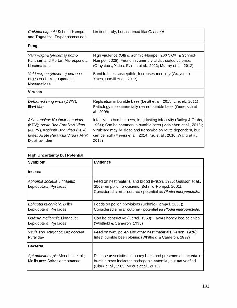
101
Crithidia expoeki Schmid-Hempel
and Tognazzo; Trypanosomatidae
Limited study, but assumed like C. bombi
Fungi
Vairimorpha (Nosema) bombi
Fantham and Porter; Microsporidia:
Nosematidae
High virulence (Otti & Schmid-Hempel, 2007; Otti & Schmid-
Hempel, 2008); Found in commercial distributed colonies
(Graystock, Yates, Evison et al., 2013; Murray et al., 2013)
Vairimorpha (Nosema) ceranae
Higes et al.; Microsporidia:
Nosematidae
Bumble bees susceptible, increases mortality (Graystock,
Yates, Darvill et al., 2013)
Viruses
Deformed wing virus (DWV);
Iflaviridae
Replication in bumble bees (Levitt et al., 2013; Li et al., 2011);
Pathology in commercially reared bumble bees (Genersch et
al., 2006)
AKI-complex: Kashmir bee virus
(KBV); Acute Bee Paralysis Virus
(ABPV), Kashmir Bee Virus (KBV),
Israeli Acute Paralysis Virus (IAPV)
Dicistroviridae
Infective to bumble bees, long-lasting infectivity (Bailey & Gibbs,
1964); Can be common in bumble bees (McMahon et al., 2015);
Virulence may be dose and transmission route dependent, but
can be high (Meeus et al., 2014; Niu et al., 2016; Wang et al.,
2018)
High Uncertainty but Potential
Symbiont Evidence
Insecta
Aphomia sociella Linnaeus;
Lepidoptera: Pyralidae
Feed on nest material and brood (Frison, 1926; Goulson et al.,
2002) on pollen provisions (Schmid-Hempel, 2001);
Considered similar outbreak potential as Plodia interpunctella.
Ephestia kuehniella Zeller;
Lepidoptera: Pyralidae
Feeds on pollen provisions (Schmid-Hempel, 2001);
Considered similar outbreak potential as Plodia interpunctella.
Galleria mellonella Linnaeus;
Lepidoptera: Pyralidae
Can be destructive (Oertel, 1963); Favors honey bee colonies
(Whitfield & Cameron, 1993)
Vitula spp. Ragonot; Lepidoptera:
Pyralidae
Feed on wax, pollen and other nest materials (Frison, 1926);
Infest bumble bee colonies (Whitfield & Cameron, 1993)
Bacteria
Spiroplasma apis Mouches et al.;
Mollicutes: Spiroplasmataceae
Disease association in honey bees and presence of bacteria in
bumble bees indicates pathogenic potential, but not verified
(Clark et al., 1985; Meeus et al., 2012)
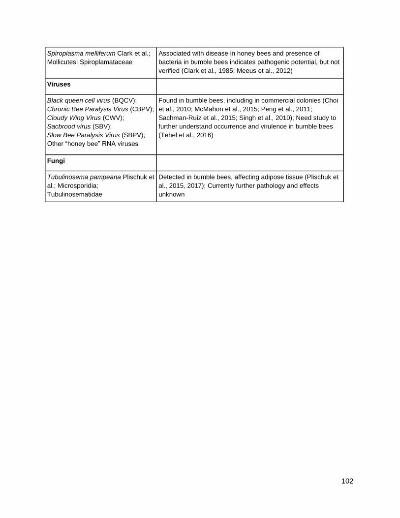
102
Spiroplasma melliferum Clark et al.;
Mollicutes: Spiroplamataceae
Associated with disease in honey bees and presence of
bacteria in bumble bees indicates pathogenic potential, but not
verified (Clark et al., 1985; Meeus et al., 2012)
Viruses
Black queen cell virus (BQCV);
Chronic Bee Paralysis Virus (CBPV);
Cloudy Wing Virus (CWV);
Sacbrood virus (SBV);
Slow Bee Paralysis Virus (SBPV);
Other “honey bee” RNA viruses
Found in bumble bees, including in commercial colonies (Choi
et al., 2010; McMahon et al., 2015; Peng et al., 2011;
Sachman-Ruiz et al., 2015; Singh et al., 2010); Need study to
further understand occurrence and virulence in bumble bees
(Tehel et al., 2016)
Fungi
Tubulinosema pampeana Plischuk et
al.; Microsporidia;
Tubulinosematidae
Detected in bumble bees, affecting adipose tissue (Plischuk et
al., 2015, 2017); Currently further pathology and effects
unknown