การควบคุมพฤติกรรมการเลี้ยงลูกโดยระบบประสาท...
TRANSCRIPT
1
การควบคุมพฤติกรรมการเลี้ยงลูกโดยระบบประสาทและระบบต่อมไร้ท่อในแม่ไก่พื้นเมืองไทย
วิทยานิพนธ์นี้เป็นส่วนหนึ่งของการศึกษาตามหลักสูตรปริญญาวิทยาศาสตรดุษฎีบัณฑิต
สาขาวิชาชีววิทยาสิ่งแวดล้อม
มหาวิทยาลัยเทคโนโลยีสุรนารี ปีการศึกษา 2555
นางสาวอรอนงค์ ไชยเชษฐ
2
NEUROENDOCRINE REGULATION OF REARING
BEHAVIOR IN THE NATIVE THAI HEN
A Thesis Submitted in Partial Fulfillment of the Requirements for the
Degree of Doctor of Philosophy in Environmental Biology
Suranaree University of Technology
Academic Year 2012
Orn-Anong Chaiyachet

IV
ACKNOWLEDGEMENTS
I owe my deepest gratitude to my advisor, Assoc. Prof. Dr. Yupaporn
Chaiseha, for her advice, guidance and patient support throughout my time as a
graduate student. I would also like to express my appreciation to my co-advisor, Prof.
Dr. Tom E. Porter, who let me experience the research in his laboratory and I would
like to thank for his advice and hospitality when I was in his laboratory. I am very
thankful to Dr. Nattiya Prakobsaeng, Dr. Natagarn Sartsoongnoen, and Dr. Sunantha
Kosonsiriluk, who were always willing to help me in any circumstances.
I would also like to thank all my colleagues and friends in Chaiseha’s and
Porter’s labs for their help and technical teaching. I am grateful to Suranaree
University Farm for providing the barn for experimental animals.
I wish to thank my family for their love, support, and encouragement that help
me overcome any difficulties.
Finally, this thesis would not have been possible without the full financial
support of the Royal Golden Jubilee Ph.D. Program, the Thailand Research Fund.
Orn-Anong Chaiyachet


CONTENTS
Page
ABSTRACT IN THAI………………………………………………………………....I
ABSTRACT IN ENGLISH…………………………………………………………..III
ACKNOWLEDGEMENTS…………………………………………………………..V
CONTENTS…………………………………………………………………….……VI
LIST OF TABLES……………………………………………………………………X
LIST OF FIGURES………………………………………………………………....XV
CHAPTER
I INTRODUCTION……………………………………………………………1
1.1 Rational of the Study……………………………………………….….……
1.2 Research Objectives………………………………………………….…….
II LITERATURE REVIEW……………………………………………...….....
2.1 Native Thai Chicken…………………………………………………….....7
2.2 Neuroendocrine Regulation of the Avian Reproductive Cycle…………..10
2.2.1 Gonadotropin Releasing Hormone/Follicle Stimulating
Hormone-Luteinizing Hormone System…………...……………
2.2.2 Vasoactive Intestinal Peptide/Prolactin System………………..17
2.3 Neuroendocrine Regulation of the Reproductive Cycle
in the Native Thai Chicken………………………………………………19
2.4 Prolactin: Structure, Function, and Regulation of Secretion……………..24
2.4.1 The Structure of Prolactin…………………………………........
1
7
6
14
24
VII
CONTENTS (Continued)
Page
2.4.2 The Function of Prolactin in Birds………………………….…..31
2.4.3 The Regulation of Prolactin Secretion…………………….……33
2.5 Vasoactive Intestinal Peptide: Structure, Function, and
Regulation of Secretion…………………………………………………..
2.5.1 The Structure of Vasoactive Intestinal Peptide………………...38
2.5.2 The Localization of Vasoactive Intestinal Peptide
in the Avian Brain…………………………………………...….
2.5.3 The Function of Vasoactive Intestinal Peptide in Birds…..….....
2.5.4 The Regulation of Vasoactive Intestinal Peptide Secretion.........
2.6 Gonadotropin Releasing Hormone: Structure, Function, and
Regulation of Secretion……………………………………………..……
2.6.1 The Structure of Gonadotropin Releasing Hormone…………...49
2.6.2 The Localization of Gonadotropin Releasing Hormone
in the Brain…………………………………………………..…
2.6.3 The Function of Gonadotropin Releasing Hormone in Birds…..
2.6.4 The Regulation of Gonadotropin Releasing Hormone
Secretion…………………………………………………..……
2.7 Maternal Behaviors in Birds………………………………………………
2.7.1 Incubation Behavior……………………………………….……
2.7.2 Neuroendocrine Regulation of Incubation Behavior…………...65
2.7.3 Rearing Behavior……………………………………………….
43
45
47
49
55
52
57
61
63
66
38
VIII
CONTENTS (Continued)
Page
2.7.4 Neuroendocrine Regulation of Rearing Behavior………….…………...
2.8 References………………………………………………………….….….
III EFFECTS OF REARING BEHAVIOR ON THE
NEUROENDOCRINE REGULATION OF THE
REPRODUCTIVE SYSTEM IN THE FEMALE
NATIVE THAI CHICKENS: ROLE OF PROLACTIN…………....…..151
3.1 Abstract………………………………………………….………............
3.2 Introduction………………………………………………………………
3.3 Materials and Methods…………………………………………………..
3.4 Results………………………………………………………………...…
3.5 Discussion……………………………………………………………….
3.6 References……………………………………………………………….
IV EFFECTS OF REARING BEHAVIOR ON NEUROENDOCRINE
REGULATION OF THE REPRODUCTIVE SYSTEM IN THE
FEMALE NATIVE THAI CHICKENS: ROLE OF VASOACTIVE
INTESTINAL PEPTIDE……………………………………...……...…..
4.1 Abstract…………………………………………………………............
4.2 Introduction……………………………………………………………..
4.3 Materials and Methods………………………………………………….
4.4 Results…………………………………………………………………..
4.5 Discussion……………………………………………………………….
69
77
151
1
152
1
159
1
163
188
1 193
1
207
1 207
1
213
1
209
1
217
1
238
1
IX
CONTENTS (Continued)
Page
4.6 References………………………………………………………….……
V EFFECTS OF REARING BEHAVIOR ON NEUROENDOCRINE
REGULATION OF THE REPRODUCTIVE SYSTEM IN THE
FEMALE NATIVE THAI CHICKENS: ROLE OF
GONADOTROPIN RELEASING HORMONE………………...............
5.1 Abstract…………………………………………………………………
5.2 Introduction……………………………………………………………..258
5.3 Materials and Methods………………………………………………….
5.4 Results………………………………………………………………..…
5.5 Discussion………………………………………………………….……
5.6 References…………………………………………………………….…
VI CONCLUSION…………………………………………………….….…...298
CURRICULUM VITAE…………………………………………………………....304
244
256
256
263
267
278
284
X
LIST OF TABLES
Table Page
3.1 Mean ± SEM of the plasma PRL levels (ng/ml) of native Thai hens
rearing chicks at different times during the rearing period (n=6).
Significant differences between means in each group at
different time points are denoted by different letters (P<0.05)
and *P<0.05 for a comparison among group at a given time point…............
3.2 The number of native Thai hens that had the F1-F5 follicles,
small yellow follicles (SYF), and small white follicles (SWF)
at different times during the rearing period and the number of
hens came back to lay in each period (n=6).………………………………..
3.3 Mean ± SEM of the ovary weight (g) of native Thai hens that
rearing chicks at different times during the rearing period (n=6).
Significant differences between means in each group at different
time points are denoted by different letters (P<0.05) and
*P<0.05 for a comparison among group at a given time point.…………….
3.4 Mean ± SEM of the oviduct weight (g) of native Thai hens that
rearing chicks at different times during the rearing period (n=6).
Significant differences between means in each group at different
time points are denoted by different letters (P<0.05) and
*P<0.05 for a comparison among group at a given time point………….…..
167
169
172
175

XI
LIST OF TABLES (Continued)
Table Page
3.5 Mean ± SEM of the plasma PRL concentrations (ng/ml) of rearing (R)
and non-rearing (NR) native Thai hens at different times of
rearing (n=6) or non-rearing their chicks (n=6). Significant
differences between means in each group at different time points
are denoted by different letters (P<0.05) and *P<0.05 for a
comparison between group at a given time point…………………………...177
3.6 The number of native Thai hens that had the F1-F5 follicles,
small yellow follicles (SYF), and small white follicles (SWF)
at different times of rearing (n=5). …………………………………………178
3.7 The number of native Thai hens that had the F1-F5 follicles,
small yellow follicles (SYF), and small white follicles (SWF)
at different times of non-rearing and the number of
hen came back to lay in each period (n=5)…………….……………………179
3.8 Mean ± SEM of the ovary weight (g) of rearing (R) and
non-rearing (NR) native Thai hens at different times of
rearing or non-rearing their chicks (n=5). Significant differences
between means in each group at different time points are denoted
by different letters (P<0.05) and *P<0.05 for a comparison between
group at a given time point………………………………………………….
183
XII
LIST OF TABLES (Continued)
Table Page
3.9 Mean ± SEM of the oviduct weight (g) of rearing (R) and
non-rearing (NR) native Thai hens at different times of rearing or
non-rearing their chicks (n=5). Significant differences between
means in each group at different time points are denoted by
different letters (P<0.05) and *P<0.05 for a comparison between
group at a given time point………………………………………………….
4.1 Abbreviations of brain areas. Nomenclature and abbreviations
are from a stereotaxic atlas of the brain of the chick
(Kuenzel and Masson, 1988)…………………………………………….….
4.2 The number of VIP-ir neurons (Mean ± SEM) in the IH-IN
of rearing (R) and non-rearing (NR) native Thai hens at different
days of rearing or non-rearing their chicks (n=6). Significant
differences between means in each group at different time points
are denoted by different letters (P<0.05) and *P<0.05 for
a comparison between group at a given time point…………………………
4.3 The number of native Thai hens that had the F1-F5 follicles,
small yellow follicles (SYF), and small white follicles (SWF)
at different days of rearing (n=6)……………………………………............
220
226
187
228

XIII
LIST OF TABLES (Continued)
Table Page
4.4 The number of native Thai hens that had the F1-F5 follicles,
small yellow follicles (SYF), and small white follicles (SWF)
at different days of non-rearing and the number of hen came back
to lay in each period (n=6)………………………………………………….
4.5 Mean ± SEM of the ovary weight (g) of rearing (R) and
non-rearing (NR) native Thai hens at different times of rearing
or non-rearing their chicks (n=6). Significant differences between
means in each group at different time points are denoted by
different letters (P<0.05) and *P<0.05 for a comparison between
group at a given time point………………………………………………….233
4.6 Mean ± SEM of the oviduct weight (g) of rearing (R) and
non-rearing (NR) native Thai hens at different days of rearing
or non-rearing their chicks (n=6). Significant differences between
means in each group at different time points are denoted by
different letters (P<0.05) and *P<0.05 for a comparison between
group at a given time point………………………………………………….237
229

XIV
LIST OF TABLES (Continued)
Table Page
5.1 Abbreviations of brain areas. Nomenclature and abbreviations
are from a stereotaxic atlas of the brain of the chick
(Kuenzel and Masson, 1988)………………………………………..………..
5.2 The number of GnRH-I-ir neurons (Mean ± SEM) in the nCPa of
rearing (R) and non-rearing (NR) native Thai hens at different days of
rearing or non-rearing their chicks (n=6). Significant differences
between means in each group at different time points are denoted by
different letters (P<0.05) and *P<0.05 for a comparison between group
at a given time point…………………………………………………………
269
277
XV
LIST OF FIGURES
Figure Page
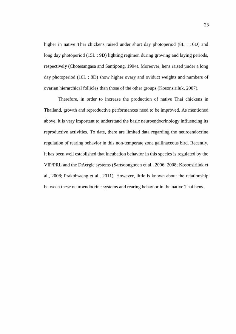
2.1 The reproductive cycle of the native Thai chickens;
non-egg laying (NL), egg laying (L), incubating eggs (INC),
and rearing chicks (R; Kosonsiriluk, 2007)….……………………………….24
2.2 The percentage of homology sequence of PRLs
among different species (Sinha, 1995). ...........................................................
2.3 Primary structures of PRLs of different species. (-) indicates
positions left blank to optimize alignment of amino acid sequences.
(*) indicates absence of residues from a genetic variant of tilapia PRL.
PD is PRL domain. PDI-PD4 indicates the four highly conserved
domains of the PRLs (Sinha, 1995)…………………………………………..
2.4 The amino acid sequences of VIP, PHI, secretin, glucagon,
and GIP. p: porcine, b: bovine, c: chicken, m: mammalian,
a: the C-terminal amino acid is in the amide form
(Rosselin et al., 1982)…………………………………………...……............
2.5 Sequence homology and amino acid similarlities (%) of VIP receptors.
Values above and under the diagonal line represent homology of
the cDNA sequence and amino acid, respectively
(Kansaku et al., 2001)........................................................................................
29
30
41
40
XVI
LIST OF FIGURES (Continued)
Figure Page
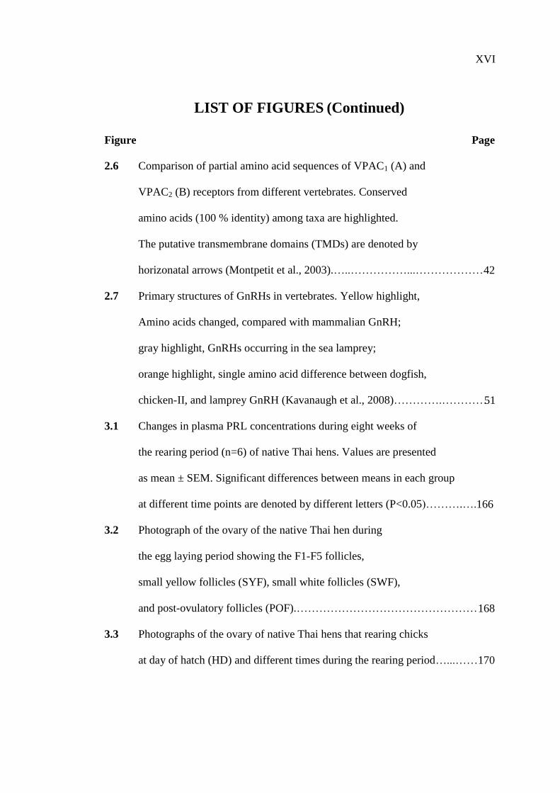
2.6 Comparison of partial amino acid sequences of VPAC1 (A) and
VPAC2 (B) receptors from different vertebrates. Conserved
amino acids (100 % identity) among taxa are highlighted.
The putative transmembrane domains (TMDs) are denoted by
horizonatal arrows (Montpetit et al., 2003).…..……………...………………
2.7 Primary structures of GnRHs in vertebrates. Yellow highlight,
Amino acids changed, compared with mammalian GnRH;
gray highlight, GnRHs occurring in the sea lamprey;
orange highlight, single amino acid difference between dogfish,
chicken-II, and lamprey GnRH (Kavanaugh et al., 2008)………….…………
3.1 Changes in plasma PRL concentrations during eight weeks of
the rearing period (n=6) of native Thai hens. Values are presented
as mean ± SEM. Significant differences between means in each group
at different time points are denoted by different letters (P<0.05)……….….
3.2 Photograph of the ovary of the native Thai hen during
the egg laying period showing the F1-F5 follicles,
small yellow follicles (SYF), small white follicles (SWF),
and post-ovulatory follicles (POF).…………………………………………
3.3 Photographs of the ovary of native Thai hens that rearing chicks
at day of hatch (HD) and different times during the rearing period…...……
51
166
168
170
42
XVII
LIST OF FIGURES (Continued)
Figure Page
3.4 Changes in the ovary weight of native Thai hens that rearing chicks.
Values are presented as means ± SEM (n=6). Significant differences
between means in each group at different time points are denoted by
different letters (P<0.05)………………………………………….…………
3.5 Photographs of the oviducts of native Thai hens that rearing chicks
at day of hatch (HD) and different times during the rearing period…...........
3.6 Changes in the oviduct weight of native Thai hens that rearing chicks.
Values are presented as means ± SEM (n=6). Significant differences
between means in each group at different time points are denoted by
different letters (P<0.05)………………………………………….…………
3.7 Changes in plasma PRL concentrations of rearing (R; n=6) and
non-rearing (NR; n=6) native Thai hens. Values are presented as
mean ± SEM. Significant differences between means in each group
at different time points are denoted by different letters (P<0.05)
and *P<0.05 for a comparison between group at a given time point……….
3.8 Photographs of the ovary of rearing (R) and non-rearing (NR)
native Thai hens at day of hatch (HD) and different times of rearing or
non-rearing their chicks…………………………………….…………...…..
180
171
173
174
176

XVIII
LIST OF FIGURES (Continued)
Figure Page
3.9 Changes in the ovary weights of rearing (R) and non-rearing (NR)
native Thai hens. Values are presented as means ± SEM (n=5).
Significant differences between means in each group at different
time points are denoted by different letters (P<0.05) and
*P<0.05 for a comparison between group at a given time point……………182
3.10 Photographs of the oviducts of rearing (R) and non-rearing (NR)
native Thai hens at day of hatch (HD) and different times of
rearing or non-rearing their chicks…………………………….…………….
3.11 Changes in the oviduct weight of rearing (R) and non-rearing (NR)
native Thai hens. Values are presented as means ± SEM (n=5).
Significant differences between means in each group at different
time points are denoted by different letters (P<0.05) and
*P<0.05 for a comparison between group at a given time point……………186
4.1 Schematic coronal brain sections showing the areas where
the expression of VIP-ir (black dots) was observed (A-D).
The sampling region for counting the number of VIP-ir neurons in
the IH-IN (C) is represented by rectangles. Coronal illustrations
were redrawn from the stereotaxic atlas of the chick brain
(Kuenzel and Masson, 1988)………………………………….…………….
184
221

XIX
LIST OF FIGURES (Continued)
Figure Page
4.2 Photomicrographs illustrating the distributions of VIP-ir neurons
and fibers in the nucleus inferioris hypothalami (IH) and nucleus
infundibuli hypothalami (IN) of the native Thai chicken (A and B).
Rectangle indicates area from which the following photomicrographs
were taken. Higher magnification of the VIP-ir neurons was
demonstrated in the IH-IN area (C and D). Scale bar = 50 µm…………….
4.3 Photomicrographs showing the distributions of VIP-ir neurons
and fibers in the IH-IN of rearing (R), and non-rearing (NR)
native Thai hens on day of hatch (HD) and different
days following the initiation of rearing or non-rearing their chicks.
Scale bar = 100 µm………………………………………………………….
4.4 Changes in the number of VIP-ir neurons in the IH-IN of rearing (R)
and non-rearing (NR) native Thai hens (n=6). Values are presented as
mean ± SEM. Significant differences between means in each group
at different time points are denoted by different letters (P<0.05)
and *P<0.05 for a comparison between group at a given time point……….
4.5 Photograph of the ovary of the native Thai hen during
the egg laying period showing the F1-F5 follicles,
small yellow follicles (SYF), small white follicles (SWF),
and post-ovulatory follicles (POF)………………………………………….
223
222
225
227
XX
LIST OF FIGURES (Continued)
Figure Page
4.6 Photographs of the ovary of rearing (R) and non-rearing (NR)
native Thai hens at day of hatch (HD) and different days
of rearing or non-rearing their chicks……………………………………….
4.7 Changes in the ovary weights of rearing (R) and non-rearing (NR)
native Thai hens. Values are presented as means ± SEM (n=6).
Significant differences between means in each group at different
time points are denoted by different letters (P<0.05) and
*P<0.05 for a comparison between group at a given time point……………
4.8 Photographs of the oviducts of rearing (R) and non-rearing (NR)
native Thai hens at day of hatch (HD) and different days of
rearing or non-rearing their chicks………………………………………….
4.9 Changes in the oviduct weight of rearing (R) and non-rearing (NR)
native Thai hens. Values are presented as means ± SEM (n=6).
Significant differences between means in each group at different
time points are denoted by different letters (P<0.05) and
*P<0.05 for a comparison between group at a given time point……………
5.1 Schematic coronal brain sections showing the areas where
the expression of GnRH-I-ir (black squares) was observed (A-B).
The sampling regions for counting the number of GnRH-I-ir neurons
in the nCPa (B) are represented by rectangles. Coronal illustrations were
redrawn from the stereotaxic atlas of the chick brain
(Kuenzel and Masson, 1988)………………………..………………………
230
232
234
236
270
XXI
LIST OF FIGURES (Continued)
Figure Page
5.2 Photomicrographs illustrating the distributions of GnRH-I-ir neurons
and fibers in the nucleus commissurae pallii (nCPa) of the native Thai
chickens (A, C). Rectangle indicates area from which the following
photomicrographs were taken. Higher magnification of the GnRH-I-ir
neurons was demonstrated in the nCPa (B, D).
Scale bar = 50 µm…………………………………………………………..
5.3 Photomicrographs illustrating the distributions of GnRH-I-ir neurons
and fibers in the hypothalamic areas of rearing (A, C, E, G, I, and K)
and non-rearing (B, D, F, H, J, and L) native Thai hens.
Scale bar = 100 µm…………………………………………………............
5.4 Photomicrographs showing the distributions of GnRH-I-ir neurons
and fibers in the nucleus commissurae pallii (nCPa) of rearing (R),
and non-rearing (NR) native Thai hens on day of hatch (HD)
and different days following the initiation of rearing or
non-rearing their chicks. Scale bar = 100 µm………………………………
5.5 Changes in the number of GnRH-I-ir neurons in the nCPa
of rearing (R) and non-rearing (NR) native Thai hens (n=6).
Values are presented as mean ± SEM. Significant differences
between means in each group at different time points are denoted
by different letters (P<0.05) and *P<0.05 for a comparison between
group at a given time point………………………………………….………
271
272
274
276

XXII
LIST OF FIGURES (Continued)
Figure Page
6.1 Changes in: A, plasma PRL concentration; B, ovary; C, oviduct
weights of native Thai hens rearing chicks during the eight weeks
of rearing period. Values are presented as mean ± SEM. Significant
differences between means in each group at different time points are
denoted by different letters (P<0.05)……………………………..…………
6.2 Changes in: A, plasma PRL concentration; B, ovary; C, oviduct
weights of rearing (R) and non-rearing (NR) native Thai hens.
Values are presented as mean ± SEM. Significant differences
between means in each group at different time points are denoted
by different letters (P<0.05) and *P<0.05 for a comparison between
group at a given time point............................................................................
6.3 Changes in: A, the number of VIP-ir neurons in the IH-IN;
B, the number of GnRH-I-ir neurons in the nCPa; C, ovary;
D, oviduct weights of rearing (R) and non-rearing (NR)
native Thai hens. Values are presented as mean ± SEM.
Significant differences between means in each group
at different time points are denoted by different letters (P<0.05)
and *P<0.05 for a comparison between group at a given time point…….…303
301
302

1
CHAPTER I
INTRODUCTION
1.1 Rational of the Study
The native Thai chicken (Gallus domesticus) belongs to genus Gallus of the
family Phasianidae. It is a small domestic animal that probably originated from the
red jungle fowl found wildly distributed throughout Southeast Asia and is
domesticated approximately 3,000 years ago. They have been raised in the
countryside of Thailand for many generations and the main objectives for raising
native Thai chickens are for consumption, sport competition, and recreation. It is not
only a main protein food source, but it also can be sold for supplemental income for
families. Its meat is firm in texture and contains high proteins as well as low fat and
cholesterol contents, resulting in high demand by consumers who prefer low fat and
antibiotic-free white meat. This provides a good opportunity for producing them in
industrial scale. Recently, the native Thai chicken has become a new economic
domestic animal of Thailand with presently growing demand and relatively high
price. The market price of the native Thai chickens is two to three times higher than
those of imported broilers. To date, there are about 76 millions native Thai chickens
in Thailand, which are raised by 2.7 millions farmers. This exported goods gains
income about 2.2 million baht per year.
The native Thai chickens can be raised under poor environmental conditions
in the backyard with local feeds, minimum care and management. Furthermore, recent

2
Thai government policies encourage the development and the use of natural resources
in supporting of His Majesty the King’s concept for self-sufficiency in agriculture.
The farmers focus on “mixed farming” that is a strategy for helping rural farmers to
increase self-sufficiency. The native Thai chicken is one of the significant resources
that needs to be developed. However, the native Thai chickens have low productivity.
The reproductive performance of native chicken is much lower than that of cross
breeds and hybrids, especially egg laying performance. One of the main causes of low
reproductive performance in the native Thai chickens is the incidence of maternal
behaviors such as incubating, brooding or rearing behaviors. The establishment of
maternal behaviors affects egg production, because it terminates egg laying. These
cause a problem for producing native Thai chickens commercially in the poultry
industry in Thailand. Presently, market demands for the native Thai chickens cannot
be met by supply, mainly because of their low egg laying performance. They tend to
lay eggs in clutches rather than evenly distributed over the year, leading to irregular
production of the chicks.
The native Thai chicken was domesticated without genetic selection and
always expresses high maternal behaviors, heritable traits from their ancestors.
Maternal behaviors are hormonal dependent, initiated with the onset of incubation
behavior, and continue through the period when the young are taking care of by the
mother (broody/rearing behavior). These behaviors constrain the number of eggs
produced. The maternal behaviors are defined as the behaviors of the mothers that
contribute to the survival of their offspring. Most mothers display the maternal
behaviors after parturition and serve the immediate provision of care and defense for
their young. In birds, the maternal behaviors are composed of incubating eggs and

3
brooding or rearing of the young. One or both parents must incubate their eggs until
hatching and then provide post-hatching care.
There are several lines of evidence indicating that neurotransmitters,
neurohormones, and hormones play an important role in the maternal behaviors of
avian species. The parental hormone, prolactin (PRL), an anterior pituitary hormone,
has been shown to be associated with the reproductive cycle in several avian species
such as turkeys, quails, bantams, ring doves, pigeons, mallard ducks, and native Thai
chickens. PRL has been implicated as a causative factor in the onset and maintenance
of maternal behaviors. Rearing behavior is maintained by high levels of PRL and
upon hatching. This pituitary hormone is widely thought to play a role in maternal
behaviors by mediating incubation behavior, crop milk production and secretion,
feeding of the young, and nest defense. In temperate zone birds, it is very well
documented that PRL is under stimulatory control by hypothalamic vasoactive
intestinal peptide (VIP), the avian PRL-releasing factor. Some evidence indicates that
the dopaminergic (DAergic) system plays an intermediary role in PRL secretion. In
many avian species, the onset and maintenance of maternal behaviors is correlated
with low levels of gonadotropin releasing hormone (GnRH), follicle stimulating
hormone (FSH), and luteinizing hormone (LH) and high levels of circulating PRL.
Recently, the presence of hypothalamic VIP-immunoreactive (-ir), tyrosine
hydroxylase (TH)-ir (as a marker for DA), and GnRH-I-ir neurons at different
reproductive stages and throughout the incubation period have been reported in the
native Thai chickens. Changes in the number of VIP-ir, TH-ir, and GnRH-I-ir neurons
in the native Thai chickens are observed across the reproductive stages and during
incubation and nest deprivation period, which is correlated directly with variations in
4
plasma PRL levels. VIP-ir neurons and fibers are extensively distributed throughout
the brain of the native Thai chickens and are predominantly expressed in the
diencephalon, where VIP-ir neurons are highly concentrated within the nucleus
inferioris hypothalami (IH) and nucleus infundibuli hypothalami (IN) areas. Changes
in numbers of VIP-ir neurons within the IH-IN are directly correlated with changing
of plasma PRL levels throughout the reproductive cycle, suggesting that VIP
expression in the IH-IN plays a regulatory role in year-round reproductive activity of
the native Thai chickens. Further studies revealed that an association exists between
DA neurons in the nucleus intramedialis (nI) and the regulation of the reproductive
system in the native Thai chickens, suggesting that the differential expression of DA
neurons in the nI might play a role in the control of VIP secretion and subsequent
PRL release in the native Thai chickens. Moreover, it has been further demonstrated
that changes in the number of VIP-ir neurons in the IH-IN are associated with
DAergic neurons within the nI and nucleus mamillaris areas, resulting in PRL release
to induce and maintain incubation behavior in the native Thai hens. It is further
suggested that nesting activity stimulates PRL secretion by the activation of the
DAergic system, which in turn stimulates the VIPergic system. These elevated PRL
levels increase nesting activity and maintain incubation behavior.
In several avian species, PRL secretion has been shown to be stimulated by the
presence of chicks. This hormone is indicated to be correlated with parental care, and
is known to decline steadily immediately after hatching in precocial bird species or
drop at the end of the rearing period in altricial bird species. In the native Thai hens,
circulating PRL levels decrease sharply during rearing stage, but changes in plasma
LH levels are not observed. This decline in plasma PRL levels corresponds with a

5
decrease in the numbers of VIP-ir neurons in hens rearing chicks. GnRH-I, the
primary hypophysiotrophic factor involved in avian reproductive regulation, has been
investigated in the native Thai chickens, revealing that the number of GnRH-I-ir
neurons decline to the lowest level during rearing stage, and plasma LH levels are
fluctuated and essentially the same levels throughout the reproductive cycle. The
distributions of the DA-ir neurons in the hypothalamic areas of the native Thai
chickens have been elucidated. The differential expression of DA-ir neurons in the nI
are observed across the reproductive cycle and the number of DA-ir neurons decrease
slightly during the transition from incubating to rearing stage, whereas plasma PRL
levels decline dramatically to the same level of non-egg laying in hens that rearing
chicks.
As aforementioned, the expression of maternal behaviors including brooding
and rearing behaviors is a costly problem, resulting in substantial loss of potential egg
production. Some evidence suggests that plasma PRL levels also play a role in
terminating egg laying and regulating clutch size in species that lay clutches of more
than two eggs. Cessation of egg laying is associated with increased PRL
concentrations in the turkey and domestic fowl. Obviously, the reproductive
efficiency of the native Thai chickens is low in comparison to those of the imported
breeds. Thus, in order to increase the production of native Thai chickens in Thailand,
it is very important to understand the neuroendocrine regulation of the maternal
behaviors. To date, it has been well established that incubation behavior in this
species is regulated by the VIP/PRL, GnRH/FSH-LH, and DAergic systems.
However, little is known about the relationship between these neuroendocrine systems
and rearing behavior in this species. Therefore, this dissertation was carried out to

6
investigate the neuroendocrine regulation of rearing behavior in the native Thai
chickens. The results gained from this study will provide an insight into the
neuroendocrine mechanism(s) underlying the regulation of rearing behavior in the
native Thai chickens. The findings gained from this dissertation will provide, for the
first time, the information of neuroendocrine regulation of rearing behavior in the
native Thai chickens, which could help to improve the productivity of the native Thai
chickens in Thailand.
1.2 Research Objectives
1.2.1 To study the changes in plasma PRL levels in the regulation of rearing
behavior in the female native Thai chickens.
1.2.2 To study the differential expression of VIP that is associated with the
neuroendocrine regulation of rearing behavior in the female native
Thai chickens.
1.2.3 To study the differential expression of cGnRH-I that is associated with
the neuroendocrine regulation of rearing behavior in the female native
Thai chickens.
7
CHAPTER II
LITERATURE REVIEW
2.1 Native Thai Chicken
The native Thai chicken or Thai indigenous chicken (Gallus domesticus),
belongs to genus Gallus of the family Phasianidae, originated from the wild jungle
fowl. It has been suggested that red jungle fowl (Gallus gallus) might be the ancestor
of all domestic chickens which is found wildly distributed throughout Southeast Asia
(Austic and Nesheim, 1990; Fumuhito et al., 1994; Hillel et al., 2003; Sawai et al.,
2010). Approximately, it was domesticated by village people 3,000 years ago. Some
characteristics of native Thai chickens inherited from the ancestry and still expressed
are maternal behaviors such as incubation and rearing behaviors (Beissinger et al.,
1998). Historically, native Thai chickens have been in the countryside of Thailand,
and the main objectives of raising native Thai chickens are for consumption, sport
competition, and recreation. Indeed, it is not only a main protein food source, but it
can be also sold for additional income for families. Generally, the native Thai
chickens are easy to raise, resistant to diseases, and acclimatized to the local
environments. However, the native Thai chicken has a slower growth rate than that of
the imported commercial broiler when raised under the same conditions, but it can be
raised with lower production costs by raising it as free range using organic local feed.
It has been reported that high performance breeds lose their advantages over native
Thai chickens in terms of weight gain when treated with local feeds (Leotarakul and
8
Pimkamlia, 1999). Moreover, the native Thai chicken is well adapted to the poor
condition of small farm or simple rural environment. Its resistance to diseases and
tolerance to heat stress are considerably higher than those of high performance or
hybrid breeds, resulting in high potential for raising native Thai chickens in rural
areas (Kajaroen et al., 1989). The native Thai chickens can adapt to high temperature,
and imported broilers are less tolerant to the high temperature than those of Thai
indigenous chickens crossbred and Thai indigenous chickens (Aengwanich, 2008).
In Thailand, to date, there are about 76 millions native Thai chickens or 24 %
of total chicken production, which are broilers 55 %, layers 15 %, commercial broiler
breeders 5 %, and commercial layer breeders 1 % (Department of Livestock
Development, 2011). Native Thai chickens provide higher meat quality with less fat
and low cholesterol than those of imported commercial broilers, resulting in high
demand by consumers (Wattanachant et al., 2004; Jaturasitha et al., 2008; Teltathum
and Mekchay, 2009). The textural characteristics of the indigenous chicken meat are
similar to spent hen meat but are much different from imported broiler meat
(Wattanachant et al., 2004; Chuaynukool et al., 2007). The indigenous chicken
muscles contain higher protein and collagen contents but lower fat content than those
of broiler muscles (Wattanachant et al., 2004; Wattanachant, 2008). In addition, the
shear values of indigenous chicken muscles either raw or cooked are higher than those
of broiler muscles (Wattanachant et al., 2004; 2005; Jaturasitha et al., 2008). The
comparison between two indigenous chicken strains, black-boned and native Thai
chickens with two imported, Bresse, and Rhode Island Red (Rhodes, a layer breed)
found that the imported breeds are heavier at slaughter and have higher contents of fat
and cholesterol than those of indigenous strains (Jaturasitha et al., 2008). Thus, there
9
are many factors such as genotype, rearing system, feed, age, muscle pH, chemical
composition, microstructure of muscle, postmortem aging, and processing methods
can influence on the quality of indigenous chicken meat (Chotesangasa and
Gongruttananun, 1999a; Jaturasitha et al., 2002; Wattanachant et al., 2005;
Wattanachant, 2008). The firm and low fat meat, free of drug residues such as
antibiotics, makes consumers prefer these meat types (Choprakarn et al., 2000). This
advantage of native Thai chicken meat leads to a higher price, about two or three
times higher than those of the imported commercial broilers in Thailand, Hong Kong,
China, and Japan (Chotesangasa and Gongruttananun, 1999a; Jaturasitha et al., 2008).
It has been further suggested that the native Thai chickens, especially Pradu Hang
Daum breed, is suitable to be developed as a meat type chicken due to its lower
genetic distance to broiler strains (Dorji et al., 2011).
The production of native Thai chickens is suited to the small farm raising
system, but improving the supply of chicks for fattening needs to be developed
(Haitook et al., 2003). The reproductive performance of native Thai chickens is much
lower than those of cross breeds and hybrids, especially egg laying performance
which is critical to secure a sufficient number of chicks for fattening (Chotesangasa et
al., 1994b). In commercial systems, hatchability is not the problem for producing the
chicks, if the number of eggs per hen is not limited. Normally, the native Thai hen
lays eggs 3-4 times per year, 4-17 eggs per clutch rather than laying eggs
continuously all year long. The hen-day egg production of the native Thai hen is
lower than that of the commercial laying hen at all times, with the peak production for
native Thai hens and commercial laying hens being 38.0 % and 75.5 %, respectively
(Chotesangasa et al., 1994b). The total number of eggs per hen of native Thai hen is
10
between 30-92 eggs per year, which is significantly lower than that of 243
eggs/hen/year of the imported commercial hen (Chotesangasa et al., 1994b). With a
hatching rate of 80-85 %, a typical hen produces 25-40 chicks annually (Klinhom et
al., 2005). The low potential in egg production of the native Thai chickens causes the
problem in order to be produced commercially in poultry industry in Thailand. The
main cause of low egg production and short egg laying period in the native Thai
chickens is the expression of the maternal behaviors (incubation and rearing
behaviors). These behaviors are high during egg laying, nesting, and brooding, which
are not desired for commercial scale production (Choprakarn and Wongpichet, 2007).
Generally, the native Thai chicken takes about 2 weeks for laying, 3 weeks for
hatching, and 6-10 weeks for taking care of the chicks. Thus, the hen spends 10-15
weeks for each reproductive cycle (Katawatin et al., 1997; Choprakarn et al., 1998).
In addition, growth rate of the native Thai chicken is significantly slower than that of
the imported chicken, taking about 4-5 months to reach marketable size with a 80-85
% carcass (Choprakarn and Wongpichet, 2007). Therefore, improving the efficiency
of native Thai chicken production would benefit the poultry industry in Thailand.
2.2 Neuroendocrine Regulation of the Avian Reproductive Cycle
The regulation of the avian reproductive system involves the interaction of
external stimuli with neuroendocrine mechanisms. The avian reproductive system is
regulated by the integration of the hypothalamus, the pituitary, and the gonads. This
system is referred to as the hypothalamo-pituitary-gonadal (HPG) axis. It is very well
documented that neurotransmitters, neurohormones, neuromodulators, and hormones
of the HPG axis play an important role in the reproductive cycle of avian species. This
11
HPG axis involves two major neuroendocrine systems controlling avian reproduction.
These systems include the gonadotropin releasing hormone/follicle stimulating
hormone-luteinizing hormone (GnRH/FSH-LH), and vasoactive intestinal
peptide/prolactin (VIP/PRL) systems and both systems are influenced by
dopaminergic (DAergic) neurotransmission (Bhatt et al., 2003; Chaiseha et al., 2003;
Chaiseha and El Halawani, 2005). In addition, in temperate zone birds, both
neuroendocrine systems depend on the photoperiod and the transduction of
photoperiodic information, resulting in either gonad recrudescence and its associated
sexual activity or gonad regression and the termination of reproductive activity. The
final common pathway regulating these GnRH/FSH-LH and VIP/PRL systems is
formed by a system of peptidergic neurons whose axons terminate around portal
capillaries in the external layer of the median eminence (eminentia mediana, ME;
Chaiseha and El Halawani, 2005). GnRH stimulates pituitary gonadotrophs to secrete
gonadotropins, FSH and LH, which in turn are responsible for ovarian follicular
growth and ovulation at the period of egg laying. In contrast, at the period of egg
incubation, VIP stimulates pituitary lactotrophs to synthesize PRL, stimulates PRL
secretion, and then regression of the gonads. In addition, GnRH and VIP can directly
affect the gonads via the appropriate gonadal receptors as well (Asem and Novero,
1993; Johnson, 2000; Sun et al., 2001).
It has been studied and well documented that FSH, LH, and PRL are associated
with the reproductive cycle in several avian species (turkeys: Mashaly et al., 1976; El
Halawani et al., 1984b; 2001; Wong et al., 1992b; mallards: Bluhm et al., 1983a;
canvasback ducks: Bluhm et al., 1983b; cockatiels: Myers et al., 1989; emperor
penguins: Lormee et al., 1999; king penguins: Mauget et al., 1994; tropical seabirds:

12
Lormee et al., 2000; geese: Boos et al., 2007; Huang et al., 2008; native Thai chickens:
Kosonsiriluk et al., 2008; Sartsoongnoen et al., 2008). During reproductively quiescent
stages (non-egg laying and rearing stages) of the native Thai chickens and turkeys,
plasma PRL levels are very low (El Halawani et al., 1984b; 1997; Karatzas et al.,
1997; Kosonsiriluk et al., 2008; Sartsoongnoen et al., 2008). At the onset of
incubation, circulating progesterone and LH levels begin to rise continuously and
reach a peak amount at about 8 to 2 hours before ovulation (Mashaly et al., 1976).
Plasma levels of FSH are low throughout the ovulatory cycle, but a significant
decrease in FSH concentrations occurs right before the preovulatory LH surge and a
significant increase occurs during 3 hours prior to oviposition as plasma LH levels
decrease (Krishnan et al., 1993). Subsequently, circulating LH levels continue to drop
during the incubating period (Myers et al., 1989). In contrast, during the periods of
laying and incubating, circulating PRL levels increase dramatically (El Halawani et
al., 1984b; Kosonsiriluk et al., 2008).
It is well documented that PRL is a causative factor for the reduced circulating
gonadotropin levels and ovarian regression, when birds shift from egg laying to
incubation behavior in bantam hens, canaries, domestic chickens, cowbirds, ducks,
mallard ducks, native Thai chickens, pheasants, pigeons, ring doves, spotted
sandpipers, turkeys, white-crowned sparrows, and wild starlings (Sharp et al., 1977;
Burke and Dennison, 1980; Goldsmith and Hall, 1980; Goldsmith et al., 1981; 1984;
Dawson and Goldsmith, 1982; Bluhm et al., 1983a; El Halawani et al., 1984b; 1997;
Oring et al., 1986; Hiatt et al., 1987; Kosonsiriluk et al., 2008; Sartsoongnoen et al.,
2008). It has been suggested that PRL acts centrally to reduce LH concentrations by
reducing GnRH concentrations in the hypothalamus (Rozenboim et al., 1993b), and

13
the abundance of LH-β subunit and PRL mRNAs expression shows an inverse
relationship in photostimulated/laying and incubating turkey hens (Wong et al.,
1992b). Ovine PRL administration suppresses the photo- and ovariectomy-induced
increases in LH secretion and delays the onset of egg laying and induces incubation
behavior in laying hens (El Halawani et al., 1991). Changes in LH and PRL
concentrations during the avian reproductive cycle are well established (Follett, 1984;
El Halawani et al., 1988b). Plasma PRL and LH levels are low in reproductively
quiescent birds, while the levels are increased in reproductively active laying hens.
During the incubating stage, circulating PRL levels are sharply elevated (El Halawani
et al., 1984b; Sharp et al., 1989; Kosonsiriluk et al., 2008; Sartsoongnoen et al., 2008),
while plasma LH levels are gradually suppressed (Lea et al., 1981; El Halawani and
Rozenboim, 1993). Abundant evidence indicates that an increased in PRL secretion is
the causative factor for the reduced circulating gonadotropins and has been observed
in several avian species. For example, in galliform birds, the onset of incubation
behavior is associated with declining levels of gonadotropins and ovarian steroids
(Sharp et al., 1979; Burke and Dennison, 1980; Bedrak et al., 1981; Lea et al., 1981)
and a dramatic rise in circulating PRL levels (Goldsmith, 1985; 1991; Lea, 1987; El
Halawani et al., 1988b; Sharp et al., 1988), and it is this rising PRL levels which has
been implicated as the cause of cessation of ovulation, ovarian regression, induction
and maintenance of incubation behavior. Subsequently, circulating PRL levels decline,
whereas circulating LH levels begin to rise when incubation behavior terminates (El
Halawani et al., 1988b; Knapp et al., 1988) and as soon as molting is ended (Bluhm et
al., 1983a; 1983b; Mauget et al., 1994). It has been further suggested that high
concentrations of PRL inhibit LH secretion (Zadworny and Etches, 1987). Studies in
14
vitro demonstrate the antigonadotropic action of PRL by reducing gonadotropic
actions of LH and FSH (Taira and Beck, 2006). Further studies have reported that the
breeding season terminates after circulating of PRL levels increase above a critical
threshold to depress GnRH neuronal and LH activities (Sharp and Blache, 2003).
Therefore, seasonal reproductive activity is inhibited by increasing circulating PRL
levels, which in turn suppresses LH secretion, inhibits follicular development, then
finally terminates egg laying (Huang et al., 2008). Furthermore, administration of
mammalian PRL into laying turkeys causes ovarian regression (Opel and Proudman,
1980; Hargis et al., 1987), and inhibits the exogenous gonadotropin-stimulated
secretion of ovarian steroids (Camper and Burke, 1977). Moreover, it has been
indicated that immunization against PRL slows down ovarian follicular development
in large white follicles into small yellow follicles and reduces hen egg laying
performance (Li et al., 2011).
2.2.1 Gonadotropin Releasing Hormone/Follicle Stimulating Hormone-
Luteinizing Hormone System
It is very well established that pituitary gonadotropins (FSH and LH) secretion
is controlled by the central nervous system (CNS) through the hypothalamus. The
hypothalamus synthesizes GnRH, which in turn stimulates the synthesis and secretion
of the pituitary gonadotropins (Ulloa-Aguirre and Timossi, 2000; Shalev and Leung,
2003). In both birds and mammals, when environmental stimuli are transduced by
specific receptors, they influence the synthesis and secretion of hypothalamic GnRH.
GnRH release occurs episodically from the mammalian hypothalamus. The frequency
and amplitude of GnRH release determine the pattern of gonadotropins secretion
15
(Levine and Ramirez, 1982; Moenter et al., 1992). Like in mammals, GnRH is
synthesized by hypothalamic neurosecretory cells, released from the ME into the
hypophysial portal vessels, and then transported to the pituitary gland, where it
stimulates the secretion of gonadotropins in birds. GnRH increases LH and FSH
secretions of the anterior pituitary cells both in vitro and in vivo (Millar et al., 1986;
Peczely, 1989). In vivo studies reveal that injection of cGnRH-I or cGnRH-II
stimulates an increase in plasma LH levels in hens (Guemene and Williams, 1999;
Proudman et al., 2006). Incubation of turkey anterior pituitary cells with GnRH
results in an increase in LH-β-subunit mRNA expression and stimulates LH secretion
(You et al., 1995a). A pulsatile pattern of GnRH release is observed from the medial
basal hypothalamus and the preoptic area (POA) in vitro (Li et al., 1994). In contrast,
GnRH inhibits FSH-stimulated steroidogenesis in chickens but enhances LH-
stimulated progesterone production (Hertelendy et al., 1982). GnRH agonists may
imitate the native hormone and induce an endogenous LH surge (Shalev and Leung,
2003). GnRH-I does not affect circulating FSH levels, but stimulates LH secretion
when administrated to 3 weeks old cockerels (Krishnan et al., 1993). Changes in
pituitary responsiveness to GnRH are negatively correlated with changes in
circulating LH levels (Balthazart et al., 1980). In addition, it has been suggested that
adrenergic stimulation at the hypothalamic level can release GnRH and subsequently
increase gonadotropins secretion (Yu et al., 1991).
In birds, the egg laying period is associated with relatively high levels of
circulating FSH, LH, and gonadal steroids levels and is regulated by hypothalamic
GnRH (El Halawani et al., 1988b). GnRH-I is the primary hypophysiotropic factor
stimulating the release of LH, since active immunization against GnRH-I causes a

16
decline in plasma LH levels and complete regression of the reproductive system
(Sharp et al., 1990). However, seasonal changes in the GnRH-II-immunoreactive (-ir)
neurons are noted, indicating an involvement of GnRH-II in the control of avian
reproduction (Teruyama and Beck, 2000; Stevenson and MacDougall-Shackleton,
2005). As mentioned above, it has been reported that GnRH neuronal activity is
regulated by photoperiod (Sharp and Blache, 2003). Photostimulatory inputs to GnRH
neurons have the potential to increase GnRH mRNA transcription and its secretion
(Dunn and Sharp, 1999) as well as increase the pituitary cells sensitivity to GnRH
(Davies and Follett, 1975). The amount of hypothalamic GnRH contents increases
during long day stimulation and decreases during photorefractoriness in many avian
species (Dawson et al., 1985; Foster et al., 1987; Bluhm et al., 1991; Rozenboim et
al., 1993a; Saldanha et al., 1994; Hahn and Ball, 1995; Dunn et al., 1996; Kang et al.,
2006). In addition, gonadal steroids, hypothalamic VIP, DA, and gonadotropin
inhibitory hormone (GnIH) are thought to be involved in the regulation of GnRH
secretion (Ramirez et al., 1984; Sharp et al., 1984; Deviche et al., 2000; Tsutsui et al.,
2000). Moreover, active VIP immunoneutralization increases pituitary contents of
LH-β and FSH-β mRNAs and is accompanied by a decline in PRL mRNA expression
(Ahn et al., 2001). Thus, GnRH plays a pivotal role in the control of avian
reproduction, and changes in hypothalamic GnRH contents are observed during the
avian reproductive cycle. GnRH contents of discrete medial preoptic, infundibulum,
and arcuate samples are higher in laying hens than those of non-laying hens (Advis et
al., 1985). In turkeys, temperate zone birds, it has been reported that GnRH-I mRNA
is abundance within the nucleus commissurae pallii (nCPa), organum vasculosum
lamina terminalis (OVLT), and nucleus septalis lateralis (SL), and is greater in laying

17
hens than those of non-photostimulated and incubating hens, while lesser GnRH-I
mRNA expression is observed in photorefractory hens (Kang et al., 2006).
2.2.2 Vasoactive Intestinal Peptide/Prolactin System
The regulation of PRL secretion in avian species involves the interaction of
external stimuli with neuroendocrine mechanisms. Critical environmental stimuli
include sensory information concerning photoperiod, ambient temperature, and the
presence of eggs and offspring. These external stimuli and the prevailing internal
steroid milieu (estrogen and progesterone) are important in initiating and maintaining
PRL secretion, although their relative importance varies with the stages of the
reproductive cycle (Curlewis, 1992). It is very well established that the regulation of
avian PRL secretion and its gene expression are influenced by hypothalamic VIP, the
avian PRL-releasing factor (PRF; El Halawani et al., 1997; 2001; Chaiseha et al.,
1998; Chaiseha and El Halawani, 1999; 2005; Kosonsiriluk et al., 2008). It has been
established for a long time that PRL secretion in birds is tonically stimulated by the
hypothalamus (Kragt and Meites, 1965; Bern and Nicoll, 1968) and that the principal
PRF is VIP (El Halawani et al., 1997; 2001), which is secreted from neurons located in
the infundibular nuclear complex (INF) of the caudo-medial hypothalamus (Sharp et
al., 1989; El Halawani et al., 1997; 2001; Chaiseha et al., 1998; Chaiseha and El
Halawani, 1999; 2005). It has been very well established that VIP is associated with
the reproductive cycle in birds (Chaiseha and El Halawani, 1999). To date, VIP is very
well accepted as the avian PRF because it meets the classical criteria for defining it as
the hypophysiotrophic PRF in birds (El Halawani et al., 1997; 2001).
18
Variations in VIP immunoreactivity, VIP mRNA steady-state levels occurring
within the hypothalamus, and VIP content in the ME as well as of VIP levels in
hypophysial portal blood are correlated with changes in the circulating PRL levels
throughout the turkey reproductive cycle (Mauro et al., 1989; Youngren et al., 1996a;
Chaiseha et al., 1998; Chaiseha and El Halawani, 1999). It also has been indicated
that the variations in PRL secretion observed across the turkey reproductive cycle are,
in part, regulated by changes in VIP receptors at the pituitary level (Chaiseha et al.,
2004). In contrast with mammals, it has been established that DAergic system
influences are involved in both stimulating and inhibiting avian PRL secretion,
depending on multiple subtypes of DA receptors (Youngren et al., 1995; 1996b;
Chaiseha et al., 1997; 2003). DA plays an intermediary role in PRL secretion,
requiring an intact VIPergic system in order to cause the release of PRL (Youngren et
al., 1996b). Dynorphin, serotonin (5-HT), DA, and VIP all appear to stimulate PRL
secretion along a pathway expressing opioid, 5-HTergic, DAergic, and VIPergic
receptors at synapses arranged serially in that functional order, with the VIPergic
system as the final mediator (El Halawani et al., 2001; 2004). In birds, VIP acts
directly on the anterior pituitary gland to stimulate PRL secretion during the
reproductive cycle (Lea and Vowles, 1986; Macnamee et al., 1986; Proudman and
Opel, 1988; El Halawani et al., 1990c; 1997; Kosonsiriluk et al., 2008).
Immunocytochemical studies have shown that hypothalamic VIP-ir neurons in the
INF and VIP-ir fibers in the ME correspond to the enhanced circulating PRL levels in
turkeys and native Thai chickens (Mauro et al., 1989; Kosonsiriluk et al., 2008).
Other studies have also shown increases in the number and cell size of VIP-ir neurons
within this region in the domesticated pigeons and ring doves during the periods of
19
elevated circulating PRL levels (Peczely and Kiss, 1988; Cloues et al., 1990).
Changes in pituitary VIP receptor mRNA expression are also observed across the
reproductive stage in the turkeys. Increased VIP receptor mRNA expression in the
pituitary gland is observed in the turkey hens with normal (laying) or high PRL
secretion (incubating), while much less VIP receptor mRNA expression is observed
in the pituitary gland of hypoprolactinemic non-photostimulated and photorefractory
turkey hens (Chaiseha et al., 2004). This suggests that the VIP receptors located in the
INF are involved in PRL secretion and indicates that PRL secretion is principally
regulated by VIP receptors at the pituitary level (Chaiseha et al., 2004). In response to
long day photoperiod, VIP/PRL secretion is increased gradually and progressively
and both their release and gene expression are up-regulated in turkey hens (Wong et
al., 1991; El Halawani et al., 1996; Tong et al., 1997; Chaiseha et al., 1998).
Activation of the GnRH/FSH-LH system in photosensitive female turkeys initiates
the reproductive activity. When gonadotropins stimulate estrogen secretion and
induce sexual receptivity, they also prime the VIP/PRL system to enhance PRL
secretion (Wineland and Wentworth, 1975; El Halawani et al., 1983; 1986).
2.3 Neuroendocrine Regulation of the Reproductive Cycle in the
Native Thai Chicken
In contrast to the temperate zone seasonal breeding species, the native Thai
chicken is a continuously breeding species found in the equatorial zone that produces
eggs all year, independent of photoperiodic cues (Konsonsiriluk, 2007;
Sartsoongnoen, 2007; Kosonsiriluk et al., 2008). The reproductive cycle of the native
Thai chicken is divided into four reproductive stages; non-egg laying (NL), egg laying
20
(L), incubating eggs (INC), and rearing chicks (R; Figure 2.1; Kosonsiriluk, 2007).
The native Thai hens highly express maternal behaviors including incubation behavior
and broodiness (Prakobsaeng et al., 2011). It has been reported that progesterone and
PRL plasma levels are related to the reproductive cycle of the native Thai chickens
(Katawatin et al., 1997; Sangkaew, 1999; Kosonsiriluk et al., 2008; Sartsoongnoen et
al., 2008). Circulating levels of progesterone and estradiol are higher in hens that have
hen-day egg production recorded more than 80 % than those of hens with their egg
production recorded below 25 % and the hen which laid no egg or non-layer
(Chotesangasa et al., 1994a). However, plasma LH levels do not change across the
reproductive stage (Kosonsiriluk et al., 2007). The secretion pattern of circulating
PRL levels of the native Thai chickens have been determined across the reproductive
cycle, revealing that plasma PRL levels are found to be low in NL, gradually elevated
in L, and reached the highest levels in INC, and then quickly dropped to a low level
again in R hens, equaling the level of NL birds (Kosonsiriluk et al., 2008;
Sartsoongnoen et al., 2008).
The presence of hypothalamic VIP-ir, tyrosine hydroxylase (TH)-ir (as a
marker for DA), and GnRH-I-ir neurons at different reproductive stage and
throughout the incubation period have been reported in the native Thai chickens.
Changes in the number of hypothalamic VIP-ir, TH-ir, and GnRH-I-ir neurons in the
native Thai chickens are observed across the reproductive cycle and during incubation
and nest deprivation period, which is correlated directly with variations in plasma
PRL levels (Sartsoongnoen et al., 2006; 2008; 2012; Kosonsiriluk et al., 2008;
Prakobsaeng et al., 2011). VIP-ir neurons and fibers are extensively distributed
throughout the brain of the native Thai chickens and are predominantly expressed in
21
the diencephalon, where VIP-ir neurons are concentrated within the nucleus inferioris
hypothalami (IH) and nucleus infundibuli hypothalami (IN) areas. Changes in
numbers of VIP-ir neurons within the IH-IN are directly correlated with changing
plasma PRL levels throughout the reproductive cycle, suggesting that VIP expression
in the IH-IN plays a regulatory role in year-round reproductive activity of the native
Thai chickens (Kosonsiriluk, 2007; Kosonsiriluk et al., 2008). Further studies reveal
an association between DA neurons in the nucleus intramedialis (nI) and the
regulation of the reproductive system in the native Thai chickens, suggesting that the
differential expression of DA neurons in the nI might play a role in the control of VIP
secretion and subsequent PRL release in the native Thai chicken, a tropical non-
seasonally breeding avian species (Sartsoongnoen et al., 2008). Moreover, it has been
further demonstrated that changes in the numbers of VIP-ir neurons in the IH-IN are
associated with DAergic neurons within the nI and nucleus mamillaris (ML) areas,
resulting in PRL release to induce and maintain incubation behavior in the native Thai
hens. It is further suggested that nesting activity stimulates PRL secretion through
activation of the DAergic system, which in turn stimulates VIPergic system. The
elevated PRL levels increase nesting activity and maintain incubation behavior
(Prakobsaeng et al., 2011).
To date, findings demonstrate the distributions and changes in the number of
GnRH-I-ir neurons are observed across the reproductive cycle and during incubation
and nest deprivation period of the native Thai chickens. The GnRH-I-ir neurons are
distributed in a discrete region lying close to the third ventricle (V III) from the level
of POA through the nucleus anterior medialis hypothalami (AM) with the greatest
number found within the nCPa. Changes in the number of GnRH-I-ir neurons within

22
the nCPa are correlated with the reproductive cycle of the native Thai chickens. The
number of GnRH-I-ir neurons in the nCPa is highest in laying hens when compared
with those in the other reproductive stages. Moreover, nest deprivation causes an
increase in the number of GnRH-I-ir neurons in the nCPa of nest-deprived hens when
compared with incubating hens, suggesting that GnRH-I expression is correlated with
the reproductive stage of native Thai chickens and may be, in part, regulated by it.
These findings confirm a pivotal role of GnRH-I in controlling avian reproduction of
this non-seasonal breeding, equatorial species (Prakobsaeng et al., 2009;
Sartsoongnoen et al., 2012).
Immunohistochemical studies of the native Thai chickens suggest that the
expression of VIP neurons in the IH-IN following hatching of the young may, in part,
account for the difference in reproductive neuroendocrine response of this bird
(Kosonsiriluk et al., 2008). It has been further demonstrated that the increase of
GnRH-ir neurons caused by nest deprivation is related to the DAergic system
(Sartsoongnoen et al., 2012). DA-ir neurons in the nI increase significantly during the
incubating stage (Sartsoongnoen et al., 2008), and nest deprivation results in
decreased DA-ir neurons in this area, paralleling decreased VIP-ir neurons in the IH-
IN (Prakobsaeng et al., 2011). These data well support an association of the neuronal
interactions between GnRH-Iergic, VIPergic, and DAergic systems in the regulation
of reproductive activity in the native Thai chickens.
The effects of photoperiod on growth, carcass quality, reproductive
development, laying performance, and reproductive efficiency have been documented
(Chotesangasa and Santipong, 1994; Chotesangasa and Gongruttananun, 1996a;
1996b; 1999a; 1999b; Kosonsiriluk, 2007; Sartsoongnoen, 2007). Egg production is
23
higher in native Thai chickens raised under short day photoperiod (8L : 16D) and
long day photoperiod (15L : 9D) lighting regimen during growing and laying periods,
respectively (Chotesangasa and Santipong, 1994). Moreover, hens raised under a long
day photoperiod (16L : 8D) show higher ovary and oviduct weights and numbers of
ovarian hierarchical follicles than those of the other groups (Kosonsiriluk, 2007).
Therefore, in order to increase the production of native Thai chickens in
Thailand, growth and reproductive performances need to be improved. As mentioned
above, it is very important to understand the basic neuroendocrinology influencing its
reproductive activities. To date, there are limited data regarding the neuroendocrine
regulation of rearing behavior in this non-temperate zone gallinaceous bird. Recently,
it has been well established that incubation behavior in this species is regulated by the
VIP/PRL and the DAergic systems (Sartsoongnoen et al., 2006; 2008; Kosonsiriluk et
al., 2008; Prakobsaeng et al., 2011). However, little is known about the relationship
between these neuroendocrine systems and rearing behavior in the native Thai hens.
24
Figure 2.1 The reproductive cycle of the native Thai chickens; non-egg laying (NL),
egg laying (L), incubating eggs (INC), and rearing chicks (R; Kosonsiriluk, 2007).
2.4 Prolactin: Structure, Function, and Regulation of Secretion
2.4.1 The Structure of Prolactin
PRL, a polypeptide hormone, was discovered by Riddle and co-workers
(1931; 1932). Its name was based on findings that an extract of bovine pituitary gland
caused the growth of crop sac and stimulated the elaboration of crop milk in pigeons
or promoted lactation in rabbits (Riddle et al., 1933; Bern and Nicoll, 1968). It is
synthesized in and secreted from the specialized cells, the lactotrophs, of the anterior
pituitary gland (Bern and Nicoll, 1968; Velkeniers et al., 1988; Freeman et al., 2000).

25
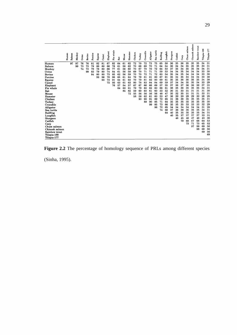
Molecular weight (MW) of the major form of PRL found in the pituitary gland is
about 23 kDa, and it is encoded by a gene consisting of 5 exons and 4 introns (Cooke
et al., 1981; Truong et al., 1984). Variants of PRL have been characterized in many
mammals. Its variants can be the result of alternative splicing of the primary
transcript, proteolytic cleavage, phosphorylation, glycosylation, and other
posttranslational modifications, thereby altering its physiological functions (Sinha,
1995). PRL is synthesized as a preprohormone consisting of 227 amino acids in most
mammals (Miller and Eberhardt, 1983). The mature hormone contains 194 to 199
amino acids, depending on species. Hormone structure is stabilized by three
intramolecular disulfide bonds. The primary structure of PRL was first reported in the
ovine (Li et al., 1970). The complete amino acid sequences of PRLs of more than 25
species have been identified (for review, see Sinha, 1995). A comparison of the amino
acid sequence from different species shows varying degrees of sequence homology,
reflecting to a great extent the order of the phylogenetic relationships. Some 32 amino
acids seem to be conserved among different species (Watahiki et al., 1989). The
homology of sequences of PRLs among different species and their primary structures
are shown in Figures 2.2 and 2.3, respectively.
PRL belongs to the families of related hormones including growth hormone
(GH) and placental lactogen (PL). Its amino acid sequence is similar to those of GH
and PL sharing genomic, structure, and biological features (Boulay and Paul, 1992;
Horseman and Yu-Lee, 1994). Genes encoding PRL, GH, and PL are evolved from a
common ancestral gene by gene duplication (Niall et al., 1971) about 500 million
years ago. Among the avian species, it has been suggested that the mechanisms,
which regulate its gene expression may be wildly conserved in this species (Kansaku

26
et al., 2005; Hiyama et al., 2009b). However, it has been demonstrated that PRL is
also synthesized by a number of extra-pituitary cells/tissues in both mammals (Ben-
Jonathan et al., 1996; Freeman et al., 2000; Soares, 2004) and birds (Berghmam et al.,
1992; Ramesh et al., 2000; Chaiseha et al., 2012), but its physiological function(s) in
these extra-pituitary tissues is poorly understood and needed to be further elucidated.
PRL is synthesized and secreted by a broad range of other cells including most
prominently various immune cells, mammary epithelium, placenta, the deciduas of
the pregnant uterus, and brain (Ben-Jonathan et al., 1996). In addition, PRL synthesis
is also found in the lacrimal gland, adrenal gland, corpus luteum, prostate gland,
testis, and pancreas (Ben-Jonathan et al., 1996; Freeman et al., 2000). To date, over
300 different physiological functions of PRL have been documented (Houdebine,
1983; Bole-Feysot et al., 1998; Harris et al., 2004) in such areas as reproduction,
water and electrolyte balance, growth and development, brain and behavior,
endocrinology and metabolism, and immunoregulation as well as behaviors like
migration, the nurturing of the young in different vertebrate species, highlighting the
importance of this pituitary hormone. Furthermore, it has been suggested that
functions and biological activities of PRL are, at least in part, regulated by
additionally post-translational modifications such as phosphorylation in the various
physiological stages (Hiyama et al., 2009a).
PRL receptor (PRLR), a single membrane-bound protein transmembrane
receptor, is a member of the Class I cytokine receptor superfamily that includes the
receptors for GH, leptin, erythropoietin, and interleukins (Bazan, 1989; 1990; Kelly et
al., 1991). PRL, PL, and primate GH bind the PRLR. PRL and GH receptors share
some structural and functional features despite their low sequence homology (30 %;
27
Goffin and Kelly, 1996). The PRLR is activated by the binding of a single ligand to
the receptor to dimerize two identical receptor subunits, leading to activation of Jak2-
kinase associated with the cytoplasmic domain, which subsequently activates a
number of signalling cascades through which PRL exerts its physiological effects (for
review, see Bole-Feysot et al., 1998; Freeman et al., 2000). Jak2 phosphorylates
tyrosine residues on different target proteins, the best identified is named signal
transducers and activators of transcription (Stats). The Jak2-Stat cascade is the major
signalling pathway of the PRLR, but other signal transducing pathways are also
involved in signal transduction by this receptor as well. Activation of mitogen-
activated protein kinases pathway has been reported in different cellular systems
under PRL stimulation (Bole-Feysot et al., 1998). In addition, activation of the
nucleotide exchange protein Vav has been reported (Clevenger et al., 1995).
Numerous PRLR isoforms have been identified in different tissues in both
mammals and birds (Davis and Linzer, 1989; Ali et al., 1991; Lesueur et al., 1991;
Pitts et al., 2000). Alternative splicing of the PRLR gene results in the multiple
isoforms which differ in the length and composition of their cytoplasmic tails and are
referred to as the short (291aa; Boutin et al., 1988) and long (591aa; Shirota et al.,
1990) PRLR isoforms (Harris et al., 2004). These isoforms are results of transcription
starting at alternative initiation sites of the different promoters and alternative splicing
of non-coding and coding exon transcripts (Hu and Dufau, 1991; Hu et al., 1998).
PRLR and its mRNA are found in the mammary gland and the ovary, the best
characterized sites of PRL biological actions in mammals (Nagano and Kelly, 1994).
cDNAs encoding the PRLR gene have been cloned in chickens (Tanaka et al., 1992),
doves, pigeons (Chen and Horseman, 1944), and turkeys (Zhou et al., 1996; Pitts et

28
al., 2000). Tissue distributions of PRLR mRNA have been reported in rats (Nagano
and Kelly, 1994; Bakowska and Morrell, 1997), turkeys (Zhou et al., 1996; Pitts et al.,
2000), and chickens (Ohkubo et al., 1998).
In mammals, the PRLR is found in the CNS and a wide range of peripheral
organs including the pituitary gland, heart, lung, thymus, spleen, liver, pancreas,
kidney, adrenal gland, uterus, skeletal muscle, prostate gland, epithelial cells, bone,
and skin (Nagano and Kelly, 1994; Nevalainen et al., 1997; Bole-Feysot et al., 1998;
Clement-Lacroix et al., 1999). In rats, PRLR mRNA expression is found in the CNS,
choroid plexus, bed nucleus of the stria terminalis (BSTM) , amygdala, central gray of
the midbrain, thalamus, hypothalamus, cerebral cortex, and olfactory bulb. The PRLR
is also extensively expressed by immune cells and some types of lymphocytes
synthesized and secreted PRL, suggesting that PRL may act as an autocrine or
paracrine modulator of immune activity (Freemark et al., 1995; 1996).
In birds, PRLR is found in the crop sac, brood patch, thyroid gland, liver,
kidney, leg, skin, large and small intestine, adipose tissue, adrenal gland, thymus,
lymphoid tissues, spleen, heart, brain, pineal gland, ovary, testis, seminal duct, and
oviduct (Tanaka et al., 1992; Chen and Horseman, 1994; Zhou et al., 1996; Ohkubo et
al., 1998; Pitts et al., 2000; Kang et al., 2007; Wang et al., 2009; Xing et al., 2011).
Indeed, it has been reported that PRLR mRNA levels are the greatest in the pineal
gland of laying and the oviduct of incubating turkey hens (Pitts et al., 2000).
29
Figure 2.2 The percentage of homology sequence of PRLs among different species
(Sinha, 1995).

30
Figure 2.3 Primary structures of PRLs of different species. (-) indicates positions left
blank to optimize alignment of amino acid sequences. (*) indicates absence of
residues from a genetic variant of tilapia PRL. PD is PRL domain. PDI-PD4 indicates
the four highly conserved domains of the PRLs (Sinha, 1995).
31
2.4.2 The Function of Prolactin in Birds
It has been well documented that PRL is associated with the reproductive
cycle in birds (turkeys: Mashaly et al., 1976; El Halawani et al., 1984a; 1997; Wong
et al., 1992b; mallards: Bluhm et al., 1983a; Boos et al., 2007; canvasback ducks:
Bluhm et al., 1983b; cockatiels: Myers et al., 1989; king penguins: Mauget et al.,
1994; emperor penguins: Lormee et al., 1999; tropical seabirds: Lormee et al., 2000;
geese: Huang et al., 2008; native Thai chickens: Kosonsiriluk et al., 2008;
Sartsoongnoen et al., 2008). During reproductively quiescent stages (non-egg laying
and rearing stages) of the native Thai chickens (Kosonsiriluk et al., 2008;
Sartsoongnoen et al., 2008) and turkeys (El Halawani et al., 1984b; 1997), plasma
PRL levels are very low. During the periods of laying and incubating, circulating PRL
levels increase dramatically (El Halawani et al., 1984b; Kosonsiriluk et al., 2008). It is
this rising PRL level that causes the cessation of ovulation, ovarian regression, and
induction and maintenance of incubation behavior. Changes in PRL gene expression
are highly correlated with the reproductive cycle in birds (Knapp et al., 1988; El
Halawani et al., 1990a; Talbot et al., 1991; Wong et al., 1991; You et al., 1995b; Tong
et al., 1997). The onset of incubation behavior is correlated with decreasing plasma
LH levels and gonadal steroids and increasing plasma PRL levels (Cogger et al.,
1979; Burke and Dennison, 1980; Lea et al., 1981; Rozenboim et al., 1993a). PRL has
been implicated as a causative factor for the reduced circulating gonadotropins and
ovarian regression, when birds shift from egg laying to incubation behavior in bantam
hens, canaries, chickens, cowbirds, ducks, mallard ducks, native Thai chickens,
pheasants, pigeons, ring doves, spotted sandpipers, turkeys, white-crowned sparrows,
and wild starlings (Riddle et al., 1935; Breitenbach and Meyer, 1959; Hohn, 1959;

32
Sharp et al., 1977; 1988; Burke and Dennison, 1980; Goldsmith and Hall, 1980;
Goldsmith and Williams, 1980; Goldsmith et al., 1981; 1984; Dawson and Goldsmith,
1982; Bluhm et al., 1983a; El Halawani et al., 1984a; 1988a; 1997; Oring et al., 1986;
Hiatt et al., 1987; Youngren et al., 1991; Kosonsiriluk et al., 2008; Sartsoongnoen et
al., 2008). PRL concentrations increase at the onset of incubation behavior and are
maintained at high levels during incubation period (Saeki and Tanabe, 1955;
Proudman and Opel, 1988) and decline when incubation behavior is terminated (El
Halawani et al., 1980; Wentworth et al., 1983). Moreover, PRL is involved in many
aspects of reproductive physiology and behaviors. It is thought to play a pivotal role
in parental behaviors by mediating increases in incubation, crop milk secretion,
feeding of young, and nest defense (Silver, 1984; Janik and Buntin, 1985; Lea et al.,
1986; Buntin et al., 1991). Active immunization against recombinant-derived PRL
reduces the incidence, delays the development, or prevents the occurrence of
incubation behavior (March et al., 1994), whereas administration of exogenous PRL
leads to increase parental behaviors (Lea and Vowles, 1986; Macnamee et al., 1986;
Pedersen, 1989; Buntin et al., 1991; Youngren et al., 1991).
Some evidence suggests that PRL plays a role in terminating egg laying,
therefore, it regulates clutch size in species that lay more than two eggs per clutch.
Cessation of egg laying is associated with an increase plasma PRL levels (Etches et
al., 1979; Burke and Dennison, 1980; Lea et al., 1981; Bluhm et al., 1983a; Hall and
Goldsmith, 1983; Silverin and Goldsmith, 1983). Several studies have been reported
that an increase in plasma PRL levels during incubating period may suppress LH
secretion (Zadworny and Etches, 1987; Porter et al., 1991; El Halawani et al., 1993;
Sharp et al., 1998). Administration of exogenous PRL suppresses concentrations of
33
gonadotropins in the turkeys (El Halawani et al., 1991) and domestic fowls (Sharp et
al., 1988). It is suggested that PRL acts centrally to reduce LH concentrations by
reducing hypothalamic GnRH levels (Rozenboim et al., 1993b). In incubating birds,
suppression of gonadotropins release involves in a mechanism independent of
increased PRL secretion (Sharp et al., 1988; 1989; Lea and Sharp, 1989; Lea et al.,
1996). In addition, PRL may directly inhibit ovarian steroidogenesis as well
(Rozenboim et al., 1993b), leading to involution of the ovary with reduced ovarian
steroidogenesis and regression of the oviduct (Porter et al., 1991). Furthermore,
seasonal reproductive activities during long day photoperiod-stimulated PRL
secretion occurred with increased PRL gene expression in the pituitary gland, which
suppress LH secretion, inhibit follicular development, terminate laying and induced
molting (Huang et al., 2008).
2.4.3 The Regulation of Prolactin Secretion
In mammals, PRL secretion is regulated by both stimulatory and inhibitory
hypothalamic factors but it is mainly under tonic inhibitory control (MacLeod and
Login, 1976; Neill, 1988; Ben-Jonathan et al., 1989; Lamberts and MacLeod, 1990).
It is well documented that the predominant mammalian PRL-inhibiting factor (PIF) is
DA, which is released from a dense network of neurons within the mediobasal
hypothalamus (MBH) named as the tuberoinfundibular dopaminergic (TIDA) neurons
and serves as the PIF of PRL secretion (Ben-Jonathan et al., 1989; Ben-Jonathan and
Hnasko, 2001). DA released from the TIDA neurons acts directly on D2 DA receptors
located on the pituitary lactotrophs (Caron et al., 1978; Civelli et al., 1991). Removal
of this DAergic inhibition can increase PRL secretion and hyperprolactinemia (Nicoll
34
and Swearingen, 1970; Nicoll, 1977). DA and its agonists inhibit the release and gene
expression of PRL and proliferation of the lactotrophs (Birge et al., 1970; Shaar and
Clemens, 1974; Pawlikowski et al., 1978; Maurer, 1981), indicating that the
regulation of PRL secretion and its gene expression are under inhibitory control of the
TIDA neurons (Pasqualini et al., 1988; Ben-Jonathan et al., 1989; Ben-Jonathan and
Hnasko, 2001). In in vivo studies, a brief fall of DA levels occurring immediately
after suckling (Chiocchio et al., 1979; Selmanoff and Wise, 1981; Demarest et al.,
1983) is necessary for PRL secretion (Grosvenor et al., 1980). Similarly, in vitro
studies indicate that pituitary PRL secretion is stimulated after short term exposure of
DA (Fagin and Neil, 1981; Denef et al., 1984). These studies confirm the
physiological role of DA as the PIF. However, it has been reported that a much lower
concentration of DA than those required for inhibition of PRL secretion can stimulate
PRL secretion in vitro (Shin, 1978; Denef et al., 1980; Burris et al., 1991; 1992;
Porter et al., 1994) and in vivo (Arey et al., 1993). These evidences suggest that all
pituitary lactotrophs have the potential to respond to the inhibitory and stimulatory
effects of DA (Kineman et al., 1994), and the two opposite effects of DA on PRL
secretion may be mediated by distinct guanine nucleotide binding proteins (G
proteins) depending on its specific receptor subtypes (Burris et al., 1992; Niimi et al.,
1993; Lew et al., 1994).
In mammals, VIP has been shown to be involved in the regulation of PRL
secretion from the pituitary gland for many decades (Kato et al., 1978; Rotsztejn et
al., 1980; Reichlin, 1988). VIP has been suggested to regulate pituitary secretion by a
neuroendocrine pathway since the presence of VIP in the hypothalamic nerve endings,
the anterior pituitary gland (Besson et al., 1979), and the hypophysial portal system
35
(Said and Porter, 1979). It is also reported that VIP can stimulate PRL release both in
vitro (Shaar et al., 1979; Enjalbert et al., 1980; Samson et al., 1980; Matsushita et al.,
1983) and in vivo (Kato et al., 1978; Frawley and Neill, 1981). Administration of VIP
antiserum inhibits PRL release induced by stress, 5-HT, or suckling (Shimatzu et al.,
1984; Abe et al., 1985; Kaji et al., 1985a; 1985b; Ohta et al., 1985). In addition, the
contents of pituitary PRL mRNA and protein appear to be regulated by VIP (Ben-
Jonathan et al., 1989; Maas et al., 1991). VIP mRNA expression is increased during
the lactation period in rats (Gozes and Shani, 1986). An increase in VIP
concentrations in the hypophysial portal blood is relative to the peripheral blood (Said
and Porter, 1979; Shimatsu et al., 1981). VIP also promotes the entry of extracellular
calcium ions into the PRL-secreting pituitary cells (Bjoro et al., 1987; Prysor-Jones et
al., 1987). As aformentioned, VIP is proposed as a mammalian PRF.
Thyrotropin-releasing hormone (TRH) acts as a hypothalamic PRF in
mammals as well. TRH stimulates PRL secretion both in vitro (Maas et al., 1991) and
in vivo (Grosvenor and Mena, 1980; de Greef and Visser, 1981; Laverriere et al.,
1988; Lafuente et al., 1994) and also PRL gene transcription (Potter et al., 1981;
Laverriere et al., 1988). The secretion of PRL by TRH occurs during a transient
depression in DAergic activity (Plotsky and Neill, 1982; Martinez de la Escalera et
al., 1988). However, there are many contradictive results that have let the researchers
to question its role as the PRF.
To date, various PRFs and PIFs have been observed both in birds and
mammals such as 5-HT (Chaiseha and El Halawani, 2005; Chaiseha et al., 2010),
angiotensin II (Malarkey et al., 1987; Opel and Proudman, 1988a; Myers and Steele,
1989; Steele, 1990), oxytocin/vasopressin (Hyde and Ben-Jonathan, 1988; 1989;

36
Johnston and Negro-Vilar, 1988), peptide histidine isoleucine (PHI; Samson et al.,
1983; Werner et al., 1983; Proudman and Opel, 1988; 1990; Chaiseha and El
Halawani, 1999; Kulick et al., 2005), and pituitary adenylate cyclase activating
polypeptide (PACAP; Miyata et al., 1989; Yamauchi et al., 1995; You et al., 2000).
The regulation of PRL in birds also involves the interaction of external stimuli
with neuroendocrine mechanisms. Critical environmental stimuli include sensory
information concerning photoperiod, ambient temperature, and the presence of eggs
and offspring. These external stimuli and steroid hormones such as estrogen and
progesterone are important in initiating and maintaining PRL secretion, although their
relative importance varies with the stages of the reproductive cycle (Curlewis, 1992).
In incubating hens, tactile stimuli from the nests and eggs maintain the elevated
circulating PRL levels and up-regulate VIP expression (Janik and Buntin, 1985; Lea
et al., 1986; Silver et al., 1988; Buntin et al., 1991; Massaro et al., 2007).
As mentioned above, the regulation of PRL secretion and its gene expression
are under the inhibitory control of the TIDA neurons in mammals (Ben-Jonathan and
Hnasko, 2001). However, this is not the case in birds, where removal of these
hypothalamic inputs results in the complete cessation of PRL secretion (Tixier-Vidal
et al., 1966; Chadwick et al., 1978; Hall et al., 1986). It has long been well established
that the secretion of PRL in birds involves in a tonic stimulatory control by the
hypothalamus rather than the inhibitory DAergic system that found in mammals
(Kragt and Meites, 1965; Bern and Nicoll, 1968; El Halawani et al., 1984a; Hall et al.,
1986). The regulation of avian PRL secretion and its gene expression is influenced by
hypothalamic VIP, the avian PRF (El Halawani et al., 1997; 2001; Chaiseha et al.,
1998; Chaiseha and El Halawani, 1999; 2005). In the past six decades, several studies

37
confirm the pivotal role of VIP as the only avian PRF. Immunoneutralization of VIP
prevents an increase in circulating PRL levels that follows photostimulation, prevents
the induction of incubation behavior, up-regulates content of LH-β- and FSH-β-
subunit mRNAs, and extends the duration of egg laying period, but does not prevent
spontaneous gonadal regression and molting (Sharp et al., 1989; El Halawani et al.,
1995; 1996; Dawson and Sharp, 1998; Ahn et al., 2001). Variations in VIP
immunoreactivity, VIP peptide contents in the INF and ME, and VIP mRNA steady-
state levels in the INF are correlated with changes in circulating PRL levels
throughout the avian reproductive cycle (Mauro et al., 1989; Chaiseha et al., 1998;
Chaiseha and El Halawani, 1999; Kosonsiriluk et al., 2008).
In contrast with mammals, DAergic influences are involved in both
stimulating and inhibiting avian PRL secretion depending on multiple DA receptor
subtypes (Youngren et al., 1995; 1996b; Chaiseha et al., 1997; 2003; Al Kahtane et
al., 2003). In turkeys, stimulatory D1 DA receptor mRNA expression has been found
to increase in the hypothalamus of hyperprolactinemic incubating hens and in the
pituitary gland of laying hens. However, inhibitory D2 DA receptor mRNA
expression increases in the pituitary gland of hypoprolactinemic photorefractory hens
(Schnell et al., 1999a; 1999b; Chaiseha et al., 2003). The stimulatory effect of DA on
PRL secretion is regulated via the D1 DA receptors residing in the INF, where the
VIP neurons are located. In contrast, DA inhibits PRL release and synthesis by
blocking the action of VIP at the pituitary level via the D2 DA receptors (Youngren et
al., 1996b; 1998; 2002; Chaiseha et al., 1997; 2003; Al Kahtane et al., 2003). Indeed,
changes in the DAergic activity during the turkey reproductive cycle parallel changes
in plasma PRL levels, number of VIP-ir neurons, VIP peptide content, and its mRNA

38
expression within the INF (El Halawani et al., 1980; 1984b; Mauro et al., 1989;
Wong et al., 1991; Chaiseha et al., 2003). It also has been indicated that the variations
in PRL secretion observed across the turkey reproductive cycle are, in part, regulated
by changes in VIP receptors at the pituitary level (Chaiseha et al., 2004). It is very
well established that DA plays an intermediary role in PRL secretion, requiring an
intact VIPergic system in order to cause the release of PRL (Youngren et al., 1996b).
In addition, there are evidence indicates that dynorphin, 5-HT, DA, and VIP all
appear to stimulate avian PRL secretion along a common pathway expressing
opioid, serotonergic, DAergic, and VIPergic receptors at synapses arranged serially in
that functional order, with the VIPergic system as the final mediator (for review, see
El Halawani et al., 2001).
2.5 Vasoactive Intestinal Peptide: Structure, Function, and
Regulation of Secretion
2.5.1 The Structure of Vasoactive Intestinal Peptide
VIP, an octacosapeptide, was first isolated from porcine duodenum (Said and
Mutt, 1970; Mutt and Said, 1974). Subsequently, it has been found to be widely
distributed in the central and peripheral nervous systems (Larsson et al., 1976; Said
and Rosenberg, 1976; Giachetti et al., 1977; Rosselin et al., 1982), with high
concentrations found in the hypothalamus (Emson et al., 1979; Samson et al., 1979;
Ceccatelli et al., 1991) and its function is considered as a neurotransmitter and
neuroendocrine substance (Larsson et al., 1976; Marley and Emson, 1982). The
discovery of a large population of VIP-ir neurons in the hypothalamus whose axons
project to the ME (Samson et al., 1978; 1979; Polak and Bloom, 1982; Lam, 1991;

39
Dalcik and Phelps, 1993) and a high concentration of VIP in the hypophysial portal
blood (Said and Porter, 1979; Shimatsu et al., 1981; Brar et al., 1985; Mutt, 1988) led
to the hypothesis that VIP involves in the regulation of anterior pituitary functions.
VIP is a neuropeptide belonged to the VIP/glucagon/secretin superfamily
including secretin, glucagon, gastric inhibitory peptide (GIP), GH releasing factor,
PHI, and PACAP. VIP exerts its biological effects by binding to its specific receptors
that are coupled to the G-proteins, whose actions mediated via the adenylate cyclase
and the production of cyclic adenosine monophosphate (cAMP; Hokfelt et al., 1980;
Couvineau et al., 1990a; Lutz et al., 1995). The peptides of this family are probably
the results of exon duplication coupled to gene duplication. VIP gene contains 7 exons
and each exon encodes a different functional domain in the final mRNA and protein.
Two adjacent exons in the genome encoding VIP and the related peptide histidine
methionine (PHM) or PHI are exon 5 and exon 4, respectively (Bodner et al., 1985;
Linder et al., 1987; Yamagami et al., 1988; Giladi et al., 1990). To date, mammalian
VIP cDNAs (Itoh et al., 1983; Nishizawa et al., 1985), chicken and turkey VIP
cDNAs (McFarlin et al., 1995; Talbot et al., 1995; You et al., 1995b) have been
cloned and characterized. The open reading frame of mammalian VIP is comprised of
165 amino acids. Chicken VIP is different from mammalian VIP in its amino acid
sequences at position 11, 13, 26, and 28, but the number of amino acid residue is the
same (Nilsson, 1975). In addition, the chicken and turkey VIP share complete amino
acid homology and are 98 % homologus at the neucleotide level. VIP mRNA may
exist with or without PHI. Both mRNA forms are found in the chicken digestive tract
and hypothalamus. The short form is found only in the turkey hypothalamus and
comprises 4 to 6 % of all VIP transcripts (You et al., 1995b). The amino acid

40
sequences of VIP and the member in VIP/glucagon/secretin family are shown in
Figure 2.4.
Figure 2.4 The amino acid sequences of VIP, PHI, secretin, glucagon, and GIP. p:
porcine, b: bovine, c: chicken, m: mammalian, a: the C-terminal amino acid is in the
amide form (Rosselin et al., 1982).
VIP receptors are purified from porcine liver with the MW is 53 kDa
(Couvineau et al., 1990b). Subsequently, it has been cloned and characterized in
mammals (Sreedharan et al., 1991; 1993; Ishihara et al., 1992; Lutz et al., 1993;
Couvineau et al., 1994; Gagnon et al., 1994; Usdin et al., 1994). Pharmacologically,
two subtypes of VIP receptors; VPAC1 and VPAC2 are expressed in a tissue specific
manner (Couvineau et al., 1994; Usdin et al., 1994; Sherward et al., 1995) and bind
both VIP and PACAP, which these receptors are high affinity receptors for VIP. VIP
receptors belong to the members of a seven transmembrane spanning domain G
protein-coupled family. The biological action of VIP is mediated via its specific
receptors that are coupled to adenylate cyclase and the production of intracellular
cAMP (Gourdji et al., 1979; Bjoro et al., 1987; Couvineau et al., 1990a; Lutz et al.,
1995). VIP receptors are presented in several organs including the lung, liver, small
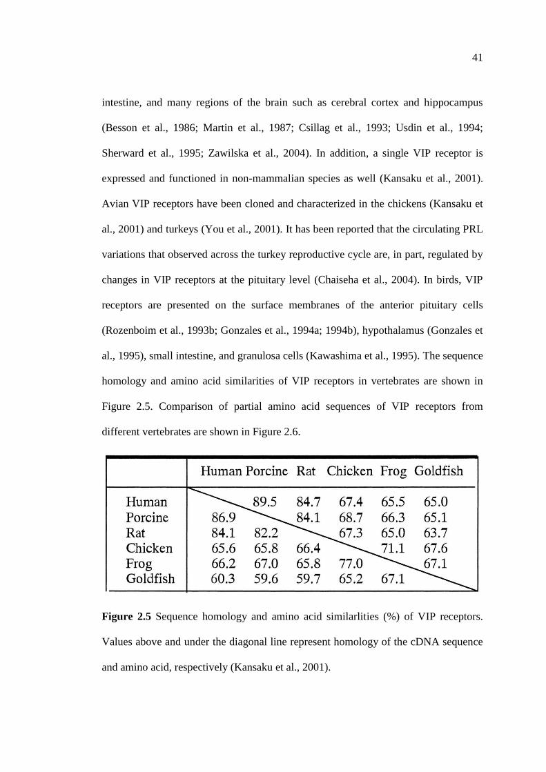
41
intestine, and many regions of the brain such as cerebral cortex and hippocampus
(Besson et al., 1986; Martin et al., 1987; Csillag et al., 1993; Usdin et al., 1994;
Sherward et al., 1995; Zawilska et al., 2004). In addition, a single VIP receptor is
expressed and functioned in non-mammalian species as well (Kansaku et al., 2001).
Avian VIP receptors have been cloned and characterized in the chickens (Kansaku et
al., 2001) and turkeys (You et al., 2001). It has been reported that the circulating PRL
variations that observed across the turkey reproductive cycle are, in part, regulated by
changes in VIP receptors at the pituitary level (Chaiseha et al., 2004). In birds, VIP
receptors are presented on the surface membranes of the anterior pituitary cells
(Rozenboim et al., 1993b; Gonzales et al., 1994a; 1994b), hypothalamus (Gonzales et
al., 1995), small intestine, and granulosa cells (Kawashima et al., 1995). The sequence
homology and amino acid similarities of VIP receptors in vertebrates are shown in
Figure 2.5. Comparison of partial amino acid sequences of VIP receptors from
different vertebrates are shown in Figure 2.6.
Figure 2.5 Sequence homology and amino acid similarlities (%) of VIP receptors.
Values above and under the diagonal line represent homology of the cDNA sequence
and amino acid, respectively (Kansaku et al., 2001).

42
Figure 2.6 Comparison of partial amino acid sequences of VPAC1 (A) and VPAC2
(B) receptors from different vertebrates. Conserved amino acids (100 % identity)
among taxa are highlighted. The putative transmembrane domains (TMDs) are
denoted by horizonatal arrows (Montpetit et al., 2003).
43
2.5.2 The Localization of Vasoactive Intestinal Peptide in the Avian Brain
Well documentedly, the distributions of VIP-containing neurons have been
conducted in the brain of many avian species including Pekin ducks (Korf and
Fahrenkrug, 1984), Japanese quails (Peczely and Kiss, 1988), turkeys (Mauro et al.,
1989; Chaiseha and El Halawani, 1999), pigeons (Cloues et al., 1990), ring doves
(Norgren and Silver, 1990), chicks (Kuenzel and Blahser, 1994; Kuenzel et al., 1997),
dark-eyed juncos (Saldanha et al., 1994), zebra finches (Bottjer and Alexander, 1995),
and native Thai chickens (Kosonsiriluk et al., 2008; Prakobsaeng et al., 2011). In
birds, VIP neurons are extensively distributed throughout the hypothalamus (Yamada
et al., 1982; Mikami and Yamada, 1984; Macnamee et al., 1986; Peczely and Kiss,
1988; Mauro et al., 1989; Hof et al., 1991; Chaiseha and El Halawani, 1999;
Kosonsiriluk et al., 2008; Prakobsaeng et al., 2011), especially in the areas of the
medial preoptic area (MPOA), medial hypothalamic region, AM, hypothalamus pars
lateralis, and INF (den Boer-Visser and Dubbeldam, 2002). Generally, three types of
VIP-ir neurons and fibers are described. The first set consists of a large number of
spindle or bipolar neurons that connected the V III to the external layer of the ME.
The second set of VIP-ir fibers extends from the infundibular nucleus to the ME. The
third set of VIP-ir neurons terminates upon small capillaries within the hypothalamus.
It has been suggested that VIP in the ME is derived from neurons located within the
INF (Macnamee et al., 1986; Mauro et al., 1989; Chaiseha and El Halawani, 1999;
Youngren et al., 2002). VIP terminals are observed in the external portion of the ME
and the majority of VIP-containing cell bodies are located in the INF. The number of
the VIP-ir neurons in the INF increases following a gonadal stimulatory photoperiod.
The hypothalami of incubating turkey hens contain more VIP-ir neurons than those of
44
non-photostimulated hens. Circulating PRL levels and hypothalamic VIP
immunoreactivity are found to reduce in nest-deprived incubating birds (Mauro et al.,
1989; Prakobsaeng et al., 2011). Fluctuations in hypothalamic VIP immunoreactivity
and its gene expression within the INF paralleled fluctuations in circulating PRL
levels during the avian reproductive cycle (Chaiseha and El Halawani, 1999). The
number, distributed area, and density of hypothalamic VIP-ir neurons are greater in
incubating hens than those of laying hens (Sharp et al., 1989; Kosonsiriluk et al.,
2008). In addition, in domesticated pigeons, increases in the number and cell size of
VIP-ir neurons within this region following the periods of elevated circulating PRL
levels have been reported (Peczely and Kiss, 1988; Cloues et al., 1990). Thus, these
VIP neurons project to the ME, where VIP is transported through the hypothalamic-
pituitary portal vessels to the anterior pituitary gland (Yamada et al., 1982; Macnamee
et al., 1986; Mauro et al., 1989). In addition, lesions in the INF can prevent the PRL
increase after photostimulation (Youngren et al., 1989).
All these above data indicate that VIP neurons in the INF are an important
factor in the stimulation of PRL secretion. It has been further established that VIP
axon terminals have been found in close apposition to GnRH neurons in the lateral
septal organ (LSO) and POA (Teruyama and Beck, 2001) and an inverse relationship
between VIP in the INF and GnRH in the POA has been reported (Deviche et al.,
2000). A subset of VIP-ir neurons within the MBH and septal region of the dove brain
has been proposed to be encephalic photoreceptors (Silver et al., 1988; Norgren and
Silver, 1990). Recently, it has been implicated that turkey melanopsin (tOPN4x) in
the hypothalamic premammillary nucleus (PMM) DA-melatonin (DA-MEL) neurons
45
acts as an important component of the photoreceptive system regulating reproductive
activity in temperate zone birds (Kang et al., 2010).
2.5.3 The Function of Vasoactive Intestinal Peptide in Birds
In birds, it has long been established that the hypothalamic control of PRL
secretion involves a stimulatory mechanism rather than the inhibitory DAergic system
found in mammals (Kragt and Meites, 1965; Bern and Nicoll, 1968; El Halawani et
al., 1984a; Hall et al., 1986). Many evidences confirm and support VIP as the most
important avian PRF (Macnamee et al., 1986; Opel and Proudman, 1988b; Mauro et
al., 1989; El Halawani et al., 1990b; 1990c; 1997; Talbot et al., 1991). It is well
established that avian pituitary PRL secretion is tonically stimulated by VIP, which is
secreted from neurons located in the INF of the caudo-medial hypothalamus (El
Halawani et al., 1997; 2001). VIP meets the classical criteria for defining it as the
hypophysiotrophic PRF in birds. These criteria are; 1) the presence of VIP-ir neurons
in the hypothalamus, 2) the secretion of VIP into hypophysial portal blood, 3) the
modulation of VIP secretion into hypophysial portal blood, 4) the presence of VIP
receptors on anterior pituitary cells, 5) the ability of VIP to regulate anterior pituitary
lactotrophs, and 6) the alteration of pituitary function, due to antagonism of VIP (for
review, see El Halawani et al., 1997). Further evidences have been provided by the
findings that immunoneutralization of endogenous VIP reduces circulating PRL levels
and its mRNA expression and totally inhibits the PRL release affected by electrical
stimulation of the MPOA (El Halawani et al., 1990b; Youngren et al., 1994) and
blocks the hormonal and behavioral characteristics of incubating hens (El Halawani et
al., 1995). Several hypothalamic neurotransmitters and neuropeptides have been

46
studied during the past six decades for their effects on PRL such as TRH, angiotensin
II, oxytocin, vasopressin, PACAP, and PHI. Only VIP is thought to be physiologically
significant PRF in birds.
VIP is a potent releaser of PRL in vivo (Lea and Vowles, 1986; Macnamee et
al., 1986; Opel and Proudman, 1988b; El Halawani et al., 1990c; Pitts et al., 1994)
and in vitro (Macnamee et al., 1986; Proudman and Opel, 1988; El Halawani et al.,
1990b; Xu et al., 1996). Thus, VIP plays a pivotal role in the regulation of PRL in
birds. VIP regulates PRL gene expression by enhancing the transcription rate of PRL
and up-regulating PRL mRNA stability (Tong et al., 1998). Variations in
hypothalamic VIP immunoreactivity, VIP contents, VIP mRNA steady-state levels,
VIP mRNA expression in the INF, VIP receptor mRNA in the pituitary cells, and VIP
concentrations in hypophysial portal blood are correlated with the changes in
circulating PRL levels in many avian species such as turkeys (Mauro et al., 1989;
Youngren et al., 1996a; Chaiseha et al., 1998; 2004; Chaiseha and El Halawani,
1999), chickens (Sharp et al., 1989a), doves (Cloues et al., 1990), and native Thai
chickens (Kosonsiriluk et al., 2008). Passive immunization with VIP anti-serum
decreases plasma PRL and pituitary mRNA levels and terminates incubation behavior
(Talbot et al., 1991). Similarly, active immunization with VIP also reduces circulating
PRL levels and prevents the expression of incubation behavior in the turkeys (El
Halawani et al., 1996; 2001). It has been suggested that the stimulatory action of VIP
occurs via specific receptors located on anterior pituitary plasma membranes, which
change throughout the reproductive cycle of the turkeys (Rozenboim and El
Halawani, 1993; Chaiseha et al., 2004). As mentioned above, it is confirmed the role
of VIP as the avian PRF.
47
Increased hypothalamic VIP mRNA and its peptide contents are associated
with gonadal regression and suppression of gonadotropins in photorefractory turkeys
(Chaiseha et al., 1998; Chaiseha and El Halawani, 1999). Immunoneutralization with
VIP up-regulates LH-β- and FSH-β-subunit mRNAs expression (Ahn et al., 2001) and
delays the onset of photorefractoriness and molt in starling (Dawson and Sharp,
1998). These findings imply that VIP also exerts an inhibitory effect on the
GnRH/FSH-LH system. In deed, it has been indicated that VIP has a central
inhibitory effect on GnRH/FSH-LH release in birds (Pitts et al., 1994).
2.5.4 The Regulation of Vasoactive Intestinal Peptide Secretion
Due to this research dissertation is conducted in the native Thai chicken, this
Section will discuss only the regulation of VIP secretion in birds. In fact, the
regulation of VIP in mammals is very well documented worldwide. It has been also
suggested that VIP mediates the effects of photoperiod on PRL secretion in the
turkeys (El Halawani et al., 1996) and quantification of hypothalamic VIP reveals an
increased VIP content following photostimulation (Mauro et al., 1992). It has been
further demonstrated that VIP peptide contents in the ME, hypothalamic cytoplasmic
VIP mRNA steady-state levels, and hypothalamic nascent VIP mRNA levels are all
increased and correlated with increased PRL secretion following photostimulation
(Chaiseha et al., 1998). These results lend support to a hypothetical scheme for
photoperiodic regulation of PRL in which VIP serves as the PRF that is intimately
linked to photoperiodic mechanisms. Furthermore, the results also imply that VIP
transcription is coupled to the photoperiodic state of the birds. However, it remains to
be clarified how photoperiodic information is transduced to VIP-ir neurons and
48
whether photoperiodic cues directly influence VIP remains an open question. Silver et
al. (1988) has shown that VIP is colocalized with an opsin-like pigment in the INF.
This area is thought to contain extra-retinal hypothalamic photoreceptors which are
important for the induction of seasonal reproductive function in birds (Oksche and
Farner, 1974; Oliver and Bayle, 1976; Oliver et al., 1977). Alternatively, photoperiod
may modulate VIP expression by acting upon unknown neuronal circuits that
influence VIP transcription. Recently, tOPN4x is found in the DA-MEL co-localized
neurons in the PMM and is implicated as an important component of the
photoreceptive system regulating reproductive activity in temperate zone birds (Kang
et al., 2010).
VIP is also inhibited by high concentrations of circulating PRL.
Intracerebroventricular (ICV) PRL injections into incubating ring doves reduce the
number of infundibular VIP-like neurons, which indicates the existence of a
hypothalamic negative feedback loop for PRL (Saldanha and Silver, 1995).
Intracranial and systemic administrations of ovine PRL into laying turkey hens reduce
circulating PRL concentrations (Youngren et al., 1991; Rozenboim et al., 1993b). In
addition, systemic PRL administration reduces hypothalamic VIP contents and the
number of anterior pituitary VIP binding sites (Rozenboim et al., 1993a), indicating
that PRL may act directly at the pituitary level. PRL binding sites have been found
within the avian hypothalamus (Buntin and Ruzycki, 1987; Buntin and Walsh, 1988)
and PRLR mRNA is also detected in the brain of chickens (Tanaka et al., 1992) and
the hypothalamus of turkeys (Zhou et al., 1996; Pitts et al., 2000). Furthermore, PRL
may cross the blood-brain barrier at the choroids plexus (Buntin and Walsh, 1988)
and binds to the PRLRs lining the V III, thus decreasing the number of hypothalamic

49
VIP-containing neurons (Saldanha and Silver, 1995). VIP immunoneutralization
averts the rise in circulating PRL levels that follows photostimulation, prevents the
induction of incubation behavior, up-regulates LH-- and FSH- subunit mRNAs
content, and extends the duration egg laying period, but does not prevent spontaneous
gonadal regression and molting (Sharp et al., 1989; El Halawani et al., 1995; 1996;
Dawson and Sharp, 1998; Ahn et al., 2001). However, despite the well established
antigonadotropic effects of PRL, it appears that the high circulating PRL levels of
laying and non-incubating birds is not the primary cause of the suppression of
GnRH/FSH-LH system and the termination of reproduction (Juss, 1993; Dawson and
Sharp, 1998).
2.6 Gonadotropin Releasing Hormone: Structure, Function, and
Regulation of Secretion
2.6.1 The Structure of Gonadotropin Releasing Hormone
GnRH, referred to as type one mammalian GnRH (mGnRH), was first isolated
and sequenced from porcine hypothalamus (Peczely, 1989; Rivier, 2001). A second
molecular form of GnRH has been later identified in several mammals and other non-
mammalian vertebrate species (for review, see Bakker and Baum, 2000). This second
form has been identified as chicken GnRH-II (cGnRH-II). GnRH is a hypothalamic
neuronal secretory decapeptide that important for the control of reproduction in
vertebrates. To date, it has been reported that GnRH consists of a family at least 25
isoforms, there are 15 and 11 isoforms of GnRH from representative vertebrate and
invertebrate species, respectively (Gorbman and Sower, 2003; Kavanaugh et al.,
2008; Zhang et al., 2008). Two to three isoforms have been identified in
50
representative species of all classes of vertebrates (Powell et al., 1994; Kah et al.,
2007). All these forms consist of 10 amino acids with conserved amino acids in
position 1, 4, 9, and 10 (Figure 2.7; Kavanaugh et al., 2008), and share at least 50 %
sequence identity (for review, see Limonta et al., 2003). The most recognized and
common structural variation among the different forms of GnRH resides in amino
acids between position 5 and 8 in the sequence. There are three types of GnRH and
two types of GnRH receptor have been found in the avian brain (Sun et al., 2001;
Shimizu and Bedecarrats, 2006). Two distinct forms of GnRH have been isolated in
chicken; cGnRH-I or GnRH-I ([Gln8]-GnRH) and cGnRH-II ([His5, Trp7, Ty8]-
GnRH; King and Millar, 1982; Miyamoto et al., 1982; 1984; Millar and King, 1984;
Sherwood et al., 1988). The gene encoding cGnRH-I has been cloned and
characterized (Dunn et al., 1993). To date, GnRH-III which is first demonstrated in
lamprey is also found in the brain of songbirds (Bentley et al., 2004). Of the three
forms, GnRH-I is the form that is directly involved in controlling reproduction in
birds (Sharp et al., 1990).
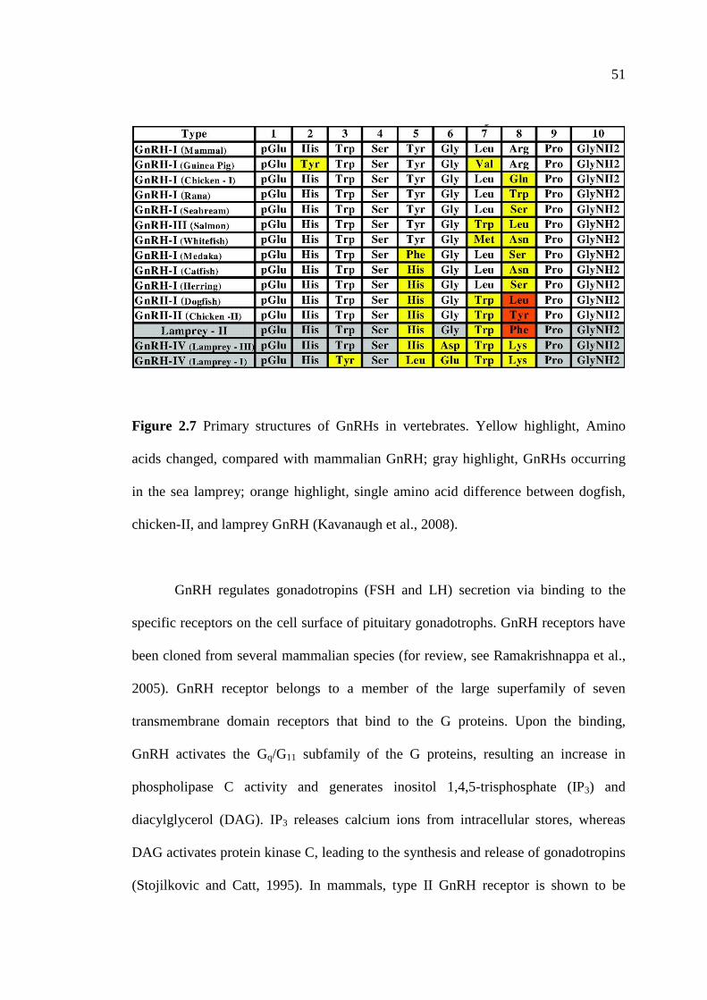
51
Figure 2.7 Primary structures of GnRHs in vertebrates. Yellow highlight, Amino
acids changed, compared with mammalian GnRH; gray highlight, GnRHs occurring
in the sea lamprey; orange highlight, single amino acid difference between dogfish,
chicken-II, and lamprey GnRH (Kavanaugh et al., 2008).
GnRH regulates gonadotropins (FSH and LH) secretion via binding to the
specific receptors on the cell surface of pituitary gonadotrophs. GnRH receptors have
been cloned from several mammalian species (for review, see Ramakrishnappa et al.,
2005). GnRH receptor belongs to a member of the large superfamily of seven
transmembrane domain receptors that bind to the G proteins. Upon the binding,
GnRH activates the Gq/G11 subfamily of the G proteins, resulting an increase in
phospholipase C activity and generates inositol 1,4,5-trisphosphate (IP3) and
diacylglycerol (DAG). IP3 releases calcium ions from intracellular stores, whereas
DAG activates protein kinase C, leading to the synthesis and release of gonadotropins
(Stojilkovic and Catt, 1995). In mammals, type II GnRH receptor is shown to be
52
highly selective for GnRH-II and is widely expressed in reproductive tissues and the
CNS (Millar et al., 2001; Neill et al., 2001). High expression of GnRH-II receptors in
the gonadotrophs suggests its role in the regulation of gonadotropin secretion. The
cloned of a novel GnRH receptor from chicken pituitary differs from the mGnRH
receptor in its primary structure, ligand selectivity, and in the agonistic behavior of
certain mGnRH receptor antagonists (Tanaka et al., 1980; Harris et al., 2004).
Particularly, some antagonists of mGnRH receptors act as agonists of the chicken
GnRH receptor, stimulating LH release from chicken pituitary cells (Kuo et al., 2002).
2.6.2 The Localization of Gonadotropin Releasing Hormone in the Brain
It is very well established that each GnRH form has its unique distributions
within the brain, indicating a difference in developmental origin and/or adult
functions (Sherwood et al., 1993; White et al., 1995; Lescheid et al., 1997; Dubois et
al., 2002). In mammals, GnRH-I neurons are distributed in a loose rostral-caudal
continuum in the ventral forebrain. The principle projection of these neurons is
toward the ME, where their terminals have been found in the hypophysial portal
plexus. However, GnRH-II-expressing neurons locate in the midbrain and the major
terminal field for these neurons is the medial habenula. In contrast to the forebrain
GnRH-I fibers, few GnRH-II fibers are observed in the ME (for review, see Bakker
and Baum, 2000).
Like in mammals, GnRH neurons and fibers are more widely distributed
throughout the avian brain. Many previous studies have examined the distributions of
cGnRH-I throughout the avian brain including chickens (Jozsa and Mess, 1982;
Sterling and Sharp, 1982; Mikami et al., 1988; Kuenzel and Blahser, 1991), ducks

53
(McNeill et al., 1976; Bons et al., 1978), white-crowned sparrows (Blahser et al.,
1986; 1989), Japanese quails (Foster et al., 1988; Mikami et al., 1988; Perera and
Follett, 1992; van Gils et al., 1993; Teruyama and Beck, 2000), European starlings
(Dawson et al., 1985; Foster et al., 1987; Goldsmith et al., 1989), garden warblers
(Bluhm et al., 1991), great tits and ring doves (Silver et al., 1992), turkeys (Millam et
al., 1993), dark-eyed juncos (Saldanha et al., 1994), house sparrows (Hahn and Ball,
1995), cockerels (Sun et al., 2001), canaries (Bentley et al., 2004), and native Thai
chickens (Sartsoongnoen et al., 2012). There are three major groups of GnRH-I-ir
neurons; 1) a telencephalic group medial to the lateral ventricles, 2) a
basotelencephalic group located ventral to the tractus septomesencephalicua (TSM)
and extending laterally and dorsocaudally, and 3) a distinctive group of neurons
located along the midline extending from the POA to septal regions (Foster et al.,
1987; Millam et al., 1993; 1998; Teruyama and Beck, 2000). Six major groups of
cGnRH-I perikarya are found including the olfactory bulb, olfactory tubercle, lobus
parolfactorius, nucleus accumbens (Ac), septal preoptic hypothalamic region (three
sub-nuclei), and lateral anterior thalamic nucleus (Kuenzel and Blahser, 1991). Fully
processed cGnRH-I mRNA and a variant transcript with a retained intron 1 are
observed in the POA, basal hypothalamus, anterior pituitary gland, and testis of the
cockerels (Sun et al., 2001). The main group of cGnRH-I cell bodies is located in the
POA with fibers extending along the V III and then entering the area of GnRH
secretion, the ME (Foster et al., 1987; Meddle and Follett, 1997). Specific GnRH-I-ir
neurons are found in several hypothlalamic areas including the POA, nucleus
preopticus medialis (POM), AM, paraventricular nucleus (PVN), and nCPa.
Additional scattered neurons are also found in the SL and around the OVLT. Several
54
studies have reported the distributions of the cGnRH-I mRNA and its protein in the
brain of several species (Millam et al., 1989; Dunn and Sharp, 1999; Sun et al., 2001;
Dawson et al., 2002; Kang et al., 2006). cGnRH-I mRNA expressions are the greatest
in the nCPa and around the OVLT. cGnRH-I mRNAs abundance within the nCPa,
OVLT, and SL are greater in laying hens than those of in non-photostimulated and
incubating turkey hens (Kang et al., 2006).
The presence of cGnRH-II in the ME has been identified, but its involvement
in the control of reproduction is remained controversial. A large group of cGnRH-II-ir
neurons in the oculomotor region and basal lateral hypothalamus are observed in the
turkeys and Japanese quails (Millam et al., 1993; 1995; 1998; Teruyama and Beck,
2000). cGnRH-II fibers are found in the ME of Japanese quails and chickens (van
Gils et al., 1993; Teruyama and Beck, 2000; Clerens et al., 2003). cGnRH-II-ir fibers
are found prominently in the POA, lateral septum (LS), both medial and lateral to the
TSM at the level of the POA and in limbic structures, olfactory areas, and forebrain
(Millam et al., 1993).
Several evidences in the Japanese quails, chickens (Mikami et al., 1988; Sharp
et al., 1990), ostriches (Powell et al., 1987), and turkeys (Millam et al., 1989) reveal
that cGnRH-I and cGnRH-II are distributed differently in the brain and other tissues.
GnRH-II-immunoreactivity has not been observed in the ME of Japanese quails,
chickens, and turkeys (van Gils et al., 1993; Millam et al., 1998; D’Hondt et al., 2001;
Clerens et al., 2003), suggesting that cGnRH-II does not directly promote pituitary
gonadotropins secretion (Millam et al., 1993). The release of cGnRH-II from the ME
has been reported (Millam et al., 1998), but this release is less amount than that of
cGnRH-I. In contrast, study in white-crowned sparrows does not find GnRH-II in the
55
ME (Meddle et al., 2006). In addition, GnRH-III neurons are distributed in the
hypothalamus and forebrain of songbirds, especially in the telencephalon including
the hippocampus and song control areas (Bentley et al., 2004).
2.6.3 The Function of Gonadotropin Releasing Hormone in Birds
Like in mammals, GnRH plays a pivotal role in the control of avian
reproduction. At the peak level of reproductive activity, birds have more GnRH-ir
neurons and fibers than those of sexually inactive or photorefractory ones (Hahn and
Ball, 1995; Parry et al., 1997; Cho et al., 1998; Sharp et al., 1990; Sartsoongnoen et
al., 2012). GnRH contents also change during the avian reproductive cycle. GnRH-I
levels decrease when birds enter the incubating period and this decrease is thought to
be regulated by the inhibitory effect of PRL (Sharp et al., 1988). GnRH-I
concentrations is significantly elevated in the POA during incubation period (Millam
et al., 1995). GnRH peptide contents in the hypothalamus during the reproductive
cycle of the turkeys (Millam et al., 1989; El Halawani et al., 1993; Rozenboim et al.,
1993a) and chickens (Dunn et al., 1996) do not change. However, the amount of
hypothalamic GnRH mRNA in incubating hens is lower than those of in laying hens
(Dunn et al., 1996). GnRH contents of discrete medial preoptic, infundibulum, and
arcuate samples are higher in laying hens than those of in non-laying hens (Advis et
al., 1985). In the turkeys, GnRH-I mRNA abundance within the nCPa, OVLT, and SL
is greater in laying hens than those of in non-photostimulated and in incubating ones,
while GnRH mRNA expression is the least in photorefractory hens, suggesting that
hypothalamic GnRH mRNA expression may be used to precisely characterize the
different reproductive stages in birds (Kang et al., 2006). Moreover, an increase in LH
56
secretion is resulted from removal of incubating hens from their nests and associated
with an increase in the amount of hypothalamic GnRH mRNA (Dunn et al., 1996).
Like in mammals, GnRH is synthesized by neurosecretory cells in the
hypothalamus, released from the ME into the hypophysial portal vessels, and then
transported to the pituitary gland, where it stimulates the secretions of FSH and LH.
GnRH increases FSH and LH secretion of the adenohypophysis both in vitro and in
vivo (Peczely, 1989). Injection of GnRH increases plasma LH levels in vivo
(Wingfield et al., 1979; McNaughton et al., 1995). Incubation of turkey anterior
pituitary cells with GnRH results in an increase in LH-β-subunit gene expression and
stimulates LH secretion (You et al., 1995a). A pulsatile pattern of GnRH release is
observed from the MBH and POA in vitro (Li et al., 1994). It has been indicated that
the expression of GnRH gene is important to maintain pituitary-ovarian function in
the chickens (Dunn et al., 1996). In contrast, GnRH inhibits FSH-stimulated
steroidogenesis in chickens, but enhances LH-stimulated progesterone production
(Hertelendy et al., 1982). GnRH does not affect circulating FSH levels, but stimulates
LH secretion in 3 weeks old cockerels (Krishnan et al., 1993). There are growing
evidences indicating GnRH influence avian gonadotropins secretion, but its ability is
different amoung the three forms of GnRH. cGnRH-I and cGnRH-II can differential
stimulate the release of FSH and LH from chicken pituitary in vitro (Millar et al.,
1986). An injection of cGnRH-I or cGnRH-II stimulates an increase in plasma LH
concentrations (Guemene and Williams, 1999).
To date, it is suggested that cGnRH-I has a physiological role in regulating of
gonadotropins secretion and cGnRH-II may not be involved in releasing avian
pituitary gonadotropins (Sharp et al., 1990), confirming by passive immunization with
57
anti-cGnRH-I, but not anti-cGnRH-II causes a decrease in the plasma LH levels and
complete regression of the reproductive system. cGnRH-II-immunoreactivity in the
ME of Japanese quails, chickens, and turkeys has not been observed (van Gils et al.,
1993; Millam et al., 1998; D’Hondt et al., 2001; Clerens et al., 2003), supporting that
cGnRH-II does not directly stimulate pituitary gonadotropins secretion (Millam et al.,
1993). However, seasonal changes in the cGnRH-II-ir neurons are noted, suggesting
an involvement of cGnRH-II in the control of reproduction (Teruyama and Beck,
2000). The role of cGnRH-II in the stimulation of female courtship behavior has been
reported since ICV infusion of cGnRH-II enhances courtship behavior in female
white-crowned sparrows, but cGnRH-I does not show the same effect (Maney et al.,
1997). The various distributions of cGnRH-II and GnRH-III in the avian brain suggest
their functional significances. cGnRH-II may act as a neurotransmitter (Jones, 1987),
and GnRH-III may act as a potential mediator in transducing song-related stimuli to
areas that control gonadotropins secretion (Bentley et al., 2004).
2.6.4 The Regulation of Gonadotropin Releasing Hormone Secretion
In birds, it has been reported that photoperiod is the main regulator of the
GnRH neuronal activities (Sharp and Blache, 2003). Photostimulatory inputs to
GnRH neurons have the potential to increase GnRH mRNA transcription and its
secretion (Dunn and Sharp, 1999) as well as pituitary sensitivity to GnRH (Davies and
Follett, 1975). Indeed, there are growing evidences to confirm that photoperiod is
associated with the GnRHergic system. The stimulatory effect of long day
photoperiod appears to be associated with an increased GnRH contents or increased
GnRH immunoreactivity in the hypothalamus and ME in birds (Dawson et al., 1985;
58
Foster et al., 1987; 1988; Goldsmith et al., 1989; Perera and Follett, 1992; Saldanha et
al., 1994; Hahn and Ball, 1995). The amount of hypothalamic GnRH increases during
long day photostimulation and decreases during photorefractoriness in many avian
species such as European starlings (Dawson et al., 1985; Foster et al., 1987; Dunn et
al., 1996), garden warblers (Bluhm et al., 1991), house sparrows (Hahn and Ball,
1995), dark-eyed juncos (Saldanha et al., 1994), and turkeys (Rozenboim et al.,
1993a; Kang et al., 2006). This decrease in hypothalamic GnRH contents occurs at a
time when the amount of hypothalamic VIP is increased (Mauro et al., 1992;
Rozenboim et al., 1993a; Saldanha et al., 1994). Providing of light pulse is shown to
induce GnRH mRNA expression in the nCPa of the reproductively quiescent turkeys
maintained under a short day lighting regimen (Kang et al., 2006). Consistency with
this finding, the nCPa responds to the photoperiod and a diet supplemented with
sulfamethazine, a compound that augments the effect of long day photostimulation,
with a significant increase in number of GnRH neurons compared with birds fed
control diets and exposed to a short day photoperiod (Kuenzel and Golden, 2006).
Taken together, these above findings support the role of photoperiod in correlated
with GnRH to regulate the reproductive system in birds. During photorefractoriness,
gonadal regression associated with a decrease in FSH and LH secretions is related to a
decrease in hypothalamic GnRH-I contents (Dawson et al., 2001; 2002; Hua, 2001).
Another study reveals that the reduction in hypothalamic GnRH contents in
photorefractory birds is associated with a reduction in GnRH precursor and
proGnRH-guanine triphosphatase activating protein, suggesting the development of
photorefractoriness is promoted by the inhibition of GnRH synthesis rather than
requiring inhibition of GnRH release from the ME (Parry et al., 1997).
59
There are other environmental cues such as access to food or local tropical
climate play a pivotal through supplemental role in regulation of the activity of GnRH
neurons (Bruggeman et al., 1998; Ciccone et al., 2006; Deviche et al., 2006; Moore et
al., 2006). The HPG axis is known to be activated by the combined environmental
factors. GnRH stimulates pituitary release of gonadotropins, which in turn increases
gonadal production of steroid hormones. Increased levels of gonadal steroids exert a
negative feedback on the GnRH/FSH-LH system. Gonadectomy increases the
synthesis of hypothalamic GnRH and the release of LH (Knight et al., 1983). In
addition, it has been established that testosterone decreases the number of cGnRH-I-ir
neurons in the POA and cGnRH-I-ir fibers in the ME (Knight et al., 1983). In juvenile
cockerels, cGnRH-I mRNA expression and its peptide contents in the POA and
cGnRH-I receptors in the pituitary cells are suppressed by estrogen (Dunn and Sharp,
1999; Sun et al., 2001).
Hypothalamic VIP is also thought to inhibit GnRH gene expression (Deviche
et al., 2000). It is very well documented that VIP receptor mRNA expression and its
peptide co-exist with cGnRH-I neurons and fibers in the LS and caudal most septal
area (Teruyama and Beck, 2001; Chaiseha et al., 2004). Furthermore, synaptic
connections between VIP axons and GnRH cell bodies or dendrites in the lateral
septal-POA are observed (Kiyoshi et al., 1998). In comparison to GnRH, the delayed
maximal photoperiodic activation of VIP neurons enables the VIPergic system to
inhibit GnRH after the annual peak of the latter is reached (Sharp and Blache, 2003).
There is an inverse relationship between VIP and GnRH peptide contents in the MBH
at the beginning of the photorefractoriness period (Deviche et al., 2000) and it lasts as
the period progresses (Rozenboim et al., 1993a). VIP immunoneutralization
60
significantly increases pituitary contents of LH-β and FSH-β mRNAs and is
accompanied by a decline in PRL mRNA expression (Ahn et al., 2001). It is
suggested that PRL acts concomitantly with VIP to inhibit LH through the reduction
of GnRH at the hypothalamic level (Rozenboim et al., 1993b).
It is well known that adrenergic stimulation at the hypothalamic level can
release GnRH and increase gonadotropins secretion (Yu et al., 1991). There are
evidences suggest an inhibitory role of DA on GnRH release in both mammals and
birds (Ramirez et al., 1984; Sharp et al., 1984). GnRH axons are terminated in the
external layer of the ME, which is closed proximity to the terminals of TIDA neurons
(Ajika, 1979; Merchenthaler et al., 1984; Ugrumov et al., 1989), but little is known
about these GnRH neuronal group(s) that project to the ME (Dawson and Goldsmith,
1997; Teruyama and Beck, 2000). In addition, DA axons and terminals are found
intermingled with VIP neurons in the INF, GnRH neurons in the POA, and with both
VIP and GnRH terminals in the external layer of the ME (Contijoch et al., 1992;
Fraley and Kuenzel, 1993). Moreover, the interactions between the VIP and GnRH
systems are innervated by VIP-ir fibers and GnRH-I-ir neurons in the LSO in the Ac,
the caudalmost septal area, and the ME (Teruyama and Beck, 2001). Activation of
DAergic neurons in the ML is associated with the activation of GnRH-I and VIP
neurons and the subsequent release of LH and PRL (Al-Zailaie et al., 2006).
Therefore, it is reasonable to consider whether any regional specificity exists in those
DA neurons that are neuroendocrine in nature, for example, controlling the secretion
and expression of VIP/PRL and GnRH/FSH-LH systems.
Another factor affecting GnRH secretion is a GnIH (Tsutsui et al., 2000). It
has been indicated that GnIH is a regulator of gonadotropins release both in vitro and

61
in vivo. A direct effect of GnIH on pituitary release of LH in the Japanese quails is
indicated in in vitro study which demonstrates that GnIH inhibits LH and FSH
syntheses and their release (Ciccone et al., 2004). GnIH also inhibits circulating LH
levels in vivo. Intraperitoneal administration of GnIH into the Japanese quails via
osmotic pump results in significantly reduced plasma LH levels (Ubuka et al., 2006).
GnIH injected concurrently with GnRH inhibits the LH surge above the baseline in
song sparrows (Osugi et al., 2004). GnIH injections also decrease plasma LH levels in
breeding free living Gamble’s white-crowned sparrows (Osugi et al., 2004). ICV
infusion of GnIH into the V III induces a sharp decrease of plasma LH levels in
photostimulated female white-crowned sparrows (Bentley et al., 2006a; 2006b).
Moreover, it has been demonstrated that GnIH also inhibits gonadotropins production
and their release (Ciccone et al., 2004; Tsutsui et al., 2005; 2006; Ubuka et al., 2006).
The expression of GnIH receptor mRNA is found in the diencephalon and the ME
closed to cGnRH-I fibers terminal in the quails (Bentley et al., 2006b) and in the
pituitary gland (Yin et al., 2005). In addition, an increase in MEL levels during a short
day photoperiod induces an increase in GnIH gene expression (Bentley et al., 2003;
Ciccone et al., 2004), suggesting that MEL seems to act directly since GnIH neurons
are equipped with MEL receptors (Ubuka et al., 2005).
2.7 Maternal Behaviors in Birds
Most mothers display maternal behaviors after parturition and serve the
immediate provision of care and defense for their offspring (Brunton and Russell,
2008). In fact, the life maintenance of the species is depended on the presence of
precise maternal care in the period that the child is dependent on the mother (Swain et

62
al., 2007). In mammals, the patterns of maternal care consist of internal incubation of
eggs during gestation, delivery of the young at parturition, and maternal care until
weaning (Rosenblatt, 2003). The mechanisms underlying the control of maternal
behaviors may be derived from the processes involving in gestation, parturition, or the
regulation of lactation in mammals including changes in circulating levels of
progesterone, estrogen, oxytocin, and PRL (Numan, 1994; Ziegler, 2000; Rosenblatt,
2002). These hormonal activities increase in the POA, the area that important for the
expression of maternal behaviors (Featherstone et al., 2000). Furthermore, the
experience that new mothers gain as they interact with their newborn young has long-
term consequences, and stimuli from the young promote maternal responsiveness and
facilitate the maternal behaviors (Fleming and Sarker, 1990).
Maternal care in birds is included incubation and brooding or rearing
behaviors. The term incubation refers to the maternal care of unhatched eggs and
brooding is the maternal care of chicks after hatching (El Halawani et al., 1988a).
Incubation behavior in birds is qualified by sitting continually on their eggs until they
hatch, while brooding or rearing behavior is directed to the care of newly hatched
chicks (Richard-Yris et al., 1983; El Halawani et al., 1988a; Ruscio and Adkins-
Regan, 2004; Sharp, 2009). Generally, the hens develope maternal behaviors
gradually in four stages; brooding, titbitting, clucking, and normal broody behavior
(Ramsay, 1953). The incidence of maternal behaviors concurs with a pause in laying
and a significant long-term fall in the plasma levels of gonadal steroids (Richard-Yris
et al., 1983; 1988). Brooding behavior or rearing behavior consists of sheltering
chicks under the wing, leading the chicks to food or away from danger, and in some
species, calling to the young (Cain et al., 1978). Birds that exhibit brooding behavior

63
allow chicks to access and remain underneath their wings, whereas birds that do not
show brooding behavior actively avoided the chicks (Ruscio and Adkins-Regan,
2004).
2.7.1 Incubation Behavior
It has been well established that the physiology and behavior associated with
incubation behavior are the complex ones. Some of the physiological changes include
elevated circulating PRL levels, reduced circulating gonadotropins levels, reduced
circulating ovarian steroids levels, ovarian regression, cessation of laying, and altered
neurotransmitter activity in the brain. The behavioral patterns that associated with
incubation behavior include nesting activity, nest protection activity, and anorexia (for
review, see El Halawani et al., 1988a).
The onset of incubation behavior is related to nesting frequency and egg
laying. Nesting frequency increases in association with the development of an
increase in PRL concentrations at night until the first day of incubation, when the
hens stop laying, nesting activity progressively extends to occupy the nest most of the
day and has transformed to full incubation behavior (Lea et al., 1981). The hens sit on
their clutches and persistently turn their eggs, rearranging the eggs to guarantee that
they are well covered. This behavior is associated with the cessation of egg laying,
clucking, and loss of feathers from the breast to form a brood patch. Normally, the
incubation behavior and the cessation of egg laying begin after the hens accumulated
a full clutch of the eggs. The bantam hens have accumulated about 10-20 eggs per
clutch. However, the turkey hens may incubate their eggs although not stop egg
laying (Lea and Sharp, 1982). In some birds, the same number of eggs is laid whether

64
or not eggs are removed from the nests, while the birds are still laying (Moss and
Watson, 1982).
Most avian species that incubate their eggs develop a defeathered, edematous,
and hyperemic area of skin, which includes most of the caudal ventral thoracic and
portion of the cranial ventral abdominal regions, named brood patch. This brood patch
develops prior to initiation of the incubation behavior and functions to facilitate heat
transfer from the hen to the eggs and the transmission of tactile stimuli to the hen (for
review, see El Halawani et al., 1988a). In the turkeys, tactile stimuli at the brood patch
appear to be mediated the suppression of PRL concentrations than by auditory or
visual stimuli (Opel and Proudman, 1985). It has been reported that anesthesia applied
to the brood patch of incubating ducks suppresses the PRL concentrations (Hall and
Goldsmith, 1983). Further evidence suggests that the brood patch formation begins
about 5 days prior to the onset of incubation behavior (Lea et al., 1981).
Administration of estrogen accompanied with PRL results in the development of the
brood patch in the canaries and white crowned sparrows (Bailey, 1952; Steele and
Hinde, 1963). Birds eat and drink very little and lose their weights during the
incubation period. This weight loss has been reported in the turkeys (Zadworny et al.,
1985), bantam chickens (Savory, 1979), geese (Akesson and Raveling, 1981), ducks
(Gatti, 1983), and native Thai chickens (Kosonsiriluk, 2007). Normally, incubation
behavior is terminated when the chicks are hatched, but may persist for a prolonged
period if the nest contains unhatched eggs. Many wild birds species that incubate
infertile eggs persist for about 50 % longer than that of normally require hatching
them (Skutch, 1962). During extended incubation period, the bantam hens
demonstrate more ingestive behavior such as feeding and drinking than searching

65
behaviors such as foraging or random walking. These behaviors are reversed when the
duration of incubation increase (Sharp, 1997).
2.7.2 Neuroendocrine Regulation of Incubation Behavior
The onset of incubation behavior is correlated with declining concentrations of
LH and gonadal steroids and increasing concentrations of PRL (Lea et al., 1981; El
Halawani et al., 1988a; El Halawani and Rozenboim, 1993). It is this rising PRL level
which has been implicated as the cause of cessation of ovulation, ovarian regression,
and induction and maintenance of incubation behavior. Subsequently, PRL level
declines, whereas LH level begins to increase when incubation behavior terminates
(El Halawani et al., 1988a; Knapp et al., 1988), and as soon as molting is stopped
(Bluhm et al., 1983a; 1983b; Mauget et al., 1994). LH concentration begins to
increase at the onset of hatching the young (Sharp et al., 1979; Goldsmith and
Williams, 1980; Hall, 1987; Zadworny et al., 1988; Kuwayama et al., 1992) or when
presence of the chicks (Richard-Yris et al., 1987a; 1987b; 1995; Sharp et al., 1988;
Leboucher et al., 1990; 1993). It has been well established that the increased in PRL
concentrations maintains incubation behavior (Sharp et al., 1988). In the turkeys,
incubation behavior is facilitated by the combined physiological action of estradiol,
progesterone, and PRL (El Halawani et al., 1986). Stimulus of nesting maintains high
circulating PRL levels in incubating hens. Removal of incubation turkeys and native
Thai hens from their nests results in a dramatic decline in plasma PRL levels (El
Halawani et al., 1980; Proudman and Opel, 1981; Prakobsaeng et al., 2009; 2011;
Prakobsaeng, 2010). It has been further reported that ovarian hormones suppress LH
secretion during incubation behavior and high level of plasma PRL supports this

66
suppression (Lea et al., 1996). The expression rate of incubation behavior and the
plasma levels of PRL and LH are depended on rearing conditions in the turkey hens
(Bedecarrats et al., 1997). In addition, the peripheral nervous inputs act on the onset
of incubation behavior (Book et al., 1991). The areas of the brain involved in the
expression of incubation behavior are the nucleus tuberis, nucleus preopticus
medialis, nucleus ovoidalis, and paleostriatum primitivum (Georgiou et al., 1995).
2.7.3 Rearing Behavior
It is well documented that maternal experience, neurotransmitters,
neurohormones, hormones, and stimuli from the young interact in a complex fashion
to promote maternal responsiveness in mammals and birds. The expression of rearing
behavior in avian species results from the presence of chicks. The influence of the
presence of chicks induces the emergence of specific maternal behaviors and
produces maternal vocalizations such as clucking and food calling. The maternal hens
display physical contact with the chicks by brooding the chicks for longer durations
after hatching, while clucking and food calling are regularly behavior emitted in hens
rearing of the young (Richard-Yris et al., 1995; 1998; Ruscio and Adkins-Regan,
2004). In many galliform birds, newly hatched chicks are precocial and can walk,
feed, see, and hear upon hatching, but they cannot effectively thermoregulate during 2
weeks after they are hatched (Mills et al., 1997). Therefore, brooding by the maternal
hens can help them to survive. Brooding behavior consists of the hens allowing the
chicks to nestle underneath its slightly raised wings, while assuming a distinct
crouching porture (Hess et al., 1976). Stimuli from the young chicks are clearly
involved in the appearance and maintenance of this maternal care behavior (Richard-

67
Yris and Leboucher, 1987; Opel and Proudman, 1989). Several galliform birds
including the chickens, turkeys, and Japanese quails can be induced brooding
behavior by introducing the newly hatched chicks to them, which the hens present
immediate maternal responses (Richard-Yris et al., 1983; Richard-Yris and
Leboucher, 1986; 1987; Opel and Proudman, 1988a; Leboucher et al., 1990; 1991;
1993; Ruscio and Adkins-Regan, 2004). It has been suggested that physical contact
between hen and chicks, alone or in combination with visual and/or auditory stimuli
originating from the chick, induces brooding behavior (Maier, 1963; Richard-Yris and
Leboucher, 1987; Richard-Yris et al., 1998). In a broody hen with chicks, a bond is
formed and the chicks learn to respond to the maternal food calling, distress call, and
to the hens purring sound (Wauters and Richard-Yris, 2002; 2003; Edgar et al., 2011).
Repeated exposure of the chicks to the hen, accompanied by food, guidance and
protection, strengthen the maternal-offspring bonds (Wauters and Richard-Yris,
2001). During embryonic development, exposure to maternal calls may be impotant
for the development of post-hatch species-specific maternal call recognition (Gottleib,
1976; Jain et al., 2004). In precocial species, birds are self-sufficient after hatching,
but parents serve an important protective function while also teaching the chicks
about food avoidance and food preference (Nicol and Pope, 1996; Nicol, 2004).
During the incubation period, the comsumption of feed and water of mother hens
decreases compared with laying hens, and then the intake of feed and water increases
rapidly after the chicks are hatched (Zadworny et al., 1985). It has been suggested that
changes in the concentrations of PRL may be related to the large changes in
intermediary and water metabolism that occurred during broodiness (Zadworny et al.,
1985). Moreover, the relationships between the mothers and the precocial young has

68
been elucidated during their first days of life, the characteristics of mothers influence
the emotional and social behavioral development of their young (Bertin and Richard-
Yris, 2005; Richard-Yris et al., 2005). Precocial chicks can be filial imprinting on
their parents in the first few days of life (Rodgers, 1995; Mills et al., 1997).
In addition to its well established role as an incubation promoting hormone,
PRL has also been implicated in the control of behavior patterns associated with care
of the young after hatching (Buntin, 1986; Vleck, 1998). PRL secretion is stimulated
by exposure of the mother hens tactile and visual stimuli from the chicks, while PRL
facilitates/stimulates the expression of maternal behaviors such as incubating,
brooding, or feeding (Angelier and Chastel, 2009). An increase in PRL secretion is
involved in the transition from sexual to parental activity (Sharp et al., 1998). Thus,
the levels of PRL are most elevated during the rearing period (Buntin, 1996), and this
is reflected in changes in circulating PRL levels in birds with different maternal care
behaviors. Depending on the species, either sharp drops or slow decreases of PRL
levels after hatching have been reported (Goldsmith and Williams, 1980; Dittami,
1981; Oring et al., 1986; Hall, 1987; Oring et al., 1988, Kosonsiriluk et al., 2008;
Sartsoongnoen et al., 2008). In many galliform birds, PRL facilitates the induction of
rearing behavior. Injection of ovine PRL induces the display of the maternal covering
stance normally adopted during brooding of chicks (Opel and Proudman, 1980). PRL
concentrations usually decline rapidly after the chicks are hatched in species with
precocial young, and the presence of the chicks can modify this rate of decline
(Dittami, 1981; Opel and Proudman, 1989). In incubating hens, the substitution of
chicks for eggs, or the appearance of chicks at hatching, is associated with an increase
in plasma LH levels and a marked decrease in plasma PRL from the high levels

69
presented during incubation period (Zadworny et al., 1988; Leboucher et al., 1991;
Richard-Yris et al., 1998). Indeed, exposure to chicks can induce maternal behavior in
the incubating, non-incubating, and ovariectomized hens, which exhibit marked
differences in circulating levels of gonadal steroids and patterns of PRL secretion
(Richard-Yris et al., 1987a; Leboucher et al., 1991; Lea et al., 1996). Unlike the
domestic chickens, which rear the precocial young, the altricial species are reared
jointly by both parents. The columbiform birds such as pigeons and doves feed the
newly hatched chicks by regurgitating crop milk, which is produced by epithelial
mucosa cells that proliferated in response to PRL and ultimately slough from the crop
sac wall (Buntin, 1996; Wang and Buntin, 1999). In addition to stimulating crop milk
production, PRL concentrations is elevated during the early post-hatching phase may
also promote the display of parental behaviors that are essential for transferring the
crop milk to the young squabs (Buntin et al., 1991). PRL concentrations begin to
decrease after the chicks achieve thermal independence and do not require constant
brooding (Goldsmith, 1991).
2.7.4 Neuroendocrine Regulation of Rearing Behavior
In avian species, PRL is involved in many aspects of reproductive physiology
and behaviors. It is thought to play a pivotal role in parental behaviors by mediating
increases in incubation, crop milk production/secretion, feeding of the young, and nest
defense (Silver, 1984; Janik and Buntin, 1985; Lea et al., 1986; Buntin et al., 1991). It
is very well established that PRL play a significant role in maternal behaviors. The
roles of PRL in the induction and maintenance of maternal behaviors have been
extensively investigated. In birds, the concentrations of PRL are low before and

70
during egg laying, increase before incubation, are maintained at high levels during
incubation and then decrease sharply to basal levels immediately after the young are
hatched (bantams: Sharp et al., 1979; mallard ducks: Goldsmith and Williams, 1980;
Japanese bantams: Zadworny et al., 1985; 1988; native Thai chickens: Kosonsiriluk et
al., 2008; Sartsoongnoen et al., 2008). In Gifujidori hens, circulating PRL levels reach
the minimum value about 1 week after hatching (Kuwayama et al., 1992).
There are abundant evidences exist to link maternal behaviors in several
species of birds with an increase in PRL secretion. High circulating PRL levels are
known to be associated with rearing behavior in the chickens (Sharp et al., 1979;
1988; Bedrak et al., 1981; Lea et al., 1981; Hoshino and Wakita, 1989), turkeys
(Burke and Dennison, 1980; Proudman and Opel, 1981), mallard ducks (Goldsmith
and Williams, 1980), and Auatralian black swans (Goldsmith, 1982). PRL is generally
accepted as a crucial factor to the onset and maintenance of broodiness in avian
species. LH secretion is depressed and PRL secretion is at its highest levels in
incubating birds (Goldsmith, 1985). It has been indicated that the patterns of PRL
secretion are regulated by the expression of parental behaviors (Dawson and
Goldsmith, 1982). PRL also acts, in part, at the gonadal level to reduce estradiol
production and causes the regression of large ovarian follicles in the chickens and
turkeys (Opel and Proudman, 1980; Zadworny et al., 1989). The studies in which
broody chickens (Sharp et al., 1979), ducks (Goldsmith and Williams, 1980), and
Auatralian black swans (Goldsmith, 1982) have been allowed to hatch and rear the
young have shown that PRL concentrations decline when the incubation period is
terminated. Further evidence in the duck suggests that this decline may occur on or
before the day of hatching (Goldsmith and Williams, 1980). Similar studies in the

71
turkeys have been reported that the drop in PRL levels at the end of incubation period
could be related to the pipping and hatching eggs, and the consequent shift to
maternal behaviors (Wentworth et al., 1983; Opel and Proudman, 1989). Moreover,
brooding behavior is also characterised by low levels of LH and gonadal steroids. LH
secretion may also be inhibited by increased plasma PRL levels, suggesting that high
levels of circulating PRL is involved in the initiation or maintenance of brooding
behavior and the possibility of an antigonaldal role in birds (Bedrak et al., 1981;
Sharp et al., 1988; Zadworny et al., 1988; 1989).
The establishment of maternal behaviors is accompanied by a significant long
term fall in plasma LH levels. The previous studies have been demonstrated the
relationship between circulating PRL levels and brooding behavior in the parents of
precocial young. The gradual decrease of plasma PRL levels is related to the decline
in brooding behavior with the age of chicks (Dittami, 1981; Opel and Proudman,
1989). In shorebirds and red-necked phalaropes, facultatively polyandrous and only
males care for the eggs and young, plasma PRL levels in broody males decline
gradually with increasing age of the broods (Gratto-Trevor et al., 1990). In several
species, circulating PRL levels decline dramatically at the time of hatch (Goldsmith
and Williams, 1980; Dittami, 1981; Goldsmith, 1982; Hall and Goldsmith, 1983;
Wentworth et al., 1983). In the pigeons and ring doves, PRL stimulates the growth
and development of specialized epithelial cells lining the crop sac, leading to
formation of crop milk, which is fed to the newly hatched. A rise in PRL
concentration is associated with the onset or maintenance of egg incubation and care
of young in a number of free-living passerine species (Goldsmith, 1991; Buntin,
1996). The expression of brooding behavior changes in endocrinological parameters
72
and production performances in the turkey hens (Guemene and Williams, 1992).
During the hens exhibit broodiness, plasma LH concentrations decrease progressively
while plasma PRL concentrations increase. High concentrations of PRL are
maintained for a long period throughout the brooding behavior and cause the decrease
in ovulation rate and egg production. In addition, in vitro study demonstrates that PRL
synthesis and release are high in the pituitary gland of broody hens. These changes in
hormonal synthesis, release, and concentrations are related to brooding behavior
(Hoshino and Wakita, 1989).
In galliform birds, PRL is involved in maternal behaviors and PRL release is
presented throughout the rearing period. The presence of chicks induces the
emergence of specific maternal behaviors in many avian species (Maier, 1963;
Richard-Yris et al., 1983; Richard-Yris and Leboucher, 1987; Leboucher et al., 1990;
1993; Wang and Buntin, 1999). Replacement of the eggs by chicks induces maternal
behaviors in incubating, non-incubating, and ovariectomized hens (Richard-Yris et
al., 1987a; 1995; 1998; Leboucher et al., 1990; 1993; Lea et al., 1996). At the same
time, PRL concentrations decline, whereas concentrations of ovarian steroid
hormones remain at low levels (Richard-Yris et al., 1987a; Sharp et al., 1988,
Leboucher et al., 1990). In addition, physical contact with newly hatched chicks
during brooding bouts slows down the decrease of PRL secretion and inhibits LH and
estradiol release in maternal hens (Leboucher et al., 1993). PRL secretion in broody
hens is facilitated by the presence of chicks and those high levels of plasma PRL
concentrations maintain rearing behavior (Sharp et al., 1988). Moreover, on the day
when chicks are introduced, brooding hens immediately show maternal responses
along with plasma estradiol levels slightly decrease. It is possible that coexistence of
73
newly hatched chicks may suppress LH secretion of the hen in the natural breeding
cycle (Kuwayama et al., 1992). When chicks grow older and become fledged,
brooding behavior progressively declines, leading to a fall in PRL concentrations
(Richard-Yris et al., 1987a; 1989; Sharp et al., 1988). Therefore, plasma PRL levels
decrease after the eggs hatched, the levels remain high for several days then decline
gradually as the young are reared, and then reach the basal levels by the time the
young are fledged. The expression of maternal behaviors results from the presence of
chicks and the introduction of chicks induces a fall in plasma PRL levels and a
moderate increase in concentrations of LH and steroid hormones in the incubating
domestic chickens (Richard-Yris et al., 1987a; 1995; Opel and Proudman, 1989;
Leboucher et al., 1990; 1993; Sharp et al., 1988; Lea et al., 1996) and turkeys (Opel
and Proudman, 1988a). These features suggest that maternal care and particularly
physical contact with the young chicks may play a key role in producing these
differences (Richard-Yris et al., 1995).
It is also very well established that the presence of chicks inhibits the
adenohypophysial-ovarian axis in incubating (Richard-Yris et al., 1987a; Sharp et al.,
1988) and non-brooding hens (Richard-Yris et al., 1983; 1987b). On the other hand,
physical contact with the chicks induces brooding behavior, an immediate drop in
PRL concentrations and a gradual rise in LH concentrations (Richard-Yris et al.,
1998). After the chicks are hatched, the levels of LH begin to increase gradually while
plasma PRL levels decline (Sharp et al., 1979; Zadworny et al., 1988). It is stated that
PRL is not secreted at an increased rate during the hens are caring for their young.
The bantam hens stop showing broody behavior between 4 to 10 weeks after the
chicks are hatched and correspond to the time when the concentrations of LH increase

74
to the levels found in laying hens (Sharp et al., 1979). In contrast, plasma PRL levels
remain at high levels after hatching and then decline when body mass and structure
size of the young are closed to those of the hen, the maternal care behavior decline
linearly with brooding behavior as well (Boos et al., 2007). In some species, stimuli
from the young or from the parent-young interactions may promote or sustain the
elevated in PRL levels. It has been suggested that a definite threshold in circulating
PRL levels is necessary to promote and/or maintain post-hatching maternal behavior
in precocial birds. On the other hand, rearing chick-hens and subsequent hens that are
removed their entire chicks exhibit an abruptly increase in plasma LH levels
concurrently with the decrease of plasma PRL levels (Leboucher et al., 1990).
Similarly with brooding Gifujidori hens, plasma PRL levels decrease rapidly on the
day of hatching, and reach minimum values about 1 week after hatching while
concentrations of LH and estradiol gradually increase after hatching and reach the
peak values immediately after the removal of chicks (Kuwayama et al., 1992).
Immunocytochemical studies have shown that the expression of progesterone
receptor (PR) immunoreactivity in the tuberal hypothalamic area (TR) declines in
brooding birds as VIP immunoreactivity increases (Askew et al., 1997; Clark et al.,
1999), suggesting that progesterone may act via the PR in the TR to inhibit VIP
secretion and subsequently to delay PRL secretion until incubation behavior has
become firmly established (Lea et al., 2001). It has been further suggested that the PR
in the POA mediates the expression of incubation behavior while PR in the TR is
involved in the control of neuroendocrine function(s) (Askew et al., 1997). During the
transition from egg laying to the parenting phase, PR immunoreactivity decreases in
the TR and significant increases in the DAergic activity and the numbers of

75
mesotocin-ir neurons (MT-ir; the avian analog of mammalian oxytocin) occurred in
specific neural regions including the PVN, nucleus dorsomedialis anterior thalami,
and nucleus supraopticus, pars ventralis (SOv) of incubating hens compared to the
laying hens (Lea et al., 2001; Thayananuphat et al., 2011). When the hens shift from
incubating eggs to brooding of the young, the majority of c-fos mRNA expression by
MT-ir neurons is observed within the PVN and SOv while the majority of c-fos
mRNA expression in the DAergic neurons is observed in the ventral part of the POM
(Thayananuphat et al., 2011). The brain areas that have been implicated in the
mediation of parental behaviors in birds are the POA, ventromedial nucleus of the
hypothalamus, and PVN (Slawski and Buntin, 1995; Schoech et al., 1998; Lea et al.,
2001). Effects of the POA lesions disrupt PRL-induced parental feeding behavior in
ring doves (Slawski and Buntin, 1995). Patterns of the expression of an immediate
early gene protein products, fos-like immunoreactivity (fos-ir), in the brains of
brooding ring doves and Japanese quails during given tactile to their young show
more fos-ir in the POA, lateral hypothalamus, LS, MPOA, and the BSTM than those
of parents not allowed to contact with their young (Ruscio and Adkins-Regan, 2004;
Buntin et al., 2006). Following ICV injections of D2 DA or oxytocin receptor
antagonists into hens brooding poults, over 80 % of those hens fail to brood their
poults and they also have less c-fos mRNA in the dorsal part of POM and the medial
part of the BSTM areas, indicating that the DAergic, through its D2 DA receptor and
MTergic systems may play a role in regulating brooding behaviors in birds
(Thayananuphat et al., 2011).
Up to date, there are limited available that describe the interrelationship and
the functional aspect of the changes in the VIPergic and the GnRH systems with those
76
in PRL levels during rearing behavior in the native Thai chickens. Therefore, this
dissertation was designed to investigate an association between the VIPergic and the
GnRH-I systems as well as the important role of PRL in the regulation of rearing
behavior in the native Thai chickens. The findings gained from this dissertation will
provide, for the first time, information on neuroendocrine regulation of rearing
behavior in the native Thai chickens, which could help to improve the productivity of
the native Thai chicken in Thailand.
77
2.8 References
Abe, H., Engler, D., Molitch, M.E., Bollinger-Gruber, J., and Reichlin, S. (1985).
Vasoactive intestinal peptide is a physiological mediator of prolactin release in
the rat. Endocrinology 116: 1383-1390.
Advis, J.P., Contijoch, A.M., and Johnson, A.L. (1985). Discrete hypothalamic
distribution of luteinizing hormone-releasing hormone (LHRH) content and of
LHRH-degrading activity in laying and nonlaying hens. Biol Reprod 32: 820-
827.
Aengwanich, W. (2008). Comparative ability to tolerate heat between Thai
indigenous chickens, Thai indigenous chickens crossbred and broilers by using
percentage of lymphocyte. Int J Poult Sci 7: 1071-1073.
Ahn, J., You, S.K., Kim, H., Chaiseha, Y., and El Halawani, M.E. (2001). Effects of
active immunization with inhibin alpha subunit on reproductive characteristics
of turkey hens. Biol Reprod 65: 1594-1600.
Ajika, K. (1979). Simultaneuos localization of LHRH and catecholamines in rat
hypothalamus. J Anat 128: 331-347.
Akesson, T.R., and Raveling, D.G. (1981). Endocrine and body weight changes of
non-nesting Canada geese. Biol Reprod 25: 792-804.
Ali, S., Pellegrini, I., and Kelly, P.A. (1991). A prolactin-dependent immune cell line
(Nb2) expresses a mutant form of prolactin receptor. J Biol Chem 266:
20110-20117.
Al Kahtane, A., Chaiseha, Y., and El Halawani, M.E. (2003). Dopaminergic
regulation of prolactin gene transcription. J Mol Endocrinol 31: 185-196.

78
Al-Zailaie, K.A., Kang, S.W., Youngren, O.M., Thayananuphat, A., Bakken, T.,
Chaiseha, Y., Millam, J.R., Proudman, J.A., and El Halawani, M.E. (2006).
Identification of dopamine, gonadotrophin-releasing hormone-I, and
vasoactive intestinal peptide neurons activated by electrical stimulation to the
medial preoptic area of the turkey hypothalamus: A potential reproductive
neuroendocrine circuit. J Neuroendocrinol 18: 514-525.
Angelier, F., and Chastel, O. (2009). Stress, prolactin and parental investment in
birds: A review. Gen Comp Endocrinol 163: 142-148.
Arey, B.J., Burris, T.P., Basco, P., and Freeman, M.E. (1993). Infusion of dopamine
at low concentrations stimulates the release of prolactin from alpha-methyl-p-
tyrosine-treated rats. Proc Soc Exp Biol Med 203: 60-63.
Asem, E.K., and Novero, R.P. (1993). Chicken gonadotropin-releasing hormones
soluble and insoluble fibronectin production by granulosa cells of the domestic
fowl in vitro. Poult Sci 72: 1961-1971.
Askew, J.A., Georgiou, G.C., Sharp, P.J., and Lea, R.W. (1997). Localization of
progesterone receptor in brain and pituitary of the ring dove: Influence of
breeding cycle and estrogen. Horm Behav 32: 105-113.
Austic, R.E., and Nesheim, M.C. (1990). Poultry Production, ed 3. Lea and Febiger,
Philadelphia, USA.
Bailey, R.E. (1952). The incubation patch of passerine birds. Condor 54: 121-136.
Bakker, J., and Baum, M.J. (2000). Neuroendocrine regulation of GnRH release in
induced ovulators. Front Neuroendocrinol 21: 220-262.
79
Bakowska, J.C., and Morrell, J.I. (1997). Atlas of the neurons that express mRNA for
the long form of the prolactin receptor in the forebrain of the female rat. J
Comp Neurol 386: 161-177.
Balthazart, J., Willems, J., and Hendrick, J.C. (1980). Changes in pituitary
responsiveness to luteinizing hormone-releasing hormone during an annual
cycle in the domestic duck, Anas platyrhynchos L. J Exp Zool 211: 113-123.
Bazan, J.F. (1989). A novel family of growth factor receptors: A common binding
domain in the growth hormone, prolactin, erythropoietin and IL-6 receptors,
and the p75 IL-2 receptor beta-chain. Biochem Biophys Res Commun 164:
788-795.
Bazan, J.F. (1990). Structural design and molecular evolution of a cytokine receptor
superfamily. Proc Natl Acad Sci USA 87: 6934-6938.
Bedecarrats, G., Guemene, D., and Richard-Yris, M.A. (1997). Effects of
environmental and social factors on incubation behavior, endocrinological
parameters, and production traits in turkey hens (Meleagris gallopavo). Poult
Sci 76: 1307-1314.
Bedrak, E., Harvey, S., and Chadwick, A. (1981). Concentrations of pituitary, gonadal
and adrenal hormones in serum of laying and broody white rock hens (Gallus
domesticus). J Endocrinol 89: 197-204.
Beissinger, S.R., Tygielski, S., and Elderd, B. (1998). Social constrains on the onset
of incubation in a neotropical parrot: A nest box addition experiment. Anim
Behav 55: 21-32.
Ben-Jonathan, N., and Hnasko, R. (2001). Dopamine as a prolactin (PRL) inhibitor.
Endocr Rev 22: 724-763.

80
Ben-Jonathan, N., Arbogast, L.A., and Hyde, J.F. (1989). Neuroendocrine regulation
of prolactin release. Prog Neurobiol 33: 399-447.
Ben-Jonathan, N., Mershon, J.L., Allen, D.L., and Steinmetz, R.W. (1996).
Extrapituitary prolactin: Distribution, regulation, functions, and clinical
aspects. Endocr Rev 17: 639-669.
Bentley, G.E., Jensen, J.P., Kaur, G.J., Wacker, D.W., Tsutsui, K., and Wingfield,
J.C. (2006a). Rapid inhibition of female sexual behavior by gonadotropin-
inhibitory hormone (GnIH). Horm Behav 49: 550-555.
Bentley, G.E., Kriegsfeld, L.J., Osugi, T., Ukena, K., O'Brien, S., Perfito, N., Moore,
I.T., Tsutsui, K., and Wingfield, J.C. (2006b). Interactions of gonadotropin-
releasing hormone (GnRH) and gonadotropin-inhibitory hormone (GnIH) in
birds and mammals. J Exp Zoolog A Comp Exp Biol 305: 807-814.
Bentley, G.E., Moore, I.T., Sower, S.A., and Wingfield, J.C. (2004). Evidence for a
novel gonadotropins-releasing hormone in hypothalamic and forebrain areas in
songbirds. Brain Behav Evol 63: 34-46.
Bentley, G.E., Perfito, N., Ukena, K., Tsutsui, K., and Wingfield, J.C. (2003).
Gonadotropin-inhibitory peptide in song sparrows (Melospiza melodia) in
different reproductive conditions, and in house sparrows (Passer domesticus)
relative to chicken-gonadotropin-releasing hormone. J Neuroendocrinol 15:
794-802.
Berghman, L.R., Grauwels, L., Vanhamme, L., Proudman, J.A., Foidart, A.,
Balthazart, J., and Vandesande, F. (1992). Immunocytochemistry and
immunoblotting of avian prolactins using polyclonal and monoclonal

81
antibodies toward a synthetic fragment of chicken prolactin. Gen Comp
Endocrinol 85: 346-357.
Bern, H.A., and Nicoll, C.S. (1968). The comparative endocrinology of prolactin.
Recent Prog Horm Res 24: 681-720.
Bertin, A., and Richard-Yris, M.A. (2005). Mothering during early development
influences subsequent emotional and social behaviour in Japanese quail. J
Exp Zool A Comp Exp Biol 303: 792-801.
Besson, J., Rotsztejn, W., Laburthe, M., Epelbaum, J., Beaudet, A., Kordon, C., and
Rosselin, G. (1979). Vasoactive intestinal peptide (VIP): Brain distribution,
subcellular localization and effect of deaferentation of the hypothalamus in
male rats. Brain Res 165: 79-85.
Besson, J., Sarrieau, A., Vial, M., Marie, J.C., Rosselin, G., and Rostene, W. (1986).
Characterization and autoradiographic distribution of vasoactive intestinal
peptide binding sites in the rat central nervous system. Brain Res 398: 329-
336.
Bertrand, D. (1994). Changes in behavior and prolactin secretion in bantam hens
during extend incubation. M.Sc. Thesis, University of Central Lancashire,
Preston, UK.
Bhatt, R., Youngren, O.M., Kang, S.W., and El Halawani, M.E. (2003). Dopamine
infusion in the third ventricle increases gene expression of hypothalamic
vasoactive intestinal peptide and pituitary prolactin and luteinizing hormone
beta subunit in the turkey. Gen Comp Endocrinol 130: 41-47.
82
Birge, C.A., Jacobs, L.S., Hammer, C.T., and Daughaday, W.H. (1970).
Catecholamine inhibition of prolactin secretion by isolated rat
adenohypophyses. Endocrinology 86: 120-130.
Bjoro, T., Ostberg, B.C., Sand, O., Gordeladze, J., Iversen, J.G., Torjesen, P.A.,
Gautvik, K.M., and Haug, E. (1987). Vasoactive intestinal peptide and peptide
with N-terminal histidine and C-terminal isoleucine increase prolactin
secretion in cultured rat pituitary cells (GH4C1) via a cAMP-dependent
mechanism which involves transient elevation of intracellular Ca2+
. Mol Cell
Endocrinol 49: 119-128.
Blahser, S., King, J.A., and Kuenzel, W.J. (1989). Testing of arg-8-gonadotropin
releasing hormone-directed antisera by immunological and
immunocytochemical methods for use in comparative studies. Histochemistry
93: 39-48.
Blahser, S., Oksche, A., and Farner, D.S. (1986). Projection of fibers immunoreactive
to an antiserum against gonadoliberin (LHRH) into the pineal stalk of the
white-crowned sparrow, Zonotrichia leucophrys gambelii. Cell Tissue Res
244: 193-196.
Bluhm, C.K., Phillips, R.E., and Burke, W.H. (1983a). Serum levels of luteinizing
hormone, prolactin, estradiol and progesterone in laying and nonlaying
mallards (Anas platyrhynchos). Biol Reprod 28: 295-305.
Bluhm, C.K., Phillips, R.E., and Burke, W.H. (1983b). Serum levels of luteinizing
hormone (LH), prolactin, estradiol, and progesterone in laying and nonlaying
canvasback ducks (Aythya valisineria). Gen Comp Endocrinol 52: 1-16.
83
Bluhm, C.K., Schwabl, H., Schwabl, I., Perera, A., Follett, B.K., Goldsmith, A.R.,
and Gwinner, E. (1991). Variations in hypothalamic gonadotrophin releasing
hormone content, plasma and pituitary LH and in vitro testosterone release in
a long distance migratory bird, the garden warbler (Sylvia borin), under
constant photoperiods. J Endocrinol 128: 339-345.
Bodner, M., Fridkin, M., and Gozes, I. (1985). Coding sequences for vasoactive
intestinal peptide and PHM-27 peptide are located on two adjacent exons in
the human genome. Proc Natl Acad Sci USA 82: 3548-3551.
Bole-Feysot, C., Goffin, V., Edery, M., Binart, N., and Kelly, P.A. (1998). Prolactin
(PRL) and its receptor: Actions, signal transduction pathways and phenotypes
observed in PRL receptor knockout mice. Endocr Rev 19: 225-268.
Bons, N., Kerdelhue, B., and Assenmacher, I. (1978). Immunocytochemical
identification of an LHRH-producing system originating in the preoptic
nucleus of the duck. Cell Tissue Res 188: 99-106.
Book, C.M., Millam, J.R., Guinan, M.J., and Kitchell, R.L. (1991). Brood patch
innervation and its role in the onset of incubation in the turkey hen. Physiol &
Behav 50: 281-285.
Boos, M., Zimmer, C., Carriere, A., Robin, J.P., and Petit, O. (2007). Post-hatching
parental care behaviour and hormonal status in a precocial bird. Behav
Processes 76: 206-214.
Bottjer, S.W., and Alexander, G. (1995). Localization of met-enkephalin and
vasoactive intestinal polypeptide in the brains of male zebra finches. Brain
Behav Evol 45: 153-177.
84
Boulay, J.L., and Paul, W.E. (1992). The interleukin-4-related lymphokines and their
binding to hematopoietin receptors. J Biol Chem 267: 20525-20528.
Boutin, J.M., Jolicouer, C., Okamura, H., Gagnon, J., Edery, M., Shirota, M.,
Banville, D., Dusanter-Fourt, I., Djiane, J., and Kelly, P.A. (1988). Cloning
and expression of the rat prolactin receptor, a member of the growth
hormone/prolactin receptor gene family. Cell 53: 69-77.
Brar, A.K., Fink, G., Maletti, M., and Rostene, W. (1985). Vasoactive intestinal
peptide in rat hypophyseal portal blood: Effects of electrical stimulation of
various brain areas, the oestrous cycle and anesthetics. J Endocrinol 106:
275-280.
Breitenbach, R.P., and Meyer, R.K. (1959). Pituitary prolactin levels in laying,
incubating and brooding pheasants (Phasianus colchicus). Proc Soc Exp Biol
Med 101: 16-19.
Bruggeman, V., D'Hondt, E., Berghman, L., Onagbesan, O., Vanmontfort, D.,
Vandesande, F., and Decuypere, E. (1998). The effect of food intake from 2 to
24 weeks of age on LHRH-I content in the median eminence and
gonadotrophin levels in pituitary and plasma in female broiler breeder
chickens. Gen Comp Endocrinol 112: 200-209.
Brunton, P.J., and Russell, J.A. (2008). The expectant brain: Adapting for
motherhood. Nat Rev Neurosci 9: 11-25.
Buntin, J.D. (1986). Role of prolactin in avian incubation behavior and care of young:
Is there a causal relationship? Ann N Y Acad Sci 474: 252-267.
Buntin, J.D. (1996). Neural and hormonal control of parental behavior in birds. Adv
Stud Behav 25: 161-213.
85
Buntin, J.D., and Ruzycki, E. (1987). Characteristics of prolactin binding sites in the
brain of the ring dove (Streptopelia risoria). Gen Comp Endocrinol 65: 243-
253.
Buntin, J.D., and Walsh, R.J. (1988). In vivo autoradiographic analysis of prolactin
binding in the brain and choroid plexus of the domestic ring dove. Cell Tissue
Res 251: 105-109.
Buntin, J.D., Becker, G.M., and Ruzycky, E. (1991). Facilitation of parental behavior
in ring doves by systemic or intracranial injections of prolactin. Horm Behav
25: 527-534.
Buntin, L., Berghman, L.R., and Buntin, J.D. (2006). Patterns of fos-like
immunoreactivity in the brains of parent ring doves (Streptopelia risoria)
given tactile and nontactile exposure to their young. Behav Neurosci 120:
651-664.
Burke, W.H., and Dennison, P.T. (1980). Prolactin and luteinizing hormone levels in
female turkeys (Meleagris gallopavo) during a photoinduced reproductive
cycle and broodiness. Gen Comp Endocrinol 41: 92-100.
Burris, T.P., Nguyen, D.N., Smith, S.G., and Freeman, M.E. (1992). The stimulatory
and inhibitory effects of dopamine on prolactin secretion involve different G-
proteins. Endocrinology 130: 926-932.
Burris, T.P., Stringer, L.C., and Freeman, M.E. (1991). Pharmacologic evidence that a
D2 receptor subtype mediates dopaminergic stimulation of prolactin secretion
from the anterior pituitary gland. Neuroendocrinology 54: 175-183.
Cain, J.R., Snodgrass, J.D., and Gore, H.G. (1978). Induced broodiness and
imprinting in wild turkeys. Poult Sci 57: 1122-1123.
86
Caron, M.G., Beaulieu, M., Raymond, V., Gagne, B., Drouin, J., Lefkowitz, R.J., and
Labrie, F. (1978). Dopaminergic receptors in the anterior pituitary gland.
Correlation of [3H] dihydroergocryptine binding with the dopaminergic
control of prolactin release. J Biol Chem 253: 2244-2253.
Chadwick, A., Bolton, N.J., and Hall, T.R. (1978). The effect of hypothalamic extract
on prolactin secretion by chicken pituitary glands in vivo and in vitro. Gen
Comp Endocrinol 34: 70.
Chaiseha, Y., and El Halawani, M.E. (1999). Expression of vasoactive intestinal
peptide/peptide histidine isoleucine in several hypothalamic areas during the
turkey reproductive cycle: Relationship to prolactin secretion.
Neuroendocrinology 70: 402-412.
Chaiseha, Y., and El Halawani, M.E. (2005). Neuroendocrinology of the female
turkey reproductive cycle. J Poult Sci 42: 87-100.
Chaiseha, Y., Kang, S.W., Leclerc, B., Kosonsiriluk, S., Sartsoongnoen, N., and El
Halawani, M.E. (2010). Serotonin receptor subtypes influence prolactin
secretion in the turkey. Gen Comp Endocrinol 165: 170-175.
Chaiseha, Y., Ngernsoungnern, P., Sartsoongnoen, N., Prakobsaeng, N., and El
Halawani, M.E. (2012). Presence of prolactin mRNA in extra-pituitary brain
areas in the domestic turkey. Acta Histochem 114: 116-121.
Chaiseha, Y., Tong, Z., Youngren, O.M., and El Halawani, M.E. (1998).
Transcriptional changes in hypothalamic vasoactive intestinal peptide during a
photo-induced reproductive cycle in the turkey. J Mol Endocrinol 21: 267-
275.
87
Chaiseha, Y., Youngren, O.M., Al-Zailaie, K.A., and El Halawani, M.E. (2003).
Expression of D1 and D2 dopamine receptors in the hypothalamus and
pituitary during the turkey reproductive cycle: Colocalization with vasoactive
intestinal peptide. Neuroendocrinology 77: 105-118.
Chaiseha, Y., Youngren, O.M., and El Halawani, M.E. (1997). Dopamine receptors
influence vasoactive intestinal peptide release from turkey hypothalamic
explants. Neuroendocrinology 65: 423-429.
Chaiseha., Y., Youngren, O.M., and El Halawani, M.E. (2004). Expression of
vasoactive intestinal peptide receptor messenger RNA in the hypothalamus
and pituitary throughout the turkey reproductive cycle. Biol Reprod 70: 593-
599.
Chen, X., and Horseman, N.D. (1994). Cloning, expression, and mutational analysis
of the pigeon prolactin receptor. Endocrinology 135: 269-276.
Chiocchio, S.R., Cannata, M.A., Cordero, J.R., and Tramezzani, I.H. (1979).
Involvement of adenohypophysial dopamine in the regulation of prolatin
release during suckling. Endocrinology 105: 544-547.
Cho, R.N., Hahn, T.P., MacDougall-Shackleton, S., and Ball, G.F. (1998). Seasonal
variation in brain GnRH in free-living breeding and photorefractory house
finches (Carpodacus mexicanus). Gen Comp Endocrinol 109: 244-250.
Choprakarn, K. and Wongpichet, K. (2007). Village chicken production systems in
Thailand. Poultry in the 21st Century, Bangkok, Thailand.
Choprakarn, K., Kachareon, S., Phararask, K., Ratanasethakul, C., and
Suriyachantratong, W. (1998). Stimulation of egg production in Thai
indigenous chickens by separation of the hen from her broods. Journal of

88
International Society for Southeast Asian Agricultural Sciences 4: 115-
122.
Choprakarn, K., Watanakul, V., Wongsvichet, K., and Suriyachantrathong, V. (2000).
Native and crossbreed chicken: Past and future. National Research
Funding and Supporting Office, Bangkok, Thailand.
Chotesangasa, R., and Gongruttananun, N. (1996a). Effects of ages at the onset of
light restriction on growth and laying performance in the native chicken.
Kasetsart J (Nat Sci) 30: 27-39.
Chotesangasa, R., and Gongruttananun, N. (1996b). Response to interruption with
short photoperiod in mid-laying of the native hen. Kasetsart J (Nat Sci) 30:
444-457.
Chotesangasa, R., and Gongruttananun, N. (1999a). Growth and carcass quality of
native chickens raised under the natural day length and the photoperiod of
twenty-three hours a day. Kasetsart J (Nat Sci) 33: 60-74.
Chotesangasa, R., and Gongruttananun, N. (1999b). Reproductive development and
performance of male native chickens raised under natural day length and
photoperiod of fifteen hours a day. Kasetsart J (Nat Sci) 33: 530-542.
Chotesangasa, R., and Santipong, P. (1994). Effects of lighting programes on growth
and laying performance of the native chicken. Kasetsart J (Nat Sci) 28:
390-401.
Chotesangasa, R., Isriyodom, S., and Gongruttananun, N. (1994a). Basal steroid
hormone profiles and reproductive organ development of the native and
commercial laying hens in different states of egg production. Kasetsart J
(Nat Sci) 28: 200-209.

89
Chotesangasa, R., Isriyodom, S., and Gongruttananun, N. (1994b). Comparative
studies on laying performance and egg components of the native and
commercial laying hens. Kasetsart J (Nat Sci) 28: 38-48.
Chuaynukool, K., Wattanachant, S., and Siripongvutikorn, S. (2007). Chemical and
physical properties of raw and cooked spent hen, broiler and Thai
indigenous chicken muscles in mixed herbs acidified soup (Tom Yum). J
Food Tech 5: 180-186.
Ciccone, N.A., Dunn, I.C., and Sharp, P.J. (2006). Increased food intake stimulates
GnRH-I, glycoprotein hormone alpha-subunit and follistatin mRNAs and
ovarian follicular numbers in laying broiler breeder hens. Domest Anim
Endocrinol 33: 62-76.
Ciccone, N.A., Dunn, I.C., Boswell, T., Tsutsui, K., Ubuka, T., Ukena, K., and Sharp,
P.J. (2004). Gonadotrophin inhibitory hormone depresses gonadotrophin alpha
and follicle-stimulating hormone beta subunit expression in the pituitary of the
domestic chicken. J Neuroendocrinol 16: 999-1006.
Civelli, O., Bunzow, J., Albert, P., Van Tol, H., and Grandy, D. (1991). Molecular
biology of the dopamine D2 receptor. NIDA Res Monogr 111: 45-53.
Clark, J.A., Belle, M.D.C., Georgiou, G.C., and Lea, R.W. (1999). Neural regulation
in the vertebrate endocrine system. Plenum Press, New York, USA.
Clement-Lacroix, P., Ormandy, C., Lepescheux, L., Ammann, P., Damotte, D.,
Goffin, V., Bouchard, B., Amling, M., Gaillard-Kelly, M., Binart, N., Baron,
R., and Kelly, P.A. (1999). Osteoblasts are a new target for prolactin: Analysis
of bone formation in prolactin receptor knockout mice. Endocrinology 140:
96-105.
90
Clerens, S., D’Hondt, E., Berghmen, L.R., Vandesande, F., and Archens, L. (2003).
Identification of cGnRH-II in the median eminence of Japanese quail
(Coturnix coturnix japonica). Gen Comp Endocrinol 131: 48-56.
Clevenger, C.V., Ngo, W., Sokol, D.L., Luger, S.M., and Gewirtz, A.M. (1995). Vav
is necessary for prolactin-stimulated proliferation and is translocated into the
nucleus of a T-cell line. J Biol Chem 270: 13246-13253.
Cloues, R., Ramos, C., and Silver, R. (1990). Vasoactive intestinal polypeptide-like
immunoreactivity during reproduction in doves: Influence of experience and
number of offspring. Horm Behav 24: 215-231.
Cogger, E.A., Burke, W.H., and Ogren, L.A. (1979). Serum luteinizing hormone,
progesterone, and estradiol levels in relation to broodiness in the turkey
(Meleagris gallopavo). Poult Sci 58: 1355-1360.
Contijoch, A.M., Gonzalez, C., Singh, H.N., Malamed, S., Troncoso, S., and Advis,
J.P. (1992). Dopaminergic regulation of luteinizing hormone-releasing
hormone release at the median eminence level: Immunocytochemical and
physiological evidence in hens. Neuroendocrinology 55: 290-300.
Cooke, N.E., Coit, D., Shine, J., Baxter, J.D., and Martial, J.A. (1981). Human
prolactin cDNA structural analysis and evolutionary comparisons. J Biol
Chem 256: 4007-4016.
Couvineau, A., Rouyer-Fessard, C., Darmoul, D., Maoret, J.J., Carrero, I., Ogier
Denis, E., and Laburthe, M. (1994). Human intestinal VIP receptor: Cloning
and functional expression of two cDNA encoding proteins with different N-
terminal domains. Biochem Biophys Res Commun 200: 769-776.

91
Couvineau, A., Rouyer-Fessard, C., Voisin, T., and Laburthe, M. (1990a). Functional
and immunological evidence for stable association of solubilized vasoactive
intestinal peptide receptor and stimulatory guanine-nucleotide-binding protein
from rat liver. Eur J Biochem 187: 605-609.
Couvineau, A., Voisin, T., Guijarro, L., and Laburthe, M. (1990b). Purification of
vasoactive intestinal peptide receptor from porcine liver by a newly designed
one-step affinity chromatography. J Biol Chem 265: 13386-13390.
Csillag, A., Hajos, F., Zilles, K., Schleicher, A., and Schroder, H. (1993). Matching
localization of vasoactive intestinal polypeptide (VIP) and VIP-receptor at
pre- and postsynaptic sites in the mouse visual cortex. J Neurocytol 22: 491-
497.
Curlewis, J.D. (1992). Seasonal prolactin secretion and its role in seasonal
reproduction: A review. Reprod Fertil Dev 4: 1-23.
D’Hondt, E., Billen, J., Berghman, L., Vandesande, F., and Arckens, L. (2001).
Chicken luteinizing hormone-releasing hormone-I and -II are located in
distinct fiber terminals in the median eminence of the quail: A light and
electron microscopic study. Belg J Zool 131: 137-144.
Dalcik, H., and Phelps, C.J. (1993). Median eminence-afferent vasoactive intestinal
peptide (VIP) neurons in the hypothalamus: Localization by simultaneous tract
tracing and immunocytochemistry. Peptides 14: 1059-1066.
Davies, D.T., and Follett, B.K. (1975). The neuroendocrine control of gonadotrophin
release in Japanese quail. I. The role of the tuberal hypothalamus. Proc R Soc
Lond B 191: 285-301.
92
Davis, J.A., and Linzer, D.I. (1989). Expression of multiple forms of the prolactin
receptor in mouse liver. Mol Endocrinol 3: 674-680.
Dawson, A., and Goldsmith, A.R. (1982). Prolactin and gonadotrophin secretion in
wild starlings (Sturnus vulgaris) during the annual cycle and in relation to
nesting, incubation, and rearing young. Gen Comp Endocrinol 48: 213-221.
Dawson, A., and Goldsmith, A.R. (1997). Changes in gonadotrophin-releasing
hormone (GnRH-I) in the pre-optic area and median eminence of starlings
(Sturnus vulgaris) during the recovery of photosensitivity and during
photostimulation. J Reprod Fertil 111: 1-6.
Dawson, A., and Sharp, P.J. (1998). The role of prolactin in the development of
reproductive photorefractoriness and postnuptial molt in the European starling
(Sturnus vulgaris). Endocrinology 139: 485-490.
Dawson, A., Follett, B.K., Goldsmith, A.R., and Nicholls, T.J. (1985). Hypothalamic
gonadotrophin-releasing hormone and pituitary and plasma FSH and prolactin
during photorefractoriness in intact and thyroidectomized starlings (Sturnus
vulgaris). J Endocrinol 105: 71-77.
Dawson, A., King, V.M., Bentley, G.E., and Ball, G.F. (2001). Photoperiodic control
of seasonality in birds. J Biol Rhythms 16: 365-380.
Dawson, A., Talbot, R.T., Dunn, I.C., and Sharp, P.J. (2002). Changes in basal
hypothalamic chicken gonadotropin-releasing hormone-I and vasoactive
intestinal polypeptide associated with a photo-induced cycle in gonadal
maturation and prolactin secretion in intact and thyroidectomized starlings
(Sturnus vulgaris). J Neuroendocrinol 14: 533-539.
93
de Greef, W.J., and Visser, T.J. (1981). Evidence for the involvement of hypothalamic
dopamine and thyrotrophin-releasing hormone in suckling-induced release of
prolactin. J Endocrinol 91: 213-223.
Demarest, K.T., McKay, D.W., Riegle, G.D., and Moore, K.E. (1983). Biochemical
indices of tuberoinfundibular dopaminergic neuronal activity during lactation-
a lack of response to prolactin. Neuroendocrinology 36: 130-137.
den Boer-Visser, A.M., and Dubbeldam, J.L. (2002). The distribution of dopamine,
substance P, vasoactive intestinal polypeptide and neuropeptide Y
immunoreactivity in the brain of the collared dove, Streptopelia decaocto. J
Chem Neuroanat 23: 1-27.
Denef, C., Baes, M., and Schramme, C. (1984). Stimulation of prolactin secretion
after short term or pulsatile exposure to dopamine in superfused anterior
pituitary cell aggregates. Endocrinology 114: 1371-1378.
Denef, C., Manet, D., and Dewals, R. (1980). Dopaminergic stimulation of prolactin
release. Nature 285: 243-246.
Department of Livestock Development, Ministry of Agriculture and Cooperatives.
(2011). Statistics of Livestock in Thailand 2011 [On-line]. Available:
http://www.dld.go.th/ict/th/images/stories/stat_web/yearly/2554/chicken54/rep
ort_chicken_54.pdf
Deviche, P., Saldanha, C.J., and Silver, R. (2000). Changes in brain gonadotropin
releasing hormone- and vasoactive intestinal polypeptide-like
immunoreactivity accompanying reestablishment of photosensitivity in male
dark-eyed junco (Junco hyemalis). Gen Comp Endocrinol 117: 8-19.

94
Deviche, P., Small, T., Sharp, P., and Tsutsui, K. (2006). Control of luteinizing
hormone and testosterone secretion in a flexibly breeding male passerine, the
Rufous-winged Sparrow, Aimophila carpalis. Gen Comp Endocrinol 149:
226-235.
Dittami, J.P. (1981). Seasonal changes in the behavior and plasma titers of various
hormones in barheaded geese, Anser indicus. Z Tierpsychol 55: 289-324.
Dorji, N., Daungjinda, M., and Phasuk, Y. (2011). Genetic characterization of Thai
indigenous chickens compared with commercial lines. Trop Anim Health
Prod 43: 779-785.
Dubois, E.A., Zandbergen, M.A., Peute, J., and Goos, H.J. (2002). Evolutionary
development of three gonadotropin-releasing hormone (GnRH) systems in
vertebrates. Brain Res Bull 57: 413-418.
Dunn, I.C., and Sharp, P.J. (1999). Photo-induction of hypothalamic gonadotrophin
releasing hormone-I mRNA in the domestic chicken: A role for oestrogen? J
Neuroendocrinol 11: 371-375.
Dunn, I.C., Beattie, K.K., Maney, D., Sang, H.M., Talbot, R.T., Wilson, P.W., and
Sharp, P.J. (1996). Regulation of chicken gonadotropin-releasing hormone-I
mRNA in incubating, nest-deprived and laying bantam hens.
Neuroendocrinology 63: 504-513.
Dunn, I.C., Chen, Y., Hook, C., Sharp, P.J., and Sang, H.M. (1993). Characterization
of the chicken preprogonadotrophin-releasing hormone-I gene. J Mol
Endocrinol 11: 19-29.
Edgar, J.L., Lowe, J.C., Paul, E.S., and Nicol, C.J. (2011). Avian maternal response to
chick distress. Proc Biol Sci 278: 3129-3134.
95
El Halawani, M.E., and Rozenboim, I. (1993). The ontogeny and control of
incubation behavior in turkeys. Poult Sci 72: 906-911.
El Halawani, M.E., Burke, W.H., and Dennison, P.T. (1980). Effect of nest-
deprivation on serum prolactin level in nesting female turkeys. Biol Reprod
23: 118-123.
El Halawani, M.E., Burke, W.H., Millam, J.R., Fehrer, S.C., and Hargis, B.M.
(1984a). Regulation of prolactin and its role in gallinaceous bird reproduction.
J Exp Zool 232: 521-529.
El Halawani, M.E., Fehrer, S.C., Hargis, B.M., and Porter, T.E. (1988a). Incubation
behavior in the domestic turkey: Physiological correlates. CRC Crit Rev
Poult Biol 1: 285-314.
El Halawani, M.E., Kang, S.W., Chaiseha, Y., Rozenboim, I., Millam, J.R., and
Youngren, O.M. (2004). Dopaminergic regulation of reproduction in the
female turkey. In The 8th
International Symposium on Avian
Endocrinology, Arizona, USA.
El Halawani, M.E., Mauro, L.J., Phillips, R.E., and Youngren, O.M. (1990a).
Neuroendocrine control of prolactin and incubation behavior in gallinaceous
birds. Prog Clin Biol Res 342: 674-684.
El Halawani, M.E., Pitts, G.R., Sun, S., Silsby, J.L., and Sivanandan, V. (1996).
Active immunization against vasoactive intestinal peptide prevents photo-
induced prolactin secretion in turkeys. Gen Comp Endocrinol 104: 76-83.
El Halawani, M.E., Silsby, J.L., and Fehrer, S.C. (1988b). Basal and hypothalamic
extract-induced luteinizing hormone and prolactin secretion by cultures

96
anterior pituitary cells from female turkeys in various stages of the
reproductive cycle. Gen Comp Endocrinol 71: 45-54.
El Halawani, M.E., Silsby, J.L., and Mauro, L.J. (1990b). Enhanced vasoactive
intestinal peptide-induced prolactin secretion from anterior pituitary cells of
incubating turkeys (Meleagris gallopavo). Gen Comp Endocrinol 80: 138-
145.
El Halawani, M.E., Silsby, J.L., and Mauro, L.J. (1990c). Vasoactive intestinal
peptide is a hypothalamic prolactin-releasing neuropeptide in the turkey
(Meleagris gallopavo). Gen Comp Endocrinol 78: 66-73.
El Halawani, M.E., Silsby, J.L., Behnke, E.J., and Fehrer, S.C. (1984b). Effect of
ambient temperature on serum prolactin and luteinizing hormone levels during
the reproductive life cycle of the female turkey (Meleagris gallopavo). Biol
Reprod 30: 809-815.
El Halawani, M.E., Silsby, J.L., Behnke, E.J., and Fehrer, S.C. (1986). Hormonal
induction of incubation behavior in ovariectomized female turkeys (Meleagris
gallopavo). Biol Reprod 35: 59-67.
El Halawani, M.E., Silsby, J.L., Fehrer, S.C., and Behnke, E.J. (1983). Effects of
estrogen and progesterone on serum prolactin and luteinizing hormone levels
in ovariectomized turkeys (Meleagris gallopavo). Gen Comp Endocrinol 52:
67-78.
El Halawani, M.E., Silsby, J.L., Foster, L.K., Rozenboim, I., and Foster, D.N. (1993).
Ovarian involvement in the suppression of luteinizing hormone in the
incubating turkey (Meleagris gallopavo). Neuroendocrinology 58: 35-41.

97
El Halawani, M.E., Silsby, J.L., Rozenboim, I., and Pitts, G.R. (1995). Increased egg
production by active immunization against vasoactive intestinal peptide in the
turkey (Meleagris gallopavo). Biol Reprod 52: 179-183.
El Halawani, M.E., Silsby, J.L., Youngren, O.M., and Phillips, R.E. (1991).
Exogenous prolactin delays photo-induced sexual maturity and suppresses
ovariectomy-induced luteinizing hormone secretion in the turkey (Meleagris
gallopavo). Biol Reprod 44: 420-424.
El Halawani, M.E., Youngren, O.M., and Chaiseha, Y. (2001). Neuroendocrinology
of PRL regulation in the domestic turkey. In Avian Endocrinology, pp 233-
244. Eds. Dawson, A., and Chaturvedi, C.M. Narosa Publishing House, New
Delhi, India.
El Halawani, M.E., Youngren, O.M., and Pitts, G.R. (1997). Vasoactive intestinal
peptide as the avian prolactin-releasing factor. In Perspectives in Avian
Endocrinology, pp 403-416. Eds. Harvey, S., and Etches, R.J. Journal of
Endocrinology, Bristol, UK.
Emson, P.C., Fahrenkrung, J., Schaffalitzky de Muckadell, O.B., Jessell, T.M., and
Iversen, L.L. (1979). Vasoactive intestinal peptide (VIP): Vesicular
localization and potassium evoked release from rat hypothalamus. Brain Res
143: 174-178.
Enjalbert, A., Arancibia, S., Ruberg, M., Priam, M., Bluet-Pajot, M.T., Rotsztejn,
W.H., and Kordon, C. (1980). Stimulation of in vitro prolactin release by
vasoactive intestinal peptide. Neuroendocrinology 31: 200-204.
98
Etches, R.J., Garbutt, A., and Middleton, A.L. (1979). Plasma concentrations of
prolactin during egg laying and incubation in the ruffed grouse (Bonasa
umbellus). Can J Zool 57: 1624-1627.
Fagin, K.D., and Neil1, J.D. (1981). The effect of dopamine on thyrotropin-releasing
hormone-induced prolactin secretion in vitro. Endocrinology 109: 1835-1840.
Featherstone, R.E., Fleming, A.S., and Ivy, G.O. (2000). Plasticity in the maternal
circuit: Effects of experience and partum condition on brain astrocyte number
in female rats. Behav Neurosci 114: 158-172.
Fleming, A.S., and Sarker, J. (1990). Experience-hormone interactions and maternal
behavior in rats. Physiol Behav 47: 1165-1173.
Follett, B.K. (1984). Reproductive cycles of vertebrates. In Physiology of
Reproduction, pp. 283-350. Ed. Lamming, G.E. Marshall’s Churchill
Livingstone, New York, USA.
Foster, R.G., Panzica, G.C., Parry, D.M., and Viglietti-Panzica, C. (1988).
Immunocytochemical studies on the LHRH system of the Japanese quail:
Influence by photoperiod and aspects of sexual differentiation. Cell Tissue
Res 253: 327-335.
Foster, R.G., Plowman, G., Goldsmith, A.R., and Follett, B.K. (1987).
Immunocytochemical demonstration of marked changes in the luteinizing
hormone releasing hormone system of photosensitive and photorefractory
European starlings. J Endocrinol 115: 211-220.
Fraley, G.S., and Kuenzel, W.J. (1993). Immunocytochemical and histochemical
analyses of gonadotrophin releasing hormone, tyrosine hydroxylase, and
99
cytochrome oxidase reactivity within the hypothalamus of chicks showing
early sexual maturation. Histochemistry 99: 221-229.
Frawley, L.S., and Neill, J.D. (1981). Stimulation of prolactin secretion in rhesus
monkeys by vasoactive intestinal polypeptide. Neuroendocrinology 33: 79-
83.
Freeman, M.E., Kanyicska, B., Lerant, A., and Nagy, G. (2000). Prolactin: Structure,
function, and regulation of secretion. Physiol Rev 80: 1523-1631.
Freemark, M., Driscoll, P., Andrews, J., Kelly, P.A., and Royster, M. (1996).
Ontogenesis of prolactin receptor gene expression in the rat olfactory system:
Potential roles for lactogenic hormones in olfactory development.
Endocrinology 137: 934-942.
Freemark, M., Nagano, M., Edery, M., and Kelly, P.A. (1995). Prolactin receptor
gene expression in the fetal rat. J Endocrinol 144: 285-292.
Fumihito, A., Miyake, T., Sumi, S., Takada, M., Ohno, S., and Kondo, N. (1994). One
subspecies of the red junglefowl (Gallus gallus gallus) suffices as the
matriarchic ancestor of all domestic breeds. Proc Natl Acad Sci USA 91:
12505-12509.
Gagnon, A.W., Aiyar, N., and Elhourbagy, N.A. (1994). Molecular cloning and
functional characterization of a human liver vasoactive intestinal peptide
receptor. Cell Signal 6: 321-327.
Gatti, K.C. (1983). Incubation weight loss in the mallard. Can J Zool 61: 565-569.
Georgiou, G.C., Sharp, P.J., and Lea, R.W. (1995). [14C]2-deoxyglucose uptake in
the brain of the ring dove (Streptopelia risoria). II. Differential uptake at the
onset of incubation. Brain Res 700: 137-141.

100
Giachetti, A., Said, S.I., Reynolds, R.C., and Koniges, F.C. (1977). Vasoactive
intestinal polypeptide in brain: Localization in and release from isolated nerve
terminals. Proc Natl Acad Sci USA 74: 3424-3428.
Giladi, I., Shani, Y., and Gozes, I. (1990). The complete structure of the rat VIP gene.
Mol Brain Res 7: 261-267.
Goffin, V., and Kelly, P.A. (1996). Prolactin and growth hormone receptors. Clin
Endocrinol 45: 247-255.
Goldsmith, A.R. (1982). The Australian black swan (Cygnus atratus): Prolactin and
gonadotrophin secretion during breeding including incubation. Gen Comp
Endocrinol 46: 458-462.
Goldsmith, A.R. (1985). Prolactin in avian reproduction: Incubation and the control
of seasonal breeding. In Prolactin: Basic and Clinical Correlates, pp 411-
425. Eds. MacLeod, R.M., Scapagini, U., and Thorner, M. Springer, Padua.
Goldsmith, A.R. (1991). Prolactin and avian reproductive strategies. Acta Congr Int
Ornithol 20: 2063-2071.
Goldsmith, A.R., and Hall, M. (1980). Prolactin concentrations in the pituitary gland
and plasma of Japanese quail in relation to photoperiodically induced sexual
maturation and egg laying. Gen Comp Endocrinol 42: 449-454.
Goldsmith, A.R., and Williams, D.M. (1980). Incubation in mallards (Anas
platyrhynchos): Changes in plasma levels of prolactin and luteinizing
hormone. J Endocrinol 86: 371-379.
Goldsmith, A.R., Burke, S., and Prosser, J.M. (1984). Inverse changes in plasma
prolactin and UI concentrations in female canaries after disruption and
reinitiation of incubation. J Endocrinol 103: 251-256.
101
Goldsmith, A.R., Edwards, C., Koprucu, M., and Silver, R. (1981). Concentrations of
prolactin and luteinizing hormone in plasma of doves in relation to incubation
and development of the crop gland. J Endocrinol 90: 437-443.
Goldsmith, A.R., Ivings, W.E., Pearce-Kelly, A.S., Parry, D.M., Plowman, G.,
Nicholls, T.J., and Follett, B.K. (1989). Photoperiodic control of the
development of the LHRH neurosecretory system of European starlings
(Sturnus vulgaris) during puberty and the onset of photorefractoriness. J
Endocrinol 122: 255-268.
Gonzales, S.M., Kawashima, M., Kamiyoshi, M., Kuwayama, T., Tanaka, K., and
Ichinoe, K. (1994a). Specific binding of vasoactive intestinal peptide receptor
in the cephalic and caudal lobe of the anterior pituitary of the incubating and
laying hens and roosters. Jpn Poult Sci 31: 300-304.
Gonzales, S.M., Kawashima, M., Kamiyoshi, M., Tanaka, K., and Ichinoe, K.
(1994b). Properties of vasoactive intestinal polypeptide receptors in the
anterior pituitary of female chickens. J Reprod Dev 30: 213-219.
Gonzales, S.M., Kawashima, M., Kamiyoshi, M., Tanaka, K., and Ichinoe, K. (1995).
Presence of vasoactive intestinal peptide receptor in the hen hypothalamus.
Endocr J 42: 179-186.
Gorbman, A., and Sower, S.A. (2003). Evolution of the role of GnRH in animal
(Metazoan) biology. Gen Comp Endocrinol 134: 207-213.
Gottleib, G. (1976). Consequences of prenatal development: Behavioural embryology.
Psychol Rev 83: 215-234.
Gourdji, D., Bataille, D., Vauclin, N., Grouselle, D., Rosselin, G., and Tixier-Vidal,
A. (1979). Vasoactive intestinal peptide (VIP) stimulates prolactin (PRL)

102
release and cAMP production in a rat pituitary cell line (GH3/B6). Additive
effects of VIP and TRH on PRL release. FEBS Lett 104: 165-168.
Gozes, I., and Shani, Y. (1986). Hypothalamic vasoactive intestinal peptide
messenger ribonucleic acid is increased in lactating rats. Endocrinology 119:
2497-2501.
Gratto-Trevor, C.L., Fivizzani, A.J., Oring, L.W., and Cooke, F. (1990). Seasonal
changes in gonadal steroids of a monogamous versus a polyandrous shorebird.
Gen Comp Endocrinol 80: 407-418.
Grosvenor, C.E., and Mena, F. (1980). Evidence that thyrotropin-releasing hormone
and a hypothalamic prolactin-releasing factor may function in the release of
prolactin in the lactating rat. Endocrinology 107: 863-868.
Guemene, D., and Williams, J.B. (1992). Changes in endocrinological parameters and
production performances in turkey hens (Meleagris gallopavo) submitted to
different broody management programs. Theriogenology 38: 1115-1129.
Guemene, D., and Williams, J.B. (1999). LH responses to chicken luteinizing
hormone-releasing hormone I and II in laying, incubating, and out of lay
turkey hens. Domest Anim Endocrinol 17: 1-15.
Hahn, T.P., and Ball, G.F. (1995). Changes in brain GnRH associated with
photorefractoriness in house sparrows (Passer domesticus). Gen Comp
Endocrinol 99: 349-363.
Haitook, T., Tawfik, E., and Zobisch, M. (2003). Options for native chicken (Gallus
domesticus) production in Northeastern Thailand. In Conference on
International Agricultural Research for Development, Gottingen,
Germany.
103
Hall, M.R. (1987). External stimuli affecting incubation behavior and prolactin
secretion in the duck (Anas platyrhynchos). Horm Behav 21: 269-287.
Hall, M.R., and Goldsmith, A.R. (1983). Factors affecting prolactin secretion during
breeding and incubation in the domestic duck (Anas platyrhynchos). Gen
Comp Endocrinol 49: 270-276.
Hall, T.R., and Goldsmith, A.R. (1983). Factors affecting prolactin secretion during
breeding and incubation in the domestic duck (Anas platyrhynchos). Gen
Comp Endocrinol 49: 270-276.
Hall, T.R., Harvey, S., and Chadwick, A. (1986). Control of prolactin secretion in
birds: A review. Gen Comp Endocrinol 62: 171-184.
Harris, J., Stanford, P.M., Oakes, S.R., and Ormandy, C.J. (2004). Prolactin and the
prolactin receptor: New targets of an old hormone. Ann Med 36: 414-425.
Hertelendy, F., Lintner, F., Asem, E.K., and Raab, B. (1982). Synergistic effect of
gonadotrophin releasing hormone on LH-stimulated progesterone production
in granulosa cells of the domestic fowl (Gallus domesticus). Gen Comp
Endocrinol 48: 117-122.
Hess, E.H., Petrovich, S.B., and Goodwin, E.B. (1976). Induction of parental behavior
in Japanese quail (Coturnix coturnix japonica). J Comp Physiol Psychol 90:
244-251.
Hiatt, E.S., Goldsmith, A.R., and Farner, D.S. (1987). Plasma levels of prolactin and
gonadotropins during the reproductive cycle of white-crowned sparrows
(Zonotrichia leucophrys). Auk 104: 208-217.
Hillel, J., Groenen, M.A., Tixier-Boichard, M., Korol, A.B., David, L., Kirzhner,
V.M., Burke, T., Barre-Dirie, A., Crooijmans, R.P., Elo, K., Feldman, M.W.,
104
Freidlin, P.J., Mäki-Tanila, A., Oortwijn, M., Thomson, P., Vignal, A.,
Wimmers, K., and Weigend, S. (2003). Biodiversity of 52 chicken populations
assessed by microsatellite typing of DNA pools. Genet Sel Evol 35: 533-557.
Hiyama, G., Kansaku, N., Kinoshita, M., Sasanami, T., Nakamura, A., Noda, K.,
Tsukada, A., Shimada, K., and Zadworny, D. (2009a). Changes in post-
translational modifications of prolactin during development and reproductive
cycles in the chicken. Gen Comp Endocrinol 161: 238-45.
Hiyama, G., Sato, T., Zadworny, D., and Kansaku, N. (2009b). Cloning of PRL and
VIP cDNAs of the Java sparrow (Padda oryzivora). Anim Sci J 80: 176-86.
Hof, P.R., Dietl, M.M., Charnay, Y., Martin, J.L., Bouras, C., Palacios, J.M., and
Magistretti, P.J. (1991). Vasoactive intestinal peptide binding sites and fibers
in the brain of the pigeon (Columba livia): An autoradiographic and
immunohistochemical study. J Comp Neurol 305: 393-411.
Hohn, E.O. (1959). Prolactin in the cowbird’s pituitary in relation to avian brood
parasitism. Nature 184: 2030.
Hokfelt, T., Johansson, O., Ljungdahl, A., Lundberg, J.M., and Schultzberg, M.
(1980). Peptidergic neurons. Nature 284: 515-521.
Horseman, N.D., and Yu-Lee, L.Y. (1994). Transcriptional regulation by the helix
bundle peptide hormones: Growth hormone, prolactin, and hematopoietic
cytokines. Endocr Rev 15: 627-649.
Hoshino, S., and Wakita, M. (1989). Increased synthesis of prolactin and growth
hormone during incubation in the pituitary of broody Nagoya hens. Horm
Metab Res 21: 480-482.
105
Houdebine, L.M. (1983). Recent data on the mechanism of action of prolactin. Ann
Endocrinol (Paris) 42: 85-100.
Hu, Z.Z., and Dufau, M.L. (1991). Multiple and differential regulation of ovarian
prolactin receptor messenger RNAs and their expression. Biochem Biophys
Res Commun 181: 219-225.
Hu, Z.Z., Zhuang, L., Meng, J., and Dufau, M.L. (1998). Transcriptional regulation of
the generic promoter III of the rat prolactin receptor gene by C/EBPbeta and
Sp1. J Biol Chem 273: 26225-26235.
Hua, M. (2001). Timing of breeding in variable environments: Tropical birds as
model system. Horm Behav 40: 281-290.
Huang, Y.M., Shi, Z.D., Liu, Z., Liu, Y., and Li, X.W. (2008). Endocrine regulations
of reproductive seasonality, follicular development and incubation in Magang
geese. Anim Reprod Sci 104: 344-358.
Hyde, J.F., and Ben-Jonathan, N. (1988). Characterization of prolactin-releasing
factor in the rat posterior pituitary. Endocrinology 122: 2533-2539.
Hyde, J.F., and Ben-Jonathan, N. (1989). The posterior pituitary contains a potent
prolactin-releasing factor: In vivo studies. Endocrinology 125: 736-741.
Ishihara, T., Shigemoto, R., Mori, K., Takahashi, K., and Nagata, S. (1992).
Functional expression and tissue distribution of a novel receptor for vasoactive
intestinal polypeptide. Neuron 8: 811-819.
Itoh, N., Obata, K., Yanaihara, N., and Okamoyo, H. (1983). Human preprovasoactive
intestinal polypeptide contains a novel PHI-27-like peptide, PHM-27. Nature
304: 547-549.

106
Jain, S., Sharma, R., and Wadhwa, S. (2004). Effect of prenatal species-specific and
music stimulation on the postnatal auditory preference of domestic chick.
Indian J Physiol Pharmacol 48: 174-183.
Janik, D.S., and Buntin, J.D. (1985). Behavioural and physiological effects of
prolactin in incubating ring doves. J Endocrinol 105: 201-209.
Jaturasitha, S., Leangwunta, V., Leotaragul, A., Phongphaew, A., Apichartsrungkoon,
T., Simasathitkul, N., Vearasilp, T., Worachai, L., and ter Meulen, U. (2002).
A comparative study of Thai native chicken and broiler on productive
performance, carcass and meat quality. In Deutscher Tropentag 2002:
Challenges to Organic Farming and Sustainable Land Use in the Tropics
and Subtropics, p 146. Ed. Deininger, A. University of Kassel,
Witzenhausen, Germany.
Jaturasitha, S., Srikanchai, T., Kreuzer, M., and Wicke, M. (2008). Differences in
carcass and meat characteristics between chicken indigenous to Northern
Thailand (black-boned and Thai native) and imported extensive breeds (Bresse
and Rhode Island Red). Poult Sci 87: 160-169.
Johnson, A.L. (2000). Reproduction in the female. In Sturkie’s Avian Physiology, pp
569-596. Ed. Whittow, C.G. Academic Press, London, UK.
Johnston, C.A., and Negro-Vilar, A. (1988). Role of oxytocin on prolactin secretion
during proestrus and in different physiological or pharmacological paradigms.
Endocrinology 122: 341-350.
Jones, S.W. (1987). Chicken II luteinizing hormone-releasing hormone inhibits the
M-current of bullfrog sympathetic neurons. Neurosci Lett 80: 180-184.

107
Jozsa, R., and Mess, B. (1982). Immunohistochemical localization of the luteinizing
hormone releasing hormone (LHRH)-containing structures in the central
nervous system of the domestic fowl. Cell Tissue Res 227: 451-458.
Juss, T.S. (1993). Neuroendocrine and neural changes associated with the
photoperiodic control of reproduction. In Avian Endocrinology, pp 47-60.
Ed. Sharp, P.J. Journal of Endocrinology, Bristol, UK.
Kah, O., Lethimonier, C., Somoza, G., Guilgur, L.G., Vaillant, C., and Lareyre, J.J.
(2007). GnRH and GnRH receptors in metazoa: A historical, comparative, and
evolutive perspective. Gen Comp Endocrinol 153: 346-364.
Kajaroen, Y., Kajaraoen, S., Theerapuntuwat, S., Sivaprapakorn, A., Saki-ya, P.,
Sripra-ya, P., Chaiput, S., and Sai-ngam, Y. (1989). Poultry on-farm trial at
the village level in Khon Kaen province: Results. The development and
improvement of small animal production for smallholders in the
Northeast. Final report, IRD Khon Kaen University and USAID, Thailand.
Kaji, H., Chihara, K., Abe, H., Kita, T., Kashio, Y., Okimura, Y., and Fujita, T.
(1985a). Effect of passive immunization with antisera to vasoactive intestinal
polypeptide and peptide histidine isoleucine amide on 5-hydroxy-L-
tryptophan-induced prolactin release in rats. Endocrinology 117: 1914-1919.
Kaji, H., Chihara, K., Kita, T., Kashio, Y., Okimura, Y., and Fujita, T. (1985b).
Administration of antisera to vasoactive intestinal polypeptide and peptide
histidine isoleucine attenuates ether-induced prolactin secretion in rats.
Neuroendocrinology 41: 529-531.
Kang, S.W., Leclerc, B., Kosonsiriluk, S., Mauro, L.J., Iwasawa, A., and El Halawani,
M.E. (2010). Melanopsin expression in dopamine-melatonin neurons of the

108
premammillary nucleus of the hypothalamus and seasonal reproduction in
birds. Neuroscience 170: 200-213.
Kang, S.W., Thayananuphat, A., Rozenboim, I., Millam, J.R., Proudman, J.A., and El
Halawani, M.E. (2006). Expression of hypothalamic GnRH-I mRNA in the
female turkey at different reproductive states and following photostimulation.
Gen Comp Endocrinol 146: 86-94.
Kang, Z., Bédécarrats, G.Y., and Zadworny, D. (2007). Expression patterns of the
prolactin receptor gene in chicken lymphoid tissues during embryogenesis and
posthatch period. Poult Sci 86: 2404-2412.
Kansaku, N., Ohkubo, T., Okabayashi, H., Guemene, D., Kuhnlein, U., Zadworny, D.,
and Shimada, K. (2005). Cloning of duck PRL cDNA and genomic DNA. Gen
Comp Endocrinol 141: 39-47.
Kansaku, N., Shimada, K., Ohkubo, T., Saito, N., Suzuki, T., Matsuda, Y., and
Zadworny, D. (2001). Molecular cloning of chicken vasoactive intestinal
peptide receptor complementary DNA, tissue distribution and chromosomal
localization. Biol Reprod 64: 1575-1581.
Katawatin, S., Sangkeow, A., Kammeng, T., and Shaiput, S. (1997). The biological
studies on reproductive cycle, ovulation cycle, oviposition, and related
behaviors in the Thai native hens: The roles of progesterone and its related to
prolactin. Annual Research Report Khon Kaen University, Khon Kaen,
Thailand.
Kato, Y., Iwasaki, Y., Iwasaki, J., Abe, H., Yanaihara, N., and Imura, H. (1978).
Prolactin release by vasoactive intestinal polypeptide in rats. Endocrinology
103: 554-558.
109
Kavanaugh, S.I., Nozaki, M., and Sower, S.A. (2008). Origins of gonadotropin-
releasing hormone (GnRH) in vertebrates: Identification of a novel GnRH in a
basal vertebrate, the sea lamprey. Endocrinology 149: 3860-3869.
Kawashima, M., Takahashi, T., Yasuoka, T., Kamiyoshi, M., and Tanaka, K. (1995).
A vasoactive intestinal peptide binding component in hen granulosa cells.
Proc Soc Exp Biol Med 209: 387-391.
Kelly, P.A., Djiane, J., Postel-Vinay, M.C., and Edery, M. (1991). The
prolactin/growth hormone receptor family. Endocr Rev 12: 235-251.
Kineman, R.D., Gettys, T.W., and Frawley, L.S. (1994). Paradoxical effects of
dopamine (DA): Gi alpha 3 mediates DA inhibition of PRL release while
masking its PRL-releasing activity. Endocrinology 135: 790-793.
King, J.A., and Millar, R.P. (1982). Structure of chicken hypothalamic luteinizing
hormone-releasing hormone. I. Structural determination on partially purified
material. J Biol Chem 257: 10722-10728.
Kiyoshi, K., Kondoh, M., Hirunagi, K., and Korf, H. (1998). Confocal laser scanning
and electron-microscopic analyses of the relationship between VIP-like and
GnRH-like-immunoreactive neurons in the lateral septal-preoptic area of the
pigeon. Cell Tissue Res 293: 39-46.
Klinhom, U., Wongsaman, C., Kanhareing, S., Treemanee, S., Suekaew, P., Utarask,
P., and Pimdee, K. (2005). Native chicken production and management by
using indigenous knowledge in the northeast of Thailand. Technical report
submitted to Thailand research fund, Bangkok, Thailand.
Knapp, T.R., Fehrer, S.C., Silsby, J.L., Porter, T.E., Behnke, E.J., and El Halawani,
M.E. (1988). Gonodal steroid modulation of basal and vasoactive intestinal

110
peptide-stimulated prolactin release by turkey anterior pituitary cells. Gen
Comp Endocrinol 76: 1141-1144.
Knight, P.G., Cunningham, F.J., and Gladwell, R.T. (1983). Concentrations of
immunoreactive luteinizing hormone releasing hormone in discrete brain
regions of the cockerel: Effects of castration and testosterone replacement
therapy. J Endocrinol 96: 471-480.
Korf, H.W., and Fahrenkrug, J. (1984). Ependymal and neuronal specializations in the
lateral ventricle of the Pekin duck, Anas platyrhynchos. Cell Tissue Res 236:
217-227.
Kosonsiriluk, S. (2007). Biological studies of the reproductive cycle and the effects
of photoperiod upon the reproductive system in the female native Thai
chicken. Ph.D. Dissertation, Suranaree University of Technology, Nakhon
Ratchasima, Thailand.
Kosonsiriluk, S., Sartsoongnoen, N., Chaiyachet, O-A., Prakobsaeng, N., Songserm,
T., Rozenboim, I., El Halawani, M.E., and Chaiseha, Y. (2008). Vasoactive
intestinal peptide and its role in continuous and seasonal reproduction in birds.
Gen Comp Endocrinol 159: 88-97.
Kosonsiriluk, S., Sartsoongnoen, N., Prakobsaeng, N., Rozenboim, I., El Halawani,
M.E., and Chaiseha, Y. (2007). Prolactin and luteinizing hormone profiles
during the reproductive cycle in the native Thai chicken. Poult Sci 86 (Suppl
1): 650.
Kragt, C.L., and Meites, J. (1965). Stimulation of pigeon pituitary prolactin release by
pigeon hypothalamic extracts in vitro. Endocrinology 76: 1169-1176.

111
Krishnan, K.A., Proudman, J.A., Bolt, D.J., and Bahr, J.M. (1993). Development of
an homologous radioimmunoassay for chicken follicle-stimulation hormone
and measurement of plasma FSH during the ovulatory cycle. Comp Biochem
Physiol Comp Physiol 105: 729-734.
Kuenzel, W.J., and Blahser, S. (1991). The distribution of gonadotropin-releasing
hormone (GnRH) neurons and fibers throughout the chick brain (Gallus
domesticus). Cell Tissue Res 264: 481-495.
Kuenzel, W.J., and Blahser, S. (1994). Vasoactive intestinal peptide (VIP)-containing
neurons: Distribution throughout the brain of the chick (Gallus domesticus)
with focus upon the lateral septal organ. Cell Tissue Res 275: 91-107.
Kuenzel, W.J., and Golden, C.D. (2006). Distribution and change in number of
gonadotropin-releasing hormone-1 neurons following activation of the
photoneuroendocrine system in the chick, Gallus gallus. Cell Tissue Res 325:
501-512.
Kuenzel, W.J., Mccune, S.K., Talbot, R.T., Sharp, P.J., and Hill, J.M. (1997). Sites of
gene expression for vasoactive intestinal polypeptide throughout the brain of
the chick (Gallus domesticus). J Comp Neurol 381: 101-118.
Kulick, R.S., Chaiseha, Y., Kang, S.W., Rozenboim, I., and El Halawani, M.E.
(2005). The relative importance of vasoactive intestinal peptide and peptide
histidine isoleucine as physiological regulators of prolactin in the domestic
turkey. Gen Comp Endocrinol 142: 267-273.
Kuo, C.B., Wu, W., Xu, X., Yang, L., Chen, C., Coss, D., Birdsall, B., Nasseri, D.,
and Walker, A.M. (2002). Pseudophosphorylated prolactin (S179D PRL)

112
inhibits growth and promotes beta-casein gene expression in the rat mammary
gland. Cell Tissue Res 309: 429-437.
Kuwayama, T., Shimada, K., Saito, N., Ohkubo, T., Sato, K., Wada, M., and Ichinoe,
K. (1992). Effects of removal of chicks from hens on concentrations of
prolactin, luteinizing hormone and oestradiol in plasma of brooding Gifujidori
hens. J Reprod Fertil 95: 617-622.
Lafuente, A., Marco, J., and Esquifino, A.I. (1994). Physiological roles of
thyrotropin-releasing hormone and vasoactive intestinal peptide on the
pulsatile secretory patterns of prolactin in pituitary-grafted female rats. J
Endocrinol 142: 581-586.
Lam, K.S.L. (1991). Vasoactive intestinal peptide in the hypothalamus and pituitary.
Neuroendocrinology 53: 45-51.
Lamberts, S.W., and MacLeod, R.M. (1990). Regulation of prolactin secretion at the
level of the lactotroph. Physiol Rev 70: 279-318.
Larsson, L.I., Fahrenkrug, J., Schffalitzky de Muckadell, O., Sundler, F., Hakanson,
R., and Rehfeld, J.F. (1976). Localization of vasoactive intestinal polypeptide
(VIP) to central and peripheral neurons. Proc Natl Acad Sci USA 73: 3197-
3200.
Laverriere, J.N., Tixier-Vidal, A., Buisson, N., Morin, A., Martial, J.A., and Gourdji,
D. (1988). Preferential role of calcium in the regulation of prolactin gene
transcription by thyrotropin-releasing hormone in GH3 pituitary cells.
Endocrinology 122: 333-340.
Lea, R.W. (1987). Prolactin and avian incubation: A comparison between galliformes
and columbiformes. Sitta 1: 117-141.
113
Lea, R.W., and Sharp, P.J. (1982). Plasma prolactin concentrations in broody turkeys:
Lack of agreement between homologous chicken and turkey prolactin
radioimmunoassays. Br Poult Sci 23: 451-459.
Lea, R.W., and Sharp, P.J. (1989). Concentrations of plasma prolactin and luteinizing
hormone following nest deprivation and renesting in ring doves (Streptopelia
risoria). Horm Behav 23: 279-289.
Lea, R.W., and Vowles, D.M. (1986). Vasoactive intestinal polypeptide stimulates
prolactin release in vivo in the ring dove (Streptopelia risoria). Experientia
42: 420-422.
Lea, R.W., Clark, J.A., and Tsutsui, K. (2001). Changes in central steroid receptor
expression, steroid synthesis, and dopaminergic activity related to the
reproductive cycle of the ring dove. Microsc Res Tech 55: 12-26.
Lea, R.W., Dods, A.S., Sharp, P.J., and Chadwick, A. (1981). The possible role of
prolactin in the regulation of nesting behavior and the secretion of luteinizing
hormone in broody bantams. J Endocrinol 91: 89-97.
Lea, R.W., Richard-Yris, M.A., and Sharp, P.J. (1996). The effect of ovariectomy on
concentrations of plasma prolactin and LH and parental behavior in the
domestic fowl. Gen Comp Endocrinol 101: 115-121.
Lea, R.W., Vowles, D.M., and Dick, H.R. (1986). Factors affecting prolactin
secretion during the breeding cycle of the ring dove (Streptopelia risoria) and
its possible role in incubation. J Endocrinol 110: 447-458.
Leboucher, G., Richard-Yris, M.A., Guemene, D., and Chadwick, A. (1993).
Respective effects of chicks and nest on behavior and hormonal concentrations
of incubating domestic hens. Physiol Behav 54: 135-140.
114
Leboucher, G., Richard-Yris, M.A., Williams, J., and Chadwick, A. (1990).
Incubation and maternal behaviour in domestic hens: Influence of the presence
of chicks on circulating luteinising hormone, prolactin and oestradiol and on
behaviour. Br Poult Sci 31: 851-862.
Leboucher, G., Richard-Yris, M.A., Williams, J., and Chadwick, A. (1991). Maternal
behavior in domestic hens: Does previous incubating and maternal experience
influence maternal responsiveness? Ethol Ecol Evol 3: 101-111.
Leotarakul, A., and Pimkamlai, O. (1999). Economic return of indigenous chicken
and crossbred indigenous and Rhode Island Red. Livestock Magazine 3: 7-
10.
Lescheid, D.W., Terasawa, E., Abler, L.A., Urbanski, H.F., Warby, C.M., Miller,
R.P., and Sherwood, N.M. (1997). A second form of gonadotropin-releasing
hormone (GnRH) with characteristics of chicken GnRH-II is present in the
primate brain. Endocrinology 138: 5618-5629.
Lesueur, L., Edery, M., Ali, S., Paly, J., Kelly, P.A., and Djiane, J. (1991).
Comparison of long and short forms of the prolactin receptor on prolactin-
induced milk protein gene transcription. Proc Natl Acad Sci USA 88: 824-
828.
Levine, J.E., and Ramirez, V.D. (1982). Luteinizing hormone-releasing hormone
release during the rat estrous cycle and after ovariectomy, as estimated with
push-pull cannulae. Endocrinology 111: 1439-1448.
Lew, A.M., Yao, H., and Elsholtz, H.P. (1994). G(i) alpha 2- and G(o) alpha-mediated
signaling in the Pit-1-dependent inhibition of the prolactin gene promoter:
115
Control of transcription by dopamine D2 receptors. J Biol Chem 269: 12007-
12013.
Li, C.H., Dixon, J.S., Lo, T.B., Schmidt, K.D., and Pankov, Y.A. (1970). Studies on
pituitary lactogenic hormone. XXX. The primary structure of the sheep
hormone. Arch Biochem Biophys 141: 705-737.
Li, Q., Tamarkin, L., Levantine, P., and Ottinger, M.A. (1994). Estradiol and
androgen modulate chicken luteinizing hormone-releasing hormone-I release
in vitro. Biol Reprod 51: 896-903.
Li, W.L., Liu, Y., Yu, Y.C., Huang, Y.M., Liang, S.D., and Shi, Z.D. (2011).
Prolactin plays a stimulatory role in ovarian follicular development and egg
laying in chicken hens. Domest Anim Endocrinol 41: 57-66.
Limonta, P., Moretti, R.M., Marelli, M.M., and Motta, M. (2003). The biology of
gonadotropin hormone-releasing hormone: Role in the control of tumor
growth and progression in humans. Front Neuroendocrinol 24: 279-295.
Linder, S., Barkhem, T., Norberg, A., Persson, H., Schalling, M., Hokfelt, T., and
Magnusson, G. (1987). Structure and expression of the gene encoding the
vasoactive intestinal peptide precursor. Proc Natl Acad Sci USA 84: 605-609.
Lormee, H., Jouventin, P., Chastel, O., and Mauget, R. (1999). Endocrine correlates
of parental care in an Antarctic winter breeding seabird, the emperor penguin,
Aptenodytes forsteri. Horm Behav 35: 9-17.
Lormee, H., Jouventin, P., Lacroix, A., Lallemand, J., and Chastel, O. (2000).
Reproductive endocrinology of tropical seabirds: Sex-specific patterns in LH,
steroids, and prolactin secretion in relation to parental care. Gen Comp
Endocrinol 117: 413-426.
116
Lutz, E.M., Mendelson, S., West, K.M., Mitchell, R., and Harmar, A.J. (1995).
Molecular characterisation of novel receptors for PACAP and VIP. Biochem
Soc Trans 23: 83S.
Lutz, E.M., Sheward, W.J., West, K.M., Morrow, J.A., Fink, G., and Harmar, A.J.
(1993). The VIP2 receptor: Molecular characterization of a cDNA encoding a
novel receptor for vasoactive intestinal peptide. FEBS Lett 334: 3-8.
Maas, D.L., Arnaout, M.A., Martinson, D.R., Erdmann, M.D., and Hagen, T.C.
(1991). Vasoactive intestinal polypeptide and thyrotropin-releasing hormone
stimulate newly synthesized, not stored, prolactin. Endocrinology 128: 1015-
1020.
MacLeod, R.M., and Login, I. (1976). Control of prolactin secretion by the
hypothalamic catecholamines. Adv Sex Horm Res 2: 211-231.
Macnamee, M.C., Sharp, P.J., Lea, R.W., Sterling, R.J., and Harvey, S. (1986).
Evidence that vasoactive intestinal polypeptide is a physiological prolactin-
releasing factor in the bantam hen. Gen Comp Endocrinol 62: 470-478.
Maier, R.A. (1963). Physical contact between mother and young. Science 139: 673-
674.
Malarkey, W.B., Zvara, B.J., and DeGroff, V.L. (1987). Angiotensin II promotes
prolactin release from normal human anterior pituitary cell cultures in a
calcium-dependent manner. J Clin Endocrinol Metab 64: 713-717.
Maney, D.L., Richardson, R.D., and Wingfield, J.C. (1997). Central administration of
chicken gonadotrophin-releasing hormone-II enhances courtship behavior in a
female sparrow. Horm Behav 32:11-18.
117
March, J.B., Sharp, P.J., Wilson, P.W., and Sang, H.M. (1994). Effect of active
immunization against recombinant-derived chicken prolactin fusion protein on
the onset of broodiness and photoinduced egg laying in bantam hens. J
Reprod Fertil 101: 227-233.
Marley, P., and Emson, P. (1982). VIP as a neurotransmitter in the central nervous
system. In Vasoactive Intestinal Peptide, pp 341-346. Ed. Said, S.I. Raven
Press, New York, USA.
Martin, J.L., Dietl, M.M., Hof, P.R., Palacios, J.M., and Magistretti, P.J. (1987).
Autoradiographic mapping of [mono[125I]iodo-Tyr10, MetO17] vasoactive
intestinal peptide binding sites in the rat brain. Neuroscience 23: 539-565.
Martinez de la Escalera, G., Guthrie, J., and Weiner, R.I. (1988). Transient removal of
dopamine potentiates the stimulation of prolactin release by TRH but not VIP:
Stimulation via Ca2+
/protein kinase C pathway. Neuroendocrinology 47: 38-
45.
Mashaly, M.M., Birrenkott, G.P., El-Begearmi, M.M., and Wentworth, B.C. (1976).
Plasma LH progesterone concentrations in the turkey hen during the ovulatory
cycle. Poult Sci 55: 1226-1234.
Massaro, M., Setiawan, A.N., and Davis, L.S. (2007). Effects of artificial eggs on
prolactin secretion, steroid levels, brood patch development, incubation onset
and clutch size in the yellow-eyed penguin (Megadyptes antipodes). Gen
Comp Endocrinol 151: 220-229.
Matsushita, N., Kato, Y., Shimatsu, A., Katakami, H., Yanaihara, N., and Imura, H.
(1983). Effects of VIP, TRH, GABA and dopamine on prolactin release from
superfused rat anterior pituitary cells. Life Sci 32: 1263-1269.

118
Mauget, R., Jouventin, P., Lacroix, A., and Ishii, S. (1994). Plasma LH and steroid
hormones in King penguin (Aptenodytes patagonicus) during the onset of the
breeding cycle. Gen Comp Endocrinol 93: 36-43.
Maurer, R.A. (1981). Transcriptional regulation of the prolactin gene by ergocryptine
and cyclic AMP. Nature 294: 94-97.
Mauro, L.J., Elde, R.P., Youngren, O.M., Phillips, R.E., and El Halawani, M.E.
(1989). Alterations in hypothalamic vasoactive intestinal peptide-like
immunoreactivity are associated with reproduction and prolactin release in the
female turkey (Meleagris gallopavo). Endocrinology 125: 1795-1804.
Mauro, L.J., Youngren, O.M., Proudman, J.A., Phillips, R.E., and El Halawani, M.E.
(1992). Effects of reproductive status, ovariectomy, and photoperiod on
vasoactive intestinal peptide in the female turkey hypothalamus. Gen Comp
Endocrinol 97: 481-493.
McFarlin, D.R., Lehn, D.A., Moran, S.M., MacDonald, M.J., and Epstein, M.L.
(1995). Sequence of a cDNA encoding chicken vasoactive intestinal peptide
(VIP). Gene 154: 211-213.
McNaughton, F.J., Dawson, A., and Goldsmith, A.R. (1995). A comparison of the
responses to gonadotrophin-releasing hormone of adult and juvenile, and
photosensitive and photorefractory European starlings, Sturnus vulgaris. Gen
Comp Endocrinol 97: 135-144.
McNeill, T.H., Kozlowski, G., Abel, J.H.Jr., and Zimmerman, E.A. (1976).
Neurosecretory pathways in the mallard duck brain. Localization by aldehyde
fuchsin and immunoperoxidase techniques for neurophysin and gonadotropin
releasing hormone. Endocrinology 99: 1323-1332.

119
Meddle, S.L., and Follett, B.K. (1997). Photoperiodically driven changes in Fos
expression within the basal tuberal hypothalamus and median eminence of
Japanese quail. J Neurosci 17: 8909-8918.
Meddle, S.L., Wingfield, J.C., Millar, R.P., and Deviche, P.J. (2006). Hypothalamic
GnRH-I and its precursor during photorefractoriness onset in free-living male
Dark-eyed Juncos (Junco hyemalis) of different year classes. Gen Comp
Endocrinol 145: 148-156.
Merchenthaler, I., Gore, T., Setalo, G., Petrusz, P., and Flerko, B. (1984).
Gonadotropin-releasing hormone (GnRH) neurons and pathways in the rat
brain. Cell Tissue Res 237: 15-29.
Mikami, S., and Yamada, S. (1984). Immunohistochemistry of the hypothalamic
neuropeptides and anterior pituitary cells in the Japanese quail. J Exp Zool
232: 405-417.
Mikami, S., Yamada, S., Hasegawa, Y., and Miyamoto, K. (1988). Localization of
avian LHRH-immunoreactive neurones in the hypothalamus of the domestic
fowl, (Gallus domesticus) and the Japanese quail, (Coturnix japonica). Cell
Tissue Res 251: 51-58.
Millam, J.R., Craig-Veit, C.B., Adams, T.E., and Adams, B.M. (1989). Avian
gonadotrophin-releasing hormones I and II in the brain and other tissues in
turkey hens. Comp Biochem and Physiol 94A: 771-776.
Millam, J.R., Craig-Veit, C.B., and Faris, P.L. (1995). Concentration of chicken
gonadotropin-releasing hormones I and II in microdissected areas of turkey
hen brain during the reproductive cycle. Domest Anim Endocrinol 12: 1-11.

120
Millam, J.R., Faris, P.L., Youngren, O.M., El Halawani, M.E., and Hartman, B.K.
(1993). Immunohistochemical localization of chicken gonadotrophin-releasing
hormones I and II (cGnRH-I and II) in turkey hen brain. J Comp Neurol 333:
68-82.
Millam, J.R., Ottinger, M.A., Craig-Veit, C.B., Fan, Y., Chaiseha, Y., and El
Halawani, M.E. (1998). Multiple forms of GnRH are released from perifused
medial basal hypothalamic/preoptic area (MBH/POA) explants in birds. Gen
Comp Endocrinol 111: 95-101.
Millar, R., Lowe, S., Conklin, D., Pawson, A., Maudsley, S., Troskie, B., Ott, T.,
Millar, M., Lincoln, G., Sellar, R., Faurholm, B., Scobie, G., Kuestner, R.,
Terasawa, E., and Katz, A. (2001). A novel mammalian receptor for the
evolutionarily conserved type II GnRH. Proc Natl Acad Sci USA 98: 9636-
9641.
Millar, R.P., and King, J.A. (1984). Structure-activity relations of LHRH in birds. J
Exp Zool 232: 425-430.
Millar, R.P., Milton, R.C., Follett, B.K., and King, J.A. (1986). Receptor binding and
gonadotropin-releasing activity of a novel chicken gonadotropin-releasing
hormone ([His5, Trp7, Tyr8]GnRH) and a D-Arg6 analog. Endocrinology
119: 224-231.
Miller, W.L., and Eberhardt, N.L. (1983). Structure and evolution of the growth
hormone gene family. Endocr Rev 4: 97-130.
Mills, A.D., Crawford, L.L., Domjan, M., and Faure, J. M. (1997). The behavior of
the Japanese or domestic quail Coturnix japonica. Neurosci Biobehav Rev
21: 261-281.

121
Miyamoto, K., Hasegawa, Y., Minegishi, T., Nomura, M., Takahashi, Y., Igarashi,
M., Kangawa, K., and Matsuo, H. (1982). Isolation and characterization of
chicken hypothalamic luteinizing hormone-releasing hormone. Biochem
Biophys Res Commun 107: 820-827.
Miyamoto, K., Hasegawa, Y., Nomura, M., Igarashi, M., Kangawa, K., and Matsuo,
H. (1984). Identification of the second gonadotropin-releasing hormone in
chicken hypothalamus: Evidence that gonadotropin secretion is probably
controlled by two distinct gonadotropin-releasing hormones in avian species.
Proc Natl Acad Sci USA 81: 3874-3878.
Miyata, A., Arimura, A., and Dahl, R.R. (1989). Isolation of a novel 38 residue
hypothalamic polypeptide which stimulates adenylate cyclase in pituitary
cells. Biochem Biophys Res Commun 164: 567-574.
Moenter, S.M., Brand, R.M., Midgley, A.R., and Karsch, F.J. (1992). Dynamics of
gonadotropin-releasing hormone release during a pulse. Endocrinology 130:
503-510.
Montpetit, C.J., Shahsavarani, A., and Perry, S.F. (2003). Localisation of VIP-binding
sites exhibiting properties of VPAC receptors in chromaffin cells of rainbow
trout (Oncorhynchus mykiss). J Exp Biol 206: 1917-1927.
Moore, I.T., Bentley, G.E., Wotus, C., and Wingfield, J.C. (2006). Photoperiod-
independent changes in immunoreactive brain gonadotropin-releasing
hormone (GnRH) in a free-living, tropical bird. Brain Behav Evol 68: 37-44.
Moss, R., and Watson, A. (1982). Heritability of egg size, hatch weight, body weight,
and viability in red grouse (Lagopus lagopus scoticus). Auk 99: 638-686.

122
Mutt, V. (1988). Vasoactive intestinal peptide and related peptides: Isolation and
chemistry. Ann NY Acad Sci 527: 1-19.
Mutt, V., and Said, S.I. (1974). Structure of porcine vasoactive intestinal
octacosapeptide. The amino-acid sequence. Use of kallikrein in its
determination. Eur J Biochem 42: 581-589.
Myers, L.S., and Steele, M.K. (1989). The brain renin-angiotensin system and the
regulation of prolactin secretion in female rats: Influence of ovarian hormones.
J Neuroendocrinol 1: 299-303.
Myers, S.A., Millam, J.R., and El Halawani, M.E. (1989). Plasma LH and prolactin
levels during the reproductive cycle of the cockatiel (Nymphicus hollandicus).
Gen Comp Endocrinol 73: 85-91.
Nagano, M., and Kelly, P.A. (1994). Tissue distribution and regulation of rat prolactin
receptor gene expression. Quantitative analysis by polymerase chain reaction.
J Biol Chem 269: 13337-13345.
Neill, J.D. (1988). Prolactin secretion and its control. In The Physiology of
Reproduction, pp 1379-1390. Eds. Knobil, E., and Neill, J.D. Raven Press,
New York, USA.
Neill, J.D., Duck, L.W., Sellers, J.C., and Musgrove, L.C. (2001). A gonadotropin-
releasing hormone (GnRH) receptor specific for GnRH II in primates.
Biochem Biophys Res Commun 282: 1012-1018.
Nevalainen, M.T., Valve, E.M., Ahonen, T., Yagi, A., Paranko, J., and Harkonen, P.L.
(1997). Androgen-dependent expression of prolactin in rat prostate epithelium
in vivo and in organ culture. FASEB J 11: 1297-1307.
123
Niall, H.D., Hogan, M.L., Sauer, R., Rosenblum, I.Y., and Greenwood, F.C. (1971).
Sequences of pituitary and placental lactogenic and growth hormones:
Evolution from a primordial peptide by gene reduplication. Proc Natl Acad
Sci USA 68: 866-870.
Nicol, C.J. (2004). Development, direction, and damage limitation: Social learning in
domestic fowl. Learn Behav 32: 72-81.
Nicol, C.J. and Pope, S.J. (1996). The maternal feeding display of domestic hens is
sensitive to perceived chick error. Anim Behav 52: 767-774.
Nicoll, C.S. (1977). Aspects in the neural control of prolactin secretion. In Frontiers
in Neuroendocrinology, pp 1-8. Eds. Martini, L., and Ganong, W.F. Oxford
University Press, London, UK.
Nicoll, C.S., and Swearingen, K.C. (1970). Preliminary observations on prolactin and
growth hormone turnover in rat adenohypophyses in vitro. In The
Hypothalamus. Eds. Martini, L., Motta, M., and Fraschini, F. Academic
Press, New York, USA.
Niimi, M., Takahara, J., Sato, M., Murao, K., and Kawanishi, K. (1993). The
stimulatory and inhibitory effects of quinpirole hydrochloride, D2-dopamine
receptor agonist, on secretion of prolactin as assessed by the reverse hemolytic
plaque assay. Life Sci 53: 305-313.
Nilsson, A. (1975). Structure of the vasoactive intestinal octacosapeptide from
chicken intestine. The amino acid sequence. FEBS Lett 60: 322-326.
Nishizawa, M., Hayakawa, Y., Yanaihara, N., and Okamoto, H. (1985). Nucleotide
sequence divergence and functional constraint in VIP precursor mRNA
evolution between human and rat. FEBS Lett 183: 55-59.

124
Norgren, R.B., and Silver, R. (1990). Distribution of vasoactive intestinal peptide-like
and neurophysin-like immunoreactive neurons and acetylcholinesterase
staining in the ring dove hypothalamus with emphasis on the question of an
avian suprachiasmatic nucleus. Cell Tissue Res 259: 331-339.
Numan, M. (1994). Maternal behavior. In Physiology and Reproduction Vol. 2, pp
221-302. Eds. Knobil, E., and Neill, J.D. Raven Press, New York, USA.
Ohkubo, T., Tanaka, M., Nakashima, K., Talbot, R.T., and Sharp, P.J. (1998).
Prolactin receptor gene expression in the brain and peripheral tissues in
broody and nonbroody breeds of domestic hen. Gen Comp Endocrinol 109:
60-68.
Ohta, H., Kato, Y., Shimatsu, A., Tojo, K., Kabayama, Y., Inoue, T., Yanaihara, N.,
and Imura, H. (1985). Inhibition by antiserum to vasoactive intestinal
polypeptide (VIP) of prolactin secretion induced by serotonin in the rat. Eur J
Pharmacol 109: 409-412.
Oksche, A., and Farner, D.S. (1974). Neurohistological studies of the hypothalamo-
hypophysial system of Zonotrichia leucophrys gambelii (Aves,
Passeriformes). Adv Anat Embryol Cell Biol 48: 1-136.
Oliver, J., and Bayle, J.D. (1976). The involvement of the preoptic-suprachiasmatic
region in the photosexual reflex in quail: Effects of selective lesions and
photic stimulation. J Physiol 72: 627-637.
Oliver, J., Herbute, S., and Bayle, J.D. (1977). Testicular response to photostimulation
by radioluminous implants in the deafferented hypothalamus of quail. J
Physiol (Paris) 73: 685-691.

125
Opel, H., and Proudman, J.A. (1980). Failure of mammalian prolactin to induce
incubation behavior in chickens and turkeys. Poult Sci 59: 2550-2558.
Opel, H., and Proudman, J.A. (1985). Effect of poults on plasma prolactin in
incubating turkey hens. Am Zool 24: 71A.
Opel, H., and Proudman, J.A. (1988a). Effects of poults on plasma concentrations of
prolactin in turkey hens incubating without eggs or a nest. Br Poult Sci 29:
791-800.
Opel, H., and Proudman, J.A. (1988b). Stimulation of prolactin release in turkeys by
vasoactive intestinal peptide. Proc Sot Exp Biol Med 187: 455-460.
Opel, H., and Proudman, J.A. (1989). Plasma prolactin levels in incubating turkey
hens during pipping of the eggs and after introduction of poults into the nest.
Biol Reprod 40: 981-987.
Oring, L.W., Fivizzani, A.J., Colwell, M.A., and El Halawani, M.E. (1988).
Hormonal changes associated with natural and manipulated incubation in the
sex-role reversed Wilson's phalarope. Gen Comp Endocrinol 72: 247-256.
Oring, L.W., Fivizzani, A.J., El Halawani, M.E., and Goldsmith, A.R. (1986).
Seasonal changes in prolactin and luteinizing hormone in the polyandrous
spotted sandpiper, Acistis macularia. Gen Comp Endocrinol 62: 394-403.
Osugi, T., Ukena, K., Bentley, G.E., O’Brien, S., Moore, I.T., Wingfield, J.C., and
Tsutsui, K. (2004). Gonadotropin-inhibitory hormone in Gambel’s white-
crowned sparrow (Zonotrichia leucophrys gambelii): cDNA identification,
transcript localization and functional effects in laboratory and field
experiments. J Endocrinol 182: 33-42.
126
Parry, D.M., Goldsmith, A.R., Millar, R.P., and Glennie, L.M. (1997).
Immunocytochemical localization of GnRH precursor in the hypothalamus of
European starlings during sexual maturation and photorefractoriness. J
Neuroendocrinol 9: 235-243.
Pasqualini, C., Bojda, F., Gaudoux, F., Guibert, B., Leviel, V., Teissier, E., Rips, R.,
and Kerdelhue, B. (1988). Changes in tuberoinfundibular dopaminergic
neuron activity during the rat estrous cycle in relation to the prolactin surge:
Alteration by a mammary carcinogen. Neuroendocrinology 48: 320-327.
Pawlikowski, M., Kunert-Radek, J., and Stepien, H. (1978). Direct antiproliferative
effect of dopamine agonists on the anterior pituitary gland in organ culture. J
Endocrinol 79: 245-246.
Peczely, P. (1989). The role of gonadotropin releasing hormone (Gn-RH) in the
regulation of gonadal functions of birds. Review article. Acta Biol Hung 40:
161-193.
Peczely, P., and Kiss, J.Z. (1988). Immunoreactivity to vasoactive intestinal
polypeptide (VIP) and thyreotropin-releasing hormone (TRH) in hypothalamic
neurons of the domesticated pigeon (Columba livia). Alterations following
lactation and exposure to cold. Cell Tissue Res 251: 485-494.
Pedersen, H.C. (1989). Effects of exogenous prolactin on parental behavior in free-
living female willow ptarmigan, Lagopus l. lagopus. Ani Behav 38: 920-934.
Perera, A.D., and Follett, B.K. (1992). Photoperiodic induction in vitro: The dynamics
of gonadotropin-releasing hormone release from hypothalamic explants of the
Japanese quail. Endocrinology 131: 2898-2908.

127
Pitts, G.R., You, S., Foster, D.N., and El Halawani, M.E. (2000). Evidence for
multiple prolactin receptor transcription in the turkey. Poult Sci 79: 355-362.
Pitts, G.R., Youngren, O.M., Silsby, J.L., Foster, L.K., Foster, D.N., Rozenboim, I.,
Phillips, R.E., and El Halawani, M.E. (1994). Role of vasoactive intestinal
peptide in the control of prolactin-induced turkey incubation behavior. I.
Acute infusion of vasoactive intestinal peptide. Biol Reprod 50: 1344-1349.
Plotsky, P.M., and Neill, J.D. (1982). Interactions of dopamine and thyrotropin-
releasing hormone in the regulation of prolactin release in lactating rats.
Endocrinology 111: 168-173.
Polak, J.M., and Bloom, S.R. (1982). Distribution and tissue localization of VIP in the
central nervous system and in seven peripheral organs. In Vasoactive
Intestinal Peptide, pp 107-120. Ed. Said, S.I. Raven Press, New York, USA.
Porter, T.E., Grandy, D., Bunzow, J., Wiles, C.D., Civelli, O., and Frawley, L.S.
(1994). Evidence that stimulatory dopamine receptors may be involved in the
regulation of prolactin secretion. Endocrinology 134: 1263-1268.
Porter, T.E., Silsby, J.L., Behnke, E.J., Knapp, T.R., and El Halawani, M.E. (1991).
Ovarian steroid production in vitro during gonadal regression in the turkey. I.
Changes associated with incubation behavior. Biol Reprod 45: 581-586.
Potter, E., Nicolaisen, A.K., Ong, E.S., Evans, R.M., and Rosenfeld, M.G. (1981).
Thyrotropin-releasing hormone exerts rapid nuclear effects to increase
production of the primary prolactin mRNA transcript. Proc Natl Acad Sci
USA 78: 6662-6666.
Powell, J.F., Zohar, Y., Elizur, A., Park, M., Fischer, W.H., Craig, A.G., Rivier, J.E.,
Lovejoy, D.A., and Sherwood, N.M. (1994). Three forms of gonadotropin-
128
releasing hormone characterized from brains of one species. Proc Natl Acad
Sci USA 91: 12081-12085.
Powell, R.C., Jach, H., Millar, R.P., and King, J.A. (1987). Identification of Gln8-
GnRH and His5,Trp7,Tyr8-GnRH in the hypothalamus and extrahypothalamic
brain of the ostrich (Struthio camelus). Peptides 8: 185-190.
Prakobsaeng, N. (2010). Effects of incubation behavior upon the neuroendocrine
regulation of the reproductive system in the female native Thai chicken.
Ph.D. Dissertation, Suranaree University of Technology, Nakhon Ratchasima,
Thailand.
Prakobsaeng, N., Sartsoongnoen, N., Kosonsiriluk, S., Rozenboim, I., El Halawani,
M.E., Porter, T.E., and Chaiseha, Y. (2009). Changes in vasoactive intestinal
peptide and gonadotropin releasing hormone-I immunoreactivity in the brain
of nest-deprived native Thai hen. Poult Sci 88 (Suppl 1): 121-122.
Prakobsaeng, N., Sartsoongnoen, N., Kosonsiriluk, S., Chaiyachet, O-A.,
Chokchaloemwong, D., Rozenboim, I., El Halawani, M.E., Porter T.E., and
Chaiseha, Y. (2011). Changes in vasoactive intestinal peptide and tyrosine
hydroxylase immunoreactivity in the brain of nest-deprived native Thai hen.
Gen Comp Endocrinol 171: 189-196.
Proudman, J.A., and Opel, H. (1981). Turkey prolactin: Validation of a
radioimmunoassay and measurement of changes associated with broodiness.
Biol Reprod 25: 573-580.
Proudman, J.A., and Opel, H. (1988). Stimulation of prolactin secretion from turkey
anterior pituitary cells in culture. Proc Soc Exp Biol Med 187: 448-454.
129
Proudman, J.A., and Opel, H. (1990). Effects of peptide histidine isoleucine on in
vitro prolactin secretion secretion in the turkey. Poult Sci 67: 1209-1214.
Proudman, J.A., Scanes, C.G., Johannsen, S.A., Berghman, L.R., and Camp, M.J.
(2006). Comparison of the ability of the three endogenous GnRHs to stimulate
release of follicle-stimulating hormone and luteinizing hormone in chickens.
Domest Anim Endocrinol 31: 141-153.
Prysor-Jones, R.A., Silverlight, J.J., and Jenkins, J.S. (1987). Vasoactive intestinal
peptide increases intracellular free calcium in rat and human pituitary tumour
cells in vitro. J Endocrinol 114: 119-123.
Ramakrishnappa, N., Rajamahendran, R., Lin, Y.M., and Leung, P.C. (2005). GnRH
in non-hypothalamic reproductive tissues. Anim Reprod Sci 88: 95-113.
Ramesh, R., Kuenzel, W.J., Buntin, J.D., and Proudman, J.A. (2000). Identification of
growth-hormone- and prolactin-containing neurons within the avian brain.
Cell Tissue Res 299: 371-383.
Ramirez, V.D., Feder, H.H., and Sawyer, C.H. (1984). The role of brain
catecholamines in regulation of LH secretion. In Frontiers in
Neuroendocrinology, pp 27-84. Eds. Ganong, W.P., and Martini, L. Raven
Press, New York, USA.
Ramsay, A.O. (1953). Variations in the development of broodiness in fowl.
Behaviour 5: 51-57.
Reichlin, S. (1988). Prolactin and growth hormone secretion in stress. Adv Exp Med
Biol 245: 353-376.
130
Richard-Yris, M.A., and Leboucher, G. (1986). Induced maternal behavior in the
domestic hen. Influence of partial or total separation on the maintenance of
maternal responsiveness. C R Acad Sci III 302: 387-390.
Richard-Yris, M.A., and Leboucher, G. (1987). Responses to successive test of
induction of maternal behavior in hens. Behav Process 15: 17-26.
Richard-Yris, M.A., Chadwick, A., Guemene, D., Grillou-Schuelke, H., and
Leboucher, G. (1995). Influence of the presence of chicks on the ability to
resume incubation behavior in domestic hens (Gallus domesticus). Horm
Behav 29: 425-441.
Richard-Yris, M.A., Garnier, D.H., and Leboucher, G. (1983). Induction of maternal
behavior and some hormonal and physiological correlates in the domestic hen.
Horm Behav 17: 345-355.
Richard-Yris, M.A., Guemene, D., Lea, R.W., Sharp, P.J., Bedecarrats, G., Foraste,
M., and Wauters, A.M. (1998). Behaviour and hormone concentrations in nest
deprived and renesting hens. Br Poult Sci 39: 309-317.
Richard-Yris, M.A., Leboucher, G., Chadwick, A., and Garnier, D.H. (1987a).
Induction of maternal behavior in incubating and non-incubating hens:
Influence of hormones. Physiol Behav 40: 193-199.
Richard-Yris, M.A., Leboucher, G., Chadwick, A., Williams, J., and Schuelke, H.
(1989). Respective influence of nest and chicks on behaviour and hormonal
levels of broody hens. International Conference Hormone Brain Behavior,
Liege, Belgium 139-140.

131
Richard-Yris, M.A., Leboucher, G., Williams, J., and Chadwick, A. (1988). Tendency
to display spontaneous incubation does not affect maternal responsiveness in
the domestic hen. Behav Neural Biol 49: 165-73.
Richard-Yris, M.A., Leboucher, G., Williams, J., and Garnier, D.H. (1987b).
Influence of food restriction and of the presence of chicks on the reproductive
system of the domestic hen. Br Poult Sci 28: 251-260.
Richard-Yris, M.A., Michel, N., and Bertin, A. (2005). Nongenomic inheritance of
emotional reactivity in Japanese quail. Dev Psychobiol 46: 1-12.
Richard-Yris, M.A., Sharp, P.J., Wauters, A.M., Guemene, D., Richard, J.P., and
Foraste, M. (1998). Influence of stimuli from chicks on behavior and
concentrations of plasma prolactin and luteinizing hormone in incubating
hens. Horm Behav 33: 139-148.
Riddle, O., and Braucher, P.E. (1931). Studies on the physiology of reproduction in
birds. Am J Physiol 97: 617-625.
Riddle, O., Bates, R.W., and Dykshorn, S.W. (1932). A new hormone of the anterior
pituitary. Proc Soc Exp Biol Med 29: 1221-1222.
Riddle, O., Bates, R.W., and Dykshorn, S.W. (1933). The preparation, identification
and assay of prolactin-a hormone of the anterior pituitary. Am J Physiol 105:
191-216.
Riddle, O., Bates, R.W., and Lahr, E.L. (1935). Prolactin induces broodiness in fowl.
Am J Physiol 111: 352-360.
Rivier, J. (2001). GnRH Agonists: The future. In GnRH Analogues: The State of
the Art 2001, pp 1-14. Ed. Lunenfeld, B. Parthenon Publishing, Lancaster,
USA.

132
Rodgers, L. (1995). The development of brain and behaviour in the chicken. CAB
International, Wallingford, UK.
Rosenblatt, J.S. (2002). Hormonal basis of parenting in mammals. In Handbook of
Parenting, Vol. 2. Biology and Ecology of Parenting, pp 31-60. Ed.
Bornstein, M.H. New Jersey: Lawrence Erlbaum, USA.
Rosenblatt, J.S. (2003). Outline of the evolution of behavioral and nonbehavioral
patterns of parental care among the vertebrates: Critical characteristics of
mammalian and avian parental behavior. Scand J Psychol 44: 265-271.
Rosselin, G., Maletti, M., Besson, J., and Rostene, W. (1982). A new neuroregulator:
The vasoactive intestinal peptide or VIP. Mol Cell Endocrinol 27: 243-262.
Rotsztejn, W.H., Benoist, L., Besson, J., Beraud, G., Bluet-Pajot, M.T., Kordon, C.,
Rosselin, G., and Duval, J. (1980). Effect of vasoactive intestinal peptide
(VIP) on the release of adenohypophyseal hormones from purified cells
obtained by unit gravity sedimentation. Inhibition by dexamethasone of VIP-
induced prolactin release. Neuroendocrinology 31: 282-286.
Rozenboim, I., and El Halawani, M.E. (1993). Characterization of vasoactive
intestinal peptide pituitary membrane receptors in turkey hens during different
stages of reproduction. Biol Reprod 48: 1129-1134.
Rozenboim, I., Silsby, J.L., Tabibzadeh, C., Pitts, G.R., Youngren, O.M., and El
Halawani, M.E. (1993a). Hypothalamic and posterior pituitary content of
vasoactive intestinal peptide and gonadotrophin releasing hormones I and II in
the turkey hen. Biol Reprod 49: 622-626.
Rozenboim, I., Tabibzadeh, C., Silsby, J.L., and El Halawani, M.E. (1993b). Effect of
ovine prolactin administration on hypothalamic vasoactive intestinal peptide
133
(VIP), gonadotropin releasing hormone I and II content, and anterior pituitary
VIP receptors in laying turkey hens. Biol Reprod 48: 1246-1250.
Ruscio, M.G., and Adkins-Regan, E. (2004). Immediate early gene expression
associated with induction of brooding behavior in Japanese quail. Horm
Behav 46: 19-29.
Saeki, Y., and Tanabe, Y. (1955). Changes in prolactin content of fowl pituitary
during broody periods and some experiments on the induction of broodiness.
Poult Sci 32: 909-919.
Said, S.I., and Mutt, V. (1970). Polypeptide with broad biological activity: Isolation
from small intestine. Science 169: 1217-1218.
Said, S.I., and Porter, J.C. (1979). Vasoactive intestinal peptide: Release into
hypophyseal portal blood. Life Sci 24: 227-230.
Said, S.I., and Rosenberg, R.N. (1976). Vasoactive intestinal polypeptide: Abundant
immunoreactivity in neural cell lines and normal nervous tissue. Science 192:
907-908.
Saldanha, C.J., and Silver, R. (1995). Intraventricular prolactin inhibits hypothalamic
vasoactive-intestinal polypeptide-expression in doves. J Neuroendocrinol 7:
881-887.
Saldanha, C.J., Deviche, P.J., and Silver, R. (1994). Increased VIP and decreased
GnRH expression in photorefractory dark-eyed juncos (Junco hyemalis). Gen
Comp Endocrinol 93: 128-136.
Samson, W.K., Lumpkin, M.D., McDonald, J.K., and McCann, S.M. (1983).
Prolactin-releasing activity of porcine intestinal peptide (PHI-27). Peptides 4:
817-819.
134
Samson, W.K., Said, S.I., and McCann, S.M. (1979). Radioimmunologic localization
of vasoactive intestinal polypeptide in hypothalamic and extrahypothalamic
sites in the rat brain. Neurosci Lett 12: 265-269.
Samson, W.K., Said, S.I., Graham, J.W., and McCann, S.M. (1978). Vasoactive
intestinal polypeptide concentration in median eminence of hypothalamus.
Lancet 2: 901-902.
Samson, W.K., Said, S.I., Snyder, G., and McCann, S.M. (1980). In vitro stimulation
of prolactin release by vasoactive intestinal peptide. Peptides 1: 325-332.
Sangkaew, A. (1999). Progesterone level in reproductive cycle of Thai native
hens. M.Sc. Thesis, Khon Kaen University, Khon Kaen, Thailand.
Sartsoongnoen, N. (2007). Neuroendocrinology of the reproductive cycle in the
female native Thai chicken: Roles of dopamine and gonadotropin
releasing hormone. Ph.D. Dissertation, Suranaree University of Technology,
Nakhon Ratchasima, Thailand.
Sartsoongnoen, N., Kosonsiriluk, S., Kang, S.W., Millam, J.R., El Halawani, M.E.,
and Chaiseha, Y. (2006). Distribution of cGnRH-I immunoreactive neurons
and fibers in the brain of native Thai chicken (Gallus domesticus). Poult Sci
85 (Suppl 1): 45.
Sartsoongnoen, N., Kosonsiriluk, S., Prakobsaeng, N., Songserm, T., Rozenboim, I.,
El Halawani, M.E., and Chaiseha, Y. (2008). The dopaminergic system in the
brain of the native Thai chicken, Gallus domesticus: Localization and
differential expression across the reproductive cycle. Gen Comp Endocrinol
159: 107-115.

135
Sartsoongnoen, N., Prakobsaeng, N., Kosonsiriluk, S., Chaiyachet, O-A.,
Chokchaloemwong, D., El Halawani, M.E., and Chaiseha, Y. (2012).
Distribution and variation in gonadotropin releasing hormone-I (GnRH-I)
immunoreactive neurons in the brain of the native Thai chicken during the
reproductive cycle. Acta Histochem 114: 409-420.
Savory, C.J. (1979). Changes in food intake and body weight of bantam hens during
breeding. Appl Anim Ethol 5: 283-288.
Sawai, H., Kim, H.L., Kuno, K., Suzuki, S., Gotoh, H., Takada, M., Takahata, N.,
Satta, Y., and Akishinonomiya, F. (2010). The origin and genetic variation of
domestic chickens with special reference to junglefowls Gallus g. gallus and
G. varius. PLoS One 5: e10639.
Schnell, S.A., You, S.K., and El Halawani, M.E. (1999a). D1 and D2 dopamine
receptor messenger ribonucleic acid in brain and pituitary during the
reproductive cycle of the turkey hen. Biol Reprod 60: 1378-1383.
Schnell, S.A., You, S.K., Foster, D.N., and El Halawani, M.E. (1999b). Molecular
cloning and tissue distribution of an avian D2 dopamine receptor mRNA from
the domestic turkey (Meleagris gallopavo). J Comp Neurol 407: 543-554.
Schoech, S.J., Ketterson, E.D., Nolan, V.Jr., Sharp, P.J., and Buntin, J.D. (1998). The
effect of exogenous testosterone on parental behavior, plasma prolactin, and
prolactin binding sites in dark-eyed juncos. Horm Behav 34: 1-10.
Selmanoff, M.K., and Wise, P.M. (1981). Decreased dopamine turnover in the median
eminence in response to suckling in the lactating rat. Brain Res 212: 101-115.
Shaar, C.J., and Clemens, J.A. (1974). The role of catecholamines in the release of
anterior prolactin in vitro. Endocrinology 95: 1202-1212.
136
Shaar, C.J., Clemens, J.A., and Dininger, N.B. (1979). Effect of vasoactive intestinal
polypeptide on prolactin release in vitro. Life Sci 25: 2071-2074.
Shalev, E., and Leung, P.C. (2003). Gonadotropin-releasing hormone and
reproductive medicine. J Obstet Gynaecol Can 25: 98-113.
Sharp, P.J. (1997). Neurobiology of the onset of incubation behavior in birds. In
Frontires in Environmental and Metabolic Endocrinology, pp 193-202.
Ed. Maitra, S.K. Burdwan University Press, West Bengal, India.
Sharp, P.J. (2009). Broodiness and broody control. In Biology of Breeding Poultry,
pp 181-205. Ed. Hocking, P.M. CAB International, Wallingford, UK.
Sharp, P.J., and Blache, D. (2003). A neuroendocrine model for prolactin as the key
mediator of seasonal breeding in birds under long- and short-day
photoperiods. Can J Physiol Pharmacol 81: 350-358.
Sharp, P.J., Culbert, J., and Wells, J.W. (1977). Variations in stored and plasma
concentrations of androgens and luteinizing hormone during sexual
development in the cockerel. J Endocrinol 74: 467-476.
Sharp, P.J., Dawson, A., and Lea, R.W. (1998). Control of luteinizing hormone and
prolactin secretion in birds. Comp Biochem and Physiol C Pharmacol
Toxicol Endocrinol 119: 275-282.
Sharp, P.J., Macnamee, M.C., Sterling, R.J., Lea, R.W., and Pedersen, H.C. (1988).
Relationships between prolactin, LH and broody behavior in bantam hens. J
Endocrinol 118: 279-286.
Sharp, P.J., Macnamee, M.C., Talbot, R.T., Sterling, R.J., and Hall, T.R. (1984).
Aspects of the neuroendocrine control of ovulation and broodiness in the
domestic hen. J Exp Zool 232: 475-483.
137
Sharp, P.J., Scanes, C.G., Williams, J.B., Harvey, S., and Chadwick, A. (1979).
Variations in concentrations of prolactin, luteinizing hormone, growth
hormone and progesterone in the plasma of broody bantams (Gallus
domesticus). J Endocrinol 80: 51-57.
Sharp, P.J., Sterling, R.J., Talbot, R.T., and Huskisson, N.S. (1989). The role of
hypothalamic vasoactive intestinal polypeptide in the maintenance of prolactin
secretion in incubating bantam hens: Observations using passive
immunization, radioimmunoassay and immunohistochemistry. J Endocrinol
122: 5-13.
Sharp, P.J., Talbot, R.T., Main, G.M., Dunn, I.C., Fraser, H.M., and Huskisson, N.S.
(1990). Physiological roles of chicken LHRH-I and -II in the control of
gonadotrophin release in the domestic chicken. J Endocrinol 124: 291-299.
Sherward, W.J., Lutz, E.M., and Harmar, A.J. (1995). The distribution of vasoactive
intestinal peptide 2 receptor messenger RNA in the rat brain and pituitary
gland as assessed by in situ hybridization. Neuroscience 67: 409-418.
Sherwood, N.M., Lovejoy, D.A., and Coe, I.R. (1993). Origin of mammalian
gonadotropin-releasing hormones. Endocr Rev 14: 241-254.
Sherwood, N.M., Wingfield, J.C., Ball, G.F., and Dufty, A.M. (1988). Identity of
gonadotropin-releasing hormone in passerine birds: Comparison of GnRH in
song sparrow (Melospiza melodia) and starling (Sturnus vulgaris) with five
vertebrate GnRHs. Gen Comp Endocrinol 69: 341-351.
Shimatsu, A., Kato, Y., Matsushita, N., Katakami, H., Yanaihara, N., and Imura, H.
(1981). Immunoreactive vasoactive intestinal polypeptide in rat hypophysial
portal blood. Endocrinology 108: 395-398.
138
Shimatsu, A., Kato, Y., Ohta, H., Tojo, K., Kabayama, Y., Inoue, T., Yanaihara, N.,
and Imura, H. (1984). Involvement of hypothalamic vasoactive intestinal
polypeptide (VIP) in prolactin secretion induced by serotonin in rats. Proc Soc
Exp Biol Med 175: 414-416.
Shimizu, M., and Bedecarrats, G.Y. (2006). Identification of a novel pituitary-specific
chicken gonadotropin-releasing hormone receptor and its splice variants. Biol
Reprod 75: 800-808.
Shin, S.H. (1978). Dopamine-induced inhibition of prolactin release from cultured
adenohypophysial cells: Spare receptors for dopamine. Life Sci 22: 67-73.
Shirota, M., Banville, D., Ali, S., Jolicoeur, C., Boutin, J.M., Edery, M., Djiane, J.,
and Kelly, P.A. (1990). Expression of two forms of prolactin receptor in rat
ovary and liver. Mol Endocrinol 4: 1136-1143.
Silver, R. (1984). Prolactin and parenting in the pigeon family. J Exp Zool 232: 617-
625.
Silver, R., Ramos, C., Machuca, H., and Silverin, B. (1992). Immunocytochemical
distribution of GnRH in the brain of adult and posthatching great tit (Parus
major) and ring dove (Streptopelia roseogrisea). Ornis Scand 23: 222-232.
Silver, R., Witkovsky, P., Horvath, P., Alones, V., Barnstable, C.J., and Lehman,
M.N. (1988). Coexpression of opsin- and VIP-like immunoreactivity in CSF-
contacting neurons of the avian brain. Cell Tissue Res 253: 189-198.
Silverin, B., and Goldsmith, A.R. (1983). Reproductive endocrinology of free living
pied flycatchers (Ficedula hypoleuca): Prolactin and FSH secretion in relation
to incubation and clutch size. J Zool 200: 119-130.
139
Sinha, Y.N. (1995). Structural variants of prolactin: Occurrence and physiological
significance. Endocr Rev 16: 354-369.
Skutch, A.F. (1962). The constancy of incubation. Wilson Bull 74: 115-151.
Slawski, B.A., and Buntin, J.D. (1995). Preoptic area lesions disrupt prolactin-induced
parental feeding behavior in ring doves. Horm Behav 29: 248-266.
Soares, M.J. (2004). The prolactin and growth hormone families: Pregnancy-specific
hormone/cytokines at the maternal-fetal interface. Reprod Biol Endocrinol 2:
51.
Sreedharan, S.P., Patel, D.R., Huang, J.X., and Goetzl, E.J. (1993). Cloning and
functional expression of a human neuroendocrine vasoactive intestinal peptide
receptor. Biochem Biophys Res Commun 193: 546-553.
Sreedharan, S.P., Robichon, A., Peterson, K.E., and Goetzl, E.G. (1991). Cloning and
expression of the human vasoactive intestinal peptide receptor. Proc Natl
Acad Sci USA 88: 4986-4990.
Steel, E.A., and Hinde, R.A. (1963). Hormonal control of brood patch and oviduct
development in domesticated canaries. J Endocrinol 26: 11-24.
Steele, M.K. (1990). Additive effects of atrial natriuretic peptide and angiotensin II on
luteinizing hormone and prolactin release in female rats. Neuroendocrinology
51: 345-350.
Stering, R.J., and Sharp, P.J. (1982). The localization of LH-RH neurons in the
diencephalons of the domestic hen. Cell Tissue Res 222: 283-298.
Stevenson, T.J., and Macdougall-Shackleton, S.A. (2005). Season- and age-related
variation in neural cGnRH-I and cGnRH-II immunoreactivity in house
sparrows (Passer domesticus). Gen Comp Endocrinol 143: 33-39.
140
Stojilkovic, S.S., and Catt, K.J. (1995). Expression and signal transduction pathways
of gonadotropin-releasing hormone receptors. Rec Prog Horm Res 50: 161-
205.
Sun, Y.M., Dunn, I.C., Baines, E., Talbot, R.T., Illing, N., Millar, R.P., and Sharp,
P.J. (2001). Distribution and regulation by oestrogen of fully processed and
variant transcripts of gonadotropin releasing hormone I and gonadotropin
releasing hormone receptor mRNAs in the male chicken. J Neuroendocrinol
13: 37-49.
Swain, J.E., Lorberbaum, J.P., Kose, S., and Strathearn, L. (2007). Brain basis of
early parent-infant interactions: Psychology, physiology, and in vivo
functional neuroimaging studies. J Child Psychol Psychiatry 48: 262-287.
Taira, H., and Beck, M.M. (2006). Activity of three-beta-hydroxysteroid
dehydrogenase in granulosa cells treated in vitro with luteinizing hormone,
follicle-stimulating hormone, prolactin, or a combination. Poult Sci 85: 1769-
1774.
Talbot, R.T., Dunn, I.C., Wilson, P.W., Sang, H.M., and Sharp, P.J. (1995). Evidence
for alternative splicing of the chicken vasoactive intestinal polypeptide gene
transcript. J Mol Endocrinol 15: 81-91.
Talbot, R.T., Hanks, M.C., Sterling, R.J., Sang, H.M., and Sharp, P.J. (1991).
Pituitary prolactin messenger ribonucleic acid levels in incubating and laying
hens: Effects of manipulating plasma levels of vasoactive intestinal
polypeptide. Endocrinology 129: 496-502.
141
Tanaka, M., Maeda, K., Okubo, T., and Nakashima, K. (1992). Double antenna
structure of chicken prolactin receptor deduced from the cDNA sequence.
Biochem Biophys Res Commun 188: 490-496.
Tanaka, T., Shiu, R.P., Gout, P.W., Beer, C.T., Noble, R.L., and Friesen, H.G. (1980).
A new sensitive and specific bioassay for lactogenic hormones: Measurement
of prolactin and growth hormone in human serum. J Clin Endocrinol Metab
51: 1058-1063.
Teltathum, T., and Mekchay, S. (2009). Proteome changes in Thai indigenous chicken
muscle during growth period. Int J Biol Sci 5: 679-685.
Teruyama, R., and Beck, M.M. (2000). Changes in immunoreactivity to anti-cGnRH-I
and -II are associated with photostimulated sexual status in male quail. Cell
Tissue Res 300: 413-426.
Teruyama, R., and Beck, M.M. (2001). Double immunocytochemistry of vasoactive
intestinal peptide and cGnRH-I in male quail: Photoperiodic effects. Cell
Tissue Res 303: 403-414.
Thayananuphat, A., Youngren, O.M., Kang, S.W., Bakken, T., Kosonsiriluk, S.,
Chaiseha, Y., and El Halawani, M.E. (2011). Dopamine and mesotocin
neurotransmission during the transition from incubation to brooding in the
turkey. Horm Behav 60: 327-335.
Tixier-Vidal, A., Bayle, J.D., and Assenmacher, I. (1966). Ultrastructural cytologic
study of the pituitary of the pigeon after ectopic autograft. Absence of
stimulation of the cells by prolactin. C R Acad Sci Hebd Seancer Acad Sci D
262: 675-678.
142
Tong, Z., Pitts, G.R., Foster, D.N., and El Halawani, M.E. (1997). Transcriptional and
post-transcriptional regulation of prolactin during the turkey reproductive
cycle. J Mol Endocrinol 18: 223-231.
Tong, Z., Pitts, G.R., You, S., Foster, D.N., and El Halawani, M.E. (1998).
Vasoactive intestinal peptide stimulates turkey prolactin gene expression by
increasing transcription rate and enhancing mRNA stability. J Mol
Endocrinol 21: 259-266.
Truong, A.T., Duez, C., Belayew, A., Renard, A., Pictet, R., Bell, G.I., and Martial,
J.A. (1984). Isolation and characterization of the human prolactin gene.
EMBO J 3: 429-437.
Tsutsui, K., Bentley, G.E., and Ciccone, N. (2005). Structure, action and functional
significance of GnIH. In Functional Avian Endocrinology, pp 73-82. Eds.
Dawson, A., and Sharp, P.J. Narosa Publishing House, New Delhi, India.
Tsutsui, K., Saigoh, E., Ukena, K., Teranishi, H., Fujisawa, Y., Kikuchi, M., Ishii, S.,
and Sharp, P.J. (2000). A novel avian hypothalamic peptide inhibiting
gonadotropin release. Biochem Biophys Res Commun 275: 661-667.
Tsutsui, K., Ubuka, T., Yin, H., Osugi, T., Ukena, K., Bentley, G.E., Ciccone, N.,
Inoue, K., Chowdhury, V.S., Sharp, P.J., and Wingfield, J.C. (2006). Mode of
action and functional significance of avian gonadotropin-inhibitory hormone
(GnIH): A review. J Exp Zool 305A: 801-806.
Ubuka, T., Bentley, G.E., Ukena, K., Wingfield, J.C., and Tsutsui, K. (2005).
Melatonin induces the expression of gonadotropin-inhibitory hormone in the
avian brain. Proc Natl Acad Sci USA 102: 3052-3057.
143
Ubuka, T., Ukena, K., Sharp, P.J., Bentley, G.E., and Tsutsui, K. (2006).
Gonadotropin-inhibitory hormone inhibits gonadal development and
maintenance by decreasing gonadotropin synthesis and release in male quail.
Endocrinology 147: 1187-1194.
Ugrumov, M., Hisano, S., and Daikoku, S. (1989). Topographic relation between
tyrosine hydroxylase- and luteinizing hormone-releasing hormone-
immunoreactive fibers in the median eminence. Neurosci Lett 102: 159-164.
Ulloa-Aguirre, A., and Timossi, C. (2000). Biochemical and functional aspects of
gonadotrophin-releasing hormone and gonadotrophins. Reprod Biomed
Online 1: 48-62.
Usdin, T.B., Bonner, T.I., and Mezey, E. (1994). Two receptors for vasoactive
intestinal polypeptide with similar specificity and complementary
distributions. Endocrinology 135: 2662-2680.
van Gils, J.A., Absil, P., Grauwels, L., Moons, L., Vandesande, F., and Balthazart, J.
(1993). Distribution of luteinizing hormone-releasing hormones I and II
(LHRH-I and -II) in the quail and chicken brain as demonstrated with
antibodies directed against synthetic peptides. J Comp Neurol 334: 304-323.
Velkeniers, B., Hooghe-Peters, E.L., Hooghe, R., Belayew, A., Smets, G., Claeys, A.,
Robberecht, P., and Vanhaelst, L. (1988). Prolactin cell subpopulations
separated on discontinuous Percoll gradient: An immunocytochemical,
biochemical, and physiological characterization. Endocrinology 123: 1619-
1630.

144
Vleck, C.M. (1998). Hormonal control of incubation/brooding behavior: Lessons
from wild birds. In Proceedings of the WPSA 10th
European Poultry
Conference, pp 163-169. Jerusalem, Israel.
Wang, J., Hou, S.S., Huang, W., Yang, X.G., Zhu, X.Y., and Liu, X.L. (2009).
Molecular cloning of prolactin receptor of the Peking duck. Poult Sci 88:
1016-1022.
Wang, Q., and Buntin, J.D. (1999). The roles of stimuli from young, previous
breeding experience, and prolactin in regulating parental behavior in ring
doves (Streptopelia risoria). Horm Behav 35: 241-253.
Watahiki, M., Tanaka, M., Masuda, N., Sugisaki, K., Yamamoto, M., Yamakawa, M.,
Nagai, J., and Nakashima, K. (1989). Primary structure of chicken pituitary
prolactin deduced from the cDNA sequence: Conserved and specific amino
acid residues in the domains of the prolactins. J Biol Chem 264: 5535-5539.
Wattanachant, S. (2008). Factors affecting the quality characteristics of Thai
indigenous chicken meat. Suranaree J Sci Tech 15: 317-322.
Wattanachant, S., Benjakul, S., and Ledward D.A. (2004). Composition, color, and
texture of Thai indigenous and broiler chicken muscles. Poult Sci 83: 123-
128.
Wattanachant, S., Benjakul, S., and Ledward, D.A. (2005). Microstructure and
thermal characteristics of Thai indigenous and broiler chicken muscles. Poult
Sci 84: 328-336.
Wauters, A.M., and Richard-Yris, M.A. (2001). Experience modulates emission of
food calls in broody hens. C R Acad Sci III 324: 1021-1027.
145
Wauters, A.M., and Richard-Yris, M.A. (2002). Mutual influence of the maternal
hen's food calling and feeding behavior on the behavior of her chicks. Dev
Psychobiol 41: 25-36.
Wauters, A.M., and Richard-Yris, M.A. (2003). Maternal food calling in domestic
hens: Influence of feeding context. C R Biol 326: 677-686.
Wentworth, B.C., Proudman, J.A., Opel, H., Wineland, M.J., Zimmerman, N.G., and
Lapp, A. (1983). Endocrine changes in the incubating and brooding turkey
hen. Biol Reprod 29: 87-92.
Werner, S., Hulting, A., Hokfelt, T., Eneroth, P., Tatemoto, K., Mutt, V., Maroder, L.,
and Wunsch, E. (1983). Effects of the peptide PHI-27 on prolactin release in
vitro. Neuroendocrinology 37: 476-478.
White, S.A., Kasten, T.L., Bond, C.T., Adelman, J.P., and Fernald, R.D. (1995).
Three gonadotropin releasing hormone genes in one organism suggest novel
roles for an ancient peptide. Proc Natl Acad Sci USA 92: 8363-8367.
Wineland, M.J., and Wentworth, B.C. (1975). Peripheral serum levels of 17 beta
estradiol in growing turkey hens. Poult Sci 54: 381-387.
Wingfield, J.C., Crim, J.W., Mattocks, P.W., and Farner, D.S. (1979). Responses of
photosensitive and photorefractory male white-crowned sparrows (Zonotrichia
leucophrys gambelii) to synthetic mammalian luteinizing hormone releasing
hormone (syn-LHRH). Biol Reprod 21: 801-806.
Wong, E.A., Ferrin, N.H., Silsby, J.L., and El Halawani, M.E. (1991). Cloning of a
turkey prolactin cDNA: Expression of prolactin mRNA throughout the
reproductive cycle of the domestic turkey (Meleagris gallopavo). Gen Comp
Endocrinol 83: 18-26.

146
Wong, E.A., Silsby, J.L., Ishii, S., and El Halawani, M.E. (1992b). Pituitary
luteinizing hormone and prolactin messenger ribonucleic acid levels are
inversely related in laying and incubating turkey hens. Biol Reprod 47: 598-
602.
Xing, G., Zhao, Q., Mao, J., Liu, T., and Wang, G. (2011). Identification and
characterization of goose prolactin receptor. Poult Sci 90: 1050-1057.
Xu, M., Proudman, J.A., Pitts, G.R., Wong, E.A., Foster, D.N., and El Halawani,
M.E. (1996). Vasoactive intestinal peptide stimulates prolactin mRNA
expression in turkey pituitary cells: Effects of dopaminergic drugs. Proc Soc
Exp Biol Med 212: 52-62.
Yamada, S., Mikami, S., and Yanaihara, N. (1982). Immunohistochemical
localization of vasoactive intestinal polypeptide (VIP)-containing neurons in
the hypothalamus of the Japanese quail, Coturnix coturnix. Cell Tissue Res
226: 13-26.
Yamagami, T., Ohsawa, K., Nishizawa, M., Inoue, C., Gotoh, E., Yanaihara, N.,
Yamamoto, H., and Okamoto, H. (1988). Complete nucleotide sequence of
human vasoactive intestinal peptide/PHM-27 gene and its inducible promotor.
Ann NY Acad Sci 527: 87-102.
Yamauchi, K., Murakami, Y., Nishiki, M., Tanaka, J., Koshimura, K., and Kato, Y.
(1995). Possible involvement of vasoactive intestinal polypeptide in the
central stimulating action of pituitary adenylate cyclase-activating polypeptide
on prolactin secretion in the rat. Neurosci Lett 189: 131-134.
Yin, H., Ukena, K., Ubuka, T., and Tsutsui, K. (2005). A novel G protein-coupled
receptor for gonadotropin-inhibitory hormone in the Japanese quail (Coturnix
147
japonica): Identification, expression and binding activity. J Endocrinol 184:
257-266.
You, S., Foster, L.K., Silsby, J.L., El Halawani, M.E., and Foster, D.N. (1995a).
Sequence analysis of the turkey LH beta subunit and its regulation by
gonadotrophin-releasing hormone and prolactin in cultured pituitary cells. J
Mol Endocrinol 14: 117-129.
You, S., Hsu, C.C., Kim, H., Kho, Y., Choi, Y.J., El Halawani, M.E., Farris, J., and
Foster, D.N. (2001). Molecular cloning and expression analysis of the turkey
vasoactive intestinal peptide receptor. Gen Comp Endocrinol 124: 53-65.
You, S., Kim, H., El Halawani, M.E., and Foster, D.N. (2000). Three different turkey
luteinizing hormone receptor (tLH-R) isoforms II: Characterization of
differentially regulated tLH-R messenger ribonucleic acid isoforms in the
ovary. Biol Reprod 62: 117-124.
You, S., Silsby, J.L., Farris, J., Foster, D.N., and El Halawani, M.E. (1995b). Tissue-
specific alternative splicing of turkey preprovasoactive intestinal peptide
messenger ribonucleic acid, its regulation, and correlation with prolactin
secretion. Endocrinology 136: 2602-2610.
Youngren, O.M., Chaiseha, Y., Al-Zailaie, K.A., Whiting, S., Kang, S.W., and El
Halawani, M.E. (2002). Regulation of prolactin secretion by dopamine at the
level of the hypothalamus in the turkey. Neuroendocrinolgy 75: 185-192.
Youngren, O.M., Chaiseha, Y., and El Halawani, M.E. (1998). Regulation of prolactin
secretion by dopamine and vasoactive intestinal peptide at the level of the
pituitary in the turkey. Neuroendocrinology 68: 319-325.

148
Youngren, O.M., Chaiseha, Y., Phillips, R.E., and El Halawani, M.E. (1996a).
Vasoactive intestinal peptide concentrations in turkey hypophysial portal
blood differ across the reproductive cycle. Gen Comp Endocrinol 103: 323-
330.
Youngren, O.M., El Halawani, M.E., Phillips, R.E., and Silsby, J.L. (1989). Effects of
preoptic and hypothalamic lesions in female turkeys during a photoinduced
reproductive cycle. Biol Reprod 41: 610-617.
Youngren, O.M., El Halawani, M.E., Silsby, J.L., and Phillips, R.E. (1991).
Intracranial prolactin perfusion induces incubation behavior in turkey hens.
Biol Reprod 44: 425-431.
Youngren, O.M., Pitts, G.R., Phillips, R.E., and El Halawani, M.E. (1995). The
stimulatory and inhibitory effects of dopamine on prolactin secretion in the
turkey. Gen Comp Endocrinol 98: 111-117.
Youngren, O.M., Pitts, G.R., Phillips, R.E., and El Halawani, M.E. (1996b).
Dopaminergic control of prolactin secretion in the turkey. Gen Comp
Endocrinol 104: 225-230.
Youngren, O.M., Silsby, J.L., Rozenboim, I., Phillips, R.E., and El Halawani, M.E.
(1994). Active immunization with vasoactive intestinal peptide prevents the
secretion of prolactin induced by electrical stimulation of the turkey
hypothalamus. Gen Comp Endocrinol 95: 330-336.
Yu, K.L., Rosenblum, P.M., and Peter, R.E. (1991). In vitro release of gonadotropin-
releasing hormone from the brain preoptic anterior hypothalamic region and
pituitary of female goldfish. Gen Comp Endocrinol 81: 256-267.

149
Zadworny, D., and Etches, R.J. (1987). Effects of ovariectomy or force feeding on the
plasma concentrations of prolactin and luteinizing hormone in incubating
turkey hens. Biol Reprod 36: 81-88.
Zadworny, D., Shimada, K., Ishida, H., and Sato, K. (1989). Gonadotropin-stimulated
estradiol production in small ovarian follicles of the hen is suppressed by
physiological concentrations of prolactin in vitro. Gen Comp Endocrinol 74:
468-73.
Zadworny, D., Shimada, K., Ishida, H., Sumi, C., and Sato, K. (1988). Changes in
plasma levels of prolactin and estradiol, nutrient intake, and time spent nesting
during the incubation phase of broodiness in the Chabo hen (Japanese
bantam). Gen Comp Endocrinol 71: 406-412.
Zadworny, D., Walton, J.S., and Etches, R.J. (1985). The relationship between plasma
concentrations of prolactin and consumption of feed and water during the
reproductive cycle of the domestic turkey. Poult Sci 64: 401-410.
Zawilska, J.B., Niewiadomski, P., and Nowak, J.Z. (2004). Receptors for vasoactive
intestinal peptide and pituitary adenylate cyclase-activating polypeptide in
turkey cerebral cortex: Characterization by [125
I]-VIP binding and effects on
cyclic AMP synthesis. Gen Comp Endocrinol 137: 187-195.
Zhang, L., Tello, J.A., Zhang, W., and Tsai, P.S. (2008). Molecular cloning,
expression pattern, and immunocytochemical localization of a gonadotropin-
releasing hormone-like molecule in the gastropod mollusk, Aplysia
californica. Gen Comp Endocrinol 156: 201-209.

150
Zhou, J.F., Zadworny, D., Guemene, D., and Kuhnlein, U. (1996). Molecular cloning,
tissue distribution, and expression of the prolactin receptor during various
reproductive states in Meleagris gallopavo. Biol Reprod 55: 1081-1090.
Ziegler, T.E. (2000). Hormones associated with non-maternal infant care: A review of
mammalian and avian studies. Folia Primatol (Basel) 71: 6-21.

151
CHAPTER III
EFFECTS OF REARING BEHAVIOR ON THE
NEUROENDOCRINE REGULATION OF THE
REPRODUCTIVE SYSTEM IN THE FEMALE NATIVE
THAI CHICKENS: ROLE OF PROLACTIN
3.1 Abstract
Prolactin (PRL) plays a significant role in reproduction, maternal care, and
parental behavior in many vertebrate species. The role of PRL in the induction and
maintenance of maternal behaviors has been extensively investigated. In birds, an
increase in circulating PRL levels has been implicated as the cause of cessation of
ovulation, ovarian regression, and induction of incubation behavior. However, there
are limited data regarding the neuroendocrine regulation of rearing behavior in the
native Thai chicken, a continuously breeding species found in the equatorial zones,
which highly expresses maternal behaviors including incubation behavior and
broodiness. The objectives of this study were to elucidate the circulating PRL levels
in hens rearing chicks during the rearing period as well as to compare the changes in
plasma PRL levels of rearing (R) hens with those of non-rearing (NR) hens. Native
Thai hens were divided into 2 groups, R hens which were allowed to rear their chicks
and NR hens which were not allowed to rear their chicks by removing the chicks
from them after hatching. Blood samples were collected in R and NR hens for
152
determining plasma PRL levels by enzyme-linked immunosorbent assay. The ovaries
and oviducts were collected, weighed, and the presences of follicles were recorded
after the hens were sacrificed. The results revealed that plasma PRL concentrations
remained at high levels on the day the chicks hatched then rapidly decreased after the
first week of hatching and remained at low levels throughout the eight weeks of the
rearing period. During the first five weeks of the rearing period, plasma PRL levels of
R hens were compared with those of NR hens. The levels of plasma PRL were
markedly decreased in hens that had their chicks removed and reached the lowest
levels by the third week of separation from chicks. In the R hens, plasma PRL levels
were high when compared to those of the NR hens. Disruption of rearing behavior by
removing their chicks increased the ovary and oviduct weights, the presence of
ovarian follicles, and the number of egg laying hens. In conclusion, this study
indicates that plasma PRL levels are associated with rearing behavior in native Thai
chickens. External cues such as physical contact with chicks and the presence of
chicks are involved in the maintenance of plasma PRL levels and rearing behavior in
this equatorial non-photoperiodic continuous breeder bird.
3.2 Introduction
Prolactin (PRL), a polypeptide hormone, is synthesized and secreted from the
lactotrophs, specialized cells of the anterior pituitary gland (Bern and Nicoll, 1968;
Velkeniers et al., 1988; Freeman et al., 2000). To date, over 300 different
physiological functions of PRL have been proposed, highlighting the importance of
this pituitary hormone (Harris et al., 2004). In mammals, PRL plays a significant role
in reproduction, maternal care, and parental behaviors. It has an essential role for

153
lactation, since it is involved in the development of mammary gland (Bern and Nicoll,
1968), milk synthesis, and maintenance of milk secretion. In birds, PRL is involved in
many aspects of reproductive physiology and behaviors. It plays a pivotal role in
parental behaviors by mediating the increases in incubation, crop milk
production/secretion, feeding of young, and nest defense (Silver, 1984; Janik and
Buntin, 1985; Lea et al., 1986; Buntin et al., 1991; Prakobsaeng et al., 2011). In
addition, PRL has been implicated as a causative factor for the reduced circulating
gonadotropins and ovarian regression, when birds shift from egg laying to incubation
behavior in chickens, turkeys, pigeons, pheasants, mallard ducks, cow birds, and
native Thai chickens (Breitenbach and Meyer, 1959; El Halawani et al., 1997;
Kosonsiriluk et al., 2008; Sartsoongnoen et al., 2008; Prakobsaeng et al., 2011). It has
been well documented that gonadotropins and PRL are associated with the
reproductive cycle in several avian species (turkeys: Mashaly et al., 1976; El
Halawani et al., 1984; 2001; Wong et al., 1992; mallards: Bluhm et al., 1983a;
canvasback ducks: Bluhm et al., 1983b; cockatiels: Myers et al., 1989; emperor
penguins: Lormee et al., 1999; king penguins: Mauget et al., 1994; tropical seabirds:
Lormee et al., 2000; geese: Boos et al., 2007; Huang et al., 2008; native Thai
chickens: Kosonsiriluk et al., 2008; Sartsoongnoen et al., 2008). Changes in
circulating PRL levels during the avian reproductive cycle are well documented (El
Halawani et al., 1988; 2001; Kosonsiriluk et al., 2008). Plasma PRL levels are low in
reproductively quiescent birds, with levels increasing in reproductively active laying
hens. Circulating PRL levels are elevated throughout incubation (El Halawani et al.,
1984; Sharp et al., 1989; Kosonsiriluk et al., 2008; Sartsoongnoen et al., 2008).

154
Abundant evidence implicating that an increase in PRL secretion is the
causative factor for the reduced circulating gonadotropins exists in several avian
species. For example, in galliform birds, the onset of incubation behavior is associated
with declining levels of gonadotropins and ovarian steroids (Sharp et al., 1979; Burke
and Dennison, 1980; Bedrak et al., 1981; Lea et al., 1981) and a dramatic rise in
circulating PRL levels (Goldsmith, 1985; 1991; Lea, 1987; El Halawani et al., 1988;
Sharp et al., 1988; Porter et al., 1991) and it is this rising PRL level which has been
implicated as the cause of cessation of ovulation, ovarian regression, induction and
maintenance of incubation behavior. Subsequently, PRL levels decline, whereas
luteinizing hormone (LH) levels begin to rise when incubation behavior terminates
(El Halawani et al., 1988; Knapp et al., 1988) and as soon as molting is ended (Bluhm
et al., 1983a; 1983b; Mauget et al., 1994). It has been suggested that high levels of
PRL inhibit LH secretion (Zadworny and Etches, 1987). Furthermore, administration
of mammalian PRL into laying turkeys causes ovarian regression (Opel and
Proudman, 1980; Hargis et al., 1987), and inhibits exogenous gonadotropin-
stimulated secretion of ovarian steroids (Camper and Burke, 1977).
The control of PRL secretion in avian species involves the interaction of
external stimuli with endocrine mechanisms. Critical environmental stimuli include
sensory information concerning photoperiod, ambient temperature, and the presence
of eggs and offspring. These external stimuli and the prevailing internal steroid milieu
(estrogen and progesterone) are important in initiating and maintaining PRL
secretion, although their relative importance varies with the stages of the reproductive
cycle (Curlewis, 1992). It is very well established that the regulation of avian PRL
secretion and PRL gene expression is influenced by hypothalamic vasoactive

155
intestinal peptide (VIP), the avian PRL-releasing factor (PRF; El Halawani et al.,
1997; Chaiseha et al., 1998; Chaiseha and El Halawani, 1999; 2005; Kosonsiriluk et
al., 2008). In birds, PRL secretion is tonically stimulated by the hypothalamus (Kragt
and Meites, 1965; Bern and Nicoll, 1968) and that principal PRF is VIP (El Halawani
et al., 1997; 2001). Variations in VIP immunoreactivity, VIP contents, and VIP
mRNA steady-state levels occurring within the hypothalamus as well as of VIP levels
in hypophysial portal blood are correlated with changes in circulating PRL levels
throughout the turkey reproductive cycle (Mauro et al., 1989; Youngren et al., 1996a;
Chaiseha et al., 1998; Chaiseha and El Halawani, 1999). It also has been indicated
that the variations in PRL secretion observed across the turkey reproductive cycle are,
in part, regulated by changes in VIP receptors at the pituitary level (Chaiseha et al.,
2004). In contrast with mammals, it has been established that dopaminergic
(DAergic) influences are involved in both stimulating and inhibiting avian PRL
secretion depending on multiple subtypes of DA receptors (Youngren et al., 1995;
1996b; Chaiseha et al., 1997; 2003). It is very well established that DA plays an
intermediary role in PRL secretion, requiring an intact VIPergic system in order to
cause the release of PRL (Youngren et al., 1996b). Dynorphin, serotonin (5-HT), DA,
and VIP all appear to stimulate avian PRL secretion along a pathway expressing
opioid, 5-HTergic, DAergic, and VIPergic receptors at synapses arranged serially in
that functional order, with the VIPergic system as the final mediator (El Halawani et
al., 2001; 2004).
It has been known for a long time that hormonal control of maternal behaviors
in various species includes PRL. The role of PRL in the induction and maintenance of
maternal behaviors has been extensively investigated. In mammals, PRL begins to

156
increase toward the end of gestation, when it is crucial for inducing milk production.
In combination with progesterone and estrogen, PRL reduces the latency of onset of
maternal behaviors (Bridges and Ronsheim, 1990). In birds, PRL concentrations are
low before and during egg laying, increase before incubation, are maintained at high
levels during incubation and then decrease rapidly to basal levels immediately after
the young are hatched (bantams: Sharp et al., 1979; mallard ducks: Goldsmith and
Williams, 1980; Japanese bantams: Zadworny et al., 1988; turkeys: El Halawani et al.,
2001; native Thai chickens: Kosonsiriluk et al., 2008). There is abundant evidence
indicating a correlation of maternal behaviors in several avian species with an
increase in PRL secretion. For example, high PRL levels are known to be associated
with incubation behavior in the chickens (Sharp et al., 1979; 1988; Bedrak et al.,
1981; Lea et al., 1981; Hoshino and Wakita, 1989), turkeys (Burke and Dennison,
1980; Proudman and Opel, 1981; Porter et al., 1991; El Halawani et al., 2001),
mallard ducks (Goldsmith and Williams, 1980) and Australian black swans
(Goldsmith, 1982a). PRL is generally accepted as a crucial factor to the onset and
maintenance of incubation behavior in avian species. LH secretion is depressed and
PRL secretion is at its highest levels in incubating birds (Goldsmith, 1985). It has
been indicated that patterns of PRL secretion are controlled by the expression of
parental behaviors (Dawson and Goldsmith, 1982), and PRL causes regression of
large ovarian follicles in the chickens and turkeys (Opel and Proudman, 1980). The
studies in which broody chickens (Sharp et al., 1979), ducks (Goldsmith and
Williams, 1980), and swans (Goldsmith, 1982a) have been allowed to hatch and rear
the young have shown that PRL levels decline at the end of the incubation period.
Further evidence in ducks suggests that this decline may occur on or before the day of
157
hatching (Goldsmith and Williams, 1980). Similar studies have been reported in the
turkeys that the decline in PRL levels at the end of incubation could be related to the
pipping and hatching of eggs and the consequent shift to maternal brooding behaviors
(Wentworth et al., 1983).
In several avian species, PRL levels decline dramatically at the time of
hatching (Goldsmith and Williams, 1980; Dittami, 1981; Goldsmith, 1982b; Hall and
Goldsmith, 1983; Wentworth et al., 1983). In pigeons and doves, PRL stimulates the
growth and development of specialized epithelial cells lining the crop sac, leading to
formation of crop milk, which is fed to the newly hatched birds. The rise in PRL
levels is associated with the onset or maintenance of egg incubation and care for the
young in a number of free-living passerine species (Goldsmith, 1991; Buntin, 1996).
In addition, the expression of incubation behavior changes endocrinological
parameters and production performances in the turkey hens (Guemene and Williams,
1992). When the hens exhibit incubation behavior, plasma LH levels decrease
progressively while plasma PRL levels increase (Porter et al., 1991). High circulating
levels of PRL are maintained for a long period throughout incubation behavior and
cause the decrease in ovulation rate and egg production. Moreover, in vitro studies
demonstrate that PRL synthesis and release are high in the pituitary gland of
incubating hens and these changes are related to incubating behavior (Hoshino and
Wakita, 1989).
In contrast of the temperate zone seasonal breeding species, the native Thai
chicken is a continuously breeding species found in the equatorial zone that produces
eggs all year, which is independent of photoperiodic cues (Kosonsiriluk, 2007;
Sartsoongnoen, 2007; Kosonsiriluk et al., 2008). The native Thai hens highly express
158
maternal behaviors including incubation behavior and broodiness (Prakobsaeng et al.,
2011). There are limited data regarding the neuroendocrine regulation of rearing
behavior in this gallinaceous bird from the non-temperate zone. To date, it has been
well established that incubation behavior in this species is regulated by the VIP/PRL,
gonadotropin releasing hormone/follicle stimulating hormone-luteinizing hormone
(GnRH/FSH-LH), and DAergic systems (Sartsoongnoen et al., 2006; 2008; 2012;
Kosonsiriluk et al., 2008; Prakobsaeng et al., 2011). Plasma PRL and LH levels
across the reproductive cycle of the native Thai chickens have been reported.
Changes in numbers of VIP-ir neurons within the nucleus inferioris hypothalami (IH)
and nucleus infundibuli hypothalami (IN) areas are directly correlated with changing
plasma PRL levels throughout the reproductive cycle, suggesting that VIP expression
in the IH-IN of the native Thai chickens plays a regulatory role in year-round
reproductive activity (Kosonsiriluk, 2007; Kosonsiriluk et al., 2008; Sartsoongnoen et
al., 2008). Moreover, it has been demonstrated that changes in the number of VIP-ir
neurons in the IH-IN are associated with DAergic neurons within the nucleus
intramedialis (nI) and nucleus mamillaris (ML) areas, resulting in PRL release to
induce and maintain incubation behavior in the native Thai chickens. It is further
suggested that nesting activity stimulates PRL secretion by the activation of the
DAergic system, which, in turn stimulates the VIPergic system. The subsequently
elevated PRL levels are thought to increase nesting activity and maintain incubation
behavior (Prakobsaeng et al., 2011).
Little is known about the relationship between the VIP/PRL and GnRH/FSH-
LH systems and the rearing behavior, in contrast to incubation behavior. Therefore,
the present study was designed to further investigate the role of PRL in the regulation
159
of rearing behavior in the native Thai chickens. Changes in plasma PRL levels as well
as ovary and oviduct weights were compared between rearing and non-rearing native
Thai hens. The findings gained from this study will provide, for the first time,
information on neuroendocrine regulation of rearing behavior in the native Thai
chickens, which could help to improve the productivity of the native Thai chicken.
3.3 Materials and Methods
3.3.1 Experimental Animals
Female and male native Thai chickens (Gallus domesticus), Pradoohangdam
breed, 22-24 weeks old, were used. They were reared and housed (9-11 females : 1
male) in floor pens equipped with basket nests under natural light (approximately 12
hrs of light and 12 hrs of darkness; 12L : 12D). Each hen was identified by wing band
number. Feed and water were given ad libitum. After the chicks were hatched, the
hens were then randomly divided into two treatment groups; hens rearing chicks (R)
and hens that had been removed from their chicks (NR). Hens in the R group were
allowed to rear their chicks naturally. Hens in the NR group were disrupted from
rearing behavior and not allowed to rear their chicks by removing them from the
chicks to another pen. Egg production, nesting activity, and other behaviors were
recorded daily throughout the experiments. The animal protocols described in this
study were approved by Suranaree University of Technology Animal Care and Use
Committee.
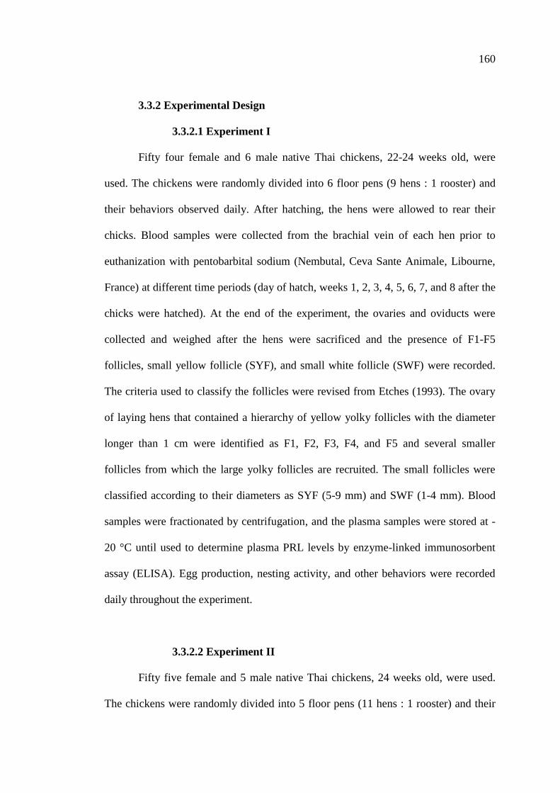
160
3.3.2 Experimental Design
3.3.2.1 Experiment I
Fifty four female and 6 male native Thai chickens, 22-24 weeks old, were
used. The chickens were randomly divided into 6 floor pens (9 hens : 1 rooster) and
their behaviors observed daily. After hatching, the hens were allowed to rear their
chicks. Blood samples were collected from the brachial vein of each hen prior to
euthanization with pentobarbital sodium (Nembutal, Ceva Sante Animale, Libourne,
France) at different time periods (day of hatch, weeks 1, 2, 3, 4, 5, 6, 7, and 8 after the
chicks were hatched). At the end of the experiment, the ovaries and oviducts were
collected and weighed after the hens were sacrificed and the presence of F1-F5
follicles, small yellow follicle (SYF), and small white follicle (SWF) were recorded.
The criteria used to classify the follicles were revised from Etches (1993). The ovary
of laying hens that contained a hierarchy of yellow yolky follicles with the diameter
longer than 1 cm were identified as F1, F2, F3, F4, and F5 and several smaller
follicles from which the large yolky follicles are recruited. The small follicles were
classified according to their diameters as SYF (5-9 mm) and SWF (1-4 mm). Blood
samples were fractionated by centrifugation, and the plasma samples were stored at -
20 °C until used to determine plasma PRL levels by enzyme-linked immunosorbent
assay (ELISA). Egg production, nesting activity, and other behaviors were recorded
daily throughout the experiment.
3.3.2.2 Experiment II
Fifty five female and 5 male native Thai chickens, 24 weeks old, were used.
The chickens were randomly divided into 5 floor pens (11 hens : 1 rooster) and their
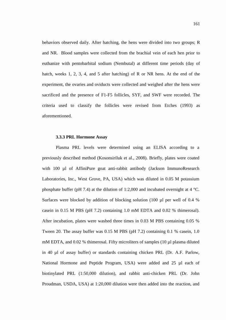
161
behaviors observed daily. After hatching, the hens were divided into two groups; R
and NR. Blood samples were collected from the brachial vein of each hen prior to
euthanize with pentobarbital sodium (Nembutal) at different time periods (day of
hatch, weeks 1, 2, 3, 4, and 5 after hatching) of R or NR hens. At the end of the
experiment, the ovaries and oviducts were collected and weighed after the hens were
sacrificed and the presence of F1-F5 follicles, SYF, and SWF were recorded. The
criteria used to classify the follicles were revised from Etches (1993) as
aforementioned.
3.3.3 PRL Hormone Assay
Plasma PRL levels were determined using an ELISA according to a
previously described method (Kosonsiriluk et al., 2008). Briefly, plates were coated
with 100 µl of AffiniPure goat anti-rabbit antibody (Jackson ImmunoResearch
Laboratories, Inc., West Grove, PA, USA) which was diluted in 0.05 M potassium
phosphate buffer (pH 7.4) at the dilution of 1:2,000 and incubated overnight at 4 °C.
Surfaces were blocked by addition of blocking solution (100 µl per well of 0.4 %
casein in 0.15 M PBS (pH 7.2) containing 1.0 mM EDTA and 0.02 % thimerosal).
After incubation, plates were washed three times in 0.03 M PBS containing 0.05 %
Tween 20. The assay buffer was 0.15 M PBS (pH 7.2) containing 0.1 % casein, 1.0
mM EDTA, and 0.02 % thimerosal. Fifty microliters of samples (10 µl plasma diluted
in 40 µl of assay buffer) or standards containing chicken PRL (Dr. A.F. Parlow,
National Hormone and Peptide Program, USA) were added and 25 µl each of
biotinylated PRL (1:50,000 dilution), and rabbit anti-chicken PRL (Dr. John
Proudman, USDA, USA) at 1:20,000 dilution were then added into the reaction, and
162
incubated overnight at 4 °C. After incubation, plates were washed and 0.1 ml of
streptavidin horseradish peroxidase (1:5,000 dilution) were added. After 2 h at room
temperature, plates were washed and 0.1 ml ABTS reagent (0.04 % 2,2’-azino-bis-3-
ethylbenzthizoline-6-sulfonic acid and 0.015 % H2O2 in 0.1 M citrate phosphate
buffer, pH 4) were added. After 1 h incubation at room temperature, the color
reaction was measured at 405 nm in a Tecan Sunrise ELISA reader (Tecan Group
Ltd., Mannedorf, Switzerland). The assay of plasma PRL levels in native Thai
chickens was validated. Pooled plasma samples of native Thai chickens produced a
dose-response curve that paralleled a chicken PRL standard curve. Plasma samples
were determined in duplicate within a single assay. The intraassay coefficient of
variation was 5.0 %, and the sensitivity was 3.9 ng/ml.
3.3.4 Statistical Analysis
Significant differences in plasma PRL levels and ovary and oviduct weights
(means ± SEM) according to each treatment group were compared utilizing one-way
analysis of variance (ANOVA). Significant differences between treatment groups
were computed utilizing Tukey’s HSD Test. P<0.05 was considered as statistically
significant. All statistical tests were analyzed employing the SPSS for Windows
Software (version 13.0, SPSS Inc., Chicago, IL, USA).
163
3.4 Results
3.4.1 Experiment I
The profiles of plasma PRL levels during the eight-week rearing period are
shown in Figure 3.1 and Table 3.1. Plasma PRL concentrations remained at high
levels on the day the chicks were hatched (P<0.05; 115.36 ± 35.56 ng/ml), then
rapidly decreased after the first week of hatching (42.23 ± 6.51 ng/ml) and remained
at low levels throughout the eight-week rearing period.
The reproductive characteristics of hens rearing chicks at different time
periods were also recorded. The presence of F1-F5 follicles, SYF, and SWF (Figure
3.2) were observed. During the rearing period, the presences of F1-F5 follicles, SYF,
and SWF at different time are shown in Table 3.2. The results showed that F1-F5
follicles were not observed in hens that were rearing their chicks for 1 to 5 weeks. A
few of the hens rearing chicks for 5 weeks exhibited the presence of SYF. However,
the presence of SWF was found in all of hens rearing chicks. The hens showed the
presence of F1-F5 follicles between weeks 6 to 8 of the rearing period. A number of
hens started to lay, the new laying cycle, in week 8 of the rearing period.
The ovaries of hens rearing chicks are shown in Figure 3.3. The ovary weights
of hens rearing chicks are shown in Figure 3.4 and Table 3.3. The ovary weights were
significantly decreased since the chicks were hatched and remained low until week 4
of the rearing period. The ovary weights of hens rearing chicks were significantly
increased and reached the highest weight at week 8 of the rearing period (P<0.05;
24.33 ± 9.72 g).
The oviducts of hens rearing chicks are shown in Figure 3.5. Similarly, the
oviduct weights of hens rearing chicks are shown in Figure 3.6 and Table 3.4. The
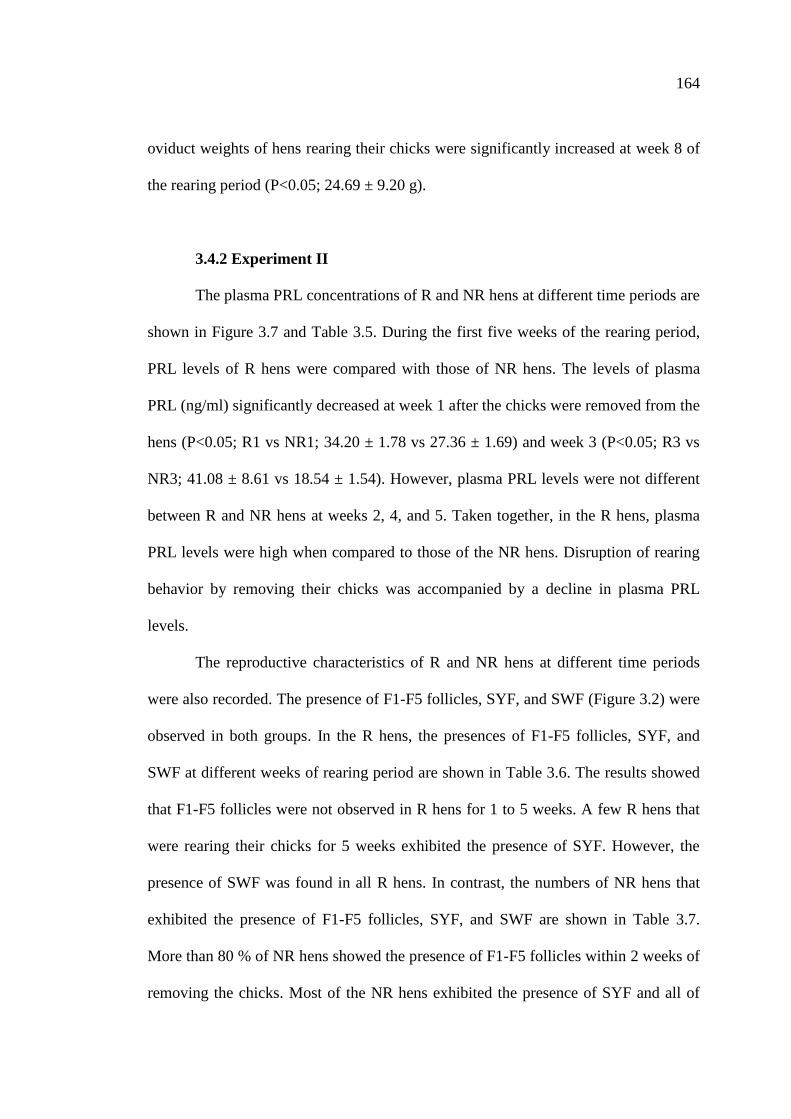
164
oviduct weights of hens rearing their chicks were significantly increased at week 8 of
the rearing period (P<0.05; 24.69 ± 9.20 g).
3.4.2 Experiment II
The plasma PRL concentrations of R and NR hens at different time periods are
shown in Figure 3.7 and Table 3.5. During the first five weeks of the rearing period,
PRL levels of R hens were compared with those of NR hens. The levels of plasma
PRL (ng/ml) significantly decreased at week 1 after the chicks were removed from the
hens (P<0.05; R1 vs NR1; 34.20 ± 1.78 vs 27.36 ± 1.69) and week 3 (P<0.05; R3 vs
NR3; 41.08 ± 8.61 vs 18.54 ± 1.54). However, plasma PRL levels were not different
between R and NR hens at weeks 2, 4, and 5. Taken together, in the R hens, plasma
PRL levels were high when compared to those of the NR hens. Disruption of rearing
behavior by removing their chicks was accompanied by a decline in plasma PRL
levels.
The reproductive characteristics of R and NR hens at different time periods
were also recorded. The presence of F1-F5 follicles, SYF, and SWF (Figure 3.2) were
observed in both groups. In the R hens, the presences of F1-F5 follicles, SYF, and
SWF at different weeks of rearing period are shown in Table 3.6. The results showed
that F1-F5 follicles were not observed in R hens for 1 to 5 weeks. A few R hens that
were rearing their chicks for 5 weeks exhibited the presence of SYF. However, the
presence of SWF was found in all R hens. In contrast, the numbers of NR hens that
exhibited the presence of F1-F5 follicles, SYF, and SWF are shown in Table 3.7.
More than 80 % of NR hens showed the presence of F1-F5 follicles within 2 weeks of
removing the chicks. Most of the NR hens exhibited the presence of SYF and all of
165
them exhibited the presence of SWF. A number of NR hens started to lay, the new
laying cycle, in week 3 after removal the chicks. Moreover, at weeks 3 to 5 of
separation the chicks from hens, 100 % of NR hens returned to lay (Table 3.7).
The ovaries of R and NR hens are shown in Figure 3.8. The ovary weights of
R and NR hens are shown in Figure 3.9 and Table 3.8. When the hens were rearing
their young, the ovary weights showed no increase throughout five weeks of the
rearing period. In contrast, in the NR group, the ovary weights displayed a significant
increase at weeks 2, 3, and 4 (P<0.05), and reached the highest weight at the third
week after removing the chicks (P<0.05; 47.32 ± 6.77 g).
The oviducts of R and NR hens are shown in Figure 3.10. The oviduct weights
of R and NR hens are shown in Figure 3.11 and Table 3.9. Similarly, the oviduct
weights of R hens showed no increase during the first five weeks of the rearing
period. Whereas, in hens that had their chicks removed after hatching, the oviduct
weights were significantly increased at week 2 after removing the chicks (P<0.05) and
reached the highest weight at week 4 (P<0.05; 55.66 ± 3.54 g). When compared
between both groups, both ovary and oviduct weights of the NR hens were
significantly increased by week 2 after removing the chicks and were higher than
those of R hens throughout week 1 to week 5 of the observation period.
166
Figure 3.1 Changes in plasma PRL concentrations during eight weeks of the rearing
period (n=6) of native Thai hens. Values are presented as mean ± SEM. Significant
differences between means in each group at different time points are denoted by
different letters (P<0.05).
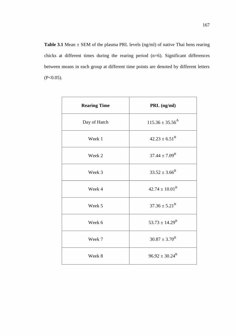
167
Table 3.1 Mean ± SEM of the plasma PRL levels (ng/ml) of native Thai hens rearing
chicks at different times during the rearing period (n=6). Significant differences
between means in each group at different time points are denoted by different letters
(P<0.05).
Rearing Time
PRL (ng/ml)
Day of Hatch
115.36 ± 35.56
A
Week 1
42.23 ± 6.51B
Week 2
37.44 ± 7.09B
Week 3
33.52 ± 3.66B
Week 4
42.74 ± 10.01B
Week 5
37.36 ± 5.21B
Week 6
53.73 ± 14.29B
Week 7
30.87 ± 3.70B
Week 8
96.92 ± 30.24B
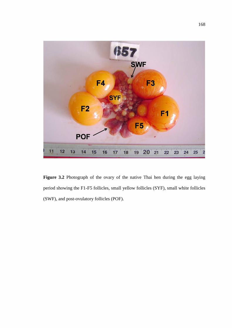
168
Figure 3.2 Photograph of the ovary of the native Thai hen during the egg laying
period showing the F1-F5 follicles, small yellow follicles (SYF), small white follicles
(SWF), and post-ovulatory follicles (POF).
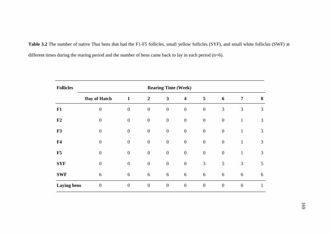
169
Table 3.2 The number of native Thai hens that had the F1-F5 follicles, small yellow follicles (SYF), and small white follicles (SWF) at
different times during the rearing period and the number of hens came back to lay in each period (n=6).
Follicles Rearing Time (Week)
Day of Hatch 1 2 3 4 5 6 7 8
F1 0 0 0 0 0 0 3 3 3
F2 0 0 0 0 0 0 0 1 3
F3 0 0 0 0 0 0 0 1 3
F4 0 0 0 0 0 0 0 1 3
F5 0 0 0 0 0 0 0 1 3
SYF 0 0 0 0 0 3 5 3 5
SWF 6 6 6 6 6 6 6 6 6
Laying hens 0 0 0 0 0 0 0 0 1
169

170
Figure 3.3 Photographs of the ovary of native Thai hens that rearing chicks at day of
hatch (HD) and different times during the rearing period.

171
Figure 3.4 Changes in the ovary weight of native Thai hens that rearing chicks.
Values are presented as means ± SEM (n=6). Significant differences between means
in each group at different time points are denoted by different letters (P<0.05).
172
Table 3.3 Mean ± SEM of the ovary weight (g) of native Thai hens that rearing
chicks at different times during the rearing period (n=6). Significant differences
between means in each group at different time points are denoted by different letters
(P<0.05).
Rearing Time Ovary Weight (g)
Day of Hatch 2.01 ± 0.27A
Week 1 1.73 ± 0.26A
Week 2 1.76 ± 0.18A
Week 3 1.39 ± 0.16A
Week 4 1.68 ± 0.18A
Week 5 2.52 ± 0.67A
Week 6 4.44 ± 1.62A
Week 7 17.54 ± 10.06AB
Week 8 24.33 ± 9.72B
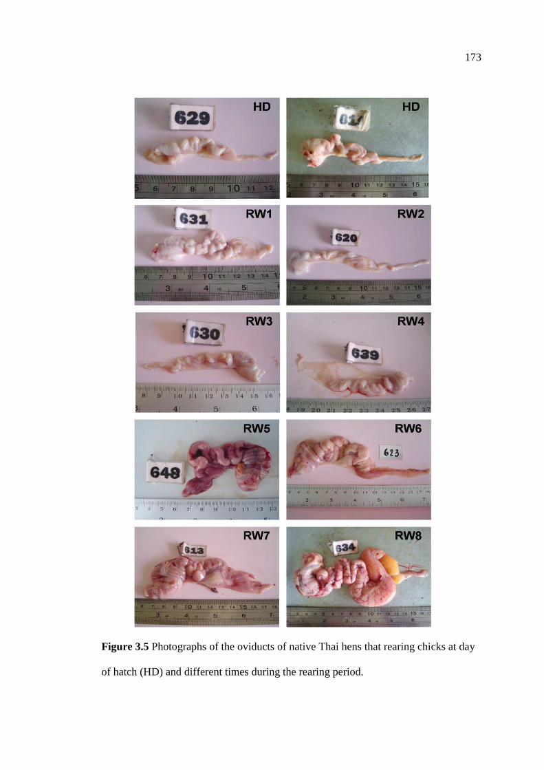
173
Figure 3.5 Photographs of the oviducts of native Thai hens that rearing chicks at day
of hatch (HD) and different times during the rearing period.

174
Figure 3.6 Changes in the oviduct weight of native Thai hens that rearing chicks.
Values are presented as means ± SEM (n=6). Significant differences between means
in each group at different time points are denoted by different letters (P<0.05).

175
Table 3.4 Mean ± SEM of the oviduct weight (g) of native Thai hens that rearing
chicks at different times during the rearing period (n=6). Significant differences
between means in each group at different time points are denoted by different letters
(P<0.05).
Rearing Time Oviduct Weight (g)
Day of Hatch 3.89 ± 0.49AB
Week 1 3.51 ± 0.55A
Week 2 2.81 ± 0.34A
Week 3 2.64 ± 0.26A
Week 4 2.41 ± 0.18A
Week 5 5.62 ± 1.81AB
Week 6 9.62 ± 3.83AB
Week 7 19.58 ± 8.97B
Week 8 24.69 ± 9.20B

176
Figure 3.7 Changes in plasma PRL concentrations of rearing (R; n=6) and non-
rearing (NR; n=6) native Thai hens. Values are presented as mean ± SEM. Significant
differences between means in each group at different time points are denoted by
different letters (P<0.05) and *P<0.05 for a comparison between group at a given
time point.
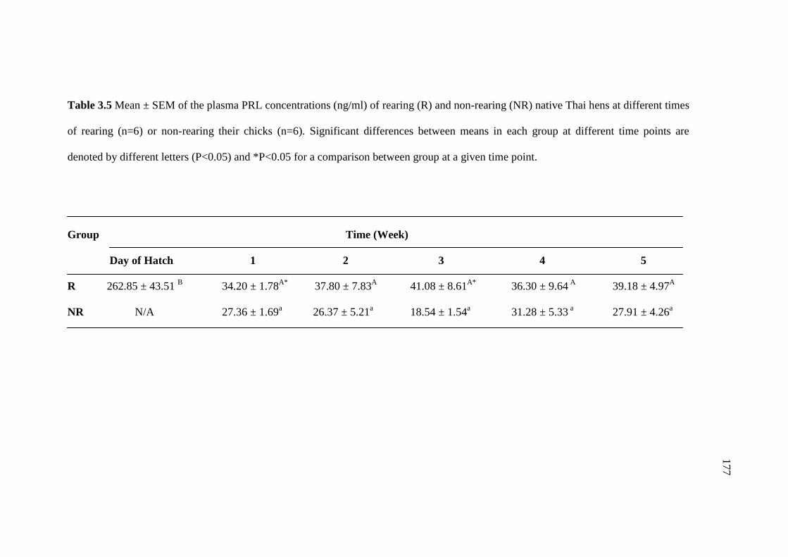
177
Table 3.5 Mean ± SEM of the plasma PRL concentrations (ng/ml) of rearing (R) and non-rearing (NR) native Thai hens at different times
of rearing (n=6) or non-rearing their chicks (n=6). Significant differences between means in each group at different time points are
denoted by different letters (P<0.05) and *P<0.05 for a comparison between group at a given time point.
Group Time (Week)
Day of Hatch 1 2 3 4 5
R 262.85 ± 43.51 B 34.20 ± 1.78
A* 37.80 ± 7.83
A 41.08 ± 8.61
A* 36.30 ± 9.64
A 39.18 ± 4.97
A
NR N/A 27.36 ± 1.69a 26.37 ± 5.21
a 18.54 ± 1.54
a 31.28 ± 5.33
a 27.91 ± 4.26
a
177
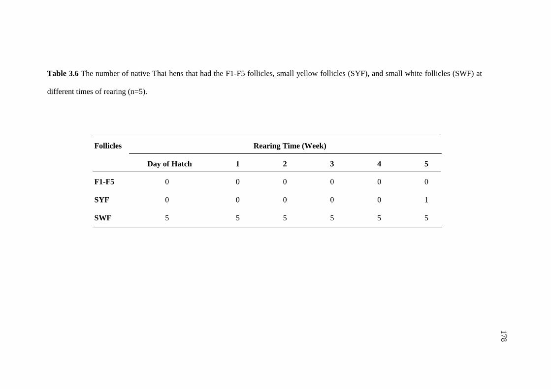
178
Table 3.6 The number of native Thai hens that had the F1-F5 follicles, small yellow follicles (SYF), and small white follicles (SWF) at
different times of rearing (n=5).
Follicles Rearing Time (Week)
Day of Hatch 1 2 3 4 5
F1-F5 0 0 0 0 0 0
SYF 0 0 0 0 0 1
SWF 5 5 5 5 5 5
178
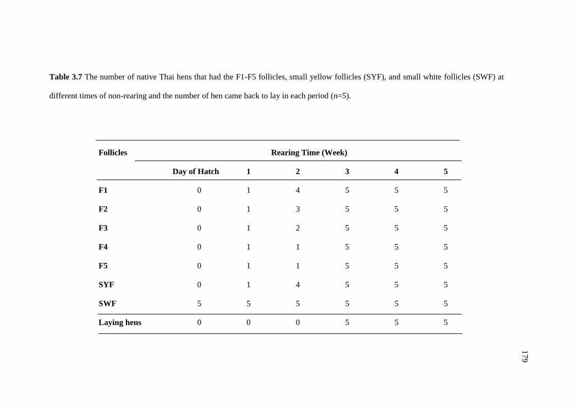
179
Table 3.7 The number of native Thai hens that had the F1-F5 follicles, small yellow follicles (SYF), and small white follicles (SWF) at
different times of non-rearing and the number of hen came back to lay in each period (n=5).
Follicles Rearing Time (Week)
Day of Hatch 1 2 3 4 5
F1 0 1 4 5 5 5
F2 0 1 3 5 5 5
F3 0 1 2 5 5 5
F4 0 1 1 5 5 5
F5 0 1 1 5 5 5
SYF 0 1 4 5 5 5
SWF 5 5 5 5 5 5
Laying hens 0 0 0 5 5 5
179

180
Figure 3.8 Photographs of the ovary of rearing (R) and non-rearing (NR) native Thai
hens at day of hatch (HD) and different times of rearing or non-rearing their chicks.

181
Figure 3.8 Photographs of the ovary of rearing (R) and non-rearing (NR) native Thai
hens at different times of rearing or non-rearing their chicks (continued).
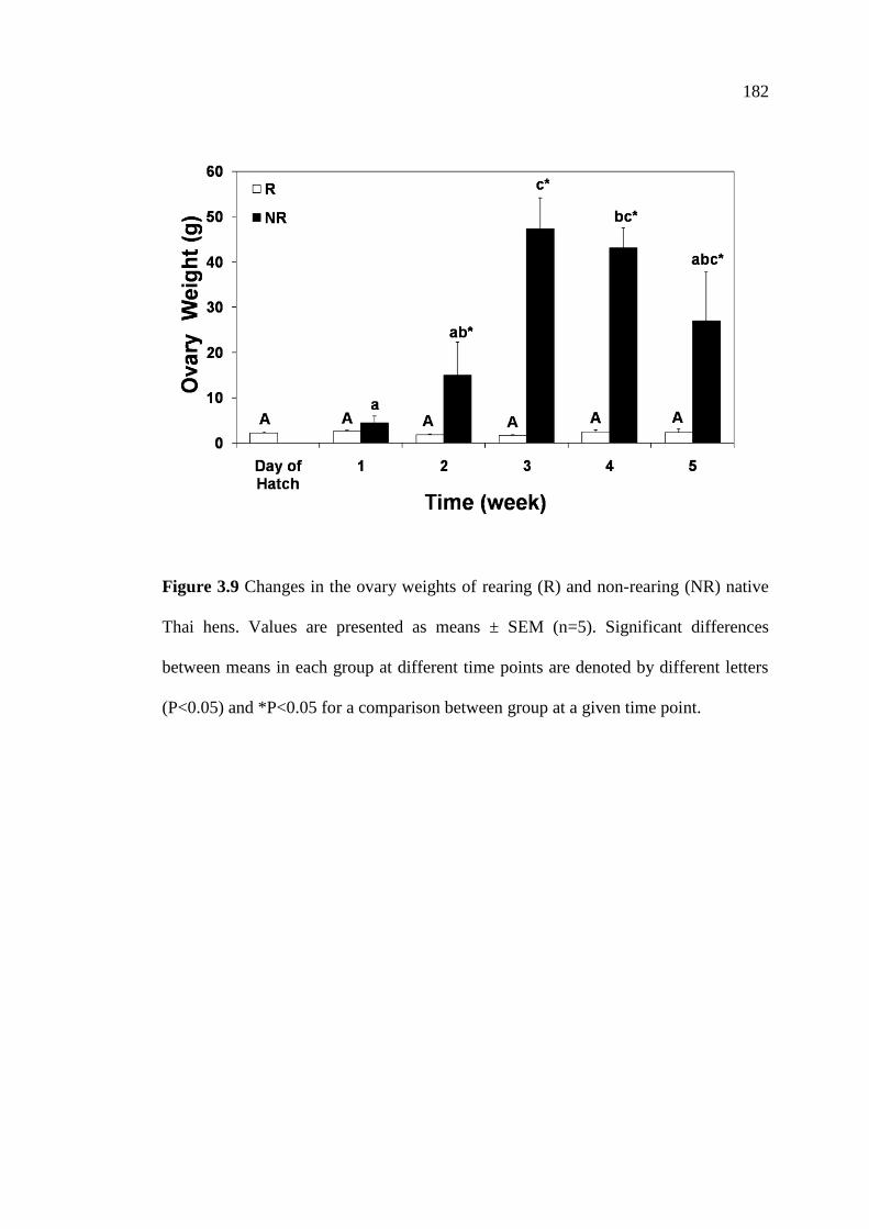
182
Figure 3.9 Changes in the ovary weights of rearing (R) and non-rearing (NR) native
Thai hens. Values are presented as means ± SEM (n=5). Significant differences
between means in each group at different time points are denoted by different letters
(P<0.05) and *P<0.05 for a comparison between group at a given time point.

183
Table 3.8 Mean ± SEM of the ovary weight (g) of rearing (R) and non-rearing (NR) native Thai hens at different times of rearing or non-
rearing their chicks (n=5). Significant differences between means in each group at different time points are denoted by different letters
(P<0.05) and *P<0.05 for a comparison between group at a given time point.
Group Rearing Time (Week)
Day of Hatch 1 2 3 4 5
R 2.25 ± 0.23A 2.55 ± 0.21
A 1.85 ± 0.13
A 1.61 ± 0.18
A 2.45 ± 0.46
A 2.27 ± 0.73
A
NR N/A 4.46 ± 1.49a 14.95 ± 7.31
ab* 47.32 ± 6.77
c* 43.12 ± 4.30
bc* 26.99 ± 10.79
abc*
183
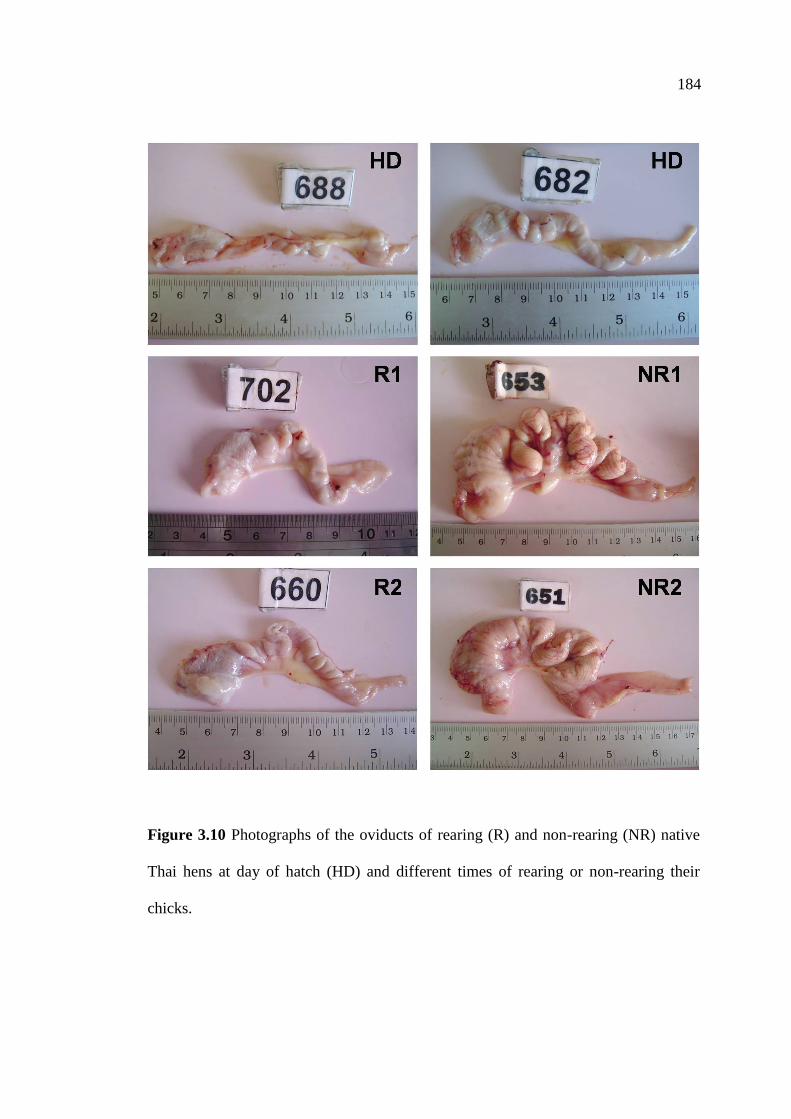
183
Figure 3.10 Photographs of the oviducts of rearing (R) and non-rearing (NR) native
Thai hens at day of hatch (HD) and different times of rearing or non-rearing their
chicks.
184

184
Figure 3.10 Photographs of the oviducts of rearing (R) and non-rearing (NR) native
Thai hens at different times of rearing or non-rearing their chicks (continued).
185

185
Figure 3.11 Changes in the oviduct weight of rearing (R) and non-rearing (NR) native
Thai hens. Values are presented as means ± SEM (n=5). Significant differences
between means in each group at different time points are denoted by different letters
(P<0.05) and *P<0.05 for a comparison between group at a given time point.
186

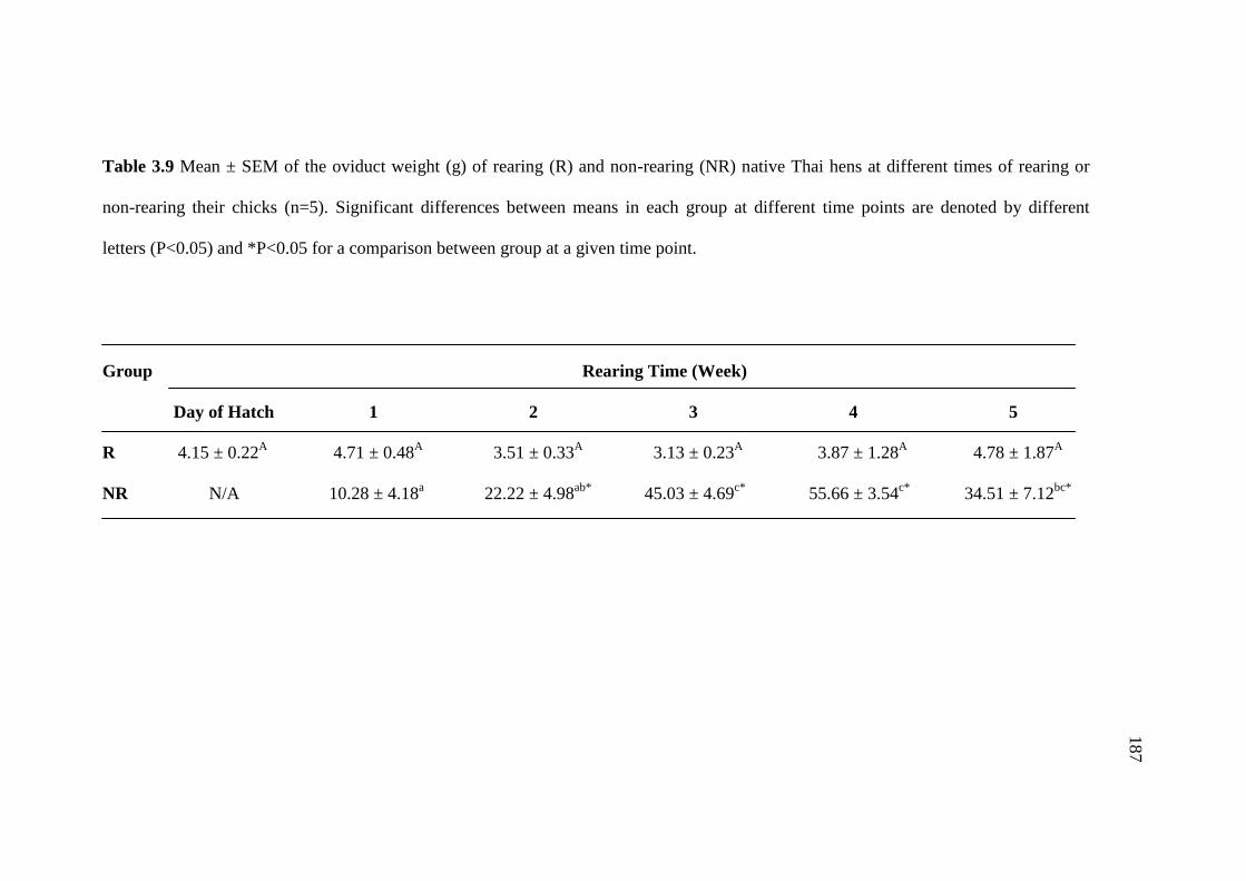
186
Table 3.9 Mean ± SEM of the oviduct weight (g) of rearing (R) and non-rearing (NR) native Thai hens at different times of rearing or
non-rearing their chicks (n=5). Significant differences between means in each group at different time points are denoted by different
letters (P<0.05) and *P<0.05 for a comparison between group at a given time point.
Group Rearing Time (Week)
Day of Hatch 1 2 3 4 5
R 4.15 ± 0.22A 4.71 ± 0.48
A 3.51 ± 0.33
A 3.13 ± 0.23
A 3.87 ± 1.28
A 4.78 ± 1.87
A
NR N/A 10.28 ± 4.18a 22.22 ± 4.98
ab* 45.03 ± 4.69
c* 55.66 ± 3.54
c* 34.51 ± 7.12
bc*
187

186
3.5 Discussion
The results of the present study revealed that plasma PRL level is associated
with rearing behavior in the native Thai chickens. During the eight weeks of the
rearing period, plasma PRL concentrations remained at high levels on the day the
chicks were hatched, and then rapidly decreased after the first week of hatching, and
remained at low levels throughout the rearing period. Comparisons of plasma PRL
levels between R and NR hens during the first five weeks of the rearing period were
elucidated. The results showed that the levels of plasma PRL were decreased in hens
that had their chicks removed and reached the lowest levels by the third week of
separation from the chicks. In the R hens, plasma PRL levels were high when
compared to those of the NR hens. Additionally, disruption of rearing behavior by
removing their chicks increased the ovary and oviduct weights, the presence of
ovarian follicles, and the number of egg laying hens. Therefore, changes in plasma
PRL levels are associated with ovarian and oviduct recrudescence and initiation of a
new laying cycle in the native Thai chickens.
In the present study, after the chicks were hatched, plasma PRL levels
remained at high levels in the R hens. After the first week of rearing period, plasma
PRL levels rapidly decreased and remained at low levels throughout the eight weeks
of the rearing period. These results are consistent with previous findings that plasma
PRL concentrations begin to fall immediately after hatching and continue to decline
for about one week after the day of the chicks were hatched (Sharp et al., 1979; Lea et
al., 1981; Zadworny et al., 1985; 1988; Leboucher et al., 1990; Kuwayama et al.,
1992; Kosonsiriluk et al., 2008). As demonstrated in previous studies of gallinaceous
species, changes in plasma PRL levels were observed across the reproductive cycle.
188
190
Plasma PRL levels were found to be low in non-laying hens, gradually increased in
laying hens, continued to rise and reached the highest levels in incubating hens, and
immediately declined to the basal levels when hens are caring for their young (El
Halawani et al., 1984; Kosonsiriluk et al., 2008).
Circulating PRL levels of R hens were compared with those of NR hens
during the first five weeks of the rearing period. The results showed differences in
plasma PRL levels between R and NR hens. In the R hens, plasma PRL levels were
higher than those of the NR hens, indicating the secretion of PRL in the R hens is
facilitated by the presence of chicks and stimulates maternal behavior which, in turn,
suppresses gonadal activity (Sharp et al., 1988; Richard-Yris et al., 1995).
Furthermore, the hens that were allowed to rear their chicks returned to lay later than
hens that were not allowed to rear the chicks, suggesting that physical contact familiar
with auditory, and/or visual stimuli from chicks during the rearing period slows down
the decrease of PRL secretion and inhibits gonadotropins and ovarian steroid
hormones (Richard-Yris and Leboucher, 1987; Richard-Yris et al., 1987a; 1998;
Leboucher et al., 1993). It has been further suggested that the presence of chicks had
no effect on PRL secretion but tends to maintain its level (Sharp et al., 1988; Richard-
Yris et al., 1995). Several studies provide evidence that PRL is not released at an
increased rate while hens are caring for their young, but it is involved in the initiation
and/or maintenance of maternal behavior and has an antigonadal role (Sharp et al.,
1979; 1988; Bedrak et al., 1981; Kuwayama et al., 1992; Leboucher et al., 1993;
Richard-Yris et al., 1995; Boos et al., 2007; Kosonsiriluk et al., 2008). In galliform
birds, PRL is involved in maternal behaviors. The presence of chicks induces the
emergence of specific maternal behaviors in many avian species (Maier, 1963;
188 189

191
Richard-Yris et al., 1983; Richard-Yris and Leboucher, 1987; Leboucher et al., 1990;
1993; Wang and Buntin, 1999). Replacement of the eggs by chicks induces maternal
behaviors in incubating, non-incubating, and ovariectomized hens (Richard-Yris et
al., 1987a; 1995; 1998; Leboucher et al., 1990; 1993; Lea et al., 1996). Physical
contact with newly hatched chicks during brooding bouts slows down the decrease of
PRL secretion and inhibits LH and estradiol release in maternal hens (Leboucher et
al., 1993). Thus, it is possible that coexistence of newly hatched chicks may suppress
LH secretion of the hen in the natural breeding cycle (Kuwayama et al., 1992).
Furthermore, it is very well demonstrated that the presence of chicks inhibits the
pituitary-ovarian axis (Richard-Yris et al., 1987a; Sharp et al., 1988) and non-
brooders (Richard-Yris et al., 1983; 1987b). In some avian species, stimuli from the
young or from parent-young interactions may promote or sustain elevated circulating
PRL levels (Buntin, 1996). It has been suggested that a definite threshold in
circulating PRL levels is necessary to promote and/or maintain post-hatching
maternal behavior in precocial birds (Boos et al., 2007). Indeed, depending on the
species, either a sharp decline or a slow decrease of PRL concentrations after
hatching has been reported (Goldsmith and Williams, 1980; Dittami, 1981; Oring et
al., 1986; 1988; Hall, 1987; Opel and Proudman, 1989; Richard-Yris et al., 1995;
1998; Setiawan et al., 2006). In those species, although PRL concentration during
rearing period is lower than that of incubation period, the level remains higher than
that of non-rearing ones (Boos et al., 2007), suggesting that PRL is likely involved in
parental care after hatching (Criscuolo et al., 2002).
The presence of F1-F5 follicles, SYF, and SWF indicates the development of
the reproductive system (Etches, 1993). In the present study, the R hens stopped
190
192
showing maternal behavior between 7 and 8 weeks after the chicks were hatched. This
corresponded to the time when the hens came back to lay again after caring for their
young. Other studies provide evidence demonstrating that when the chicks grow older
and become fledged, brooding behavior progressively declines, leading to a drop in
PRL levels (Richard-Yris et al., 1987a; 1989; 1998; Sharp et al., 1988; Boos et al.,
2007). In good agreement with these results are findings in bantams, in which the
hens stopped broody behavior between 4 to 10 weeks after the chicks were hatched,
corresponding to the time when circulating LH levels had increased to the levels
found in laying hens (Sharp et al., 1979). Whereas, the NR hens returned to lay at the
third week after removal of the chicks. Similarly in bantam hens, Gifujidori hens and
native Thai hens, the effects of removal of chicks from the care of hens induced
ovarian recrudescence and egg laying between week 3 and 4 of separation from the
chicks (Sharp et al., 1979; Kuwayama et al., 1992; Kosonsiriluk et al., 2008). On the
other hand, rearing chick hens and subsequent hens that were removed from their
chicks exhibit an abrupt increase in plasma LH levels concurrent with the decrease of
plasma PRL levels (Leboucher et al., 1990). It has been reported that circulating LH
and estradiol levels gradually increased after hatching or when chicks were present
and reached peak levels immediately after removal of the chicks (Sharp et al., 1979;
Richard-Yris et al., 1998; Kuwayama et al., 1992). In addition, the establishment of
maternal behavior was accompanied by a significant long-term fall in the plasma
levels of LH and gonadal steroids (Bedrak et al., 1981; Richard-Yris et al., 1988).
Interestingly, LH secretion may also be inhibited by increased plasma PRL levels, and
high levels of PRL have been suggested to be involved in the initiation and/or
maintenance of broodiness and have an antigonaldal role in birds (Bedrak et al., 1981;
191
193
Sharp et al., 1988; Zadworny et al., 1988). Further evidence in support of these results
indicates that immunization against PRL slows down ovarian follicular development
and reduces hen egg laying performance, suggesting that PRL plays a stimulatory role
in ovarian follicular development in chicken hens (Li et al., 2011).
In this present study, after removing the chicks from the hens, plasma PRL
levels were decreased in the NR hens when compared with those of R hens and
reached the lowest levels by the third week, which corresponded with an increase in
ovary and oviduct weights at the time when hens returned to lay. These findings
corresponded with the decreases in the number of VIP-ir neurons in IH-IN of nest-
deprived native Thai hens, which paralleled the decreases in plasma PRL levels, and
they came back into lay within 18 days of nest deprivation (Prakobsaeng et al., 2011).
Further evidence supporting the above findings includes the demonstrated changes in
the number of GnRH-I-ir neurons that were observed in the nucleus commissurae
pallii (nCPa) of native Thai hens, which were correlated with the reproductive stages.
The greatest number of GnRH-I-ir neurons was found in laying hens, then slightly
decreased in incubating hens, and declined to the lowest levels during the rearing
stage. Furthermore, studies in nest-deprived native Thai hens showed a significant
increase in the number of GnRH-I-ir neurons in the nCPa (Sartsoongnoen et al.,
2012), while VIP-ir neurons decreased and ovary and oviduct weights increased as
hens started the new laying cycle (Prakobsaeng et al., 2011). Moreover, removal of
the hens from their eggs resulted in an increase in LH secretion and hypothalamic
contents of cGnRH-I mRNA (Dunn et al., 1996). These findings, taken together with
the results of the present study, clearly demonstrate that PRL is involved in the
192
194
initiation and maintenance of rearing behavior. In addition, changes in this pituitary
hormone affect reproductive characteristics of the native Thai chickens.
In summary, this present study indicates that plasma PRL levels are associated
with rearing behavior in the native Thai chickens. The external cues such as physical
contact with chicks and the presence of chicks are involved in the maintenance of
plasma PRL levels and rearing behavior. Removal of chicks from the care of hens
disrupts rearing behavior, decreases plasma PRL concentrations, induces ovarian
recrudescence, increases ovary and oviduct weights, and finally induces the hens to
come back to lay in the new cycle.
3.6 References
Bedrak, E., Harvey, S., and Chadwick, A. (1981). Concentrations of pituitary, gonadal
and adrenal hormones in serum of laying and broody white rock hens (Gallus
domesticus). J Endocrinol 89: 197-204.
Bern, H.A., and Nicoll, C.S. (1968). The comparative endocrinology of prolactin.
Recent Prog Horm Res 24: 681-720.
Bluhm, C.K., Phillips, R.E., and Burke, W.H. (1983a). Serum levels of luteinizing
hormone, prolactin, estradiol and progesterone in laying and nonlaying
mallards (Anas platyrhynchos). Biol Reprod 28: 295-305.
Bluhm, C.K., Phillips, R.E., and Burke, W.H. (1983b). Serum levels of luteinizing
hormone (LH), prolactin, estradiol, and progesterone in laying and nonlaying
canvasback ducks (Aythya valisineria). Gen Comp Endocrinol 52: 1-16.
193

195
Boos, M., Zimmer, C., Carriere, A., Robin, J.P., and Petit, O. (2007). Post-hatching
parental care behaviour and hormonal status in a precocial bird. Behav
Processes 76: 206-214.
Breitenbach, R.P., and Meyer, R.K. (1959). Pituitary prolactin levels in laying,
incubating and brooding pheasants (Phasianus colchicus). Proc Soc Exp Biol
Med 101: 16-19.
Bridges, R.S., and Ronsheim, P.M. (1990). Prolactin (PRL) regulation of maternal
behavior in rats: Bromocriptine treatment delays and PRL promotes the rapid
onset of behavior. Endocrinology 126: 837-848.
Buntin, J.D. (1996). Neural and hormonal control of parental behavior in birds. Adv
Stud Behav 25: 161-213.
Buntin, J.D., Becker, G.M., and Ruzycki, E. (1991). Facilitation of parental behavior
in ring doves by systemic or intracranial injections of prolactin. Horm Behav
25: 424-444.
Burke, W.H., and Dennison, P.T. (1980). Prolactin and luteinizing hormone levels in
female turkeys (Meleagris gallopavo) during a photoinduced reproductive
cycle and broodiness. Gen Comp Endocrinol 41: 92-100.
Camper, P.M., and Burke, W.H. (1977). The effects of prolactin on the gonadotropin
induced rise in serum estradiol and progesterone of the laying turkey. Gen
Comp Endocrinol 32: 72-77.
Chaiseha, Y., and El Halawani, M.E. (1999). Expression of vasoactive intestinal
peptide/peptide histidine isoleucine in several hypothalamic areas during the
turkey reproductive cycle: Relationship to prolactin secretion.
Neuroendocrinology 70: 402-412.
194
196
Chaiseha, Y., and El Halawani, M.E. (2005). Neuroendocrinology of the female
turkey reproductive cycle. J Poult Sci 42: 87-100.
Chaiseha, Y., Tong, Z., Youngren, O.M., and El Halawani, M.E. (1998).
Transcriptional changes in hypothalamic vasoactive intestinal peptide during a
photo-induced reproductive cycle in the turkey. J Mol Endocrinol 21: 267-
275.
Chaiseha, Y., Youngren, O.M., Al-Zailaie, K.A., and El Halawani, M.E. (2003).
Expression of D1 and D2 dopamine receptors in the hypothalamus and
pituitary during the turkey reproductive cycle: Colocalization with vasoactive
intestinal peptide. Neuroendocrinology 77: 105-118.
Chaiseha, Y., Youngren, O.M., and El Halawani, M.E. (1997). Dopamine receptors
influence vasoactive intestinal peptide release from turkey hypothalamic
explants. Neuroendocrinology 65: 423-429.
Chaiseha, Y., Youngren, O.M., and El Halawani, M.E. (2004). Expression of
vasoactive intestinal peptide receptor mRNA in the hypothalamus and
pituitary throughout the turkey reproductive cycle. Biol Reprod 70: 593-599.
Criscuolo, F., Chastel, O., Gabrielsen, G.W., Lacroix, A., and Le Maho, Y. (2002).
Factors affecting plasma concentrations of prolactin in the common eider
Somateria mollissima. Gen Comp Endocrinol 125: 399-409.
Curlewis, J.D. (1992). Seasonal prolactin secretion and its role in seasonal
reproduction: A review. Reprod Fertil Dev 4: 1-23.
Dawson, A., and Goldsmith, A.R. (1982). Prolactin and gonadotrophin secretion in
wild starlings (Sturnus vulgaris) during the annual cycle and in relation to
nesting, incubation, and rearing young. Gen Comp Endocrinol 48: 213-221.
195
197
Dittami, J.P. (1981). Seasonal changes in the behavior and plasma titers of various
hormones in barheaded geese, Anser indicus. Z Tierpsychol 55: 289-324.
Dunn, I.C., Beattie, K.K., Maney, D., Sang, H.M., Talbot, R.T., Wilson, P.W., and
Sharp, P.J. (1996). Regulation of chicken gonadotropin-releasing hormone-I
mRNA in incubating, nest-deprived and laying bantam hens.
Neuroendocrinology 63: 504-513.
El Halawani, M.E., Burke, W.H., Millam, J.R., Fehrer, S.C., and Hargis, B.M. (1984).
Regulation of prolactin and its role in gallinaceous bird reproduction. J Exp
Zool 232: 521-529.
El Halawani, M.E., Fehrer, S.C., Hargis, B.M., and Porter, T.E. (1988). Incubation
behavior in the domestic turkey: Physiological correlates. CRC Crit Rev
Poult Biol 1: 285-314.
El Halawani, M.E., Kang, S.W., Chaiseha, Y., Rozenboim, I., Millam, J.R., and
Youngren, O.M. (2004). Dopaminergic regulation of reproduction in the
female turkey. In The 8th
International Symposium on Avian
Endocrinology, Arizona, USA.
El Halawani, M.E., Youngren, O.M., and Chaiseha, Y. (2001). Neuroendocrinology
of PRL regulation in the domestic turkey. In Avian Endocrinology, pp 233-
244. Eds. Dawson, A., and Chaturvedi, C.M. Narosa Publishing House, New
Delhi, India.
El Halawani, M.E., Youngren, O.M., and Pitts, G.R. (1997). Vasoactive intestinal
peptide as the avian prolactin-releasing factor. In Perspectives in Avian
Endocrinology, pp 403-416. Eds. Harvey, S., and Etches, R.J. Journal of
Endocrinology, Bristol, UK.
196

198
Etches, R.J. (1993). Reproduction in poultry. In Reproduction in Domesticated
Animals. Ed. King, G.J. Elsevier, Huddersfield, UK.
Freeman, M.E., Kanyicska, B., Lerant, A., and Nagy, G. (2000). Prolactin: Structure,
function, and regulation of secretion. Physiol Rev 80: 1523-1631.
Goldsmith, A.R. (1982a). The Australian black swan (Cygnus atratus): Prolactin and
gonadotrophin secretion during breeding including incubation. Gen Comp
Endocrinol 46: 458-462.
Goldsmith, A.R. (1982b). Plasma concentrations of prolactin during incubation and
parental feeding throughout repeated breeding cycles in canaries (Serinus
canarius). J Endocrinol 94: 51-59.
Goldsmith, A.R. (1985). Prolactin in avian reproduction: Incubation and the control
of seasonal breeding. In Prolactin: Basic and Clinical Correlates, pp 411-
425. Eds. MacLeod, R.M., Scapagini, U., and Thorner, M. Springer, Padua.
Goldsmith, A.R. (1991). Prolactin and avian reproductive strategies. Acta
Congressus Internationalis Ornithologici XX 4: 2063-2071.
Goldsmith, A.R., and Williams, D.M. (1980). Incubation in mallards (Anas
platyrhynchos): Changes in plasma levels of prolactin and luteinizing
hormone. J Endocrinol 86: 371-379.
Guemene, D., and Williams, J.B. (1992). Changes in endocrinological parameters and
production performances in turkey hens (Meleagris gallopavo) submitted to
different broody management programs. Theriogenology 38: 1115-1129.
Hall, M.R. (1987). External stimuli affecting incubation behavior and prolactin
secretion in the duck (Anas platyrhynchos). Horm Behav 21: 269-287.
197

199
Hall, M.R., and Goldsmith, A.R. (1983). Factors affecting prolactin secretion during
breeding and incubation in the domestic duck (Anas platyrhynchos). Gen
Comp Endocrinol 49: 270-276.
Hargis, B.M., El Halawani, M.E., and Porter, T.E. (1987). Ovarian regression and
incubation behavior induced by ovine prolactin (oPrl) in Nicholas turkey hens.
Poult Sci 66 (Suppl. 1): 111.
Harris, J., Stanford, P.M., Oakes, S.R., and Ormandy, C.J. (2004). Prolactin and the
prolactin receptor: New targets of an old hormone. Ann Med 36: 414-425.
Hoshino, S., and Wakita, M. (1989). Increased synthesis of prolactin and growth
hormone during incubation in the pituitary of broody Nagoya hens. Horm
Metab Res 21: 480-482.
Huang, Y.M., Shi, Z.D., Liu, Z., Liu, Y., and Li, X.W. (2008). Endocrine regulations
of reproductive seasonality, follicular development and incubation in Magang
geese. Anim Reprod Sci 104: 344-358.
Janik, D.S., and Buntin, J.D. (1985). Behavioural and physiological effects of
prolactin in incubating ring doves. J Endocrinol 105: 201-209.
Knapp, T.R., Fehrer, S.C., Silsby, J.L., Porter, T.E., Behnke, E.J., and El Halawani,
M.E. (1988). Gonodal steroid modulation of basal and vasoactive intestinal
peptide-stimulated prolactin release by turkey anterior pituitary cells. Gen
Comp Endocrinol 76: 1141-1144.
Kosonsiriluk, S. (2007). Biological studies of the reproductive cycle and the effects
of photoperiod upon the reproductive system in the female native Thai
chicken. Ph.D. Dissertation, Suranaree University of Technology, Nakhon
Ratchasima, Thailand.
198
200
Kosonsiriluk, S., Sartsoongnoen, N., Chaiyachet, O-A., Prakobsaeng, N., Songserm,
T., Rozenboim, I., El Halawani, M.E., and Chaiseha, Y. (2008). Vasoactive
intestinal peptide and its role in continuous and seasonal reproduction in birds.
Gen Comp Endocrinol 159: 88-97.
Kragt, C.L., and Meites, J. (1965). Stimulation of pigeon pituitary prolactin release by
pigeon hypothalamic extract in vitro. Endocrinology 76: 1169-1176.
Kuwayama, T., Shimada, K., Saito, N., Ohkubo, T., Sato, K., Wada, M., and Ichinoe,
K. (1992). Effects of removal of chicks from hens on concentrations of
prolactin, luteinizing hormone and oestradiol in plasma of brooding Gifujidori
hens. J Reprod Fertil 95: 617-622.
Lea, R.W. (1987). Prolactin and avian incubation: A comparison between galliformes
and columbiformes. Sitta 1: 117-141.
Lea, R.W., Dods, A.S., Sharp, P.J., and Chadwick, A. (1981). The possible role of
prolactin in the regulation of nesting behavior and the secretion of luteinizing
hormone in broody bantams. J Endocrinol 91: 89-97.
Lea, R.W., Richard-Yris, M.A., and Sharp, P.J. (1996). The effect of ovariectomy on
concentrations of plasma prolactin and LH and parental behavior in the
domestic fowl. Gen Comp Endocrinol 101: 115-121.
Lea, R.W., Vowles, D.M., and Dick, H.R. (1986). Factors affecting prolactin
secretion during the breeding cycle of the ring dove (Streptopelia risoria) and
its possible role in incubation. J Endocrinol 110: 447-458.
Leboucher, G., Richard-Yris, M.A., Guemene, D., and Chadwick, A. (1993).
Respective effects of chicks and nest on behavior and hormonal concentrations
of incubating domestic hens. Physiol Behav 54: 135-140.
199
201
Leboucher, G., Richard-Yris, M.A., Williams, J., and Chadwick, A. (1990).
Incubation and maternal behaviour in domestic hens: Influence of the presence
of chicks on circulating luteinising hormone, prolactin and oestradiol and on
behaviour. Br Poult Sci 31: 851-862.
Li, W.L., Liu, Y., Yu, Y.C., Huang, Y.M., Liang, S.D., Shi, Z.D. (2011). Prolactin
plays a stimulatory role in ovarian follicular development and egg laying in
chicken hens. Domest Anim Endocrinol 41: 57-66.
Lormee, H., Jouventin, P., Chastel, O., and Mauget, R. (1999). Endocrine correlates
of parental care in an Antarctic winter breeding seabird, the emperor penguin,
Aptenodytes forsteri. Horm Behav 35: 9-17.
Lormee, H., Jouventin, P., Lacroix, A, Lallemand, J., and Chastel, O. (2000).
Reproductive endocrinology of tropical seabirds: Sex-specific patterns in LH,
steroids, and prolactin secretion in relation to parental care. Gen Comp
Endocrinol 117: 413-426.
Maier, R.A. (1963). Maternal behavior in the domestic hen: The role of physical
contact. J Comp Physiol Psychol 56: 357-361.
Mashaly, M.M., Birrenkott, G.P., EL-Begearmi, M.M., and Wentworth, B.C. (1976).
Plasma LH progesterone concentrations in the turkey hen during the ovulatory
cycle. Poult Sci 55: 1226-1234.
Mauget, R., Jouventin, P., Lacroix, A., and Ishii, S. (1994). Plasma LH and steroid
hormones in king penguin (Aptenodytes patagonicus) during the onset of the
breeding cycle. Gen Comp Endocrinol 93: 36-43.
Mauro, L.J., Elde, R.P., Youngren, O.M., Phillips, R.E., and El Halawani, M.E.
(1989). Alterations in hypothalamic vasoactive intestinal peptide-like
200

202
immunoreactivity are associated with reproduction and prolactin release in the
female turkey (Meleagris gallopavo). Endocrinology 125: 1795-1804.
Myers, S.A., Millam, J.R., and El Halawani, M.E. (1989). Plasma LH and prolactin
levels during the reproductive cycle of the cockatiel (Nymphicus hollandicus).
Gen Comp Endocrinol 73: 85-91.
Opel, H., and Proudman, J.A. (1980). Failure of mammalian prolactin to induce
incubation behavior in chickens and turkeys. Poult Sci 59: 2550-2558.
Opel, H., and Proudman, J.A. (1989). Plasma prolactin levels in incubating turkey
hens during pipping of the eggs and after introduction of poults into the nest.
Biol Reprod 40: 981-987.
Oring, L.W., Fivizzani, A.J., Colwell, M.A., and El Halawani, M.E. (1988).
Hormonal changes associated with natural and manipulated incubation in the
sex-role reversed Wilson's phalarope. Gen Comp Endocrinol 72: 247-256.
Oring, L.W., Fivizzani, A.J., El Halawani, M.E., and Goldsmith, A.R. (1986).
Seasonal changes in prolactin and luteinizing hormone in the polyandrous
spotted sandpiper, Acistis macularia. Gen Comp Endocrinol 62: 394-403.
Porter, T.E., Silsby, J.L., Behnke, E.J., Knapp, T.R., and El Halawani, M.E. (1991).
Ovarian steroid production in vitro during gonadal regression in the turkey. I.
Changes associated with incubation behavior. Biol Reprod 45: 581-586.
Prakobsaeng, N., Sartsoongnoen, N., Kosonsiriluk, S., Chaiyachet, O-A.,
Chokchaloemwong, D., Rozenboim, I., El Halawani, M.E., Porter T.E., and
Chaiseha, Y. (2011). Changes in vasoactive intestinal peptide and tyrosine
hydroxylase immunoreactivity in the brain of nest-deprived native Thai hen.
Gen Comp Endocrinol 171: 189-196.
201
203
Proudman, J.A., and Opel, H. (1981). Turkey prolactin: Validation of a
radioimmunoassay and measurement of changes associated with broodiness.
Biol Reprod 25: 573-580.
Richard-Yris, M.A., and Leboucher, G. (1987). Effects of exposure to chicks on
maternal behaviour in domestic chickens. Bird Behav 7: 31-36.
Richard-Yris, M.A., Chadwick, A., Guemene, D., Grillou-Schuelke, H., and
Leboucher, G. (1995). Influence of the presence of chicks on the ability to
resume incubation behavior in domestic hens (Gallus domesticus). Horm
Behav 29: 425-441.
Richard-Yris, M.A., Garnier, D.H., and Leboucher, G. (1983). Induction of maternal
behavior and some hormonal and physiological correlates in the domestic hen.
Horm Behav 17: 345-355.
Richard-Yris, M.A., Guemene, D., Lea, R.W., Sharp, P.J., Bedecarrats, G., Foraste,
M., and Wauters, A.M. (1998). Behaviour and hormone concentrations in nest
deprived and renesting hens. Br Poult Sci 39: 309-317.
Richard-Yris, M.A., Leboucher, G., Chadwick, A., and Garnier, D.H. (1987a).
Induction of maternal behavior in incubating and non-incubating hens:
Influence of hormones. Physiol Behav 40: 193-199.
Richard-Yris, M.A., Leboucher, G., Chadwick, A., Williams, J., and Schuelke, H.
(1989). Respective influence of nest and chicks on behaviour and hormonal
levels of broody hens. In International Conference Hormone Brain
Behavior, pp 139-140. Liege, Belgium.
202

204
Richard-Yris, M.A., Leboucher, G., Williams, J., and Chadwick, A. (1988). Tendency
to display spontaneous incubation does not affect maternal responsiveness in
the domestic hen. Behav Neural Biol 49: 165-173.
Richard-Yris, M.A., Leboucher, G., Williams, J., and Garnier, D.H. (1987b).
Influence of food restriction and of the presence of chicks on the reproductive
system of the domestic hen. Br Poult Sci 28: 251-260.
Sartsoongnoen, N., Kosonsiriluk, S., Kang, S.W., Millam, J.R., El Halawani, M.E.,
and Chaiseha, Y. (2006). Distribution of cGnRH-I immunoreactive neurons
and fibers in the brain of native Thai chicken (Gallus domesticus). Poult Sci
85 (Suppl 1): 45.
Sartsoongnoen, N., Kosonsiriluk, S., Prakobsaeng, N., Songserm, T., Rozenboim, I.,
El Halawani, M.E., and Chaiseha, Y. (2008). The dopaminergic system in the
brain of the native Thai chicken, Gallus domesticus: Localization and
differential expression across the reproductive cycle. Gen Comp Endocrinol
159: 107-115.
Sartsoongnoen, N., Prakobsaeng, N., Kosonsiriluk, S., Chaiyachet, O-A.,
Chokchaloemwong, D., El Halawani, M.E., and Chaiseha, Y. (2012).
Distribution and variation in gonadotropin releasing hormone-I (GnRH-I)
immunoreactive neurons in the brain of the native Thai chicken during the
reproductive cycle. Acta Histochem 114: 409-420.
Setiawan, A.N., Davis, L.S., Darby, J.T., Lokman, P.M., Young, G., Blackberry,
M.A., Cannell, B.L., and Martin, G.B. (2006). Hormonal correlates of parental
behavior in yellow-eyed penguins (Megadyptes antipodes). Comp Biochem
Physiol A Mol Integr Physiol 145: 357-362.
203
205
Sharp, P.J., Macnamee, M.C., Sterling, R.J., Lea, R.W., and Pedersen, H.C. (1988).
Relationships between prolactin, LH and broody behavior in bantam hens. J
Endocrinol 118: 279-286.
Sharp, P.J., Scanes, C.G., Williams, J.B., Harvey, S., and Chadwick, A. (1979).
Variations in concentrations of prolactin, luteinizing hormone, growth
hormone and progesterone in the plasma of broody bantams (Gallus
domesticus). J Endocrinol 80: 51-57.
Sharp, P.J., Sterling, R.J., Talbot, R.T., and Huskisson, N.S. (1989). The role of
hypothalamic vasoactive intestinal polypeptide in the maintenance of prolactin
secretion in incubating bantam hens: Observations using passive
immunization, radioimmunoassay and immunohistochemistry. J Endocrinol
122: 5-13.
Silver, R. (1984). Prolactin and parenting in the pigeon family. J Exp Zool 232: 617-
625.
SPSS Inc. (2004). SPSS Base 13.0 Users Guide. Prentice Hall, New Jersey, USA.
Velkeniers, B., Hooghe-Peters, E.L., Hooghe, R., Belayew, A., Smets, G., Claeys, A.,
Robberecht, P., and Vanhaelst, L. (1988). Prolactin cell subpopulations
separated on discontinuous Percoll gradient: An immunocytochemical,
biochemical, and physiological characterization. Endocrinology 123: 1619-
1630.
Wang, Q., and Buntin, J.D. (1999). The roles of stimuli from young, previous
breeding experience, and prolactin in regulating parental behavior in ring
doves (Streptopelia risoria). Horm Behav 35: 241-253.
204
206
Wentworth, B.C., Proundman, J.A., Opel, H., Wineland, M.J., Zimmerman, N.G., and
Lapp, A. (1983). Endocrine changes in the incubating and brooding turkey
hen. Biol Reprod 29: 87-92.
Wong, E.A., Silsby, J.L., Ishii, S., and El Halawani, M.E. (1992). Pituitary luteinizing
hormone and prolactin messenger ribonucleic acid levels are inversely related
in laying and incubating turkey hens. Biol Reprod 47: 598-602.
Youngren, O.M., Chaiseha, Y., Phillips, R.E., and El Halawani, M.E. (1996a).
Vasoactive intestinal peptide concentrations in turkey hypophysial portal
blood differ across the reproductive cycle. Gen Comp Endocrinol 130: 323-
330.
Youngren, O.M., Pitts, G.R., Phillips, R.E., and El Halawani, M.E. (1995). The
stimulatory and inhibitory effects of dopamine on prolactin secretion in the
turkey. Gen Comp Endocrinol 98: 111-117.
Youngren, O.M., Pitts, G.R., Phillips, R.E., and El Halawani, M.E. (1996b).
Dopaminergic control of prolactin secretion in the turkey. Gen Comp
Endocrinol 104: 225-230.
Zadworny, D., and Etches, R.J. (1987). Effects of ovariectomy or force feeding on the
plasma concentrations of prolactin and luteinizing hormone in incubating
turkey hens. Biol Reprod 36: 81-88.
Zadworny, D., Shimada, K., Ishida, H., Sumi, C., and Sato, K. (1988). Changes in
plasma levels of prolactin and estradiol, nutrient intake, and time spent nesting
during the incubation phase of broodiness in the Chabo hen (Japanese
bantam). Gen Comp Endocrinol 71: 406-412.
205
207
Zadworny, D., Walton, J.S., and Etches, R.J. (1985). The relationship between plasma
concentrations of prolactin and consumption of feed and water during the
reproductive cycle of the domestic turkey. Poult Sci 64: 401-410.
206

207
CHAPTER IV
EFFECTS OF REARING BEHAVIOR ON
NEUROENDOCRINE REGULATION OF THE
REPRODUCTIVE SYSTEM IN THE FEMALE NATIVE
THAI CHICKENS: ROLE OF VASOACTIVE
INTESTINAL PEPTIDE
4.1 Abstract
It is well established that the initiation and maintenance of maternal behavior
is correlated with plasma prolactin (PRL) levels. Vasoactive intestinal peptide (VIP),
an octacosaneuropeptide, plays a pivotal role in the regulation of PRL secretion in
birds, and is defined as the avian PRL-releasing factor. Changes in the number of
hypothalamic VIP neurons within the nucleus inferioris hypothalami (IH) and nucleus
infundibuli hypothalami (IN) are directly correlated with circulating PRL levels
throughout the reproductive cycle in native Thai chicken, a continuously breeding
species found in the equatorial zone. Further studies indicated an association between
VIP neurons in the IH-IN and dopaminergic neurons in the nucleus intramedialis (nI)
and nucleus mamillaris lateralis (ML) with the expression of incubation behavior. To
date, no data are available that describe the interrelationship and the functional aspect
of the changes in VIPergic system during rearing behavior in the native Thai chicken.
Therefore, this study was designed, utilizing an immunohistochemistry technique, to
208
compare the differential expression of VIP-immunoreactive (VIP-ir) neurons within
the hypothalamic areas of rearing (R) native Thai hens with those of non-rearing
(NR) ones. The results revealed that the hypothalamic VIP-ir neurons and fibers were
not observed in the nucleus anterior medialis hypothalami, nucleus
suprachaiasmaticus, pars medialis, nucleus periventricularis hypothalami, regio
lateralis hypothalami, nucleus ventromedialis hypothalami, nI, and ML areas of both
R and NR hens. The expression of VIP-ir neurons were found in the IH-IN in both
groups. Changes in the number of VIP-ir neurons in R and NR hens were observed in
the IH-IN. The greatest density of VIP-ir neurons was found in the IH-IN of R hens.
The number of VIP-ir neurons in the IH-IN remained high after the day the chicks
were hatched until day 7 of the rearing period then sharply decreased from day 10 to
day 21. When the chicks were deprived from the hens, VIP-ir neurons counted were
markedly decreased on day 4 and continued to be lower than those of R hens through
day 21. The effects of removal of chicks from the care of hens induced ovarian and
oviduct recrudescence and the initiation of a new laying cycle. Taken together, the
present findings indicate an association between VIPergic system and rearing
behavior in the native Thai chicken. The differential expression of VIP neurons in the
IH-IN might play a regulatory role in year-round reproductive activity and subsequent
PRL release in this equatorial bird. Disruption of rearing behavior by removing the
chicks from the hens markedly decreases in the number of VIP-ir neurons in the IH-
IN, suggesting that the VIPergic system in the IH-IN may be involved in the
regulation of the reproductive neuroendocrine system and the initiation and
maintenance of rearing behavior in native Thai chickens. It is further indicated that
the VIP/PRL system is not only a key regulator of incubation behavior as it is well

209
established but it may also be involved in the regulation of rearing behavior in the
native Thai chicken. These findings suggest that the presence of chicks and/or
physical contact with chicks induced maternal behavior in the native Thai hens as
well. The relationship between hens and chicks while hens took care of their young
might slow down the decreases in circulating PRL levels and the number of VIP-ir
neurons in the IH-IN which, in turn, facilitate, stimulate or maintain maternal care
behavior in the native Thai chickens.
4.2 Introduction
In avian species, it is well known that two major neuroendocrine systems play
a pivotal role in the reproductive cycle. One system involves chicken gonadotropin
releasing hormone-I (cGnRH-I or GnRH) and the subsequent secretion of follicle
stimulating hormone (FSH) and luteinizing hormone (LH), known as GnRH/FSH-LH
system (Peczely, 1989; Sharp et al., 1990; 1998). Another system involves vasoactive
intestinal peptide (VIP) and the subsequent secretion of prolactin (PRL), also known
as VIP/PRL system (El Halawani et al., 1997; 2001; Chaiseha and El Halawani, 2005).
Both systems are influenced by dopaminergic (DAergic) neurotransmission (Bhatt et
al., 2003; Chaiseha et al., 2003). Changes in LH and PRL concentrations during the
avian reproductive cycle are well documented (Follett, 1984; El Halawani et al.,
1988). Plasma PRL and LH levels are low in reproductively quiescent birds, while the
levels are increased in reproductively active laying hens. During the incubating stage,
circulating PRL levels are sharply elevated (El Halawani et al., 1984; Sharp et al.,
1989; Kosonsiriluk et al., 2008; Sartsoongnoen et al., 2008), while plasma LH levels
are gradually suppressed (Lea et al., 1981; El Halawani and Rozenboim, 1993). In

210
birds, PRL has been implicated as a causative factor for the reduced circulating
gonadotropin and ovarian regression, when birds shift from egg laying to incubation
behavior in chickens, turkeys, pigeons, pheasants, mallard ducks, and cow birds (El
Halawani et al., 1997). Similarly in gallinacous-temperate zone birds,
hyperprolactinemia is associated with incubation behavior and ovarian regression in
the native Thai chicken, a tropical non-seasonally breeding avian species
(Kosonsiriluk et al., 2008; Sartsoongnoen et al., 2008; Prakobsaeng et al., 2009; 2011).
It is very well established that PRL secretion is tonically stimulated (Kragt and Meites,
1965; Bern and Nicoll, 1968; Ben-Jonathan et al., 1989) and VIP is the avian PRL
releasing factor (PRF), which is secreted from neurons located in the infundibular
nuclear complex (INF) of the caudo-medial hypothalamus (Sharp et al., 1989; El
Halawani et al., 1997; Chaiseha et al., 1998; Chaiseha and El Halawani, 1999; 2005).
VIP, an octacosapeptide, was first isolated from porcine duodenum (Said and
Mutt, 1970). Subsequently, it has been found to be widely distributed in the central
and peripheral nervous systems (Larsson et al., 1976; Said and Rosenberg, 1976;
Giachetti et al., 1977; Rosselin et al., 1982), with a high concentrations are found in
the hypothalamus (Emson et al., 1979; Samson et al., 1979; Ceccatelli et al., 1991)
and is considered to function as a neurotransmitter and neuroendocrine substances
(Larsson et al., 1976; Marley and Emson, 1982; Lam, 1991). In mammals, VIP
regulates the release of anterior pituitary hormones such as PRL (Kato et al., 1978;
Rotsztejn et al., 1980; Frawley and Neill, 1981), growth hormone (Chihara et al.,
1982; 1984), and adrenocorticotropic hormone (Oliva et al., 1982; White et al., 1982).
It is well documented that VIP stimulates PRL release both in vivo (Kato et al., 1978;
Frawley and Neill, 1981) and in vitro (Samson et al., 1980; Matsushita et al., 1983).
211
Several studies have been reported that VIP is a physiological PRL-releasing factor in
many species of mammals (Frawley and Neill, 1981; Kato et al., 1984; Abe et al.,
1985). In addition, VIP also regulates the amount of pituitary PRL mRNA and its
protein expression (Ben-Jonathan et al., 1989; Maas et al., 1991). Furthermore, the
regulatory effects on the circulatory, immune, reproductive, and gastrointestinal
systems have been reported (Grossman, 1974; Andersson et al., 1982; Said, 1982;
Gressens et al., 1993; Bakken et al., 1995; Gomariz et al., 2001).
In birds, VIP acts directly on the anterior pituitary gland to stimulate PRL
secretion during the reproductive cycle (Lea and Vowles, 1986; Macnamee et al.,
1986; Proudman and Opel, 1988; El Halawani et al., 1990; 1997; Kosonsiriluk et al.,
2008). In turkeys, the relationship between VIP content in the median eminence
(eminentia mediana; ME) and VIP mRNA steady-state levels occurring within the
hypothalamus as well as of VIP levels in hypophysial portal blood and changes in
circulating PRL levels are correlated with the reproductive stages (Youngren et al.,
1996; Chaiseha et al., 1998; Chaiseha and El Halawani, 1999). Immunocytochemical
studies have shown that hypothalamic VIP-immunoreactive (VIP-ir) neurons in the
INF and VIP-ir fibers in the ME correspond to the enhanced circulating PRL levels in
turkeys and native Thai chickens (Mauro et al., 1989; Kosonsiriluk et al., 2008).
Other studies have also shown increases in the number and cell size of VIP-ir neurons
within this region in the domesticated pigeons and ring doves during periods of
elevated circulating PRL levels (Peczely and Kiss, 1988; Cloues et al., 1990). It also
has been indicated that the variations in PRL secretion observed across the turkey
reproductive cycle are, in part, regulated by changes in VIP receptors at the pituitary
level (Chaiseha et al., 2004).

212
Recently, it has been reported that VIP-ir neurons and fibers are extensively
distributed throughout the brain of the native Thai chickens and are predominantly
expressed in the diencephalon, where VIP-ir neurons are concentrated within the
nucleus inferioris hypothalami (IH) and nucleus infundibuli hypothalami (IN) areas.
Changes in the number of VIP-ir neurons within the IH and IN are directly correlated
to plasma PRL levels throughout the reproductive cycle of native Thai chickens
(Kosonsiriluk et al., 2008). The greatest number of VIP-ir neurons within the IH-IN
found during incubation period, when the greatest plasma level of PRL observed
(Kosonsiriluk et al., 2008; Prakobsaeng et al., 2009; 2011). Further studies in native
Thai chicken indicates an association between VIP neurons in the IH-IN and DA
neurons in the nucleus intramedialis (nI) and nucleus mamillaris lateralis (ML) areas
with the degree of hyperprolactinemia during the incubating state, suggesting that the
expression of incubation behavior in birds is regulated by the DAergic system which,
in turn, stimulate VIP and subsequent PRL release (Prakobsaeng et al., 2011).
In contrast to the temperate zone seasonal breeding species, the native Thai
chicken is a continuously breeding species found in the equatorial zone that produces
eggs all year, independently on photoperiodic cues (Konsonsiriluk, 2007;
Sartsoongnoen, 2007; Kosonsiriluk et al., 2008). To date, no data are available that
describe the interrelationship and the functional aspect of the changes in VIPergic
system during rearing behavior in the native Thai chickens. Therefore, the objectives
of this study were to investigate changes in the number of VIP-ir neurons within the
hypothalamic areas between rearing and non rearing native Thai hens. The findings of
differential expression of VIP-ir neurons within the individual hypothalamic areas

213
may provide an insight of the role of VIP in the regulation of rearing behavior in the
native Thai chickens.
4.3 Materials and Methods
4.3.1 Experimental Animals
Female and male native Thai chickens (Gallus domesticus), Pradoohangdam
breed, 22-24 weeks old, were used. They were reared and housed (7-8 females : 1
male) in floor pens equipped with basket nests under natural light (approximately 12
hrs of light and 12 hrs of darkness; 12L : 12D). Each hen was identified by wing band
number. Feed and water were given ad libitum. The native Thai hens were randomly
divided into two treatment groups; hens rearing chicks (R) and hens that had been
removed from their chicks (NR). Hens in the R group were allowed to rear the chicks
naturally. Hens in the NR group were disrupted from rearing behavior and not
allowed to rear the chicks by removing them from their chicks to another pen. Egg
production, nesting activity, and other behaviors were recorded daily throughout the
experiments. The animal protocols described in this study were approved by
Suranaree University of Technology Animal Care and Use Committee.
4.3.2 Experimental Design
Seventy eight female and 10 male native Thai chickens, 22-24 weeks old,
were used. The chickens were randomly divided into 10 floor pens (7-8 hens : 1
rooster) and observed for their daily behaviors. After hatching, the hens were divided
into two groups; R and NR. The hens were then sacrificed at different time periods
(day of hatch, day 4, 7, 10, 14, 17, and 21; n=6) after they started to rear their chicks
214
or after the chicks were removed. To visualize and localize VIP-ir neurons within the
hypothalamic areas, the brain of each hen was fixed by pressure perfusion with 4%
paraformaldehyde, sectioned with a cryostat, and then processed by
immunohistochemistry (IHC). A postmortem examination of each hen was performed
to confirm its reproductive status. The ovaries and oviducts were collected and
weighed after the hens were sacrificed. The presence of F1-F5 follicles, small yellow
follicle (SYF), and small white follicle (SWF) were recorded. The criteria that used to
classify the follicles were revised from Etches (1993). The ovary of laying hen that
contains a hierarchy of yellow yolky follicles with the diameter longer than 1 cm
were identified as F1, F2, F3, F4, and F5 and several smaller follicles from which the
large yolky follicles are recruited. The small follicles were classified according to
their diameters as SYF (5-9 mm) and SWF (1-4 mm).
4.3.3 Processing of Tissues for Immunohistochemistry
Prior to perfusion, the hens were intravenously injected with 3,000 units of
heparin (Baxter Healthcare Corporation, Deerfield, IL, USA), and then euthanized
with pentobarbital sodium (Nembutal, Ceva Sante Animale, Libourne, France). The
head was removed and immediately fixed by pressure-perfusion via the carotid
arteries with phosphate buffered saline (PBS, pH 7.4) 100 ml for 3-5 min, followed
by a freshly prepared 4 % paraformaldehyde in 650 ml of 0.1 M PBS (pH 7.4) for 30
min according to the method described by Kosonsiriluk et al. (2008). The brain was
then dissected intact from the skull, and soaked in 20 % sucrose in PBS at 4 °C for 48
hrs or until it is saturated for cryoprotection. The brain was then frozen in powdered
dry ice for 1 hr, and stored at -35 °C until sectioned. Frozen brains were sectioned in
215
the coronal plane at a thickness of 16 µm using a cryostat (Leica CM1850, Leica
Instruments GMbH, Nussioch, Germany). Sections were mounted on chrome alum-
gelatin-coated glass slides with two sections per slide and stored desiccated at -20 °C
until used. Four adjacent sections of each individual hypothalamic area were
processed by IHC in order to visualize and analyze changes in the number of VIP-ir
neurons. The stereotaxic atlas of the brain of the chick (Kuenzel and Masson, 1988)
was used to choose the sections containing the hypothalamic nuclei analyzed. For
each area, the sections were chosen starting with the most rostral section that
contained each nucleus and every subsequent section until the nucleus disappeared
from view. The plane that expressed the greatest density of VIP-ir neurons was
chosen to analyze. These planes of sections have been published in earlier reports
(Kosonsiriluk et al., 2008; Prakobsaeng et al., 2011).
4.3.4 Immunohistochemistry
Changes in the number of hypothalamic VIP-ir neurons of R and NR hens
were determined by IHC according to the previously described method (Kosonsiriluk
et al., 2008). The primary and secondary antibodies used for detecting VIP-ir neurons
were VIP primary antibody (polyclonal anti-chicken VIP antiserum; VIP4-DYC8,
generously provide by Dr. M.E. El Halawani, University of Minnesota, USA) and
CyTM
3-conjugated AffiniPure donkey anti-rabbit IgG (Jackson ImmunoResearch
Laboratories, Inc., West Grove, PA, USA), respectively. Four adjacent sections from
R and NR hens in the individual hypothalamic areas were thawed to room
temperature prior to use. The sections were rehydrated in PBS for 30 min at room
temperature. After removing from PBS, the sections were then incubated with 60 µl
216
of the primary antibody diluted with PBS (pH 7.4) containing 1 % bovine serum
albumin and 0.3 % Triton-X 100 at 1:1000 dilution for overnight at 4 °C in a moist
chamber. Subsequently, the sections were then washed three times with PBS (pH 7.4)
for 5 min each. After washing, 60 µl of the secondary antibody at 1:500 dilution was
applied onto the sections under dark condition. Slides were further incubated in a
moist dark chamber at room temperature for 1 hr, washed with PBS (pH 7.4) three
times for 5 min each, and then mounted with DPX mountant (Sigma-Aldrich, Inc.,
Steinheim, Germany). Microscopic images of brain sections were visualized and
further analyzed.
4.3.5 Image Analysis
Microscopic images of the brain sections of the hens were visualized with a
fluorescence microscope (Olympus IX71, Tokyo, Japan) using a cooled digital color
camera (Olympus DP70, Tokyo, Japan). The images were captured and stored by
DP70-BSW Software (Olympus, Tokyo, Japan). The differential expression of VIP-ir
neurons in each individual area of the brain was visualized and analyzed. The number
of VIP-ir neurons of four adjacent sections was counted manually to determine
changes in the number of VIP-ir neurons. To aid in the documentation of
neuroanatomical results, the nomenclature and schematic diagrams from the
stereotaxic atlas of the brain of the chick (Kuenzel and Masson, 1988) and the
chicken hypothalamus (Kuenzel and van Tienhoven, 1982) were used to illustrate
VIP immunoreactivity. The specificity of the anti-VIP antibody was tested by
omission of the primary antibody during that step of immunohistochemistry. No
immunostaining of VIP was observed in control sections.
217
4.3.6 Statistical Analysis
Significant differences in the number of VIP-ir neurons (means ± SEM) in the
individual hypothalamic areas according to each treatment group were compared
using one-way analysis of variance (ANOVA) followed by Tukey’s HSD Test. A
probability less than 0.05 (P<0.05) indicated a significant difference. All statistical
tests were analyzed employing the SPSS for Windows Software (version 13.0, SPSS
Inc., Chicago, IL, USA).
4.4 Results
The distributions of VIP-ir neurons were determined across the hypothalamic
areas; the nucleus anterior medialis hypothalami (AM), nucleus suprachaiasmaticus,
pars medialis (SCNm), nucleus periventricularis hypothalami (PHN), regio lateralis
hypothalami (LHy), nucleus ventromedialis hypothalami (VMN), IH, IN, ME,
nucleus intramedialis (nI), and nucleus mamillaris lateralis (ML) areas (Figure 4.1).
No VIP-ir neurons were observed in the AM, SCNm, PHN, LHy, VMN, nI, ML of R
and NR hens. A dense accumulation of the VIP-ir fibers were located in the ME of
both R and NR groups. The expression of VIP-ir neurons were found in the IH and IN
areas in both groups (Figure 4.2). The greatest density of VIP-ir neurons was found in
the IH-IN of R hens. The numbers of VIP-ir neurons were markedly lower when hens
were removed from their chicks. The differential expression of VIP-ir neurons in the
IH-IN of R and NR hens are shown in Figure 4.3. Changes in the number of VIP-ir
neurons in the IH-IN of R and NR hens are shown in Figure 4.4 and Table 4.2. The
results revealed that the number of VIP-ir neurons in the IH-IN remained high after
the day the chicks were hatched until day 7 of the rearing period then sharply
218
decreased from day 10 to day 21. In addition, the greatest density of VIP-ir neurons
was observed in the IH-IN of R hens, but the numbers were markedly lower. When
the chicks were removed from the hens, VIP-ir neurons counted were markedly and
significantly decreased (P<0.05) on day 4 and continued to be lower than those of R
hens through day 21; day 4 (R4 vs NR4; 54.29 ± 2.84 vs 30.33 ± 2.91 cells), day 7
(R7 vs NR7; 32.17 ± 1.41 vs 13.42 ± 4.35 cells), day 17 (R17 vs NR17; 13.38 ± 1.56
vs 5.46 ± 0.84 cells), and day 21 (R21 vs NR21; 7.04 ± 0.96 vs 2.92 ± 0.76 cells).
However, the number of VIP-ir neurons did not differ between R and NR groups at
day 10 and day 14.
The reproductive characteristics of R and NR hens at different time periods
were also recorded. The presence of F1-F5 follicles, SYF, and SWF (Figure 4.5) were
observed in both groups. In the R hens, the presences of F1-F5 follicles, SYF, and
SWF at different days of the rearing period are shown in Table 4.3. The results
showed that F1-F5 follicles and SYF were not observed in the R hens that were
rearing their chicks from day of hatch to day 21. However, the presence of SWF was
found in all R hens. In contrast, the numbers of NR hens that exhibited the presence of
F1-F5 follicles, SYF, and SWF are shown in Table 4.4. After the chicks were
removed, the NR hens showed the presence of F1-F5 follicles within 17 days. Most of
the NR hens exhibited the presence of SYF at day 14 after removing the chicks and all
of them exhibited the presence of SWF. A number of NR hens started to lay, the new
laying cycle, in day 21 after removal the chicks. Moreover, all NR hens came back
into lay within 21 days after chick removal.
The ovaries of R and NR hens are shown in Figure 4.6. The ovary weights of
R and NR hens are shown in Figure 4.7 and Table 4.5. When the hens were rearing

219
their young, the ovary weights showed no increase throughout 21 days of the rearing
period. In contrast, the ovary weights in the NR hens displayed a significant increase
(P<0.05) at days 7, 14, 17 and 21 when compared to those the R hens, and reached the
highest weight at day 21 after removing the chicks (P<0.05; 46.82 ± 6.73 g).
The oviducts of R and NR hens are shown in Figure 4.8. The oviduct weights
of R and NR hens are shown in Figure 4.9 and Table 4.6. Similarly, the oviduct
weights of R hens showed no increase during 21 days of the rearing period. Whereas,
in hens that had their chicks removed after hatching, the oviduct weights were
significantly increased at day 7 after removal of chicks (P<0.05) and reached the
highest weight at day 21 (P<0.05; 42.95 ± 3.60 g). In addition, after removal of chicks
from the care of hens induced ovarian recrudescence within 7 days, appearance of
yellow follicles by day 14, and egg laying by day 21. Whereas, the ovary and oviduct
weights of hens were caring for their young did not differ at a different day of
observation periods.

220
Table 4.1 Abbreviations of brain areas. Nomenclature and abbreviations are from a
stereotaxic atlas of the brain of the chick (Kuenzel and Masson, 1988).
AM Nucleus anterior medialis hypothalami
SCNm Nucleus suprachaiasmaticus, pars medialis
PHN Nucleus periventricularis hypothalami
LHy Regio lateralis hypothalami
VMN Nucleus ventromedialis hypothalami
IH Nucleus inferioris hypothalami
IN Nucleus infundibuli hypothalami
ME Eminentia mediana (Median eminence)
nI Nucleus intramedialis
ML Nucleus mamillaris lateralis
V III Ventriculus tertius (Third ventricle)

221
Figure 4.1 Schematic coronal brain sections showing the areas where the expression
of VIP-ir (black dots) was observed (A-D). The sampling region for counting the
number of VIP-ir neurons in the IH-IN (C) is represented by rectangles. Coronal
illustrations were redrawn from the stereotaxic atlas of the chick brain (Kuenzel and
Masson, 1988).
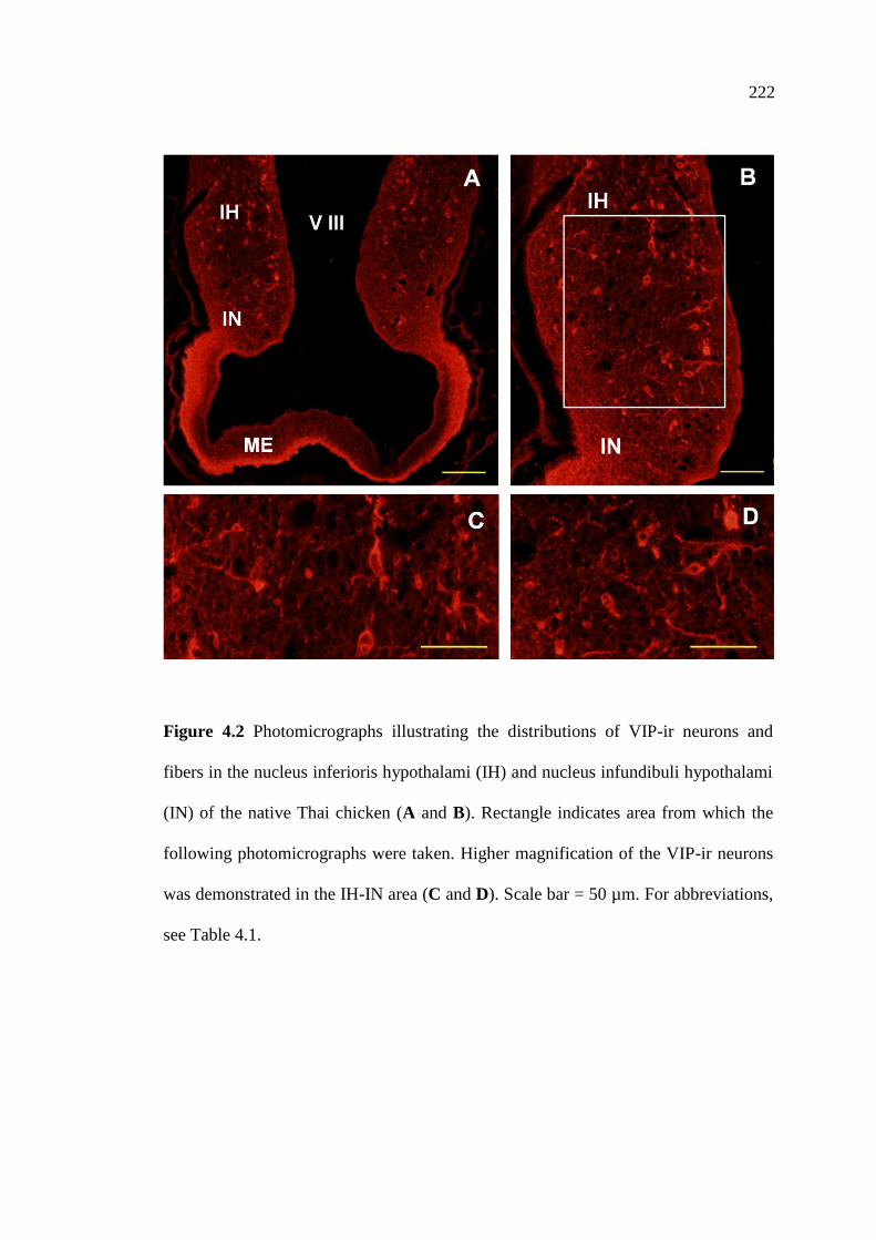
222
Figure 4.2 Photomicrographs illustrating the distributions of VIP-ir neurons and
fibers in the nucleus inferioris hypothalami (IH) and nucleus infundibuli hypothalami
(IN) of the native Thai chicken (A and B). Rectangle indicates area from which the
following photomicrographs were taken. Higher magnification of the VIP-ir neurons
was demonstrated in the IH-IN area (C and D). Scale bar = 50 µm. For abbreviations,
see Table 4.1.
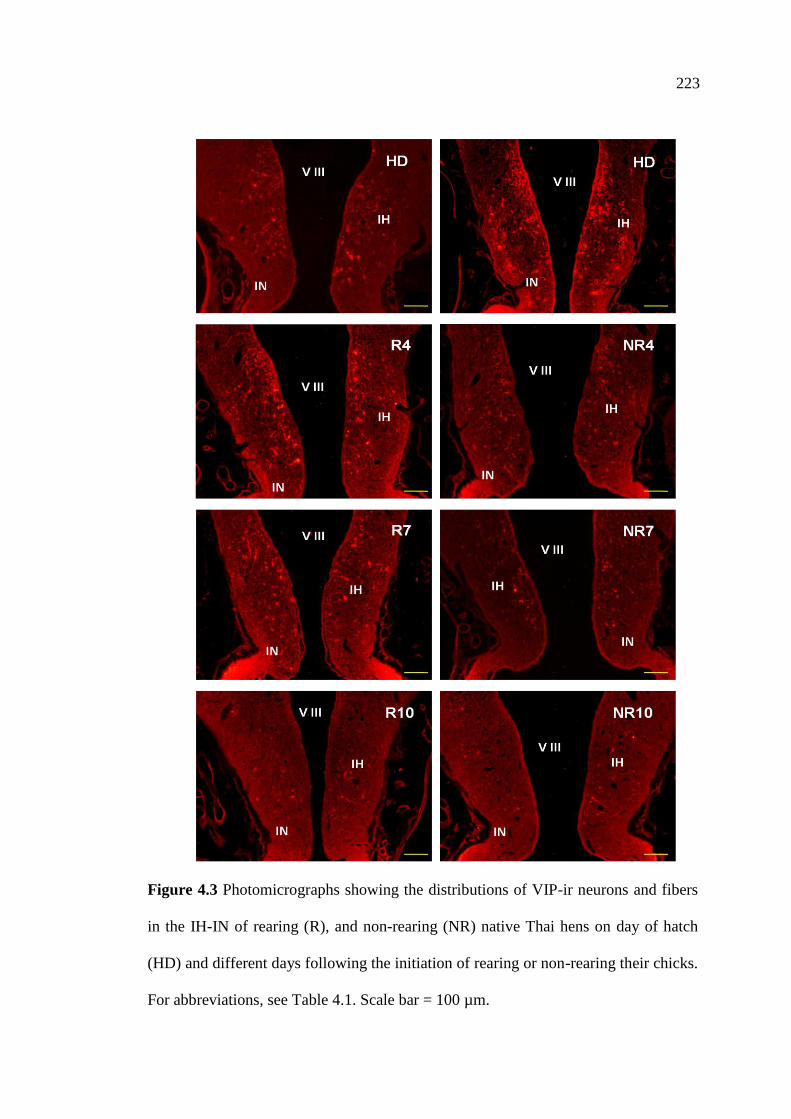
223
Figure 4.3 Photomicrographs showing the distributions of VIP-ir neurons and fibers
in the IH-IN of rearing (R), and non-rearing (NR) native Thai hens on day of hatch
(HD) and different days following the initiation of rearing or non-rearing their chicks.
For abbreviations, see Table 4.1. Scale bar = 100 µm.

224
Figure 4.3 Photomicrographs showing the distributions of VIP-ir neurons and fibers
in the IH-IN of rearing (R) and non-rearing (NR) native Thai hens on different days
following the initiation of rearing or non-rearing their chicks. For abbreviations, see
Table 4.1. Scale bar = 100 µm (continued).
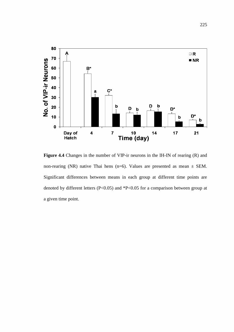
225
Figure 4.4 Changes in the number of VIP-ir neurons in the IH-IN of rearing (R) and
non-rearing (NR) native Thai hens (n=6). Values are presented as mean ± SEM.
Significant differences between means in each group at different time points are
denoted by different letters (P<0.05) and *P<0.05 for a comparison between group at
a given time point.
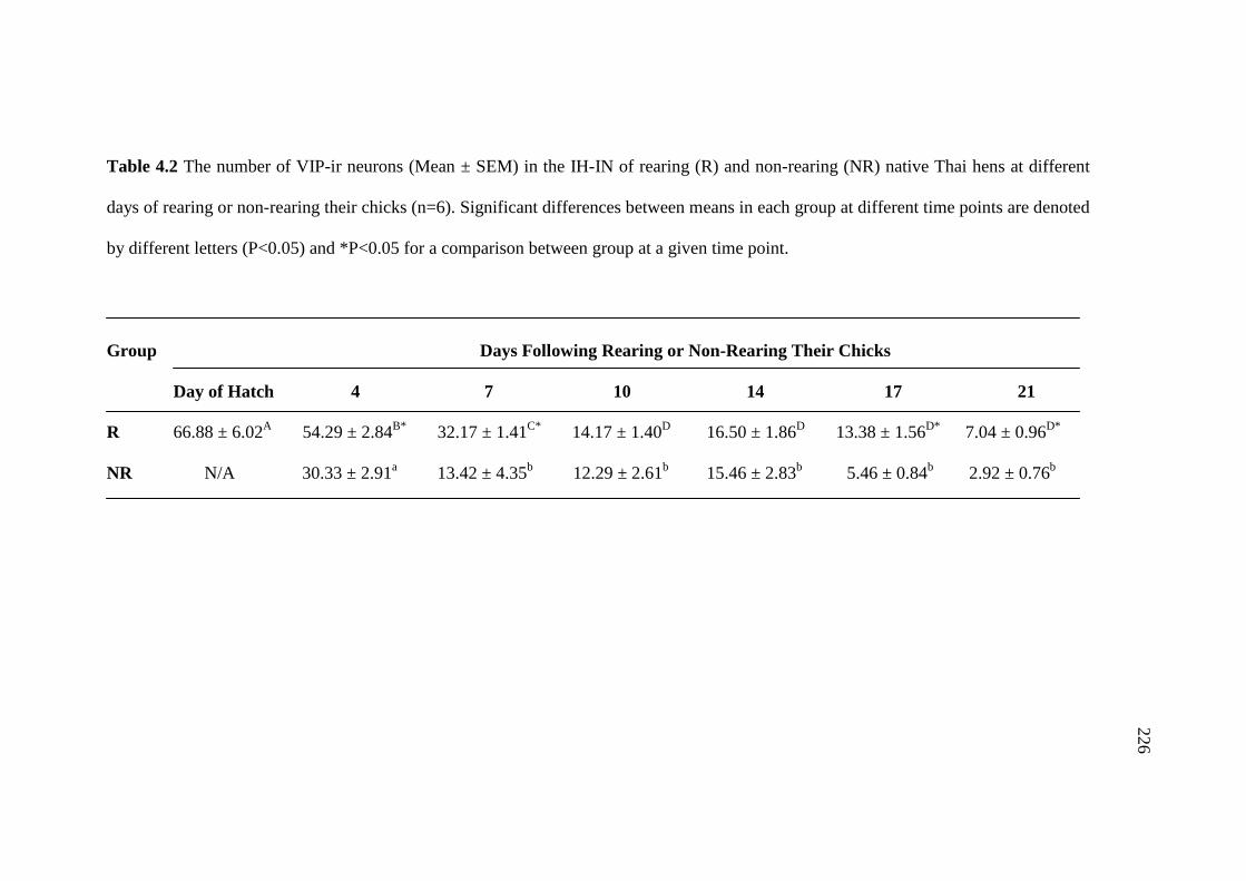
224
Table 4.2 The number of VIP-ir neurons (Mean ± SEM) in the IH-IN of rearing (R) and non-rearing (NR) native Thai hens at different
days of rearing or non-rearing their chicks (n=6). Significant differences between means in each group at different time points are denoted
by different letters (P<0.05) and *P<0.05 for a comparison between group at a given time point.
Group Days Following Rearing or Non-Rearing Their Chicks
Day of Hatch 4 7 10 14 17 21
R 66.88 ± 6.02A 54.29 ± 2.84
B* 32.17 ± 1.41
C* 14.17 ± 1.40
D 16.50 ± 1.86
D 13.38 ± 1.56
D* 7.04 ± 0.96
D*
NR N/A 30.33 ± 2.91a 13.42 ± 4.35
b 12.29 ± 2.61
b 15.46 ± 2.83
b 5.46 ± 0.84
b 2.92 ± 0.76
b
226
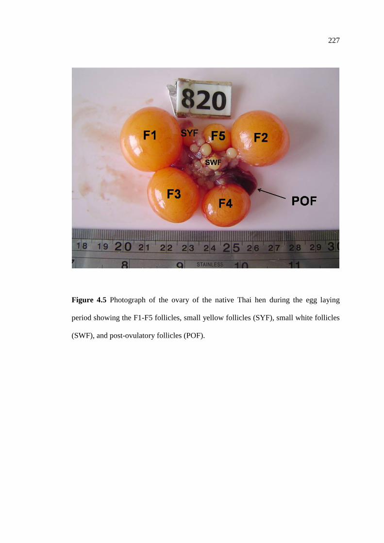
227
Figure 4.5 Photograph of the ovary of the native Thai hen during the egg laying
period showing the F1-F5 follicles, small yellow follicles (SYF), small white follicles
(SWF), and post-ovulatory follicles (POF).

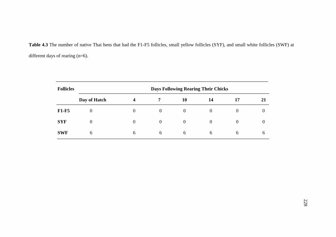
224
Table 4.3 The number of native Thai hens that had the F1-F5 follicles, small yellow follicles (SYF), and small white follicles (SWF) at
different days of rearing (n=6).
Follicles Days Following Rearing Their Chicks
Day of Hatch 4 7 10 14 17 21
F1-F5 0 0 0 0 0 0 0
SYF 0 0 0 0 0 0 0
SWF 6 6 6 6 6 6 6
228

230
Table 4.4 The number of native Thai hens that had the F1-F5 follicles, small yellow follicles (SYF), and small white follicles (SWF) at
different days of non-rearing and the number of hen came back to lay in each period (n=6).
Follicles Days Following Non-Rearing Their Chicks
Day of Hatch 4 7 10 14 17 21
F1 0 0 0 0 0 6 6
F2 0 0 0 0 0 6 6
F3 0 0 0 0 0 6 6
F4 0 0 0 0 0 6 6
F5 0 0 0 0 0 6 6
SYF 0 0 0 0 5 6 6
SWF 6 6 6 6 6 6 6
Laying hens 0 0 0 0 0 0 6
229

230
Figure 4.6 Photographs of the ovary of rearing (R) and non-rearing (NR) native Thai
hens at day of hatch (HD) and different days of rearing or non-rearing their chicks.

231
Figure 4.6 Photographs of the ovary of rearing (R) and non-rearing (NR) native Thai
hens at different days of rearing or non-rearing their chicks (continued).

232
Figure 4.7 Changes in the ovary weights of rearing (R) and non-rearing (NR) native
Thai hens. Values are presented as means ± SEM (n=6). Significant differences
between means in each group at different time points are denoted by different letters
(P<0.05) and *P<0.05 for a comparison between group at a given time point.
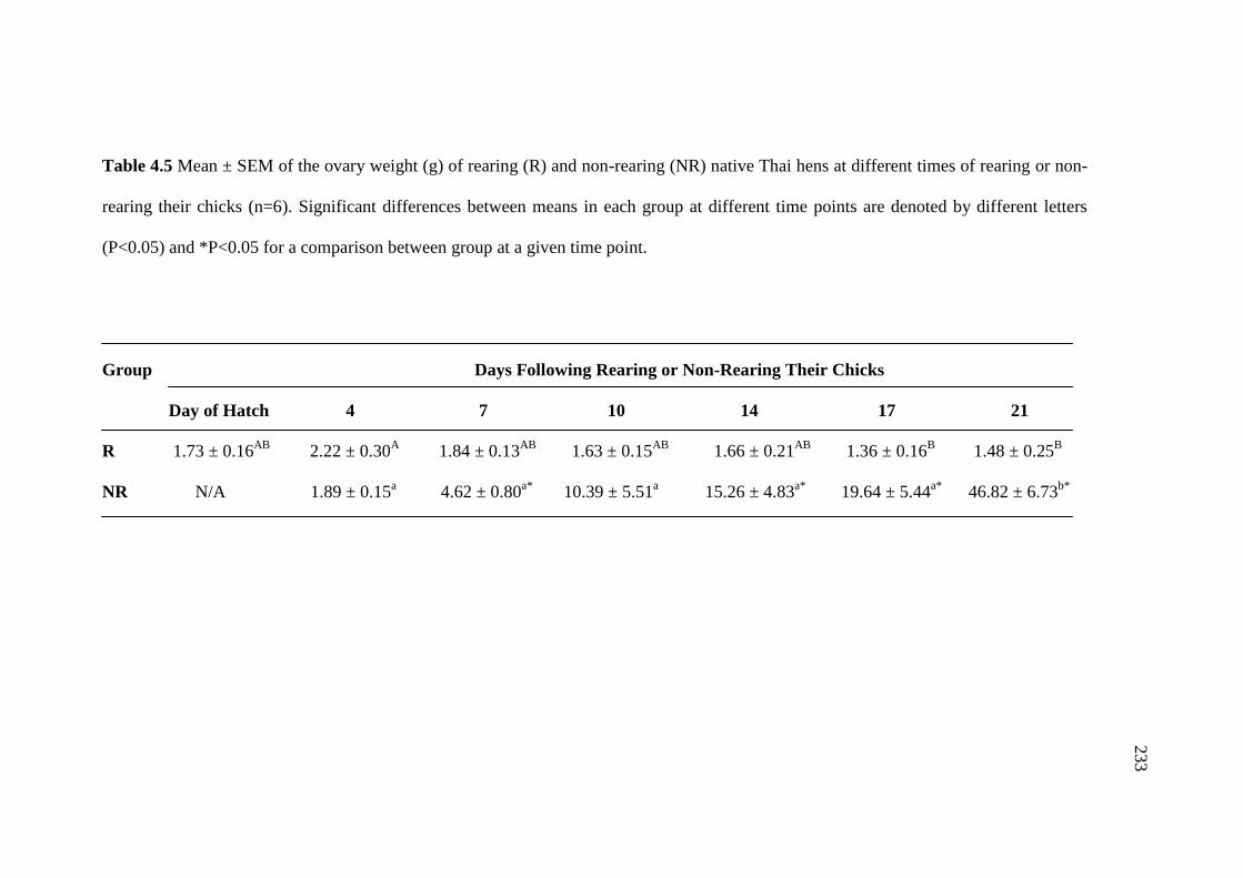
233
Table 4.5 Mean ± SEM of the ovary weight (g) of rearing (R) and non-rearing (NR) native Thai hens at different times of rearing or non-
rearing their chicks (n=6). Significant differences between means in each group at different time points are denoted by different letters
(P<0.05) and *P<0.05 for a comparison between group at a given time point.
Group Days Following Rearing or Non-Rearing Their Chicks
Day of Hatch 4 7 10 14 17 21
R 1.73 ± 0.16AB
2.22 ± 0.30A 1.84 ± 0.13
AB 1.63 ± 0.15
AB 1.66 ± 0.21
AB 1.36 ± 0.16
B 1.48 ± 0.25
B
NR N/A 1.89 ± 0.15a 4.62 ± 0.80
a* 10.39 ± 5.51
a 15.26 ± 4.83
a* 19.64 ± 5.44
a* 46.82 ± 6.73
b*
233
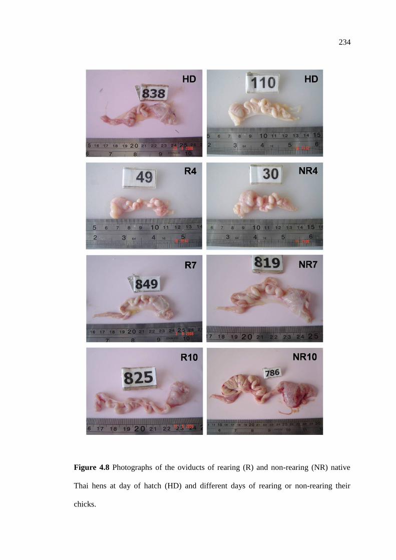
279
Figure 4.8 Photographs of the oviducts of rearing (R) and non-rearing (NR) native
Thai hens at day of hatch (HD) and different days of rearing or non-rearing their
chicks.
234

280
Figure 4.8 Photographs of the oviducts of rearing (R) and non-rearing (NR) native
Thai hens at different days of rearing or non-rearing their chicks (continued).
235
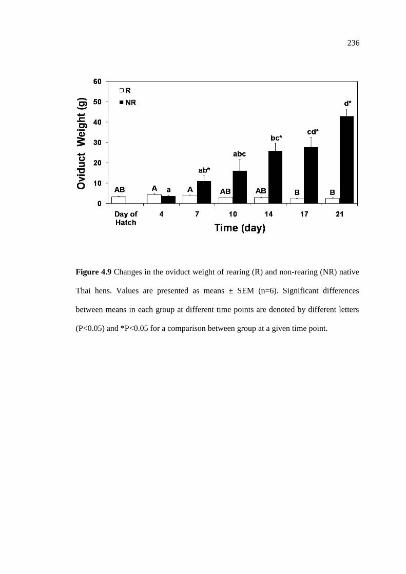
281
Figure 4.9 Changes in the oviduct weight of rearing (R) and non-rearing (NR) native
Thai hens. Values are presented as means ± SEM (n=6). Significant differences
between means in each group at different time points are denoted by different letters
(P<0.05) and *P<0.05 for a comparison between group at a given time point.
236


283
Table 4.6 Mean ± SEM of the oviduct weight (g) of rearing (R) and non-rearing (NR) native Thai hens at different days of rearing or non-
rearing their chicks (n=6). Significant differences between means in each group at different time points are denoted by different letters
(P<0.05) and *P<0.05 for a comparison between group at a given time point.
Group Days Following Rearing or Non-Rearing Their Chicks
Day of Hatch 4 7 10 14 17 21
R 3.24 ± 0.31AB
4.28 ± 0.72A 4.02 ± 0.19
A 3.06 ± 0.05
AB 2.83 ± 0.26
AB 2.36 ± 0.17
B 2.44 ± 0.30
B
NR N/A 3.66 ± 0.34a 10.99 ± 2.76
ab* 16.07 ± 5.54
abc 25.88 ± 3.83
bc* 27.71 ± 4.59
bc* 42.95 ± 3.60
d*
237
245
4.5 Discussion
The results from the present study revealed that the hypothalamic VIP-ir
neurons and fibers were not observed in the AM, SCNm, PHN, LHy, VMN, nI, ML
areas of both R and NR hens. The expression of VIP-ir neurons and fibers were found
in the IH-IN area in both groups. Changes in the number of VIP-ir neurons within the
IH-IN area were compared between R and NR hens. The greatest density of VIP-ir
neurons was found in the R group and the neuronal densities were decreased in the
NR group. The results of this present study clearly demonstrate the association
between the hypothalamic VIPergic system and rearing behavior in native Thai
chickens, revealing markedly differences in the number of VIP-ir neurons within the
IH-IN between R and NR hens.
The number of VIP-ir neurons in the IH-IN remained high after the day the
chicks were hatched until day 7 of the rearing period then sharply decreased from day
10 to day 21. When the chicks were removed from the hens, VIP-ir neurons were
markedly decreased on day 4 and continued to be lower than those of R hens through
day 21. In addition, the effects of removal of chicks from the care of hens induced
ovarian and oviduct recrudescence and the initiation of a new laying cycle. The
results of the present study provide evidence indicates a pivotal role of the VIPergic
system in the regulation of rearing behavior in this equatorial bird.
In the current study, VIP-ir neurons and fibers were not observed in other
hypothalamic areas except within the IH-IN of both R and NR hens. Furthermore, it
has been reported that changes in the number of VIP-ir neurons within the IH-IN are
correlated with changing concentrations of circulating PRL during the rearing period
(Chaiyachet et al., 2010), suggesting that hypothalamic VIP expression in the IH-IN
238
246
and subsequent PRL release play a regulatory role in rearing behavior in the native
Thai chickens. The results from this study are in good agreement with the previous
findings that a large group of VIP-ir neurons found in the IH-IN and its differential
expression may play a role in regulating the reproductive activity in birds (Yamada et
al., 1982; Mikami and Yamada, 1984; Macnamee et al., 1986; Mikami, 1986; Peczely
and Kiss, 1988; Silver et al., 1988; Mauro et al., 1989; Kuenzel and Blahser, 1994;
Kosonsiriluk et al., 2008; Prakobsaeng, 2010; Prakobsaeng et al., 2011). These
findings are well supported by immunohistochemical studies illustrated changes in
the number of VIP-ir neurons within the IH-IN are associated with the shifts in
circulating PRL levels throughout the reproductive cycle of equatorial continuously
breeding, non-photoperiodic birds (Kosonsiriluk et al., 2008). Furthermore, previous
study reported that the expression of incubation behavior is activated by DA neurons
in the nI and ML to VIP neurons in the IH-IN and subsequently stimulates PRL
secretion, resulting in regression of the reproductive system (Prakobsaeng et al.,
2011). In turkeys, a seasonal breeder, increases in the number of VIP-ir neurons
within the INF paralleling increases in levels of plasma PRL. In addition, it has been
reported that the group of VIP neurons in the INF stimulates the release of pituitary
PRL both in vitro (Proudman and Opel, 1983; Macnamee et al., 1986) and in vivo
(Macnamee et al., 1986) and is associated with the reproductive cycle in birds (Mauro
et al., 1989; Sharp et al., 1989; El Halawani and Rozenboim, 1993; Chaiseha and El
Halawani, 1999). Moreover, the increased VIP mRNA expression in the INF is
correlated with increased levels of circulating PRL level and LH-β mRNA expression
in the anterior pituitary (Bhatt et al., 2003). More evidence of hypothalamic VIP is
associated with reproduction and PRL secretion have been studied in ring doves,
239
247
increases in number of VIP neurons and cell size were detected in the ventral
infundibulum, anticipating the increases in plasma PRL levels (Cloues et al., 1990).
The number of hypothalamic VIP-ir neurons were low in non-laying but the number
of neurons and cell size were increased during laying and incubation stages. There
were markedly increased during the incubating phase, then decreased during the
rearing period (Mauro et al., 1989; Sharp et al., 1989; Cloues et al., 1990;
Kosonsiriluk et al., 2008).
Changes in the number of VIP-ir neurons within the IH-IN of R and NR hens
were compared and revealed that the numbers of VIP-ir neurons in the rearing hens
were greater than those of hens not rearing their chicks, suggesting that the presence
and physical contact with chicks induced maternal behavior, the larger number of
VIP-ir neurons maintains circulating PRL levels which may, in part, necessary to
facilitate/stimulate the expression of rearing behavior in the native Thai hens. It is
well documented that, VIP is the avian PRF (El Halawani et al., 1997). In hens are
caring for their young, tactile stimuli from the chicks or in combination with visual
and/or auditory stimuli from newly hatched chicks maintain the circulating PRL
levels and up regulate VIP expression (Sharp et al., 1988; 1989; Silver et al., 1988;
Buntin et al., 1991; Leboucher et al., 1993). The increased number of VIP-ir neurons
has been shown to be correlated with the up-regulation of VIP peptide contents and
its mRNA expression and subsequently PRL concentrations (Mauro et al., 1989;
1992; Chaiseha and El Halawani, 1999).
In the present study, the numbers of VIP-ir neurons in the IH-IN of the hens at
the day the chicks were hatched remained high until rearing day 7 then sharply
decreased and continued to decline during the rest of observed rearing period. These
240
248
results are supported by the findings that the number of VIP-ir neurons within the IH-
IN increased in the incubating phase and when the hens shifted from incubating eggs
to rearing chicks, the number of VIP-ir neurons were sharply dropped, then returned
to low level as found in non-laying hens (Kosonsiriluk et al., 2008). Similarly in ring
doves, VIP-like immunoreactivity cells size and staining intensity in the ventral
portion of the infundibular region in the brain were detected, VIP cell sizes were
increased from about incubation day 14 to rearing day 14, and steady decreased
during the remaining post-hatching period (Cloues et al., 1990). In addition, increases
in the number and cell size of VIP-ir neurons within the medio-basal hypothalamus
when PRL concentrations are high have been demonstrated in the domesticated
pigeons and ring doves during the initiation of crop milk secretion and feeding the
offspring (Peczely and Kiss, 1988; Cloues et al., 1990). Moreover, it has been well
established that VIP in the caudo-medial hypothalamus might relate to the control of
pituitary functions by projecting fibers to the external layer of the ME and influencing
the pituitary PRL secretion (Mikami, 1986). Furthermore, in the current study, the R
hens came back into lay later than the NR hens. In good agreement with these results
are the findings in which incubating bantam hens were deprived of their nests by
replacing eggs with chicks induced maternal behavior, resulting to start new laying
cycle later than hens not allowed to rear their chicks (Sharp et al., 1988).
In the present study, after removing the chicks from the hens, the number of
VIP-ir neurons in the IH-IN was sharply decreased from day 4 toward the end of
observed times in the NR hens when compared with those of R hens and reached
minimum values at day 21, which corresponded with an increase in ovary and oviduct
weights at the time when hens returned to lay after removal of chicks. Similarly in
241
249
bantam hens, Gifujidori hens, and native Thai hens, the effects of removal of chicks
from the care of hens induced ovarian recrudescence and egg laying between days 21
to 28 of separation from chicks (Sharp et al., 1979; Kuwayama et al., 1992;
Kosonsiriluk et al., 2008). These findings are corresponded with the decreases in the
number of VIP-ir neurons in IH-IN of nest-deprived native Thai hens paralleled the
decreases in the plasma PRL levels, and they came back into lay within 18 days of
nest deprivation (Prakobsaeng et al., 2011). Further evidence supported the above
findings have been demonstrated that changes in the number of GnRH-I-ir neurons
were observed in the nucleus commissurae pallii (nCPa) of native Thai hens, which
correlated with the reproductive stages. The greatest number of GnRH-I-ir neurons
found in laying hens, then slightly decreased in incubating hens, and declined to the
lowest levels during the rearing stage (Sartsoongnoen et al., 2012). Further studies in
nest-deprived native Thai hens showed a significant increase in the number of GnRH-
I-ir neurons in the nCPa (Sartsoongnoen et al., 2012), decrease in VIP-ir neurons,
accompanied by an increase in ovary and oviduct weights, following by the hens
started the new laying cycle (Prakobsaeng et al., 2011). The increased neuroendocrine
activity of the VIP/PRL system has been shown to suppress the GnRH/FSH-LH
system (Sharp et al., 1998), reduce ovarian steroids secretion (Zadworny et al., 1988),
terminate egg laying, and induce ovarian regression (Zadworny et al., 1988; Youngren
et al., 1991). On the other hand, rearing chick hens and subsequent that hens had been
removed their entire chicks exhibit an abrupt increase in plasma LH levels concurrent
with the decrease of plasma PRL levels (Leboucher et al., 1990). It has been reported
that circulating of LH and estradiol levels gradually increased after hatching or when
chicks were present and reached peak values immediately after removal of chicks
242
250
(Sharp et al., 1979; 1988; Kuwayama et al., 1992; Richard-Yris et al., 1998). Thus, it
is possible that PRL may also be importance in the increased the number of VIP-ir
neurons in the IH-IN observed in rearing native Thai hens. These findings taken
together with the results in the present study clearly implicate the enhanced activity of
the VIP/PRL system in the initiation and maintenance of rearing behavior in native
Thai hens. Indeed, in this present study, disruption of rearing behavior by removing
the chicks from the hens markedly decreases in the number of VIP-ir neurons in the
IH-IN.
In conclusion, the present findings indicate an association between the
VIPergic system and rearing behavior in the native Thai chickens. The differential
expression of VIP neurons in the IH-IN might play a regulatory role in year-round
reproductive activity and subsequent PRL release in this equatorial bird, non
photoperiodic species. Disruption of rearing behavior decreases the number of VIP-ir
neurons in the IH-IN. Therefore, the VIPergic system in the IH-IN may be involved
in the regulation of the reproductive neuroendocrine system and the initiation and
maintenance of rearing behavior in native Thai chickens. It is further indicated that
the VIP/PRL system is not only a key regulator of incubation behavior as it is well
established but it may also be involved in the regulation of rearing behavior in the
native Thai chicken. These findings also suggest that the presence of chicks and/or
physical contact with chicks induced maternal behavior in the native Thai hens. The
relationship between hens and chicks while hens took care of their young might slow
down the decreases in circulating PRL levels and the number of VIP-ir neurons in the
IH-IN which, in turn, facilitate, stimulate or maintain maternal care behavior in this
equatorial bird.
243
251
4.6 References
Abe, H., Engler, D., Molitch, M.E., Bollinger-Gruber, J., and Reichlin, S. (1985).
Vasoactive intestinal peptide is a physiological mediator of prolactin release in
the rat. Endocrinology 116: 1383-1390.
Andersson, P.O., Bloom, S.R., Edwards, A.V., and Jarhult, J. (1982). Effects of
stimulation of the chorda tympani in bursts on submaxillary responses in the
cat. J Physiol (Lond) 322: 469-483.
Bakken, I.J., Vincent, M.B., Sjaavaag, I., and White, L.R. (1995). Vasodilation in
porcine ophthalamic artery: Peptide interaction with acetylcholine and
endothelial dependence. Neuropeptides 29: 69-75.
Ben-Jonathan, N., Arbogast, L.A., and Hyde, J.F. (1989). Neuroendocrine regulation
of prolactin release. Prog Neurobiol 33: 399-447.
Bern, H.A., and Nicoll, C.S. (1968). The comparative endocrinology of prolactin.
Recent Prog Horm Res 24: 681-720.
Bhatt, R., Youngren, O.M., Kang, S.W., and El Halawani, M.E. (2003). Dopamine
infusion in the third ventricle increases gene expression of hypothalamic
vasoactive intestinal peptide and pituitary prolactin and luteinizing hormone
beta subunit in the turkey. Gen Comp Endocrinol 130: 41-47.
Buntin, J.D., Becker, G.M., and Ruzycky, E. (1991). Facilitation of parental behavior
in ring doves by systemic or intracranial injections of prolactin. Horm Behav
25: 527-534.
Ceccatelli, S., Fahrenkrug, J., Villar, M.J., and Hokfelt, T. (1991). Vasoactive
intestinal polypeptide/peptide histidine isoleucine immunoreactive neuron
systems in the basal hypothalamus of the rat with special reference to the
244
252
portal vasculature: An immunohistochemical and in situ hybridization study.
Neuroscience 43: 483-502.
Chaiseha, Y., and El Halawani, M.E. (1999). Expression of vasoactive intestinal
peptide/peptide histidine isoleucine in several hypothalamic areas during the
turkey reproductive cycle: Relationship to prolactin secretion.
Neuroendocrinology 70: 402-412.
Chaiseha, Y., and El Halawani, M.E. (2005). Neuroendocrinology of the female
turkey reproductive cycle. J Poult Sci 42: 87-100.
Chaiseha, Y., Tong, Z., Youngren, O.M., and El Halawani, M.E. (1998).
Transcriptional changes in hypothalamic vasoactive intestinal peptide during a
photo-induced reproductive cycle in the turkey. J Mol Endocrinol 21: 267-
275.
Chaiseha, Y., Youngren, O.M., Al-Zailaie, K.A., and El Halawani, M.E. (2003).
Expression of D1 and D2 dopamine receptors in the hypothalamus and
pituitary during the turkey reproductive cycle: Colocalization with vasoactive
intestinal peptide. Neuroendocrinology 77: 105-118.
Chaiseha. Y., Youngren, O.M., and El Halawani, M.E. (2004). Expression of
vasoactive intestinal peptide receptor messenger RNA in the hypothalamus
and pituitary throughout the turkey reproductive cycle. Biol Reprod 70: 593-
599.
Chaiyachet, O-A., Chokchaloemwong, D., Prakobsaeng, N., Sartsoongnoen, N.,
Kosonsiriluk, S., Rozenboim, I., El Halawani, M.E., Porter, T.E., and
Chaiseha, Y. (2010). Neuroendocrine regulation of rearing behavior in the
native Thai hen. Poult Sci 89 (Suppl 1): 679.
245

253
Chihara, K., Iwasaki, J., Minamitani, N., Kaji, H., Matsukura, S., Tamaki, N.,
Matsumota, S., and Fujita, T. (1982). Effect of vasoactive intestinal
polypeptide on growth hormone secretion in perfused acromegalic pituitary
adenoma tissues. J Clin Endocrinol Metab 54: 773-779.
Chihara, K., Kaji, H., Minamitani, N., Kodama, H., Kita, T., Goto, B., Chiba T., Coy,
D.H., and Fujita, T. (1984). Stimulation of growth hormone by vasoactive
intestinal polypeptide in acromegaly. J Clin Endocrinol Metab 58: 81-86.
Cloues, R., Ramos, C., and Silver, R. (1990). Vasoactive intestinal polypeptide-like
immunoreactivity during reproduction in doves: Influence of experience and
number of offspring. Horm Behav 24: 215-231.
El Halawani, M.E., and Rozenboim, I. (1993). The ontogeny and control of
incubation behavior in turkeys. Poult Sci 72: 906-911.
El Halawani, M.E., Burke, W.H., Millam, J.R., Fehrer, S.C., and Hargis, B.M. (1984).
Regulation of prolactin and its role in gallinaceous bird reproduction. J Exp
Zool 232: 521-529.
El Halawani, M.E., Fehrer, S.C., Hargis, B.M., and Porter, T.E. (1988). Incubation
behavior in the domestic turkey: Physiological correlates. CRC Crit Rev
Poult Biol 1: 285-314.
El Halawani, M.E., Silsby, J.L., and Mauro, L.J. (1990). Enhanced vasoactive
intestinal peptide-induced prolactin secretion from anterior pituitary cells of
incubating turkeys (Meleagris gallopavo). Gen Comp Endocrinol 80: 138-
145.
El Halawani, M.E., Youngren, O.M., and Chaiseha, Y. (2001). Neuroendocrinology
of PRL regulation in the domestic turkey. In Avian Endocrinology, pp 233-
246

254
244. Eds. Dawson, A., and Chaturvedi, C.M. Narosa Publishing House, New
Delhi, India.
El Halawani, M.E., Youngren, O.M., and Pitts, G.R. (1997). Vasoactive intestinal
peptide as the avian prolactin-releasing factor. In Perspectives in Avian
Endocrinology, pp 403-416. Eds. Harvey, S., and Etches, R.J. Journal of
Endocrinology LTD, Bristol, UK.
Emson, P.C., Fahrenkrung, J., Schaffalitzky de Muckadell, O.B., Jessell, T.M., and
Iversen, L.L. (1979). Vasoactive intestinal peptide (VIP): Vesicular
localization and potassium evoked release from rat hypothalamus. Brain Res
143: 174-178.
Etches, R.J. (1993). Reproduction in poultry. In Reproduction in Domesticated
Animals. Ed. King, G.J. Elsevier, Huddersfield, UK.
Follett, B.K. (1984). Reproductive cycles of vertebrates. In Physiology of
Reproduction, pp. 283-350. Ed. Lamming, G.E. Marshall’s Churchill
Livingstone, New York, USA.
Frawley, L.S., and Neill, J.D. (1981). Stimulation of prolactin secretion in rhesus
monkeys by vasoactive intestinal polypeptide. Neuroendocrinology 33: 79-
83.
Giachetti, A., Said, S.I., Reynolds, R.C., and Koniges, F.C. (1977). Vasoactive
intestinal polypeptide in brain: Localization in and release from isolated nerve
terminals. Proc Natl Acad Sci USA 74: 3424-3428.
Gomariz, R.P., Martinez, C., Abad, C., Leceta, J., and Delgado, M. (2001).
Immunology of VIP: A review and therapeutical perspectives. Curr Pharm
Des 7: 89-111.
247
255
Gressens, P., Hill, J.M., Gozes, I., Fridkin, M., and Brenneman, D.E. (1993). Growth
factor function of vasoactive intestinal peptide in whole cultured mouse
embryos. Nature 362: 155-157.
Grossman, M.I. (1974). Candidate hormones of the gut. I. Introduction.
Gastroenterology 67: 730-731.
Kato, Y., Iwasaki, Y., Iwasaki, J., Abe, H., Yanaihara, N., and Imura, H. (1978).
Prolactin release by vasoactive intestinal polypeptide in rats. Endocrinology
103: 554-558.
Kato, Y., Shimatsu, A., Matsushita, N., Ohta, H., and Imura, H. (1984). Role of
vasoactive intestinal polypeptide (VIP) in regulating the pituitary function in
man. Peptides 5: 389-394.
Kosonsiriluk, S. (2007). Biological studies of the reproductive cycle and the effects
of photoperiod upon the reproductive system in the female native Thai
chicken. Ph.D. Dissertation, Suranaree University of Technology, Nakhon
Ratchasima, Thailand.
Kosonsiriluk, S., Sartsoongnoen, N., Chaiyachet, O-A., Prakobsaeng, N., Songserm,
T., Rozenboim, I., El Halawani, M.E., and Chaiseha, Y. (2008). Vasoactive
intestinal peptide and its role in continuous and seasonal reproduction in birds.
Gen Comp Endocrinol 159: 88-97.
Kragt, C.L., and Meites, J. (1965). Stimulation of pigeon pituitary prolactin release by
pigeon hypothalamic extracts in vitro. Endocrinology 76: 1169-1176.
Kuenzel, W.J., and Blahser, S. (1994). Vasoactive intestinal peptide (VIP)-containing
neurons: Distribution throughout the brain of the chick (Gallus domesticus)
with focus upon the lateral septal organ. Cell Tissue Res 275: 91-107.
248

256
Kuenzel, W.J., and Masson, M. (1988). A stereotaxic atlas of the brain of the chick
(Gallus domesticus). Johns Hopkins University Press, Baltimore, Maryland,
USA.
Kuenzel, W.J., and van Tienhoven, A. (1982). Nomenclature and location of avian
hypothalamic nuclei and associated circumventricular organs. J Comp Neurol
206: 293-313.
Kuwayama, T., Shimada, K., Saito, N., Ohkubo, T., Sato, K., Wada, M., and Ichinoe,
K. (1992). Effects of removal of chicks from hens on concentrations of
prolactin, luteinizing hormone and oestradiol in plasma of brooding Gifujidori
hens. J Reprod Fertil 95: 617-622.
Lam, K.S. (1991). Vasoactive intestinal peptide in the hypothalamus and pituitary.
Neuroendocrinology 53 (Suppl 1): 45-51.
Larsson, L.I., Fahrenkrug, J., Schffalitzky de Muckadell, O., Sundler, F., Hakanson,
R., and Rehfeld, J.F. (1976). Localization of vasoactive intestinal polypeptide
(VIP) to central and peripheral neurons. Proc Natl Acad Sci USA 73: 3197-
3200.
Lea, R.W., and Vowles, D.M. (1986). Vasoactive intestinal polypeptide stimulates
prolactin release in vivo in the ring dove (Streptopelia risoria). Experientia
42: 420-422.
Lea, R.W., Dods, A.S., Sharp, P.J., and Chadwick, A. (1981). The possible role of
prolactin in the regulation of nesting behavior and the secretion of luteinizing
hormone in broody bantams. J Endocrinol 91: 89-97.
249
257
Leboucher, G., Richard-Yris, M.A., Guemene, D., and Chadwick, A. (1993).
Respective effects of chicks and nest on behavior and hormonal concentrations
of incubating domestic hens. Physiol Behav 54: 135-140.
Leboucher, G., Richard-Yris, M.A., Williams, J., and Chadwick, A. (1990).
Incubation and maternal behaviour in domestic hens: Influence of the presence
of chicks on circulating luteinising hormone, prolactin and oestradiol and on
behaviour. Br Poult Sci 31: 851-862.
Maas, D.L., Arnaout, M.A., Martinson, D.R., Erdmann, M.D., and Hagen, T.C.
(1991). Vasoactive intestinal polypeptide and thyrotropin-releasing hormone
stimulate newly synthesized, not stored, prolactin. Endocrinology 128: 1015-
1020.
Macnamee, M.C., Sharp, P.J., Lea, R.W., Sterling, R.J., and Harvey, S. (1986).
Evidence that vasoactive intestinal polypeptide is a physiological prolactin-
releasing factor in the bantam hen. Gen Comp Endocrinol 62: 470-478.
Marley, P., and Emson, P. (1982). VIP as a neurotransmitter in the central nervous
system. In Vasoactive intestinal peptide, pp 341-346. Ed. Said, S.I. Raven
Press, New York, USA.
Matsushita, N., Kato, Y., Shimatsu, A., Katakami, H., Yanaihara, N., and Imura, H.
(1983). Effects of VIP, TRH, GABA and dopamine on prolactin release from
superfused rat anterior pituitary cells. Life Sci 32: 1263-1269.
Mauro, L.J., Elde, R.P., Youngren, O.M., Phillips, R.E., and El Halawani, M.E.
(1989). Alterations in hypothalamic vasoactive intestinal peptide-like
immunoreactivity are associated with reproduction and prolactin release in the
female turkey (Meleagris gallopavo). Endocrinology 125: 1795-1804.
250

258
Mauro, L.J., Youngren, O.M., Proudman, J.A., Phillips, R.E., and El Halawani, M.E.
(1992). Effects of reproductive status, ovariectomy, and photoperiod on
vasoactive intestinal peptide in the female turkey hypothalamus. Gen Comp
Endocrinol 97: 481-493.
Mikami, S. (1986). Immunocytochemistry of the avian hypothalamus and
adenohypophysis. Int Rev Cytol 103: 189-248.
Mikami, S., and Yamada, S. (1984). Immunohistochemistry of the hypothalamic
neuropeptides and anterior pituitary cells in the Japanese quail. J Exp Zool
232: 405-417.
Oliva, D., Nicosia, S., Spada, A., and Giannattasio, G.L. (1982). VIP stimulates
ACTH release and adenylate cyclase in human ACTH secreting pituitary
adenomas. Eur J Pharmacol 83: 101-105.
Peczely, P. (1989). The role of gonadotropin releasing hormone (Gn-RH) in the
regulation of gonadal functions of birds. Acta Biol Hung 40: 161-193.
Peczely, P., and Kiss, J.Z. (1988). Immunoreactivity to vasoactive intestinal
polypeptide (VIP) and thyreotropin-releasing hormone (TRH) in hypothalamic
neurons of the domesticated pigeon (Columba livia). Alterations following
lactation and exposure to cold. Cell Tissue Res 251: 485-494.
Prakobsaeng, N. (2010). Effects of incubation behavior upon the neuroendocrine
regulation of the reproductive system in the female native Thai chicken.
Ph.D. Dissertation, Suranaree University of Technology, Nakhon Ratchasima,
Thailand.
Prakobsaeng, N., Sartsoongnoen, N., Kosonsiriluk, S., Rozenboim, I., El Halawani,
M.E., Porter, T.E., and Chaiseha, Y. (2009). Changes in vasoactive intestinal
251
259
peptide and gonadotropin releasing hormone-I immunoreactivity in the brain
of nest-deprived native Thai hen. Poult Sci 88 (Suppl 1): 121-122.
Prakobsaeng, N., Sartsoongnoen, N., Kosonsiriluk, S., Chaiyachet, O-A.,
Chokchaloemwong, D., Rozenboim, I., El Halawani, M.E., Porter T.E., and
Chaiseha, Y. (2011). Changes in vasoactive intestinal peptide and tyrosine
hydroxylase immunoreactivity in the brain of nest-deprived native Thai hen.
Gen Comp Endocrinol 171: 189-196.
Proudman, J.A., and Opel, H. (1983). Stimulation of prolactin and growth hormone
secretion from turkey pituitary cells. Poult Sci 62: 1484-1485.
Proudman, J.A., and Opel, H. (1988). Stimulation of prolactin secretion from turkey
anterior pituitary cells in culture. Proc Soc Exp Biol Med 187: 448-454.
Richard-Yris, M.A., Guemene, D., Lea, R.W., Sharp, P.J., Bedecarrats, G., Foraste,
M., and Wauters, A.M. (1998). Behaviour and hormone concentrations in nest
deprived and renesting hens. Br Poult Sci 39: 309-317.
Rosselin, G., Maletti, M., Besson, J., and Rostene, W. (1982). A new neuroregulator:
The vasoactive intestinal peptide or VIP. Mol Cell Endocrinol 27: 243-262.
Rotsztejn, W.H., Benoist, L., Besson, J., Beraud, G., Bluet-Pajot, M.T., Kordon, C.,
Rosselin, G., and Duval, J. (1980). Effect of vasoactive intestinal peptide
(VIP) on the release of adenohypophyseal hormones from purified cells
obtained by unit gravity sedimentation. Inhibition by dexamethasone of VIP-
induced prolactin release. Neuroendocrinology 31: 282-286.
Said, S.I. (1982). Vasoactive intestinal polypeptide. J Endocrinol Invest 9: 191-200.
Said, S.I., and Mutt, V. (1970). Polypeptide with broad biological activity: Isolation
from small intestine. Science 169: 1217-1278.
252
260
Said, S.I., and Rosenberg, R.N. (1976). Vasoactive intestinal polypeptide: Abundant
immunoreactivity in neural cell lines and normal nervous tissue. Science 192:
907-908.
Samson, W.K., Said, S.I., and McCann, S.M. (1979). Radioimmunologic localization
of vasoactive intestinal polypeptide in hypothalamic and extrahypothalamic
sites in the rat brain. Neurosci Lett 12: 265-269.
Samson, W.K., Said, S.I., Snyder, G., and McCann, S.M. (1980). In vitro stimulation
of prolactin release by vasoactive intestinal peptide. Peptides 1: 325-332.
Sartsoongnoen, N. (2007). Neuroendocrinology of the reproductive cycle in the
female native Thai chicken: Roles of dopamine and gonadotropin
releasing hormone. Ph.D. Dissertation, Suranaree University of Technology,
Nakhon Ratchasima, Thailand.
Sartsoongnoen, N., Kosonsiriluk, S., Prakobsaeng, N., Songserm, T., Rozenboim, I.,
El Halawani, M.E., and Chaiseha, Y. (2008). The dopaminergic system in the
brain of the native Thai chicken, Gallus domesticus: Localization and
differential expression across the reproductive cycle. Gen Comp Endocrinol
159: 107-115.
Sartsoongnoen, N., Prakobsaeng, N., Kosonsiriluk, S., Chaiyachet, O-A.,
Chokchaloemwong, D., El Halawani, M.E., and Chaiseha, Y. (2012).
Distribution and variation in gonadotropin releasing hormone-I (GnRH-I)
immunoreactive neurons in the brain of the native Thai chicken during the
reproductive cycle. Acta Histochem 114: 409-420.
Sharp, P.J., Dunn, I.C., Main, G.M., Sterling, R.J., and Talbot, R.T. (1990).
Gonadotrophin-releasing hormones: Distribution and function. In
253
261
Endocrinology of Birds: Molecular to Behavioral, pp 31-42. Eds. Wada,
M., Ishii, S., Scanes, C.G. Sciences Society Press-Springer, Tokyo-Berlin,
Japan.
Sharp, P.J., Dawson, A., and Lea, R.W. (1998). Control of luteinizing hormone and
prolactin secretion in birds. Comp Biochem and Physiol C Pharmacol
Toxicol Endocrinol 119: 275-282.
Sharp, P.J., Macnamee, M.C., Sterling, R.J., Lea, R.W., and Pedersen, H.C. (1988).
Relationships between prolactin, LH and broody behavior in bantam hens. J
Endocrinol 118: 279-286.
Sharp, P.J., Scanes, C.G., Williams, J.B., Harvey, S., and Chadwick, A. (1979).
Variations in concentrations of prolactin, luteinizing hormone, growth
hormone and progesterone in the plasma of broody bantams (Gallus
domesticus). J Endocrinol 80: 51-57.
Sharp, P.J., Sterling, R.J., Talbot, R.T., and Huskisson, N.S. (1989). The role of
hypothalamic vasoactive intestinal polypeptide in the maintenance of prolactin
secretion in incubating bantam hens: Observations using passive
immunization, radioimmunoassay and immunohistochemistry. J Endocrinol
122: 5-13.
Silver, R., Witkovsky, P., Horvath, P., Alones, V., Barnstable, C.J., and Lehman,
M.N. (1988). Coexpression of opsin- and VIP-like immunoreactivity in CSF-
contacting neurons of the avian brain. Cell Tissue Res 253: 189-198.
SPSS Inc. (2004). SPSS Base 13.0 Users Guide. Prentice Hall, New Jersey, USA.
White, M.C., Adams, E.F., Loizou, M., and Mashiter, K. (1982). Vasoactive intestinal
peptide stimulates adrenocorticotropin release from human corticotropinoma
254
262
cells in culture: Interaction with arginine vasopressin and hydrocortisone. J
Clin Endocrinol Metab 55: 967-972.
Yamada, S., Mikami, S., and Yanaihara, N. (1982). Immunohistochemical
localization of vasoactive intestinal polypeptide (VIP)-containing neurons in
the hypothalamus of the Japanese quail, Coturnix coturnix. Cell Tissue Res
226: 13-26.
Youngren, O.M., Chaiseha, Y., Phillips, R.E., and El Halawani, M.E. (1996).
Vasoactive intestinal peptide concentrations in turkey hypophysial portal
blood differ across the reproductive cycle. Gen Comp Endocrinol 103: 323-
330.
Youngren, O.M., El Halawani, M.E., Silsby, J.L., and Phillips, R.E. (1991).
Intracranial prolactin perfusion induces incubation behavior in turkey hens.
Biol Reprod 44: 425-431.
Zadworny, D., Shimada, K., Ishida, H., Sumi, C., and Sato, K. (1988). Changes in
plasma levels of prolactin and estradiol, nutrient intake, and time spent nesting
during the incubation phase of broodiness in the Chabo hen (Japanese
bantam). Gen Comp Endocrinol 71: 406-412.
255

256
CHAPTER V
EFFECTS OF REARING BEHAVIOR ON
NEUROENDOCRINE REGULATION OF THE
REPRODUCTIVE SYSTEM IN THE FEMALE NATIVE
THAI CHICKENS: ROLE OF GONADOTROPIN
RELEASING HORMONE
5.1 Abstract
There are two major neuroendocrine systems that play an important role in the
avian reproductive cycle. One system involves gonadotropin releasing hormone-I
(GnRH-I) and the subsequent release of follicle stimulating hormone (FSH) and
luteinizing hormone (LH), named as GnRH/FSH-LH system. The other system
involves the avian prolactin (PRL) releasing factor, vasoactive intestinal peptide
(VIP) and the subsequent release of PRL, known as VIP/PRL system. The
establishment of maternal behavior in birds is accompanied by a significant long term
fall in plasma levels of LH and gonadal steroids, and the presence of chicks slows
down the decrease in plasma PRL levels. The synthesis and secretion of FSH and LH
are regulated by GnRH, a decapeptide which is secreted from hypothalamic neuronal
cells. Changes in the number of hypothalamic GnRH-I neurons within the nucleus
commissurae pallii (nCPa) are correlated with the reproductive cycle of native Thai
chickens. Moreover, nest deprivation of incubating native Thai hens increases the
257
number of GnRH-I neurons in the nCPa. To date, there is no study describing the
interrelationship and functional aspects of the GnRH system and rearing behavior in
the native Thai chicken, non-temperate zone birds. Thus, to further understand the
role of GnRH-I in the regulation of the reproductive cycle, especially the rearing
behavior in this bird, native Thai chickens were divided into two groups; rearing (R)
and non-rearing (NR) hens. The differential expression of GnRH-I-immunoreactive
(GnRH-I-ir) neurons in the hypothalamus of R and NR hens were compared utilizing
immunohistochemical techniques. Expression of hypothalamic GnRH-I-ir neurons
within the nucleus anterior medialis hypothalami, nucleus suprachaiasmaticus, pars
medialis, nCPa, nucleus septalis lateralis, nucleus paraventricularis magnocellularis,
and regio lateralis hypothalami areas were observed in both groups. High expression
of GnRH-I-ir neurons was found in the nCPa of NR hens, whereas fewer GnRH-I-ir
neurons were observed in the nCPa of R hens. The number of GnRH-I-ir neurons in
the nCPa was low in the R group and significantly increased by day 17 after removal
of chicks from the hens. These findings indicate, for the first time, an association of
the GnRH/FSH-LH system with rearing behavior in this non-photoperiodic,
continuously breeding avian species. The expression of rearing behavior of native
Thai chickens might be regulated, in part, by the differential expression of GnRH-I
neurons in the nCPa. This study also confirms a pivotal role of GnRH-I in controlling
of reproduction of this non-seasonally breeding, equatorial avian species.
258
5.2 Introduction
Avian reproductive is regulated by the integration of hormonal inputs by the
hypothalamus, the pituitary, and the gonads. This system is referred as the
hypothalamo-pituitary-gonadal (HPG) axis. It is very well documented that
neurotransmitters, neurohormones, neuromodulators, and hormones of the HPG axis
play an important role in the reproductive cycle of avian species. This HPG axis
involves two major neuroendocrine systems controlling avian reproduction. These
neuroendocrine systems include the gonadotropin releasing hormone/follicle
stimulating hormone-luteinizing hormone (GnRH/FSH-LH), and vasoactive intestinal
peptide/prolactin (VIP/PRL) systems, and both systems are influenced by
dopaminergic (DAergic) neurotransmission (Bhatt et al., 2003; Chaiseha et al., 2003;
Chaiseha and El Halawani, 2005).
GnRH is a hypothalamic neuronal secretory decapeptide important for the
regulation of reproduction in many vertebrates. GnRH was first isolated from porcine
hypothalamus and has been sequenced (Peczely, 1989; Rivier, 2001). It consists of a
family of at least 25 isoforms, and 15 isoforms are found in various vertebrate species
(Gorbman and Sower, 2003; Kavanaugh et al., 2008). In avian species, three subtypes
of GnRH and two subtypes of GnRH receptors have been found (Sun et al., 2001;
Shimizu and Bedecarrats, 2006). Two distinct forms of GnRH have been isolated in
chicken, cGnRH-I or GnRH-I and cGnRH-II (King and Millar, 1982; Miyamoto et al.,
1982; Sherwood et al., 1988). The gene encoding cGnRH-I has been cloned and
characterized (Dunn et al., 1993). GnRH-III is first demonstrated in lamprey and is
also found in the brain of songbirds (Bentley et al., 2004).
259
In birds, cGnRH-I is responsible for the HPG axis, which is directly involved
in controlling reproduction (Sharp et al., 1990). Growing evidence indicates that the
three forms of GnRH influence avian gonadotropins secretion, but their abilities differ
(Katz et al., 1990; Sharp et al., 1990; Guemene and Williams, 1992; Bentley et al.,
2004; Stevenson and MacDougall-Shackleton, 2005; Proudman et al., 2006). Like in
mammals, GnRH is synthesized by neurosecretory cells in the hypothalamus, released
from the median eminence (ME) into the hypophysial portal vessels, and then
transported to the pituitary gland, where it stimulates the secretion of the
gonadotropins, FSH and LH. GnRH increases LH and FSH secretions of the anterior
pituitary both in vitro and in vivo (Millar et al., 1986; Peczely, 1989). In vivo studies
reveal that injection of cGnRH-I or cGnRH-II stimulates an increase in plasma LH
concentrations in hens (Guemene and Williams, 1999; Proudman et al., 2006).
Incubation of turkey anterior pituitary cells with GnRH results in an increase in LH-β-
subunit mRNA expression and stimulates LH secretion (You et al., 1995). A pulsatile
pattern of GnRH release is observed from the medial basal hypothalamus and the
preoptic area (POA) in vitro (Li et al., 1994). In contrast, GnRH inhibits FSH-
stimulated steroidogenesis in chickens but enhances LH-stimulated progesterone
production (Hertelendy et al., 1982).
The egg laying period in birds is associated with relatively high levels of
circulating FSH, LH, and gonadal steroids and is regulated by hypothalamic GnRH
(El Halawani et al., 1988). cGnRH-I is the primary hypophysiotropic factor
stimulating the release of LH, since active immunization against cGnRH-I causes a
decline in plasma LH concentrations and complete regression of the reproductive
system (Sharp et al., 1990). However, seasonal changes in the cGnRH-II-
260
immunoreactive neurons are noted, indicating an involvement of cGnRH-II in the
control of reproduction (Teruyama and Beck, 2000; Stevenson and MacDougall-
Shackleton, 2005). The various distributions of cGnRH-II and GnRH-III in avian
brains suggest their functional significances. It is reported that cGnRH-II may act as a
neurotransmitter (Jones, 1987; Katz et al., 1990), and GnRH-III may act as a potential
mediator in transducing song-related stimuli to areas that control gonadotropin
secretion (Bentley et al., 2004).
In birds, it has been reported that GnRH neuronal activity is regulated by
photoperiod (Sharp and Blache, 2003). Photostimulatory inputs to GnRH neurons
have the potential to increase GnRH mRNA transcription and GnRH release (Dunn
and Sharp, 1999) as well as increase pituitary cell sensitivity to GnRH (Davies and
Follett, 1975). The amount of hypothalamic GnRH increases during long day
stimulation and decreases during photorefractoriness in many avian species (Dawson
et al., 1985; Foster et al., 1987; Bluhm et al., 1991; Rozenboim et al., 1993a; Saldanha
et al., 1994; Hahn and Ball, 1995; Dunn et al., 1996; Kang et al., 2006). In addition,
gonadal steroid hormones, hypothalamic VIP, DA, and gonadotropin inhibitory
hormone are thought to be involved in the regulation of GnRH secretion (Ramirez et
al., 1984; Sharp et al., 1984; Deviche et al., 2000; Tsutsui et al., 2000). Moreover,
active VIP immunoneutralization increases pituitary LH-β and FSH-β mRNA and is
accompanied by a decline in PRL mRNA expression (Ahn et al., 2001).
In mammals, it is very well documented that GnRH regulates LH secretion in
both spontaneous and induced ovulating species (Bakker and Baum, 2000). Similarly,
GnRH plays a pivotal role in the control of avian reproduction as well. Changes in
GnRH content are observed during the avian reproductive cycle. GnRH content of

261
discrete medial preoptic, infundibulum, and arcuate samples are higher in laying hens
than that of non-laying hens (Advis et al., 1985). In turkeys, a temperate zone bird, it
has been reported that GnRH-I mRNA is abundance within the nucleus commissurae
pallii (nCPa), organum vasculosum, lamina terminalis, and nucleus septalis lateralis
(SL) is greater in laying hens than in non-photostimulated and incubating hens, while
lower mRNA expression is observed in photorefractory hens (Kang et al., 2006).
Furthermore, an association between hypothalamic GnRH-I mRNA, GnRH-I peptide
content, and pituitary gonadotropin secretion during the avian reproductive cycle has
been documented (Millam et al., 1989; El Halawani et al., 1993; Rozenboim et al.,
1993a; Dunn et al., 1996).
In mammals, GnRH perikarya axons terminate in the external layer of the ME,
which is closed proximity to the terminals of tuberoinfundibular DA neurons (Ajika,
1979; Merchenthaler et al., 1984; Ugrumov et al., 1989). Like mammals, GnRH
perikarya and fibers are more extensively distributed throughout the avian brains.
GnRH-I localization studies have been conducted in the brains of many avian species,
such as chickens (Kuenzel and Blahser, 1991), ducks (Bons et al., 1978), white-
crowned sparrows (Blahser et al., 1989), Japanese quail (Teruyama and Beck, 2000),
European starlings (Goldsmith et al., 1989), garden warblers (Bluhm et al., 1991),
great tits and ring doves (Silver et al., 1992), turkeys (Millam et al., 1993), dark-eyed
juncos (Saldanha et al., 1994), house sparrows (Hahn and Ball, 1995), cockerels (Sun
et al., 2001), canaries (Bentley et al., 2004), and native Thai chickens (Sartsoongnoen
et al., 2006; 2012). The main group of cGnRH-I cell bodies is located in the POA,
with fibers extending along the third ventricle and then entering the ME, the area of
GnRH secretion (Meddle and Follett, 1997). Furthermore, several studies have
262
reported the distribution of the cGnRH-I mRNA and its protein in the avian brain
(Millam et al., 1989; Dunn and Sharp, 1999; Sun et al., 2001; Dawson et al., 2002;
Kang et al., 2006). It has been reported that the greatest cGnRH-I mRNA expression
occurs in the nCPa and around the organum vasculosum laminae terminalis (OVLT),
which is more abundant in the nCPa, OVLT, and SL of the laying turkey hens than
those of the non-photostimulated and incubating hens (Kang et al., 2006).
The differential expression of GnRH-I neurons has been reported in many
temperate zone species, but little is known regarding neuroendocrine regulation in
non-temperate zone gallinaceous birds. In contrast to the temperate zone seasonal
breeding species, the native Thai chicken is a continuously breeding species found in
the equatorial zone that produces eggs all year, independent of photoperiodic cues
(Konsonsiriluk, 2007; Sartsoongnoen, 2007; Kosonsiriluk et al., 2008). Previous
findings indicate that the distributions and the number of GnRH-I-ir neurons change
across the reproductive cycle and during the incubation and nest deprivation periods
of the native Thai chickens. The GnRH-I-ir neurons are distributed in a discrete
region lying close to the third ventricle from the level of the POA through the anterior
hypothalamus, with the greatest abundance found within the nCPa. Changes in the
number of GnRH-I-ir neurons within the nCPa are correlated with the reproductive
cycle of the native Thai chickens (Sartsoongnoen et al., 2012). Moreover, nest
deprivation causes an increase in the number of GnRH-I-ir neurons in the nCPa of
nest-deprived hens when compared with incubating hens, suggesting that GnRH-I
expression is correlated with the reproductive stage of this non-seasonal breeding,
equatorial species (Prakobsaeng et al., 2009; Sartsoongnoen et al., 2012).
263
To date, limited studies describe the interrelationship and functional aspects of
the GnRH system and rearing behavior in the native Thai chickens. Therefore, the
aim of this study was to investigate whether the differential expression of GnRH-I-ir
neurons within the hypothalamic areas were associated with rearing behavior in the
native Thai chickens. Differences in the number of GnRH-I-ir neurons within the
hypothalamic areas of rearing hens with those of non-rearing hens were compared.
The findings of differential expression of GnRH-I within individual hypothalamic
areas may provide an insight into the mechanism(s) underlying the regulation of
rearing behavior in the native Thai chickens.
5.3 Materials and Methods
5.3.1 Experimental Animals
Female and male native Thai chickens (Gallus domesticus), Pradoohangdam
breed, 22-24 weeks old, were used. They were reared and housed (7-8 females : 1
male) in floor pens equipped with basket nests under natural light (approximately 12
hrs of light and 12 hrs of darkness; 12L : 12D). Each hen was identified by wing band
number. Feed and water were given ad libitum. The native Thai hens were randomly
divided into two treatment groups; hens rearing chicks (R) and hens that had been
removed from their chicks (NR). Hens in the R group were allowed to rear the chicks
naturally. Hens in the NR group were disrupted from rearing behavior and not
allowed to rear their chicks by removing them from the chicks to another pen. Egg
production, nesting activity, and other behaviors were recorded daily throughout the
experiments. The animal protocols described in this study were approved by
Suranaree University of Technology Animal Care and Use Committee.
264
5.3.2 Experimental Design
Seventy eight female and 10 male native Thai chickens, 22-24 weeks old,
were used. The chickens were randomly divided into 10 floor pens (7-8 hens : 1
rooster) and observed for their daily behaviors. After hatching, the hens were divided
into two groups; R and NR. The hens were then sacrificed at different time periods
(day of hatch, day 4, 7, 10, 14, 17, and 21; n=6) after they started to rear their chicks
or after the chicks were removed. To visualize and localize GnRH-I-ir neurons within
the hypothalamic areas, the brain of each hen was fixed by pressure perfusion with
4 % paraformaldehyde, sectioned with a cryostat, and then processed by
immunohistochemistry (IHC). A postmortem examination of each hen was performed
to confirm its reproductive status.
5.3.3 Processing of Tissues for Immunohistochemistry
Prior to perfusion, the hens were intravenously injected with 3,000 units of
heparin (Baxter Healthcare Corporation, Deerfield, IL, USA), and then euthanized
with pentobarbital sodium (Nembutal, Ceva Sante Animale, Libourne, France). The
head was removed and immediately fixed by pressure-perfusion via the carotid
arteries with 100 ml of phosphate buffered saline (PBS, pH 7.4) for 3-5 min, followed
by 650 ml of a freshly prepared 4 % paraformaldehyde in 0.1 M PBS (pH 7.4) for 30
min according to a previously described method (Sartsoongnoen et al., 2012). The
brain was then dissected intact from the skull, and soaked in 20 % sucrose in PBS at 4
°C for 48 hrs or until saturated for cryoprotection. The brain was then frozen in
powdered dry ice for 1 hr, and stored at -35 °C until sectioned. Frozen brains were
sectioned in the coronal plane at a thickness of 16 µm using a cryostat (Leica

265
CM1850, Leica Instruments GMbH, Nussioch, Germany). Sections were mounted on
chrome alum-gelatin-coated glass slides with two sections per slide and stored
desiccated at -20 °C. Four adjacent sections of each individual hypothalamic area
were processed by IHC in order to visualize and analyze changes in the number of
GnRH-I-ir neurons. The stereotaxic atlas of the brain of the chick (Kuenzel and
Masson, 1988) was used to choose the sections containing the hypothalamic nuclei
analyzed. For each area, the sections were chosen starting with the most rostral
section that contained each nucleus and every subsequent section until the nucleus
disappeared from view. The plane that expressed the greatest density of GnRH-I-ir
neurons was chosen to analyze. These planes of sections have been published in
earlier reports (Sartsoongnoen et al., 2012).
5.3.4 Immunohistochemistry
Changes in the number of hypothalamic GnRH-I-ir neurons of R and NR hens
were determined by IHC according to a previously described method (Sartsoongnoen
et al., 2012). The primary and secondary antibodies used for detecting GnRH-I-ir
neurons were primary rabbit monoclonal antibody directed against GnRH-I
(generously provide by Dr. J.R. Millam, University of California, Davis, USA) and
CyTM
3-conjugated AffiniPure donkey anti-rabbit IgG secondary antibody (Jackson
ImmunoResearch Laboratories, Inc.), respectively. Four adjacent sections from R and
NR hens in the individual hypothalamic area were thawed at room temperature prior
to use. The sections were rehydrated in PBS for 30 min at room temperature. After
removing from PBS, the sections were then incubated with 60 µl of primary antibody
at 1:1,000 dilution in PBS (pH 7.4) containing 1 % bovine serum albumin and 0.3 %
266
Triton-X 100 at 4 °C for overnight in a moist chamber, then washed three times with
PBS (pH 7.4) for 5 min each. After washing, 60 µl of secondary antibody at 1:500
dilution in PBS was applied under dark conditions onto the sections. Slides were
further incubated in a moist dark chamber at room temperature for 1 hr, washed with
PBS (pH 7.4) three times for 5 min each, and then mounted with DPX mountant
(Sigma-Aldrich, Inc., Steinheim, Germany). Microscopic images of brain sections
were visualized and further analyzed.
5.3.5 Image Analysis
Microscopic images of the brain sections of the hens were visualized under a
fluorescence microscope (Olympus IX71, Tokyo, Japan) using a cooled digital color
camera (Olympus DP70, Tokyo, Japan). The images were captured and stored by
DP70-BSW Software (Olympus, Tokyo, Japan). The differential expression of
GnRH-I-ir neurons in each individual area of the brain was visualized and analyzed.
The number of GnRH-I-ir neurons of four adjacent sections was counted manually to
determine changes in the numbers of GnRH-I-ir neurons. To aid in the documentation
of neuroanatomical results, the nomenclature and schematic diagrams from the
stereotaxic atlas of the brain of the chick (Kuenzel and Masson, 1988) and the
chicken hypothalamus (Kuenzel and van Tienhoven, 1982) were used to illustrate
GnRH-I immunoreactivity. The specificity of the anti-GnRH-I antibody was tested by
omission of the primary antibody during that step of immunohistochemistry. No
immunostaining of GnRH-I was observed in control sections.

267
5.3.6 Statistical Analysis
Significant differences in the number of GnRH-I-ir neurons (means ± SEM) in
the individual hypothalamic areas according to each treatment group were compared
utilizing one-way analysis of variance (ANOVA). Significant differences between
treatment groups were computed utilizing Tukey’s HSD Test. A probability less than
0.05 (P<0.05) indicated a significant difference. All statistical tests were analyzed
employing the SPSS for Windows Software (version 13.0, SPSS Inc., Chicago, IL,
USA).
5.4 Results
The distributions of GnRH-I-ir neurons and fibers were determined across the
hypothalamic areas of R and NR hens; the nucleus anterior medialis hypothalami
(AM), nucleus suprachaiasmaticus, pars medialis (SCNm), nCPa, SL, nucleus
paraventricularis magnocellularis (PVN), and regio lateralis hypothalami (LHy) areas
were analyzed (Figures 5.1). A high density of GnRH-I-ir neurons was found in the
nCPa of NR hens (Figure 5.2), whereas less expression was observed in the nCPa of
R hens. Some GnRH-I-ir fibers were observed in the AM, SCNm, SL, PVN, and LHy
of both R and NR hens, but differences were not noted (Figure 5.3). The differential
expression of GnRH-I-ir neurons in the nCPa of R and NR hens is shown in Figure
5.4. Changes in the number of GnRH-I-ir neurons in the nCPa of R and NR hens at
different time periods are shown in Figure 5.5 and Table 5.2. In the comparison of R
and NR groups, the number of GnRH-I-ir neurons in the nCPa was low in the R
group. The number of GnRH-I-ir neurons in the nCPa was increased when the hens
were deprived from their chicks, showing a significant difference (P<0.05) at day 17

268
(R17 vs NR17; 0.25 ± 0.13 vs 3.38 ± 1.28 cells), and day 21 (R21 vs NR21; 0.71 ±
0.30 vs 2.46 ± 0.67 cells) of observation.
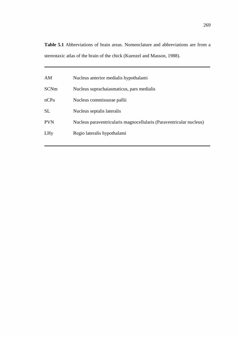
269
Table 5.1 Abbreviations of brain areas. Nomenclature and abbreviations are from a
stereotaxic atlas of the brain of the chick (Kuenzel and Masson, 1988).
AM Nucleus anterior medialis hypothalami
SCNm Nucleus suprachaiasmaticus, pars medialis
nCPa Nucleus commissurae pallii
SL Nucleus septalis lateralis
PVN Nucleus paraventricularis magnocellularis (Paraventricular nucleus)
LHy Regio lateralis hypothalami

270
Figure 5.1 Schematic coronal brain sections showing the areas where the expression
of GnRH-I-ir (black squares) was observed (A-B). The sampling regions for counting
the number of GnRH-I-ir neurons in the nCPa (B) are represented by rectangles.
Coronal illustrations were redrawn from the stereotaxic atlas of the chick brain
(Kuenzel and Masson, 1988). For abbreviations, see Table 5.1.
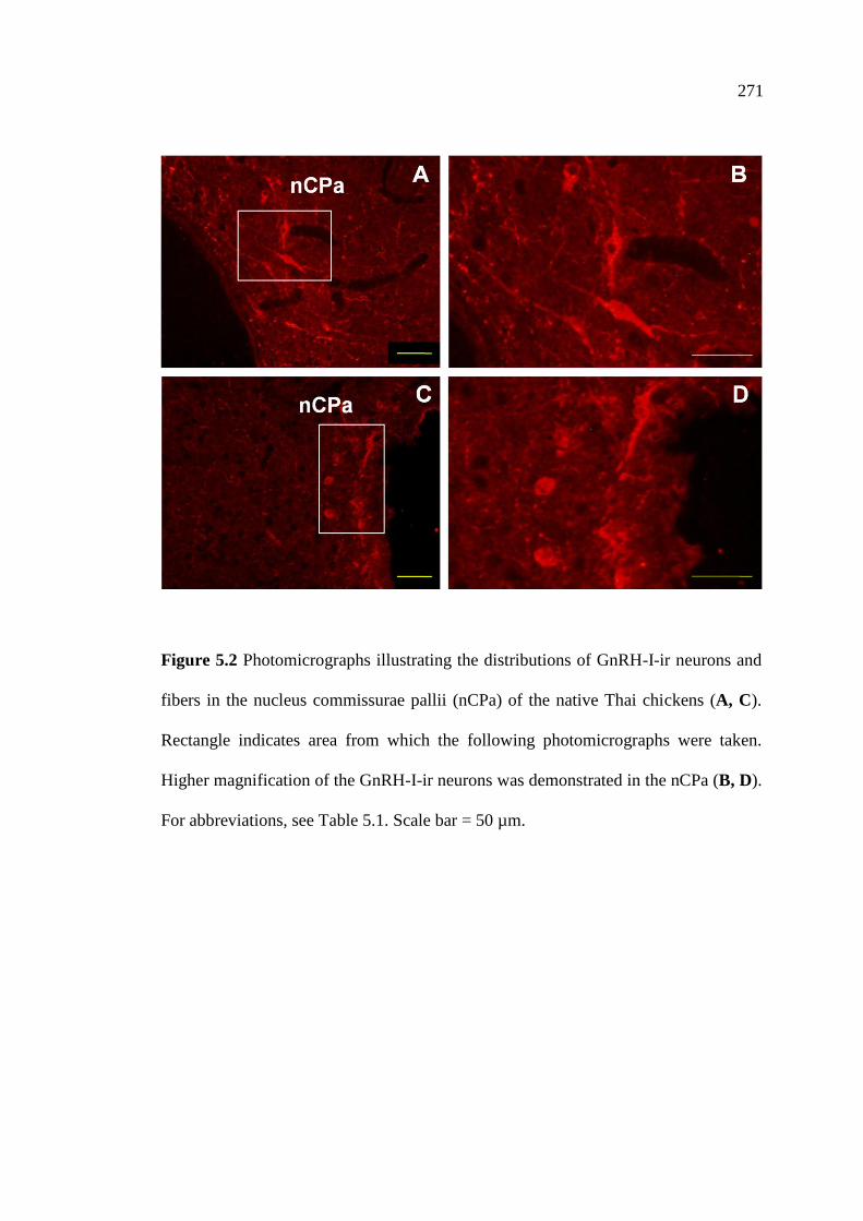
271
Figure 5.2 Photomicrographs illustrating the distributions of GnRH-I-ir neurons and
fibers in the nucleus commissurae pallii (nCPa) of the native Thai chickens (A, C).
Rectangle indicates area from which the following photomicrographs were taken.
Higher magnification of the GnRH-I-ir neurons was demonstrated in the nCPa (B, D).
For abbreviations, see Table 5.1. Scale bar = 50 µm.
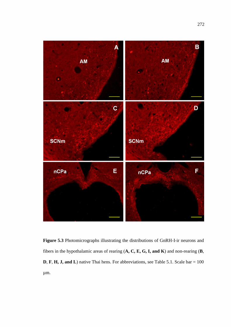
272
Figure 5.3 Photomicrographs illustrating the distributions of GnRH-I-ir neurons and
fibers in the hypothalamic areas of rearing (A, C, E, G, I, and K) and non-rearing (B,
D, F, H, J, and L) native Thai hens. For abbreviations, see Table 5.1. Scale bar = 100
µm.
273
Figure 5.3 Photomicrographs illustrating the distributions of GnRH-I-ir neurons and
fibers in the hypothalamic areas of rearing (A, C, E, G, I, and K) and non-rearing (B,
D, F, H, J, and L) native Thai hens. For abbreviations, see Table 5.1. Scale bar = 100
µm (continued).
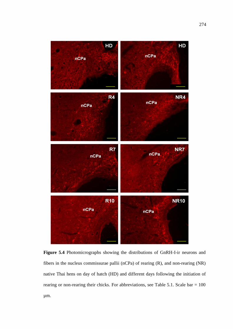
274
Figure 5.4 Photomicrographs showing the distributions of GnRH-I-ir neurons and
fibers in the nucleus commissurae pallii (nCPa) of rearing (R), and non-rearing (NR)
native Thai hens on day of hatch (HD) and different days following the initiation of
rearing or non-rearing their chicks. For abbreviations, see Table 5.1. Scale bar = 100
µm.

275
Figure 5.4 Photomicrographs showing the distributions of GnRH-I-ir neurons and
fibers in the nucleus commissurae pallii (nCPa) of rearing (R), and non-rearing (NR)
native Thai hens on different days following the initiation of rearing or non-rearing
their chicks. For abbreviations, see Table 5.1. Scale bar = 100 µm (continued).

276
Figure 5.5 Changes in the number of GnRH-I-ir neurons in the nCPa of rearing (R)
and non-rearing (NR) native Thai hens (n=6). Values are presented as mean ± SEM.
Significant differences between means in each group at different time points are
denoted by different letters (P<0.05) and *P<0.05 for a comparison between group at
a given time point.

277
Table 5.2 The number of GnRH-I-ir neurons (Mean ± SEM) in the nCPa of rearing (R) and non-rearing (NR) native Thai hens at
different days of rearing or non-rearing their chicks (n=6). Significant differences between means in each group at different time points
are denoted by different letters (P<0.05) and *P<0.05 for a comparison between group at a given time point.
Group Days Following Rearing or Non-Rearing Their Chicks
Day of Hatch 4 7 10 14 17 21
R 0.21 ± 0.12A 0.54 ± 0.21
A 0.29 ± 0.12
A 0.54 ± 0.36
A 0.83 ± 0.36
A 0.25 ± 0.13
A 0.71 ± 0.30
A
NR N/A 0.79 ± 0.31ab
1.21 ± 0.48ab
1.08 ± 0.37ab
2.75 ± 0.95ab
3.38 ± 1.28b*
2.46 ± 0.67ab*
277
278
5.5 Discussion
The results from the present study demonstrate that GnRH-I-ir neurons and
fibers were distributed throughout the hypothalamic areas of R and NR hens and were
predominantly expressed in the nCPa area. GnRH-I-ir fibers were observed within the
AM, SCNm, SL, PVN, and LHy of both R and NR hens. A high accumulation of
GnRH-I-ir neurons was found in the nCPa of NR hens. Changes in the number of
GnRH-I-ir neurons within the hypothalamic areas of R and NR hens were also
observed in the nCPa. The number of GnRH-I-ir neurons in the nCPa was low in the
R hens and significantly increased by day 17 after the chicks were removed from the
hens. These findings indicate, for the first time, an association of the GnRH/FSH-LH
system with rearing behavior in this non-photoperiodic, continuously breeding avian
species.
In the present study, the distributions of GnRH-I-ir neurons and fibers were
observed throughout the hypothalamus of R and NR hens, with the greatest density
found within the nCPa area and the differential expression of GnRH-I neurons
observed in both groups. High expression of GnRH-I-ir neurons was found in the
nCPa of NR hens, whereas lower numbers of them were observed in the nCPa of R
hens, suggesting that GnRH-I expression in this hypothalamic area may be involved
in the reproductive regulatory system of the native Thai chickens. The obvious group
of GnRH-I-ir neurons and fibers found in the nCPa in this study corresponded with
previous studies in the native Thai chickens, demonstrating that the majority of
GnRH-I-ir neurons and fibers are distributed primarily in the preoptic region
throughout the caudal end of the hypothalamus, where the greatest density of GnRH-
I-ir neurons was found within the nCPa (Sartsoongnoen, 2007; Prakobsaeng et al.,
279
2009; Prakobsaeng, 2010; Sartsoongnoen et al., 2012). Further evidence supporting
these findings are studies in Japanese quails, domestic chickens, and turkeys,
indicating that the greatest numbers of GnRH-I-ir neurons and GnRH-I mRNA
expression are found in the nCPa (Teruyama and Beck, 2000; Kang et al., 2006;
Kuenzel and Golden, 2006). The extensive distribution of GnRH-I immunoreactivity
in the present study is consistent with the important role played by this hypothalamic
neuropeptide in gonadotropin secretion and in the control of reproductive activity, as
previously reported in other avian species (Mikami et al., 1988; Katz et al., 1990;
Silver et al., 1992; Millam et al., 1993; van Gils et al., 1993; Dawson and Goldsmith,
1997; Sharp et al., 1998).
Changes in the number of GnRH-I-ir neurons were observed in the nCPa of R
and NR hens, and the numbers of GnRH-I-ir neurons were compared. The number of
GnRH-I-ir neurons in the nCPa was low in the R hens and did not differ at different
times during observation of the rearing period. These results correspond with the
changes of these hypothalamic neurons in the nCPa throughout the reproductive cycle
of the native Thai chickens. It was reported that the greatest number of GnRH-I-ir
neurons in the nCPa is found in laying hens, which then decreases in incubating and
non-laying birds, with the lowest numbers found during the rearing stage. Moreover,
nest deprivation of incubating native Thai hens increases the number of GnRH-I-ir
neurons in the nCPa (Sartsoongnoen et al., 2012). Furthermore, changes in GnRH
synthesis and secretion are correlated with the reproductive stage of other birds
(Sharp et al., 1990; Rozenboim et al., 1993a; 1993b; Dunn et al., 1996; Kang et al.,
2006). It has been reported that birds at the peak level of reproductive activity have
more GnRH-I-ir neurons and fibers than those of sexually inactive or photorefractory

280
birds (Sharp et al., 1990; Sharp, 1993; Hahn and Ball, 1995; Parry et al., 1997; Cho et
al., 1998; Stevenson and MacDougall-Shackleton, 2005; Stevenson and Ball, 2009).
Hypothalamic GnRH mRNA expression is greater in laying hens than that of
incubating hens (Dunn et al., 1996; Kang et al., 2006) and is lowest in photorefractory
hens (Kang et al., 2006). In addition, GnRH peptide contents of discrete medial
preoptic, infundibulum, and arcuate samples are higher in laying hens than those of
non-laying hens (Advis et al., 1985). Recently, it has been suggested that changes in
the number of GnRH-I neurons in the nCPa are key regulators of the avian
reproductive cycle, whether birds are photoperiodic or equatorial breeders
(Sartsoongnoen et al., 2012).
Disruption of rearing behavior by removing the chicks from the hens results in
an immediate increase in plasma LH and estradiol concentrations and a marked
decrease in plasma PRL levels (Sharp et al., 1979; Leboucher et al., 1990; Kuwayama
et al., 1992; Chaiyachet et al., 2010). In the present study, the numbers of GnRH-I-ir
neurons in the nCPa of the NR hens were gradually increased at day 4 and
significantly increased by day 17 when compared with the R hens during observation
periods. These results are supported by the studies indicating that after rearing chicks
hens and subsequent that hens had been removed their entire chicks exhibit an abrupt
increase in plasma LH levels concurrent with a decrease in plasma PRL levels
(Leboucher et al., 1990). Indeed, it has been reported that the secretion of LH and
estradiol are gradually increased after hatching or the presence of chicks and reach
peak values immediately after removal of chicks (Sharp et al., 1979; 1988;
Kuwayama et al., 1992; Dunn et al., 1996; Richard-Yris et al., 1998). This increase in
plasma LH levels is associated with a significant increase in hypothalamic cGnRH-I
281
mRNA (Dunn et al., 1996; Dunn and Sharp, 1999). Additionally, it also has been
reported that ovary mass of birds is positively correlated with GnRH-I mRNA levels
in the brain of sexually mature passerine songbirds (Ubuka and Bentley, 2009).
Removal of chicks from the care of hens results in an increase in the number
of GnRH-I-ir neurons in the nCPa (this study) and a decrease in VIP-ir neurons in the
nucleus inferioris hypothalami (IH) and nucleus infundibuli hypothalami (IN) and a
marked decline in plasma PRL levels (Chaiyachet et al., 2010). In addition, disruption
of rearing behavior increases ovary and oviduct weights, and hens resume egg laying
within 21 days after removing of chicks. Therefore, the number of GnRH-I-ir neurons
in the nCPa, which decrease in the R hens but increase in the NR hens, may be
involved in the regulation of PRL and LH secretions and rearing behavior in the
native Thai chickens. These findings are further supported by studies in nest-deprived
native Thai hens showing a significant increase in the number of GnRH-I-ir neurons
in the nCPa (Sartsoongnoen et al., 2012), a decrease in VIP-ir neurons in the IH-IN,
and a precipitous decline in plasma PRL levels, which are accompanied by an
increase in ovary and oviduct weights following the start of the new laying cycle
(Prakobsaeng et al., 2011).
In avian species, it has been suggested that the presence of newly hatched
chicks may suppress LH secretion and prevent decreases in plasma PRL levels, and
depressed LH secretion is associated with a decrease in the expression of
hypothalamic GnRH-I. This decrease is thought to be regulated by the inhibitory
effect of PRL (Sharp et al., 1988; Kuwayama et al., 1992; Dunn et al., 1996).
Moreover, PRL acts concomitantly with VIP to inhibit LH by means of reduction of
GnRH content in the hypothalamus (Rozenboim et al., 1993b). Immunoneutralization
282
of VIP increases pituitary LH-β and FSH-β mRNAs and is accompanied by a decline
in PRL mRNA expression (Ahn et al., 2001). The neuronal interactions between the
DAergic, VIPergic and GnRH-I neuronal systems in the regulation of reproductive
activities in birds have been well established. The relationship between the DAergic
and the GnRH-I systems has been demonstrated by photostimulation in turkeys,
revealing that during the photoinducible phase, the number of activated DA neurons
in the nucleus premamillaris and GnRH-I neurons in the nCPa are increased, as well
as an up-regulation of GnRH-I mRNA expression (Thayananuphat et al., 2007). It has
been reported that GnRH perikarya axons terminate in the ME, which is in close
proximity to the terminals of tuberoinfundibular DA neurons (Ajika, 1979;
Merchenthaler et al., 1984; Ugrumov et al., 1989). It has also been proposed that DA
from the tuberoinfundibular area may be one of the putative neurotransmitters
responsible for the increased activity of GnRH within the ME of chicks showing
precocious puberty (Fraley and Kuenzel, 1993). Furthermore, DA axons and terminals
are found intermingled with VIP neurons in the infundibular nuclear complex, GnRH
neurons in the POA, and with both VIP and GnRH terminals in the external layer of
the ME (Contijoch et al., 1992; Fraley and Kuenzel, 1993). Moreover, it has been
suggested that the interactions between the VIP and the GnRH systems are innervated
by VIP-ir fibers and GnRH-I-ir neurons in the lateral septal organ in the nucleus
accumbens, the caudal-most septal area, and the ME (Teruyama and Beck, 2001).
Activation of DAergic neurons in the nucleus mamillaris lateralis is associated with
the activation of GnRH-I and VIP neurons and the release of LH and PRL (Al-Zailaie
et al., 2006). Immunohistochemical studies of the native Thai chickens suggested that
the expression of VIP neurons in the IH-IN following hatching of the young may, in
283
part, account for the difference in reproductive neuroendocrine responses of this
equatorial bird (Kosonsiriluk et al., 2008). It has been further demonstrated that the
increase in GnRH-I-ir neurons caused by nest deprivation is related to the DAergic
system (Sartsoongnoen et al., 2012). It has been reported that DA-ir neurons in the
nucleus intramedialis increase significantly during the incubating stage
(Sartsoongnoen et al., 2008), and nest deprivation results in decreased DA-ir neurons
in this area, paralleling decreased VIP-ir neurons in the IH-IN (Prakobsaeng et al.,
2011). These data well support an association of the neuronal interactions between
GnRH-Iergic, VIPergic, and DAergic systems in the regulation of reproductive
activity in the native Thai chickens.
In summary, this present study clearly demonstrates that disruption of rearing
behavior by removing the chicks from the hens increases the number of GnRH-I-ir
neurons in the nCPa, suggesting that GnRH-I neurons in this hypothalamic area may
be involved in the suppression of rearing behavior in the native Thai chickens.
Furthermore, the initiation of maternal care behavior in the native Thai chickens
might be, in part, regulated by the differential expression of GnRH-I neurons in the
nCPa. Alternatively, the changes noted may be restricted to the reinitiation of
reproductive activity and egg laying that follows removal of chicks from the hen.
284
5.6 References
Advis, J.P., Contijoch, A.M., and Johnson, A.L. (1985). Discrete hypothalamic
distribution of luteinizing hormone-releasing hormone (LHRH) content and of
LHRH-degrading activity in laying and nonlaying hens. Biol Reprod 32: 820-
827.
Ahn, J., You, S.K., Kim, H., Chaiseha, Y., and El Halawani, M.E. (2001). Effects of
active immunization with inhibin alpha subunit on reproductive characteristics
of turkey hens. Biol Reprod 65: 1594-1600.
Ajika, K. (1979). Simultaneuos localization of LHRH and catecholamines in rat
hypothalamus. J Anat 128: 331-347.
Al-Zailaie, K.A., Kang, S.W., Youngren, O.M., Thayananuphat, A., Bakken, T.,
Chaiseha, Y., Millam, J.R., Proudman, J.A., and El Halawani, M.E. (2006).
Identification of dopamine, gonadotrophin-releasing hormone-I, and
vasoactive intestinal peptide neurones activated by electrical stimulation to the
medial preoptic area of the turkey hypothalamus: A potential reproductive
neuroendocrine circuit. J Neuroendocrinol 18: 514-525.
Bakker, J., and Baum, M.J. (2000). Neuroendocrine regulation of GnRH release in
induced ovulators. Front Neuroendocrinol 21: 220-262.
Bentley, G.E., Moore, I.T., Sower, S.A., and Wingfield, J.C. (2004). Evidence for a
novel gonadotropins-releasing hormone in hypothalamic and forebrain areas in
songbirds. Brain Behav Evol 63: 34-46.
Bhatt, R., Youngren, O.M., Kang, S.W., and El Halawani, M.E. (2003). Dopamine
infusion in the third ventricle increases gene expression of hypothalamic

285
vasoactive intestinal peptide and pituitary prolactin and luteinizing hormone
beta subunit in the turkey. Gen Comp Endocrinol 130: 41-47.
Blahser, S., King, J.A., and Kuenzel, W.J. (1989). Testing of arg-8-gonadotropin
releasing hormone-directed antisera by immunological and
immunocytochemical methods for use in comparative studies. Histochemistry
93: 39-48.
Bluhm, C.K., Schwabl, H., Schwabl, I., Perera, A., Follett, B.K., Goldsmith, A.R.,
and Gwinner, E. (1991). Variations in hypothalamic gonadotrophin releasing
hormone content, plasma and pituitary LH and in vitro testosterone release in
a long distance migratory bird, the garden warbler (Sylvia borin), under
constant photoperiods. J Endocrinol 128: 339-345.
Bons, N., Kerdelhue, B., and Assenmacher, I. (1978). Immunocytochemical
identification of an LHRH-producing system originating in the preoptic
nucleus of the duck. Cell Tissue Res 188: 99-106.
Chaiseha, Y., and El Halawani, M.E. (2005). Neuroendocrinology of the female
turkey reproductive cycle. J Poult Sci 42: 87-100.
Chaiseha, Y., Youngren, O.M., Al-Zailaie, K.A., and El Halawani, M.E. (2003).
Expression of D1 and D2 dopamine receptors in the hypothalamus and
pituitary during the turkey reproductive cycle: Colocalization with vasoactive
intestinal peptide. Neuroendocrinology 77: 105-118.
Chaiyachet, O-A., Chokchaloemwong, D., Prakobsaeng, N., Sartsoongnoen, N.,
Kosonsiriluk, S., Rozenboim, I., El Halawani, M.E., Porter, T.E., and
Chaiseha, Y. (2010). Neuroendocrine regulation of rearing behavior in the
native Thai hen. Poult Sci 89 (Suppl 1): 679.
286
Cho, R.N., Hahn, T.P., MacDougall-Shackleton, S., and Ball, G.F. (1998). Seasonal
variation in brain GnRH in free-living breeding and photorefractory house
finches (Carpodacus mexicanus). Gen Comp Endocrinol 109: 244-250.
Contijoch, A.M., Gonzalez, C., Singh, H.N., Malamed, S., Troncoso, S., and Advis,
J.P. (1992). Dopaminergic regulation of luteinizing hormone-releasing
hormone release at the median eminence level: Immunocytochemical and
physiological evidence in hens. Neuroendocrinology 55: 290-300.
Davies, D.T., and Follett, B.K. (1975). The neuroendocrine control of gonadotrophin
release in Japanese quail. I. The role of the tuberal hypothalamus. Proc R Soc
Lond B 191: 285-301.
Dawson, A., and Goldsmith, A. R. (1997). Changes in gonadotrophin-releasing
hormone (GnRH-I) in the pre-optic area and median eminence of starlings
(Sturnus vulgaris) during the recovery of photosensitivity and during
photostimulation. J Reprod Fertil 111: 1-6.
Dawson, A., Follett, B.K., Goldsmith, A.R., and Nicholls, T.J. (1985). Hypothalamic
gonadotrophin-releasing hormone and pituitary and plasma FSH and prolactin
during photorefractoriness in intact and thyroidectomized starlings (Sturnus
vulgaris). J Endocrinol 105: 71-77.
Dawson, A., Talbot, R.T., Dunn, I.C., and Sharp, P.J. (2002). Changes in basal
hypothalamic chicken gonadotropin-releasing hormone-I and vasoactive
intestinal polypeptide associated with a photo-induced cycle in gonadal
maturation and prolactin secretion in intact and thyroidectomized starlings
(Sturnus vulgaris). J Neuroendocrinol 14: 533-539.

287
Deviche, P.J., Saldanha, C.J., and Silver, R. (2000). Changes in brain gonadotropin
releasing hormone- and vasoactive intestinal polypeptide-like
immunoreactivity accompanying reestablishment of photosensitivity in male
dark-eyed junco (Junco hyemalis). Gen Comp Endocrinol 117: 8-19.
Dunn, I.C., and Sharp, P.J. (1999). Photo-induction of hypothalamic gonadotrophin
releasing hormone-I mRNA in the domestic chicken: A role for oestrogen? J
Neuroendocrinol 11: 371-375.
Dunn, I.C., Beattie, K.K., Maney, D., Sang, H.M., Talbot, R.T., Wilson, P.W., and
Sharp, P.J. (1996). Regulation of chicken gonadotropin-releasing hormone-I
mRNA in incubating, nest-deprived and laying bantam hens.
Neuroendocrinology 63: 504-513.
Dunn, I.C., Chen, Y., Hook, C., Sharp, P.J., and Sang, H.M. (1993). Characterization
of the chicken preprogonadotrophin-releasing hormone-I gene. J Mol
Endocrinol 11: 19-29.
El Halawani, M.E., Silsby, J.L., and Fehrer, S.C. (1988). Basal and hypothalamic
extract-induced luteinizing hormone and prolactin secretion by cultures
anterior pituitary cells from female turkeys in various stages of the
reproductive cycle. Gen Comp Endocrinol 71: 45-54.
El Halawani, M.E., Silsby, J.L., Foster, L.K., Rozenboim, I., and Foster, D.N. (1993).
Ovarian involvement in the suppression of luteinizing hormone in the
incubating turkey (Meleagris gallopavo). Neuroendocrinology 58: 35-41.
Foster, R.G., Plowman, G., Goldsmith, A.R., and Follett, B.K. (1987).
Immunocytochemical demonstration of marked changes in the luteinizing

288
hormone releasing hormone system of photosensitive and photorefractory
European starlings. J Endocrinol 115: 211-220.
Fraley, G.S., and Kuenzel, W.J. (1993). Immunocytochemical and histochemical
analyses of gonadotrophin releasing hormone, tyrosine hydroxylase, and
cytochrome oxidase reactivity within the hypothalamus of chicks showing
early sexual maturation. Histochemistry 99: 221-229.
Goldsmith, A.R., Ivings, W.E., Pearce-Kelly, A.S., Parry, D.M., Plowman, G.,
Nicholls, T.J., and Follett, B.K. (1989). Photoperiodic control of the
development of the LHRH neurosecretory system of European starlings
(Sturnus vulgaris) during puberty and the onset of photorefractoriness. J
Endocrinol 122: 255-268.
Gorbman, A., and Sower, S.A. (2003). Evolution of the role of GnRH in animal
(Metazoan) biology. Gen Comp Endocrinol 134: 207-213.
Guemene, D., and Williams, J.B. (1992). In-vitro and in-vivo responses to chicken
LHRH-I and chicken LHRH-II in male turkeys (Meleagris gallopavo). J
Endocrinol 132: 387-393.
Guemene, D., and Williams, J.B. (1999). LH responses to chicken luteinizing
hormone-releasing hormone I and II in laying, incubating, and out of lay
turkey hens. Domest Anim Endocrinol 17: 1-15.
Hahn, T.P., and Ball, G.F. (1995). Changes in brain GnRH associated with
photorefractoriness in house sparrows (Passer domesticus). Gen Comp
Endocrinol 99: 349-363.
Hertelendy, F., Lintner, F., Asem, E.K., and Raab, B. (1982). Synergistic effect of
gonadotrophin releasing hormone on LH-stimulated progesterone production

289
in granulosa cells of the domestic fowl (Gallus domesticus). Gen Comp
Endocrinol 48: 117-122.
Jones, S.W. (1987). Chicken II luteinizing hormone-releasing hormone inhibits the
M-current of bullfrog sympathetic neurons. Neurosci Lett 80: 180-184.
Kang, S.W., Thayananuphat, A., Rozenboim, I., Millam, J.R., Proudman, J.A., and El
Halawani, M.E. (2006). Expression of hypothalamic GnRH-I mRNA in the
female turkey at different reproductive states and following photostimulation.
Gen Comp Endocrinol 146: 86-94.
Katz, I.A., Millar, R.P., and King, J.A. (1990). Differential regional distribution and
release of two forms of gonadotropin-releasing hormone in the chicken brain.
Peptides 11: 443-450.
Kavanaugh, S.I., Nozaki, M., and Sower, S.A. (2008). Origins of gonadotropin-
releasing hormone (GnRH) in vertebrates: Identification of a novel GnRH in a
basal vertebrate, the sea lamprey. Endocrinology 149: 3860-3869.
King, J.A., and Millar, R.P. (1982). Structure of chicken hypothalamic luteinizing
hormone-releasing hormone. I. Structural determination on partially purified
material. J Biol Chem 257: 10722-10728.
Kosonsiriluk, S. (2007). Biological studies of the reproductive cycle and the effects
of photoperiod upon the reproductive system in the female native Thai
chicken. Ph.D. Dissertation, Suranaree University of Technology, Nakhon
Ratchasima, Thailand.
Kosonsiriluk, S., Sartsoongnoen, N., Chaiyachet, O-A., Prakobsaeng, N., Songserm,
T., Rozenboim, I., El Halawani, M.E., and Chaiseha, Y. (2008). Vasoactive

290
intestinal peptide and its role in continuous and seasonal reproduction in birds.
Gen Comp Endocrinol 159: 88-97.
Kuenzel, W.J., and Blahser, S. (1991). The distribution of gonadotropin-releasing
hormone (GnRH) neurons and fibers throughout the chick brain (Gallus
domesticus). Cell Tissue Res 264: 481-495.
Kuenzel, W.J., and Golden, C.D. (2006). Distribution and change in number of
gonadotropin-releasing hormone-1 neurons following activation of the
photoneuroendocrine system in the chick, Gallus gallus. Cell Tissue Res 325:
501-512.
Kuenzel, W.J., and Masson, M. (1988). A stereotaxic atlas of the brain of the chick
(Gallus domesticus). Johns Hopkins University Press, Baltimore, Maryland,
USA.
Kuenzel, W.J., and van Tienhoven, A. (1982). Nomenclature and location of avian
hypothalamic nuclei and associated circumventricular organs. J Comp Neurol
206: 293-313.
Kuwayama, T., Shimada, K., Saito, N., Ohkubo, T., Sato, K., Wada, M., and Ichinoe,
K. (1992). Effects of removal of chicks from hens on concentrations of
prolactin, luteinizing hormone and oestradiol in plasma of brooding Gifujidori
hens. J Reprod Fertil 95: 617-622.
Leboucher, G., Richard-Yris, M.A., Williams, J., and Chadwick, A. (1990).
Incubation and maternal behaviour in domestic hens: Influence of the presence
of chicks on circulating luteinising hormone, prolactin and oestradiol and on
behaviour. Br Poult Sci 31: 851-862.

291
Li, Q., Tamarkin, L., Levantine, P., and Ottinger, M.A. (1994). Estradiol and
androgen modulate chicken luteinizing hormone-releasing hormone-I release
in vitro. Biol Reprod 51: 896-903.
Meddle, S.L., and Follett, B.K. (1997). Photoperiodically driven changes in Fos
expression within the basal tuberal hypothalamus and median eminence of
Japanese quail. J Neurosci 17: 8909-8918.
Merchenthaler, I., Gore, T., Setalo, G., Petrusz, P., and Flerko, B. (1984).
Gonadotropin-releasing hormone (GnRH) neurons and pathways in the rat
brain. Cell tissue Res 237: 15-29.
Mikami, S., Yamada, S., Hasegawa, Y., and Miyamoto, K. (1988). Localization of
avian LHRH-immunoreactive neurons in the hypothalamus of the domestic
fowl, Gallus domesticus, and the Japanese quail, Coturnix coturnix. Cell
Tissue Res 251: 51-58.
Millam, J.R., Craig-Veit, C.B., Adams, T.E., and Adams, B.M. (1989). Avian
gonadotrophin-releasing hormones I and II in the brain and other tissues in
turkey hens. Comp Biochem and Physiol 94: 771-776.
Millam, J.R., Faris, P.L., Youngren, O.M., El Halawani, M.E., and Hartman, B.K.
(1993). Immunohistochemical localization of chicken gonadotrophin-releasing
hormones I and II (cGnRH-I and II) in turkey hen brain. J Comp Neurol 333:
68-82.
Millar, R.P., Milton, R.C., Follett, B.K., and King, J.A. (1986). Receptor binding and
gonadotropin-releasing activity of a novel chicken gonadotropin-releasing
hormone ([His5, Trp7, Tyr8]GnRH) and a D-Arg6 analog. Endocrinology
119: 224-231.
292
Miyamoto, K., Hasegawa, Y., Minegishi, T., Nomura, M., Takahashi, Y., Igarashi,
M., Kangawa, K., and Matsuo, H. (1982). Isolation and characterization of
chicken hypothalamic luteinizing hormone-releasing hormone. Biochem
Biophys Res Commun 107: 820-827.
Parry, D.M., Goldsmith, A.R., Millar, R.P., and Glennie, L.M. (1997).
Immunocytochemical localization of GnRH precursor in the hypothalamus of
European starlings during sexual maturation and photorefractoriness. J
Neuroendocrinol 9: 235-243.
Peczely, P. (1989). The role of gonadotropin releasing hormone (Gn-RH) in the
regulation of gonadal functions of birds. Acta Biol Hung 40: 161-193.
Prakobsaeng, N. (2010). Effects of incubation behavior upon the neuroendocrine
regulation of the reproductive system in the female native Thai chicken.
Ph.D. Dissertation, Suranaree University of Technology, Nakhon Ratchasima,
Thailand.
Prakobsaeng, N., Sartsoongnoen, N., Kosonsiriluk, S., Rozenboim, I., El Halawani,
M.E., Porter, T.E., and Chaiseha, Y. (2009). Changes in vasoactive intestinal
peptide and gonadotropin releasing hormone-I immunoreactivity in the brain
of nest-deprived native Thai hen. Poult Sci 88 (Suppl 1): 121-122.
Prakobsaeng, N., Sartsoongnoen, N., Kosonsiriluk, S., Chaiyachet, O-A.,
Chokchaloemwong, D., Rozenboim, I., El Halawani, M.E., Porter T.E., and
Chaiseha, Y. (2011). Changes in vasoactive intestinal peptide and tyrosine
hydroxylase immunoreactivity in the brain of nest-deprived native Thai hen.
Gen Comp Endocrinol 171: 189-196.
293
Proudman, J.A., Scanes, C.G, Johannsen, S.A., Berghman, L.R., and Camp, M.J.
(2006). Comparison of the ability of the three endogenous GnRHs to stimulate
release of follicle-stimulating hormone and luteinizing hormone in chickens.
Domest Anim Endocrinol 31: 141-153.
Ramirez, V.D., Feder, H.H., and Sawyer, C.H. (1984). The role of brain
catecholamines in regulation of LH secretion. In Frontiers in
Neuroendocrinology, pp 27-84. Eds. Ganong, W.P., and Martini, L., Raven
Press, New York, USA.
Richard-Yris, M.A., Guemene, D., Lea, R.W., Sharp, P.J., Bedecarrats, G., Foraste,
M., and Wauters, A.M. (1998). Behaviour and hormone concentrations in nest
deprived and renesting hens. Br Poult Sci 39: 309-317.
Rivier, J. (2001). GnRH Agonists: The future. In GnRH Analogues: The State of
the Art 2001, pp 1-14. Ed. Lunenfeld, B. Parthenon Publishing, Lancaster,
USA.
Rozenboim, I., Silsby, J.L., Tabibzadeh, C., Pitts, G.R., Youngren, O.M., and El
Halawani, M.E. (1993a). Hypothalamic and posterior pituitary content of
vasoactive intestinal peptide and gonadotropin releasing hormones I and II in
the turkey hen. Biol Reprod 49: 622-626.
Rozenboim, I., Tabibzadeh, C., Silsby, J.L., and El Halawani, M.E. (1993b). Effect of
ovine prolactin administration on hypothalamic vasoactive intestinal peptide
(VIP), gonadotropin releasing hormone I and II content, and anterior pituitary
VIP receptors in laying turkey hens. Biol Reprod 48: 1246-1250.
294
Saldanha, C.J., Deviche, P.J., and Silver, R. (1994). Increased VIP and decreased
GnRH expression in photorefractory dark-eyed juncos (Junco hyemalis). Gen
Comp Endocrinol 93: 128-136.
Sartsoongnoen, N. (2007). Neuroendocrinology of the reproductive cycle in the
female native Thai chicken: Roles of dopamine and gonadotropin
releasing hormone. Ph.D. Dissertation, Suranaree University of Technology,
Nakhon Ratchasima, Thailand.
Sartsoongnoen, N., Kosonsiriluk, S., Kang, S.W., Millam, J.R., El Halawani, M.E.,
and Chaiseha, Y. (2006). Distribution of cGnRH-I immunoreactive neurons
and fibers in the brain of native Thai chicken (Gallus domesticus). Poult Sci
85 (Suppl 1): 45.
Sartsoongnoen, N., Kosonsiriluk, S., Prakobsaeng, N., Songserm, T., Rozenboim, I.,
El Halawani, M.E., and Chaiseha, Y. (2008). The dopaminergic system in the
brain of the native Thai chicken, Gallus domesticus: Localization and
differential expression across the reproductive cycle. Gen Comp Endocrinol
159: 107-115.
Sartsoongnoen, N., Prakobsaeng, N., Kosonsiriluk, S., Chaiyachet, O-A.,
Chokchaloemwong, D., El Halawani, M.E., and Chaiseha, Y. (2012).
Distribution and variation in gonadotropin releasing hormone-I (GnRH-I)
immunoreactive neurons in the brain of the native Thai chicken during the
reproductive cycle. Acta Histochem 114: 409-420.
Sharp, P.J. (1993). Photoperiodic control of reproduction in the domestic hen. Poult
Sci 72: 897-905.
295
Sharp, P.J., and Blache, D. (2003). A neuroendocrine model for prolactin as the key
mediator of seasonal breeding in birds under long- and short-day
photoperiods. Can J Physiol Pharmacol 81: 350-358.
Sharp, P.J., Dawson, A., and Lea, R.W. (1998). Control of luteinizing hormone and
prolactin secretion in birds. Comp Biochem Physiol C Pharmacol Toxicol
Endocrinol 119: 275-282.
Sharp, P.J., Macnamee, M.C., Sterling, R.J., Lea, R.W., and Pedersen, H.C. (1988).
Relationships between prolactin, LH and broody behavior in bantam hens. J
Endocrinol 118: 279-286.
Sharp, P.J., Macnamee, M.C., Talbot, R.T., Sterling, R.J., and Hall, T.R. (1984).
Aspects of the neuroendocrine control of ovulation and broodiness in the
domestic hen. J Exp Zool 232: 475-483.
Sharp, P.J., Scanes, C.G., Williams, J.B., Harvey, S., and Chadwick, A. (1979).
Variations in concentrations of prolactin, luteinizing hormone, growth
hormone and progesterone in the plasma of broody bantams (Gallus
domesticus). J Endocrinol 80: 51-57.
Sharp, P.J., Talbot, R.T., Main, G.M., Dunn, I.C., Fraser, H.M., and Huskisson, N.S.
(1990). Physiological roles of chicken LHRH-I and -II in the control of
gonadotrophin release in the domestic chicken. J Endocrinol 124: 291-299.
Sherwood, N.M., Wingfield, J.C., Ball, G.F., and Dufty, A.M. (1988). Identity of
gonadotropin-releasing hormone in passerine birds: Comparison of GnRH in
song sparrow (Melospiza melodia) and starling (Sturnus vulgaris) with five
vertebrate GnRHs. Gen Comp Endocrinol 69: 341-351.
296
Shimizu, M., and Bedecarrats, G.Y. (2006). Identification of a novel pituitary-specific
chicken gonadotropin-releasing hormone receptor and its splice variants. Biol
Reprod 75: 800-808.
Silver, R., Ramos, C., Machuca, H., and Silverin, B. (1992). Immunocytochemical
distribution of GnRH in the brain of adult and posthatching great tit (Parus
major) and ring dove (Streptopelia roseogrisea). Ornis Scand 23: 222-232.
SPSS Inc. (2004). SPSS Base 13.0 Users Guide. Prentice Hall, New Jersey, USA.
Stevenson, T.J., and Ball, G.F. (2009). Anatomical localization of the effects of
reproductive state, castration, and social milieu on cells immunoreactive for
gonadotropin-releasing hormone-I in male European starlings (Sturnus
vulgaris). J Comp Neurol 517: 146-155.
Stevenson, T.J., and Macdougall-Shackleton, S.A. (2005). Season- and age-related
variation in neural cGnRH-I and cGnRH-II immunoreactivity in house
sparrows (Passer domesticus). Gen Comp Endocrinol 143: 33-39.
Sun, Y.M., Dunn, I.C., Baines, E., Talbot, R.T., Illing, N., Millar, R.P., and Sharp,
P.J. (2001). Distribution and regulation by oestrogen of fully processed and
variant transcripts of gonadotropin releasing hormone I and gonadotropin
releasing hormone receptor mRNAs in the male chicken. J Neuroendocrinol
13: 37-49.
Teruyama, R., and Beck, M.M. (2000). Changes in immunoreactivity to anti-cGnRH-I
and -II are associated with photostimulated sexual status in male quail. Cell
Tissue Res 300: 413-426.

297
Teruyama, R., and Beck, M.M. (2001). Double immunocytochemistry of vasoactive
intestinal peptide and cGnRH-I in male quail: Photoperiodic effects. Cell
Tissue Res 303: 403-414.
Thayananuphat, A., Kang, S.W., Bakken, T., Millam, J.R., and El Halawani, M.E.
(2007). Rhythmic dependent light induction of gonadotrophin-releasing
hormone-I expression and activation of dopaminergic neurones within the
premammillary nucleus of the turkey hypothalamus. J Neuroendocrinol 19:
399-406.
Tsutsui, K., Saigoh, E., Ukena, K., Teranishi, H., Fujisawa, Y., Kikuchi, M., Ishii, S.,
and Sharp, P.J. (2000). A novel avian hypothalamic peptide inhibiting
gonadotropin release. Biochem Biophys Res Commun 275: 661-667.
Ubuka, T., and Bentley, G.E. (2009). Identification, localization, and regulation of
passerine GnRH-I messenger RNA. J Endocrinol 201: 81-87.
Ugrumov, M., Hisano, S., and Daikoku, S. (1989). Topographic relation between
tyrosine hydroxylase- and luteinizing hormone-releasing hormone-
immunoreactive fibers in the median eminence. Neurosci Lett 102: 159-164.
van Gils, J., Absil, P., Grauwels, L., Moons, L., Vandesande, F., and Balthazart, J.
(1993). Distribution of luteinizing hormone-releasing hormones I and II
(LHRH-I and -II) in the quail and chicken brain as demonstrated with
antibodies directed against synthetic peptides. J Comp Neurol 334: 304-323.
You, S., Foster, L.K., Silsby, J.L., El Halawani, M.E., and Foster, D.N. (1995).
Sequence analysis of the turkey LH beta subunit and its regulation by
gonadotrophin-releasing hormone and prolactin in cultured pituitary cells. J
Mol Endocrinol 14: 117-129.

298
CHAPTER VI
CONCLUSION
Native Thai chickens highly express maternal behaviors including incubation
behavior and broodiness or rearing behavior. These behaviors affect the production of
the native Thai chickens because they reduce the number of eggs produced.
Therefore, in order to improve the productivity of this chicken, it is very important to
understand the basic neuroendocrinology influencing its maternal behaviors,
especially the rearing behavior of the native Thai chickens. The avian reproductive
system is regulated by the hypothalamo-pituitary-gonadal axis. The two
neuroendocrine systems that play a pivotal role in the avian reproductive system are
the gonadotropin releasing hormone/follicle stimulating hormone-luteinizing hormone
(GnRH/FSH-LH) and vasoactive intestinal peptide/prolactin (VIP/PRL) systems.
Both systems are influenced by dopaminergic neurotransmission. Thus, the aims of
this dissertation were to investigate the roles of PRL, VIP, and GnRH on the
neuroendocrine regulation of rearing behavior in native Thai chickens.
The results revealed that plasma PRL concentrations remained at high levels
on the day the chicks were hatched, then rapidly decreased after the first week of
hatching and then remained at low levels throughout the eight weeks of the rearing
period (Figure 6.1A). The ovary and oviduct weights of hens rearing their chicks were
significantly decreased since the chicks were hatched then showed a significant
increase at week 8 of the rearing period (Figures 6.1B and C). During five weeks of
299
the rearing period, plasma PRL levels of hens rearing chicks (R) were compared with
those of non-rearing chicks hens (NR). The levels of plasma PRL were markedly
decreased in hens that had their chicks removed and reached the lowest levels by the
third week of separation from chicks. In the R hens, plasma PRL levels were high
when compared to those of the NR hens during the different observation periods
(Figure 6.2A). Disruption of rearing behavior by removing their chicks increased the
ovary and oviduct weights, the presence of ovarian follicles, and the number of egg
laying hens (Figures 6.2B and C).
The decrease of plasma PRL concentrations in the NR hens paralleled the
decline in the number of VIP-immunoreactive (VIP-ir) neurons in the nucleus
inferioris hypothalami (IH) and nucleus infundibuli hypothalami (IN; Figure 6.3A).
The number of VIP-ir neurons in the IH-IN remained high after the day the chicks
were hatched until day 7 of the rearing period and then sharply decreased from day 10
to day 21. When the chicks were deprived from the hens, VIP-ir neurons were
markedly decreased on day 4 and continued to be lower than those of R hens through
day 21 (Figure 6.3A). In contrast with the levels of plasma PRL and the numbers of
VIP-ir neurons in the IH-IN, the number of GnRH-I-ir neurons in the nucleus
commissurae pallii (nCPa) was low in the R group and significantly increased by day
17 after removal of chicks from the hens (Figure 6.3B). In addition, the effects of
removal of chicks from the care of hens induced ovarian and oviduct recrudescence
within 7 days, appearance of yellow follicles by day 14, and the initiation of a new
laying cycle by day 21 (Figures 6.3C and D).
In summary, the present study indicated an association between plasma PRL
levels, VIP neurons in the IH-IN, and GnRH-I neurons in the nCPa, suggesting that
300
the expression of rearing behavior in the native Thai chickens might be, in part,
regulated by the differential expression of VIP neurons in the IH-IN and GnRH-I
neurons in the nCPa. The relationship between hens and chicks while hens took care
of their young might slow down the decreases in circulating PRL levels and the
number of VIP-ir neurons in the IH-IN which, in turn, facilitate, stimulate or maintain
rearing behavior in the native Thai chickens. Taken together, the present findings
indicate that disruption of rearing behavior by removing the chicks from the hens
markedly decreases plasma PRL levels and the number of VIP-ir neurons in the IH-
IN, and is accompanied by an increase in the number of GnRH-I-ir neurons in the
nCPa, suggesting that the VIPergic system in the IH-IN and the GnRH system in the
nCPa may be involved in the regulation of the reproductive neuroendocrine system
and the initiation and maintenance of rearing behavior in native Thai chickens. It is
further indicated that the VIP/PRL system is not only a key regulator of incubation
behavior as it is well established, but it may also be involved in the regulation of
rearing behavior in the native Thai chicken. These findings further suggest that the
presence of chicks and/or physical contact with chicks induced maternal care
behavior in the native Thai hens as well. These findings also indicate, for the first
time, an association of GnRH/FSH-LH system with rearing behavior in this non-
photoperiodic, continuously breeding avian species. This confirms a pivotal role of
hypothalamic GnRH-I in controlling of avian reproduction of this equatorial species.

301
Figure 6.1 Changes in: A, plasma PRL concentration; B, ovary; C, oviduct weights
of native Thai hens rearing chicks during the eight weeks of rearing period. Values
are presented as mean ± SEM. Significant differences between means in each group
at different time points are denoted by different letters (P<0.05).

302
Figure 6.2 Changes in: A, plasma PRL concentration; B, ovary; C, oviduct weights
of rearing (R) and non-rearing (NR) native Thai hens. Values are presented as mean ±
SEM. Significant differences between means in each group at different time points
are denoted by different letters (P<0.05) and *P<0.05 for a comparison between
group at a given time point.

303
Figure 6.3 Changes in: A, the number of VIP-ir neurons in the IH-IN; B, the number
of GnRH-I-ir neurons in the nCPa; C, ovary; D, oviduct weights of rearing (R) and
non-rearing (NR) native Thai hens. Values are presented as mean ± SEM. Significant
differences between means in each group at different time points are denoted by
different letters (P<0.05) and *P<0.05 for a comparison between group at a given time
point.

CURRICULUM VITAE
NAME: Miss Orn-Anong Chaiyachet
DATE OF BIRTH: January 03, 1980
PLACE OF BIRTH: Roi Et, Thailand
NATIONALITY: Thai
EDUCATION:
2006-present Ph.D. Candidate (Environmental Biology)
Suranaree University of Technology, Thailand
2002-2005 M.Sc. (Biotechnology)
Khon Kaen University, Thailand
1998-2001 B.Sc. (2st Degree Honors, Biology)
Mahasarakham University, Thailand
AWARDS AND HONORS:
Scholarship from the Royal Golden Jubilee Ph.D. Program, the Thailand Research
Fund, Thailand
PUBLICATIONS:
4 international journals and 4 international meeting abstracts