diabetic foot infections: a comprehensive overview · ble revascularization, 3) relief of pressure...
TRANSCRIPT

26
Abstract. – Diabetic foot ulcers (DFUs), a mi-cro-vascular complication, are associated with a substantial increase in morbidity and mortal-ity. DFUs are a complicated mixture of neurop-athy, peripheral arterial diseases, foot deformi-ties, and infections. Foot infections are frequent and potentially devastating complications. In-fection prospers in more than half of all foot ul-cers and is the factor that most often leads to lower extremity amputation. The complications of microbial flora span the spectrum from su-perficial cellulitis to chronic osteomyelitis and gangrenous extremity lower limb amputations. Wounds without confirmed soft tissue or bone infections do not require antibiotic therapy. Mild and moderate infections need empiric therapy covering Gram-positive cocci, while severe in-fections caused by drug-resistant organisms require broad-spectrum anti-microbials target-ing aggressive Gram-negative aerobes and obli-gate anaerobes.
Key Words:Diabetes mellitus, Diabetic foot ulcer, Infection, Am-
putation, Antibiotics, Mmicrobiology.
Introduction
Infections in ulcerated feet in patients with dia-betes are a primary cause of morbidity, including discomfort, and reduced physical and mental qual-
ity of life, and they give rise to a need for visits by health-care providers, wound care, antimicro-bial therapy, and often surgical procedures/de-bridements. As such, these infections comprise the most frequent grounds for both diabetes-associat-ed hospitalization and lower extremity losses,. In an acute presentation with diabetic foot infection (DFI), there is frequently a delay in the recognition of the causative organism, which may compel the use of empirical antibiotics,. According to Peters, the incidence of foot infections in people with dia-betes ranges from an overall lifetime risk of 4% to a yearly risk of 7%. If infection advances to deeper structures, including the underlying bone, diabet-ic foot osteomyelitis (DFO) develops. DFIs are the most frequent diabetes-related complication re-quiring hospitalization, and DFO is present in 44-68% of patients with DFIs admitted to the hospital.
Infections in foot lesions should be clinically defined by the presence of inflammation or pu-rulence, and then classified by severity. This approach helps clinicians make decisions about which patients to hospitalize, send for imaging procedures or recommend for surgical interven-tions. Many organisms, alone or in combination, can cause DFIs, but Gram-positive cocci (GPC), especially staphylococci, are the most common.
To achieve more successful outcomes and ulti-mately avoid amputations, a systematic approach to the management of DFIs must be adopted. If
European Review for Medical and Pharmacological Sciences
D. PITOCCO1, T. SPANU2, M. DI LEO1, R. VITIELLO3, A. RIZZI1, L. TARTAGLIONE1, B. FIORI2, S. CAPUTO1, G. TINELLI4, F. ZACCARDI1, A. FLEX5, M. GALLI3, A. PONTECORVI6, M. SANGUINETTI2
1Diabetes Care Unit, Endocrinology, University Hospital “A. Gemelli”, Catholic University of the Sacred Heart, Rome, Italy2Institute of Microbiology, University Hospital “A. Gemelli”, Catholic University of the Sacred Heart, Rome, Italy3Institute of Orthopedic Surgery, Internal Medicine, University Hospital “A. Gemelli”, Catholic University of the Sacred Heart, Rome, Italy4Institute of Vascular Surgery, University Hospital “A. Gemelli”, Catholic University of the Sacred Heart, Rome, Italy5Institute of Internal Medicine, University Hospital “A. Gemelli”, Catholic University of the Sacred Heart, Rome, Italy6Institute of Endocrinology, University Hospital “A. Gemelli”, Catholic University of the Sacred Heart, Rome, Italy
Corresponding Author: Dario Pitocco, MD; e-mail: [email protected]
Diabetic foot infections: a comprehensive overview
2019; 23(2 Suppl.): 26-37

Diabetic foot infections: a comprehensive overview
27
not treated promptly and appropriately, DFI can become incurable or even lead to septic gangrene. At least 60% of non-traumatic lower limb ampu-tations occur among people with diabetes.
The existence of osteomyelitis further raises the costs of hospitalization because of the need for additional diagnostic studies, prolonged med-ical treatment and surgeries. Notably, the use of antibiotics in such cases is at least doubled. When
amputation is needed, a high-level (i.e., transtib-ial) procedure is often indicated, more because of irreversible ischaemia than because of uncon-trolled infection. However, most amputations reflect the multimodal foot problems related to diabetes, which highlights the need for a multidis-ciplinary approach (Figure 1). All clinicians reg-ularly seeing persons with diabetes should have an understanding of how to prevent, diagnose
Figure 1. Multimodal foot problems related to diabetes and the need for a multidisciplinary approach.

D. Pitocco, T. Spanu, M. Di Leo, R. Vitiello, A. Rizzi, et al
28
and treat DFIs. Given the increasing amount of research in this area, this review aims to make cli-nicians aware of recent developments in this field.
Pathogenesis of Diabetic Foot Infection The foot is the crossroad for many pathological
processes in diabetics and it is an area in which almost all gears of the lower limb are involved: skin, subcutaneous tissue, muscles, bones, joints, nerves, and blood vessels. DFI is more often the consequence than the cause of diabetic foot ul-cers. These infections usually begin with a split in the cutaneous envelope, typically in a site of trauma (mechanical/ thermal) or ulceration. In-fection is best defined as an invasion by micro-or-ganisms and their multiplication in host tissues induces inflammatory responses. This is followed by tissue destruction. DFI is defined by infection in soft tissue or bone anywhere below the malleo-li in a diabetic person. Several factors predispose diabetic patients to developing a DFI, including neuropathy, vasculopathy, immunopathy, and foot biomechanics.
Sensory loss due to peripheral neuropathy in the diabetic foot is always considered to be the earliest developed and most prominent threat, and features in the development of ulcers. About 60% of diabetic patients with foot ulcers have neurop-athy. Nerve dysfunction in diabetic patients may be described as sensory, motor, or autonomic. The lack of balances in the musculature of the foot due to motor neuropathy result in atrophy with mus-cle wastage, dislocation of fat pads and associ-ated foot deformities, such as foot drop, clawed and hammerhead toes, and equinus deformities, creating areas susceptible to trauma. As a conse-quence of the reduced sensation, insults to lower extremities often go unnoticed, which progres-sively worsens the state, as the affected lesion is subjected to repetitive plantar pressure and shear forces from ambulation and weight bearing that damage the sensory nerves of extremities. With the loss of sweat and oil gland functions, the di-abetic foot becomes dry and keratinized. Auto-nomic neuropathy leads to sudomotor function and abnormal blood flow to the soles of the feet. With displacement in functions of the foot’s sweat and sebaceous glands, the skin becomes dry and keratinizes, so that it more easily cracks, generat-ing a portal for infection,.
Diabetic angiopathy is the most frequent cause of morbidity and mortality in diabetic patients. Macroangiopathy reveals as diffuse multi-seg-mental involvement of the lower limb vessels. It
is also connected to a damage of collateral cir-culation. This is considered to be an atheroscle-rotic obstructive disease of large vessels, which leads to peripheral arterial disease of the lower extremities. Little is known about the biology of peripheral arterial disease (PAD) in individuals with diabetes, but it is believed that the vascular changes observed with other manifestations of atherosclerotic disease are also applicable to pa-tients with both peripheral arterial disease and di-abetes. Peters et al suggest that previous amputa-tion, peripheral vascular disease, and neuropathy are significant risk factors for DFI. Microangiop-athy results in capillary basal membrane thick-ening, altered nutrient exchange, tissue hypoxia and microcirculation ischemia. There is evidence of diabetic foot from as early as ancient Egyptian times, as mummies with prosthetic toes have been discovered. Pryce reported a case of a foot ulcer associated with diabetes in 1887.
A lack of attention to foot hygiene and the use of poorly fitting footwear are the major factors that are preventable in the development of infection. Diabetic foot infection may range from fungal in-fections of the nail to severe necrotising limb- or life-threatening infections. Early diagnosis and prompt definitive treatment may be delayed due to a lack of foot sensation, the patient’s poor eyesight, and poor judgement by the physician. Abrasions, rashes and loss of skin integrity can be the initiat-ing factors in the development of diabetic foot in-fection. Approximately 60% of foot infections start in webbed spaces and 30% in nails, while 10% are secondary to punctures. In a diabetic, the clinical presentation ranges from acute cellulitis to life threatening necrotising fasciitis.
Debridement must be meticulous and repeated debridement is often necessary. Treatment prior-ities are: 1) aggressive treatment of infections, 2) diagnosis of ischemia and evaluation for possi-ble revascularization, 3) relief of pressure on the wound and 4) improvement of the wound envi-ronment with debridement dressing and advanced care treatments. Treatment of a complicated dia-betic foot ulcer can involve numerous pathways. After a complete ulcer evaluation, including mea-surements, x-rays and fundoscopy, an arterial Doppler ultrasound can be utilized.
Immunopathy has been implicated in the di-abetic patient’s inherent susceptibility to infec-tion as well as the potential to mount a normal inflammatory response. Impaired host defences secondary to hyperglycaemia include defects in leukocyte function and morphologic changes to

Diabetic foot infections: a comprehensive overview
29
macrophages. Bagdade et al demonstrated that leukocyte phagocytosis was significantly reduced in patients with poorly controlled diabetes, and that an improvement in microbiocidal rates was directly correlated with the correction of hyper-glycaemia. Decreased chemotaxis of growth factors and cytokines, coupled with an excess of metalloproteinases, impede normal wound heal-ing by creating a prolonged inflammatory state. Fasting hyperglycaemia and the presence of an open wound create a catabolic state. A negative nitrogen balance ensues secondary to insulin deprivation, caused by gluconeogenesis from protein breakdown. This metabolic dysfunction impairs the synthesis of proteins, fibroblasts and collagen, and further systemic deficiencies are propagated, which lead to nutritional compro-mise. A research indicates impairment of the im-mune system at serum glucose levels ≥150 ml/dl. Patients with diabetes tolerate infections poorly and infections adversely affect diabetic control. This repetitive cycle leads to uncontrolled hy-perglycaemia, which further affects the host’s re-sponse to infection.
Foot Architecture The distinctive framework of the foot, which
has several interconnected bony compartments, favours the spread of infection via proximal cal-caneal convergence or direct perforation of septae. In addition, the soft tissues of the foot, like plantar tendons, aponeurosis, muscles sheaths, and fascia, cannot resist infections. Infection of the bone is an outcome of the contiguous spreading of infection to cortex (osteis) or bone marrow (osteomyelitis). A sterile metal probe is inserted into the ulcer. If it penetrates to the bone, it nearly always indicates the presence of infected bone. Ulcers >60 mm2 in size, chronic discharge from the sinus tract, an erythrocyte sedimentation rate >70 mm/hour, or the presence of sausage toe, suggest the presence of underlying osteomyelitis. Plain radiographs are a cost-effective method for confirming osteomyeli-tis. Other sensitive techniques include CT scans, MRI, and radioisotope scans providing high-reso-lution images of bone and soft tissues,. Some inde-pendent risk factors for DFI include wounds that penetrate bones, recurrent lesions, and a history of amputations, neuropathy, wounds with traumatic etiologies, and the presence of PAD.
Assessment of Infection Clinical assessment requires appropriate de-
bridement to remove necrotic sections and cal-
luses to fully visualize the wound. The diag-nosis of infection is based on the presence of purulence, or at least two classic symptoms or signs of inflammation (e.g., erythema, edema, warmth, tenderness, pain or induration). How-ever, in some cases, patients with diabetes may have a dull neuro-inflammatory response, such that they do not manifest typical signs of infec-tion. Secondary signs in cases of neuropathic foot include friable or discoloured granulation tissue, a foul odour, non-purulent discharges, and delayed wound healing. A proper emphasis should be placed on evaluating the risk factors of DFI, including positive probe-to-bone (PTB), the presence of an ulceration for more than 30 days, a history of recurrent foot ulcers, a traumatic eti-ology, the presence of peripheral arterial disease in the involved limb, a history of lower extremity amputation, a lack of protective sensations, renal insufficiency or a history of walking barefoot. An adequate description of ulcer characteristics, such as size, depth, base, margins, appearance, and location, is necessary for mapping progress during treatment. A thorough assessment of the presence of granulation tissue or slough should be made in the floor of the ulcer to determine subsequent management. Patients with a diabetic foot infection should also be properly evaluated for arterial insufficiency and neuropathic condi-tions on a structured schedule based on defined risk factors. The presence of fever, tachycardia or tachypnea may indicate an infected wound. The vascular status should be documented by palpat-ing all peripheral pulses, or by using a hand-held Doppler for non-palpable or faint pedal pulses. ABI is a common non-invasive tool used in diag-nosing PAD, but false elevations due to calcified arteries warrant more vascular studies. Neuro-logical examinations are also needed to clinical-ly manage diabetic foot ulcers and healing.
Microbiology of Diabetic Foot Ulcers The management of diabetic foot disease pri-
marily focuses on avoiding amputation of the lower extremities. The basic principles of wound healing apply equally to DFU patients and pa-tients with wounds at any other site. The heal-ing of DFUs will occur in the presence of three conditions: adequate arterial inflow, appropriate control of infection, and the offloading of the wound site and the immediate surrounding area infection is defined as invasion and colonization by pathogenic microbes in a foot wound, which causes local tissue damage favoured by hyper-
D. Pitocco, T. Spanu, M. Di Leo, R. Vitiello, A. Rizzi, et al
30
glycaemia-mediated deranged host defences. Infections begin as minor problems and later progress to involve deep tissues, joints, or bones, especially if unmanaged.
Exploring the microbial etiology is an import-ant aspect of DFI management. Foot wounds in diabetics lack many of the protective barriers and mechanisms associated with intact skin, there-by providing a portal for invasive microorgan-isms. The presence of non-replicating pathogens is termed “contamination”, while wounds with fast-dividing microbes are “colonized”. Criti-cal colonization or a state of transition between colonization and invasion delays wound healing, and altered microbial-host interactions increase virulence. Diffused immune responses accelerate the process. When a colonized wound progresses to an infected wound, a microbiological analysis should be undertaken to evaluate the underlying pathogen(s). Management of a clinically overt di-abetic foot infection requires apposite systemic antibiotic therapy, which is best guided by iden-tifying the causative pathogens. Proper specimen collection, such as deep-tissue samples, can reveal the true flora and are preferred over wound swabs, as the latter may reveal colonizing agents and pro-vide false results. A curettage or tissue scraping from the base of the ulcer provides a more accu-rate result if promptly sent for aerobic and anaer-obic analysis. Swab or tissue specimens should be evaluated for phenotypic testing in line with CLSI guidelines. This can be attained through a culture of a specimen using selective or standard growth media along with antimicrobial sensitivity testing. Traditional microscopy and staining techniques, such as the Gram-stained smear, can provide ad-ditional organism characterization. Disadvantag-es of these techniques include the fact that they take at least a couple of days to process, they miss some facultative organisms, and they are less use-ful in patients undergoing antibiotic therapy.
Longstanding DFUs with severe infections are usually polymicrobial. In a clinico-microbiolog-ical study of 80 diabetic foot patients by Gade-palli et al32, 82.5% demonstrated polymicrobial flora with an average of 2.3 species per patient and an aerobic to anaerobic ratio of 5.5. The most commonly isolated pathogens were Staph-ylococcus aureus, Proteus spp, and Escherich-ia coli. Among anaerobes, Peptostreptococcus spp, Veilonella species, and Bacteroides species were predominant. In another study, Zubair et al reported polymicrobial etiology in 65% cases of DFI with a predominance of Escherichia coli and
Staphylococcus aureus among the aerobes and Peptostreptococcus spp among the anaerobes.
Infection in previously untreated DFUs is caused by Gram-positive cocci, mostly in a mo-no-microbial state, whereas chronic or severely infected lesions harbour polymicrobial strata with a mix of Gram-negative aerobes and anaerobes. Breen et al demonstrated that Staphylococcus au-reus is the most important pathogen in DFIs and a component of polymicrobial etiology. Among the Gram-negative group, Escherichia coli, Klebsiel-la pneumoniae, and Proteus species are the most common pathogens, followed by Pseudomonas aeruginosa. Chronic or previously treated wounds harbour Gram-negative bacilli, especially from the Enterobacteriace family. Wounds involving deep tissues or ischemic necrosis are invaded by obligate anaerobes. Pseudomonas infections are common in wounds soaked in wet dressings and frequently seen in warmer regions. In foot ulcers, the methicillin-resistant staphylococcus aureus (MRSA) was high but MSRA was eradicated by regular debridement and topical treatments.
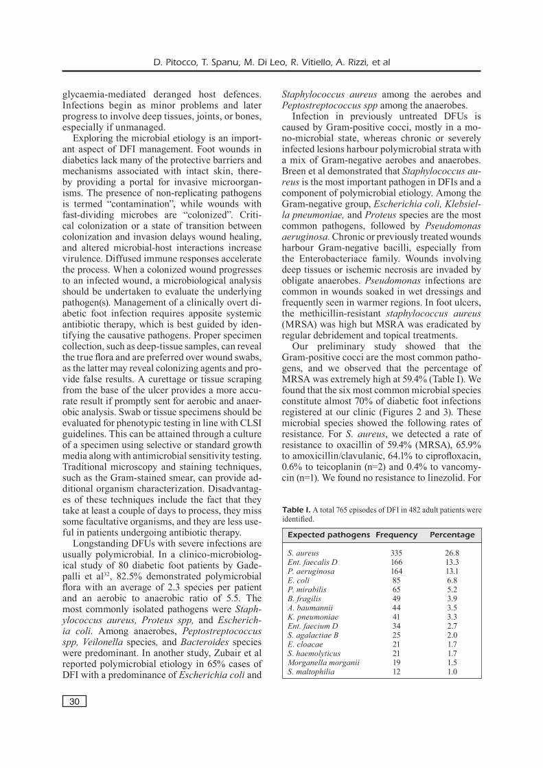
Our preliminary study showed that the Gram-positive cocci are the most common patho-gens, and we observed that the percentage of MRSA was extremely high at 59.4% (Table I). We found that the six most common microbial species constitute almost 70% of diabetic foot infections registered at our clinic (Figures 2 and 3). These microbial species showed the following rates of resistance. For S. aureus, we detected a rate of resistance to oxacillin of 59.4% (MRSA), 65.9% to amoxicillin/clavulanic, 64.1% to ciprofloxacin, 0.6% to teicoplanin (n=2) and 0.4% to vancomy-cin (n=1). We found no resistance to linezolid. For
Table I. A total 765 episodes of DFI in 482 adult patients were identified.
Expected pathogens Frequency Percentage
S. aureus 335 26.8Ent. faecalis D 166 13.3P. aeruginosa 164 13.1E. coli 85 6.8P. mirabilis 65 5.2B. fragilis 49 3.9A. baumannii 44 3.5K. pneumoniae 41 3.3Ent. faecium D 34 2.7S. agalactiae B 25 2.0E. cloacae 21 1.7S. haemolyticus 21 1.7Morganella morganii 19 1.5S. maltophilia 12 1.0

Diabetic foot infections: a comprehensive overview
31
P. aeruginosa, no antibiotics with a sensitivity of 100% were observed. We found the following rates of resistance to other antibiotics: 58.5% to ciprofloxacin, 44.5% to gentamicin, 34.1% to ce-fotazidime, 39.1% to imipenem, 28.2% to mero-penem, and 18.3% to amikacin. For P. mirabilis, no resistance to meropenem was observed, while we detected the following rates of resistance to other antibiotics: 79.7% to ampicillin, 60.9% to ciprofloxacin, 45.2% to cefotaxime, 42.6% to amoxicillin/clavulanate, 37.5% to cefotazidime, and 14.1% to amikacin. For E. faecalis D, no re-sistance to linezolid was observed. We detected the following rates of resistance to other antibi-otics: 4.3% to vancomycin, 4.2% to teicoplanin, and 3% to ampicillin. For E. coli, no resistance to imipenem and meropenem was observed, but we detected the following rates of resistance to other antibiotics: 80% to ampicillin, 69.4% to cip-rofloxacin, 25% to amoxicillin/clavulanate, 37.6% to cefotaxime, 35.3% to gentamicin, 31.8% to ce-
fotazidime, and 1.2% to amikacin. For B. fragilis, we observed resistance of 21.4% to piperacillin, but no resistance to amoxicillin/clavulanate, imi-penem or meropenem.
The prevention of ulcers infected by multi-drug resistant organisms (MDRO) should be in focus and resistance patterns should be careful-ly monitored. A mild or moderate DFI can be treated with oral antimicrobials, while chronic infection requires inpatient antimicrobial therapy or surgical treatment, as well as controlled met-abolic derangements. Patients with DFI should initially be treated with an empirical regime cov-ering Gram-positive cocci. The spectrum can be broadened to cover Gram-negative aerobes in chronic infections. Wounds that are necrotic, foul smelling or gangrenous require anti-anaerobic microbials. A definite diagnosis of bone infection usually requires histological findings consistent with bone infection along with microbiological examinations of an aseptically obtained bone
Figure 2. Overall antibiotic resistance.

D. Pitocco, T. Spanu, M. Di Leo, R. Vitiello, A. Rizzi, et al
32
sample. However, this is typically only necessary when the diagnosis is in doubt or determining the causative pathogen’s antibiotic susceptibility is crucial. The primary treatment for bone infection should be parenteral and can be prolonged up to six weeks. Chronic osteomyelitis requires surgi-cal intervention (i.e., removal of bone).
Classification of Diabetic Foot Infections An adequate description of ulcer characteris-
tics, such as size, depth, appearance, and location, allows for mapping of progress during treatment. The evaluation should determine the etiology of the ulcer and ascertain whether the lesion is neu-ropathic, ischemic or neuro-ischemic. Various classification systems have been used to evaluate the severity of diabetic foot lesions. These sys-tems attempt to encompass different characteris-tics of the ulcer, including size, depth, ischemia, infection, and neuropathy.
One of the most commonly used classification systems is the Wagner-Meggit system. Although
it was devised for dysvascular foot, it has been used for lesion classification for the past 25 years. This six-grade classification system takes into consideration the depth of the ulcer, the presence of gangrene, and the extent of tissue necrosis. Even though Wagner-Meggit’s grading is one of the most widely used classification systems, it does not take into account important clinical parameters, such as ischemia, infection, or other co-morbid factors (Table II).
The University of Texas system primarily grades ulcers based on depth, and then, it stages which di-vide patients who have clean ulcers and those who are infected. More specifically, grade 0 in the Texas System classification (Table III) represents a pre- or postulcerative site. Grade 1 ulcers are superficial wounds through either the epidermis or the epider-mis and dermis, but that do not penetrate to ten-don, capsule, or bone. Grade 2 wounds penetrate to tendon or capsule, but the bone and joints are not complicated. Grade 3 wounds infiltrate bone or in-to a joint. Each wound grade consisted of 4 stages:
Figure 3. Prevalence (%) of antibiotic resistance, by year.

Diabetic foot infections: a comprehensive overview
33
clean wounds (A), nonischemic infected wounds (B), ischemic wounds (C), and infected ischemic wounds (D). The S(AD) SAD classification (Table IV) grades 5 ulcer features (size, depth, sepsis, ar-teriopathy, and denervation) on a 4-point scale (0-3). Similarly, the International Working Group on the Diabetic Foot has suggested the PEDIS clas-sification (Perfusion (ischaemia), Extent (area), Depth, Infection, Sensation (neuropathy)), which grades the wound on a 5-feature basis: perfusion (arterial supply), extent (area), depth, infection, and sensation. Finally, according to the Infectious Dis-eases Society of America guidelines, the infected diabetic foot is subclassified into the categories of uninfected, mild (restricted involvement of only skin and subcutaneous tissues), moderate (more extensive or affecting deeper tissues), and severe (accompanied by systemic signs of infection or metabolic instability).
In an uncomplited clinical classification approach, diabetic foot ulcers can be described as neuropathic,
ischemic, or neuroischemic, determined by how complications such as peripheral neuropathy and ar-terial disease affect the ulcer’s etiology.
Choosing an Appropriate AntibioticAs soon as a diagnosis of DFI is established,
antibiotic treatment should be initiated. NICE (2016) guidelines state that all primary care set-tings should have care pathways in place for man-aging DFIs with specific antibiotic regimens that take local resistance issues into account. The anti-biotic choice should be based on the likely proven causative pathogens, the severity of the infection and evidence of efficacy for DFIs while being cognisant of cost. The NICE guidelines also rec-ommend that the choice of antibiotic treatment should be influenced by the care setting, patient preferences, the clinical situation, and the pa-tient’s medical history.
The IWGDF and NICE make specific recom-mendations concerning antimicrobial therapy for DFIs depending on the severity: - For mild infections, initially offer oral antibiot-
ics effective against Gram-positive organisms. - A one- to two-week course of antibiotic ther-
apy is usually sufficient for mild infections. - For moderate and severe infections, admin-
ister antibiotics effective against Gram-posi-tive and Gram-negative organisms, including anaerobic bacteria.
Table II. Wagner classification system.
0 Pre-ulcerative, with no open lesion or cellulitis 1 Superficial ulcer 2 Deep ulcer upto tendons and joint tissue 3 Deep ulcer with abscess, osteomyelitis, and joint sepsis 4 Localized gangrene of forefoot or heel 5 Gangrene of entire foot/global gangrene
Table III. University of Texas Classification System.
0 1 2 3
A No open lesion Superficial wound Affected tendons/capsules Affected bone/joint B With infection With infection With infection With infection C Ischemic Ischemic Ischemic Ischemic D Infection/Ischemia Infection/Ischemia Infection/Ischemia Infection/Ischemia
Table IV. Diabetic foot infection classification schemes: Infectious Diseases Society of America (IDSA).
Clinical description IDSA IWGDF
Wound without purulence or any manifestations of inflammation Uninfected 1 ≥ 2 Manifestations of inflammation (purulence or erythema, pain, tenderness, warmth, or induration); any cellulitis or erythema extends 52 cm Mild 2around ulcer, and infection is limited to skin or superficial subcutaneous tissues; no local complications or systemic illness Infection in a patient who is systemically well and metabolically stable but has ≥ 2 cm; Moderate 3lymphangitis; spread beneath fascia; deep tissue abscess; gangrene; muscle, tendon, joint, or bone involvement Infection in a patient with systemic toxicity or metabolic instability Severe 4(e.g., fever, chills, tachycardia, hypotension, confusion, vomiting, leukocytosis,acidosis, hyperglycemia, or azotemia)

D. Pitocco, T. Spanu, M. Di Leo, R. Vitiello, A. Rizzi, et al
34
- For moderate infections, offer oral or initial parental administration depending on the clinical situation and choice of antibiotic.
- For severe infections, administer parental therapy with a switch to oral therapy based on the clinical situation and the response to treatment.
- For DFO, offer six weeks of antibiotic ther-apy according to local protocols for patients who do not undergo surgical resection of the infected bone.
- For those who have undergone surgical inter-vention and all infected bone are resected, offer no more than one week of antibiotic therapy37. Lipsky et al43 do not recommend prophylactic treatment of clinically uninfected wounds with antimicrobial therapy, and they advise against the use of any specific type of dressing for DFI with the aim of preventing an infection or im-proving its outcome.
Treatment of Diabetic Foot InfectionsFounded on the results of the available studies,
no single drug or combination of agents seems to be superior to any others. We have several antibi-otic agents for treating DFIs, both by the oral and parenteral routes. It is important to bear in mind that while antibiotics are necessary for treating a DFI, they are not usually sufficient. All patients will need appropriate wound care (debridement, dressings, and pressure off-loading) and most will need some surgical interventions. An internation-al survey found that developing a stewardship pro-gramme was associated with reductions in 96% of hospitals for inappropriate prescribing, 86% for broad-spectrum antibiotic use, 80% for antibiotic expenditure, 71% for healthcare-acquired infec-tions, 65% for length of stay or mortality, and 58% for bacterial resistance. MRSA infections protract wound healing times and hospitalization stays, in-crease the need for surgical procedures, and re-sult in treatment failure. The antibiotic regimen should take account of an agent active against Gram-positive cocci having a care for MRSA in high-risk patients. Treatment for previously treat-ed or severe DFI should include extended cover-age for Gram-negative bacilli and enterococcus species. Gangrenous and foul-smelling wounds may necessitate anti-anaerobic therapy. In a ran-domized study of ampicillin/sulbactam versus imipenem/cilastatin for the treatment of mod-erately severe DFI in 90 patients, there were no significant differences between the treatments in terms of clinical success rate, adverse-event fre-
quency, duration of study antibiotic treatment, or length of hospitalization. In another randomized study, double-blinded, multicentre trial in diabet-ic adults (n=586) with a foot infection classified as moderate-to-severe it was found that ertapenem were equivalent to those for patients treated with piperacillin/tazobactam. The once-daily ertape-nem is advantageous in the DFI setting, in spite of the fact that ertapenem does not cover most enterococci or Pseudomonas aeruginosa. Usual-ly, moderate and severe DFI are typically treated with intravenous antibiotic therapy for two to four weeks, with four to six weeks of therapy for os-teomyelitis.
Operating intervention of moderate to severe DFI is often essential, and includes aggressive incision, drainage and debridement of non-viable soft tissue and bone. With an increasing infection severity, there was a statistically significant trend toward an increased risk for amputation, an in-creased anatomic level of amputation. An increas-ing infection severity was associated to a signifi-cant trend toward increasing risk for experiencing other diabetic foot-related complications, such as neuropathy, vascular disease, and history of am-putation. Foot infections can extend proximally into the leg through the tarsal tunnel, resulting in rapidly ascending limb- and life-threatening infec-tions. Early surgical treatment of DFI may reduce the need for major amputations. An aggressive surgical approach against foot infection in diabet-ic patients may reduce the need for above-ankle amputation. Treatment of diabetic foot infection requires the combination of early surgical treat-ment and antimicrobial therapy. Many surgeons still advocate a transtibial amputation (TTA) as the primary surgical option for non-healing foot ulcer-ations. A bioabsorbable calcium sulphate antibiotic beads into the surgical wound can increase the ef-fects of TMA for diabetic ulcerations of the fore-foot. This method could have a significant impact on the management of diabetic forefoot ulcerations by preventing additional hospital stays for opera-tive revisions, and thereby improving the patient’s quality of life.
Adjunctive therapies include the use of antibi-otic-impregnated beads, the application of nega-tive-pressure wound therapy. Adjunctive HBOT has a positive effect on wound healing in diabetic foot with infection. Conservative treatment, in-cluding prolonged, culture-guided parenteral and oral antibiotics, is efficacious without amputation in a large percentage of diabetic patients admitted for a foot skin ulcer or suspected osteomyelitis55.

Diabetic foot infections: a comprehensive overview
35
Predictors of Treatment Failure in Diabetic Foot Infections
Clinical failure rates were 46% for patients with risk factors (elevated white blood cell count, C-reactive protein or erythrocyte sedimentation rate; high wound severity score; inpatient treat-ment; low serum albumin; male sex; and skin temperature of affected foot >10°C above that of unaffected foot) compared with 10% for patients with no risk factors and 16-17% for patients with one risk factor56. Increased WBC and severe UT wounds (grades 2 and 3) were significant inde-pendent risk factors for clinical failure in patients treated for DFI in the SIDESTEP study46. Clinical failure was noted in 23% of the patients with UT wounds 2B,D and 3B,D at baseline, which can be compared to 11% for a wound stage of 0 or 1. The mean WBC was 9,777 cells/mm3 for those patients who failed treatment, compared to 7,933 cells/mm3 for those with a favourable response. CRP and ESR values greater than 9.1 and 54.4, respectively, were associated with treatment fail-ure. A meta-analysis of data57 from randomized controlled trials on DFIs showed a treatment fail-ure rate of 22.7% in 18 studies. The isolation of MRSA was found to be a significant factor associ-ated with treatment failure, although the presence or absence of OM did not affect the outcome. In a retrospective cohort study46 of the outcomes of conservatively treated DFIs, fever, increased se-rum creatinine, prior hospitalization for DFI and gangrenous lesions were independent factors as-sociated with treatment failure.
Conclusions
The prevalence of MRSA is high and the in-correct use of antibacterials, hospital environ-ment, osteomyelitis, and nasal carriage of MRSA give to infection with MRSA. Understanding the pathophysiology and promptly identifying risk factors for DFI are essential. A thorough evalua-tion of DFI that utilises a multidisciplinary team is recommended to achieve optimal outcomes. It is important to classify accurately DFI in order to guide treatment regimens, facilitate consistent communication among health-care providers, and predict patient outcomes. The IDSA and UT classifications provide relatively simplistic and objective methods for classifying DFI. Prompt recognition and treatment of DFI are mandatory for ensuring maximal limb salvage.
Conflict of InterestThe authors have not received any funding or benefits from the industry to conduct this study.
Reference
1) Singh n, ArmStrong Dg, LipSky BA. Preventing foot ulcers in patients with diabetes. JAMA 2005; 293: 217-228.
2) nAtionAL DiABeteS fAct Sheet, 2011. US Department of Health and Human Services. Center for Dis-ease Control and Prevention, Atlanta, GA (2011).
3) LipSky BA. Empirical therapy for diabetic foot infec-tions: are there clinical clues to guide antibiotic selection? Clin Microbiol Infect 2007; 13: 351‐353.
4) richArD JL, Sotto A, LAvigne Jp. New insights in diabetic foot infection. World J Diabetes 2011; 2: 24‐32.
5) peterS eJ. Pitfalls in diagnosing diabetic foot infec-tions. Diabetes Metab Res Rev 2016; 32 (Supp 1): 254-260.
6) LipSky BA, ArAgon-SAnchez J, DiggLe m, emBiL J, kono S, LAvery L, SenneviLLe É, UrBAnčič-rovAn v, vAn ASten S; International Working Group on the Diabetic Foot, Pe-ters EJ. IWGDF guidance on the diagnosis and man-agement of foot infections in persons with diabetes. Diabetes Metab Res Rev 2016; 32 (Suppl 1): 45-74.
7) ArmStrong Dg, LipSky BA. Diabetic foot infections: stepwise medical and surgical management. Int Wound J 2004; 1: 123-132.
8) centerS for DiSeASe controL AnD prevention, US De-pArtment of heALth AnD hUmAn ServiceS. National diabetes fact sheet: general information and na-tional estimates on diabetes in the United States, 2003. Atlanta: US Centers for Disease Control and Epidemiology, 2003.
9) mUtLUogLU m, SivriogLU Ak, erogLU m, UzUn g, tUrhAn v, Ay h, LipSky BA. The implications of the presence of osteomyelitis on outcomes of infected diabetic foot wounds. Scand J Infect Dis 2013; 45: 497-503.
10) fAgLiA e, cLerici g, cAminiti m, cUrci v, SomALvico f. Influence of osteomyelitis location in the foot of diabetic patients with transtibial amputation. Foot Ankle Int 2013; 34: 222-227.
11) DArBeLLAy p, UçkAy i, DomingUez D, mUgnAi D, fiLtri L, Lew D, ASSAL m. Diabetic foot infection: a multi-disciplinary approach. Rev Med Suisse 2011; 7: 894-897.
12) ArmStrong Dg, LAvery LA. Diabetic foot ulcers: prevention, diagnosis and classification. Am Fam Phys 1998; 57: 1325-1332.
13) reiBer ge, viLeikyte L, Boyko eJ, DeL AgUiLA m, Smith Dg, LAvery LA. Causal pathways for incident low-er-extremity ulcers in patients with diabetes from two settings. Diabetes Care 1999; 22: 157-162.
14) noor S, zUBAir m, AhmAD J. Diabetic foot ulcer–a re-view on pathophysiology, classification and microbial etiology. Diabetes Metab Syndr 2015; 9: 192-199.

D. Pitocco, T. Spanu, M. Di Leo, R. Vitiello, A. Rizzi, et al
36
15) AhmAD J. The diabetic foot. Diabetes Metab Syndr Clin Res Rev 2016; 10: 48-60.
16) JoSeph wS, Lefrock JL. The pathogenesis of diabet-ic foot infections immunopathy, angiopathy. J Foot Surg 1987; 26: 7-11.
17) peterS eJ, LAvery LA, ArmStrong Dg. Diabetic lower extremity infection: influence of physical, psycho-logical and social factors. J Diabetes Complica-tions 2005; 19: 107-112.
18) pryce tD. A case of perforating ulcers of both feet associated with diabetes and ataxic symptoms. Lancet 1887; 130: 11-12.
19) LipSky BA, pecorAro re, LArSon SA, hAnLey me, Ahroni Jh. Outpatient management of uncompli-cated lower extremity infections in diabetic pa-tients. Arch Intern Med 1990; 150: 790-797.
20) BAgDADe JD, root rk, BULger rJ. Impaired leuko-cyte function in patients with poorly controlled di-abetes. Diabetes 1974; 23: 9-15.
21) inzUcchi Se. Clinical practice. Management of hy-perglycemia in the hospital setting. N Engl J Med 2006; 355: 1903-1911.
22) hoStetter mk. Perspectives in diabetes. Handicaps to host defense. Effects of hyperglycemia on C3 and Candida albicans. Diabetes 1990; 39: 271-275.
23) Sentochnik De, eLiopoULoS gm. Infection and diabe-tes. In: Kahn CR, Weir GC, editors. Joslin’s diabe-tes mellitus. 13th ed. Philadelphia: Lea & Febiger, 1994; pp. 867-868.
24) keiDAr z, miLitiAnU D, meLAmeD e, BAr-ShALom r, iS-rAeL o. The diabetic foot: initial experience with 18F-FDG PET/CT. J Nucl Med 2005; 46: 444-449.
25) BASU S, chrySSikoS t, hoUSeni m, Scot mALAy D, ShAh J, zhUAng h, ALAvi A. Potential role of FDG PET in the setting of diabetic neuro-osteoar-thropathy: can it differentiate uncomplicated Charcot’s neuroarthropathy from osteomyelitis and soft tissue infection. Nucl Med Commun 2007; 28: 465-472.
26) LipSky BA, peterS eJ, BerenDt Ar, SenneviLLe e, BAk-ker k, emBiL Jm, LAvery LA, UrBAnčič-rovAn v, Jeff-coAte wJ; International Working Group on Diabetic Foot. Specific guidelines for the treatment of di-abetic foot infections 2011. Diabetes Metab Res Rev 2012; 28 (Suppl. 1): 234-235.
27) AmericAn DiABeteS ASSociAtion. Peripheral arterial disease in people with diabetes. Diabetes Care 2003; 26: 3333-3341.
28) BoULton AJ, ArmStrong Dg, ALBert Sf, frykBerg rg, heLLmAn r, kirkmAn mS, LAvery LA, LemASter Jw, miLLS JL Sr, mUeLLer mJ, SheehAn p, wUkich Dk; American Diabetes Association; American Association of Clinical Endocrinologists. Com-prehensive foot examination and risk assess-ment: a report of the task force of the foot care interest group of the American Diabetes Asso-ciation, with endorsement by the American As-sociation of Clinical Endocrinologists. Diabetes Care 2008; 31: 1679-1685.
29) eDwArDS r, hArDing kg. Bacteria and wound heal-ing. Curr Opin Infect Dis 2004; 17: 91-96.
30) ABBAS zg, LUtALe Jk, iLonDo mm, ArchiBALD Lk. The utility of Gram stains and culture in the manage-ment of limb ulcers in persons with diabetes. Int Wound J 2012; 9: 677-682.
31) gADepALLi r, DhAwAn B, SreenivAS v, kApiL A, Ammini Ac, chAUDhry r. A clinicomicrobiological study of diabetic foot ulcers in an Indian tertiary care hos-pital. Diabetes Care 2006; 29: 1727-1732.
32) zUBAir m, ABiDA m, JAmAL A. Incidence, risk factors for amputation among patients with diabetic foot ulcer in a North Indian tertiary hospital. Foot 2011; 22: 24-30.
33) Breen JD, kArchmer Aw. Staphylococcus aureus in-fections in diabetic patients. Infect Dis Clin North Am 1995; 9: 11-24.
34) DAng cn, prASAD yD, BoULton AJ, JUDe eB. Methi-cillin-resistant Staphylococcus aureus in the dia-betic foot clinic: a worsening problem. Diabet Med 2003; 20: 159-161.
35) pitocco D, SpAnU t, Di Leo m, vitieLLo r, rizzi A, tArtAgLione L, cApUto S, tineLLi g, zAccArDi f, fLex A, gALLi m, pontecorvi A, SAngUinetti m. Spectrum of germs and their antibiotic pattern in patients with infected diabetic foot ulcers: an observational study of five years. Data not published.
36) kAnDemir o, AkBAy e, SAhin e, miLcAn A, gen r. Risk factors for infection of the diabetic foot with multi-antibiotic resistant microorganisms. J Infect 2007; 54: 439-445.
37) wAgner fw Jr. The dysvascular foot: a system for diagnosis and treatment. Foot Ankle 1981; 2: 64-122.
38) LAvery LA, ArmStrong Dg, hArkLeSS LB. Classifica-tion of diabetic foot wounds. J Foot Ankle Surg 1996; 35: 528-531.
39) mAcfArLAne rm, JeffcoAte wJ. Classification of di-abetic foot ulcers: the S(AD) SAD system. Diabet Foot 1999; 2: 123-131.
40) SchAper nc. Diabetic foot ulcer classification sys-tem for research purposes: a progress report on criteria for including patients in research stud-ies. Diabetes Metab Res Rev 2004; 20 Suppl 1: S90-S95.
41) LAvery LA, ArmStrong Dg, mUrDoch Dp, peterS eJg, LipSky BA. Validation of the infectious diseases So-ciety of America’s diabetic foot infection classifi-cation system. Clin Infect Dis 2007; 44: 562-565.
42) NICE (2016) Diabetes in Adults NICE Quality Standards (QS6) (online) Available at: https://www.nice.org.uk/guidance/ QS6/chapter/Intro-duction (accessed 20.08.2016)
43) LipSky BA, ArAgon-SAnchez J, DiggLe m, emBiL J, kono S, LAvery L, SenneviLLe É, UrBAnčič-rovAn v, vAn AS-ten S; International Working Group on the Diabetic Foot, Peters EJ. IWGDF guidance on the diagno-sis and management of foot infections in persons with diabetes. Diabetes Metab Res Rev 2016; 32 (Suppl 1): 45-74.

Diabetic foot infections: a comprehensive overview
37
44) vArDAkAS kz, horiAnopoULoU m, fALAgAS me. Fac-tors associated with treatment failure in patients with diabetic foot infections: An analysis of data from randomized controlled trials. Diabetes Res Clin Pract 2008; 80: 344-351.
45) BArriere SL. Clinical, economic and societal impact of antibiotic resistance. Expert Opin Pharmacoth-er 2015; 16: 151-153.
46) eLeftheriADoU i, tentoLoUriS n, ArgiAnA v, JUDe e, BoULton AJ. Methicillin-resistant Staphylococcus aureus in diabetic foot infections. Drugs 2010; 70: 1785-1797.
47) mckinnon pS, pALADino JA, grAySon mL, giBBonS gw, kArchmer Aw. Cost-effectiveness of ampicil-lin/sulbactam versus imipenem/cilastatin in the treatment of limb-threatening foot infections in di-abetic patients. Clin Infect Dis 1997; 24: 57-63.
48) LipSky BA, ArmStrong Dg, citron Dm, tice AD, morgen-Stern De, ABrAmSon mA. Ertapenem versus piperacillin/tazobactam for diabetic foot infections (SIDESTEP): prospective, randomised, controlled, double-blinded, multicentre trial. Lancet 2005; 366: 1695-1703.
49) LAvery LA, ArmStrong Dg, mUrDoch Dp, peterS eJ, LipSky BA. Validation of the Infectious Diseases Society of America’s diabetic foot infection classi-fication system. Clin Infect Dis 2007; 44: 562-565.
50) tAn JS, frieDmAn nm, hAzeLton-miLLer c, fLAnA-gAn Jp, fiLe tm. Jr. Can aggressive treatment of diabetic foot infections reduce the need for
above-ankle amputation? Clin Infect Dis 1996; 23: 286-291.
51) SAnDerS LJ, DUnLAp g. Transmetatarsal amputa-tion: a successful approach to limb salvage. J Am Pod Med Assoc 1992; 82: 129-130.
52) krAUSe fg, DevrieS g, meAkin c, kALLA tp, yoUnger AS. Outcome of transmetatarsal amputations in diabetics using antibiotic beads. Foot Ankle Int 2009; 30: 486-493.
53) chen ce, ko Jy, fong cy, JUhn rJ. Treatment of di-abetic foot infection with hyperbaric oxygen ther-apy. Foot Ankle Surg 2010; 16: 91-95.
54) pittet D, wySSA B, herter-cLAveL c, kUrSteiner k, vAUcher J, Lew pD. Outcome of diabetic foot in-fections treated conservatively: a retrospective cohort study with long-term follow-up. Arch Intern Med 1999; 159: 851-856.
55) chen z, cheng L, feng g. Bone inflammation and chronic recurrent multifocal osteomyelitis. Eur Rev Med Pharmacol Sci 2018; 22: 1380-1386.
56) LipSky BA, SheehAn p, ArmStrong Dg, tice AD, poLiS AB, ABrAmSon mA. Clinical predictors of treatment failure for diabetic foot infections: data from a pro-spective trial. Int Wound J 2007; 4: 30-38.
57) vArDAkAS kz, horiAnopoULoU m, fALAgAS me. Fac-tors associated with treatment failure in patients with diabetic foot infections: an analysis of data from randomized controlled trials. Diabetes Res Clin Pract 2008; 80: 344-351.