management of sternal wound dehiscence · management of sternal wound dehiscence ... based on wound...
TRANSCRIPT
Management of sternal wound dehiscence
Fouzia Choukairi, Joseph Ring, Joe Thekkudan, Stuart Enoch
Fouzia Choukairi is a Speciality Trainee in Plastic Surgery, University Hospital of South Manchester; Joseph Ring is a Speciality Trainee in Orthopaedic Surgery, Royal Oldham Hospital; Joe Thekkudan is a Registrar in Cardiothoracic Surgery, New Cross Hospital, Wolverhampton; Stuart Enoch is Programme Director — Education and Research at the Doctors’ Academy and Specialist Registrar in Plastic Surgery, University Hospital of South Manchester
Sternal wound complications may result from median sternotomy procedures following cardiothoracic surgery and can represent a significant management problem, for example, infections and dehiscence can increase hospital stay, morbidity and mortality. Morbidity from sternal wound dehiscence was close to 50% until recently, but with recent advances, including sternal fixation techniques and various reconstructive surgical options, this has improved. This article outlines the problems of wound dehiscence and delayed healing following median sternotomy and provides an overview of the pertinent management options.
Sternal wound complications may result from median sternotomy procedures following
cardiothoracic surgery. The median sternotomy is the incision of choice for most procedures that involve the heart and great vessels, including trauma, bypass, valve replacement, heart failure and transplant surgery. In addition, it allows access for many pulmonary procedures (Falor and Traylor, 1982). When compared to approaching the chest cavity from the lateral side (lateral thoracotomy), the median sternotomy provides excellent exposure and results in less postoperative pain (Falor and Traylor, 1982).
as negative pressure wound therapy (NPWT) has reduced the morbidity rate to approximately 10% (Miller and Nahai, 1989; Jones et al, 1997; Sjögren et al, 2006).
Sternal wound infections and dehiscence also prolong recovery time as well as increasing hospital stay, re-operative rates, morbidity and mortality (Losanoff et al, 2002b).
This article aims to highlight the problems of sternal wound dehiscence and delayed healing following median sternotomy, as well as providing an overview of the management options.
Median sternotomyMedian sternotomy was introduced in the 1950s (Julian et al, 1957) and has revolutionised the world of cardiothoracic surgery. The midline incision is made through the bony sternum and provides excellent access to the thoracic organs as well as permitting a range of procedures to be performed on the thoracic structures (Grevious, 2009).
Other incisions exist and newer minimally invasive approaches are being developed, however, the median sternotomy remains the incision of choice for many procedures since it provides the best access to the mediastinal structures as well as access into both hemithoracies. It is considered
Clinical PRACTICE DEVELOPMENTClinical PRACTICE DEVELOPMENT
99Wounds uk, 2011, Vol 7, No 1
KEY WORDSSternal wound dehiscenceMediastinitis reconstructionSternotomySternal instability
However, postoperative wound infection of the sternum is common and if severe, chronic or untreated, can lead to sternal wound dehiscence. The incidence of sternal wound infections following median sternotomy procedures is in the region of 0.5–8.4%
... postoperative wound infection of the sternum is common and if severe, chronic or untreated, can lead to sternal wound dehiscence.
(Grevious, 2009). This can be further complicated by development of osteomyelitis of the sternum which has a reported incidence of 0.8–2.5%.
Sternal wound dehiscence can lead to mediastinitis, which is where the superficial wound infection travels deeper and affects the mediastinal structures. It has an incidence of 1–5% of median sternotomies (Grevious, 2009) and results in a chronic inflammatory state and infection, both on the surface (sternal wound) and the mediastinal structures.
Morbidity from sternal wound dehiscence and its associated complications ran at nearly 50% until recently, but advances in management options and medical devices such
Choukairi.indd 3 15/03/2011 14:25
100
Clinical PRACTICE DEVELOPMENT
Wounds uk, 2011, Vol 7, No 1
to be less painful and provides a lower incidence of respiratory complications when compared to other incisions (Farhat et al, 2004).
ComplicationsPotential wound complications following median sternotomy include superficial or deep wound infections, desiccation of wound margins, wound breakdown, wound dehiscence, osteomyelitis, mediastinitis, and exposure of vital structures and major vessels.
Long-term problems include sternal instability, paradoxical motion of the chest wall and chronic pain. These symptoms may have significant effects on the patient’s quality of life.
Pairolero and Arnold (1984) classified sternal wound infections into three types based on timing and presentation (Table 1). This system does not necessarily indicate the type of treatment required, however type II and III will generally require specialist input and surgical reconstruction (Pairolero and Arnold, 1984). More recently, El Oakley and Wright (1996) proposed another classification for mediastinitis based on wound assessment during surgical debridement, which helps to determine a management approach.
Delayed healingFollowing a median sternotomy, the integrity of the sternum and the surrounding structures are significantly compromised. There is little muscle or soft tissue overlying the sternum, and consequently the area does not have a generous blood supply. The close proximity of the bone to the skin also exacerbates the problem of recalcitrance and a superficial wound infection can soon enter the bone leading to osteomyelitis. Furthermore, the area is subject to high stresses and almost constant movement, both due to respiration and activities of the shoulder girdle. Some recognised patient and surgical factors in the aetiology of delayed sternal wound healing are listed in Table 2.
Initial assessment for sternal wound dehiscenceAn accurate clinical evaluation is
with surrounding progressive cellulitis, systemic infection or sepsis. However, if the infection is localised and minimal, it is reasonable to withhold antibiotics until microbiological swab results (provisional or definitive) are available, or at least after obtaining tissue samples (either in ward or theatre) for microbiological analysis.
The key aim once infection is identified is to perform swift and thorough surgical debridement, following which further management can be decided upon. The aim of surgical debridement is to remove all non-viable tissue back to bleeding tissue and produce a clean wound that allows definitive management to be performed
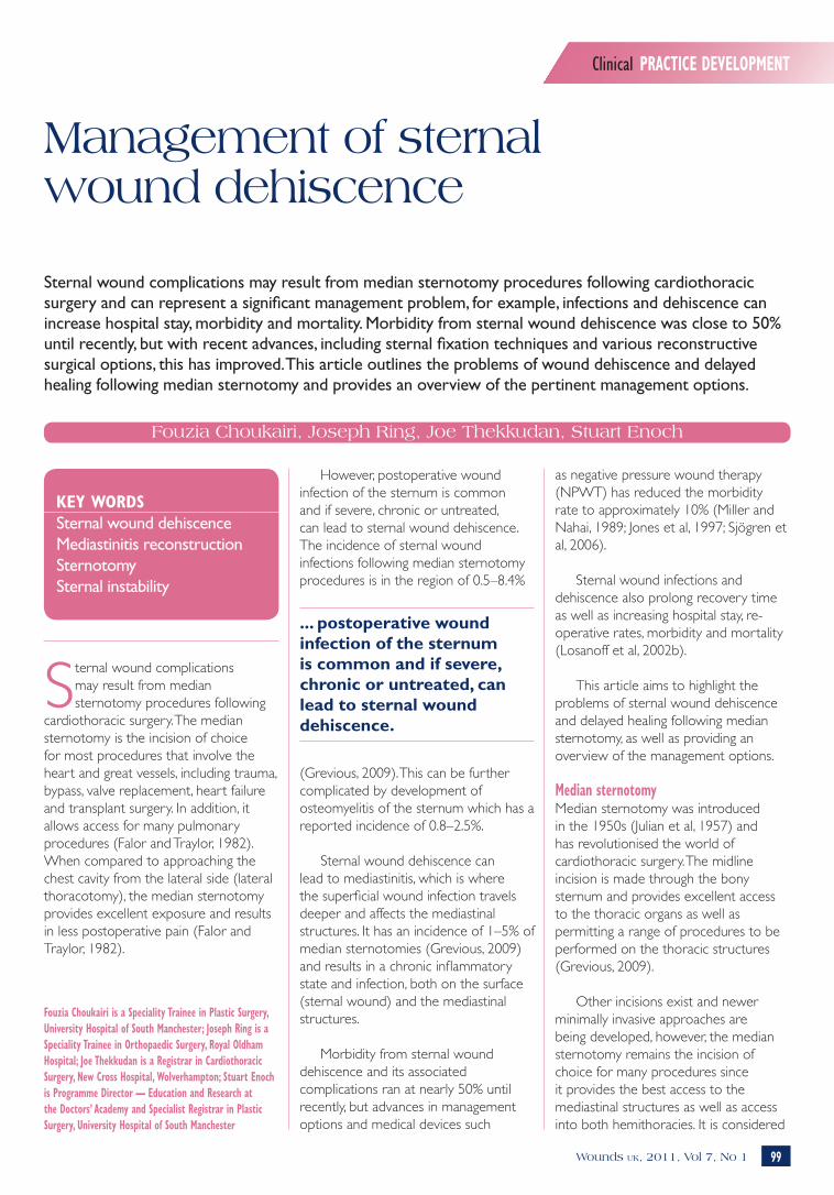
essential to recognise the early signs and symptoms of sternal wound dehiscence (Figure 1). Clinicians should be alert for signs of local wound changes such as erythema or increased exudate, pain, systemic signs such as pyrexia, and respiratory symptoms (e.g. pleural effusions). Clinicians should be extra-alert to the possibility of complications in high-risk patients, such as increased risk of infection, delayed wound healing and wound dehiscence (Losanoff et al, 2002a).
If infection is suspected, appropriate swabs, samples and cultures should be taken (Grevious, 2009). Empirical broad-spectrum antibiotics may be started if the infection is overwhelming
Table 1Pairolero and Arnold Classification System of Mediastinitis (Pairolero and Arnold, 1984)
Type 1 8Occurs within a few days of initial surgery8There is serosanguinous discharge but no evidence of cellulitis, osteomyelitis or costochondritis8There are no signs of wound breakdown or instability
Type 2 8 Occurs in the first few weeks following surgery8 Characterised by drainage, cellulitis and positive cultures8 Usually classed as fulminant mediastinitis, often with bony involvement
Type 3 8 Chronic wounds occurring months or years after surgery8 Incorporate signs of chronic infection with sinuses, cellulitis and osteomyelitis8 Medistinal involvement is uncommon with type III infections
Table 2Risk factors for sternal wound complications
Patient risk factors Surgical factors
8 Increasing age8 Female sex8Diabetes mellitus8Obesity 8Smoking 8Malnutrition8Sepsis 8Renal failure 8Immunosuppression 8Osteoporosis8Chronic obstructive pulmonary disease (COPD)
8 Poor technique 8 Extended operative time8 Prolonged ventilation8 Insufficient sternal fixation8 Excessive haemorrhage8 Internal mammary artery harvest8 Cardiopulmonary bypass8 Hypoperfusion 8 Use of bone wax
Choukairi.indd 4 15/03/2011 14:25
Clinical PRACTICE DEVELOPMENT
101Wounds uk, 2011, Vol 7, No 1
immediately or at a later time (Grevious, 2009). If there is evidence of mediastinitis and extensive disruption of the sternum, more radical surgery and reconstruction should be considered.
Before surgery, baseline preoperative investigations (for example, full blood count, electrolytes, inflammatory markers such as C-reactive protein [CRP] and serum albumin levels) are needed to ensure the patient’s fitness for general anaesthesia.
Plain radiography of the sternum may also demonstrate mediastinal air
or separation of the sternal half (when performing a median sternotomy, the sternal bone is longitudinally [vertically] sawn in half and the ‘sternal half ’ refers to these two halves of bone) (Gualdi et al, 2005). Ultrasound may be used to identify any collections of fluid. Computerised tomography (CT) scans are useful for detecting sternal disruption, the location of fluid, and any underlying lung or mediastinal pathology.
If it is suspected that the infection has extended into the bones, this can be established by a magnetic resonance imaging (MRI) scan. However, the effectiveness of any MRI may be limited by the presence of sternal metalwork (Gualdi et al, 2005). Intraoperatively, both superficial wound swabs and deep tissue biopsy may be obtained for microbiological analysis (O’Brien Norris, 1995).
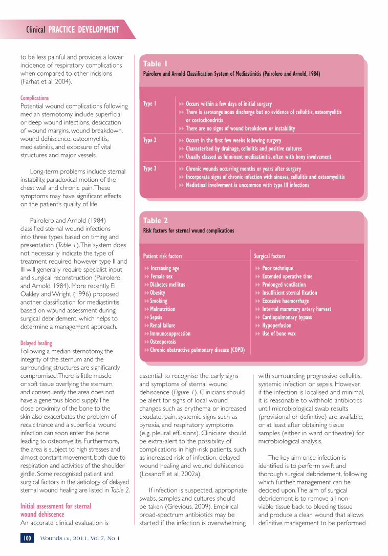
ManagementA number of factors need to be considered when evaluating and managing non-healing sternal wounds, including: 8 Presence and depth of infection8 Any evidence of underlying
osteomyelitis (Figure 2)8 Anatomy of any tissue defects8 Sternal stability 8 General health of the patient and
any associated comorbidities.
In the 1950s, dehisced and non-healing sternal wounds were mostly left to heal by secondary intention, which included debridement, packing and open drainage. However, with these techniques alone, morbidity was close to 50%. In 1963, the concept of catheter antibiotic irrigation of sternal wounds was introduced, leading to a reduction in morbidity from 50% to approximately 20% (Shumacker and Mandelbaum, 1963). Although this reduction was considered to be a significant step forward, it was associated with a major complication — the erosion of major vessels by the catheter, leading in some cases to fatal haemorrhage (Grevious, 2009). This resulted in the drive for better and alternative management options.
Current treatment options include: 8 Promoting healing by secondary
intention through the use of NPWT8 Split-skin grafting if appropriate8 Sternal stabilisation procedures8 Reconstructive procedures.
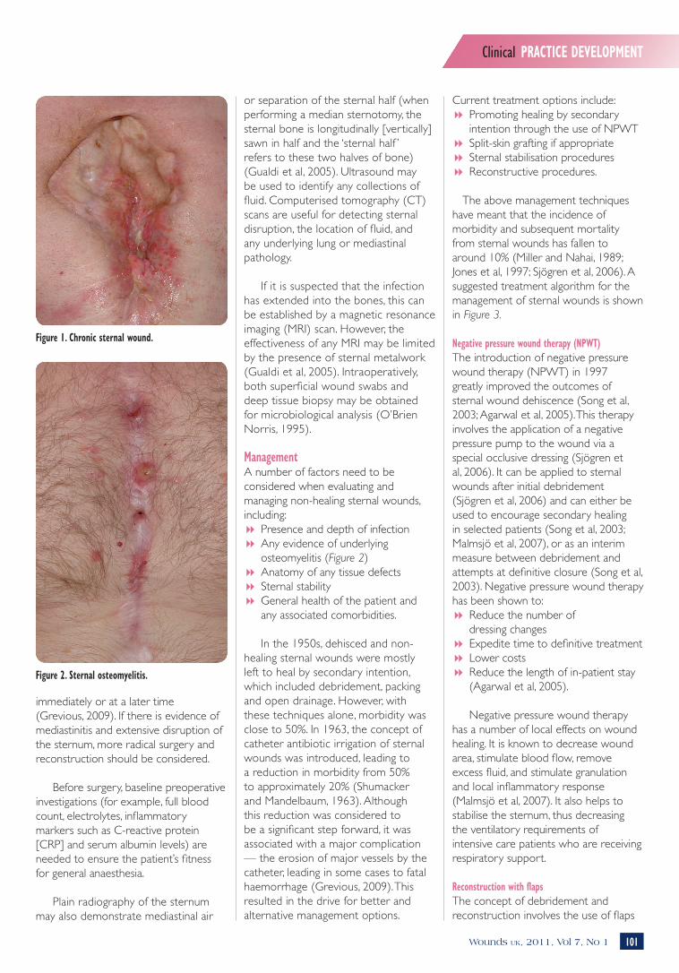
The above management techniques have meant that the incidence of morbidity and subsequent mortality from sternal wounds has fallen to around 10% (Miller and Nahai, 1989; Jones et al, 1997; Sjögren et al, 2006). A suggested treatment algorithm for the management of sternal wounds is shown in Figure 3.
Negative pressure wound therapy (NPWT)The introduction of negative pressure wound therapy (NPWT) in 1997 greatly improved the outcomes of sternal wound dehiscence (Song et al, 2003; Agarwal et al, 2005). This therapy involves the application of a negative pressure pump to the wound via a special occlusive dressing (Sjögren et al, 2006). It can be applied to sternal wounds after initial debridement (Sjögren et al, 2006) and can either be used to encourage secondary healing in selected patients (Song et al, 2003; Malmsjö et al, 2007), or as an interim measure between debridement and attempts at definitive closure (Song et al, 2003). Negative pressure wound therapy has been shown to:8 Reduce the number of
dressing changes8 Expedite time to definitive treatment8 Lower costs 8 Reduce the length of in-patient stay
(Agarwal et al, 2005).
Negative pressure wound therapy has a number of local effects on wound healing. It is known to decrease wound area, stimulate blood flow, remove excess fluid, and stimulate granulation and local inflammatory response (Malmsjö et al, 2007). It also helps to stabilise the sternum, thus decreasing the ventilatory requirements of intensive care patients who are receiving respiratory support.
Reconstruction with flapsThe concept of debridement and reconstruction involves the use of flaps
Figure 1. Chronic sternal wound.
Figure 2. Sternal osteomyelitis.
Choukairi.indd 5 15/03/2011 14:25
102
Clinical PRACTICE DEVELOPMENT
Wounds uk, 2011, Vol 7, No 1
to reduce the dead space in the sternal defect as well as to provide bulk (muscle mass or omentum) to encourage healing (José, 1999). Previous irradiation of the chest wall or abdominal surgery may preclude the use of certain types of flaps. Therefore, full assessment of the patient is essential before embarking on a specific type of reconstruction (O’Brien Norris, 1995; Grevious, 2009).
Lee et al introduced the omental flap in 1976 (Lee et al, 1976; Jurkiewicz and Arnold, 1977), and later more complex myocutaneous flaps were developed following the work of Jurkiewicz (Jurkiewicz and Arnold, 1977). These include the pectoralis major, rectus abdominis and latissimus dorsi flaps (Jurkiewicz et al, 1980; Tizian et al, 1985).
Defects of the upper third are most easily and successfully covered using the pectoralis flap (Jones et al, 1997). Defects of the mid and lower third and larger defects can be covered using the rectus abdominis flap (Greig et al, 2007). The two techniques can be combined for larger defects, resulting in better outcome.
A major study by Castelló et al (1999) that examined sternal wounds demonstrated that pectoralis flaps were the most commonly used flap in sternal reconstruction, with the second choice being pectoralis combined with rectus abdominus flaps, and then the rectus abdominus muscle alone. Latissmus dorsi or omental flaps were only used in a small number of cases. The study showed that there was an overall reduction in mortality using these techniques (José et al, 1999).
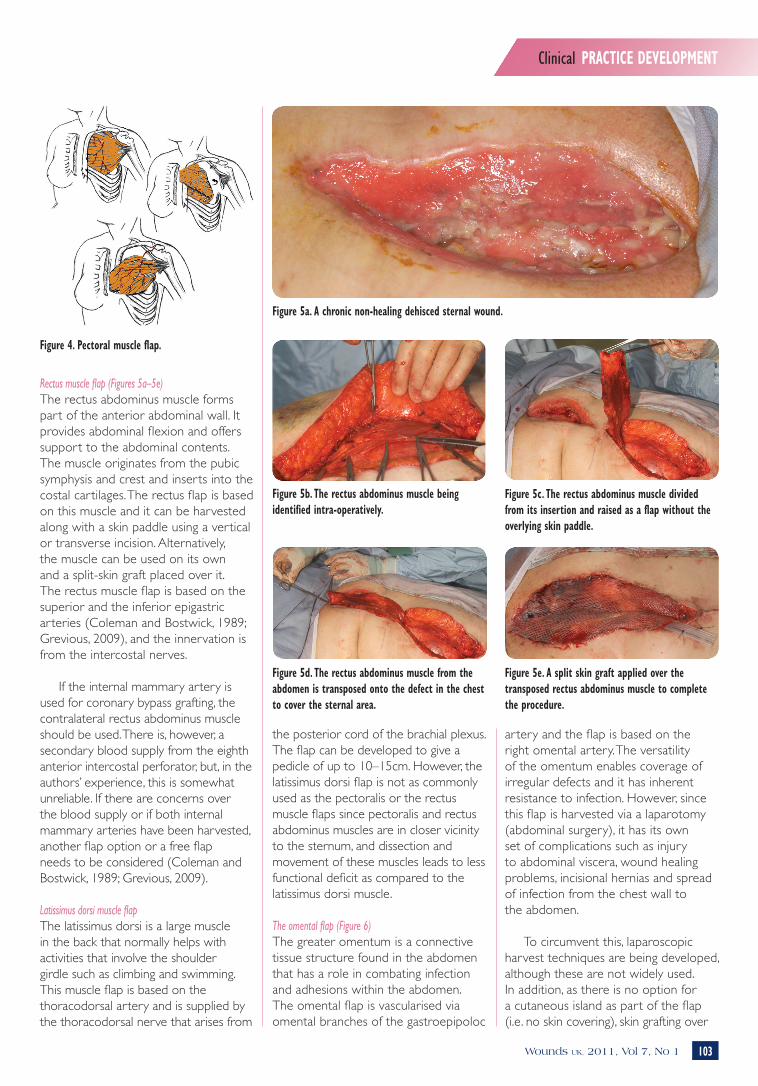
Pectoralis muscle flaps (Figure 4)Sternal wounds, particularly in the upper third, can be covered using the pectoralis muscle flap (Greig et al, 2007). It is often the first choice due to its proximity to the defect requiring coverage. The muscle is harvested through a midline incision. The flap is based either on the thoracoacromial pedicle or on the internal mammary perforators (a flap involves taking a piece of tissue — usually fascia or muscle with or without the overlying skin — to cover a defect or wound. In order for the ‘tissue’ to survive and healing to take place, the flap needs a blood supply. This comes from the blood vessels supplying the original tissue, i.e. fascia or muscle, and the term ‘based’ is used to denote the blood vessel that supplies the flap. These ‘flaps’ can remain attached by their blood supply to their original location [pedicled flaps], or be divided and re-attached by microsurgery to a new location [free flap]) (Tobin, 1989; Grevious, 2009). This flap can be used for ‘simpler’ wounds by dissecting only the medial portion of the muscle from the ribs and moving this medially over the defect. One disadvantage of the pectoralis muscle flap is the resulting compromise in the functionality of the shoulder girdle.Figure 3. Flow chart summarising the management of sternal wounds.
Sternal wound breakdown
Swab for cultures to identify if wound infected or associated osteomyelitis
Positive microbiology
Does the wound need surgical input?
Heal by secondary intention +/- VAC
therapy
Does it need further surgery or
reconstruction
Leave to heal by secondary intention with regular dressing
changes +/- VAC
Reconstruction with flaps +/- sternal fixation
Further debridement/change
of VAC
Continued infection/non-healing wound
No
Yes
No
Yes
Initial debridement +/- VAC
Wound not cleanWound clean
Pre-operative assessment
Re-swab after 5–7 days
Patient fit for GA Patient not fit for GA
Negative microbiology
Discuss with microbiologist and
commence antibiotics
Management of sternal wounds: An algorithm
Choukairi.indd 6 15/03/2011 14:25
Clinical PRACTICE DEVELOPMENT
103Wounds uk, 2011, Vol 7, No 1
Rectus muscle flap (Figures 5a–5e)The rectus abdominus muscle forms part of the anterior abdominal wall. It provides abdominal flexion and offers support to the abdominal contents. The muscle originates from the pubic symphysis and crest and inserts into the costal cartilages. The rectus flap is based on this muscle and it can be harvested along with a skin paddle using a vertical or transverse incision. Alternatively, the muscle can be used on its own and a split-skin graft placed over it. The rectus muscle flap is based on the superior and the inferior epigastric arteries (Coleman and Bostwick, 1989; Grevious, 2009), and the innervation is from the intercostal nerves.
If the internal mammary artery is used for coronary bypass grafting, the contralateral rectus abdominus muscle should be used. There is, however, a secondary blood supply from the eighth anterior intercostal perforator, but, in the authors’ experience, this is somewhat unreliable. If there are concerns over the blood supply or if both internal mammary arteries have been harvested, another flap option or a free flap needs to be considered (Coleman and Bostwick, 1989; Grevious, 2009).
Latissimus dorsi muscle flapThe latissimus dorsi is a large muscle in the back that normally helps with activities that involve the shoulder girdle such as climbing and swimming. This muscle flap is based on the thoracodorsal artery and is supplied by the thoracodorsal nerve that arises from
the posterior cord of the brachial plexus. The flap can be developed to give a pedicle of up to 10–15cm. However, the latissimus dorsi flap is not as commonly used as the pectoralis or the rectus muscle flaps since pectoralis and rectus abdominus muscles are in closer vicinity to the sternum, and dissection and movement of these muscles leads to less functional deficit as compared to the latissimus dorsi muscle.
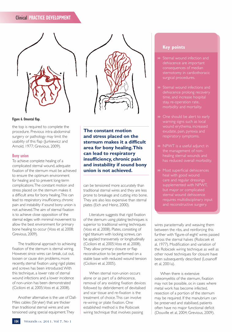
The omental flap (Figure 6)The greater omentum is a connective tissue structure found in the abdomen that has a role in combating infection and adhesions within the abdomen. The omental flap is vascularised via omental branches of the gastroepipoloc
artery and the flap is based on the right omental artery. The versatility of the omentum enables coverage of irregular defects and it has inherent resistance to infection. However, since this flap is harvested via a laparotomy (abdominal surgery), it has its own set of complications such as injury to abdominal viscera, wound healing problems, incisional hernias and spread of infection from the chest wall to the abdomen.
To circumvent this, laparoscopic harvest techniques are being developed, although these are not widely used. In addition, as there is no option for a cutaneous island as part of the flap (i.e. no skin covering), skin grafting over
Figure 5a. A chronic non-healing dehisced sternal wound.
Figure 5b. The rectus abdominus muscle being identified intra-operatively.
Figure 5c. The rectus abdominus muscle divided from its insertion and raised as a flap without the overlying skin paddle.
Figure 5d. The rectus abdominus muscle from the abdomen is transposed onto the defect in the chest to cover the sternal area.
Figure 5e. A split skin graft applied over the transposed rectus abdominus muscle to complete the procedure.
Figure 4. Pectoral muscle flap.
Choukairi.indd 7 15/03/2011 14:25
Wound care SCIENCE
104 Wounds uk, 2011, Vol 7, No 1
Clinical PRACTICE DEVELOPMENT
Key points
8 Sternal wound infection and dehiscence are important consequences of median sternotomy in cardiothoracic surgical procedures.
8 Sternal wound infections and dehiscence prolong recovery time, and increase hospital stay, re-operation rate, morbidity and mortality.
8 One should be alert to early warning signs such as local wound erythema, increased exudate, pain, pyrexia and respiratory symptoms.
8 NPWT is a useful adjunct in the management of non-healing sternal wounds and has reduced overall morbidity.
8 Most superficial dehiscences heal with good wound care and regular dressings supplemented with NPWT, but major or complicated sternal wound dehiscence requires multidisciplinary input and reconstructive surgery.
the top is required to complete the procedure. Previous intra-abdominal surgery or pathology may limit the usability of this flap (Jurkiewicz and Arnold, 1977; Grevious, 2009).
Bony unionTo achieve complete healing of a complicated sternal wound, adequate fixation of the sternum must be achieved to ensure the optimum environment for healing and to prevent long-term complications. The constant motion and stress placed on the sternum makes it a difficult area for bony healing. This can lead to respiratory insufficiency, chronic pain and instability if sound bony union is not achieved. The aim of sternal fixation is to achieve close opposition of the sternal edges with minimal movement to allow the best environment for primary bone healing to occur (Voss et al, 2008; Grevious, 2009).
The traditional approach to achieving fixation of the sternum is sternal wiring. However, since wires can break, cut out, loosen or cause skin problems, more recently, sternal fixation using rigid plates and screws has been introduced. With this technique, a lower rate of sternal wound infections and a lower incidence of non-union has been demonstrated (Cicilioni et al, 2005; Voss et al, 2008).
Another alternative is the use of Dall-Miles cables (Stryker) that are thicker than traditional sternal wires and are tensioned using special equipment. They
wires parasternally and weaving them between the ribs, and reinforcing this further with ‘figure-of-eight’ wires passed across the sternal halves (Robicsek et al, 1977). Modification and variation of the Robicsek wiring technique as well as other novel techniques for closure have been subsequently described (Losanoff et al, 2001a).
When there is extensive osteomyelitis of the sternum, fixation may not be possible, or, in cases where metal work has become infected, resection of a portion of the sternum may be required. If the manubrium can be preserved and stabilised, patients often have no major functional deficit (Douville et al, 2004; Grevious, 2009).
Figure 6. Omental flap.
The constant motion and stress placed on the sternum makes it a difficult area for bony healing. This can lead to respiratory insufficiency, chronic pain and instability if sound bony union is not achieved.
can be tensioned more accurately than traditional sternal wires and they are less prone to breakage and cutting into bone. They are also less expensive than sternal plates (Eich and Heinz, 2000).
Literature suggests that rigid fixation of the sternum using plating techniques is superior to traditional wiring techniques (Voss et al, 2008). Plates, consisting of rigid titanium with locking screws, can be applied transversely or longitudinally (Cicilioni et al, 2005; Voss et al, 2008). They allow primary closure or flap reconstruction to be performed on a stable base with reduced wound tension (Cicilioni et al, 2005).
When sternal non-union occurs alone or as part of a dehiscence, removal of any existing fixation devices followed by debridement of devitalised and scar tissue and re-fixation is the treatment of choice. This can involve re-wiring or plate fixation. One established method is the Robicsek wiring technique that involves passing
Choukairi.indd 8 15/03/2011 14:25
Wound care SCIENCE
105Wounds uk, 2011, Vol 7, No 1
Clinical PRACTICE DEVELOPMENT
ConclusionSternal wound complications exist on a spectrum of severity but, fortunately, a large proportion heal with appropriate conservative management. Most superficial dehiscence heals with regular dressings and good wound care, but major or complicated sternal wound dehiscence requires multidisciplinary input and surgery.
All risk factors should be addressed before median sternotomy surgery, especially in patients in the high-risk group. Careful patient selection and meticulous surgical technique is essential to prevent complications. Postoperatively, diligent observations for any early signs of wound breakdown or infection are vital.
ReferencesAgarwalJP,OgilvieM,WuLC,LohmanRF,GottliebLJ,FranczykM,SongDH(2005)Vacuum-assistedclosureforsternalwounds:afirst-linetherapeuticmanagementapproach.Plast Reconstr Surg116(4)1035–40
AratariC,ManchéA,FerrettiL,FusellaManchébM,FerretticL,FusellacM(2009)Cardiopulmonarybypasslinesternalwrappingforprotectionandhaemostasis.Interact CardioVasc Thorac Surg9:147–9
CastellóJ,CentellaT,GarroL,etal(1999)Muscleflapreconstructionforthetreatmentofmajorsternalwoundinfectionsaftercardiacsurgery:a10-yearanalysis.Scand J Plast Reconstr Surg Hand Surg3(1):17–24
CicilioniOJ,StiegFH,PapanicolaouG(2005)Sternalwoundreconstructionwithtransverseplatefixation.Plast Reconstr Surg115(5): 1297–303
ColemanJJ,BostwickJ(1989)Rectusabdominusmuscle-myocutaneousflapforchestwallreconstruction.Surg Clin NorthAm69(5):1007–27
DouvilleEC,AsaphJW,DworkinRJ,etal(2004)Sternalpreservation:abetterwaytotreatmoststernalwoundcomplicationsaftercardiacsurgery.Ann Thorac Surg78(5):1659–64
EichBS,HeinzTR(2000)TreatmentofsternalnonunionwiththeDall-Milescablesystem.Plast Reconstr Surg106(5):1075–8
ElOakleyRM,WrightJE(1996)Postoperativemediastinitis:Classification
LosanoffJE,RichmanBW,JonesJW(2002b)Disruptionandinfectionofmediansternotomy:acomprehensivereview.Eur J Cardiothorac Surg21(5):831–9
McDonaldWS,BrameM,SharpC,EggerstedtJ(1989)Riskfactorsformediansternotomydehiscenceincardiacsurgery.South Med J82(11):1361–4
MalmsjöM,IngemanssonR,SjögrenJ(2007)Mechanismsgoverningtheeffectsofvacuum-assistedclosureincardiacsurgery.Plast Reconstr Surg120(5): 1266–75
MillerJI,NahaiF(1989)Repairofthedehiscedmediansternotomyincision.Surg Clin North Am69(5):1091–2
O’BrienNorrisS(1995)Sternal Wound Infection: Guidelines for critical care nursing.Mosby,Oxford
PairoleroPC,ArnoldPG(1984)Managementofrecalcitrantmediansternotomywounds.J Thorac Cardiovasc Surg88(3):357–64
RobicsekF,DaughertyHK,CookJW(1977)Thepreventionandtreatmentofsternumseparationfollowingopenheartsurgery.Coll Works Cardiopulm Dis21:61–3
ShumackerHB,MandelbaumI(1963)Continuousantibioticirrigationinthetreatmentofinfection.Arch Surg86:384
SjögrenJ,MalmsjöM,GustafssonR,IngemanssonR(2006)Poststernotomymediastinitis:areviewofconventionalsurgicaltreatments,vacuum-assistedclosuretherapyandpresentationoftheLundUniversityHospitalmediastinitisalgorithm.Eur J Cardiothorac Surg30(6):898–905
SongDH,WuLC,LohmanRF,GottliebLJ,FranczykM(2003)Vacuumassistedclosureforthetreatmentofsternalwounds:thebridgebetweendebridementanddefinitiveclosure.Plast Reconstr Surg111(1):92–7
TobinGR(1989)Pectoralismajormuscle-myocutaneousflapforchestwallreconstruction.Surg Clin North Am69(5): 991–1006
TizianC,BorstHG,BergerA(1985)Treatmentoftotalsternalnecrosisusingthelatissimusdorsimuscleflap.Plast Reconstr Surg76(5): 703–7
VossB,BauernschmittR,WillA,KraneM,KrossR,BrockmannG(2008)Sternalreconstructionwithtitaniumplatesincomplicatedsternaldehiscence.Eur J Cardiothorac Surg34(1):139–45
andmanagement.Ann Thorac Surg61(3): 1030–6
FalorWH,TraylorR(1982)Extendedindicationsforthemediansternotomyincision.Am Surg48(11)582–3
FarhatF,MettonO,JegadenO(2004)Benefitsandcomplicationsoftotalsternotomyandministernotomyincardiacsurgery.Surg Technol Int13: 199–205
GreigAV,GehJL,KhandujaV,ShibuM(2007)Choiceofflapforthemanagementofdeepsternalwoundinfection—ananatomicalclassification. J Plast Reconstr Aesthet Surg60(4):372–8
GreviousMA(2009)Chest Reconstruction, Sternal Dehiscence.Availableonlineat:http://emedicine.medscape.com/article/1278627-overview(accessed29November,2010)
GualdiGF,BertiniL,ColaiacomoMC,LanciottiS,CascianiE,PolettiniE(2005)Imagingofmediansternotomycomplications.Clin Ter156(1-2): 19–22
JonesG,JurkiewiczMJ,BostwickJ,etal(1997)Managementoftheinfectedmediansternotomywoundwithmuscleflaps:TheEmory20-yearexperience.Ann Surg225(6):766–76
JoséR,CastellóJR,CentellaT,etal(1999)Muscleflapreconstructionforthetreatmentofmajorsternalwoundinfectionsaftercardiacsurgery:a10-yearanalysis.Scand J Plast Reconstr Surg Hand Surg33(1):17–24
JulianOC,Lopez-BelloM,DyeWS,etal(1957)Themediansternalincisioninintracardiacsurgerywithextracorporealcirculation:ageneralevaluationofitsuseinheartsurgery.Surgery42: 753–61
JurkiewiczMJ,ArnoldPG(1977)Theomentum:anaccountofitsuseinthereconstructionofthechestwall.Ann Surg185(5):548–54
JurkiewiczMJ,BostwickJ,HesterTR,etal(1980)Infectedmediansternotomywound.Successfultreatmentbymuscleflaps.Ann Surg191(6):738–44
LeeABJr,SchimertG,ShaktinS,SeigelJH(1976)Totalexcisionofthesternumandthoracicpedicletranspositionofthegreateromentum;usefulstrategemsinmanagingseveremediastinalinfectionfollowingopenheartsurgery.Surg80(4): 433–6
LosanoffJE,JonesJW,RichmanBW(2002a)Primaryclosureofmediansternotomy:techniquesandprinciples.Cardiovasc Surg10(2):102–10
Wuk
Choukairi.indd 9 15/03/2011 14:25