diabetes mellitus - cartercenter.org · the prevalence of diabetes mellitus is declining in recent...
TRANSCRIPT
MODULE
\
Diabetes Mellitus
For the Ethiopian Health Center Team
Dereje Abebe, Yayehirad Tassachew, Jemal Adem, Nejmudin Reshad, and Sintayehu Delelegn
Debub University
In collaboration with the Ethiopia Public Health Training Initiative, The Carter Center, the Ethiopia Ministry of Health, and the Ethiopia Ministry of Education
2005

Funded under USAID Cooperative Agreement No. 663-A-00-00-0358-00.
Produced in collaboration with the Ethiopia Public Health Training Initiative, The Carter Center, the Ethiopia Ministry of Health, and the Ethiopia Ministry of Education.
Important Guidelines for Printing and Photocopying Limited permission is granted free of charge to print or photocopy all pages of this publication for educational, not-for-profit use by health care workers, students or faculty. All copies must retain all author credits and copyright notices included in the original document. Under no circumstances is it permissible to sell or distribute on a commercial basis, or to claim authorship of, copies of material reproduced from this publication. ©2006 by Dereje Abebe, Yayehirad Tassachew, Jemal Adem, Nejmudin Reshad, and Sintayehu Delelegn All rights reserved. Except as expressly provided above, no part of this publication may be reproduced or transmitted in any form or by any means, electronic or mechanical, including photocopying, recording, or by any information storage and retrieval system, without written permission of the author or authors.
This material is intended for educational use only by practicing health care workers or students and faculty in a health care field.

1
ACKNOWLEDGMENTS
The authors are grateful to The Carter Center for its financial, material, moral and expert
assistance without which it would have been impossible to develop this module.
We could like to extend our gratitude to our university /Health Science College/ for
keeping the atmosphere conductive for the preparation of this module.
Finally, it is our pleasure to acknowledge all those, who have directly and/or indirectly
provided us with administrative and logistic support that ultimately facilitated the
development and preparation of the module.
2
TABLE OF CONTENTS TOPIC PAGE
Acknowledgments ............................................................................................ i
Table of contents..............................................................................................ii
UNIT ONE: Introduction ................................................................................ 1
1.1 Purpose and use of the module...................................................... 1
1.2. Direction for using the module....................................................... 1
UNIT TWO: The core module........................................................................ 2
2.1 Pre and post tests............................................................................. 2
2.2 Significance and brief description about measles ............................. 9
2.3 Learning objectives........................................................................... 9
2.4 Learning Activity ............................................................................... 9
Definition ......................................................................................... 11
Epidemiology ................................................................................... 9
Clinical features ............................................................................... 13
Diagnosis ......................................................................................... 14
Management..................................................................................... 16
Prevention and control..................................................................... 21
UNIT THREE: Satellite modules .................................................................. 36
3.1 Satellite module for Public Health Officers ...................................... 36
3.2 Satellite module for Public Health Nurses ....................................... 43
3.3 Satellite module for Medical Laboratory Technologists ................... 70
3.4 Satellite module for Environmental Health Officers ........................ 105
Satellite module for Health Service Extension workers ....................... 111
Table – Knowledge ............................................................................ 123 Table – Attitude ................................................................................. 124 Table – Practice ................................................................................. 125 Reference .......................................................................................... 126 Key answers ...................................................................................... 127

1
UNIT ONE INTRODUCTION
1.1 Purposes and Use of the Module This module is intended to serve as a general learning material for diabetes mellitus by
the health center team.
This module can also be used by other categories of health professionals. It should be
kept in mind, though, that it is not a substitute for standard textbooks.
1.2 Directions for Using the Module Before starting to read this module, please follow the instruction given below.
• Start with the pre-test before going through the core module.
• Use a separate sheet of paper to write your answers. The pretest contains to
parts: Part One and Part Two.
o Part one contains common questions to be attempted by all categories of
the health center team.
o Part two questions are prepared for the specific categories; health officers,
nurses, environmental health technologists, and medical laboratory
technologists. Select and answer questions that apply to you.
• Having gone through the core module, proceed to read the satellite module that
corresponds to your profession of interest.
• Study the task analysis for the health center team members in comparison with
that of your own.
2
UNIT TWO CORE MODULE
2.1 Pre test Answer the questions as appropriate on a separate answer sheet.
2.1.1 Pretest for all categories of the health center team
Write true or false for questions 1-3 and give short answers for questions 4 through 8.
1. The prevalence of diabetes mellitus is declining in recent years due to improved
management of cases.
2. Diabetes mellitus is a curable illness.
3. Diabetes mellitus is a disease of adults.
4. How is diabetes mellitus classified currently?
5. What are the laboratory tests that could be carried out to make a diagnosis of
diabetes mellitus?
6. What are the acute metabolic complications of diabetes mellitus?
7. Compare and contrast type 1 and type 2 diabetes mellitus.
8. Mention the goals of long-term treatment of patients with diabetes mellitus.
2.1.2 Pretest for Specific Categories of the Health Center Team 2.1.2.1 Health Officers
1. What are the salient features in the clinical evaluation of a patient suspected to
have diabetes that aid you in labeling him/her as having type 1 or type 2 diabetes
mellitus?
2. List some oral antihyperglycemic agents that are in common use.
3. What are the signs and symptoms that may be seen in a patient with
hypoglycemia? What should the first step be in managing a known diabetic when
he /she presents with loss of consciousness in the absence of a laboratory facility
that could help you determine the random blood sugar?

3
2.1.2.2 Bsc Nurses
Answer the following questions on the separate sheet
1. Which of the following is the best time for short acting insulin administration?
A. Morning before meal
B. Morning after meal
C. At any time through a day
D. Evening only
2. Which action would be inappropriate to include in diabetic teaching plan?
A. Changing position hourly to increase circulation
B. Inspect legs and feet’s daily for any change
C. Keep legs elevated on two pillows
D. Keep insulin not in use in the refrigerator
3. Which statement is true regarding diabetes?
A. Diabetes is an acute disorder that responds only to insulin treatment
B. Diabetes is chronic disorder that responds only to insulin treatment
C. Diabetes is an abnormality of carbohydrate, fat, protein metabolism
D. All of the above
4. One of the following is not the site for subcutaneous injection during management of
diabetes mellitus.
A. Outer aspects of the upper arms
B. Anterior thigh
C. Abdomen
D. All
E. None
5. A boy age 7 recently was diagnosed with type 1 diabetes mellitus .He takes NPH and
regular insulin. His mother asks the nurse if he can go on an after noon foot ball
playing during an upcoming weekend. Which response by the Nurse would be the
best?
A. He should have a snack, such as cheese, sandwich and a glass of milk, an
hour before the play and should carry a fast acting source of glucose

4
B. He should not go on to play because the possible side effects of extraordinary
activates are just unpredictable
C. He should increase morning dosage of NPH insulin by approximately 1/3 to
cover the increased metabolic rate during the play
D. B and C
6. WHO diagnostic criteria for DM in non pregnant women and male adults is
A. Random blood sugar>140mg/dl
B. Random blood sugar >110mg/dl
C. Random blood sugar >200mg/dl
D. Random blood sugar >180ml/dl
7. The majority of calories of a diabetic patient should be obtained from
A. Complex carbohydrate
B. Simple carbohydrate
C. Proteins
D. Fats
E. C&D
8. There seems to be a positive association between type 2 DM and
A. Hypotension
B. Kidney dysfunction
C. Obesity
D. Sex
E. None
9. The nurse should encourage exercise in a diabetic patients because it
A. Decreases total triglycirid level
B. Improves insulin utilization
C. Lowers blood glucose
D. Accomplishes all of the above
E. None

5
Part II True / False questions a. There is no cure for Diabetes T/F
b. Glucose is mainly made in the kidney T/F
c. ‘A’ cells in the Islets of Langerhans produce insulin T/F
d. Many complications of Diabetes are avoidable T/F
e. Diabetes is more common in obese people T/F
f. Glucagon is used to treat hyperglycemia T/F
g. In infections the blood sugar level goes down T/F
h. Raised blood pressure should always be treated in the
i. Diabetic patient T/F
j. Diabetics should routinely test their urine for ketones T/F
k. Ketoacidosis and vomiting in a diabetic is a life-threatening
l. situation T/F
m. Short-acting insulin acts for about 1 hour T/F
n. The main problem to address in diabetes is the normalization
o. of blood sugar levels T/F
p. Blisters on a diabetic foot are often painless T/F
q. All available insulin’s contain 100U per ml T/F
r. Refined carbohydrates are unrestricted in a diabetic diet T/F
s. Fiber is unrestricted in a diabetic patient T/F
t. Hypertension is only important when proteinuria is present T/F
u. The ischaemic foot is characterized by absent pulses T/F
v. Diabetic Autonomic Neuropathy can cause impotence T/F
w. The feet should be checked at every follow up visit T/F
x. Infection can cause loss of glycaemic control T/F
Part II Case study
10. Ato Kebede, a newly diagnosed type1 patient is admitted to the medical ward. You
further assessed him and found that patient has polyphagia polydypsia and weight
loss. The physician ordered lente insulin for him.

6
A. You planned to teach Ato kebede about self-injection of insulin. What are the
important points that should be included in your teaching plan?
B. One of the acute complications of diabetic mellitus is hypoglycemia. What are the
causes of hypoglycemia in a diabetic patient like Ato Kebede?
C. How do you explain the signs of hypoglycemia for Ato Kebede?
D. How do you prevent the complication of hypoglycemia?
E. It is known that majority of lower extremity amputation are performed in a diabetic
patient like Ato kebede. What are the diabetic complications contributing to foot
infections?
F. Mention at least 6-foot care instruction to be given for Ato Kebede.
2.1.2.3 Medical laboratory technologists Instructions: choose the appropriate answer from the alternatives given for each
question and write the answers on a separate sheet of paper.
1. Why is there a discrepancy between the whole blood glucose concentration and
the plasma glucose concentration?
A. Because there is a different distribution of Glucose in whole blood and
plasma
B. Because there is a high amount of water in plasma
C. Because the cellular component in whole blood use glucose frequently
D. None
2. One of the following methods of Glucose determination does use enzymatic
reaction
A. Folin- MU copper Reduction method
B. Alkaline ferric cyanide method
C. Hexokinase n.v. method
D. Somogyi-Nelson method

7
3. Which of the following method is highly specific for glucose determination
A. Alkaline ferric cyanide method
B. Copper Reduction method
C. Glucose oxidase method
D. O-Toluidine method
4. When does glucose appear in the wine
A. When the urine glucose level higher than blood glucose level
B. When blood glucose level is between 60-11omg/de
C. When the blood glucose level is greater than 180-200 mg/dl
D. When a person is started
5. One of the following methods for urinary glucose determination is highly specific
A. Copper reduction method
B. O-Toluidine method
C. Reagent strip Tests
D. A and B
6. Sodium fluoride additive used in a specimen collected for Glucose
A. Inhibits glycol tic enzymes from destroying the glucose
B. Precipitates the protein present
C. Prevents non glucose reducing substances from interfering with the
testing
D. None of the above
7. In a person with normal glucose metabolism, the blood glucose level usually
increases rapidly after carbohydrates are ingested, but returns to a normal level
after
A. 30 minute
B. 60 minute
C. 90 minute
D. 15 minute
8. Which of the following organs uses glucose from digested carbohydrates and
stores it as glycogen for later use as a source of immediate energy by the
muscles?

8
A. Kidneys
B. Liver
C. Pancreas
D. Thyroid
9. Which of the following samples good for Glucose determination
A. Serum/ plasma
B. Whole blood
C. Urine
D. All
10. To say the oral Glucose Tolerance test normal
A. The fasting blood sugar level should be 60-110mg/dl
B. The fasting blood sugar level should be higher than 110mg/dl
C. The fasting blood sugar level should be normal or slightly elevated
D. The fasting blood sugar level should be always less than the lower
limit.
2.1.2.4 Environmental health officers 1. What situation makes difficult the study of causation of environmental factors and
to link conclusively with DM?
2. Why diabetes mellitus patients are most susceptible for different kinds of skin
infections?
3. What is the basic reason for the fact that E.H.Os are supposed to be highly
concerned to make the working places free of any possible causalities for DM
patients?
4. What are the known environmental factors that are thought to cause DM?
5. What parcel of Health information is highly beneficial for the family or community
with strong DM history?

9
2.2 Significance and Brief Description of Diabetes Mellitus Diabetes mellitus is a chronic illness that affects more than 170 million people world
wide. It is an important cause of morbidity and mortality.
It is
• Responsible for many cases of ESRD
• An important cause of blindness
• A leading cause of non-traumatic lower limb amputations
• Closely related with cardiovascular disease which is
o A major cause of diabetes related deaths
o 2-4 times more common in patients with diabetes mellitus than in the
general population
• Associated with an increased risk of cerebrovascular accidents
• Associated with reduced life expectancy by as much as 5-10 years in middle
aged patients
2.3 Learning Objectives • After reading the module, one will be able to
• Explain the importance of diabetes mellitus as a public health problem
• Describe diabetes mellitus, its classification and clinical presentation.
• Outline the diagnostic tests for diabetes mellitus.
• Describe the logic behind appropriately employed treatment.
• Describe the role played by each member of the health center team.
• Describe the overall principles of management.
2.4 Learning Activity 2.4.1 Case Study Wogemo Olijura is a 16-year-old boy, who presented to Leku health center on
20/11/2002 with complaints of excessive thirst, frequent urination, nausea, vomiting

10
abdominal pain, easy fatigability and blurred vision of one-week duration. His condition
slowly deteriorated in the period of time that he has been ill and he was drowsy.
The health officer on duty examined him and the findings were an acutely sick looking
boy who was conscious and in respiratory distress. He also noticed a fruity odor on the
boy’s breath. He had a right radial pulse rate of 126 per minute, which was feeble.
Blood pressure was 80/50 with the measurement taken in the supine position from the
right arm. Respiratory rate was 32 per minute and it was deep. His eyes were sunken
and his tongue was dry.
The health officer ordered the following investigations with the results shown.
RBS =450mg/dl
WBC =17,500 with 78% of the cells being PMNs
Urine analysis revealed glycosuria of 4+ and ketonuria of 4+.
On further questioning it was found out that he lives in a one room thatched roofed
house with his seven siblings and parents. There is no window in the house; the cattle
are kept in the same room and firewood is burned in the same room.
The house is very badly lit with the only source of light being the daylight coming
through the door. He comes from a rural village 15Km far from the health center and
had to be carried all the way to the health center by his relatives.
There is no history of diabetes mellitus in the family.
Questions related to the case study
1. What are the diagnostic possibilities?
2. Did the health officer request appropriate investigations?
3. What is the first step to be taken in managing the above patient?
4. What management difficulties do you anticipate in managing this patient?

11
DEFINITION Diabetes Mellitus is a clinical syndrome comprising a heterogeneous group of metabolic
diseases that are characterized by chronic hyperglycemia and disturbances in
carbohydrate, fat and protein metabolism secondary to defects in insulin secretion,
insulin action or both.
CLASSIFICATION OF DIABETES MELLITUS Based on the pathologic process considered to be responsible for hyperglycemia,
diabetes mellitus can be classified into
• Type 1 Diabetes Mellitus o Autoimmune destruction of the pancreatic islet β-cells with absolute loss of
insulin secretion
o In few patients the pathogenesis remains idiopathic
• Type 2 Diabetes Mellitus
o is a heterogeneous group of disorders usually characterized by variable
degrees of insulin resistance, impaired insulin secretion, β-cell dysfunction
and dysregulated hepatic glucose production
• Other specific subtypes of Diabetes Mellitus
� Genetic defects of β-cell function
� Genetic defects of insulin action
� Diseases of the exocrine pancreas
• when the majority of pancreatic islets (>80%) are destroyed as in
o pancreatitis
o pancreatectomy

12
� Endocrinopathies
o acromegaly
o Cushing's disease
o Pheochromocytoma
o Glucagonoma
o Etc
� Drug or chemical induced
� Infection
o Congenital rubella
o Cytomegalovirus
� Etc
• Gestational Diabetes Mellitus Is glucose intolerance that develops and first becomes recognized during pregnancy
and resolves following delivery.
o Insulin resistance related to the metabolic changes of late pregnancy increases
insulin requirements and may lead to hyperglycemia or impaired glucose
tolerance.
NB The terms insulin-dependent diabetes mellitus (IDDM) and noninsulin-dependent
diabetes mellitus (NIDDM) are obsolete.
Epidemiology The prevalence of diabetes mellitus has risen dramatically in the past two decades; it is
also projected that the number of individuals with diabetes mellitus will continue to
increase in the near future.
The prevalence of diabetes mellitus is reaching epidemic proportions, in large part
because of obesity and sedentary life style in both adults and children
The incidence and prevalence of diabetes mellitus in the general Ethiopian population
are unknown. A population based study done near Gondar on 2381 individuals using
glycosuria screening with blood glucose confirmation showed glucose intolerance in

13
only 0.5%. 86% of the study subjects were under 20 years of age, however, and the
figure for those above 40 was found to be 2.4%.
CLINICAL FEATURES Classical symptoms
• Thirst
• Polyuria
• Nocturia
• Rapid weight loss
• Increased susceptibility to infection in patients with uncontrolled diabetes
• Chronic fatigue and malaise
Signs
• Signs related to acute and chronic complications
General Comparison of type 1 and type 2 diabetes mellitus
Type 1 Type 2
Previous terminology IDDM NIDDM
Relative frequency 5-10% of the diabetic
population
90-95% of the diabetic
population
Age at onset <30 years Usually >40 years
Precipitating and
associated risk factors
Largely unknown;
microbial, chemical, dietary
etc
Age, obesity, sedentary lifestyle,
previous gestational diabetes
Endogenous insulin
reserve
Low or absent Usually present
Stress, withdrawal of
insulin
Ketoacidosis Nonketotic hyperosmolar state
Human leukocyte
antigen association
Positive Negative
Family history of
Diabetes mellitus
Infrequent Frequent
14
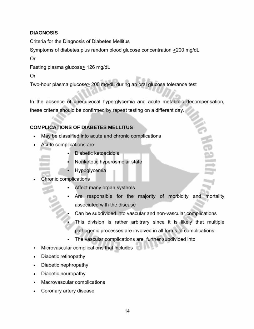
DIAGNOSIS Criteria for the Diagnosis of Diabetes Mellitus
Symptoms of diabetes plus random blood glucose concentration >200 mg/dL
Or
Fasting plasma glucose> 126 mg/dL
Or
Two-hour plasma glucose> 200 mg/dL during an oral glucose tolerance test
In the absence of unequivocal hyperglycemia and acute metabolic decompensation,
these criteria should be confirmed by repeat testing on a different day.
COMPLICATIONS OF DIABETES MELLITUS • May be classified into acute and chronic complications
• Acute complications are
Diabetic ketoacidois
Nonketotic hyperosmolar state
Hypoglycemia
• Chronic complications
Affect many organ systems
Are responsible for the majority of morbidity and mortality
associated with the disease
Can be subdivided into vascular and non-vascular complications
This division is rather arbitrary since it is likely that multiple
pathogenic processes are involved in all forms of complications.
The vascular complications are further subdivided into
Microvascular complications that includes
• Diabetic retinopathy
• Diabetic nephropathy
• Diabetic neuropathy
Macrovascular complications
• Coronary artery disease
15
• Peripheral vascular disease
• Cerebrovascular disease
The non-vascular complications are
Gastroparesis
Sexual dysfunction
Skin changes
Laboratory Evaluation Aims
• To determine degree of metabolic control
• Define associated complications
Extent of tests shall be individualized.
Tests to be Carried Out • Generally included are
• Fasting blood glucose (FBS)
• Random blood sugar (RBS)
• HbA1c
• Urinalysis
- Ketones
- Glycosuria
- Protein
� Serum lipid profile
� Serum BUN and creatinine
� Baseline electrocardiography
16
MANAGEMENT OVERALL PRINCIPLES
The goals of therapy for patients with type 1 or type 2 diabetes mellitus are to:
• eliminate symptoms related to hyperglycemia,
• reduce or eliminate the long-term microvascular and macrovascular complications of
diabetes mellitus and
• allow the patient to achieve as normal a life-style as possible
The care of an individual with either type 1 or type 2 diabetes mellitus requires a
multidisciplinary team.
Patient education, dietary management and exercise play a central role in managing
diabetic patients in addition to pharmacologic therapy.
Patient Education
• It should be viewed as a continuing process with regular visits for reinforcement and
not just a one-time affair.
• Involves education of the patient and family members about a number of issues
important for optimal diabetes care, including
- self-monitoring of blood glucose
- urine ketone monitoring ( for those with type 1 diabetes mellitus)
- insulin administration, if necessary
- guidelines for diabetes management during illnesses
- management of hypoglycemia
- foot and skin care
- diabetes management before, during, and after exercise
- risk factor-modifying activities

17
Dietary Management This involves optimal coordination of caloric intake with other aspects of diabetes
therapy like insulin, exercise and weight loss
Aims of Dietary Management
• Abolish symptoms of hyperglycemia
• Avoid hypoglycemia associated with therapeutic agents (insulin, oral
glucose lowering agents)
• Reduce overall blood glucose and minimize fluctuations
• Avoid atherogenic diets or those which may aggravate diabetic
complications (e.g. high protein intake in nephropathy)
• For a patient with type 1 diabetes mellitus the aim of dietary
management is to coordinate and match the caloric intake, both
temporally and quantitatively, with the appropriate amount of insulin.
• In type 2 patients, it should address the greatly increased prevalence
of cardiovascular risk factors (hypertension, dyslipidemia, obesity) and
disease in this population. The majority of these individuals are obese,
and weight loss is strongly encouraged and should remain an
important goal
• Food intake must be spread evenly throughout the waking hours and
taken at regular times in relation to the insulin dose.
• Patients should be advised to spread whatever food is available
through the day and the reasons explained.
• The diet should be balanced in relation to its composition of fats
(<30%), protein (10-20%), and carbohydrates (50-60%).
• Simple sugars that are rapidly absorbed should be avoided.
• Soluble fiber in the diet that delays the absorption and dampens
postprandial hyperglycemia should be taken.

18
General Dietary Instructions
Food items the diabetic should avoid (rapidly absorbed carbohydrates)
� Sugar, honey, jams, candy, marmalade
� Cakes, Sweet Biscuits
� Soft drinks (Coca Cola, Mirinda etc )
� Alcohols
Foods which the diabetic can take with restrictions
� Food items from grains: enjera, bread, kinche, kita, atmit
� Foods items prepared from peas, beans, lentils, chick peas
� Potato, sweet potato, kocho, bulla
Food items the diabetic can take freely or with minimal restrictions
� Lean meat and fish
� Eggs, milk, cottage cheese
� Green leafy vegetables (cabbage, tomato, pumpkin, carrots, onion)
� Tea, coffee and lemon juice without sugar, Ambo water, other mineral
waters
� Spices: pepper, garlic, ‘berbere’
Exercise It has multiple positive benefits (cardiovascular benefits, reduced blood pressure,
maintenance of muscle mass, reduction in body fat, weight loss, etc.).
Despite its benefits, exercise presents several challenges for individuals with diabetes
mellitus because they lack the normal glucoregulatory mechanisms.
Individuals with type 1 DM are prone to either hyperglycemia or hypoglycemia during
exercise, depending on the pre-exercise plasma glucose, the circulating insulin level,
and the level of exercise-induced catecholamines.
If the insulin level is too low, the rise in catecholamines may increase the plasma
glucose excessively, promote ketone body formation, and possibly lead to ketoacidosis.

19
If the circulating insulin level is excessive, this relative hyperinsulinemia may reduce
hepatic glucose production (decreased glycogenolysis, decreased gluconeogenesis)
and increase glucose entry into muscle, leading to hypoglycemia.
To avoid exercise-related hyper- or hypoglycemia, individuals with type 1 diabetes
should
• monitor blood glucose before, during, and after exercise
• delay exercise if blood glucose is > 250 mg/dL, <100 mg/d), or if ketones are present
• eat a meal 1 to 3 hours before exercise and take supplemental carbohydrate
feedings at least every 30 min during vigorous or prolonged exercise
• decrease insulin doses (based on previous experience) before exercise and inject
insulin into a nonexercising area.
In individuals with type 2 DM, exercise-related hypoglycemia is less common but can
occur in individuals taking either insulin or sulfonylureas.
MANAGEMENT OF THE TYPE 1 DIABETIC PATIENT Insulin Therapy in Type 1 Diabetes Mellitus Type 1 diabetic patients have an absolute requirement for insulin.
In general, they require 0.5-1.0 U/Kg per day of Insulin.
Insulin formulations are available as U-100 (1ml of solution equivalent to 100 units) or
U-40 (1ml of solution equivalent to 40units). It is very important that one designs and implements an insulin regimen that mimics
physiologic insulin secretions.
Twice daily administration of a short acting and intermediate acting insulin, given in
combination before breakfast and the evening meal, is the simplest and most commonly
used regimen.
Two thirds of the dose is given in the morning and one third is given in the evening.
Side effects of insulin therapy
� Hypoglycemia
� Weight gain
� Peripheral edema (in the short term)

20
� Insulin antibodies
� Local allergy
� Lipodystrophy at insulin injection sites
Insulin Preparations
Main types of therapeutic insulin
Species Bovine
Porcine
Human
Purity Conventional
Single peak
Highly purified
Duration of action Short
Intermediate
Long
MANAGEMENT OF THE TYPE 2 DIABETIC PATIENT Goals of Therapy • Improved glycemic control
• Treatment of conditions associated with type 2 diabetes mellitus
• Obesity
• hypertension
• Dyslipidemia
• Cardiovascular disease
• Detection and management of diabetes mellitus related complications
In a newly diagnosed type 2 diabetic, one should resort first to dietary management and
exercise before embarking on pharmacologic measures.
Glycemic control is reassessed and if response is not achieved, pharmacologic agents
may be tried.
21
Oral glucose lowering agents are preferred as the initial choices to lower serum glucose
levels.
As type 2 diabetes is a progressive illness, monotherapy is seldom successful in the
long term.
Therapy is initiated with one class of agent, depending on patient characteristics and a
second agent is added if adequate glycemic control is not achieved.
Groups of Oral Antidiabetic Agents with Examples Sulphonylureas
� Glibenclamide
Biguanides
� Metformin
Alpha-glucosidase inhibitors
� Acarbose
Thiazolidinediones
� Troglitazone
PREVENTION AND CONTROL Screening Many patients with diabetes mellitus are unaware that they have diabetes mellitus and
type 2 diabetes mellitus may be present for up to a decade before diagnosis.
Many patients with type 2 diabetes mellitus have one or more of diabetes mellitus
related complications at diagnosis.
For the above reasons, it is recommended to screen those at risk of developing
diabetes mellitus using fasting blood glucose.
This includes
• Those above 45 years of age every three years
• Those with family history of diabetes mellitus (parent or sibling with type 2 diabetes
mellitus)
• Obesity as evidenced by BMI> 27Kg/m2

22
• History of delivering a baby weighing above 4Kg or previous episode of gestational
diabetes mellitus
• Hypertension
A number of lifestyle modification and pharmacologic agents are suggested to prevent
or delay its onset.
High risk individuals should be encouraged to
• Maintain a normal body mass index
• Engage in regular physical exercise
The morbidity and mortality of diabetes mellitus related complications can be greatly
reduced if detected and treated at an early stage. These screening procedures are
indicated for all patients with diabetes mellitus.
Included under these procedures are
• Self monitoring of blood glucose
• HbA1c testing (2-4 times/ year)
• Annual patient education
• Screening for hypertension and dislypidemia
• Annual comprehensive eye examination by a qualified ophthalmologist
• Annual foot examination
COMPLICATIONS OF DIABETES MELLITUS
DIABETIC KETOACIDOSIS (DKA) DKA is a major cause of medical emergency and a serious cause of morbidity.
It is most commonly seen in patients with type 1 diabetes mellitus, but it can also be
seen in type 2 diabetics especially during acute illness.
Clinical Features
Symptoms • Nausea/vomiting
• Thirst/Polyuria

23
• Abdominal pain
• Shortness of breath
• Blurred vision
• Weight loss
• Altered mental state
Physical findings
• Tachycardia
• Hypotension
• Tachypnea/respiratory distress
• Kussmaul’s respiration (deep, fast breathing)
• Fever/hypothermia
• Dry mucous membranes/reduced skin turgor
• Abdominal tenderness
• Lethargy/obtundation/ possibly coma
Signs of infection, which may precipitate DKA, should be sought on physical
examination, even in the absence of fever.
Abdominal pain may be severe and sometimes may be mistaken for an acute
abdominal condition like pancreatitis or ruptured viscous.
Pathophysiology
DKA results from insulin deficiency combined with counterregulatory hormone excess
(glucagon, catecholamines, cortisol, and growth hormone). Both insulin deficiency and
glucagon excess, in particular, are necessary for DKA to develop.
The hyperglycemia of DKA results from
• increased hepatic glucose production via
• gluconeogenesis
• glycogenolysis

24
• Impaired peripheral glucose utilization
Ketosis results from a marked increase in free fatty acid release from adipocytes, with a
resulting shift toward ketone body synthesis in the liver. Reduced insulin levels, in
combination with elevations in catecholamines and growth hormone, lead to an increase
in lipolysis and release of free fatty acids. Normally, these free fatty acids are converted
to triglycerides or very low density lipoproteins (VLDL) in the liver, but in DKA,
hyperglucagonemia alters hepatic metabolism to favor ketone body formation.
Average losses of Fluid and Electrolyte in DKA of moderate severity
• Water: 6 Liters
• Sodium: 500 mmol
• Chloride: 400 mmol
• Potassium:350 mmol
Precipitating Events • Inadequate insulin administration
• Infection (pneumonia, UTI, gastroenteritis, sepsis)
• Infarction (cerebral, coronary, mesenteric, peripheral)
• New onset diabetes
• Drugs
Laboratory Abnormalities and Diagnosis
DKA is characterized by
• Hyperglycemia RBS>250mg/dL,
• Ketosis
• Ketone bodies positive at serum dilution of >1:2
• Ketonuria of 2+ or above
• Metabolic acidosis (increased anion gap)
• PH of 7.3 or lower and a bicarbonate level of <15 mEq/L
Despite a total-body potassium deficit, the serum potassium at presentation is typically
at the high end of the normal range or mildly elevated secondary to the acidosis.

25
TREATMENT • Rehydration
• Insulin
• Treatment of the precipitating cause when applicable
• Management of acid base disturbance
• Management of electrolyte imbalance
• Supportive care
• Resumption of subcutaneous insulin therapy once patient is out of the state of DKA
• Patient education
With appropriate therapy, the mortality of DKA is low (<5%).
Mortality is related more to the underlying or precipitating event, such as infection or
myocardial infarction.
Complications of DKA • The major non-metabolic complication of DKA therapy is cerebral edema, which
most often develops in children as DKA is resolving.
• Over replacement of free water should be avoided.
• Venous thrombosis and adult respiratory distress syndrome occasionally complicate
DKA.
• Disseminated intravascular coagulation (rare)
• Acute circulatory failure
NON-KETOTIC HYPEROSMOLAR COMA
It is the second major clinical presentation of uncontrolled diabetes mellitus.
It is almost exclusively seen in patients older than 60.
Precipitating factors include
� Drugs like beta-blockers
� Parenteral and enteral feeding
� Excessive intravenous glucose administration
As with DKA, osmotic diuresis is central to the pathogenesis but this develops more
slowly.

26
Dehydration is made worse by limited access to water and by the reduced perception of
thirst in the elderly.
Non-ketotic heperosmolar coma is characterized by marked hyperglycemia and loss of
water up to 25% of body weight in severe cases.
Differences between DKA and Non-Ketotic Hyperosmolar Coma (HONK)
DKA HONK
Age Any age >60
Presentation Hours to days Days or weeks
Mortality 5%overall 50%
Serum glucose High Very high
Serum osmolality High Very high
Serum sodium Normal or low Normal or high
Bicarbonate Low Normal or slightly low
Ketonuria Present Absent
The principles of treatment of HONK are similar to those for DKA.
Rehydration is the most important initial treatment and isotonic saline is the fluid of
choice. Less insulin is needed as compared to DKA early in treatment because acidosis
is not a feature and rehydration alone lowers blood glucose.
HYPOGLYCEMIA Hypoglycemia is defined as a recorded blood glucose concentration less than normal.
• Sometimes defined as a plasma glucose level <45 to 50 mg/dl
The glucose thresholds for hypoglycemia-induced symptoms and physiologic
responses vary widely, depending on the clinical setting.
Clinically significant hypoglycemia is based on the demonstration of Whipple's triad that includes
• symptoms consistent with hypoglycemia
• a low plasma glucose concentration(<45mg/dL)
• relief of symptoms after the plasma glucose level is raised
27
CAUSES Most commonly occurs as a side effect of the treatment of diabetes mellitus. Incidence
increases with attempts to achieve euglyemia with tight control of glucose
concentrations
Other causes in patients with diabetes include
• Overdose of insulin or oral agents
• ill timed administration of insulin or oral agents
• administration of the wrong type of insulin
• Missed or delayed meals or snacks
• Uncompensated exercise
• Alcohol consumption
• Concomitant chronic renal failure
• insulin clearance is reduced in patients with chronic renal failure
Hypoglycemia can cause significant morbidity and can be lethal, if severe or prolonged.
It should be considered in any patient who presents with confusion, altered level of
consciousness, or seizures.
The central nervous system can not synthesize glucose or store enough glycogen for
more than a few minutes’ glucose supply.
The brain cannot use free fatty acids as an energy source, and ketone bodies, which
are generated late, are not useful in acute hypoglycemia. Significant hypoglycemia,
therefore, can cause both acute and chronic brain dysfunction.
Morbidity related to severe hypoglycemia in diabetic patients
CNS
• Coma
• Convulsions
• Brain damage
• Impaired cognitive function, Intellectual decline
• Vascular events: TIA, stroke

28
Heart
� Cardiac arrhythmias
� Myocardial ischemia
Eye
• Vitreous hemorrhage
• ? worsening of retinopathy
Others
• Hypothermia
• Accidents
CLINICAL MANIFESTATIONS The various signs and symptoms of hypoglycemia appear at different glycemic
thresholds, related to different mechanisms.
They are subdivided into autonomic and neuroglycopenic manifestations.
The autonomic signs and symptoms occur with a relatively milder degree of
hypoglycemia. With worsening of the hypoglycemia, and consequent CNS glucose
deprivation, the neuroglycopenic symptoms and signs set in.
Autonomic signs and symptoms Result from increased autonomic nervous system activity
They include
• Palpitations
• Tremor or shaking
• Nervousness, Anxiety
• Irritability
• Sweating
• Hunger
• Nausea, vomiting
• Tingling, Paresthesias
• Tachycardia
• Hypertension

29
Adrenergic symptoms are mediated by norepinephrine released from sympathetic
postganglionic neurons and the release of epinephrine from the adrenal medullae.
Increased sweating is mediated by cholinergic sympathetic nerve fibers.
Neuroglycopenic signs and symptoms Neuroglycopenic symptoms are the direct result of central nervous system neuronal
glucose deprivation.
Signs and symptoms include
• Confusion
• Odd behavior
• Inability to concentrate
• Drowsiness
• Visual disturbance
• Tingling around the mouth
• Convulsions
• Focal neurologic deficits e.g. hemiplegia
• Coma
TREATMENT Urgent treatment is necessary in patients with suspected hypoglycemia.
Blood should be drawn, whenever possible, before the administration of glucose to
allow documentation of the plasma glucose level.
Oral treatment with glucose tablets or glucose-containing fluids, candy, or food is
appropriate if the patient is able and willing to take these. A reasonable initial dose is 20
g of glucose. If neuroglycopenia precludes oral feedings, parenteral therapy is
necessary. Intravenous glucose (25 g) should be given using a 50% solution followed
by a constant infusion of 5 or 10% dextrose. If intravenous therapy is not practical,
subcutaneous or intramuscular glucagon can be used, particularly in people with type 1
diabetes mellitus. Because it acts primarily by stimulating glycogenolysis, glucagon is
ineffective in glycogen-depleted individuals (e.g., those with alcohol-induced
hypoglycemia). These treatments raise plasma glucose concentrations only transiently,
30
and patients should be encouraged to eat as soon as they are alert in order to prevent a
recurrence.
CHRONIC COMPLICATIONS OF DIABETES MELLITUS The microvascular complications of both type 1 and type 2 DM result from chronic
hyperglycemia. It is also suspected that a genetic susceptibility for developing particular
complications exists.
Evidence implicating a causative role for chronic hyperglycemia in the development of
macrovascular complications is less conclusive.
Dyslipidemia and hypertension also play important roles in macrovascular
complications.
Three major theories have been proposed to explain how hyperglycemia might lead to
the chronic complications of diabetes mellitus. These are
• formation of advanced glycosylation end products (AGEs) via the nonenzymatic
glycosylaton of cellular protein
• increased glucose metabolism via the sorbitol pathway
• increased formation of diacylglycerol leading to activation of certain isoforms of
protein kinase C (PKC), which, in turn, affect a variety of cellular events that lead to
diabetes mellitus-related complications
Studies have provided definitive proof that reduction in chronic hyperglycemia can
prevent many of the early complications of type 1 DM.
The development of chronic complications correlates with the duration of diabetes and
glycemic control
Ophthalmologic Complications of Diabetes Mellitus Diabetes mellitus is a leading cause of blindness in the working population in the
developed world
Blindness is primarily the result of progressive diabetic retinopathy and clinically
significant macular edema.
Diabetic retinopathy is classified into two stages. These are non-proliferative and
proliferative retinopathy.
31
Proliferative retinopathy
• retinal vascular micaroaneurysms
• blot hemorrhages
• cotton wool spots
Non-proliferative retinopathy
• Neovascularization is the hallmark.
The most effective treatment for diabetic retinopathy is prevention.
Intensive glycemic control will delay the development or slow the progression of diabetic
retinopathy.
There may be a transient, paradoxical worsening of established diabetic retinopathy,
during the first 6 to 12 months of improved glycemic control.
Regular, comprehensive eye examinations for all individuals with diabetes mellitus are
required, and these should be performed by an experienced ophthalmologist.
Other Ocular Problems � Cataract
� Develop early and progress rapidly in diabetic subjects
� Glaucoma
� Ocular palsies
� Sudden visual loss
Renal Complications of Diabetes Mellitus Diabetic Nephropathy
• One of the commonest causes of end stage renal failure Nephropathy progresses
through the following stages.
• Hyperfiltration
• Associated with increased glomerular size and kidney volume
• Increased glomerular filtration rate
32
• Microalbuminuria
• defined as excretion in a 24-h period of 30 to 300 mg/d of albumin
(incipient nephropathy)
• Established nephropathy
Overt proteinuria (Urinary protein >300 mg/d)
Dipstick positive
• End stage renal disease
Renal failure that requires dialysis or transplantation
The optimal therapy for diabetic nephropathy is prevention.
Interventions effective in slowing progression from microalbuminuria to overt
nephropathy include near normalization of glycemia, strict blood pressure control and
administration of ACE inhibitors.
Diabetic Neuropathy Manifests as polyneuropathy, mononeuropathy, and/or autonomic neuropathy. The
most common form of diabetic neuropathy is distal symmetric polyneuropathy often
described as having a glove and stocking distribution.
Most frequently presents with distal sensory loss. Hyperesthesia, parathesia, and pain
also occur. Physical examination reveals sensory loss, loss of ankle reflexes, and
abnormal position sense.
Diabetic polyradiculopathy is a syndrome characterized by severe disabling pain in
the distribution of one or more nerve roots. It may be accompanied by motor weakness.
Mononeuropathy presents with pain and motor weakness in the distribution of a single
nerve.Involvement of the third cranial nerve is most common and is heralded by
diplopia. Physical examination reveals ptosis and opthalmoplegia with normal papillary
constriction to light. Sometimes cranial nerves IV, VI, or VII (Bell's palsy) are affected.
Peripheral mononeuropathies or simultaneous involvement of more than one nerve
(mononeuropathy multiplex) may also occur.
Autonomic Neuropathy in diabetes can involve multiple systems, including: the
cardiovascular, gastrointestinal, genitourinary, sudomotor, and metabolic systems.
Autonomic neuropathies affecting the cardiovascular system cause a resting

33
tachycardia and orthostatic hypotension. Gastroparesis and bladder-emptying
abnormalities are also likely related to the autonomic neuropathy seen in DM (discussed
below). Hyperhidrosis of the upper extremities and anhidrosis of the lower extremities
result from sympathetic nervous system dysfunction. Anhidrosis of the feet can promote
dry skin with cracking, which increases the risk of skin ulceration. Autonomic
neuropathy may reduce counterregulatory hormone release, leading to an inability to
sense hypoglycemia appropriately (hypoglycemia unawareness, thereby subjecting the
patient to the risk of severe hypoglycemia and complicating efforts to improve glycemic
control.
One should consider other possible causes of neuropathy before ascribing signs and
symptoms to be due to diabetic neuropathy as other causes of neuropathy may present
in a similar manner.
Diabetic foot disease
Different types of diabetic tissue damage interact and combine in the feet, giving a wide
variety of lesions ranging from relatively harmless dysaesthesiae to fulminating
infections and widespread ulceration and gangrene.
Factors that play important roles in the pathogenesis of diabetic foot ulcers include,
• Neuropathy
o Predisposes patient to repetitive trauma to the feet
• Reduction in blood flow
o Delays wound healing
o Serves as a good medium for bacterial multiplication
• Deformity in the feet
o This leads to abnormal foot mechanics with misdistribution of pressure
over parts of the feet.
• Reduced anti-infective activity of leucocytes of hyperglycaemic patients
• Poor eye sight of diabetics
34
Preventive management is as important as correct treatment of established lesions.
The main aim is to prevent excessive pressure on particular areas of skin.
• Look for corns and callosities on the soles or heels.
Patients should be advised to inspect their feet regularly.
If the diabetic cannot see his feet properly, someone else must look at them at least
weekly or, if there is marked loss of pinprick sensation, thrice weekly.
The principles of treatment of diabetic foot ulcers involve the elimination of infection by
draining pus or removing infected bone, by removing dead tissue likely to provide a
focus for infection, and by using antibiotics if necessary.
Healing is speeded by encouraging the greatest possible blood flow, and protecting the
foot from trauma.
Debridement must be thorough and extensive.
Amputation may be required in case of life threatening infections by gas forming
organisms or in cases where there is of dead tissue.
Antibiotics choice should be guided by culture of either the local lesion or blood when
infection is widespread.
The control of glucose levels should be as strict as possible, as hyperglycemia is bound
to bring about dysfunction of the leucocytes.
DIABETIC HEART DISEASE
Several different processes contribute to diabetic heart disease. These include
• Abnormalities in plasma lipids
• Increased liability to hypertension in diabetics
• Structural narrowing of the lumina of vessels
• The presence of a state that favors coagulation
Patients may present with either clinical features of ischeamic heart disease or
congestive heart failure in the absence of ischeamia (diabetic cardiomyopathy).

35
A greater proportion of cardiac infarcts seem painless in patients with diabetes than in
non-diabetics.
Heart disease is the major cause of death among both type 1 and type 2 diabetics, and
ultimately is likely to affect about 60 per cent of patients.
The management of ischaemic heart disease among diabetics differs little from that
generally employed.
Life style modification plays a big role in the management. Patients should be advised
to stop smoking.

36
UNIT THREE SATELLITE MODULES
3.1 SATELLITE MODULE FOR PUBLIC HEALTH OFFICRES
Diabetes Mellitus
I. Definition A group of metabolic disorders characterized by hyperglycemia resulting from defects in
insulin secretion, insulin action, or both
II. Classification 1. Type 1 diabetes Mellitus
Previously called insulin dependent DM or Juvenile onset DM due to its usual onset in
adolescence or childhood
It includes about 5-10% of all diabetic patents and is caused by autoimmune destruction
of the B-cells of the pancreas. The resulting hyperglycemia is responsible for the acute
and chronic complications of the disease.
In few patients with type 1 DM the pathogenesis remains idiopathic.
2. Type 2 DM
This class of diabetes also known as non-insulin dependent DM accounts for 90-95% of
the population with DM. It is common in people older than 40yrs and results from
variable combinations of insulin resistance and defects in insulin secretion.
Learning Activity In the exercise in the core module,
1. Identify the key historical findings suggesting the diagnosis.
2. Identify the key physical findings supporting the diagnosis of type 1DM

37
3. In the hypothetical case already mentioned the health officer requested the
following laboratory investigations with the results shown below.
RBS = 450mg/dL
U/A - glucose = 4+
- ketones =4+
- Albumin =negative
3.1. Do the findings on laboratory investigation support (refute) your clinical
suspicion?
3.2. What additional investigation would you request if resources permit?
III. Diagnosis
1. Clinical features
The presentation of patients depends on the type of diabetes and the stage of
pathologic process.
1.1. Type 1 DM
Patients with type 1 DM commonly present with the classic acute symptoms of
hyperglycemia: excessive thirst (polydipsia), polyuria, polyphagia and weight loss.
Twenty five percent of type 1 diabetics present for the first time with diabetic
ketoacidosis (DKA) characterized by hyperglycemia, ketosis and acidosis.
1.2. Type 2 DM
The presentation of type 2 diabetes is less acute than type 1 with “poly” symptoms and
accompanying lethargy and fatigue.
The disease is often present for many years before the diagnosis and chronic
hyperglycemia may be responsible for susceptibility to infections (eg. vaginitis)

38
2. Criteria for the Diagnosis of DM
DM can be diagnosed in the presence of any of the following
2.1. “Poly” symptoms plus casual plasma glucose greater than or equal to 200mg/dl
2.2. Fasting blood glucose (FBG) greater than or equal to 126 mg/dl
2.3 Two hours plasma glucose of greater than or equal to 200 mg/dl during an oral
glucose tolerance test (OGTT).
(OGTT – Plasma glucose measurement after 75g of anhydrous glucose load).
NB: In the absence of unequivocal hyperglycemia the criteria should be confirmed by
repeat testing on a different day.
IV. Management The goals of management are:
1. Short term – immediate treatment to relieve the symptoms such as polydipsia,
polyuria, or acute infection.
2. Intermediate – to return the patient to physiologic state and social life.
3. Long term – to prevent the development or delay progression of complications of
diabetes
The treatment of diabetes can be categorized as non-drug therapy and drug therapy.
1. Non – Drug Therapy
1.1. Regular Physical exercise
This results in improvements in the sense of well being, cardiovascular fitness, blood
pressure, insulin sensitivity, weight reduction and glycemic control. Regular physical
exercise for at least 30 minutes a day is recommended.
Blood glucose should ideally be measured before any exercise which shouldn’t be
undertaken with FBS of 300 mg/dl and above on the other hand if FBS is less than
100mg/dl exercise may precipitate hypoglycemia and carbohydrate should be
39
consumed in advance medical evaluation is advised to determine the level of fitness
and appropriate exercise based on the presence and degree of macrovascular and
cardiovascular complications.
1.2. Dietary Control
A general dietary recommendation includes consumption of a balanced health diet
composed of:
- 10 – 20% protein
- 30% fat
- 50-60% carbohydrate
Patients should be advised to avoid dimple sugars like table sugar, honey etc and low
saturated fat and cholesterol white high fiber diet is recommended.
2. Drug Therapy
2.1. Type 1 DM
Patients with type 1 diabetes have on absolute requirement of insulin for survival.
Insulin is also used in type 2 diabetics when a combination of oral agents fails to
achieve glucose targets and temporarily in patients with serious infection or surgery.
The types of insulin available are rapid acting, short acting, intermediate acting and long
acting.
Standard insulin therapy consists of one to two injections per day using intermediate or
long acting insulin with or without regular insulin. Starting insulin does vary from 0.15 to
0.50/kg (as high as 1.5 u/kg in cases of severe insulin resistance) depending on patient
size and degree of glycemia.
Adults of normal weight may be started with 20-25 u/d of intermediate acting insulin and
increased to maintain a blood sugar level of 80-120 mg/dl.

40
2.2. Type 2 DM
Provided pharmacologic therapy is not required immediately all patients should be given
at least a one month trial of diet, exercise and weight management. If this regimen does
not lead to adequate blood glucose control, oral antihyperglycemic agents with or
without insulin are indicated. Insulin may be needed in symptomatic patients who have
type 2 DM with FBG values greater than 250 mg/dl. The common antihyperglycemic
agents in use are discussed below.
a. Glibenclamide
Dosage
• 2.5– 20mg daily or in two divided doses
Side effect
• hypoglycemia.
Contraindications
• hepatic and renal impairment.
Drug interactions
• alcohol – flushing
Dosage form
• tablets of 5mg
b. Metformin
Dosage
• 500 – 2000 mg PO daily in divided doses
Side effects
• anorexia, nausea, vomiting, abdominal discomfort and diarrhea
Contraindications
• renal disease, hepatic disease, alcoholism
Dosage forms
• tablets of 500mg.

41
V. Complications The complications of DM can be divided into acute and chronic complications
1. Acute complications
1.1 Diabetic ketoacidosis (DKA)
It is a clinical condition that may be defined as a triad of
- Hyperglycemia
- Ketosis
- Acidosis
It usually occurs in the setting of type 1 DM and is primarily caused by relative or
absolute insulin deficiency.
Common precipitating factors are infection and omission of insulin dose. Patients may
also come with DKA on initial presentation.
Symptoms include nausea, vomiting, polydipsia, polyuria, abdominal pain and
weakness. On examination, signs include tachycardia, orthostatic hypotension, poor
skin turgor, warm or dry skin and mucous membranes, deep and fast breathing
(Kussmaul’s respiration), hypothermia or normothermia, acetone breath, and altered
mental status or coma.
Investigations:
- Blood glucose greater than 250 mg/dl
- Ketosis: ketonuria of 2+ to 3+
Treatment
- Non drug treatment: correct or treat the precipitating factor
- Drug treatment includes insulin, fluid replacement, and potassium replacement
� Insulin
� 20U of regular insulin (10IM/10IV) followed by 5U IM every hour in
adults (0.1U/kg/h in children)
Blood glucose should be checked every two hours. If after the first two hours
blood glucose level has not fallen significantly, dose of IM insulin can be
doubled. When the patient is completely out of ketosis (evidenced by absent

42
urinary ketones) regular insulin is given 6 hours subcutaneously according to
random blood sugar (RBS) level as follows:
RBS> 250mg/dl- 12u
RBS: 180-250mg/dl-8u
RBS – 120-180mg/dl –4u
RBS < 120 but >70 mg/dl-70mg/dL-no insulin
RBS<70mg/dL- Hold insulin and give juice or meal and recheck blood glucose
in one hour.
Fluid Replacement Normal saline IV should be given rapidly as soon as the patient arrives.
Total fluid given may be as high as 10 litres depending on the patient’s response &
urine output.
Fluid replacement may proceed in the following manner.
2-3L of 0.9% saline over first 1-3 hour (5-10mL/Kg per hour); subsequently, 0.45%
saline at 150-300mL/hr; change to 5% glucose and 0.45% saline at 100-200mL/hr when
plasma glucose reaches 250mg/dL.
Electrolyte Replacement Potassium replacement should be according to serum potassium values. Potassium, 20
meg 1h is generally safe if renal function is normal
SE. renal failure
Dosage forms, injection 20 meg /10 ml ampoule of kc/

43
3.2 SATELLITE MODULE FOR BSC NURSES Directions for using the module Before starting to read this module, please follow the directions given below
Go through all the contents of the Core Module by starting with the pre test
Use a separate sheet of paper to write your answers and label it as pre-test
answers
Learning objectives
On completion of this module, the learner will able to
1. Differentiate between type 1 and type 2 diabetes
2. Describe etiologic factors associated with diabetics
3. Understand the function of glucose and insulin
4. Relate the clinical manifestation of diabetic mellitus to the associated
pathophysiologic alteration
5. Recognize the seriousness of DM with reference to morbidity and mortality
6. Identify the diagnostic and clinical significance of blood glucose tests
7. Describe the various type of insulin
8. Explain the dietary modification used for management of person with diabetes
9. Describe the relationship between diet, exercises and modification for persons with
diabetes
10. Develop a plan for teaching insulin self administration
11. Learn on the pharmacological calculation of insulin to reach on accurate dose (units
to milliliter from a vial containing 40,80 or100 units)
12. Differentiate between hypoglycemia and Diabetic ketoacidocis and HHNS
13. Describe the major macrovascular, microvascular and neuropathic complication of
diabetic and self care behavior important in the prevention
14. Explain why the are of such importance
15. Use the Nursing process as a frame work for care of the patient with diabetes

44
Diabetes Mellitus
Definition: - is a chronic multifactorial, systemic metabolic disorder characterized by
hyperglycemia and abnormal insulin production and /or action.
Insulin is a hormone produced by the pancreas. it controls the level of glucose in the
blood by regulating the production and storage of glucose.
-Beta cells are responsible for production and secreting insulin and glucagons.
-It is anabolic or storage hormone
When a meal is eaten, insulin secretion increases and moves glucose
from the blood into the muscle, liver, and fat cell.
In those cells insulin has the following effect.
o Stimulate storage of glucose in the liver and muscle
o Enhance storage of dietary fat in the adipose tissue
o Accelerate transport of amino acid in to the cells
Also inhibit the break down of stored glucose, protein and fat
Glucagon-a relative or absolute excesses of glucagons are an essential factor in the
development of DM. It increases blood glucose concentrations
Classification of diabetes Type 1-IDDM
Type 2-NIDDM
Other specific subtype- like malnutrition related Diabetic Mellitus (MRDM), gestational
DM
Characteristics of type 1 & type 2 DM (compare& contrast) See the Core Module
Etiology Type 1 DM
- Is characterized by destruction of the pancreatic beta cells
- The exact cause is unknown

45
- But it is thought that a combination of
Genetic susceptibility
Environmental factors that contribute to beta cell destruction and
Genes regulating immune response are involved
Type 2 DM
- Is related to insulin resistance (a decreased sensitivity to insulin) and
impaired insulin secretion
- But the exact mechanism is unknown
- Risk factors include:
Age (insulin resistance tends to increase with age over 65)
Obesity
Family history is strongly associated or environmental factors, e.g. Viruses
Diagnostic Criteria for Diabetic Mellitus Fasting plasma glucose (FPG) >126mg/dl
Random blood glucose (RBS) >200mg/dl with symptoms
2hr post load glucose >200mg/dl
See the Core Module for the details
Management Goal: - to try to normalize insulin activity & blood glucose levels in an attempt to reduce
the development of the vascular & Neuropathic complications.
There are five components of management for diabetes: -
- Diet
- Exercise
- Monitoring blood glucose
- Medication (as needed)
- Education

46
I. Dietary Management Goal: - provision of all the essential food constitutes (e.g., carbohydrate, proteins, fat,
vitamins, minerals)
- Achievement and maintenance of reasonable weight
- Meeting energy needs
- Prevention of wide daily fluctuations in blood glucose levels with Blood
glucose level as close to normal as is safe and practical
- Decrease blood lipid levels, if elevated
A. Calories The most important objective in dietary management of DM is control the total calorie
intake and to attain or maintain a reasonable body weight and control of blood glucose
levels.
The general recommendation include consumption of a balanced healthy diet composed of the following
• 50% to 60% of calories to be derived from carbohydrates
• Less than 30% obtained from fat and
• The remaining 10% to 20% from protein
*Food which diabetic should avoid (rapidly absorbed carbohydrate/simple sugar)
1) Sugar, honey, jam, marmalade and candy
2) Cakes and sweet biscuits
3) Soft drink (Fanta, coca cola, etc)
4) Alcohol (Cognac, tej, araki, whisky)
There are types of alcohols which are allowed in moderation, that is less
sweat drinks i.e. light beer or dry wine (not more than 2 drinks for men,1 drink for
women/day). Alcoholic beverage equivalent to12 oz beer, 5 oz wine and 1.5 oz spirit. It
should be always taken with food.

47
*Foods which diabetic should take with restriction (cereals or starch 50 - 60 %) a. Foods from grain e.g. injera, bread, kinche, dabo kolo, kita ,atemit
b. Foods prepared from peas, beans, lentils
c. Potato, sweat potato, kocho, bulla
d. All fruits except lemons and grape fruit
e. Macaroni, pasta, rice
Foods, which diabetics can take freely or with minimal restriction (protein 10-20%)
A) Lean meat and fish (with minimal restriction)
B) Eggs and milk (with minimal restriction)
C) Green leafy vegetables (kale, salad, cabbage
D) Lemon, grape fruit
E) Tea, coffee, lemon juice without sugar, ambo water, other mineral water and
clear soup
F) Spices pepper, berbere
G) Tomato, pumpkin, carrot, Onion, chili pepper
II. Exercise
- Is extremely important in the management of diabetes because of its
effect on lowering blood glucose and reducing cardiovascular risk factors
- Lowers blood glucose level by increasing the uptake of glucose by body
muscles and by improving insulin utilization
- Pre or post exercise snack may be required to prevent hypoglycemia after
exercise
- Patients should be taught to do regular, moderate exercise at the same
time and in the same amount for at least 30 minutes each day. Exercise
recommendations must be altered as necessary for patients with diabetic
complications
- Blood glucose level should be measured before any exercise activity is
initiated.

48
- Exercise should not be initiated with fasting plasma glucose>250mg/dl
or<100mg/dl (because it may precipitate diabetic ketoacidosis and
hypoglycemia respectively)
-Patient is advised to:
• Use proper footwear and if appropriate other
Protective equipment
• Avoid exercise in extreme heat or cold
• Inspect feet daily after exercise
• Avoid exercising during periods of poor metabolic control.
III. Monitoring of Glucose and Ketones
• Blood glucose level should be assessed frequently by the patient or by having
follow up in the health unit
• Urine and ketone checks are appropriate if blood glucose is greater or equal
to 250 mg/dl
IV. Medications
Insulin therapy
• In type 1 diabetes, the body loses the ability to produce insulin, thus,
exogenous insulin must be administered indefinitely. A standard insulin
treatment consists of one or two injection/day, using intermediate or long
acting insulin with or with out regular insulin.
• In type 2 diabetes, insulin may be necessary on a long term basis to control
glucose levels if diet and oral agents have failed. In addition, some patients
whose type 2 diabetes is usually controlled by diet alone or diet and an oral
agent may require insulin temporarily during illness, infection, pregnancy,
surgery or some other stressful events.
Insulin preparation
A number of insulin preparation are available. They vary according to four main
characteristics, that is

49
1) Concentration –U-40
-U-80
-U-100
-U-500
2) Species (source)- Human source
-Animal source (beef/pork)
3) Manufacturer –Lilly
-Novo nordisk companies
4) Time course of action –Rapid acting(regular)
-Intermediate acting (NPH and lente)
-Long acting(ultra lente)
-Mixed (e.g 70%NPH/30%Reg)
Time course
Agent
Onset
Peak
Duration
Indications
Short acting Regular (R) ½ -1
hr
2-3hr 4-6hr Usually
administered 20-
30 minutes
before meal
Intermediate
acting
NPH (Neutal
protamine
Hagedorn)Lent
e(L)
3-4hr 4-12hr 16-20 hr Usually taken
after food
Long acting Ultralente (“UL”) 6-8 hr 12-16 hr 20-30hr Used primarily to
control fasting
glucose level
50
Patient education -about Insulin Injection
- Insulin injections are administered into the subcutaneous tissue
- Equipment: - Insulin
- Short acting insulin is clear in appearance and long acting
insulin are cloudy and white
- The long acting must be mixed (gently inverted or rolled in the hands)
before use
- Before injection it should have room temperature, which may require
rolling it in the hands or removing it from a refrigerator for a time before
the injection. Actually there is no significant difference in the biologic
activity between insulin put in the refrigerator and in the temperature (25-
34oc). It would seem safe to conclude that unless insulin in Africa is
stored for a long period at very high temperature, there is no potential
problem (5).
- If a frosted, adherent coating is present, some of the insulin is bound and
should not be used
Syringes - Should be matched with the insulin concentration
- 1 ml syringes – hold 100 units
- ½ ml syringes – hold 50 units
- 3/10 ml syringes – hold 30 units
Preparing the injection Mixing insulin: - when short and long acting insulin is to be mixed first withdraw
the regular insulin (short acting)
Administering the injection Allow alcohol to evaporate from the skin before injection or avoid use of alcohol for
cleansing
- Four main areas
• Abdomen

51
• Arms (posterior surface)
• Thighs (anterior surface)
• Hips
Absorption is greatest in abdomen and decreases progressively in the arm, thigh, and
hips.
Rotation - Rotation of injection site is required to prevent lipodystrophy, localized changes in fatty
tissue,
The patient is instructed as:
1. Do not use a site > once every 4 to 6 weeks
2. Sites should be 1 to 1 ½ inches apart
3. Use all sites in one geographic area, then move to the next area
4. Document site use
Side effects of insulin injections 1. Local allergic reactions.
- This appears in the form of redness, swelling, tenderness, and indurations
or a 2 to 4 cm wheal may appear at the injection site 1to 2 hrs after
injection
- Usually occur during the beginning stage of therapy and disappear with
continued use of insulin
- Antihistamine will be given 1 hr before injection
- If alcohol is used to clean the area the skin should be allowed to dry
- A local reaction is usually not dangerous unless it becomes more
extensive over time
2. Systemic allergic reaction-are rare
- Can be life threatening
- Local skin reaction that gradually spreads in to generalized urticaria which
can include laryngeal edema with respiratory distress

52
Treatment involves: - desensitization, gradually increasing the amount of insulin
under cautious observation.
3. Insulin lipodystrophy
- Refers to a localized disturbance of fat metabolism in the form of
lipoatrophy (loss of subcutaneous fat and appears as slight dimpling or
more serious pitting of subcutaneous fat) or lipohyperthrophy (is the
development of fibro fatty masses at the injection site and is caused by
the repeated use of injection site)
- If insulin is injected in to scarred areas the absorption may be delayed
Treatment: Patient should avoid injection on the areas and prevent by rotating injection
sites
3. Insulin Resistance
- Insulin requirements up to 1u/kg can be seen with obesity, stress, aging
- Modest insulin resistance-2-3u/kg wt-can be seen frequently with type 2
- Extreme insulin resistance (>3u/kg)-is rare and may be caused by a variety of
autoimmune and genetic disorder
Oral Anti diabetic agents Effective for type 2 DM patients who do not respond to diet and exercise alone and who
are able to produce some insulin
A. Glibenclamide Dosage: 2.5 – 20mg daily or in two divided doses
Side effect: hypoglycemia.
Contraindications: hepatic and renal impairment.
Drug interactions occur with alcohol leading to flushing
Dosage form: tablet 5mg

53
B. Metformin Dosage, 500 – 2000 mg Po daily in divided doses
Side effects: anorexia, nausea, vomiting, abdominal discomfort and diarrhea.
Contra-indication: renal disease, hepatic disease alcoholism
Dosage form: tablet, 500mg and 850mg
Fig1: Algorithm for the control of type 2(NIDDM)
New NIDDM
Diet and life style advice
Still symptomatic Start oral agents
Obese- metformin None obese-glibenclamide
Increase dose monthly, if necessary to maximum dose
-Patient well and asymptomatic
- Continue at PHC clinic
Still not controlled, give combin Metformin and glibenclamide
Again, increase dose to the top
Still not controlled refer to the Dr.
54
A. Acute complications of diabetes 1. Hypoglycemia (Insulin Reactions)
- Occurs when blood glucose level falls below 50 to 60 mg /dl (2.7 to 3.3 mmol/L )
Caused by: Too much insulin or hypoglycemic agents
-Too little food or
-Excessive physical exercise or excessive alcohol
-Occurs also if meals are delayed or snacks are omitted
Symptoms includes
Mild hypoglycemia
- Sweating
- Tremor
- Tachycardia
- Palpitation
- Nervousness and
- Hunger
Moderate hypoglycemia
- Inability to concentrate
- Headache
- Light headedness
- Confusion
- Memory lapses
- Numbness of the lips and tongue
- Slurred speech
- In coordination
- Emotional changes
- Irrational behavior
- Double vision and drowsiness
Severe hypoglycemia
- Seizures
- Difficulty arousing from sleep or
- Loss of consciousness

55
Treatment for Mild and moderate hypoglycemia
10 to 15 mg of a fast acting sugar orally
2-3tsp 4 to6 tsp 2 to4
2-3tsp
Hard candies sugar, honey , fruit juice or glucose tablets
Treatment for Severe hypoglycemia The initial treatment of a confused, comatose patient is to infuse a bolus of 50 ml of
50% glucose; preferably after a blood sample for lab analysis has been obtained. This
bolus is followed by the continuous infusion of 5 to 10 % of glucose at a rate sufficient to
keep the plasma glucose level> 100mg/dl
Patient education: - prevented by following a regular pattern for eating, administering
insulin, and exercising
- Because unexpected hypoglycemia may occur all patients treated with
insulin should wear an identification bracelet or tag indicating that they
have diabetes and should keep sugar or candy in their pocket
- Patient and family members should be aware of signs of hypoglycemia
2. Diabetic Ketoacidosis (DKA) DKA is caused by an absence or markedly inadequate amount of insulin. Patients with
sever DKA can become severely dehydrated with the loss of electrolyte. Volume loss is
highly variable as is Na+ and K + decreasing. Paradoxically potassium appear elevated
as a response to acidosis, though this is a temporary shift of potassium from intra to
extra cellular space
Sign and symptoms: - anorexia, nausea, and vomiting & abdominal pain
- Acetone breathe
- Kussmaul respiration (very deep& and fast respiration)
-Lab.value : Blood glucose level 300 to 800 mg /dL
Causes: - A reduced or missed dose of insulin, an illness or infection
56
Treatment of DKA includes
• Fluid replacement if kidney functions is normal and there is no concern
for heart disease
- 0.9% normal saline of a very high rate usually 0.5 to l lit /hr for 2 to 3 hrs,
then the decrease the rate to 250-500ml/hr as orthostatic change
disappear.
- Fluid may need to be given cautiously in renal impaired or older patients
who may have underlying new problems
- Hypo tonic normal saline (0.45%) for hypertensive
- K- replacement – Give none with the first liters of saline, then 20 mmol
hourly in saline up to a total of 80 mmol
- Insulin IV at a slow, continuous rate (start with a 10u bolus of regular
insulin IV, followed immediately by a continuous IV insulin infusion at 0.1u
/kg/hr if weight is known, if not 6u/hr. when the patient starts to eat and
drink change to subcutaneous 30 min before meal and discontinue insulin
infusion 30-60 min after meal.
- Hourly monitoring of blood glucose level, urine glucose level, blood
ketone level Alter insulin if necessary accordingly
- Bicarbonate infusion- doesn’t give routinely. Give 50 mmol slow IV only if
patients condition is critical.
3. Hyperglycemic Hyperosmolar Nonketotic syndrome (HHNK) - is a situation in which hyperosmolarity and hyperglycemia predominate, with alteration
of the sensorium (sense of awareness) .See the core module for the details
Clinical manifestations
* Symptoms of hypotension
- Profound dehydration
- Tachycardia and
- Neurologic signs (e.g. altered sensorium, seizures, hemiparesis )

57
Causes: - Occurs most frequently in older people (50-70 yrs) who had no previous
history of DM or only mild type 2 diabetes and renal impairment
Precipitating events
- Acute illness
- Ingestion of medication known to provoke insulin insufficiency (thiazide
diuretics, propranolol) or
- Therapeutic procedures
Management: - Similar with diabeticketoacidosis (DKA)
Fluid, electrolyte and insulin replacement
1) Water loss is usually more severe that electrolyte depletion give patient 1-2 litter
0.9% normal saline, if hypotensive followed by .45% normal saline.
2) Infusion rate should be based on renal and cardiac status
3) Insulin requirements are often low.
B. Long term Complications of diabetes - Affect almost all organ systems of the body
- Generally categorized as Macro vascular and Micro vascular
1. Macro vascular Complications This complication are cardiovascular& cerbrovascular disease including hypertension
myocardial infarction ischemia, stroke& peripheral vascular disease
- Prevention and treatments of modifiable risk factors is recommended (Smoking,
obesity & initiation of safe exercise.)
2. Micro vascular Complications involve retina, kidney and nerves - Are unique to diabetes
- Is characterized by capillary basement membrane thickening
- 3 places where impaired capillary function may have devastating effects
are the microcirculation of the retina of the eye (Retinopathy - twenty five
times greater risk of blindness) & the kidney (Nephropathy - seventeen

58
times more likely to experience kidney failure) and blood supply to
peripheral nerves (Neuropathy).
Foot and leg problems
-50-70% of lower extremity amputations are performed on people with diabetes. 50% of
which are preventable, provided patients are taught preventive foot care on daily basis
-Diabetic complications contributing to foot infections are: Neuropathy, peripheral
vascular disease and immunocompromised.
o Neuropathy A group of disease that affect all types of nerves including peripheral, autonomic and
spinal nerves
- Leading to loss of pain and pressure sensation and autonomic neuropathy
- Leads to increased dryness and fissuring of the skin 2o to increased sweating)
o Peripheral Vascular diseases Poor circulation of the lower extremities contributes to poor wound healing and the
development of gangrene
o Immunocompromised Hyperglycemia impairs the ability of specialized leukocytes to destroy bacteria
Foot Care Instructions for a patient with DM
1. Assess your feet daily for sensation, reddened areas, or broken skin
2. Wash and dry your feet daily, especially between the toes
3. If the skin is dry, apply a thin coat of lubricating oil
4. If callus formation is present, rub areas with a pumice stone when the feet are
wet then rub with a towel. Avoid use of chemical agents
5. Immediately after bathing while the toenails are soft, clip the nails straight across,
and smooth them to the shape of the toe.
6. Wear shoes and stockings that give room for movement of the toes. Wear shoes
with only moderately high heels
7. Tie shoes loosely but firmly
8. If your feet perspire, change shoes and stockings during the day

59
9. Measures that increase circulation to the lower extremities should be instituted,
including
- Avoid smoking
- Avoid crossing legs when sitting
- Protect extremities when exposed to cold
- Avoid immersing feet in cold water
- Use socks or stockings that do not apply pressure to the legs at specific
sites or constrict.
- Apply pressure to the legs at specific sites
- Institute an exercise regimen
10. Use a light when walking at night
11. Do not place feet near sources of heat
e.g fireplace, heater , hot water bottle, etc.
12. Wear shoes when outdoors that protects toes and soles of feet from cuts and
bruises.
13. Referral to a specialist when necessary.
Nursing Process
The Patient with Newly Diagnosed Diabetic Mellitus
Assessment
- The history and physical assessment focus on
• sign and symptom of prolonged hyperglycemia and
• physical, social and emotional factors that may affect the patient ability to
learn and perform diabetes self care activities
- The patient is interviewed and asked for a description of
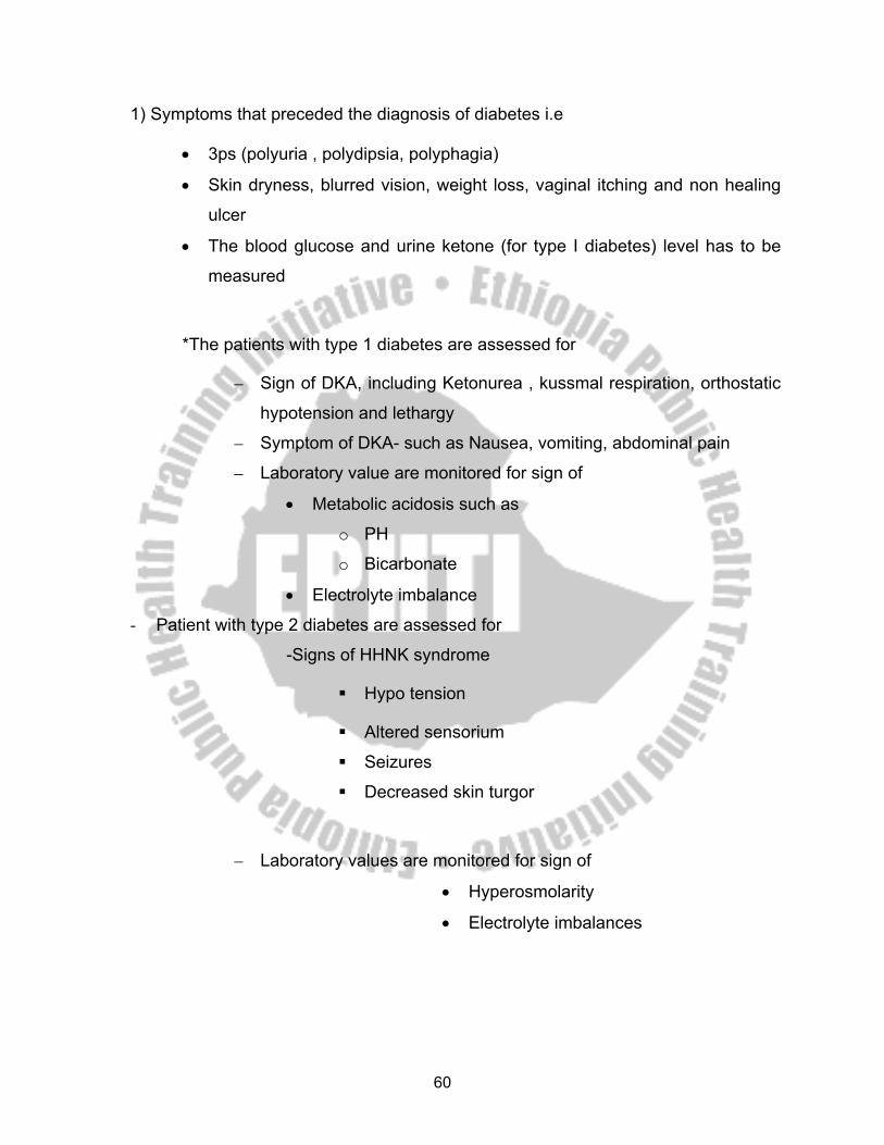
60
1) Symptoms that preceded the diagnosis of diabetes i.e
• 3ps (polyuria , polydipsia, polyphagia)
• Skin dryness, blurred vision, weight loss, vaginal itching and non healing
ulcer
• The blood glucose and urine ketone (for type I diabetes) level has to be
measured
*The patients with type 1 diabetes are assessed for
– Sign of DKA, including Ketonurea , kussmal respiration, orthostatic
hypotension and lethargy
– Symptom of DKA- such as Nausea, vomiting, abdominal pain
– Laboratory value are monitored for sign of
• Metabolic acidosis such as
o PH
o Bicarbonate
• Electrolyte imbalance
- Patient with type 2 diabetes are assessed for
-Signs of HHNK syndrome
Hypo tension
Altered sensorium
Seizures
Decreased skin turgor
– Laboratory values are monitored for sign of
• Hyperosmolarity
• Electrolyte imbalances

61
2) Physical factors -that may impair ability to learn or perform self-care skills, or result in
complications such as
• Visual deficit
• Deficits in motor coordinator (patient is observed eating or performing other
tasks)
• Neurological defect- assessed for aphasia, mental status, sensation in feet
3) Social situation -that may influence the diabetes treatment and educational plan
• Decreased literacy
• Limited financial resource /lack of health insurance
• Presence or absence of family support
• Typical daily schedule
4) Emotional status -is assessed through observation of
General demeanor- anxiety, withdrawn
Body language - avoid eye contact
- Pt asked for major concern and Fear about D.M (this is to see his/her conceptions
or identify any misinformation)
-Coping skill- by asking to patient how to deal with difficult situations
Not all patients may have similar nursing diagnosis because nursing process is client specific and individualized. But possible N/ Dxs include:
Based on the assessment data, the patients major nursing diagnosis may include the
following.
- Risk for fluid volume deficit( FVD) related to polyuria and dehydration
- Altered Nutrition related to ( r/t) imbalance of insulin, food and physical activity
- Knowledge deficit about diabetes self care skills/ information
- Potential self care deficit r/t physical impairment or social factors

62
Collaborative problem /potential complications
• Fluid over load, pulmonary edema congestive heart failure
• Hypokalemia
• Hyperglycemia and ketoacidosis
• Hypoglycemia
• Cerebral edema
Nursing care Plan
- Attainment of fluid and electrolyte balance
- Optimal control of blood glucose
- Improving nutritional intake and regaining weight loss
- Ability to perform basic diabetic skills and self care activity
- Reduction in anxiety
- Absence of complications
Nursing Interventions
1. Maintaining fluid and electrolyte balance
- Measuring Intake and output
- Administering I/v fluid and electrolytes as ordered
- Encouraging oral fluid intake
- Monitor lab values of serum electrolyte ( esp, Na and k)
- Vital sign monitoring
2. Improving nutritional intake
- Diet is planned for the control of glucose
• Take in to consideration the patients life style, cultural back ground,
activity level and food preference
- Patient is encouraged to eat full meals and snack as based on the kcal need.
- Arrangement are made with the dietitian for an extra snack before increased
physical activities

63
3. Improving self care
- Patient teaching to prepare for self care
- Special equipment is used for instruction on diabetic injection skill
- Low literacy information is used
- Families are instructed to enable them to assist in diabetic management
to profile syringe
to monitor blood glucose
- Follow up education is arranged
- Consideration is given for financial limitation or physical limitation (such as center
for visually impaired)
Other members of the health care team are informed about variation in the timing of
meal and the work schedule (e.g. if pt works at night or in the evening and sleeps during
the day / so that the diabetes treatment regimen can be adjusted accordingly.
4. Reducing Anxiety
- Nurse provide emotional support and gives time for client
- Patient and family are assisted to focus on learning self care behavior
- Encouraged to perform the skills that are most feared and reassured and self
injection and puncturing a finger for glucose monitoring
5. Patient education and Home care considerations to prevent complications
DM is a chronic life long illness requiring a lifetime of self-management behaviors
- The patient is taught survival skill including
1. Simple pathophysiology
- Definition
- Normal blood glucose level
- Effects of insulin and exercise
- Effects of food and stress, including illness and infection
64
B. Treatment modalities
- Simple patho physiology
- Treatment modalities (diet, insulin administration, monitoring BG, Urine ketone)
C. Recognition, treatment, and prevention of acute complications
- Hypoglycemia
- Hyperglycemia
D. Pragmatic information
- Where to buy and store insulin, syringes, glucose monitoring supplies when to
call the Nurse or physician.
- When and how to reach to health unit
In depth / continuing education during follow up
Preventive measures for the avoidance of long-term complications
- Foot care
- Eye care
- General hygiene (of skin care oral hygiene)
- Risk factor management. eg control of BP.
Monitoring and managing potential Complication
1. Fluid over load caused by administration of large volume at a rapid rate
- This risk is increased in elderly patient and in those with preexisting cardiac
disease
Nursing care – Monitor the pt closely during treatment for
Vital sign at frequent interval
Intravenous (IV) in take and keep careful records of I/v and
other fluid intake along with urine out put measurement
- Physical exam with focuses on cardiac rate, rhythm breath sound, venous
distension skin torpor and urine output
2. Ortho static hypotension secondary to dehydration

65
Hypokalemia
- A potential complication (cpx) during treatment of DKA as K is lost from body
store
Cause- Dehydration
-Increased urinary execration of K
-Movement of potassium form extra cellular fluid( ECF) in to the cell with insulin
administration of
Mgt – cautious replacement of potassium
- Ensuring proper kidney functioning before the administration
- Monitoring of cardiac rate, rhythm& elctrocardiogram and serum potassium level
3. Hyperglycemia and ketoacidosis
- Monitor blood glucose level and urine ketonuria
- Medication are administered as prescribed
- Pt is monitored for sign and Symptom of impending hypergly cemia and keto
acidosis
4. Hypoglycemia
Cause – skip or delay meal
- Not follow the prescribed diet
- Greatly increase the amount of exercise with out modifying diet or insulin
Management- Juice or glucose tablet
- Encourage the pt to eat full meal and snacks as prescribed per diabetic diet
- See the above descriptions for the details
5. Cerebral edema
Rare problem, which commonly encountered in children
Evaluation
Expected outcome

66
1. Achieve fluid and electrolyte balance
a, Demonstrate I/o balance
b, Exhibit electrolyte values that are with in normal limit
c, Vital signs remain stable
2. Achieves metabolic balance
a, Avoid extremes of glucose level( Hpo/hyperglycemia
b, Demonstrate rapid resolution of hypoglycemia episode
c, Avoid further weight loss
3. Demonstrate verbalizes diabetic survival skill
Simple pathophysiology
a. Define diabetes as a condition in with high blood glucose is present
b. State normal blood glucose range
c. Identifies factors that cause the blood glucose level
- to fall (insulin, exercise)
- to rise ( food, illness and infection )
d- describes the major treatment modalities
- diet
- Exercise
- Monitoring
- Medication
- Education
Treatment Modalities (insulin, diet, monitoring, Education)
a, Demonstration proper technique for drawing up and injecting insulin
b, Verbalize insulin injection rotation plan
c, Verbalize understanding of classification of food group
d, Verbalize appropriate schedule for eating snacks and meals
e .demonstrate proper technique – for monitoring blood glucose

67
Demonstrate proper technique - for disposing of needles used for blood
glucose monitoring and insulin injection
- for urine ketone testing and verbalize
appropriate time to assess for ketones
3.1 Acute Complication (Hypoglycemia and Hyperglycemia)
a. Verbalizes symptoms of hypoglycemia (shakiness, sweating headache, hunger,
Numbness or tingling of lips or finger, weakness, fatigue, difficult concentration,
Change of mood and dangers of untreated hypoglycemia (seizure and coma)
b. Identify appropriate Rx of hypoglycemia, including 10 to 15 gm simple
Carbohydrate (of 2to 4 glucose table, 4+6 of juice , 2to 3 TSP sugar or 6to 10 life
savers) followed by a snack of protein and CHO, such as cheese and cracker or
milk or by a regularly scheduled meal
c. Identify potential causes of Hypoglycemia
i. too much insulin,
ii. delayed or decreased food intake
iii. increase physical activity
d. Verbalize preventive behavior i.e monitoring of blood glucose, taking snake
before exercises, verbalize importance of wearing medical identification and
carrying a source of simple CHO at all time
e. Verbalizes symptom prolonged hyperglycemia- increased thrust and urination
3.2 Pragmatic information
a. Verbalize where to purchase and store insulin, syringe and glucose monitoring
supplies
b. Identify appropriate circumstance for calling the physician eg- when ill, when
glucose level repeatedly increasing
4. Absence of complication
a. Exhibit normal cardiac rate and rhythm and normal breath sound
b. Jugular venous pressure and distention with in normal limit
c. Blood glucose and urine ketones with in normal limits

68
d. Exhibit no manifestation of hypo or hyper glycemia
e. Mental status improved with out sign of cerebral edema
f. States measures to prevent occurrence of complications
Keys for the pretest and post test questions for Nurses
1. A
2. C
3. C
4. E
5. A
6. C
7. A
8. C
9. D
10. A- site of injection
-Preparations of medication
-Rotations
-About syringe and needle
-Some problems with insulin injections
B)-Too much insulin
-Too little food or
-Excessive physical exercise
-Delay of meal or omitting of snacks
C) Sweating
-Tremor
-Tachypnea
-Confusion
-Seizure
-Loss of consciousness

69
D) Having snack, not delaying the meal, right dose of medications, having Candies at
hand
F)-assess foot daily for sensation, redness and broken skins
-Wash dry feet daily
- If skin is dry apply a thin coat of lubricating oil
-Tie shoes loosely but firmly
-If your feet perspire, change shoe and stocking during the day
-Wear shoe and stocking that gives room for the movement of the toe
Part-ii True or false
A.T K. F T.T
B. F L. T U.T
C. T M.T
D. T N.F
E. T O.F
F. F P.T
H.T Q.F
I. T R.T
J. T S.T
REFERENCES
1) Brunner and Sudarths,Text books of Medical-Surgical eight ed. Lippincott
company,Philadephia
2) ì¼R ywYN /rG flq tÆƶ ßéØsR ½ ySµ*R HmM½ /Ml@ 1995 ›¼M
3) 3)Andreoli,cecil Essentials of Medicine 5th ed.aharcourt Health
Company,london
4) 4)Fraces T. Lester ,Management of Diabetes Mellitus , 2nd ed.,1991
5) 5)J.Abdulkadir,Diabetic Melitus in Africa.
70
3.3 SATELLITE MODULE FOR MEDICAL LABORATORY TECHNOLOGISTS
Introduction
1. Purpose of the module
Diabetes mellitus is a diverse group of hyper glycemic disorders with different etiologies
and clinical pictures; there fore timely diagnosis and management based on true
laboratory results are crucial.
This Satellite Module on Diabetes Mellitus is intended to resolve the critical shortage of
clinical chemistry reference materials both for students and for other professionals of
the same field working in different health institutions
1.1 Directions for using the satellite Module - After completion of the Core Module, try to answer all pre- test questions and
write the answers on a separate sheet of paper
- Read the rest of this Satellite Module
- Refer to the Core Module whenever necessary
- Answer the post- test questions
- Compare the results of the pre- test questions with the key given at the end.

71
2. Pre- test questions Instructions: choose the appropriate answer from the alternatives given for each
question and write the answers on a separate sheet of paper.
1. Why is there a discrepancy between the whole blood glucose concentration and the
plasma glucose concentration?
A. Because there is a different distribution of Glucose in whole blood and
plasma
B. Because there is a high amount of water in plasma
C. Because the cellular component in whole blood use glucose frequently
D. None
2. One of the following methods of Glucose determination does use enzymatic reaction
A. Folin- MU copper Reduction method
B. Alkaline ferric cyanide method
C. Hexokinase n.v. method
D. Somogyi-Nelson method
3. Which one of the following methods is highly specific for glucose determination
A. Alkaline ferric cyanide method
B. Copper Reduction method
C. Glucose oxidase method
D. O-Toluidine method
4. When does glucose appear in the urine?
A. When the urine glucose level higher than blood glucose level
B. When blood glucose level is between 60-110 mg/dl
C. When the blood glucose level is greater than 180-200 mg/dl
D. When a person is starved
5. One of the following methods for urinary glucose determination is highly specific
A. Copper reduction method
B. O-Toluidine method
C. Reagent strip Tests
D. A and B

72
6. Sodium fluoride additive used in a specimen collected for Glucose
A. Inhibits glycolytic enzymes from destroying the glucose
B. Precipitates the protein present in the sample
C. Prevents non glucose reducing substances from interfering with the
testing
D. None of the above
7. During GGT test in a person with normal glucose metabolism, the blood glucose
level usually increases rapidly after carbohydrates are ingested, but returns to a
normal level after
A. 30 minute
B. 60 minute
C. 90 minute
D. 15 minute
8. Which one of the following organs uses glucose from digested carbohydrates
and stored it as glycogen for later use as a source of immediate energy by the
muscles?
A. Kidneys
B. Liver
C. Pancreas
D. Thyroid
9. Which one of the following samples good for Glucose determination
A. Serum/ plasma
B. Whole blood
C. Urine
D. All
10. To say the Oral Glucose Tolerance test is normal
A. The fasting blood sugar level should be 60-110 mg/dl
B. The fasting blood sugar level should be higher than 110 mg/dl
C. The fasting blood sugar level should be normal or slightly elevated
D. The fasting blood sugar level should be always less than the lower limit.

73
3. Learning objectives
After studying this satellite module the student will be able to:-
Collect, preserve or prepare the correct specimens for diagnosis of
diabetes mellitus
Perform different clinical chemistry tests in management of diabetes
mellitus
Practice different quality control procedures in laboratory diagnosis of
diabetes mellitus
4. Laboratory tests for diagnosis and management of diabetes mellitus - Determination of - Blood glucose
- Serum/ plasma glucose
- Urine glucose
- Determination of urine ketone bodies
- Determination of urine protein
- Determination of BUN and creatinin
4.1 Types and collection of different sample
Laboratory diagnosis of Diabetes Mellitus may be performed on specimens of:
• Fasting/random whole blood
• Plasma / serum (free of hemolysis)
• Urine
But, serum or plasma is more preferable for the determination of glucose due to the
following reasons:
• Since plasma or serum contains approximately 10 to 15% more water than
whole blood the total glucose in plasma or serum is about 10 to 15% greater
than in whole blood.
• It is easier to interpret values obtained from a single component system
(plasma) than a two component system (whole blood)

74
• Because there are several substances in blood (particularly in red cells) that
interfere with tests for blood glucose either because they are measured as
glucose or because they interfere in enzyme procedures.
• As indicated above, values based on whole blood tend to vary with the
hematocrit.
• Glucose is more stable in plasma or serum than in whole blood, as many
glycolytic enzymes are present in RBC.
• Plasma or serum is easier to handle, to pipette precisely and to store than is
whole blood.
The blood specimen can be collected both from vein or capillary, it depends on the type
of sample the test procedure needs, and if serum or plasma is needed Venus blood
should be collected with clean, dry, capped test tube, and with or with out anticoagulant.
Urine samples are also possible to collect using, a clean, dry, free of any disinfectant,
large and wide mouth container so as to do both qualitative and quantitative
determination of glucose and others
4.2 Types of blood specimen for Glucose determination 4.2.1 Fasting blood specimen
The term fasting in this case means that the patient has had no food or drink for 8 to 12
hours, no drugs that might affect the blood glucose level, and no emotional disturbances
that might cause liberation of glucose into the blood.
4.2.2 Two Hours post prandial specimen
A blood specimen drawn 2 hours after a meal is know as a two- hour post prandial
specimen. To more completely detect diabetes mellitus, stressing the system with a
defined glucose load tests carbohydrate metabolic capacity. To do this, a high-
carbohydrate drink or meal is given to the patient, blood is collected 2 hours after
ingestion, and the glucose concentration is determined.

75
4.2.3Capillary blood specimen An advantage of using whole blood is the convenience of measuring glucose directly on
capillary blood, such as:
• That taken from infants
• In mass screening programs for detection of diabetes mellitus, or
• In the home monitoring being done by so many diabetes patients. In the fasting state the arterial (capillary) blood glucose concentration is 5 mg/dl higher
than the venous concentration.
5. Preparation and preservation Samples for glucose determination
The following factors which affects the stability of glucose in body fluid must be take in
to account, such as:
• Those glycolytic enzymes found particularly in the red cells, which under goes
glycolysis at an average rate of approximately 10 mg/dl/hr in whole blood or 5
mg/dl/hr in sufficiently centrifuged plasma which still contain leukocytes
Keeping these considerations in mind, there are several ways to prevent or retard
glycolysis in specimen to be analyzed. For example:
• Sample for glucose analysis should be delivered to the laboratory as soon as
possible after being drowned from the patient.
• Refrigeration or addition of small amount of sodium fluoride to the fluid may
retard glycolysis for a few hours
• If plasma or serum is to be used for the glucose determination, it must be
separated from the cells or it will clot with in 30 minutes after the blood is drown
unless a specific additive is used.
Note When certain enzymatic glucose methods are used, fluoride anticoagulated
blood should not be used, as the fluoride might inhibit the enzyme. Use of
serum separator gel tubes, processed as quickly as possible with in thirty
minutes if possible- is preformed for these methods.

76
6. Diagnostic criteria of DM based on Glucose determination
Fasting and Two- hour postprandial tests when glucose metabolism is being monitored,
glucose is commonly determined on fasting blood specimens and on 2-hour
postprandial specimens. For non pregnant adults, the fasting serum or plasma glucose
concentration should normally be less than 110 mg/dl and the serum or plasma glucose
taken 2 hours 140 mg/dl. Including in the new criteria from the expert committee on the
diagnosis and classification of Diabetes Mellitus. There are three diagnostic criteria for
diabetes Mellitus:
1. A fasting serum or plasma glucose level equal to or greater than 126 mg/dl; or
2. A random blood glucose (blood drawn with out considering time since the last
meal) equal to or greater than 200 mg/dl, along with symptoms of diabetes
(polyuria, polydipsia, and un explained weight loss),
3. A 2- hour post load glucose level equal to or greater than 200 mg/dl during an
oral glucose tolerance test. Impaired glucose tolerance (IGT) is indicated if the
fasting glucose is between 110 and 126 mg/dl and one postprandial glucose
level is greater than 200 mg/dl.
7. Methods for quantitative determination of Glucose.
The various methods for the quantitative determination of glucose can be divided in to
three general categories.
• Enzymatic Methods,
• Oxidation Reduction methods, and
• Aromatic amine methods.
Of these, enzymatic methods using hexokinase or glucose oxidase methodology are
most commonly used

77
7.1 Enzymatic methods
Almost all currently used glucose Methods utilize enzymatic techniques. The use of
enzymes is a means of achieving absolute specificity in the determination of glucose
concentration. The two most widely used automated enzyme glucose methods are
based on the enzymes hexokinase and glucose oxidase. Glucose oxidase is also used
in the most common manual methods.
7.1.1 Glucose oxidase method
Sample: whole blood, serum, plasma, and other body fluids
Principle: β-D- glucose in serum (specimen) is oxidized to gluconic acid by a specific
enzyme glucose oxidase. When glucose is oxidized proportionally H2O2 is released. In
the presence of peroxidase enzyme the released H2O2 reacts with oxygen acceptors
like aminophenazene and phenol forming a rose colored quinone rose derivative. The
intensity of color is directly proportional to the amount of glucose present in the sample
and the absorbance is read at 546nm. The absorbance of test is compared with that of
standard. In this procedure uric acid, ascorbic acid and glutathione compete with amino
phenazine for H2O2. Urine sample cannot be used directly as urine may contain
electrolysis those inhibit enzyme activity
N.B Mutase is added to convert α, D-glucose to β-D-glucose
1. Glucose + H2O Glucose H2O2+ gluconic acid
oxidase
2. H2O2+ aminophenazene + phenol peroxidase quinone imine (rose)
Glucose oxidase is specific to β,D- glucose so mutase is present to convert α ,D-
glucose to β – configuration
7.1.2. Hexokinase U.V Method
Sample: whole blood, serum, plasma and other body fluids Principle: Glucose is phosphorylated in the presence of ATP by catalytic activity of
hexokinase forming glucose 6-phosphate and ADP. Glucose-6-phosphate

78
dehydrogenase oxidizes glucose 6-phosphate in the presence of hydrogen acceptor
NADP+. Proportionally NADP+ is reduced to NADPH. The amount of formed NADPH is
directly proportional to the amount of glucose present in the sample. The absorbance
due to NADPH is read at 340nm every minute and change in absorbance of the test is
compared with that of the standard.
1. Glucose + ATP hexokinase Glucose – 6- phosphate + ADP+
2. Glucose-6- phosphate +NADP+ G6PDH NADPHH++ 6-
A A= absorbance due to NADPH+
C= Glucose concentration
C
Fig 1. The relation ship between absorbance and concentration
7.2 Oxidation- Reduction Methods
Oxidation methods for blood glucose depend on the fact that glucose contains an
aldehyde group as part of its chemical structure. The presence of this aldehyde gives
glucose its reducing properties. Other substances in blood also have reducing
properties. Some of these non glucose reducing substances are other sugars and
metabolic compounds and materials such as uric acid, creatinine, ascorbic acid, certain
amino acids, creatine, and phenol. The oxidation- reduction methods for determining
blood glucose differ primarily in the way they handle the non-glucose reduction
substances. When the non-glucose reducing substances are removed as part of a
glucose determination, the resulting value is called the true glucose value.
7.2.1 Alkaline ferric cyanide method
Sample: serum, plasma, and whole blood
phosphogluconate

79
Principle: In hot alkaline solution ferric cyanide ion is reduced by glucose to
ferrocyanide ion. Before reduction the color of ferric cyanide solution is yellow and
after reduction it is colorless. The decrease in yellow color is directly proportional to
the concentration of glucose present in the sample and the absorbance is taken at
420 nm. This method is not specific for glucose since any reducing substance
participates in this reaction.
Glucose + Ferric cyanide Alkaline Ferrous cyanate + Oxidized glucose
1000C
CT = AT AST
A= Absorbance before Reaction minute Absorbance after reaction
7.2.2 Copper Reduction Method.
Sample: serum, plasma, whole blood and other body fluids. Principle In hot alkaline solution, glucose readily reduces cupric ions to cuprous ions
with formation of mainly cuprous oxide (CU2O). On the addition of phosphomolybdic
acid (arsenomolybdic acid) to the reaction mixture the cuprous ions quantitatively
reduces molybdic acid to lower oxides of molybdium which is blue in color. The intensity
of blue color is directly proportional to the concentration of glucose present in the
sample and the absorbance of the solution is read at 680 nm.
CT= AT xCST AST= absorbance of standard
Ast CST= concentration of standard
CT= concentration of test
AT= absorbance of test
This method has many drawbacks:
Lacks specificity due to the reducing ability of uric acid, glutathione and others.
x CST

80
It needs special tubes, constricted follin- MU tubes
It needs color stabilizers, NO2 SO4, which prevents reoxidation of cu+
It needs protein precipitants like tungstic acid, Ba (OH)2 and Zn SO4
The method is highly sensitive, though it is laborious
Glucose + Cu2+ hot Cu+ oxidized Glucose
OH
Cu+ + arsenomolybodic acid Cu++ + reduced arsenomolybdate
(Blue color)
7.2.3.a. Folin- Mu copper Reduction Method Reagents 1. Protein free filtrate reagent, aqueous solution
a. H2SO4 ………………………….. 0.67N
b. B Na2 O4.2H2O……………..100g/l
2. Copper reagent
a. Na2CO3……………………40g
b. Tartaric acid………………. 7.5g
c. CuSO4.5H2O …………….. 4.5g
To prepare this reagent dissolve Na2 CO3 in 500 ml of distilled water. With stirring
transfer tartaric acid to Na2CO3 solution. CuSO4.5H2O is first dissolved in 100 ml of
water and transfer the whole solution to Na2CO3 – tartaric acid solution with stirring.
Dilute the solution to 1000ml. The reagent is stable indefinitely at 200C.
3 Phosphomolybdic acid reagen
a. Na2 WO2.2H2O…………..10gm
b. Molybdic acid (MOO3)…..70gm
c. 10%W/v NaOH solution… 400ml

81
NaOH solution is added with stirring Boil the solution For 30-40 minutes and cool to
room temperature.
d. Add distilled water to………….700ml
e. Concentrated H3PO4………….. 250ml
f. Dilute the solution to ………….1000ml
Procedure
Arrange 3 test tubes labeled blank, standard & test
RB ST T
Glucose standard 200mg%
Water
Serum
0.67 NH2SO4
10% Na2WO4
-
1.0ml
-
0.75ml
0.25ml
0.5ml
0.5ml
-
0.75ml
0.25ml
-
0.5ml
0.5ml
0.75ml
0.25 ml
Mix and centrifuge. Transfer 1.0 ml of each of the blank, standards and each of filtrate
to folin- Mu sugar tubes graduated at the 12ml mark. To the 3 test tubes transfer 1.0ml
copper reagent and heat the tubes in a boiling water bath or heating black at 1000C for
8 minutes and cool to room temperature. To each tube transfer 1.0ml of
phosphomolybdic acid with Mixing. Dilute the tubes to 12 ml with water and mix. Read
the absorbance of the test (T) and standard (ST) against reagent blank (RB) at 680nm.

82
7.2.3.b. Somogyi- Nelson Method
1. Zinc sulfate solution
ZnSo4 .7H2O …………….. 50 gm
Water to ………………….. 1000ml
2. Barium hydroxide solution (0.3N)
Ba (OH)2. 8H2O…………… 50 gm
Water to ……………………. 1000ml
After 3 days filter and keep in poly ethylene bottle
3. Copper sulfate solution
- Na2HPO4 ……………………… .29 gm
- K-Na-C4H4O6.4H2O…………. 4ogm
- Water to ………………………... 700ml
- 1 N NaOH………………………. 100ml
- 10% W/V cuso4.5H2O……….. . 80 ml
cuso4 is added with stirring
- Na2SO4……………………….180 gm
- Water to……………………….1000ml
Let stand for two days and filter and precipitate. Keep at 20-250C
4. Arsenomolybdic soltuon
5. Water………………………….... 900ml
6. Conc. H2SO4 …………………… 42ml
7. Ammonium molybdate …………. 50 gm
8. 12 gm% W/V sodium arsenate ... 50ml
Mix and incubate at 370C for 48 hours. Store in a brown bottle

83
Procedure
- Blood, serum or
CSF
0.5ml
- Water 7.5ml
- Ba (OH) 2 1ml
- Mix and let stand
for 1 minute
- ZnSO4 1ml
- Mix and let stand
for 2 minutes
Separate the protein free filtrate from the precipitate. Arrange 3 folin- Mutubes labeled
as blank (B), standard (ST) and Test) (T).
B ST T 1 Filtrate (ml) - - 2.0
2 Distilled water (ml) 2.0 - -
3 Standard (ml) - 2.0 -
4 CuSO4 solution (ml) 1.0 1.0 1.0
Mix and incubate the tubes in boiling water both or heating block (at 1000C)
for 10 minutes. Cool in cold water by immersing the tubes in cold water bath
5 Arsenomolybdate 1.0 1.0 1.0
Allow to stand for 2 minute at 200C and dilute to 25ml with water and measure
the absorbance of the standard and test against reagent blank at 490 nm
CT= AT x CST AST

84
7.3 Aromatic amine methods
Aromatic amine methods depend on the fact that various aromatic amine methods
depend on the fact that various aromatic amines react with glucose in hot acetic acid to
form colored derivatives.
7.3.1 O- Toludine Method
Sample: plasma, serum, and other body fluids
Principle: Aromatic amines react with glucose in hot acetic acid solution to produce a
colored derivative. In this specific method glucose reacts with O-toludine in hot acetic
acid and in the presence of thiourea, forming a green colored derivative, which is called
shiff- base (glucosylamine). The intensity of color is read at 630 nm and it is directly
proportional to the amount of glucose present in the sample
O H2N CH3 CH3
H C + Acetic acid HC = N
H – C – OH 1000C
C= N
CH3
Glucosyn amine
(Blue colored)
O-Tolidine Glucose

85
Reagent
1. O- Toludin reagent
Thiourea …………………………. 1.5gm
Aceticacid (pure)………………… 940ml
O-Toluidine………………………. 60ml
Mix well and keep the solution in brown bottle in the dark at 200C stable for two
months
2. Glucose standard (stock)
Glucose powder …………..…….20gm
1gm% W/V benzoic acid to…… 1000ml
Stable for one year if kept at 4-100C
3. Glucose working standard
Glucose stock standard…………………….5ml
1gm% benzoic acid to…………………….. 100ml
Stable for Six months if kept at 4-100C. Both stock and working glucose standards
should be stored (kept) at 4-100C
Procedure
B St T Distilled water (ml) 0.1 - -
Glucose standard (ml)
- 0.1 -
Serum (ml)
- - 0.1
O-Toluidine reagent (ml) 7 7 7
Mix well and incubate at 1000C for 10 minute. Cool and read the absorbance at 630 nm

86
A
C
7.4 Glucose Tolerance Test (GTT)
In the detection and treatment of diabetes it is sometimes necessary to have more
information than can be obtained from only testing the fasting specimen for glucose.
There fore the GTT is usually performed:
- When a person has been found to have a fasting serum or plasma glucose
concentration above that of most non diabetic persons (about 110 mg/dl)
- To identify hypoglycemia, an abnormal response to a glucose load that
results in a serum or plasma glucose concentration much below the normally
accepted range
Since early detection and management of diabetes are important to avoid the many
complications of the disease, it is desirable to detect these early cases of diabetes or
pre diabetes. For these reasons, the physician may request a glucose tolerance test.
a. Oral glucose Tolerance Test.
To conduct oral glucose tolerance test on a subject, first blood and urine samples are
taken in fasting state, and. the blood glucose level is determined. The urine sample is
qualitatively tested for glucose. If blood glucose level is normal and urine sample is
negative the individual will be loaded with 75 gm glucose dissolved in 300 ml of water.
Blood and urine samples are collected every 30 minutes four times for a period of 3
hours. The blood sample are analyzed for glucose concentration and the urine samples
are qualitatively tested for glucose.
CT = ATxCsT AsT
Fig 2. The relation ship between Absorbance (A) and concentration (C)

87
Note:- In malabsorption of carbohydrate the fasting blood glucose level is always less than
the lower limit of the normal rang. During GTT, blood glucose level will not rise
even with in 2 hours, above the normal range. Most probably slight elevation
may be observed after 3 hours then falls down below the normal range with in
one or one half hours
b. Intravenous Glucose Tolerance Test: this method is less commonly used,
although it eliminates the variable factors involved in the rate of intestinal absorption in
different individuals. 0.5g. Glucose is given per kg body weight by infusing 20gm% W/V
glucose solution and the infusion will be completed with in half an hour. In normal
individuals the blood glucose concentration does not rise to more than 250 mg% at the
end of infusion and falls below the fasting level with in 2 hours. In case of latent and
severe diabetes mellitus glucose concentration pattern will be as in oral GTT.
Factors affecting (altering) carbohydrate tolerance
1. Quantity of glucose administered: The peak level doesn’t increase if the quantity
of glucose administered is greater than 50 gm, since glucose is absorbed at a
steady rate by the intestine and it may take a longer time to return to fasting level
even in normal GTT.
2. The rate of absorption may be low in conditions of mal absorption and high in
hyperthyroidism. This may result in low peak level or an early high peak.
3. Diet preceding the day of test: Starvation or carbohydrate free diet will decrease
the carbohydrate tolerance of the individual and cause hyperglycemia and
glycosuria when the individual is subjected to GTT. There fore, in order to get the
correct diet of the carbohydrate tolerance (GTT) the subject must be on a
carbohydrate diet for preceding 3 days
4. Age: Elderly people show a decrease carbohydrate tolerance
5. Endocrine: Insulin increases carbohydrate tolerance while all other hormones
considered earlier tend to decrease its tolerance.

88
8. Self- Monitoring of glucose
Many patients (especially those with type 1(insulin- dependent diabetes mellitus)) now
regularly monitor their own blood glucose concentrations on the advice of their health
care provider, using reagent test strips and reflectance meter. Several companies
manufacture reagent test strips for monitoring blood glucose, and most of these
companies make reflectance meters to be used to electronically read the test result.
Instruments include one Touch,* Accu- chek Easy, and Glucometer Elite. The strips
used for these tests are impregnated with the enzyme glucose oxidase, enzyme
peroxidase and an indicator to give a color change that is detectable. The color change
can be read in a reflectance meter on which the result (in mg/dl) is visualized. Although
blood is tested, results are converted to plasma glucose values by the instrument.
Eg. Glucose oxidase reaction (Vitros Method)
Ex β- D- glucose + H2O glucose D- gluconic acid + H2O2
oxidase
2H2O2 + 4 aminopyrine + 1,7- dehydroxynapthylene peroxidase H2O + a red dye
9. Urine Glucose determinations
Chemical screening tests for glucose (dextrose) are generally included in every routine
urinalysis. The occurrence of glucose in the urine indicates that the metabolic disorder
diabetes mellitus should be suspected, although several other conditions result in
glycosuria (glucosuria).
The lowest blood glucose concentration that will result in glycosuria is termed the renal
threshold (180-200 mg/dl). It is possible to use both enzymatic technique and oxidation-
reduction technique to determine urine glucose
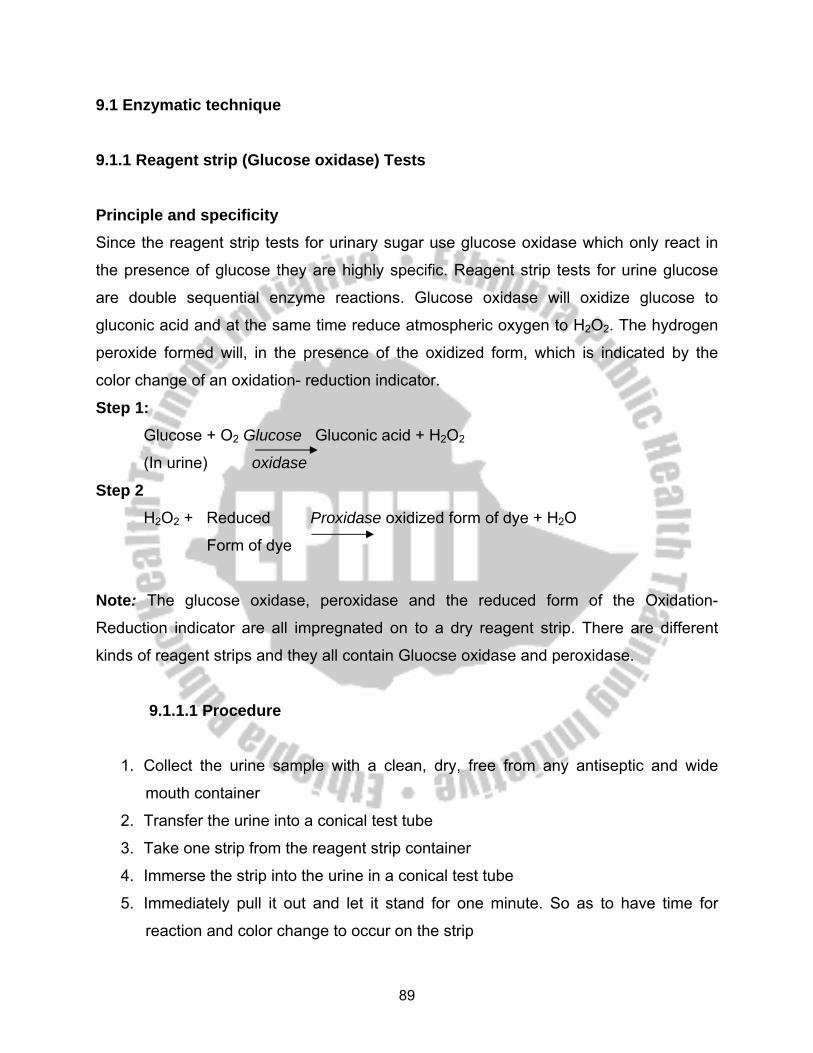
89
9.1 Enzymatic technique 9.1.1 Reagent strip (Glucose oxidase) Tests
Principle and specificity Since the reagent strip tests for urinary sugar use glucose oxidase which only react in
the presence of glucose they are highly specific. Reagent strip tests for urine glucose
are double sequential enzyme reactions. Glucose oxidase will oxidize glucose to
gluconic acid and at the same time reduce atmospheric oxygen to H2O2. The hydrogen
peroxide formed will, in the presence of the oxidized form, which is indicated by the
color change of an oxidation- reduction indicator.
Step 1: Glucose + O2 Glucose Gluconic acid + H2O2
(In urine) oxidase
Step 2 H2O2 + Reduced Proxidase oxidized form of dye + H2O
Form of dye
Note: The glucose oxidase, peroxidase and the reduced form of the Oxidation-
Reduction indicator are all impregnated on to a dry reagent strip. There are different
kinds of reagent strips and they all contain Gluocse oxidase and peroxidase.
9.1.1.1 Procedure
1. Collect the urine sample with a clean, dry, free from any antiseptic and wide
mouth container
2. Transfer the urine into a conical test tube
3. Take one strip from the reagent strip container
4. Immerse the strip into the urine in a conical test tube
5. Immediately pull it out and let it stand for one minute. So as to have time for
reaction and color change to occur on the strip
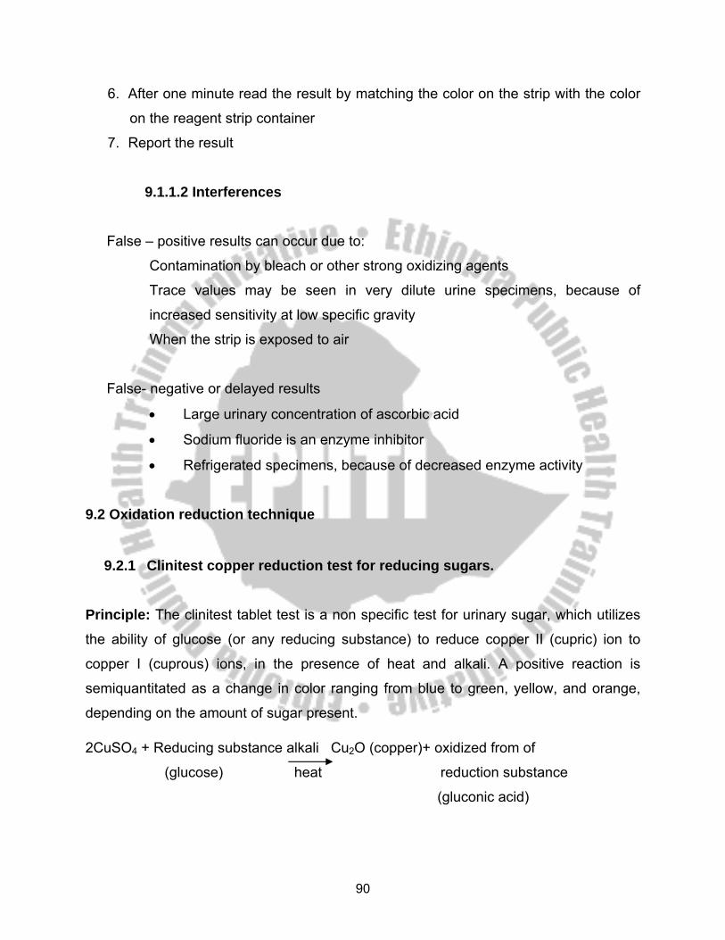
90
6. After one minute read the result by matching the color on the strip with the color
on the reagent strip container
7. Report the result
9.1.1.2 Interferences
False – positive results can occur due to:
Contamination by bleach or other strong oxidizing agents
Trace values may be seen in very dilute urine specimens, because of
increased sensitivity at low specific gravity
When the strip is exposed to air
False- negative or delayed results
• Large urinary concentration of ascorbic acid
• Sodium fluoride is an enzyme inhibitor
• Refrigerated specimens, because of decreased enzyme activity
9.2 Oxidation reduction technique
9.2.1 Clinitest copper reduction test for reducing sugars.
Principle: The clinitest tablet test is a non specific test for urinary sugar, which utilizes
the ability of glucose (or any reducing substance) to reduce copper II (cupric) ion to
copper I (cuprous) ions, in the presence of heat and alkali. A positive reaction is
semiquantitated as a change in color ranging from blue to green, yellow, and orange,
depending on the amount of sugar present.
2CuSO4 + Reducing substance alkali Cu2O (copper)+ oxidized from of
(glucose) heat reduction substance
(gluconic acid)
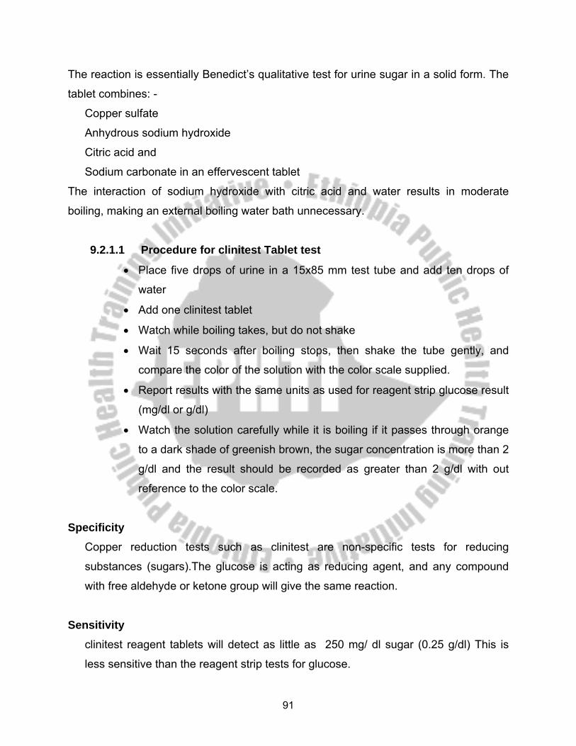
91
The reaction is essentially Benedict’s qualitative test for urine sugar in a solid form. The
tablet combines: -
Copper sulfate
Anhydrous sodium hydroxide
Citric acid and
Sodium carbonate in an effervescent tablet
The interaction of sodium hydroxide with citric acid and water results in moderate
boiling, making an external boiling water bath unnecessary.
9.2.1.1 Procedure for clinitest Tablet test
• Place five drops of urine in a 15x85 mm test tube and add ten drops of
water
• Add one clinitest tablet
• Watch while boiling takes, but do not shake
• Wait 15 seconds after boiling stops, then shake the tube gently, and
compare the color of the solution with the color scale supplied.
• Report results with the same units as used for reagent strip glucose result
(mg/dl or g/dl)
• Watch the solution carefully while it is boiling if it passes through orange
to a dark shade of greenish brown, the sugar concentration is more than 2
g/dl and the result should be recorded as greater than 2 g/dl with out
reference to the color scale.
Specificity Copper reduction tests such as clinitest are non-specific tests for reducing
substances (sugars).The glucose is acting as reducing agent, and any compound
with free aldehyde or ketone group will give the same reaction.
Sensitivity clinitest reagent tablets will detect as little as 250 mg/ dl sugar (0.25 g/dl) This is
less sensitive than the reagent strip tests for glucose.

92
9.2.1.2 Interferences False positive results occurs:
• Since it is reducing substance, the presence of extremely large
amount of ascorbic acid
• Specimens that have a low specific gravity and contain glucose may
give slightly elevated result
• Large quantities of nalidixic acid, cephalosporins and probenacid.
False negative results
• Mixing the test tube before the 15 second wait after boiling stops, due
to reoxidation of the cuprous ions to cupric ions by atmospheric oxygen
10. Determination of ketone bodies in urine
ketone bodies are a group of three related substances: acetone, aceto acetic acid, and
β – hydroxyl butyric acid. When ever fat (rather than carbohydrate) is used as the major
source of energy, ketosis and ketonuria may result. The two out standing causes of
ketone accumulation are diabetes mellitus and starvation In diabetes mellitus, the body
is unable to use carbohydrate as an energy source and attempts to compensate by
resorting to fat catabolism, which results accumulation of ketone more than normal, that
the body is unable to utilize it. The clinical result is an increased concentration of
ketones in the blood (ketonemia) and in the urine (ketonuria.) Since the presence of
ketone bodies in urine is an early indication of lack of adequate insulin control, reagent
strips that combine tests for glucose and ketone are often used.
10.1 Dipstick test Principle: the reagent strip tests for ketone bodies are based on legal’s (Rothera’s)
test, a color reaction with sodium nitroprusside (nitro ferricyanide). Acetic acid will react
with sodium nitro prusside in an alkaline medium to form a purple color.

93
10.1.1 Procedure 1. After collecting the urine sample from the patients, transfer into a clean, dry and free
of disinfectant test tube
2. Then immerse the dipstick into the urine
3. Then drain and let it stand for certain seconds for the reaction to take place
4. Read the result by comparing the color produced with the standard on the strip
container
Note acetone and aceto acetic acid can be detected by different dip stick tests,
but there is no reagent strip test for β - hydroxyl butyric acid
10.1.2 Interferences - The presence of various:
• Pigments
• Drugs or
• Urine specimens presents problems in reading results
- False-positive may result due to:
• Specimens containing phthaleins, very large amounts of phenyl ketones or the
preservative β-hydroxy quinoline
• Highly concentrated urine specimens
- False -negative
• Conversion of acetoacetic acid to acetone with subsequent evaporation from the
specimen in improperly stored urine specimen.
11 Determination of urine protein Microalbuminuria
• Diabetes mellitus causes progressive changes to the kidneys and ultimately
results in diabetic renal nephropathy. This complication progresses over a period
of years and may be delayed by aggressive glycemic control
• An early sign that nephropathy is occurring is an increase in urinary albumin
• It is thought that the early development of renal complications can be predicted
by the early detection of consistent micro albuminuria. And this early detection is

94
desirable, as better control of blood glucose levels may delay the progression of
renal disease
11.1 Methods of measurement
Test for urinary protein are of two major types:
a. Tests that are based on the use of the protein error of PH indicators
- This is the methodology employed in the various reagent strip tests
- They are more sensitive to the presence of albumin than to other proteins.
b. Tests that are based on the precipitation of protein by chemical or coagulation by
heat
- This test will detect all proteins, including albumin, glycoproteins, globulins,
Bence Jones protein & hemoglobin
11.1. a Reagent strip test Principle: Reagent strip tests for urinary protein involves the use of PH indicators
substances that have characteristic colors at specific PH values. The phenomenon of
showing different color at different PH is called. “The protein error of indicators” The PH
of the urine is held constant by means of buffer, so that any change of color of the
indicator will indicate the presence of protein.
11. 1.a. 1 Procedure It is the similar with other reagent strip test procedure. (But the reading time can vary
manufacture to manufacturer instruction on the leaf late)
Specificity The reagent strip tests for urinary protein are more sensitive to the presence of albumin
than they are to other proteins

95
Sensitivity (minimum Detectable level:) Manufacturer’s value
- Multistrx /Albustix…………………..15 to 30 mg/dl albumin
- Chemstrip……………………………. 6 mg/dl albumin
- Etc
11.10.2 Interferences - If the urine is strongly pigmented, there may interference with the color reaction.
False- positive results
- if the urine is exposed to the reagent strip for too long, the buffere may be
washed out of the strip, resulting in the formation of blue color whether protein is
present or not
- If a urine specimen is exceptionally alkaline or highly buffered, the reagent strip
tests may give a positive result in the absence of protein
False – Negative results
- When proteins other than albumin are present, the reagent strip will give a
negative result in the presence of protein
11.1. b. Confirmatory tests (sulfosalicylic acid (SSA) test SSA test or another protein precipitation method may be used to confirm the presence
of protein when reactions indicating a trace or more are obtained or when reagent strip
results are in doubt.
SSA test
Principle: This test is based on the cold precipitation of protein with a strong acid,
namely sulfosalicylic acid.
11.1b.1 procedure for SSA test for urine protein
• Centrifuge a 12 ml aliquot urine
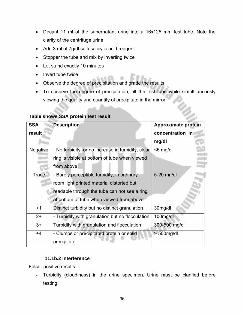
96
• Decant 11 ml of the supernatant urine into a 16x125 mm test tube. Note the
clarity of the centrifuge urine
• Add 3 ml of 7g/dl sulfosalicylic acid reagent
• Stopper the tube and mix by inverting twice
• Let stand exactly 10 minutes
• Invert tube twice
• Observe the degree of precipitation and grade the results
• To observe the degree of precipitation, tilt the test tube while simult ancously
viewing the quality and quantity of precipitate in the mirror
Table shows SSA protein test result
SSA result
Description Approximate protein concentration in mg/dl
Negative - No turbidity, or no increase in turbidity, clear
ring is visible at bottom of tube when viewed
from above
<5 mg/dl
Trace - Barely perceptible turbidity, in ordinary
room light printed material distorted but
readable through the tube can not see a ring
at bottom of tube when viewed from above
5-20 mg/dl
+1 Distinct turbidity but no distinct granulation 30mg/dl
2+ - Turbidity with granulation but no flocculation 100mg/dl
3+ Turbidity with granulation and flocculation 300-500 mg/dl
+4 - Clumps or precipitated protein or solid
precipitate
> 500mg/dl
11.1b.2 Interference False- positive results
- Turbidity (cloudiness) in the urine specimen. Urine must be clarified before
testing

97
False- Negative results
- The occurrence of highly buffered alkaline urine if the buffer is sufficient to
neutralize the acid in SSA.
12. Determination of creatinine and BUN
Diabetes mellitus can have profound effects on the renal system. In insulin- dependent
diabetes mellitus (IDDM, type I) patients suffer from a deficit of insulin activity.
Approximately 40% to 50% of these patients will develop progressive deterioration of
kidney function (diabetic nephropathy) with in 15 to 20 years after their diagnosis.
The lesions are primarily glomerular, but they may affect all other kidney structures as
well, they are theorized to be caused by the abnormally hyper glycemic environment
than constantly bathes the vascular system. In this case we will do a renal function
tests, such as
- Determination of blood and urine creatinine
- Determination of Blood urea nitrogen & urea
12.1 Determination of blood urea nitrogen and urea It is customary, in most laboratories, to express urea as BUN, but urea is quit different
from BUN. The structure of urea is NH2-CO-NH2 having a molecular weight of 60. the
two nitrogen atoms represent 28. There fore urea represents 28 gm urea nitrogen
(BUN), or 2.14 gm urea stands for 1gm BUN. BUN is converted to urea by multiplying
the value of BUN by 2.14 and urea is converted to BUN by dividing the value of urea by
2.14
12.1.1 Determination of urea There are two methods, such as:-
12.1.1.1 colorimetric methods for urea determination eg. Bertholet method
12.1.1.2 U.V enzymatic method in urea determination

98
Bertholet method Specimen: serum, plasma, urine and other body fluid
Principle:- urea is hydrolyzed to NH4HCO3 by urease enzyme at 370C. The released
NH4HCO3 is converted to NH3, CO2 and H2O by making the reaction mixture alkaline.
This NH3 reacts with phenol in the presence of sodium hypochloride and sodium
nitropreside SNP (catalyst). The reaction product is a blue colored derivative
indophenol. The intensity of blue color is directly proportional to the concentration of
urea present in the sample and the absorbance is read it 560 nm and compared with
the standard.
1. NH2-C- NH2+ 2H2O urease (NH4) 2 CO3 + H+
370C
2. 2NH4+ NaOH 2NH3 + H2O
OH 3. NH3+ 2 + NaOCl SNP Indophenol N
O-
SNP= sodium nitropruside
Reagents 1. Urease buffer, pH 6.5
Urease powder 300,000 IU 15 mg
EDTA powder 100 mg
Water to 100 ml
Before making the volume to 100 ml adjust the pH to 6.5 The reagent is stable for
six months at 4-100C
2. Phenol reagent
Phenol 50 g
Sodium nitropruside 250 mg
Distilled water to 1000 ml
O
indophenol

99
If it is stored in brown bottles at 4-100C the reagent is stable for two months.
3. Alkaline hypochlorite reagent,
NaOH pellets 25g
Sodium hypochlorite (NaOCl) or 2mg
Purex bleach 20 ml
Distilled water to 1000 ml
N.B If NaOCl is not available use 20 ml purex bleach (10%)
Urea standard Stock standard- Dissolve 1.0717 gm pure urea in about 50 ml of distilled water in
100 ml volumetric flask. Add 0.1 gm sodium azide as preservative. Make the volume
to 100 ml with ammonia free distilled water. This stock standard provides you 500
mg BUN per 100 ml and 1.072 gm urea/ 100 ml.
Working standard- 50mg/100 ml BUN or 107.2 mg/100ml Urea. Dilute 10ml of stock
standard to 100ml using ammonia free water.
Procedure B S T
Urea Buffer (ml) 0.2 0.2 0.2
Distilled water (µl) 20 - -
Urea working standard (µl) - 20 -
Serum /plasma (µl) - - 20
Mix well and incubate at 370 C for 15 min
Phenol reagent (ml) 5 5 5
Alkaline hypochlorite (ml) 5 5 5
If the reagent is stored at 4-100C in brown bottles it is stable for two months. Mix well
and incubate at 370C for 15 minute. Read the absorbance of the tests and standard
at 560 nm against reagent blank
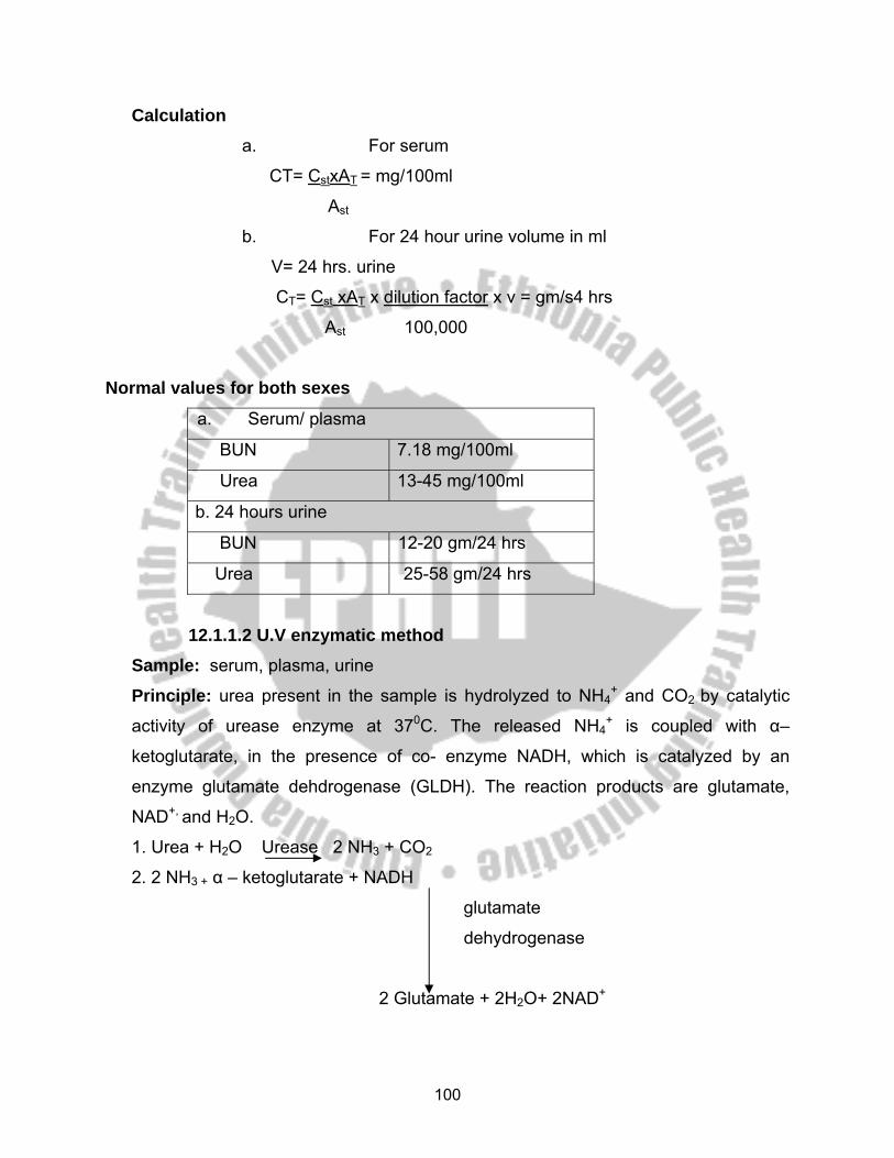
100
Calculation a. For serum
CT= CstxAT = mg/100ml
Ast
b. For 24 hour urine volume in ml
V= 24 hrs. urine CT= Cst xAT x dilution factor x v = gm/s4 hrs Ast 100,000
Normal values for both sexes
a. Serum/ plasma
BUN 7.18 mg/100ml
Urea 13-45 mg/100ml
b. 24 hours urine
BUN 12-20 gm/24 hrs
Urea 25-58 gm/24 hrs
12.1.1.2 U.V enzymatic method Sample: serum, plasma, urine
Principle: urea present in the sample is hydrolyzed to NH4+ and CO2 by catalytic
activity of urease enzyme at 370C. The released NH4+ is coupled with α–
ketoglutarate, in the presence of co- enzyme NADH, which is catalyzed by an
enzyme glutamate dehdrogenase (GLDH). The reaction products are glutamate,
NAD+, and H2O.
1. Urea + H2O Urease 2 NH3 + CO2
2. 2 NH3 + α – ketoglutarate + NADH
glutamate
dehydrogenase
2 Glutamate + 2H2O+ 2NAD+

101
In this reaction NADH is converted to NAD+, and the amount of NADH consumed is
directly proportional to the amount of urea present in the sample. The decrease in
concentration due to NADH is read at 340 nm every minute. Decrease in
absorbance per minute is directly proportional to the concentration of urea present in
the sample. For 5 consecutive minutes the absorbance is taken. Change in
absorbance is calculated. The average change in absorbance is evaluated
CT= AT x Cst
Ast
12.1.2 Determination of creatinine Specimen: plasma, serum, urine, whole blood and other body fluids can be used
Stability: creatinine is stable in serum and urine for a few days at 4-100C and for
a longer period if the sample is frozen.
12.1.2.1 Method: Jaffe Reaction Principle: Creatinine present in a protein free filtrate or diluted urine reacts with alkaline
picrate forming a golden- brown color of a tautomer of creatinine picrate. The intensity
of the golden brown color is directly proportional to the concentration of creatinine
present in the sample and the absorbance is read at 550nm.
A
C
Calculation
a. For serum
b. For urine
CT = AT x Cst x dilution factor Ast = mg/100ml
CT = AT x Cst x dilution factor x V Ast 100,000 = gm/24 hours
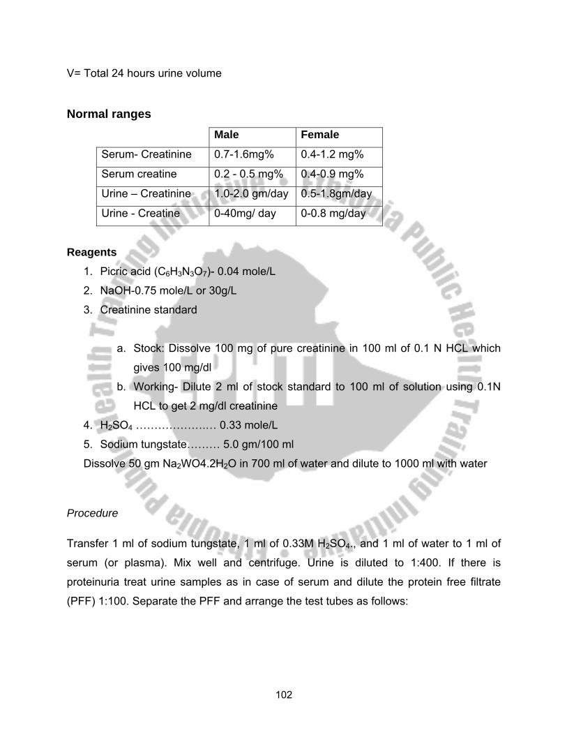
102
V= Total 24 hours urine volume
Normal ranges
Male Female
Serum- Creatinine 0.7-1.6mg% 0.4-1.2 mg%
Serum creatine 0.2 - 0.5 mg% 0.4-0.9 mg%
Urine – Creatinine 1.0-2.0 gm/day 0.5-1.8gm/day
Urine - Creatine 0-40mg/ day 0-0.8 mg/day
Reagents 1. Picric acid (C6H3N3O7)- 0.04 mole/L
2. NaOH-0.75 mole/L or 30g/L
3. Creatinine standard
a. Stock: Dissolve 100 mg of pure creatinine in 100 ml of 0.1 N HCL which
gives 100 mg/dl
b. Working- Dilute 2 ml of stock standard to 100 ml of solution using 0.1N
HCL to get 2 mg/dl creatinine
4. H2SO4 ……………….… 0.33 mole/L
5. Sodium tungstate……… 5.0 gm/100 ml
Dissolve 50 gm Na2WO4.2H2O in 700 ml of water and dilute to 1000 ml with water
Procedure
Transfer 1 ml of sodium tungstate, 1 ml of 0.33M H2SO4., and 1 ml of water to 1 ml of
serum (or plasma). Mix well and centrifuge. Urine is diluted to 1:400. If there is
proteinuria treat urine samples as in case of serum and dilute the protein free filtrate
(PFF) 1:100. Separate the PFF and arrange the test tubes as follows:

103
Blank Test Standard
Distilled water (ml) 4 2 3.5
Creatinine working standard
(ml)
- - 0.5
Protein free filtrate of diluted
urine (ml)
- 2 -
Picric acid (ml) 1 1 1
NaOH solution (ml) 1 1 1
Incubate at 20-250C for 15 min., and read the absorbance at 500 nm
A. For serum
B. For urine
Quality control
- For producing Laboratory results in determination of different analyst in different
clinical samples for the diagnosis of diabetes mellitus and its complications
quality control procedures are mandatory practicing
- Commonly to produce correct laboratory results we will control the following
factors.
1. Pre analytic factors This factors are commonly appear before the analysis of the analyte. For example
during
CT= ATxCstx4 Ast
CT= ATxCst x400 x4 x V Ast x 100 x 1000 = gm/24 hrs
104
A. Patient preparation
B. Sample collection
C. Sample handling and storage
A. Patient preparation- during this time a lot of things can be done for example. The
patient should be asked about his nutritional status, a recent mead. Alcohol,
drugs etc. Which all affect the value of the anlyte.
B. Sample collection:- During sample collection the laboratory personnel should be
a ware of the type of sample, time of collection, area of collection (ream or
capillary) , etc.
C. Sample handling and storage here the type of test tube, anti coagulants and
storage temperature with respect to the type of sample should be considered.
2. Analytical factors The laboratory is more able to control the analytical factors, which depend heavily on
instrumentation and reagents
A. Instrumentation
- Instrument function checks that are to be routinely performed should be detailed
in procedure manual and their performance should be documented
B. Reagents and kits
Reagents and kits should be dated when received and also when opened.
New lots of reagents should be run in parallel with old reagent lots before
being used for analysis.
3. Post analytical factors The post analytical factors consist of the recording and reporting of patient data to
the physician with in the appropriate time interval
Post test
Go back to the pretest questions & do them carefully
105
3.4 SATELLITE MODULE FOR ENVIRONMENTAL HEALTH OFFICERS
1. Direction for using this module Before reading this satellite module be sure that you have completed the pre- test and
studied the core module
2. Learning objectives At the end of reading this satellite module the E.H.Os will be able to:
a. describe the role of environmental health students in DM prevention, controlling
and rehabilitative activities.
b. list the possible environmental predisposing factors of DM
c. inspect the sanitation of hospital wards which are dedicated for admitting DM
cases and take necessary measures on the defects found.
d. inspect the working places of DM patients to check whether they are prone to
any kind of possible causalities.
e. educate the community on how to avoid/ decrease the predisposing factors DM;
chemicals compounds, Biological factors, obesity, sedentary working styles, etc
f. educate patients about the controlling and rehabilitative measures of DM cases
at the health facilities, at annual DM day, etc.
3. Introduction Up to the 1950s it was thought and accepted that DM is the headache of public health
of the very developed countries. But today literature and statistical data clearly depicted
that these cases of diabetes are getting high with alarming incidence rate even in
developing nations. And Ethiopia is one of the developing countries where by the
prevalence is increasing from time to time.
Numerous environmental events have been proposed to trigger the autoimmune
process in genetically susceptible individuals; however, none has been conclusively
linked to diabetes.

106
Identification of an environmental trigger has been difficult because the event may
precede the onset of DM by several years. Though this is the case, it is strongly
believed that there are environmental factors which have a link with DM like, chemical
compounds (Rodenticides, heavy metals virus, rarely exposure to bovine milk proteins),
physical factors (penetrative short- wave length rays) etc.
4. Why the pattern of prevalence of DM has changed across the globe?
For this new change of the pattern of prevalence of diabetes many factors were
considered as the culprit. Among the factors that aid the increment of the prevalence
diabetes even in the developing, countries the following are some:
1. Most people are living in very hectic environment where by the housing condition
is predominated with substandard housing condition that doesn’t usually meet
the physiological and psychological requirement of the dwellers. And this leads
the people to live or to expend much of their time in very stressful environment.
2. People are more ignorant about the healthy style of nutrition at the family and
community level in particular and at large respectively. In an area where there is
no good understanding about the malnourishment consequences, especially
obesity, the high prevalence occurrence of DM in that specific community will be
highly inevitable.
3. Long term exposure to some specific type of organic and inorganic chemicals,
recently it came to understand that they will cause DM by affecting the pancreas
and they interfere with the right physiological secretion of insulin.
4. Exposure to some biological factor (coxsakie and rubella most prominently)
cause DM by trigger the autoimmune system so that the destruction of β -cell of
the pancreas will occur.
5. IN rarest case, DM may happen because of the destruction β – cell s of the
pancreas by trauma, accidents, chronic inflammation.

107
6. Many jobs are becoming sedentary rather than exercise/movement demanding
and in turn these furnish the ground for the people to become more obese.
7. Physical exercise is not taken as a routine life activity among the people
especially living in developing country where the living places are not comfortable
to make exercises at continual basis.
5. Environmental factors linked to DM Putative environmental triggers include viruses (coxsackie and rubella most
prominently) early exposure to bovine milk proteins, and Nitrosourea compounds.
Epidemiological studies have noted an association between exposure to bovine milk
intake and type 1 A DM; studies are ongoing to investigate a possible relationship
between exposure to bovine milk and the autoimmune process of type 1 A DM.
6. Diabetes in Africa: some of the factors related to the development of diabetes in
Africa include:
• Genetic Factors: Family history
• Environmental Factors: such as infection, dietary changes.
• Chronic calcific pancreatitis, malnutrition and environmental toxins(oral iron
overload)
• Thrifty phenotypes and genotypes - NIDDM
• Plenty food - reduced exercise - insulin resistance a sin urbanization
• Low birth weight - fetal malnutrition - reduction of effective Beta cells - NIDDM.
7. Expected tasks to be accomplished by Environmental Health Officers regarding DM prevention, controlling and rehabilitative actions.
1. The environmental health officers will take great share of tasks of advocacy on
proper nutrition so that obesity can be attacked.

108
2. Construction of standard housing with local materials will be advocated and
technically commented and regularly inspected by environmental Health officers.
Thus the requirements of physiological and psychological health of the dwellers
will be met and this consequently will alleviate the potential stressful
environment.
3. Environmental health officers technically suggest comments and follow its
implementation to make the working places more comfortable. E.g. a DM patient
working in steel processing industry is often time advised to wear very thick and
laceration proof gloves at any time of working to avoid the incidence of lacerated
wound.
4. Working places, institutions, houses will be inspected at continual basis for
whether they are comfortable for DM patients or not, and whether they have the
culprit factors (predisposing chemicals, especially Rodenticides (vacor),
Biological factors (viruses) physical agents (radiations).
5. Enclosing short wave length ray emanating sites with radiation proof construction
materials.
6. Promoting personal hygiene for any DM patients as they are most susceptible
and potentially be affected with different kinds of secondary skin infections.
7. Routine hospital sanitation inspection will be beneficial in preventing the admitted
DM cases from nosocomial infections.
8. Health information promotion regarding DM is the crucial area where the
environmental health officers are expected to play a vital role.
9. The environmental health officers are most needed here to apply their expertise
knowledge of housing and institutional sanitation, nutrition and food hygiene and
safety, environmental chemistry. Organic chemistry and environmental quality
control courses in conformity with the preventing and controlling strategies of
DM.
10. It has to be emphasized that E.H.Os should have deep concern about safe
injection of insulin and proper disposal of used needles. No room should be left
for contamination that is allowing the occurrence of secondary infections.

109
11. The EHOs are expected to be involved in research activities, to examine and
analyze the environment for its physical, chemical and biological qualities that will
help in modifying the environment to make ideal for DM cases.
12. Give refresher trainings on DM for environmental health technicians and health
extension workers from environmental health point of view.
Post – test
First try to look and do the pretest again, then keep on attempting the following
questions.
1. What situation makes difficult the study of causation of environmental factors and
to link conclusively with DM?
2. Why diabetes mellitus patients are most susceptible for different kinds of skin
infections?
3. What is the basic reason for the fact that E.H.Os are supposed to be highly
concerned to make the working places free of any possible causalities for DM
patients?
4. What are the known environmental factors that are thought to cause DM?
5. What parcel of Health information is highly beneficial for the family or community
with strong DM history?
Multiple Questions 1. DM can be transmitted through the following ways except
A. Feco-oral route
B. Aerosol respiration
C. contact
D. Unsafe sexual activity
E. None of the above
2. DM can be caused by one of the following factor except A. β- cell affection by autoimmune system
B. Chemicals

110
C. Biological agents
D. taking high amount of sugar in the diet
E. None of the above
3. From the following alternatives one can be identified as one risk group to develop Dm
except A. Obese person
B. a person with strong DM family history
C. a person that had > 4 kg body weight during delivery
D. a person who has married Dm patient
E. none of the above
4. From the following set of types of rays one possibly could not cause DM compared
with others
A. IR rays
B. Gamma rays
C. Beta rays
D. Radioactive rays
E. none of the above
5. The main causative agent of DM is
A. Bacteria
B. Viruses
C. Fungus
D. Parasites
E. none of the above

111
SATELLITE MODULE FOR HEALTH SERVICE EXTENSION WORKERS
Introduction Every nation in the world will have well developed and vividly written national health
policy likewise Ethiopia as a country has clear health policy which is intended to be
implemented in every corner of the country. This policy is preventive led. Thus, today
there has been a strong commitment from the government side to realize the policy and
protects the public health. It is one part of the strategies of national health policy to train
the health extension workers as a front line community health personnel in the regional
health institutions with the intent that after the end of their training they will go near to
the community that is rural areas and they fight with the nation public health challenges
together with their professional colleagues in the interdisciplinary approach.
Therefore this module will have the great benefit for the health extension workers to
know more about and operationally/procedurally to be instructed then what to do about
the different aspects of DM in the health station, even more in the community. Moreover
the health extension workers will be very much clear on their role the promotive,
preventive, therapeutic tasks regarding DM after they have thoroughly read this module.
Purpose of the module This module is intended to be used by health extension package workers. It provides
basic information on different aspects of diabetes so that they participate in early case
detection, case management and prevention of complications as front line health
workers.
Directions for using the module
Before starting to read this module, please follow the instruction given below.
• Start with the pre-test before going through the core module.
• Use a separate sheet of paper to write your answers.

112
• Having gone through the module, try to attempt the presented questions.
• Study the task analysis for the health center team members in comparison with
that of your own.
Objectives 2.3 Learning Objectives After reading the module, one will be able to
• Explain the importance of diabetes mellitus as a public health problem
• Describe diabetes mellitus, its classification and clinical presentation.
• Outline the diagnostic tests for diabetes mellitus.
• Describe the logic behind appropriately employed treatment.
• Describe the role played by each member of the health center team.
• Describe the overall principles of management.
Diabetes mellitus Definition Diabetes Mellitus is a clinical syndrome comprising a heterogeneous group of metabolic
diseases that are characterized by chronic hyperglycemia and disturbances in
carbohydrate, fat and protein metabolism secondary to defects in insulin secretion,
insulin action or both Types
Type 1And type 2 CLINICAL FEATURES Classical symptoms
• Thirst
• Polyuria
• Nocturia

113
• Rapid weight loss
• Increased susceptibility to infection in patients with uncontrolled diabetes
• Chronic fatigue and malaise
Signs
• Signs related to acute and chronic complications
Possible laboratory tests at Health center & Health post level for the investigation of Diabetics Mellitus
1. Self- Monitoring of glucose
Many patients (especially those with type 1(insulin- dependent diabetes mellitus)) now
regularly monitor their own blood glucose concentrations on the advice of their health
care provider, using reagent test strips and reflectance meter. Several companies
manufacture reagent test strips for monitoring blood glucose, and most of these
companies make reflectance meters to be used to electronically read the test result.
Instruments include one Touch,* Accu- chek Easy, and Glucometer Elite. The strips
used for these tests are impregnated with the enzyme glucose oxidase, enzyme
peroxidase and an indicator to give a color change that is detectable. The color change
can be read in a reflectance meter on which the result (in mg/dl) is visualized. Although
blood is tested, results are converted to plasma glucose values by the instrument.
Eg. Glucose oxidase reaction (Vitros Method)
2. Urine Glucose determinations
Chemical screening tests for glucose (dextrose) are generally included in every routine
urinalysis. The occurrence of glucose in the urine indicates that the metabolic disorder
diabetes mellitus should be suspected, although several other conditions result in
glycosuria (glucosuria).

114
The lowest blood glucose concentration that will result in glycosuria is termed the renal
threshold (180-200 mg/dl). It is possible to use both enzymatic technique and oxidation-
reduction technique to determine urine glucose
2.1 Enzymatic technique 2.1.1 Reagent strip (Glucose oxidase) Tests
Principle and specificity Since the reagent strip tests for urinary sugar use glucose oxidase which only react in
the presence of glucose they are highly specific. Reagent strip tests for urine glucose
are double sequential enzyme reactions. Glucose oxidase will oxidize glucose to
gluconic acid and at the same time reduce atmospheric oxygen to H2O2. The hydrogen
peroxide formed will, in the presence of the oxidized form, which is indicated by the
color change of an oxidation- reduction indicator.
Note: The glucose oxidase, peroxidase and the reduced form of the Oxidation-
Reduction indicator are all impregnated on to a dry reagent strip. There are different
kinds of reagent strips and they all contain Gluocse oxidase and peroxidase.
2.1.1.1 Procedure
1. Collect the urine sample with a clean, dry, free from any
antiseptic and wide mouth container
2. Transfer the urine into a conical test tube
3. Take one strip from the reagent strip container
4. Immerse the strip into the urine in a conical test tube
5. Immediately pull it out and let it stand for one minute.
So as to have time for reaction and color change to
occur on the strip
6. After one minute read the result by matching the color
on the strip with the color on the reagent strip container
7. Report the result

115
3. Determination of ketone bodies in urine ketone bodies are a group of three related substances: acetone, aceto acetic acid,
and β – hydroxyl butyric acid.
When ever fat (rather than carbohydrate) is used as the major source of energy,
ketosis and ketonuria may result.
The two out standing causes of ketone accumulation are diabetes mellitus and
starvation
In diabetes mellitus, the body is unable to use carbohydrate as an energy source
and attempts to compensate by resorting to fat catabolism, which results
accumulation of ketone more than normal, that the body is unable to utilize it.
The clinical result is an increased concentration of ketones in the blood (ketonemia)
and in the urine (ketonuria.)
Since the presence of ketone bodies in urine is an early indication of lack of adequate
insulin control, reagent strips that combine tests for glucose and ketone are often used.
3.1 dipstick test Principle: the reagent strip tests for ketone bodies are based on legal’s (Rothera’s)
test, a color reaction with sodium nitroprusside (nitro ferricyanide). Acetic acid will react
with sodium nitro prusside in an alkaline medium to form a purple color.
3.1.1 Procedure 1. After collecting the urine sample from the patients, transfer into a clean, dry and free
of disinfectant test tube
2. Then immerse the dipstick into the urine
3. Then drain and let it stand for certain seconds for the reaction to take place
4. Read the result by comparing the color produced with the standard on the strip
container
Note acetone and aceto acetic acid can be detected by different dip stick tests,
but there is no reagent strip test for β - hydroxyl butyric acid

116
4. Determination of urine protein Microalbuminuria
• Diabetes mellitus causes progressive changes to the kidneys and ultimately
results in diabetic renal nephropathy. This complication progresses over a period
of years and may be delayed by aggressive glycemic control
• An early sign that nephropathy is occurring is an increase in urinary albumin
• It is thought that the early development of renal complications can be predicted
by the early detection of consistent micro albuminuria. And this early detection is
desirable, as better control of blood glucose levels may delay the progression of
renal disease
4.1 Methods of measurement Test for urinary protein are of two major types:
a. Tests that are based on the use of the protein error of PH indicators
- This is the methodology employed in the various reagent strip tests
- They are more sensitive to the presence of albumin than to other proteins.
b. Tests that are based on the precipitation of protein by chemical or coagulation by
heat
- This test will detect all proteins, including albumin, glycoproteins, globulins,
Bence Jones protein & hemoglobin
4.1.1 Reagent strip test Principle: Reagent strip tests for urinary protein involves the use of PH indicators
substances that have characteristic colors at specific PH values. The phenomenon of
showing different color at different PH is called. “the protein error of indicators” The PH
of the urine is held constant by means of buffer, so that any change of color of the
indicator will indicate the presence of protein.
4.1.1.1 Procedure It is the similar with other reagent strip test procedure. (But the reading time can vary
manufacture to manufacturer instruction on the leaf late)

117
Complications
• Classified into acute and chronic complications
• Acute complications are
o Diabetic ketoacidois
o Nonketotic hyperosmolar state
o Hypoglycemia
• Chronic complications
o Affect many organ systems
o Are responsible for the majority of morbidity and mortality associated with
the disease
o Can be subdivided into vascular and non-vascular complications
o The vascular complications are further subdivided into
Microvascular complications that includes
• Diabetic retinopathy
• Diabetic nephropathy
• Diabetic neuropathy
Macrovascular complications
• Coronary artery disease
• Peripheral vascular disease
• Cerebrovascular disease
o The non-vascular complications are
Gastroparesis
Sexual dysfunction
Skin changes
Therapeutic approach
There are four components of management for diabetes, which is carried by the health
extension workers: -
- Diet
- Exercise
- Monitoring
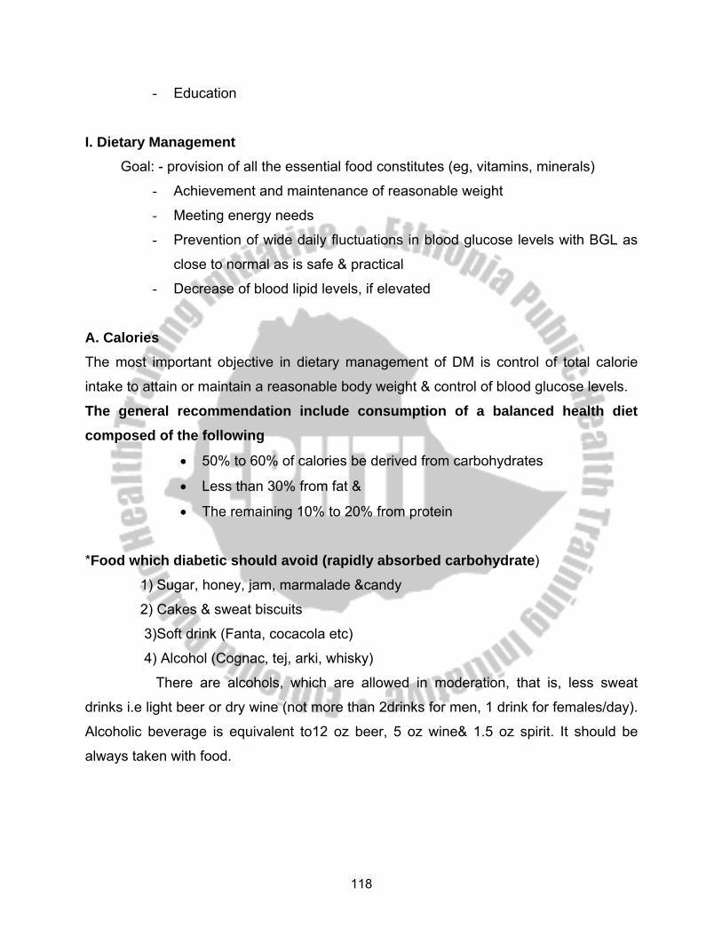
118
- Education I. Dietary Management Goal: - provision of all the essential food constitutes (eg, vitamins, minerals)
- Achievement and maintenance of reasonable weight
- Meeting energy needs
- Prevention of wide daily fluctuations in blood glucose levels with BGL as
close to normal as is safe & practical
- Decrease of blood lipid levels, if elevated
A. Calories The most important objective in dietary management of DM is control of total calorie
intake to attain or maintain a reasonable body weight & control of blood glucose levels.
The general recommendation include consumption of a balanced health diet composed of the following
• 50% to 60% of calories be derived from carbohydrates
• Less than 30% from fat &
• The remaining 10% to 20% from protein
*Food which diabetic should avoid (rapidly absorbed carbohydrate)
1) Sugar, honey, jam, marmalade &candy
2) Cakes & sweat biscuits
3)Soft drink (Fanta, cocacola etc)
4) Alcohol (Cognac, tej, arki, whisky)
There are alcohols, which are allowed in moderation, that is, less sweat
drinks i.e light beer or dry wine (not more than 2drinks for men, 1 drink for females/day).
Alcoholic beverage is equivalent to12 oz beer, 5 oz wine& 1.5 oz spirit. It should be
always taken with food.

119
*Foods which diabetic should take with restrictions A) Foods from grain eg injera, bread, kinche , dabo kolo, kita ,atemit
B) Foods prepared from peas, beans, and lentils
C) Potato, sweat potato, kocho, bulla
D) All fruits except lemons& grap fruit
E) Macaroni, pasta, rice
Foods, which diabetics can take freely or with minimal restriction A) Lean meat &fish
B) Eggs and milk
C) Green leafy vegetables (kale, salad, cabbage D) Lemon, grape fruit
E) Tea, coffee, lemon juice with out sugar, ambo water, and other mineral
water & clear soup
F) Spices pepper ,berberi
G) Tomato, pumpkin, carrot, onion and chilipeper
II. Exercise - Is extremely important in the management of diabetes because of its
effect on
lowering blood glucose and
reducing cardiovascular risk factors
-Lowers blood glucose level by increasing the uptake of glucose by body
muscles and by improving insulin utilization
- Pre or post exercise snack may be required to prevent hypoglycemia after
exercise
- Patients should be thought to do regular, moderate exercise at the same
time (preferably when blood glucose level are at their peak) and in the
same amount for at least 30 minutes each day.
-Patient is advised:- to use proper footwear and if appropriate other
protective equipment
- avoid exercise in extreme heat or cold
120
- inspect feet daily after exercise
- avoid exercising during periods of poor metabolic control .
III. Monitoring of Glucose and Ketones
Blood glucose level and urine for ketone and glucose should be assessed frequently by
self or by having follow up in the health unit
Pt education -about Insulin Injection - Insulin injections are administered into the subcutaneous tissue
- Equipment: - Insulin
- Short acting insulin is clear in appearance and long acting insulin
are cloudy and white
- The long acting must be mixed (gently inverted or rolled in the hands)
before use
- Before injection it should have room To which may require rolling it in the
hands or removing it from a refrigerator for a time before the injection
- If a frosted, adherent coating is present, some of the insulin is bound and
should not be used
Syringes - should be matched with the insulin concentration
- 1 ml syringes – hold 100 units
- ½ ml syringes – hold 50 units
- 3/10 ml syringes – hold 30 units
Administering the injection
-Avoid use of alcohol for cleansing
- Four main areas
• Abdomen
• Arms (posterior surface)
• Thighs (anterior surface)
• Hips

121
Absorption is greatest in abdomen and decreases progressively in the arm, thigh, and
hips Rotation - Rotation of injection site is required to prevent lipodystrophy, localized changes in fatty
tissue,
Pt is instructed as: -
1. Do not use a site > once every 4 to 6 weeks
2. Sites should be 1 to 1 ½ inches apart
3. Use all sites in one geographic area, then move to the next area
4. Document site use
Side effects of insulin injections 1. Local allergic reactions.
- in the form of redness, swelling , tenderness , and indurations or a 2 to
4 cm wheal may appear at the injection site 1to 2 hrs after injection
- usually occur during the beginning stage of therapy and disappear with
continued use of insulin
- antihistamine will be given 1 hr before injection
- if alcohol is used to clean the area the skin should be allowed to dry
2. Systemic allergic reaction
- are rare
- local skin reaction that gradually spreads in to generalized urticaria
Treatment:- desensitization , gradually increasing the amount of insulin
3. Insulin lipodystrophy
- Refers to a localized disturbance of fat metabolism in the form of loss of
sc fat and appears as slight dimpling or more serious pitting of sc fat or is
the development of fibrofatty masses at the injection site and is caused by
the repeated use of injection site
- if insulin is injected in to scarred areas the absorption may be delayed
Treatment: Pt should avoid injection on the areas and prevent by rotating

122
Injection sites
Risk identification • Those above 45 years of age every three years
• Those with family history of diabetes mellitus (parent or sibling with type 2
diabetes mellitus)
• Obesity as evidenced by BMI> 27Kg/m2
• History of delivering a baby weighing above 4Kg in a lady or previous episode of
gestational diabetes mellitus
• Hypertension
Prevention:
• Screening • A number of lifestyle modification and pharmacologic agents are suggested to
prevent or delay its onset.
High-risk individuals should be encouraged to
• Maintain a normal body mass index
• Engage in regular physical exercise
No specific intervention is proven to prevent type 2 diabetes mellitus.
Role and Task Analysis
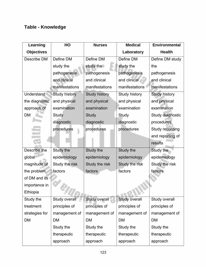
123
Table - Knowledge
Learning Objectives
HO Nurses Medical Laboratory
Environmental Health
Describe DM Define DM
study the
pathogenesis
and clinical
manifestations
Define DM
study the
pathogenesis
and clinical
manifestations
Define DM
study the
pathogenesis
and clinical
manifestations
Define DM study
the
pathogenesis
and clinical
manifestations
Understand
the diagnostic
approach of
DM
Study history
and physical
examination
Study
diagnostic
procedures
Study history
and physical
examination
Study
diagnostic
procedures
Study history
and physical
examination
Study
diagnostic
procedures
Study history
and physical
examination
Study diagnostic
procedures
Study recording
and reporting of
results
Describe the
global
magnitude of
the problem
of DM and its
importance in
Ethiopia
Study the
epidemiology
Study the risk
factors
Study the
epidemiology
Study the risk
factors
Study the
epidemiology
Study the risk
factors
Study the
epidemiology
Study the risk
factors
Study the
treatment
strategies for
DM
Study overall
principles of
management of
DM
Study the
therapeutic
approach
Study overall
principles of
management of
DM
Study the
therapeutic
approach
Study overall
principles of
management of
DM
Study the
therapeutic
approach
Study overall
principles of
management of
DM
Study the
therapeutic
approach
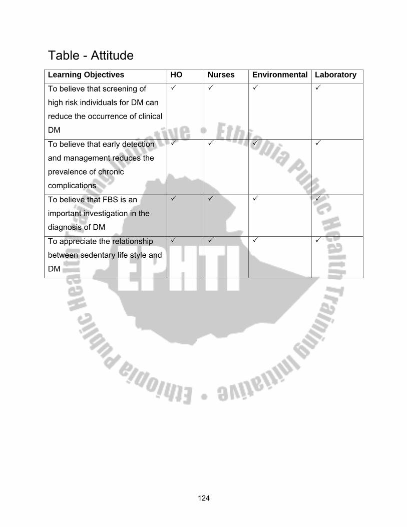
124
Table - Attitude Learning Objectives HO Nurses Environmental Laboratory
To believe that screening of
high risk individuals for DM can
reduce the occurrence of clinical
DM
To believe that early detection
and management reduces the
prevalence of chronic
complications
To believe that FBS is an
important investigation in the
diagnosis of DM
To appreciate the relationship
between sedentary life style and
DM

125
Table - Practice Learning Objectives HO Nurses Environment
al Laboratory
Demonstrate methods
and techniques of
Diabetic patient
examination
Label patients as high
and low risk and follow
them accordingly
Manage diabetics
related complication
Identify risk factors
Develop necessary
skills on laboratory
investigations
Follow the standard
reporting and recoding
technique
perform the
appropriate
laboratory
tests
- Follow the
scientific
procedures to do
the tests
- order routine lab
investigations
performappropriate
laboratory tests
- Follow the
scientific
procedures to do
the tests
- order routine lab
investigations
perform the
appropriate
laboratory
tests
- Follow the
scientific
procedures
to do the
tests
perform the
appropriate
laboratory
tests
- Follow the
scientific
procedures
to do the
tests
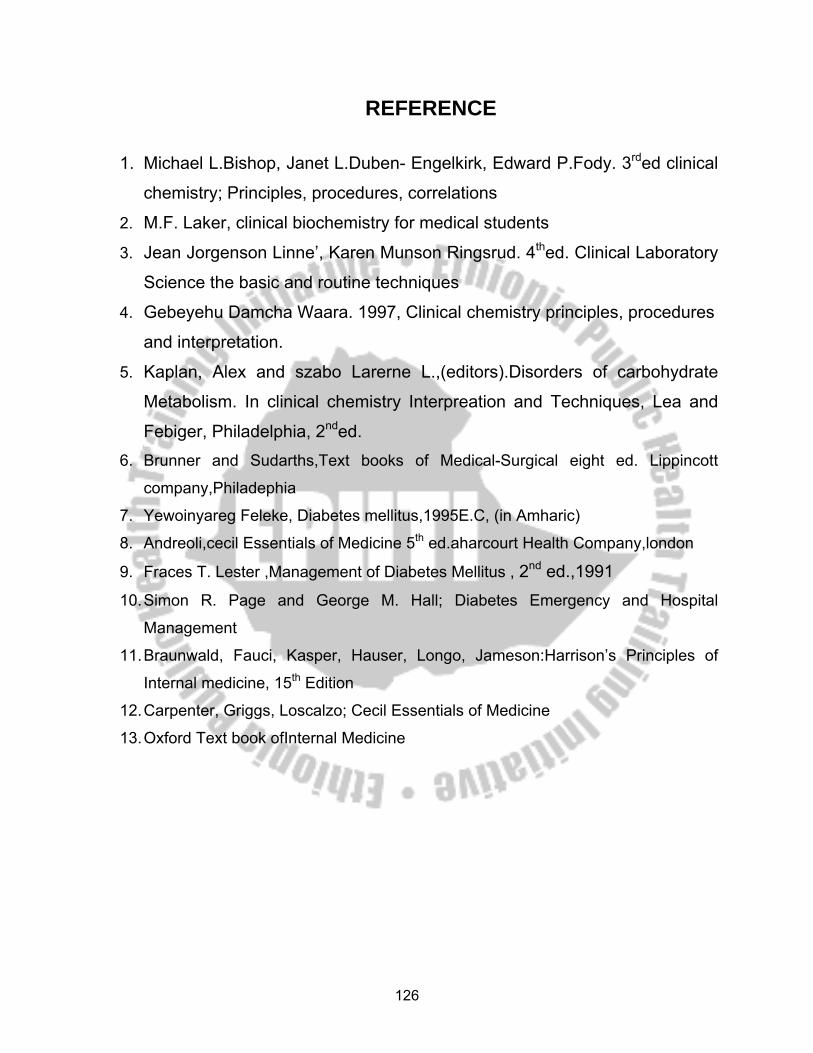
126
REFERENCE
1. Michael L.Bishop, Janet L.Duben- Engelkirk, Edward P.Fody. 3rded clinical
chemistry; Principles, procedures, correlations
2. M.F. Laker, clinical biochemistry for medical students
3. Jean Jorgenson Linne’, Karen Munson Ringsrud. 4thed. Clinical Laboratory
Science the basic and routine techniques
4. Gebeyehu Damcha Waara. 1997, Clinical chemistry principles, procedures
and interpretation.
5. Kaplan, Alex and szabo Larerne L.,(editors).Disorders of carbohydrate
Metabolism. In clinical chemistry Interpreation and Techniques, Lea and
Febiger, Philadelphia, 2nded. 6. Brunner and Sudarths,Text books of Medical-Surgical eight ed. Lippincott
company,Philadephia
7. Yewoinyareg Feleke, Diabetes mellitus,1995E.C, (in Amharic)
8. Andreoli,cecil Essentials of Medicine 5th ed.aharcourt Health Company,london
9. Fraces T. Lester ,Management of Diabetes Mellitus , 2nd ed.,1991
10. Simon R. Page and George M. Hall; Diabetes Emergency and Hospital
Management
11. Braunwald, Fauci, Kasper, Hauser, Longo, Jameson:Harrison’s Principles of
Internal medicine, 15th Edition
12. Carpenter, Griggs, Loscalzo; Cecil Essentials of Medicine
13. Oxford Text book ofInternal Medicine
127
Keys for the pretest and post test questions for Nurses 1. A
2. C
3. C
4. E
5. A
6. C
7. A
8. C
9. D
10. a- site of injection
-preparations of medication
-Rotations
-about syringe and needle
-some problems with insulin injections
b) -too much insulin
-too little food or
-excessive physical exercise
-delay of meal or omitting of snacks
c) -sweating
-tremor
-tachypnea
-confusion
-seizure
-loss of consciousness
d) Having snack, not delaying the meal, right dose of medications, having candies at
hand
f)-assess foot daily for sensation, redness and broken skins
-wash dry feet daily
- If skin is dry apply a thin coat of lubricating oil

128
-tie shoes loosely but firmly
-If your feet perspire, change shoe and stocking during the day
-wear shoe and stocking that gives room for the movement of the toe
Key For Laboratory Technology
1. B 6. A
2. C 7. C
3. C 8. B
4. C 9. A
5. C 10.A