developing brain and systemic inflammation: a “toll … · developing brain and systemic...
TRANSCRIPT

Developing brain and systemic inflammation: a “Toll-like” link
with consequences
Amin Mottahedin
Department of Physiology, Institute of Neuroscience and Physiology at Sahlgrenska Academy University of Gothenburg Gothenburg, Sweden, 2017

Cover illustration by Amin Mottahedin. Two-photon image of mouse choroid plexus immunolabeled for occludin (red) and CD31 (green). Developing brain and systemic inflammation: a “Toll-like” link with consequences © 2017 Amin Mottahedin [email protected] ISBN 978-91-629-0207-0 Printed in Gothenburg, Sweden 2017 Ineko AB

To my dear family


Developing brain and systemic inflammation: a “Toll-like” link with consequences
Amin Mottahedin Department of Physiology, Institute of Neuroscience and Physiology Sahlgrenska
Academy at University of Gothenburg
ABSTRACT
The developing brain is vulnerable to external insults, and perinatal brain injury (PBI) is a major cause of life-long neurological syndromes such as cerebral palsy. Currently, no pharmaceutical intervention is available. Hy-poxia/ischemia (HI), infections and inflammation are implicated in the path-ogenesis of PBI. However, the crosstalk between these etiologies is not fully understood. Toll-like receptors (TLR) 3 and TLR2 are responsible for sens-ing viral and bacterial infections and initiating the inflammatory response. The aim of this thesis was to investigate the effect of systemic inflammation induced by activation of these TLRs on neonatal HI brain injury. We demon-strate that intraperitoneal administration of TLR3 and TLR2 ligands (PolyI:C and P3C, respectively) prior to HI increases the brain injury in ne-onatal mice. PolyI:C and P3C induced neuroinflammation and altered mi-croglial phenotype as assessed by RT-qPCR, multiplex cytokine assay or flow cytometry. PolyI:C also upregulated the pro-apoptotic gene, Fasl, ex-pression and reduced activation of pro-survival signaling molecule Akt. On the other hand, P3C suppressed mitochondrial respiration, a major mecha-nism of cellular energy production. P3C, unlike other TLR agonists, induced marked infiltration of leukocytes to the cerebral spinal fluid and brain of neonatal mice and rats. Confocal microscopy, Cre recombinase-mediated gene targeting and in vitro cell transmigration assay revealed the choroid plexus as a site of leukocyte entry. RNA sequencing of the choroid plexus followed by transcriptome cluster analysis and Ingenuity Pathway Analysis revealed potential mechanisms of leukocyte infiltration, including a specific chemotaxis signature and cytoskeleton-related pathways. Finally, we show that N-acetylcysteine treatment inhibits TLR2-mediated leukocyte traffick-ing in vivo and in vitro.
To conclude, this thesis describe a TLR-mediated link between systemic inflammation and developing brain with detrimental consequences on HI brain injury, suggesting potential novel therapeutic strategies.
Keywords: neonatal brain injury, hypoxia-ischemia, inflammation, infec-tion, Toll-like receptor, choroid plexus ISBN (Print): 978-91-629-0207-0 ISBN (PDF): 978-91-629-0208-7

Sammanfattning på svenska Hjärnskador hos nyfödda barn är den vanligaste orsaken till neurologiska problem (såsom cerebral pares) bland barn. I Sverige diagnostiseras 2 av 1000 spädbarn med cerebral pares. Dessa hjärnskador uppstår i både fullgångna och för tidigt födda barn, men förekomsten är betydligt högre bland barn som fötts förtidigt. En vanlig orsak till hjärnskadorna hos spädbarn är brist på syre (hypoxi) och/eller lågt blodflöde (ischemi) till hjärnan. Ännu en riskfaktor är infektioner hos spädbarnet eller mamman någon gång runt födseln. Hur infektioner kan öka risken för hjärnskada i nyfödda var den huvudsakliga frågan vi ville försöka svara på i denna avhan-dling. Infektioner känns igen av vårt immunförsvar genom specifika recep-torer som kallas Toll-liknande receptor (TLR) som finns på ytan av alla immunceller. Virus aktiverar framförallt TLR3 och en viss grupp bakterier aktiverar TLR2, och genom aktivering av dessa receptorer startar en immu-nologisk reaktion i kroppen. Vi använder oss av en modell av hypoxi/ischemi (HI) hos nyfödda möss som producerar hjärnskador som liknar dem som kan ses hos spädbarn. Genom att administrera specifika ämnen aktiverades TLR3 och TLR2 innan HI. Vi upptäckte att aktivering av dessa receptorer ökar hjärnskadorna i både grå substans (nervceller) och vit substans (nervfibrer). Det visar på att virus- och bakterieinfektioner gör hjärnan känsligare för HI. Vi undrade sedan vad det är som orsakar denna ökade känslighet. Vi upptäckte att aktivering av TLR3 leder till en kraftig inflammatorisk respons i hjärnan, vilket ledde till specif-ika cellförändringar som pekade på minskad överlevnadsfunktion samt att hjärnans egna immunceller (s.k. mikroglia) blev mer reaktiva. Aktiveringen av TLR2 ledde till invasion av vita blodkroppar till hjärnan från blodet och minskad mitokondriefunktion (mitokondrierna ger celler energi) i hjärnans celler. Vi såg att de vita blodkropparna tar sig in i hjärnan via ett litet organ i hjärnan som kallas plexus choroidea. Därför analyserade vi genförän-dringar i plexus choroidea efter TLR2-stimulering och kunde fastställa de molekylära mekanismer som kan göra det möjligt för vita blodkroppar att ta sig in i hjärnan. Slutligen visade vi att behandling av neonatala råttor med N-acetylcystein, ett antioxidantläkemedel, blockerar invasionen av vita blodkroppar till hjärnan i denna modell. Sammanfattningsvis fann vi att vi-rus- och bakterieinfektioner orsakar inflammation i hjärnan och gör hjärnan känsligare för HI-relaterade hjärnskador. Minskning av inflammationen i hjärnan genom riktade behandlingar mot plexus choroidea eller genom behandling med N-acetylcystein kan minska risken för hjärnskador hos nyfödda.

List of papers This thesis is based on the following studies, referred to in the text by their Roman numerals.
I . Stridh L, Mottahedin A, Johansson ME, Valdez RC, Northington F, Wang X, Mallard C. Toll-like receptor-3 activation increases the vulnerability of the neonatal brain to hypoxia-ischemia. Journal of Neuroscience, 2013. 33(29): p. 12041-51.
I I . Mottahedin A, Svedin P, Nair S, Mohn CJ, Wang X, Hagberg H, Ek J, Mallard C. Systemic activation of Toll-like receptor 2 suppresses mitochondrial respiration and exacerbates hypoxic-ischemic injury in the developing brain. Journal of Cerebral Blood Flow and Metabolism. 2017 Jan 1:271678X17691292.
I I I . Mottahedin A, Smith PL, Hagberg H., Ek CJ, Mallard C. TLR2-mediated leukocyte trafficking to the developing brain. Journal of Leukocyte Biology. 2017 Jan;101(1):297-305.
IV. Mottahedin A, Ek J, Truvé K, Hagberg H, Mallard C. Differential analysis of TLR2- versus TLR4-induced alterations in transcriptome of choroid plexus reveals leukocyte trafficking mechanisms. Manuscript.
V. Mottahedin A, Blondel S, Ek J, Babikian A, Hagberg H, Mallard C, Ghersi Egea JF, Strazielle N. N-acetylcysteine inhibits TLR2-mediated neutrophil transmigration through the choroid plexus. Manuscript.


C ONTENT ix
Content Abbreviations 1 Introduction 1 Brain development 2 Brain interfaces 4 Brain immune cells 5 Perinatal brain injury 6 Hypoxic-ischemic brain injury 8 From systemic inflammation to neuroinflammation 8 Toll-like receptors 10 Perinatal systemic inflammation 12 Inflammatory cells in circulation 12 Neuroinflammation 14 Brain vasculature in mediating inflammation 15 Choroid plexus in mediating inflammation 16 Immune cells in neuroinflammation 17 Inflammation and brain injury 20 Aims 21 Methodological considerations 21 Laboratory animals 22 TLR agonists 23 Hypoxic-ischemic brain injury model 24 Immunohistochemistry and histology 26 Brain injury assessment 27 Quantitative reverse transcription PCR 27 Flow cytometry 28 Multiplex cytokine assay 29 Magnetic activated cell sorting 29 Western blot 30 Mitochondrial respirometry 31 Brain barrier permeability test 32 Cre-recombinase mediated gene targeting in the choroid plexus 33 RNA sequencing 34 Transcriptome analysis 35 In vitro model of neutrophil transmigration through choroid plexus 36 Statistics

x C ONTENT
38 Results summary 38 Systemic activation of viral (TLR3) or bacterial (TLR2) receptors aggravates HI brain injury in neonatal mice 39 TLR3 activation induces neuroinflammation 39 TLR3 activation suppresses cell survival pathways in the brain 40 TLR3 and TLR2 stimulation alters the microglia phenotype 41 Systemic TLR2 activation suppresses brain mitochondrial respiration 42 Systemic activation of TLR2 but not TLR4 induces neutrophil and monocyte invasion of the CNS 43 Choroid plexus is a route of TLR2-induced leukocytes trafficking 43 TLR2 induces specific chemotaxis and cytoskeleton regulating pathways in the choroid plexus 44 N-acetyl cysteine blocks TLR2-mediated leukocyte infiltration to the brain 45 Discussion 45 Sensitization of the developing brain to HI by systemic inflammation 46 How does systemic inflammation sensitize the brain to HI injury? 46 ○ TLR-mediated neuroinflammation 47 ○ Neuroinflammation and cell death pathways 49 ○ Neuroinflammation and cerebral energy metabolism 50 TLR2-mediated leukocyte trafficking to the CNS through choroid plexus 50 ○ What is special about TLR1/2 signalling? 51 ○ Why does TLR2 activation results in leukocyte migration to the CNS? 53 Conclusion and future perspective 55 Acknowledgements 57 References

AB BR EVI ATIONS xi
Abbreviations ANOVA analysis of variance ATP adenosine triphosphate BBB blood-brain barrier BCSFB blood-cerebrospinal fluid barrier CMV cytomegalovirus CNS central nervous system CP cerebral palsy CPEC choroid plexus epithelial cell CSF cerebrospinal fluid CVO circumventricular organ DAB 3,3′-Diaminobenzidine DAMP damage-associated molecular pattern dsRNA double-stranded RNA EAE experimental autoimmune encephalomyelitis EONS early-onset neonatal sepsis ETC electron transfer chain FADH flavin adenine dinucleotide FCCP carbonyl cyanide-4-(trifluoromethoxy) phenylhydrazone FIRS fetal inflammatory response syndrome GBS group B streptococcus G-CSF granulocyte-colony stimulating factor GRK2 G protein-coupled receptor kinase 2 GW gestational week HI hypoxia/ischemia HIE hypoxic/ischemic encephalopathy ICV intracerebroventricular IgG immunoglobulin gamma IHC immunohistochemistry IL interleukin iNOS induced nitric oxide synthase IVH intraventricular hemorrhage JNK c-Jun N-terminal kinase LONS late-onset neonatal sepsis LPS lipopolysaccharide MACS magnetic activated cell sorting MAP2 microtubule-associated protein 2 MBP myelin basic protein

xii ABBR EVI ATIONS
MCP-1 monocyte chemoattractant protein 1 MD2 myeloid differentiation protein-2 MEGF10 multiple epidermal growth factor-like domains protein 10 MERTK mer tyrosine kinase MHCII major histocompatibility complex MIP1-a macrophage inflammatory protein 1 alpha MIP1-b macrophage inflammatory protein 1 beta miRNA microRNA MyD88 myeloid differentiation primary response gene 88 NAC N-acetylcysteine NADH nicotinamide adenine dinucleotide NE neonatal encephalopathy NEC necrotizing entrocolitis NF-κB nuclear factor kappa-light-chain-enhancer of activated B cells PAMP pathogen-associated molecular pattern PFA paraformaldehyde PND postnatal day Poly I:C polyinosinic:polycytidylic acid PRR pattern-recognition receptor PRX3 peroxiredoxin 3 PVL periventricular leukomalacia RANTES regulated on activation, normal T cell expressed and secreted RNAi interfering RNA ROS reactive oxygen species RT-qPCR reverse transcription quantitative polymerase chain reaction SOD2 superoxide dismutase 2 SVZ sub-ventricular zone TGF-β transforming growth factor beta TIR Toll-like/IL1R TLR toll-like receptor TNFα tumor necrosis factor alpha TRIF TIR-domain-containing adapter-inducing interferon-β WB Western blot WBC white blood cell

INTR ODUC TION 1
Introduction Brain development The brain is the most complex organ in the body; hence, its development consist of intricate processes including formation, differentiation, migration and connec-tion of neurons. The cerebral cortex grey matter that constitutes more than 80% of the adult human brain mass contains 19% of the total brain neurons (16 billion), while cerebellum that has 10% of brain mass contains 80% of all neurons (69 billion). Almost half of the brain cells are non-neuronal, cells which are mainly located in white matter (15 billion) and a small proportion in grey matter (1.5 bil-lion) (Azevedo et al., 2009). In humans, brain development starts as early as week 3 of gestation (GW3) when the neural plate emerge from neuroepithelial cells of the ectoderm (Stiles and Jernigan, 2010). This is followed by formation and clo-sure of the neural tube that is the scaffold structure for development of the central nervous system (CNS). The neural tube is also eventually transformed into the ventricular system of the brain.
Progenitor cells, such as radial glial cells, line the internal side of the neural tube and start dividing and giving rise to pyramidal neural cells by GW5 (Budday et al., 2015). The human brain developmental time course from GW5 onwards is depicted in figure 1. The newborn neurons migrate radially from the subventricu-lar zone (SVZ) outward to form the cortical plate (Budday et al., 2015). Differen-tiation of the migrated neurons give rise to two main inhibitory and excitatory neuronal types and several subtypes, of which sixteen has been identified by sin-gle-cell nucleus RNA sequencing (Lake et al., 2016). All six layers of cortex are shaped by GW18, and thereafter, dendritic and axonal growth and synaptogenesis begins (Budday et al., 2015). These processes are guided by intrinsic and extrinsic cues, and local mRNA translation plays a key role (Holt and Schuman, 2013). The migration of neurons continues postnatally in humans until 1.5-2 years of age (Sanai et al., 2011; Paredes et al., 2016). A large population of neurons migrate from SVZ to the frontal lobe during the first 5 months of human life and differen-tiate into inhibitory neurons (Paredes et al., 2016). Synaptogenesis is followed postnatally by elimination of unnecessary synapses by microglia and astrocytes in a process mediated by components from the complement system, MEGF10 (mul-tiple epidermal growth factor-like domains protein 10) and MERTK (mer tyrosine kinase) phagocytic pathways (Stevens et al., 2007; Chung et al., 2013). Synaptic

2 INTR ODUC TION
pruning is a critical mechanism of sculpting neural circuit and brain plasticity (Riccomagno and Kolodkin, 2015). Astrocytes emerge from either radial glial cells prenatally or progenitor cells in SVZ postnatally (Ge et al., 2012). They are detected in the human brain by GW15 (Roessmann and Gambetti, 1986). Micro-glia, the professional immune cells of the brain, originate from yolk sac primitive macrophages and migrate to the developing brain as early as GW5 (Monier et al., 2007; Ginhoux et al., 2010). Therefore, both microglia and astrocytes are instru-mental in shaping the developing brain from early stages (Reemst et al., 2016). The progenitors of oligodendrocytes (OL), the myelinating cells, are generated in the fetal brain around GW17 and continue to proliferate and differentiate into ma-ture OL until early childhood (Rakic and Zecevic, 2003; Yeung et al., 2014). My-elination, therefore, begins after OL appear in the brain around mid-gestation (Tosic et al., 2002; Yeung et al., 2014). Myelination in adult brain occurs as one of the mechanisms of brain plasticity induced by neural activity (Liu et al., 2012). Brain vasculature starts to form from GW5 (Budday et al., 2015) and choroid plexus from GW7 (Dziegielewska et al., 2001) and both contain the barrier struc-tures that separates the CNS from the periphery.
Brain interfaces
Brain vasculature, choroid plexus and circumventricular organs (CVOs) constitute
Figure 1. The time course of events in developing brain of human and potential perinatal challenges. EL=elimination

INTR ODUC TION 3
the major brain interfaces. The CNS is protected from unwanted compounds, in-cluding immune system components, by the blood-brain barrier (BBB) in the brain vasculature and blood-cerebrospinal fluid barrier (BCSFB) in the choroid plexus as well as barrier structures surrounding CVOs. Essential compounds such as nu-trients and ions pass these barriers into the CNS by, for example, passive diffusion, energy-dependent active transport and energy-independent facilitated diffusion (Banks, 2016). In addition, some large molecules such as albumin has been shown to access the CNS in small amounts through extracellular pathways (functional leaks) such as via vessels of the pial surface of the meninges and some CVOs (e.g. median eminence) (Banks, 2016).
Brain microvasculature comprises of pial arteries in the subarachnoid space, arte-rioles in the Virchow-Robin space, penetrating arterioles and capillaries. All these structures are equipped with BBB; however, capillaries lack the smooth muscle layer (Kulik et al., 2008). BBB consists of various junction proteins between the endothelial cells of brain vasculature, while similar structures are present between epithelial cells of the choroid plexus that constitutes the BCSFB (Redzic, 2011a). The structure includes adherence junctions, tight junctions, and gap junctions. These can be transmembrane proteins or cytoplasmic plaque proteins that interact with cell scaffolds to form the functional barrier (Redzic, 2011b). Claudins are a family of transmembrane proteins that are believed to have a central role in the function of tight junctions. It is known that claudin 3, 5 and 12 dominate in the
Figure 2. Left panel: two-photon image of a P9 mouse choroid plexus immunolabeled for tight junction protein occludin (red) of epithelial cells and CD31 (green) of vessels. Right panel: transmission electron microscopy image of the lateral ventricle choroid plexus of a P9 mouse.

4 INTR ODUC TION
BBB, while claudin 1, 2, 3 and 11 dominate in the choroid plexus epithelium bar-rier (Redzic, 2011b). BBB is supported and regulated by astrocyte end-feet and pericytes, which together with neurons, constitute the neurovascular unit (Haw-kins and Davis, 2005; Armulik et al., 2010). Both BBB and BCSFB are formed and become functional early during development in humans, non-human primates and rodents (Saunders et al., 2012; Ek et al., 2015). In humans, BBB tight junction proteins occludin and claudin-5 were detected in the fetal brain at GW 16 (Ballabh et al., 2005). Moreover, peripherally injected dye (trypan blue) in aborted human fetuses (from ~GW12) did not diffuse into the CNS, supporting a functional BBB early in development (Grontoft, 1954; Saunders et al., 2012). The choroid plex-uses are located in the brain ventricles and their development in humans starts as early as GW7 (Fig. 1). It consists of fenestrated vessels surrounded by a monolayer of epithelial cells (CPECs)(Mortazavi et al., 2013). The tight junction structure between the CPECs constitutes the BCSFB (Fig. 2). The stroma between CPEC and blood vessels contain some resident macrophages and dendritic cells (Quin-tana et al., 2015). The apical side (CSF side) of the choroid plexus is villous, which increases the choroid plexus surface area, and consequently, increases CSF pro-duction.(Lun et al., 2015). Special macrophages called Kolmer’s epiplexus cells crawl on the microvilli (Fig. 2). Tufts of primary or motile cilia are also present on the apical side (Fig. 2) with roles in CSF circulation and osmo-/chemosensation (Wolburg and Paulus, 2010; Lun et al., 2015). The principal function of the cho-roid plexus is to produce of CSF. In addition to water and ions, CSF contains many proteins (e.g. neurotropic factors), metabolites, hormones and microRNAs, of which many are transported from peripheral blood and some are produced by the choroid plexus itself (Lehtinen et al., 2011; Lun et al., 2015). Therefore, the cho-roid plexus has a pivotal role in the CNS development and physiological functions. As an essential gatekeeper to the brain, the malfunction of the choroid plexus can-contribute to several CNS pathologies, including some neurodegenerative diseases (Balusu et al., 2016a).
Brain immune cells
Under normal physiological conditions, microglia cells are the sole professional immune cells in the brain parenchyma, playing important roles in brain develop-ment and maintenance. For some decades, it was assumed that microglia are dormant cells that become active only when there is an infection or injury. How-ever, a substantial interest in microglia biology in the last decade has shed light on many functions of microglia in the normal brain. Synaptic pruning and remod-eling during development and phagocytosis of apoptotic neurons without inflam-mation are among emerging functions attributed to microglia under physiological

INTR ODUC TION 5
conditions (Takahashi et al., 2005; Paolicelli et al., 2011; Schafer et al., 2012). Microglia originate from a subset of precursor monocytes in the yolk sac during early stages of embryo development (Ginhoux et al., 2010; Kierdorf et al., 2013). Recently, it was discovered that microglia cells express regional diversity in the healthy brain (Grabert et al., 2016). Other resident macrophages are also present in the meninges, choroid plexus and perivascular spaces. Meningeal and peri-vascular macrophages originate from embryonic hematopoietic precursors, and similar to microglia, have a long life span. However, choroid plexus macrophages, although having the same origin, are short-lived and replenished by blood mono-cytes after birth (Goldmann et al., 2016). Furthermore, a small number of T cells, mostly effector-memory T cells from adaptive immunity, are present in the CSF surveying the CNS for potential antigens (Engelhardt and Ransohoff, 2012). Re-cently, it was discovered that the brain is connected to the lymphatic system, chal-lenging the long-held dogma that the brain is an “immune-privileged” organ (Louveau et al., 2015). However, the fact that immune homeostasis of the brain is strictly controlled remains unswerving.
Perinatal brain injury As depicted in figure 1, the perinatal period, is a critical time for brain develop-ment and is vulnerable to external challenges. Injury to the perinatal brain can occur in 4 clinical settings: neonatal encephalopathy (NE) of term infants, perina-tal stroke, preterm brain injury and systemic infections (Hagberg et al., 2015; Hag-berg et al., 2016). NE occurs in 3/1000 live births and is attributed mainly to hypoxia and/or ischemia (HI), although this has been recently questioned (Badawi et al., 1998; McIntyre et al., 2015). Therefore, it has been suggested that the term “hypoxic/ischemic encephalopathy” (HIE) should be used only when criteria for intrapartum asphyxia, such as marked acidosis, is present (Volpe, 2012).
The clinical symptoms such as seizures, feeding difficulties, irritability and altered tonus, together with an altered electroencephalogram pattern, is associated with HIE in term infants (Pierrat et al., 2005; Kurinczuk et al., 2010; Stridh et al., 2011). Neonates that develop HIE more often present with low APGAR score, and um-bilical cord acidosis at birth (Allen and Brandon, 2011). Insufficient supply of oxygen (hypoxia) and/or blood (ischemia) to the fetus/newborn can be due to an-tepartum, intrapartum and postpartum complications such as maternal hypoten-sion, uterine rupture, cord prolapse, placental abruption or systemic inflammation. Moderate hypothermia (33.5ºC) started within 6 hour of birth and continued for 3 days is currently the only available treatment of HIE. Hypothermia significantly

6 INTR ODUC TION
increases the survival rate and improves the disability outcome, although not all infants benefit from this treatment (Edwards et al., 2010; Shankaran et al., 2012). The pattern of brain injury differs between term and preterm infants. In preterm infants, the injury appears most commonly in the form of white matter damage (e.g. if severe, periventricular leukomalacia; PVL) and intraventricular hemor-rhage (IVH). In the term infants, focal grey matter injury in basal ganglia, thala-mus, and cortex is most common (Mallard and Vexler, 2015b).
Perinatal stroke has two subtypes, perinatal arterial ischemic stroke and cerebral sinovenous thrombosis, with occurrence rate of 1/1600-1/5000 and 0.6-12/100000 live births respectively (deVeber et al., 2001; Lee et al., 2005; Laugesaar et al., 2007; Berfelo et al., 2010; van der Aa et al., 2014). Systemic infection, such as neonatal sepsis, is another clinical compromise that is associated with brain injury in the perinatal period (Mallard and Wang, 2012).
Perinatal brain injury can lead to permanent neurological impairment that affects motor and cognitive functions. Cerebral palsy (CP) is the leading cause of motor disability in children affecting 17 million people worldwide (Graham et al., 2016). The prevalence of CP in a large European population was 1.9 per 1000 live births in 1980 and 1.77/1000 in 2003, showing a slight decrease over time (Sellier et al., 2016). In western Sweden, 206 children who were born between 2003 and 2006 were later diagnosed with CP. This number indicats a prevalence of 2.18 infants per 1000 births. Strikingly, the CP prevalence was up to 71 per 1000 births for children born premature in GW<28 (Himmelmann and Uvebrant, 2014). Ex-tremely preterm infants also present a higher prevalence of different cognitive dis-abilities when the outcome was measured at 2.5 or 6.5 years of age in a Swedish study (Serenius et al., 2013; Serenius et al., 2016). Preterm birth occurs in 5-18% of live births, and up to 10% of extremely premature infants suffer brain injury (Hagberg et al., 2015; Lancet, 2016; Serenius et al., 2016)
Hypoxic-ischemic brain injury
HI brain injury has a complex pathophysiology that consists of three phases. The primary phase of injury is due to the immediate energy failure after deficit in blood or oxygen supply. Lack of energy causes sodium/potassium pump failure leading to an influx of Na+ ions, depolarization of neurons, and excessive release of the excitatory neurotransmitter glutamate (Hagberg et al., 2016). The glutamate-me-diated activation of neurons results in a massive increase in intracellular calcium ions that activates apoptotic cell death pathways and induces the production of

INTR ODUC TION 7
reactive oxygen species (ROSs). The activation of these pathways causes mito-chondrial damage (Hagberg et al., 2014), BBB disruption and brain edema, which altogether contributes to neural cell death by necrosis (if HI is severe) and apop-tosis (Allen and Brandon, 2011; Thornton et al., 2012b). Following reperfusion, there is a latent or transient recovery period during which the oxygenation and metabolism of the brain returns to normal (blood reperfusion) (Gilland et al., 1998b; Thornton et al., 2012a). Following acute ischemia, this period normally lasts for 6-12 hour. Next, if the initial insult is severe, a secondary energy failure occurs that lasts from hours to several days. It is suggested that neuronal loss dur-ing this phase of brain injury is due to a combination of inflammation, oxidative stress and excitotoxicity, however the exact mechanisms are yet to be elucidated (Allen and Brandon, 2011; Hagberg et al., 2016). A tertiary phase of injury has recently been suggested to be important in the clinical outcome after HIE that in-volves chronic inflammation, gliosis, epigenetic changes and tissue remodeling (Bennet et al., 2012; Fleiss and Gressens, 2012). The role of inflammation is piv-otal in secondary and tertiary phases of brain injury, suggesting it as a potential target for therapeutic intervention (Hagberg et al., 2015).

8 INTR ODUC TION
From systemic inflammation to neuroinflam-mation Toll-like receptors mediate inflammation Inflammation is a response to a stimulus, which can be a microbial component, an injured cell component, or a toxic compound. These stimuli all express unique and conserved molecular patterns such as: pathogen-associated molecular patterns (PAMPs) or damage-associated molecular patterns (DAMPs), which are recog-nized by host, pattern recognition receptors (PRRs). The main family of PRRs are Toll-like receptors (TLRs) (Takeuchi and Akira, 2010). The name was coined due to the similarity between the first discovered TLR gene sequence and toll gene in drosophila melanogaster (O'Neill et al., 2013).
Figure 3. Toll-like receptor signalling. Reprinted by permission from Macmillan Publish-ers Ltd: [Nature Reviews Immunology] (O'Neill et al., 2013), copyright (2013)

INTR ODUC TION 9
TLR proteins are transmembrane glycoproteins that share an intracellular Toll-like/IL1R (TIR) domain and have various leucine-rich repeat motifs in the extra-cellular domain (Akira et al., 2006). They are located on the cell surface or asso-ciated with organelles in the cytoplasm. There are 10 TLRs in humans and 13 in mice, each recognizing certain PAMPs and DAMPs (Fig. 3). For example, TLR4 recognize the lipopolysaccharide (LPS) of the outer membrane of gram-negative bacteria, TLR2 recognizes the lipopeptides present in the cell wall of both gram-positive and gram-negative bacteria, and TLR3 recognizes the double-stranded RNA of viruses (Akira et al., 2006). Some TLRs need to form homo- or heterodi-mers in order to recognize specific ligands. For instance, the TLR1/TLR2 hetero-dimer recognize triacylated lipoproteins of Mycoplasma pneumoniae (Shimizu et al., 2007) as well as diacylated lipopeptides, while TLR2/TLR6 heterodimers can only recognize diacylated lipopeptides (Jin and Lee, 2008). TLR2 has also been shown to form heterodimer with TLR4 to respond to hemoglobin during in-tracerebral hemorrhage (Wang et al., 2014). TLR3 is located on endosomal mem-branes in the cytoplasm. Upon binding of the ligand, TLR3 form homodimers and a signal is initiated (Jin and Lee, 2008). TLR4 extracellular domain is in a complex with adapter molecule myeloid differentiation protein-2 (MD2) and form homodi-mers upon LPS binding (Kim et al., 2007).The various dimerization diversifies the immune responses in different immune cells (Depaolo et al., 2008; Netea et al., 2008; DePaolo et al., 2012).TLRs are expressed in the CNS. In the mouse brain the mRNA of TLR1-9 has been detected and shown to be developmentally regu-lated (Kaul et al., 2012; Stridh et al., 2013). The protein expression of some of these TLRs have also been reported in different brain cell types of neonatal mouse (Stridh et al., 2011). In the human brain TLR1-9 mRNA can be detected: TLR1-9 were detected in microglia and TLR2-3 in astrocytes and oligodendrocytes (Bsibsi et al., 2002). Some TLRs are also expressed on neurons of different spe-cies including human and mouse (Rietdijk et al., 2016). In addition to their prin-cipal function in the immune response, different TLRs play critical roles in neurogenesis and can have negative effects on regulation of axonal growth and neural progenitor proliferation (Cameron et al., 2007; Rolls et al., 2007; Lathia et al., 2008). Downstream of TLRs are various adapter molecules, which transfer the signal to corresponding transcriptions in the cytoplasm. The adapter molecules for TLR2 and TLR3 signaling are MyD88 (Myeloid differentiation primary response gene 88) and TRIF (TIR-domain-containing adapter-inducing interferon-β) re-spectively. Activation of transcription factors (such as NF-κB) by protein modifi-cation lead to their nuclear translocation and transcription of genes encoding, for example, various cytokines, including type I and II of interferons (Fig. 3) (Akira et al., 2006). Cytokines are signaling proteins produced by immune and non-im-mune cells. Chemokines are chemotactic cytokines involved in cell migration.

10 INTR ODUC TION
They are categorized into several subfamilies (e.g. CC and CXC) based on the arrangement of their N-terminal cysteine residues (Zlotnik and Yoshie, 2000). Some chemokines have other conventional names than the CC/CXC-based names. For example, CCL3 is also called MIP1a. Interleukins are a large family of cyto-kines with pro-inflammatory, anti-inflammatory or immunoregulatory functions (Turner et al., 2014). Tumor necrosis factor (TNF) constitutes another family of cytokines with various functions in immunity and cell death/survival (Brenner et al., 2015).
Perinatal systemic inflammation Maternal and neonatal infections are the main cause of systemic inflammation in the perinatal period. Maternal infections that affect the umbilical cord and fetal membranes, such as funisitis and chorioamnionitis often originate from the lower genital tracts and are caused by multiple microorganisms, most commonly from the mycoplasma family, which are part of the vaginal flora in most women (Tita and Andrews, 2010). Other causative pathogens are gram-variable Gardnerella vaginalis, gram-negative bacteroides and gram-postive Group B streptococcus (Tita and Andrews, 2010). Human and animal studies show that maternal infec-tions induce an elevation in certain inflammatory cytokines in maternal and fetal blood, as well as in the neonate’s brain (Dammann and Leviton, 1997; Laborada and Nesin, 2005). For example, chorioamnionitis in humans cause an elevation of interleukin 6 (IL6) and granulocyte-colony stimulating factor (G-CSF) in both ma-ternal and fetal blood, and an increase in IL6 and IL8 in the newborn’s CSF (Dam-mann and Leviton, 1997; Laborada and Nesin, 2005). Maternal infection may also lead to fetal inflammatory response syndrome (FIRS) in preterm infants, an acute systemic inflammation characterized by elevated IL6 in cord blood plasma and neutrophilia (Bonadio, 2016). Among the causes of inflammation are also viral infections. Many neurotropic viruses including herpes-simplex virus, herpes virus, cytomegalovirus (CMV) and Epstein-bar viruses are able to pass the placenta, and increase the risk of preterm birth and cerebral palsy (Gibson et al., 2006). CMV is the most common congen-ital viral infection in developed countries. It is transmitted from the mother with primary and recurrent CMV infection to the fetus during pregnancy with a trans-mission rate of 32% and 1.4% respectively (Kenneson and Cannon, 2007). CMV induces a pro-inflammatory cytokine release in the placenta and amniotic fluid of pregnant women (Scott et al., 2012). It also induces an interferon response in the developing brain of mice (van den Pol et al., 2007). Congenital CMV infection is associated with permanent disabilities such as hearing loss and mental retardation
INTR ODUC TION 11
(Naing et al., 2016). Neonatal infections are major causes of mortality and morbidity in the first month of life. Approximately 1% of all newborns in developed countries are affected by infections in the neonatal period (Vergnano et al., 2011). These infections often lead to sepsis, meningitis and/or pneumonia (Heath and Jardine, 2014). Sepsis has recently been re-defined as “a life-threatening organ dysfunction caused by a dysregulated host response to infection” (Singer et al., 2016). In other words, sep-sis is a severe and serious systemic inflammation in response to infections. Neo-natal sepsis and meningitis were the causative factors for ~7% (0.4 million) of all global deaths of children under the age of 5 in 2015, while preterm birth remained the leading cause of mortality in this group (~16%; 0.9 million) (Liu et al., 2016). Preterm infants are, in particular, susceptible to invasive infections (Strunk et al., 2014). Neonatal sepsis is divided into two categories: early-onset (EONS, 0-7 days of age) and late-onset (LONS, 8-28 days of age). EONS is believed to originate from the placenta or maternal genital tracts, while LONS can be nosocomial or from the community environment (Dong and Speer, 2014). In developed coun-tries, the major pathogens for EONS are Group B streptococcus (GBS) and Esch-erichia coli (E-coli) (Simonsen et al., 2014; Schrag et al., 2016) and for LONS coagulase-negative staphylococci such as Staphylococcus epidermidis. In devel-oping countries Klebsiella species, E-coli, Staphylococcus aureus, GBS are the main causes of EONS and gram-positives such as Streptococcus and Staphylococ-cus species are the pathogens causing LONS (Obiero et al., 2015). The very low birth weight infants are more vulnerable to EONS particularly to gram-negative infections (Wynn and Levy, 2010). Necrotizing entrocolitis (NEC) is a severe intestinal disease, mostly in preterm infants, and associated with a systemic inflammatory response (Neu and Walker, 2011). Another cause of systemic inflammation, predominantly affecting the very preterm infant, is severe lung disease requiring mechanical ventilation. In addition to inflammation, respiratory support may cause fluctuations in cerebral blood flow that add to the increased risk of brain injury (Polglase et al., 2014). Severe respir-atory conditions in the term infants, such as meconium aspiration, are also fol-lowed by a systemic inflammatory response (Reuter et al., 2014).

12 INTR ODUC TION
Inflammatory cells in circulation In addition to a surge of various cytokines into the circulation, systemic inflam-mation is associated with a marked change in the diversity and quantity of blood leukocytes. The reference range for white blood cell counts in newborns in their first month of life are: 9100-34000 white blood cells (WBC)/µm3, 32-67% seg-mented neutrophils, 0-8% band neutrophils, 25-37% lymphocyte, 0-9% mono-cytes, 0-2% eosinophils and 0-1% basophils (Andropoulos, 2012). Many factors might influence the values including: infant age in hours and gender, as well as maternal factors (Thomas et al., 2010). The reference is of limited value for pre-term infants due to large variabilities (Maheshwari, 2014). There is also diversity in the subtype of cells present within different populations of leukocytes. For ex-ample, circulating monocytes of human and mouse are categorized into inflam-matory (CD14+CD16+ and CD14+CD16−, human) and patrolling (CD14dimCD16+, human) based on their surface marker expression. The former has a role in in-flammatory response to bacterial pathogens, while the latter has roles in tissue repair and inflammatory response to viruses (Cros et al., 2010; van de Veerdonk and Netea, 2010). TLRs are expressed on human neonatal blood monocytes, neutrophils and lym-phocytes at comparable levels to adults (Viemann et al., 2005; Dasari et al., 2011), however, some differences, for example in TLR3 expression, has been reported between neonatal and adult leukocytes (Slavica et al., 2013). During neonatal sep-sis TLR2 (but not TLR4) expression is transiently increased on monocytes (Vie-mann et al., 2005). However, the phagocytic activity of neutrophils and monocytes is defective in newborns in their first 3 days of life (Filias et al., 2011). Moreover, exposure to pathogens also reduces the phagocytic activity of these cells, predis-posing the neonates to sepsis (Silveira-Lessa et al., 2016). Newborns with infec-tions have lower WBC and neutrophils count and larger proportion of immature neutrophils when assessed <24h of life compared to the age-matched non-infected newborns (Thomas et al., 2010). The number of CD14dimCD16+ and CD14highCD16+ monocytes is increased in newborns with sepsis (Skrzeczyñska et al., 2002). Lymphocyte count >95th percentile or <5th percentile is also associated with EONS (Christensen et al., 2012). Neuroinflammation What conditions lead to the breach of the tightly homeostatic system of the brain and neuroinflammation? CNS infections leading to meningitis or encephalitis are
INTR ODUC TION 13
naturally the first plausible cause of neuroinflammation. Meningitis is the inflam-mation of CNS meningeal membranes in response to infections (Kim, 2010). Ne-onatal meningitis is usually associated with sepsis, particularly EONS (Simonsen et al., 2014). GBS, E-coli, Listeria monocytogenes, Streptococcus pneumoniae are main pathogens causing meningitis. With development of vaccines, meningitis in-cidence due to Neisseria meningitidis and Haemophilus influenza, the classical meningitis pathogens, has decreased (Kim, 2010). Any local injury (e.g. HI) in the CNS also triggers an inflammatory response. Moreover, some autoimmune diseases such as multiple sclerosis also involves the CNS, resulting in severe in-flammation in the brain (Becher et al., 2017).
Peripheral inflammation, particularly systemic inflammatory conditions like sep-sis, can affect the immune homeostasis of the CNS and its function. Adult sepsis frequently leads to septic encephalopathy (Dal-Pizzol et al., 2014) and long-term cognitive decline (Annane and Sharshar, 2015). However, direct evidence for an association of systemic inflammation with neuroinflammation is scarce in hu-mans, as accessing relevant tissue is difficult.. In postmortem brains of adult pa-tients, that died of septic shock, an increased expression of the inflammatory cytokine TNF-a was observed in glial cells and induced nitric oxide synthase (iNOS) was detected in the vasculature, which was associated with apoptosis in neurons (Sharshar et al., 2003). In another study, an elevation of chemokines CXCL8, CXCL10, CXCL12, CCL13 and CCL22 was detected in brains of pa-tients that died of sepsis complications (Warford et al., 2017). In preterm infants with EONS, the level of TNF-a and IL1-b was higher in plasma and CSF com-pared to age-matched controls. (Basu et al., 2015). There are more studies on hu-man preterm infants with documented neuroinflammation; however, it is not possible to attribute the effect only to systemic inflammation or sepsis due to the often-multifactorial nature of the pathology and death. There are a few experi-mental studies in adult humans connecting systemic inflammation to brain func-tion, however the presence of neuroinflammation was not investigated (Krabbe et al., 2005; van den Boogaard et al., 2010; Kullmann et al., 2014). For example, systemic administration of a low dose E-coli endotoxin (0.2 ng/kg) in humans did not change the plasma level of the stress hormone cortisol, but increased the cyto-kines TNF-a and IL6, which was associated with a decline in declarative memory performance (Krabbe et al., 2005). It should also be noted that the dosages used in these endotoxemia models are understandably far lower than those measured in plasma of sepsis patients (Marshall et al., 2002).
Altogether, the human data suggest that peripheral systemic inflammation can in-

14 INTR ODUC TION
duce neuroinflammation and alter the brain function. However, there is more sup-porting evidence from data in experimental animal models. In adult mouse and rat models of sepsis, induced by cecal ligation and perforation, increased leukocyte trafficking was observed in brain vasculature in addition to release of several cy-tokines and chemokines in the brain (Comim et al., 2011). This was followed by an increase in BBB permeability in several regions of the brain and brain autono-mous dysfunction. Likewise, systemic infection of neonatal mouse with Staphy-lococcus epidermidis, without bacteria presence in the CSF, significantly upregulated expression level of several inflammatory genes in the brain (Bi et al., 2015). In an LPS (5mg/kg)-induced sepsis model in the adult mouse, a sustained activation of microglia cells and release of pro-inflammatory cytokines was iden-tified in the brain (Weberpals et al., 2009). Similar inflammatory responses have been reported with LPS administration in neonatal rat, mouse and sheep (Mallard et al., 2003; Smith et al., 2014; Patil et al., 2016). Moreover, intra-amniotic in-flammation is associated with subsequent neuroinflammation in neonates in ex-perimental animal models (Bell and Hallenbeck, 2002; Schmidt et al., 2016). Thus, these experimental studies show that systemic inflammation can induce neu-roinflammation.
Several mechanisms have been suggested to communicate the systemic inflam-mation to the CNS (Fig. 4). Systemic infections (e.g. sepsis) result in the release of several PAMPs and DAMPs, as well as several inflammatory cytokines in the circulation. These compounds can stimulate the PRRs at the brain interfaces (e.g. choroid plexus), leading to the release of inflammatory mediators into the CNS and/or leukocyte recruitment. In support, it has been shown that activation of TLR4 in the adult mouse can induce neuroinflammation independent of peripheral inflammation suggesting that activation of TLR4 at the brain interfaces is suffi-cient for the inflammatory effect in the CNS (Chakravarty and Herkenham, 2005).
In addition, these compounds can cause alterations in the brain barriers, leading to increased permeability that might facilitate the communication of inflammatory molecules and cells. This is further discussed in the next sections.
Figure 4. The possible mechanisms of neuroinflammation due to systemic inflamma-tion/infections.
INTR ODUC TION 15
Brain vasculature in mediating inflammation
Inflammatory mediators in the periphery can reach the CNS via an intact, altered or disrupted BBB (Varatharaj and Galea, 2017). Some cytokines can be trans-ported from blood to the brain by specific saturable transporters. The rate of transport is dependent on the cytokine type and the brain region (Banks, 2005). Polarized nature of endothelial cells also enables them to act as communicators between the luminal (blood side) and abluminal (brain side) sides. Therefore, stim-ulation of the luminal side with PAMPs leads to the release of cytokines to the abluminal side, and vice versa (Banks et al., 2009). Systemic infection/inflamma-tion such as in sepsis is also associated with disruption of BBB that might further enhance the neuroinflammation (Gofton and Young, 2012).
Leukocytes can pass the BBB in various brain pathologies. The transmigration occurs mainly at capillaries and post-capillary venules, however, other parts of brain vasculature may also be a route of entry (Larochelle et al., 2011). Lep-tomeningeal vessels has been recently shown to be the main route of effector T cell migration to the CSF and brain parenchyma during experimental autoimmune encephalomyelitis (EAE), an animal model for multiple sclerosis (Schläger et al., 2016).
Choroid plexus in mediating inflammation
There are several studies supporting the role of choroid plexus in mediating pe-ripheral inflammation to the CNS and contributing to neuroinflammation. In re-sponse to peripheral LPS administration, the expression of several inflammatory cytokines and chemokines was upregulated in adult mouse choroid plexus (Marques et al., 2007; Marques et al., 2009b; Marques et al., 2009a). Moreover, choroid plexus epithelial cells produce a panel of cytokines and chemokines in response to meninigitis-causing bacteria, such as Neisseria meningitidis (Stein-mann et al., 2013; Borkowski et al., 2014). Recently, it was demonstrated in two adult mouse models of systemic inflammation (LPS injection or cecum ligation) that extracellular vesicles containing microRNA are secreted by the choroid plexus into the CSF. These microRNAs are taken up by microglia and astrocytes leading to up-regulation of pro-inflammatory genes (Balusu et al., 2016b). The choroid plexus has also been suggested as an invasion route of inflammatory leu-kocytes into the CNS in many pathologies such as meningitis, multiple sclerosis and brain trauma (Reboldi et al., 2009; Wewer et al., 2011; Schmitt et al., 2012;
16 INTR ODUC TION
Szmydynger-Chodobska et al., 2012). However, recent advances revealed that the choroid plexus could also act as an educational gate for trafficking of anti-inflam-matory and beneficial immune cells to the CNS during, for example, spinal cord injuries (Shechter et al., 2013; Schwartz and Baruch, 2014). The role of TLRs in the choroid plexus in the communication between systemic inflammation and the brain has not been investigated. Expression of many TLRs has been shown in mouse choroid plexus and human choroid plexus papilloma cells, but to date no data is available from healthy human choroid plexus (Stridh et al., 2013; Borkow-ski et al., 2014; Schwerk et al., 2015).
Immune cells in neuroinflammation
As mentioned, microglia cells are the most abundant professional immune cells in the brain parenchyma. Similar to peripheral macrophages, microglia have conven-tionally been categorized into two phenotypes, M1 and M2, based on expression of specific cell markers. The M1 phenotype is considered pro-inflammatory and is identified by expression of markers such as TNF-a, iNOS, major histocompati-bility complex (MHC II), IL-6, and CD86. The M2 anti-inflammatory reparative phenotype is identified by markers such as arginase-1, CD206, transforming growth factor beta (TGF-b) and IL-10. M2 phenotype is involved in phagocytosis of debris and dead cells, resolution of inflammation, remodeling of axons, oli-godendrogenesis and neurogenesis (Hu et al., 2015). In response to external stim-uli, microglia develop different phenotypes with various functions (Chhor et al., 2013). However, the paradigm of M1/M2 phenotypes of microglia that has arisen from in vitro studies has been criticized for oversimplification of the biologically complex immune state of microglia in vivo (Hu et al., 2015). In support, neonatal HI induces various microglia phenotypes in the brain, including mixed classical (M1) and alternative (M2) phenotypes (Hellstrom Erkenstam et al., 2016).
There are several studies showing that systemic inflammation induced by periph-eral administration of LPS or bacteria leads to microglia activation and upregula-tion of pro-inflammatory genes such as TNF-a and IL1-b (systematically reviewed in (Hoogland et al., 2015)). Recently, it was discovered that activated microglia cells release TNF-a, IL1-a and C1q, which induce a reactive phenotype of astro-cytes termed A1 which is dysfunctional, toxic to neurons and oligodendrocytes and destructive to synapses (Liddelow et al., 2017). Both microglia and astrocytes maintain the activated phenotype long after neonatal HI (Bona et al., 1999). In-duction of cytokines by microglia is dependent on expression of Peli1, a regulator of TLR signaling (Xiao et al., 2013).

INTR ODUC TION 17
Microglia and astroglia cells might also mediate leukocyte recruitment to the CNS when there is a local or systemic infection/injury (Babcock et al., 2003). For ex-ample, in the adult mouse with inflammatory liver injury, circulating TNF-a acti-vates microglia cells which in turn produce CCL2 (monocyte chemoattractant protein 1, MCP-1) leading to recruitment of peripheral monocytes to the brain (D'Mello et al., 2009). Infiltrating leukocytes can promote the neuroinflammatory milieu by production of inflammatory cytokines and paving the way for further communication between the peripheral immune system and the CNS. For in-stance, it has been suggested that CNS-invading T cells in a multiple sclerosis animal model, EAE, might mediate recruitment of peripheral myeloid cells to the brain and also activate CNS resident macrophages (Becher et al., 2017). The in-filtrating monocytes during EAE contribute to the pathology and differentiate into resident macrophages and dendritic cells. (King et al., 2009). HI in neonatal mice is also associated with long-lasting T cell infiltration to the brain (Winerdal et al., 2012). Leukocyte infiltration to the CSF is also a hallmark of meningitis in new-borns and adults (Kim, 2010). Moreover, 9% of preterm infants with abnormalities observed in their brain scans have increased number of leukocytes in their CSF (Viscardi et al., 2004).
Inflammation and brain injury
The notion of inflammation/infection being involved in neonatal encephalopathy stems from several clinical studies. Maternal infections such as funisitis, chori-oamnionitis, urinary and respiratory tract infections increase the risk of cerebral palsy in newborns (Grether and Nelson, 1997; Miller et al., 2013; Bear and Wu, 2016). Maternal infections also increase the risk of EONS, pneumonia, asphyxia, intraventricular hemorrhage (IVH) and white matter damage in the newborn (Tita and Andrews, 2010; Nelson and Penn, 2015). Chorioamnionitis is a common preg-nancy complication associated with 32% of preterm births (Yoon et al., 2001) and up to 13% of term births (Tita and Andrews, 2010). Maternal inflammation has been associated with increased risk of neurodevelopmental disorders such as schizophrenia and autism (Knuesel et al., 2014). Moreover, preterm infants with FIRS have a higher risk of suffering from severe forms of PVL and IVH (Hofer et al., 2013). Preterm infants with sepsis or NEC have a higher risk of white matter injury and motor impairment at 2 years of age (Shah et al., 2008; Basu et al., 2015). Term neonates who develop cerebral palsy also show significant elevation of chemokines such as CCL3 and CCL4 (also known as MIP1-a and MIP1-b, respec-tively), RANTES (regulated on activation, normal T cell expressed and secreted)

18 INTR ODUC TION
and MCP-1 in blood (sampled at 1-18 days of age), suggesting ongoing systemic inflammation (Nelson et al., 1998). Children with cerebral palsy also show an al-tered immune system with high level of inflammatory molecules in the blood (Grether and Nelson, 1997). Asphyxiated neonates have an increased amount of inflammatory cytokines IL-6 and IL-8 in the CSF that is associated with more severe HIE (Savman et al., 1998) suggesting a neuroinflammatory component of the injury process. Altogether, human studies suggest inflammation as a major risk factor for perinatal brain injury including HIE (reviewed in (Hagberg et al., 2015)). The underlying mechanisms of inflammation-induced injury have been studied in various animal models. Neonatal mice injected intraperitoneally with various in-flammatory cytokines, such as IL1-b, developed larger injuries to subsequent ex-citotoxic insult (by intracerebral injection of glutamate analogues), an effect that was mediated by prostaglandin production in the brain (Dommergues et al., 2000; Favrais et al., 2007). The inflammation-induced increase in excitotoxicity was due to downregulation of G protein-coupled receptor kinase 2 (GRK2) a key molecule in regulating glutamate receptors (Degos et al., 2013). Systemic activation of TLR4 by LPS before HI sensitizes the brain to the injury through the MyD88 pathway by inducing a systemic inflammation (Eklind et al., 2001; Eklind et al., 2005; Wang et al., 2009a). The mouse pups that received LPS prior to HI had greater brain tissue loss and poor myelination, suggesting aggravated injury in both grey and white matter. Activation of microglial TLR4 leads to axonal and neuronal loss in vitro and in vivo in a mouse model of neonatal HI (Lehnardt et al., 2003). However, the effect of LPS depends on the interval between endotoxin injection and HI, and the LPS dosage. LPS injection 6 hours or 72 hours before HI increases the brain injury, while LPS administration 24 hour before HI induce preconditioning effect with improved HI outcome (Eklind et al., 2005). The pro-tective effect was associated with up-regulation of corticosterone in rat pups (Ikeda et al., 2006). The link between LPS-induced systemic inflammation and increased vulnerability of the neonatal brain to HI is not fully understood. How-ever, neuroprotective effect of N-acetylcysteine (NAC), a common antioxidant reagent, in a LPS-sensitized HI model suggests a role for ROS in meditating sen-sitization to the brain injury (Wang et al., 2007). In addition, blocking lymphocyte trafficking in the same model ameliorated the injury, suggesting a contribution of peripheral immune cells to brain damage (Yang et al., 2014), which is consistent with stroke model studies in adult mouse (Liesz et al., 2011; Lee et al., 2014). The LPS sensitization effect was also abolished in neonatal mice lacking TNF gene cluster, suggesting a detrimental role of these cytokines (Kendall et al., 2011).

INTR ODUC TION 19
It has been shown that systemic activation of TLR3 induces neural apoptosis and accelerates prion-induced neurodegeneration (Field et al., 2010) and also nigro-striatal dopaminergic degeneration similar to Parkinson disease (Deleidi et al., 2010). However, TLR3 deficiency is not neuroprotective in an adult mice stroke model (Famakin et al., 2011). Maternal immune activation by TLR3 ligand, Poly-inosinic:polycytidylic acid (Poly I:C), causes changes in mouse fetal brain leading to exploratory and social deficits in offspring; an effect that is mediated by IL-6 (Smith et al., 2007). However, the impact of TLR3 activation on neonatal HI has not been studied.
TLR2 activation by intrathecal administration of Pam3CSK4 (P3C) causes leuko-cyte infiltration to the brain and neuronal loss due to apoptosis (Hoffmann et al., 2007). However, in vitro treatment of neurons with the TLR2 ligand did not lead to neural death suggesting the effect is mediated by inflammation rather than di-rect cytotoxicity of P3C (Hoffmann et al., 2007). Interestingly, TLR2 deficiency exacerbates ischemic brain injury in adult mice (Choi et al.; Bohacek et al., 2012). In contrast, activation of TLR2 and TLR4 of brain infiltrating macrophages by peroxiredoxin proteins released from necrotic neurons increases the ischemic in-jury, suggesting peroxiredoxin as a potential target in treatment of strokes (Shi-chita et al., 2012). However, TLR2 deficiency is neuroprotective in a mouse model of HI (Stridh et al., 2011). Whether activation of TLR2 contributes to brain injury in neonatal HI, and the underlying mechanisms, has not been investigated.
Systemic inflammation might also affect the functions of other peripheral organs leading to CNS sensitization to injury. For instance, it might prime or exacerbate respiratory distress, contributing to hypoxia (Speer, 2009, 2011; Forsberg et al., 2016). Furthermore, there is evidence for a detrimental effect of systemic inflam-mation on mitochondrial function (Jeger et al., 2013) suggesting another potential link between inflammation and HI brain injury .

20 AIM S
Aims
The overall aim of the thesis project is to address the question: how is systemic inflammation communicated to the developing brain and affects the hypoxic/is-chemic brain injury?
The specific aims are:
• To test the hypothesis that systemic activation of TLR3 signalling has an impact on neonatal HI
• To test the hypothesis that systemic activation of TLR2 signalling has an impact on neonatal HI
• To test the hypothesis that activation of different TLRs in the periphery induce a specific inflammatory profile in the CNS

M ETHODO LOGIC AL C ONSIDERATIONS 21
Methodological considerations Laboratory animals Neonatal mice (Paper I-IV) and rats (V) were used as animal models in research projects for this thesis. In Paper III, we also used adult mice in one experiment. The use of mice and rats to model human diseases has revolutionized the biomed-ical sciences in the last century. Particularly, using transgenic mice has substan-tially contributed to our understanding of molecular mechanisms of many diseases. However, in spite of remarkable similarity between the human and ro-dent genome, there are considerable differences at the organ, cellular and molec-ular level between these species. The adult mouse brain weighs ca 0.5 gr, almost 3000 times lighter than the human brain (1500 gr). It contains 71 million neurons, which is almost 1200 times less than that of human brain (86 billion) (Azevedo et al., 2009). The mouse brain lacks gyri, the special cortical folding that is highly consistent between human individ-uals and increases the cortical grey matter surface area and volume (White et al., 2010). Moreover, the white/grey matter ratio and cerebral blood flow regulation is different in rodents compared to humans (Mallard and Vexler, 2015a). Rodents also are born relatively more immature, and many developmental events in the brain occur postnatally compared to humans. We used pups at postnatal day (PND) 8-9, which in terms of brain development roughly corresponds to human infants at near-term age based on several anatomical and functional factors (Hag-berg et al., 2002b; Craig et al., 2003; Workman et al., 2013). The immune system of rodents have many similarities, but also some important differences compared to humans. This has recently been debated after a study pub-lished by Seok et al claimed that human gene responses to inflammatory condi-tions are poorly mimicked in mouse models (Seok et al., 2013). Two years later Takao and Miyakawa re-evaluated the same data as Seok et al and concluded that that the gene response to inflammatory conditions in mouse models highly mimics human conditions with 59.5–93.2% of gene changes in the same direction (Takao and Miyakawa, 2015). The difference between human and mouse immune re-sponses in addition to the genetic factors, might be influenced by the laboratory conditions. It has been shown that a pathogen-free environment impedes the full development of the mouse immune system, making them resemble the immature

22 M ETHODOLOGIC AL C ONSIDER ATIONS
immune system of the human neonate (Beura et al., 2016). However, normalizing the environment for the mice enhance the similarities to that of the adult human (Beura et al., 2016). The standardization of these “normalized” conditions might be a challenging impediment requiring extensive studies. Therefore in our study, the mice and rats were housed in standard pathogen-free ventilated cages with ad libitum access to food and water and 12h light/dark cycle. The following mice and rat strains were used in this thesis:
- C57Bl6/J is the most used inbred wild type mouse strain (Paper I-V) - TRIF knock-out (KO) (C57BL/6J–Ticam1Lps2/J; Jackson Laboratory)
are the mice lacking the adapter molecule for TLR3, TRIF (Paper I) - TLR2 KO (B6.129-Tlr2tm1Kir/J; Jackson Laboratory) (Paper II, III) - Lys-EGFP-ki are mice in which peripheral myeloid cells express the
green fluorescence protein (Paper III) - MyD88 KO (B6.129P2(SJL)-Myd88tm1.1Defr/J; Stock No: 009088;
Jackson Laboratory) are mice lacking the adapter molecule downstream of TLR2, TLR4 and IL1R (Paper III)
- MyD88 flox (B6.129P2(SJL)-Myd88tm1Defr/J; Stock No: 008888; Jackson Laboratory) are mice in which two inserted loxP genes flanks the MyD88 gene enabling Cre recombinase-mediated gene knock-out (Paper IV)
- Sprague-Dawley and Wistar rats are outbred rats widely used in biomed-ical research (Paper V)
For experiments on transgenic knockout mice, littermates were used in order to assure that genetic background of the pups and the environment remain compara-ble (Holmdahl and Malissen, 2012). We bred female and male mice that were heterozygous for the knockout gene, and the pups that were gene knockout or wild type were identified by genotyping and used in the studies.
TLR agonists Poly I:C (paper I) is a synthetic mimic of viral double-stranded RNA (dsRNA). A class of viruses have dsRNA as their genomic component (e.g. Reoviridae). In single-stranded positive-sense RNA viruses (e.g. picornavirus) and DNA viruses (e.g. Herpesviridae ), dsRNA is a bi-product of viral replication (Weber et al., 2006). The TLR3 ecotodomain binds the dsRNA of at least 40-50 base pair length in cytoplasmic endosomes (Liu et al., 2008).

M ETHODO LOGIC AL C ONSIDERATIONS 23
Pam3CSK4 (P3C, paper II-V) is a synthetic lipopeptide constructed of three fatty acid pamitoyl molecules linked to amino acids cysteine, serine and lysine. Lipopeptides are cell wall components of gram-positive and gram-negative bacte-ria with various roles in bacterial growth, colonization, antigenicity and signal transduction (Kovacs-Simon et al., 2011). The lipid chain of P3C induces hetero-dimerization of TLR1 and TLR2, which in turn induces dimerization of their TIR domain and initiates intracellular signaling (Jin et al., 2007). Lipopolysaccharide (LPS, paper III and IV) is the major component of the outer membrane in gram-negative bacteria. It consists of three elements: lipid A, core oligosaccharide and O antigen polysaccharide. The O antigen is structurally vari-able which gives rise to several serotypes (Maldonado et al., 2016). Lipid A makes a hydrogen bound to MD2 of the TLR4-MD2 complex leading to TLR4-mediated activation (Park et al., 2009). We used ultra-pure LPS from E.coli serotype O55:B5 which is a common enteropathogenic strain (Rodrigues et al., 1996). Due to batch-to-batch variations associated with purified LPS, we used the same batch in all experiments. All TLR agonists were injected intraperitoneally at a consistent volume of 10 µl/g body weight. After the injection, the pups were held in the hand briefly to assure that there was no leak-out of injected agonsits. The dosages were selected based on our or others’ previous studies.
Hypoxic-ischemic brain injury model The HI brain injury model used in this thesis is induced by permanent ligation of the left carotid artery in mouse pups at PND9 followed by an hour of rest with the dam and then exposure to hypoxia. The length of the surgical operation should be consistent and kept at a minimum since prolonged exposure to the anesthesia com-pound isoflurane might exert a neuroprotective effect (Chen et al., 2011; Burchell et al., 2013). The operated mice are kept on a warm pad (35°C) to avoid hypother-mia. After the rest period, pups are placed in a chamber with a tightly controlled temperature of 36°C. The chamber is first ventilated for 10 minutes with humidi-fied warm air, followed by 50 minutes of hypoxia (air mixed with nitrogen to adjust the oxygen to 10%) and then 10 minutes of air only. The neonatal model, often referred to as the Rice-Vannucci model, was first described in rats (Rice et al., 1981) and then modified for use in mice (Ditelberg et al., 1996). The ad-vantages of the model are its unilateral form of injury that leaves the contralateral hemisphere as an internal control and that the extent of the injury can be controlled by the length of the hypoxia, although the model does produce high inter-animal

24 M ETHODOLOGIC AL C ONSIDER ATIONS
variability. The injury pattern is fairly similar between PND9 mouse HI and hu-man term infant HIE. Basal ganglia and thalamus are the main sites of injury in HIE infants; the cortex can be involved if the injury is severe (Miller and Ferriero, 2009). These regions are also affected in the mouse HI model; however, the loss of hippocampal tissue is usually more prominent in the mouse. The damage to white matter is also present in both human and mouse HI brain injury (Miller and Ferriero, 2009; Mallard and Vexler, 2015b).
Immunohistochemistry and histology Immunohistochemistry (IHC) is a method in which a target protein is detected visually in a tissue by immunolabelling. The tissues in this thesis were processed in different ways prior to IHC: In paper I and II, paraformaldehyde (PFA)-fixed brain tissues were dehydrated and then embedded in paraffin. Paraffin-embedded tissues has the advantage that cutting thin sections (10 µm in paper I and II) is possible and that the tissue morphology is highly preserved. In paper III, PFA-fixed brain tissue were cryopreserved in sucrose and then frozen. Thick sections (40 μm) were cut on a cryostat and were kept in -20°C freezer in a cryoprotectant solution. Free-floating immunostaining has the advantage of better antibody pen-etration in a thick section but handling the tissue can be troublesome. IHC on thick sections provides more information on the tissues three-dimensional structure and the protein localization. In paper V, whole choroid plexus tissue was fixed in PFA and then immunolabeled, which helped to identify limited numbers of infiltrating cells in a small tissue sample. The localization of the target protein and an estimation of its expression level can be acquired by IHC. A simple IHC procedure include antigen retrieval (optional), blocking of non-specific binding sites, primary antibody incubation, secondary antibody incubation and visualization. The primary antibody is raised in and puri-fied from an animal species, and usually is an IgG class of immunoglobulins. The secondary antibody is raised against the primary Ig molecule (usually the class-specific heavy chain of Ig) in a different species, therefore it reacts with the pri-mary antibody at several sites and amplifies the signal. The secondary antibody is modified in a way that it can be visualized by microscopy. This modification is commonly a fusion with a fluorescent molecule or an enzyme that reacts with cer-tain substrates, producing a color. Tissue processing is an important step in IHC. The tissue is usually immediately fixed with a fixative to preserve the protein structures. PFA is the most commonly used fixative, which acts by cross-linking proteins. The cross-linking is initiated

M ETHODO LOGIC AL C ONSIDERATIONS 25
by covalently linking primary amines, purines and thiols (24-48h) and is continued by involving other functional groups such as amides (Thavarajah et al., 2012). The cross-links can be reversed by a procedure called antigen retrieval that exposes the antigenic sites of the protein to the primary antibody. Antigen retrieval is achieved by heating the fixed tissue sections or/and the use of detergents (Fowler et al., 2011). In Paper I, II and III, the antigen retrieval was performed by heating the sections in citric buffer. The non-specific binding site was blocked by incubat-ing the sections with blood serum containing various proteins or a serum purified protein such as albumin. After incubation with primary antibodies, a biotinylated secondary antibody was added. Next, an avidin/streptavidin-conjugated horserad-ish peroxidase was incubated with the sections. Avidin/streptavidin form a very strong non-covalent binding to biotin. Peroxidase in presence of hydrogen perox-ide oxidizes the substrate, 3,3′-Diaminobenzidine (DAB), producing a visible re-action product. Free-floating sections were stained using fluorescent-conjugated secondary anti-bodies. In paper V whole choroid plexus was dissected out, briefly fixed in PFA and stained using fluorescent-conjugated antibodies. We used Triton X-100, a strong detergent, to enhance tissue permeability and antigen availability in free-floating brain sections and whole-mount choroid plexus staining. To visualize the staining, a bright-field microscope was used for DAB stained thin sections and a confocal fluorescence microscope for free-floating or whole-mount samples stained with fluorescent antibodies. The confocal microscope has some advantages over conventional fluorescence microscopes including less out-of-fo-cus blurring, better resolution and greater focal depth. In an unpublished experiment related to Paper II, we performed IHC to visualize microglia cells. Microglia cells were stained for Iba-1 (Wako polyclonal rabbit anti-Iba1; 1:2000) following a standard protocol used in Paper I and II. In a histology experiment related to Paper II but not previously published (Fig. 10), brain tissue sections were stained with thionin/acid fuchsin. Briefly, after de-paraffinization and rehydrating the sections, slides were dipped in 1% thionin/to-luidine solution for 4 minutes, followed by 30 seconds dip in acid fuchsin. After rinsing in water, sections were dehydrated and mounted with coverslips. Neuro-pathology scoring was performed as described previously (Wang et al., 2009a).

26 M ETHODOLOGIC AL C ONSIDER ATIONS
Brain injury assessment For measuring the extent of injury, we performed IHC to visualize microtubule-associated protein 2 (MAP2) and myelin basic protein (MBP). MAP2 is a specific marker for neurons and thus is commonly used to measure neural loss (Gilland et al., 1998a). MAP2 is involved in stabilizing the microtubule structure in neurons. There are three isoforms of MAP2 (Dehmelt and Halpain, 2005) and the HM2 clone of the antibody used in our study reacts with all of them. MBP constitute 30% of the total protein content of the neural myelin sheath and is one of the most abundant proteins in the CNS (Boggs, 2006). MBP staining is commonly used to detect damage to the white matter. The neural tissue damage was calculated as a percentage of neural loss in relation to the contralateral (uninjured) hemisphere as: Tissue loss percentage = (Ac-Ai)/ Ac x 100 Ac is MAP2-stained area in contralateral hemisphere and Ai is MAP2-stained area in ipsilateral (injured hemisphere). By normalizing the tissue loss to the area of the uninjured hemisphere, the varia-bility in section/tissue size that might be introduced during tissue processing is avoided. Five coronal sections at five different levels of the brain were analyzed from each animal which enabled us to obtain an overall assessment of the brain injury in different regions of the brain. The white matter damage or myelin loss (MBP) was calculated using the same formula but only three levels of the brain were analyzed. The MBP-positive area was measured in subcortical white matter. The number of animals used in this experiment is based on our previous studies using the same model. In an experiment related to Paper II (Fig. 11), we stained for microglia cells in the brain sections. To count the cells in an unbiased manner, we performed stereology which is a method to obtain quantitative information with three-dimensional prop-erty from a two-dimensional section (Mühlfeld et al., 2010). A Leica DM600 mi-croscope was used together with a stereology software, Stereo Investigator. A Fractionator method was used for quantification.
M ETHODO LOGIC AL C ONSIDERATIONS 27
Quantitative reverse transcription PCR Quantitative reverse transcription polymerase chain reaction (RT-qPCR) is the most commonly used method for quantification of gene expression. We performed a two-step RT-qPCR in paper I to quantify the mRNA of several genes in the mouse brain. The first step is the reverse transcription of the isolated RNA to DNA using a transcriptase enzyme and oligo-dT and random primers. The next step is the PCR amplification of the target gene using specific primers in a thermocycler. A fluorescent dye (e.g. SYBR Green in our study) is bound to the double-stranded DNA and is detected in real time. When the fluorescence intensity of the DNA product reaches the detection threshold, Ct value or threshold cycle is determined, which reflects the amount of the DNA template when the efficiency of the reaction is optimal (assessed by making a standard curve of serial dilutions of a cDNA sample). The DNA quantity of the sample is determined in comparison to the standard curve of the standard sample. The standard sample is ideally a sample with known quantity of the target gene or a sample estimated to have a substantial amount of the target gene. We used the latter, thus the values we obtained by this method are relative rather than absolute. A key step of the calculations is to nor-malize the obtained value of gene expression to the expression of the reference gene which should not change in response to the stimuli. This way, the results are presented as target gene/reference gene ratio. In some conditions, finding a stable reference gene might be challenging. Therefore, it is important to use exactly the same amount of RNA from all samples in the cDNA synthesis step with several replicates. RT-qPCR is an extremely sensitive method for detection of a few copy numbers of DNA. Therefore, accurate pipetting of the reagents and samples in the tube is a crucial step. Moreover, a negative control sample containing the reagents only should be included to verify that the reagents are not contaminated with DNA. The melting temperature analysis is also another critical control step to ensure that obtained signal comes from a single PCR product specific to the target template.
Flow cytometry The transcribed mRNA of genes does not always translate into a protein product due to post-transcriptional regulation of gene expression by, for example, interfer-ing RNAs (RNAi). Therefore, it is important to also measure the protein expres-sion and not just gene expression. Conventional methods such as Western blot and IHC are very reliable but have some limitations such as being semi-quantitative in

28 M ETHODOLOGIC AL C ONSIDER ATIONS
addition to limitation of number of targets that can be detected at the same time. Flow cytometry is a method for identification, characterization and quantification of cell phenotypes by detecting and measuring expression of different proteins labelled with fluorescent molecules (e.g. fluorescent antibodies) in single cells in a flow system. The advantage of this method is that the expression of several pro-teins can be quantified simultaneously by using an array of antibodies tagged with different fluorochromes, which is particularly powerful when phenotyping cells. In paper I and III we performed flow cytometry to identify and characterize dif-ferent immune cells in the brain. In paper I, the brain tissue was dissociated me-chanically to obtain single cell suspension. In paper III, the enzymatic digestion of the brain preceded the mechanical dissociation in order to enhance the cell vi-ability and integrity. White blood cells in blood and CSF were also analyzed by flow cytometry in Paper III. Prior to flow cytometry, red blood cells were lyzed by a hypertonic salt solution. After tissue dissociation and quantification of the single cells, non-specific binding sites were blocked and cells were incubated with primary and fluorescent secondary antibodies. For the CSF samples that do not contain numerous cells, the washing steps were minimized to avoid cell loss. The analysis of flow cytometry data is based on a gating strategy to distinguish and quantify different cell populations based on their characteristics. First, cells are gated based on the physical characteristics, their size and granularity. This information is obtained based on how the light is scattered by the cells straight forward or to the sides. Next, the cells are usually gated based on viability and singularity. This is followed by gating based on the expression of a lineage marker such as CD45 which is a surface protein expressed by all leukocytes. The gating continues based on other proteins expressed by specific cells in order to identify and quantify cell subtype specific populations. The results are presented as pro-portion of subtypes of parent populations.
Multiplex cytokine assay Multiplex cytokine assay is an immunoassay for simultaneous quantification of multiple target proteins in a sample. There is a significant advantage of this tech-nique for small-volume samples such as CSF from rodents. In paper III and V, we used this method to measure several cytokines in plasma, CSF and the choroid plexus epithelial cell culture medium. In this method, magnetic microbeads (the size of a mammalian cells) labelled with a certain fluorophore is covalently bound to an antibody specific to a target molecule, e.g. a cytokine. The cytokine in the sample thus binds the corresponding antibody-coupled bead and the unbound pro-teins are removed by washing. Then a biotinylated antibody targeting another

M ETHODO LOGIC AL C ONSIDERATIONS 29
epitope of the cytokine is added followed by addition of fluorescent-tagged strep-tavidin that binds biotin. By this technique, the final sandwich product emits two fluorescent lights, one coming from the bead identifying the target cytokine and the second originating from the bound antibody-biotin-streptavidin correlating to the amount of the cytokine. The fluorescent intensity is translated to a concentra-tion by relating to the values on a standard curve. The detecting machine is also a flow cytometer (Bio-plex 200 System in our case).
Magnetic activated cell sorting (MACS) In unpublished work related to Paper II, we isolated microglia cells from PND9 mice by performing MACS. The method involves identifying target cell types in a suspension based on a cell surface marker using a specific antibody conjugated to magnetic beads. The cells then pass through the magnetic columns and the tar-get cells are bound to the magnets and then eluted. To make the cell suspension, brain tissue was enzymatically digested using 0.01% papain, 0.1% dispase, 0.01% DNase and 12.4mM MgSO4 in phosphate buffer saline (37°C for 10 min) followed by mechanical dissociation by triturating. For MACS isolation of microglia cells CD11b magnetic bead-conjugated antibody (Milteny) was used following the manufacturer protocol and using autoMACS Pro Separator (Milteny). Posseld2 program was used in which the cells pass through two magnetic columns instead of one to enhance the purity.
Western blot In another unpublished work related to paper II (Fig. 12), Western blot (WB) was performed on brain isolated mitochondria. Brain mitochondria isolation was per-formed according to the previously described Method A in (Wang et al., 2011). WB is a gold standard immunoassay for detection and relative quantification of the target proteins. Isolated mitochondria were frozen and thawed followed by a brief sonication. The lysed mitochondria were mixed with Laemmli Sample buffer (containing 10% beta-Mercaptoethanol, Bio-Rad) and heated at 95°C for 5 min before being loaded to the 8–16% Criterion™ TGX™ Precast Protein Gel. The proteins were transferred to a PVDF membrane in a Trans Blot Turbo system (Bi-orad). Following blocking in 5% milk for 1h RT, the membranes were incubated with primary antibodies against superoxide dismutase 2 (SOD2, abcam; 1:2000) or peroxiredoxin 3 (PRX3; abcam; 1:2000) overnight at 4°C. After incubation with corresponding secondary antibodies (Vector) visualization was performed using Clarity ECL substrate (Bio-Rad) in a ChemiDoc XRS+ System (Bio-Rad),

30 M ETHODOLOGIC AL C ONSIDER ATIONS
and total protein normalization was performed following the manufacturer’s pro-tocol.
Mitochondrial respirometry Mitochondria are the power plants of the cells, producing the majority of the cell high-energy phosphate molecule ATP through their electron transfer chain (ETC) and ATP synthase (Fig. 5) (West et al., 2011). The glycolysis (cytoplasm) and Krebs cycle are other cellular pathways producing small numbers of ATP mole-cules. The glycolysis by-product pyruvate enters the mitochondria and through the Krebs cycle, the electron carrier molecules nicotinamide adenine dinucleotide (NADH) and flavin adenine dinucleotide (FADH2) are produced and can enter the ETC. The ETC consists of four transmembrane enzyme complexes in the inner membrane (between the matrix and intermembrane space) of the mitochondria, of which three act as proton pumps as well. NADH and FADH2 electrons are trans-ferred to the complex I and complex II of the ETC respectively, and then via the complex II to complex III and complex IV, leading to H+ protons being pumped out of the membrane into the intermembrane space. The electrons are received back in the matrix via oxygen molecules and water molecules are formed. As the concentration of H+ protons increases, the gradient forces relay of the protons to complex V, the ATP synthase, and back to the matrix during which ATP is formed from ADP. This is called coupled respiration or ox-idative phosphorylation. If the protons do not enter complex V to make ATP but
Figure 5. Mitochondria elec-tron transport chain. . Re-printed by permission from Macmillan Publishers Ltd: [Nature Reviews Immunol-ogy] (West et al., 2011).

M ETHODO LOGIC AL C ONSIDERATIONS 31
just leak back to the matrix (e.g. in the absence of ADP) or transfer through un-coupling proteins it is called leak or uncoupled respiration, respectively. Through-out the mitochondrial respiration, ROS are also produced of which a proportion are neutralized by mitochondrial antioxidant enzymes such as superoxide dis-mutase (SOD) (Fig. 5). Mitochondrial respiration is assessed by measuring the oxygen consumption in a closed chamber. We used a high-resolution low-noise respirometry system, O2K (Oroboros Instruments). In the respirometer, the oxygen amount is first obtained as a current voltage between a gold cathode and silver anode with potassium chlo-ride as the electrolyte. The current is the result of the oxygen reduction at the cath-ode. The voltage is then translated to O2 flux (pmol.s-1.ml-1). Each of the ETC enzyme complexes can be fed with their substrates, at saturating concentration, and O2 consumption can be measured in real-time, which is the main advantage of the O2K system, in addition to the highly sensitive low-noise O2 sensors. The respirometry can be performed on cells or isolated mitochondria. The advantage of respirometry on isolated mitochondria is that the respiration is not affected by other cellular factors, thus easier to interpret the mitochondrial function. There-fore, we isolated mitochondria from the brain using an established method. The complex I substrates pyruvate and malate were added to the respirometer cham-bers with mitochondria which leads to the leak or basal respiration. Addition of ADP rapidly stimulates oxidative phosphorylation and ATP synthesis. Adding the compound FCCP (Carbonyl cyanide-4-(trifluoromethoxy)phenylhydrazone) which permeabilizes the inner membrane to the H+ protons leads to breakage of the proton gradient and uncoupling of ETC from oxidative phosphorylation. The O2 consumption rate was normalized to the protein content of the sample.
Brain barriers permeability test The BBB and BCSFB are impermeable to peripheral molecules with certain phys-ical and biochemical properties. In addition, efflux system at the barriers removes undesired molecules. This is a major obstacle in developing drugs that can pene-trate into the CNS. Although it is generally believed that molecules larger than 500 Da or/and low lipophilic are not passively diffused to the CSF, several excep-tions makes this general assumption irresolute (Banks, 2009). We tested the brain barriers permeability to 14C sucrose, a passive permeability marker, with molecu-lar weight of 340 Da. In paper III, the samples were collected 30 minutes after sucrose i.p. injection and the plasma/brain tissue or plasma/CSF ratio of radioac-tivity was obtained as permeability values for BBB and BCSFB respectively. The

32 M ETHODOLOGIC AL C ONSIDER ATIONS
brain tissue concentrations were corrected for blood residual which is estimated to be ~1% of brain volume (Gregoriadis, 1993). In paper V, first a concentration-time curve was obtained by collecting plasma 3-30 minutes after 14C sucrose i.p. injection and measuring the radioactivity in samples. The area under the curve (AUC) was used to calculate the 14C sucrose CSF influx constant as: K in csf = Ct/AUC0→t The Ct is the sucrose concentration in the CSF at sampling time and AUC0→t is the 14C sucrose concentration in the plasma from time zero to the CSF sampling time.
Cre-recombinase mediated gene targeting in the choroid plexus Cre recombinase/loxP system has been widely used for tissue-specific knock out of genes in laboratory animals. Cre recombinase is an enzyme that was first dis-covered in P1 bacteriophages with a function of catalysing the DNA recombina-tion at the recognitions sites of loxP (Nagy, 2000). Cre recombinase/loxP system in mice involves a mouse that expresses Cre and a mouse that has loxP sites flank-ing a specific gene. The Cre gene is transgenically inserted into the mouse genome and replaces a gene which is specifically expressed in a tissue of interest. The loxP sequence is inserted on the sides of a target gene. Breeding the Cre mouse to loxP mouse for several generations leads to excision of the target gene in that specific tissue. Since the choroid plexus is an epithelial tissue which is abundant in the body, it is challenging to specifically target the choroid plexus using Cre/loxP system as described. A solution would be to expose the target cells directly to the Cre recombinase. However, cells are not permeable to the Cre enzyme. To over-come this challenge, a study described how Cre can be fused to the HIV protein Tat (Cre-Tat) to enhance the cell penetration (Wadia et al., 2004). To target the
Figure 6. Intracerebroventricular injection sites on a P3 mouse. Left panel the injection site coordinates; right panel the coronal cut of the brain after the dye injection.

M ETHODO LOGIC AL C ONSIDERATIONS 33
choroid plexus cells, we injected Cre-Tat directly into the ventricles to access the epithelial cells of choroid plexus as described by (Spatazza et al., 2013) in the adult mouse. We modified the method for P3 pups. The coordinates of the intrac-erebroventricular (ICV) injection sites was determined after several test injections of a dye, Evans blue (Fig. 6). The Cre-Tat dosage was also determined in pilot experiments based on the manufacturer data from in vitro tests.
RNA sequencing RNA sequencing ( RNA seq) is known as a “revolutionary tool” that has trans-formed the biomedical science in the past decade by revealing the whole transcrip-tome of cells in health and diseases and in different animal species (Wang et al., 2009b). RNA seq has some advantages over older transcriptomic tools such as microarray. The microarray transcriptomic techniques are limited to known tran-scripts of genes, while with RNA seq it is possible to discover new gene transcripts or novel spliced forms. High sensitivity for detection of low number of transcripts and great reproducibility are other advantages of RNA seq (Wang et al., 2009b). Using this method, different species of RNA (e.g. mRNA, miRNA) can be se-quenced and quantified. In Paper IV, we performed RNA seq on an Illumina plat-form. In our study, the RNA was extracted from the choroid plexus and the quantity and quality of the RNA was assessed using a Bioanalyser system. In this system, RNA is electrophoretically separated on a microfabricated chip. All RNA bands, including the ribosomal RNA (18S and 28S) are automatically analyzed (Fig. 7) and a RNA integrity number is obtained (RIN score, scale of 0-10). The average RIN score of the samples in our study was 9.2, suggesting a high quality of RNA.
Figure 7. RNA quality as-sessment by Bioanalyser sys-tem on samples in paper V. RNA bands on electrohoretic gel (right panel) are ana-lyzed by the system. The large peaks represent bands corresponding to 18S and 28S ribosomal RNA (left panel).
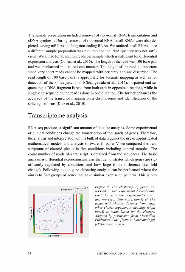
34 M ETHODOLOGIC AL C ONSIDER ATIONS
The sample preparation included removal of ribosomal RNA, fragmentation and cDNA synthesis. During removal of ribosomal RNA, small RNAs were also de-pleted leaving mRNAs and long non-coding RNAs. We omitted small RNAs since a different sample preparation was required and the RNA quantity was not suffi-cient. We aimed for 30 million reads per sample which is sufficient for differential expression analysis (Conesa et al., 2016). The length of the read was 100 base pair and was performed in a paired-end manner. The length of the read is important since very short reads cannot be mapped with certainty and are discarded. The read length of 100 base pairs is appropriate for accurate mapping as well as for detection of the splice junctions (Chhangawala et al., 2015). In paired-end se-quencing, a DNA fragment is read from both ends in opposite directions, while in single-end sequencing the read is done in one direction. The former enhances the accuracy of the transcript mapping on a chromosome and identification of the splicing isoforms (Katz et al., 2010).
Transcriptome analysis RNA seq produces a significant amount of data for analysis. Some experimental or clinical conditions change the transcription of thousands of genes. Therefore, the analysis and interpretation of this bulk of data requires the use of sophisticated mathematical models and analysis software. In paper V we compared the tran-scriptome of choroid plexus in five conditions including control samples. The count number of reads of a transcript is obtained from the sequencer. The basic analysis is differential expression analysis that demonstrates which genes are sig-nificantly regulated by conditions and how large is the difference (i.e. fold change). Following this, a gene clustering analysis can be performed where the aim is to find groups of genes that have similar expression patterns. This is per-
Figure 8. The clustering of genes ex-pressed in two experimental conditions. Each dot represents a gene and x and y axis represent their expression level. The genes with shorter distance from each other cluster together. A heatmap (right panel) is made based on the clusters. Adapted by permission from Macmillan Publishers Ltd: [Nature biotechnology] (D'Haeseleer, 2005)

M ETHODO LOGIC AL C ONSIDERATIONS 35
formed by calculating the mathematical distance between them. As shown in fig-ure 8, the genes that are closer together form a cluster (D'Haeseleer, 2005). Then, these groups of genes with similar expression patterns can be further ana-lyzed by functionally annotating the gene clusters. This is performed by GO on-tology profiling of genes. In the GO ontology database, each gene has been assigned a GO term (e.g. a biological pathway/function such as apoptosis) based on the knowledge or predictive methods (Yon Rhee et al., 2008). By mathemati-cally comparing the number of significantly regulated genes that are related to a GO term in a dataset (e.g. 4 out of 1000 genes related to X term in our dataset) with all genes related to that GO term in a species (e.g. 500 out of 2300 mouse genes related to X term), an enrichment score and a significance value is obtained. We used DAVID Functional Annotation Tool v 6.8 for this purpose (Huang da et al., 2009b, a). The GO ontology is a useful tool to gain an overall biological interpretation about the regulated genes in a dataset. However, this tool does not take into account the interactions between various gene products in a biological system and direction-ality of these interactions. Therefore, we used the Ingenuity Pathway Analysis (IPA) which is a knowledge-based platform for analysis of omics data. The IPA knowledge database is constructed and continuously updated by curators, which extract all the information about the interactions between biological molecules from research studies and their relationship to biological pathways and functions. Based on this database and mathematical models, IPA predicts what biological pathways are regulated (up or down) by the gene changes in the dataset. Moreover, upstream or downstream regulators of pathways are also predicted.
In vitro model of neutrophil transmigration through choroid plexus Modelling choroid plexus in vitro has been a challenge for several reasons:
- choroid plexus consists of various cell types and a stroma - choroid plexus is a highly polarized tissue - choroid plexus epithelial cell barrier function should be mimicked - choroid plexus of rodents such as mouse and rat is a small tissue, which
means several animals need to be sacrificed for an in vitro experiment The current choroid plexus in vitro models include a monolayer of epithelial cells

36 M ETHODOLOGIC AL C ONSIDER ATIONS
(choroid plexus epithelial cells; CPEC), the principal cell component of the cho-roid plexus tissue. To date, there is no co-culture model of choroid plexus epithe-lial and endothelial cells that comprises the choroid plexus vessels. However, most other properties of the choroid plexus can be recapitulated in vitro. For CPEC culture, both primary and immortalized cells have been used. However, the im-mortalized cell lines fail to reproduce some critical functions and characteristics of the choroid plexus such as the barrier function (Kläs et al., 2010; Lazarevic and Engelhardt, 2016). Therefore, in Paper V we used a method of primary culture of neonatal rat CPEC which was first described by (Tsutsumi et al., 1989) and later was optimized by (Strazielle and Ghersi-Egea, 1999). In this method, the choroid plexus tissue is first enzymatically and mechanically dissociated and then the ep-ithelial cells are separated by sedimentation and differential adhesion to a plastic surface. The isolated cells are grown for approximately one week on a filter coated with extracellular matrix protein, laminin. For leukocyte trafficking studies the filter insert is inverted and cells are seeded before placing it back to its position in the well (Fig. 9). Before the transmigration experiment, the effect of different dos-ages of P3C on the CPEC permeability was assessed to insure the integrity of the barrier.
We isolated leukocytes from neonatal rat blood after hypertonic lysis of red blood cells. The leukocytes were added to the basal side of the choroid plexus and were allowed to transmigrate for five hours. The transmigrated cells were then collected from the apical side by centrifuging the medium and the cells were counted in a Bürker chamber or fixed on a slide for immunocytochemistry.
Statistics In general, we used Student's t-test when comparing two groups of samples and one-way analysis of variance (ANOVA) together with a post-hoc test when com-paring multiple groups. The significance was set at p<0.05. These tests are based on the assumption that the sample populations follow a Gaussian distribution.
Figure 9. Inverted and non-inverted model of choroid plexus epithelial cell culture on the filter insert.

M ETHODO LOGIC AL C ONSIDERATIONS 37
However, the tests perform well even if the distribution is not ideally normal es-pecially for large sample size (Lumley et al., 2002). Therefore, we did not test for normal distribution. The sample size for the HI experiments was fairly large and based on our previous studies and understanding of the injury variability. The data are presented as mean ± SEM or mean ± SD.
38 R ESULTS SUM M AR Y
Results summary Systemic activation of viral (TLR3) or bacterial (TLR2) receptors aggravates HI brain injury in ne-onatal mice Infection and inflammation in the perinatal period is a major risk factor for brain injury in newborns (Hagberg et al., 2015). Our group previously showed for the first time that systemic activation of TLR4, a receptor for bacterial LPS, worsens the HI injury in the neonatal rats (Eklind et al., 2001). TLR4 is the main immune receptor for gram-negative bacteria. However, a large proportion of perinatal in-fections are caused by gram-positive bacteria and mycoplasma (Tita and Andrews, 2010) for which TLR2 plays a key role in recognition and in the inflammatory response (Takeuchi et al., 1999). Moreover, viral infections are another cause of inflammation in the perinatal period (Silasi et al., 2015) with TLR3 being critical for initiating the response. Therefore, we asked whether systemic activation of these immune receptors impacts on the HI brain injury. We used a neonatal mouse model of HI at an age corresponding to brain development in near term infants. Poly I:C and P3C were used as agonists for TLR3 and TLR2 respectively. Sys-temic administration of TLR3 and TLR2 agonists 14h prior to HI increased the neuronal tissue (MAP2+ tissue loss) loss by 91% (Paper I) and 46% (Paper II) respectively compared to the control group which received saline. Moreover, TLR3 stimulation increased the white matter (MBP+ tissue loss) injury at hippo-campal and striatal levels by 430% and 140% respectively (Paper I). TLR2 acti-vation also increased the total subcortical white matter injury by 32% (Paper II). Neonatal mice lacking the TLR3 adapter molecules TRIF were protected against the sensitizing effect of PolyI:C (Paper I). Likewise, TLR2 knockout mice were protected against the sensitizing effect of P3C (Paper II). We also asked whether specific regions of the brain were more vulnerable to P3C-sensitized HI injury. Neuropathology scoring did not show a regional specificity of P3C sensitization (Fig. 10A). However, TLR2 knock out mice were most pro-tected in the cerebral cortex (Fig. 10B).
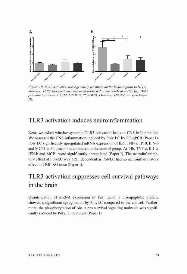
R ESULT S SUM M AR Y 39
TLR3 activation induces neuroinflammation Next, we asked whether systemic TLR3 activation leads to CNS inflammation. We assessed the CNS inflammation induced by Poly I:C by RT-qPCR (Paper I). Poly I:C significantly upregulated mRNA expression of IL6, TNF-a, IP10, IFN-b and MCP1 at 6h time point compared to the control group. At 14h, TNF-a, IL1-a, IFN-b and MCP1 were significantly upregulated (Paper I). The neuroinflamma-tory effect of PolyI:C was TRIF dependent as PolyI:C had no neuroinflammatory effect in TRIF KO mice (Paper I).
TLR3 activation suppresses cell survival pathways in the brain Quantification of mRNA expression of Fas ligand, a pro-apoptotic protein, showed a significant upregulation by PolyI:C compared to the control. Further-more, the phosphorylation of Akt, a pro-survival signaling molecule was signifi-cantly reduced by PolyI:C treatment (Paper I).
Figure 10. TLR2 activation homogenously sensitizes all the brain regions to HI (A), however, TLR2 knockout mice are most protected in the cerebral cortex (B). Data presented as mean ± SEM. *P<0.05, **p<0.01, One-way ANOVA, n= (see Paper II).

40 R ESULTS SUM M AR Y
TLR3 and TLR2 stimulation alter the microglia phenotype Microglia cells are the most abundant professional immune cells in the brain pa-renchyma, and develop different phenotypes in response to stimuli including a M1-like pro-inflammatory and M2-like reparative phenotype. Therefore, we asked whether TLR stimulation alters the microglia phenotype using flow cytom-etry on the brain single cell suspension (Paper I) and qPCR on isolated microglia. Poly I:C reduced the proportion of microglia with a reparative M2 phenotype. Likewise, neonatal mice treated with Poly I:C and exposed to HI had a signifi-cantly lower number of M2 microglia cells in the contralateral hemisphere com-pared to control group. In the ipsilateral hemisphere Poly I:C+HI mice also had a lower number of M2 microglia than saline+HI mice, although the difference was not statistically significant (Paper I). On the other hand, microglia cells isolated from brain by magnetic-activated cell sorting at 14h after P3C treatment showed a mixed inflammatory phenotype char-acterized by upregulation of mRNA expression of both pro-inflammatory cyto-kines (e.g. IL1a, IL1b, TNFa ) and anti-inflammatory cytokines (e.g. IL10) (data not shown). Moreover, the number of microglia cells increased after HI in the ipsilateral hem-isphere, however, prior PolyI:C (Paper I) or P3C (Fig. 11A) treatment did not af-fect the microgliosis. TLR2 deficiency also had no effect on microgliosis after P3C/saline+HI (Fig. 11B).
Figure 11. Number of Iba-1+ microglia cells increase in ipsilateral hemisphere after HI. Microgliosis is not affected by prior P3C treatment (A) or TLR2 deficiency (B). ). Data presented as mean ± SEM. *P<0.05, **p<0.01, ****p<0.0001, One-way ANOVA, n= (see in Paper II).

R ESULT S SUM M AR Y 41
Systemic TLR2 activation suppresses brain mito-chondrial respiration HI brain injury is primarily induced by general energy failure. Therefore, we asked whether systemic inflammation alters the function of the cellular powerhouse, the mitochondrion. We used a high-resolution respirometry system to assess respira-tion of mitochondria isolated from the brain 14h after systemic administration of P3C or saline. Oxidative phosphorylation induced by ADP was significantly sup-pressed by 23% in the P3C group compared to the saline group. Moreover, uncou-pled respiration induced by FCCP showed a 21% reduction in the P3C group although the difference was not statistically significant (paper II). We also asked whether P3C-induced systemic inflammation induces a mitochon-drial oxidative stress and change in mitochondrial antioxidant enzymes. There-fore, we performed western blotting to measure the amount of SOD2 and PRX3 from brain-isolated mitochondria 14h after P3C or saline administration. We did not observe any significant change between P3C and control group (Fig. 12).
Figure 12. P3C does not change the protein expression of mitochondrial antioxidant en-zymes SOD2 and PRX3 analyzed 14h after treatment. Representative image of western blotting for SOD2 and PRX3 (left) and the band analysis (right). Data presented as mean±SEM, n=8. No significant difference was observed between groups as assessed by Student t-test.

42 R ESULTS SUM M AR Y
Systemic activation of TLR2 but not TLR4 induces neutrophil and monocyte invasion of the CNS TLR2 and TLR4 are both receptors that are activated by certain bacteria and acti-vation of either leads to phosphorylation and nuclear translocation of transcription factor NF-κB and expression of inflammatory cytokines. Surprisingly, i.p. injec-tion of P3C (TLR2 agonist) but not LPS (TLR4) induced massive leukocyte infil-tration to the CSF of neonatal and adult mice (Paper III). Likewise, P3C injection in neonatal rats caused a similar effect (Paper V). Infiltrating leukocytes were de-tected in the CSF as early as 6h after the P3C administration and peaked at 14h in neonatal mice. The number of cells gradually decreased at 24h and 48h time points (Paper III). Two dosages of P3C (1 mg/kg and 5 mg/kg) had similar effect. At the 48h time point, LPS injected at 1mg/kg (but not 0.3 mg/kg) slightly increased the number of leukocytes in the CSF. Flow cytometry revealed that the P3C-induced CSF-infiltrating leukocytes in neonatal mice were mainly neutrophils at 6h and inflammatory monocytes at 14h and 24h time points (Paper III). However, in ne-onatal rats infiltrating leukocytes were mainly neutrophils at 14h, as assessed by immunocytochemistry (Paper V). To understand the striking difference between P3C and LPS, we first hypothesized that P3C at the administered dosages induced a stronger inflammatory response than LPS. We measured 23 cytokines in the plasma and spleen and found that LPS at either high or low dosage induced higher or similar level of cytokines as P3C 6h after injection. Therefore, we asked if P3C induces a specific neuroinflamma-tory profile. Multiplex cytokine assay showed significant release of IL1a, IL12, KC, MCP1, MIP1a, MIP1b and G-CSF in the brain 6h after administration of 5 mg/kg P3C. High dose LPS, on the other hand, induced significant release of G-CSF, IL6, KC, MCP1 and RANTES in the brain, while cytokine induction by low dose LPS was not significantly regulated (Paper III). Next, we asked if P3C administration changed the CNS barrier permeability. To achieve this, we injected 14C-sucrose intraperitoneally following P3C treatment and measured the accumulation rates of sucrose from blood to different CNS com-partments including the CSF to assess changes in CNS barrier permeability. P3C increased the permeability of both the BBB and BCSFB 10h after administration (Paper III). The effect of P3C on BSCFB permeability was also tested and repro-duced in neonatal rats (Paper V).

R ESULT S SUM M AR Y 43
Choroid plexus is a route of TLR2-induced leuko-cytes trafficking Next, we asked where in the brain the infiltrating leukocytes localize. To address this question, we first performed flow cytometry and found that in P3C-injected neonatal mice there were significantly higher numbers of CD11b+CD45hi infil-trating leukocytes (Paper III). Then, we performed immunohistochemistry using LysM-EGFP mice in which peripheral myeloid cells express GFP. The results showed that infiltrating neutrophils and monocytes localize in periventricular re-gions of the brain including the choroid plexus and median eminence as well as in the subarachnoid space (Paper III). Therefore, we hypothesized that choroid plexus is a route of leukocyte entry. We tested this hypothesis in vivo and in vitro: We first asked whether knocking down TLR2 adapter molecule MyD88 specifi-cally in choroid plexus would inhibit the P3C-induced leukocyte infiltration to the CSF. To address this question, we injected Cre-Tat or saline intracerebroventric-ularly to P3 MyD88-flox mice. In response to P3C injection at P8, mice that had received Cre-TAT demonstrated 59% reduction in the number of infiltrated leu-kocytes in the CSF compared to the mice that received saline (Paper V). To investigate whether leukocytes can transmigrate through the choroid plexus we performed studies in vitro. Leukocytes were isolated from neonatal rats after P3C injection and were added to the monolayer of CPEC pre-treated with P3C or sa-line. Neutrophils could transmigrate through the CPEC pre-treated with P3C but not saline (Paper V). Altogether, the results showed that the choroid plexus is a route of leukocyte entry after P3C administration.
TLR2 induces specific chemotaxis and cytoskele-ton regulating pathways in the choroid plexus To understand the mechanism of leukocyte transmigration through choroid plexus we performed RNA sequencing on choroid plexus isolated from mice 6h and 14h after injection with P3C or LPS. We hypothesized that both P3C and LPS induce an inflammatory response in the choroid plexus; however, genes that are specifi-cally induced by P3C might be responsible for regulating the leukocyte transmi-gration. We selected the dosages of LPS and P3C that induced a similar systemic inflammatory response (Paper II).

44 R ESULTS SUM M AR Y
Differential expression analysis of RNA seq data revealed that both P3C and LPS significantly regulated expression of thousands of genes in the choroid plexus (Fig. 13, paper IV). The number of genes regulated by LPS was higher than by P3C at both time points. There were 832 and 241 genes specifically regulated by P3C at 6h and 14h, respectively (Fig. 13). Gene cluster analysis and Ingenuity Pathway Analysis revealed P3C-specific effects on chemotaxis and cytoskeleton-regulating pathways (Paper IV).
N-acetyl cysteine blocks TLR2-mediated leuko-cyte infiltration to the brain Reactive oxygen species (ROS) have a profound role in immune response as well as in cytoskeleton remodeling (Staiculescu et al., 2014). We asked whether target-ing ROS has an impact on TLR2-mediated leukocyte migration to the CSF. We used N-acetyl cysteine (NAC) to target ROS as it has previously been shown to be effective in neonatal LPS-induced inflammation (Wang Ann Neurol 2007). NAC treatment almost completely blocked leukocyte infiltration to the CSF in neonatal rats treated with P3C (Paper V). Likewise, NAC treatment of CPEC sig-nificantly diminished P3C-mediated neutrophil transmigration. Multiplex cyto-kine assay revealed that the effect of NAC was largely independent of peripheral and central cytokine induction (Paper V).
Figure 13. A Venn diagram showing the number and percentage of genes significantly regulated by P3C or LPS in the choroid plexus at different times after treatment.

DISC USSION 45
Discussion Sensitization of the developing brain to HI by sys-temic inflammation Infection and inflammation are known risk factors for human neonatal brain in-jury. Previous experimental studies also support a contributory role of inflamma-tion in neonatal HI injury (Hagberg et al., 2012; Hagberg et al., 2015). These studies have also shown that the inflammation-induced sensitization of the devel-oping brain to the injury is time-dependent and dose-dependent. In other words, the effect of the inflammation on the brain injury depends on the temporal stage of the inflammation and its magnitude. For example, when LPS is injected 4h, 6h, or 72h before HI in neonatal rats the brain injury is increased (Eklind et al., 2001; Eklind et al., 2005). Likewise, LPS injection 14h before HI on P9 mice exacer-bated the brain injury (Wang et al., 2009a). However, LPS injection 24h before HI leads to a pre-conditioning effect and reduced the injury (Eklind et al., 2005). Furthermore, low dose LPS injected 24h before HI reduces the neuroinflammation and protects the neonatal rat brain while high dose did not (Lin et al., 2009). The preconditioning is a general effect following a subtle insult that protects against a major insult. For example, a period of hypoxia 24h before the HI protects the brain of neonatal rats and improve the long-term outcome of sensory and cognitive func-tions (Gustavsson et al., 2005). The mechanism of the LPS preconditioning is yet to be elucidated; however, some components of cell death/survival pathways such as Bcl-2 family, JNK pathway, trophic factor pathways and inositol triphosphate kinase/Akt pathway has been suggested to be involved (Gustavsson et al., 2007; Mallard and Hagberg, 2007).
Overall, previous experimental studies suggest that inflammation with sufficient strength during an acute phase can sensitize the brain to HI injury. However, less is known about how different inflammatory stimuli sensitize to HI injury. As men-tioned, the effect of the TLR4 agonist LPS on neonatal HI brain injury has been studied. LPS is the major component of the cell wall of gram-negative bacteria. However, gram-positive bacterial infections and viral infections are other im-portant causes of perinatal inflammation that stimulate an inflammatory response via TLR2 and TLR3 respectively. In the present thesis we demonstrated that sys-temic activation of either TLR3 (Paper I) or TLR2 (Paper II) exacerbated the HI brain injury both in the grey and white matter.
While preparing this thesis a study was published by (Falck et al., 2017) showing that systemic activation of TLR2 in neonatal rats also sensitize the brain to HI

46 DISC USSION
injury. Interestingly, the authors found that hypothermia was neuroprotective in P3C+HI model while it had no effect on the LPS+HI model suggesting a patho-gen-dependent mechanism of injury and protection.
How does systemic inflammation sensitize the brain to HI injury? We showed that systemic activation of TLR3 and TLR2 sensitizes the developing brain to HI injury. Both grey matter and white matter were affected. The grey matter tissue loss is the result of neural cell death. White matter loss was examined by loss of area of MBP staining. This may reflect white matter injury due to dam-age of the myelin sheath, axons and/or the oligodendrocytes (Hagberg et al., 2002a; Back and Rosenberg, 2014). In the present thesis detailed anatomical changes of the brain injury were not examined. However, potential mechanisms that may be involved in the injury progression was examined by addressing mainly three aspects:
1. TLR-mediated neuroinflammation We found that both systemic TLR3 and TLR2 stimulation caused inflammation in the brain represented by expression of various inflammatory cytokines and chem-okines, although, the neuroinflammatory profile was highly different between the two TLR agonists. PolyI:C induced interferon-related genes as well as genes reg-ulated by NF-κB transcription factor such as IL1b. On the other hand, P3C induced release of several cytokines and chemokines in the brain leading to the CNS leu-kocyte invasion. One way to investigate experimentally whether these inflamma-tory changes in the brain are essential for sensitization, is to block the communication of systemic inflammation to the brain. We did found that inhibit-ing MyD88 activity in the choroid plexus ameliorated the P3C-induced neuroin-flammation, however, the effect on HI injury remains to be investigated. Likewise, in paper V, we showed that targeting ROS activity by NAC reduced the neuroin-flammation induced by P3C. However, we did not assess the effect of NAC on the HI injury. What factors other than neuroinflammation may underlie the sensitization effect? One possible mechanism could be inflammation-induced effects on the cerebral blood flow. However, a previous study did not find that LPS affects the cerebral blood flow in an LPS+HI model in neonatal rats (Eklind et al., 2001). Another possible mechanism might be the changes in body temperature induced by sys-temic inflammation, since hyperthermia is associated with activation of apoptotic

DISC USSION 47
pathways in the developing brain (Khan and Brown, 2002) and increased brain injury in adult stroke (Ginsberg and Busto, 1998). Conversely, hypothermia is neuroprotective in human neonates with HIE and animal models (Davidson et al., 2015). The neuroprotective effect of hypothermia might also affect neuroinflam-mation. However, P3C administration did not change the rectal temperature dur-ing HI, ruling out the possibility of hyperthermia-induced sensitization (Paper II). This is in agreement with the study by Falck et al where P3C had no effect on neonatal rat rectal temperature, however, hypothermia protected against the sen-sitized brain injury. Other systemic effects of inflammation on lung, kidney and liver function might also affect circulation metabolites, nutrients and gases that can play a role in sensitizing HI injury. However, it can still be assumed that neu-roinflammation is the key component.
2. Neuroinflammation and cell death pathways Does neuroinflammation sensitize neurons to injury? First, it should be considered that microglia and astrocytes play key roles in inducing a neurotoxic inflammatory milieu. We found a switch in microglia phenotype by PolyI:C and P3C. It was recently discovered that microglia release of TNF, IL1a and C1q induces a neuro-toxic phenotype of astrocyte (called A1), although the secreted neurotoxin by A1 astrocytes remain to be identified. (Liddelow et al., 2017). In our study, PolyI:C induced TNFa mRNA upregulation in the brain. P3C, on the other hand, increased the mRNA expression of both TNFa and IL1a in the microglia and IL1a protein in the whole brain. Therefore, it is plausible that the neuroinflammation-induced sensitization might be partly due to microglia-dependent induction of A1 astro-cytes. In support, it has been shown that microglia are required for LPS-induced oligodendrocyte injury and hypomyelination in the developing rat brain (Lehnardt et al., 2002). In addition to aforementioned hypothesis, neuroinflammation might directly affect the cell death pathways. Generally, cell death occurs through three major mechanisms: necrosis, apoptosis and autophagy. The necrotic and apoptotic pathways have some overlaps during HI leading to a “continuum” form of cell death (Northington et al., 2011a). Necrosis was previously considered as a non-programmed accidental cell death mechanism. However, recent advances revealed several regulated forms of necrosis including necroptosis, ferroptosis and parthan-atos (Conrad et al., 2016). Necroptosis and parthanatos has been implicated in neonatal HI injury (Hagberg et al., 2004; Chavez-Valdez et al., 2012). Inhibiting necroptosis pathway by necrostatin reduced neuroinflammation and protected ne-onatal mice against HI brain injury (Northington et al., 2011b). Necroptosis is in-duced by TNF, FAS ligand, TNF-related apoptosis-inducing ligand (TRAIL) as well as TLR3 and TLR4 ligands (Weinlich et al., 2017). We showed that PolyI:C

48 DISC USSION
upregulated the Fas ligand and TNFa expression in the brain (Paper I). P3C also upregulated the expression of TNF mRNA in the choroid plexus, however, it did not significantly change the TNFa protein in the brain. Therefore, there might be a link between TLR3 and TLR2 activation and the necroptosis pathway. Parthanatos is a caspase-independent mechanism of cell death mediated by poly (ADP-ribose)-polymerase (PARP) and involving mitochondrial protein AIF (Fa-tokun et al., 2014). Interestingly, in EAE mice a cholesterol derivative activates PARP1 in microglia and astrocytes in a TLR2-dependent manner suggesting a link between these signaling pathways (Farez et al., 2009). Therefore, TLR2 activation affecting parthanatos in the neonatal brain might be plausible and warrants further investigation. Both TNF and Fas can also trigger apoptosis. Triggering apoptosis by these death receptor ligands as well as by excitotoxicity leads to oligomerization of BAK and BAX mitochondrial proteins which mediates permeabilization of mitochondria outer membrane, release of cytochrome c to the cytosol, formation of apoptosome and activation of the executioner caspases (Fuchs and Steller, 2015). Inhibition of Bax activity by intracerebroventricular injection of a Bax-inhibiting peptide is neuroprotective in neonatal mouse HI (Wang et al., 2010). Therefore, mitochon-dria plays a central role in HI brain injury (Hagberg et al., 2014). TLR3 activation has been associated with apoptosis mediated via death receptors and mitochondria in an endothelial cell line (Sun et al., 2011). Likewise, intrathecal administration of P3C is also associated with neural apoptosis that is mediated by microglia cells (Hoffmann et al., 2007). Therefore, apoptosis priming might be one of the sensi-tizing mechanisms of PolyI:C and P3C. In support, we showed that PolyI:C inhib-its the activation of anti-apoptotic kinase Akt (Paper I). In addition, PolyI:C activated NF-κB signaling (Paper I) that might in turn induce the pro-apoptotic p53 signaling (Fleiss et al., 2015). We also showed mitochondrial dysfunction by P3C in paper II which might prime the HI-induced apoptosis. As it is shown in some conditions, mitochondrial dysfunction precedes apoptosis probably via in-duction of ROS (Green and Leeuwenburgh, 2002; Griffiths et al., 2008; Zanotto-Filho et al., 2011). Moreover, reduction in mitochondrial respiration in cancer cells is associated with increased apoptosis mediated by ROS (Pelicano et al., 2003; Kwong et al., 2007). We did not measure ROS induction in the brain after TLR3 or TLR2 stimulation, and assessment of brain mitochondrial antioxidant enzymes did not show any changes in response to P3C administration which may or may not reflect the oxidative status of the brain. Finally, it is known that HI-induced excitotoxicity is enhanced by prior IL1b exposure (Degos et al., 2013). The expression of this cytokine was significantly upregulated by PolyI:C in the

DISC USSION 49
brain and by P3C in the choroid plexus (Paper I and II) suggesting another mech-anism of sensitization. Autophagy is a cellular intrinsic mechanism for homeostasis by which undesired macromolecules and organelles undergo lysosomal degradation. Whether autoph-agy is a mechanism or a sequela (or both) of cell death has been a matter of debate in the last few years. However, growing evidence support the hypothesis of au-tophagy as a cell death mechanism (Kroemer and Levine, 2008). Autophagy has been detected in dying thalamic neurons of asphyxiated newborns (Ginet et al., 2014). In adult ischemic brain, however, autophagy has been suggested as a neu-roprotective mechanism (Papadakis et al., 2013). Autophagy also plays a key role in inflammation and immunity (Levine et al., 2011). Although, we did not inves-tigate the effect of TLR3 and TLR2 activation on brain autophagy-related pro-teins, there is evidenc for a link between TLR signaling and autophagy (Shi and Kehrl, 2008). Altogether, neuroinflammation might prime cell death pathways leading to sensitization of the brain to HI.
3. Neuroinflammation and cerebral energy metabolism HI is essentially an energy failure condition. The energy failure occurs in two phases. During the primary insult a transient reduction in cerebral blood flow causes primary energy failure and is followed by a latent phase where metabolism recovers, as assessed by measurement of high-energy phosphates by magnetic resonance spectroscopy in asphyxiated human newborns (Hope et al., 1984) and piglets (Lorek et al., 1994). The latent phase is followed by secondary energy fail-ure, the mechanism of which is not fully understood; however, excitotoxicity, mi-tochondrial dysfunction, inflammation and oxidative stress have been implicated (Allen and Brandon, 2011; Hagberg et al., 2014). In paper II we showed that sys-temic activation of TLR2 impairs complex-I mediated mitochondrial respiration. The respirometry was performed on mitochondria isolated from the brain, at the time when HI would have been induced. In other words, our results suggest that mitochondria are already dysfunctional at the time of primary energy failure. Therefore, TLR2 activation might have an accumulative effect on the HI-induced primary energy failure. Moreover, TLR-2 mediated mitochondrial dysfunction might potentiate the secondary energy failure as well. These hypotheses could be tested by measuring e.g. ATP at several time points after P3C/saline+HI. Further-more, we did not investigate the underlying cause of mitochondria dysfunction, nor did we test the respiratory function of other complexes in the ETC. The enzy-matic activity measurement of different ETC complexes would be a complemen-tary experiment to elucidate the mitochondrial status change by systemic TLR2

50 DISC USSION
activation. Several studies have shown a link between systemic inflammatory con-ditions such as sepsis and mitochondrial dysfunction in the brain (Brealey et al., 2002; d'Avila et al., 2008; Jeger et al., 2013) supporting a potential link between inflammation, mitochondrial function and HI injury.
TLR2-mediated leukocyte trafficking to the CNS through choroid plexus We showed that systemic administration of the TLR2 ligand P3C induces marked infiltration of neutrophils and monocytes into the CNS. By performing several in vivo and in vitro experiments in two animal species, we found that choroid plexus is the most likely route of leukocyte entry into the CNS. Here I discuss some as-pects of these results:
1- What is special about TLR1/2 signalling?
We found that systemic activation of TLR1/2, but not TLR2/6 and TLR4 results in significant leukocyte infiltration to the CSF and periventricular regions of the brain. Previously, we reported that TLR3 activation does not cause the same effect in leukocyte trafficking (Stridh et al., 2013). What is special about activation of TLR1/2? Both TLR2 and TLR4 signal through adapter molecule MyD88 leading to activation and translocation of transcription factor NF-κB leading to the expres-sion of different inflammatory cytokines. TLR4, in addition, signals through an-other adapter molecule, TRIF, leading to activation of interferon-regulating factors (IRFs). IRFs are transcription factors that when activated result in an in-duction of interferon responses. We showed that LPS caused higher or similar level of systemic inflammation as P3C. However, the neuroinflammatory profile was strikingly different. One potential mechanism underlying this difference might be the differential expression of TLRs on immune cells and at the brain interface. TLR2 and TLR4, but not TLR3, are expressed in neutrophils and mon-ocytes of neonatal and adult human and mice (Levy, 2007; Melvan et al., 2010; Dasari et al., 2011; Kollmann et al., 2012; Thomas and Schroder, 2013). In adult human monocytes the expression of TLR2 is higher than TLR4 (Hornung et al., 2002). TLR2-4 mRNA are also expressed on brain endothelial cells as well as in the choroid plexus (Nagyőszi et al., 2010; Mallard, 2012; Stridh et al., 2013). Therefore, it is not likely that different expression patterns of TLRs underlie the different neuroinflammatory effects. A TLR2 specific molecular mechanism must be behind its special effect on leukocyte transmigration. There are some studies comparing the gene expression and metabolic profiles induced by TLR4 versus

DISC USSION 51
TLR2 agonists in leukocyte subtypes (Blankley et al., 2014; Lachmandas et al., 2016). The principal difference observed in the transcriptomic studies was that both TLR2 and TLR4 induce NF-κB-related genes but only TLR4 activation leads to expression of interferon-related genes as expected. Lachmandas et al recently reported that TLR2 and TLR4 stimulation induce different metabolic programs in human monocytes (Lachmandas et al., 2016). However, when the comparison was made between dosages of agonists which induced a similar level of inflammatory response, no significant difference was observed in the metabolic programs. Therefore, it is crucial to consider the dose-dependent nature of responses to dif-ferent TLRs. Hence, in the RNA seq study (Paper IV) we used dosages of LPS and P3C that induce comparable inflammatory responses in the circulation. To our knowledge, no other studies have compared the effect of different TLR agonists on the brain interfaces at whole transcriptome level. However, recently, a qPCR-based study showed that lipoteichoic acid, a TLR2 agonist, downregulated mRNA expression of tight junction gene claudin 5 while LPS upregulated it (Mayerhofer et al., 2017). Moreover, the study showed that both agonists downregulated oc-cludin which is in agreement with our transcriptomic results.
We demonstrated that P3C induces a specific cytokine profile in the choroid plexus although expression of many cytokines was similar between the two ago-nists. Moreover, a cytoskeleton-related pathway was also induced specifically by P3C. Therefore, P3C induces changes at the choroid plexus that potentially are required for the recruitment of leukocytes and facilitation of transmigration.
Reactive oxygen species influence many functions in cells including inflammatory responses (Mittal et al., 2014) and cytoskeleton/tight junctions rearrangement (Schreibelt et al., 2007). We showed that targeting ROS with NAC inhibited P3C-induced leukocyte migration through choroid plexus, which was independent of the peripheral and central cytokine response. Therefore, the results suggest that ROS activity within the migrating cells or epithelial cells at the choroid plexus is required for TLR2-mediated cell trafficking. Altogether, the results provide evi-dence for specific molecular mechanisms downstream of TLR2 such as chemo-taxis and cytoskeleton pathways that might be involved in leukocyte trafficking to the brain.
2- Why does TLR2 activation results in leukocyte migration to the CNS? Not all bacterial infections in the periphery leads to a massive leukocyte migration into the CNS. In fact, the effect of P3C in inducing leukocyte infiltration to the CSF best resembles clinical bacterial meningitis. P3C is a synthetic mimic of lip-oproteins that are present in the bacterial cell wall of both gram-positive and gram-

52 DISC USSION
negative bacteria. Localization and surface projection of these lipoproteins and therefore their associated TLR2 activation is strain-dependent (Kovacs-Simon et al., 2011). Moreover, it was recently demonstrated that bacteria-induced activation of TLR2 is dependent on the shedding of lipoproteins from the bacterial cell wall (Hanzelmann et al., 2016). However, many bacterial strains that activate TLR2 do not cause leukocyte infiltration to the CSF. This difference in response might be due to the diversity of lipoprotein structure (Kovacs-Simon et al., 2011; Nakayama et al., 2012) in different bacterial strains and whether they bind TLR1/2 or TLR2/6 at the brain interfaces, especially at the choroid plexus. Interestingly, some bacte-rial strains can hijack the TLR2 signaling pathway in order to alter the epithelial barrier function and access the target tissue (Chun and Prince, 2009; Clarke et al., 2011). Therefore, there might be an evolutionary competition between the im-mune system and pathogenic bacteria related to the function of TLR2.

C ONC LUSION AND FUTUR E PER SPEC TIVE 53
Conclusion and future perspective
In this thesis, I investigated the effect of systemic inflammation on the developing brain in the context of HI injury (Fig. 14)
We showed for the first time that systemic inflammation induced by intraperito-neal injection of agonists for TLR3 (PolyI:C) and TLR2 (P3C) sensitize the neo-natal mouse brain to subsequent HI injury. TLR3 activation augmented pro-apoptotic and suppressed pro-survival pathways. TLR2 activation, on the other hand, suppressed mitochondrial respiration in the brain. Both agonists induced a phenotypic switch in microglia cells that might compromise their reparative func-tion. We also demonstrated that P3C induces a substantial infiltration of leuko-cytes to the CNS of neonatal mice and rats. Several in vivo and in vitro experiments suggested choroid plexus as the route of the leukocyte entry. By transcriptome analysis of choroid plexus, we demonstrate potential mechanism underlying the P3C-induced leukocyte trafficking including a chemotaxis signa-ture and cytoskeleton reorganization.
Figure 14. Schematic figure summarizing the mechanism of TLR3- (left) and TLR2-induced (right) sensitization of developing brain to HI injury.

54
Finally, we demonstrated that the antioxidant compound, NAC, inhibits the leu-kocyte infiltration without substantially affecting the cytokine/chemokine re-sponse. Infection and inflammation are major risks factors for neonatal HI injury, and this study reveals some mechanisms underlying this association and proposes novel drug targets.
In treatment of pregnancy complication such as perinatal viral infections and as-phyxia, TLR3 might be a potential drug target to attenuate neonatal brain injury. Currently, no TLR3 inhibitor is clinically in use. However, some small-molecule probes have been described with antagonistic effect on TLR3 (Cheng et al., 2011). Moreover, this thesis and a previous study (Stridh et al., 2011) suggest TLR2 as a potential drug target, and some progress has been made in recent years in devel-oping an inhibitor of TLR2 (Mistry et al., 2015; Patra and Choi, 2016). Therefore, the next step in translating our results into a therapy is to test the efficacy of these TLR inhibitors in pre-clinical neonatal models of HI brain injury. Moreover, we showed that NAC blocks the TLR2-mediated leukocyte invasion of the CNS. However, whether NAC diminishes the TLR2-sensitized HI injury remains to be investigated. We previously reported that NAC reduced the brain injury in an LPS+HI model in neonatal mouse, supporting its potential use in inflammation-sensitized brain injury (Wang et al., 2007). Fortunately, the pharmacokinetics of NAC in mothers with chorioamnionitis has been described and its safety has been confirmed in two pilot trials that paves the way for further clinical investigations (Wiest et al., 2014; Jenkins et al., 2016). We also demonstrated that the choroid plexus communicates the peripheral inflammation to the brain. Whether inhibiting this communication alleviates the brain injury is a question that needs to be ad-dressed in future experimental studies. Hence, in the future, choroid plexus-tar-geted therapy might be considered as a strategy in treatment of inflammation-sensitized brain injuries.

AC KNOW LEDGEM ENT 55
Acknowledgement I have been greatly fortunate to receive very kind support and help from many wonderful people during the past years working on this thesis project. I am in-debted to all of them and would love to express my sincere gratitude to them:
First of all my main supervisor, Carina Mallard, for your kind constant support, being an inspiring source of knowledge and motivation as well as patience and humor, especially during some tough days, and for keeping the door always open for questions and discussion.
My co-supervisor, Joakim Ek, for being both a supervisor and a good friend, for your great help in doing different experiments, and your kind generosity in teach-ing me several techniques.
My co-supervisor, Henrik Hagberg for being an inspiration with your wealth of knowledge, dedication and passion for science, and for your educational inputs on the project.
My co-supervisor, Karin Sävman, for your excellent inputs on this thesis and brining clinical insight into our basic researches.
Jean-Francois Ghersi-Egea and Nathalie Strazielle, for your great contribution to this thesis with your excellent expertise and giving me the opportunity to join your lab to learn new things and enjoy the wonderful Lyon!
Xiaoyang Wang, for all insightful talks on the new ideas, your great advices on the projects and future career, and also for giving me the opportunity to gain ex-perience in organizing a course for students.
Anna-Lena Leverin and Pernilla Svedin, for being the best lab managers and kindly teaching me different methods, and being a great help with experiments.
All the co-authors of the papers without whom this thesis wouldn’t exist: Linnea Stridh, Maria Johansson, Raul Chavez Valdez, Frances Northington, Peter Smith, Pernilla Svedin, Syam Nair, Carl-Johan Mohn, Katarina Truvé, San-drine Blondel, Anaid Babikian.
Mona Svedman and Kajsa for your great help in EBM animal facility, Katarina Truvé and Marcela Davila at GU Genomics Core facility, Maria Smedh (the cover image couldn’t be captured without you!) and Massimo Micaroni at Imag-ing Core facility, and Markus Johansson and Oskar Bergström in IT depart-ment.

56
Pete Smith for your kind friendship during these years in Göteborg and all the helps and supports; Syam Nair for being a great office-mate and friend, and shar-ing the mitochondria love; Ana Baburamani and Kristina Sobotka my favorite Aussies for brining joy and laughter anywhere and being just awesome! Special thanks again to Kristina for kindly reading the text and excellent suggestions. Ve-ronica Golubinskaya for being my weekend’s lab-mate and for being the most kind and caring person. Elodie Saudrais and other friends in Lyon for your kind hospitality and all the helps. All other former and current colleagues and friends from our research group and the neighbor groups: Aditi, Agneta, Ali, Alina, Anna, Anna-Maj, Arshed, Aura, Barbara, Cristiano, Dan, Dile, Dimitra, Emelie, Eridan, Gabriella, Gaga, Gisela, Holger, Jacqueline, Jaspal, Jenny, Kerstin, Lisa, Lovisa, Marie, Markus, Maryam, Masako, Massimo, Mats, Min, Nina, Peidi, Peter, Roberto, Ruijin, Sabrina, Sha, Tanya and Xiaoli for making the best work environment with your friendliness and helpfulness!
To my family: Maman, Babajoon, Amir, Farina, Elham, Azadeh and Hamed for your support and love!♥
R EFER ENC ES 57
References Akira S, Uematsu S, Takeuchi O (2006) Pathogen recognition and innate
immunity. Cell 124:783-801. Allen KA, Brandon DH (2011) Hypoxic Ischemic Encephalopathy:
Pathophysiology and Experimental Treatments. Newborn and infant nursing reviews : NAINR 11:125-133.
Andropoulos DB (2012) Appendix B: Pediatric Normal Laboratory Values. In: Gregory's Pediatric Anesthesia, pp 1300-1314: Wiley-Blackwell.
Annane D, Sharshar T (2015) Cognitive decline after sepsis. The Lancet Respiratory Medicine 3:61-69.
Armulik A, Genove G, Mae M, Nisancioglu MH, Wallgard E, Niaudet C, He L, Norlin J, Lindblom P, Strittmatter K, Johansson BR, Betsholtz C (2010) Pericytes regulate the blood-brain barrier. Nature 468:557-561.
Azevedo FAC, Carvalho LRB, Grinberg LT, Farfel JM, Ferretti REL, Leite REP, Filho WJ, Lent R, Herculano-Houzel S (2009) Equal numbers of neuronal and nonneuronal cells make the human brain an isometrically scaled-up primate brain. The Journal of Comparative Neurology 513:532-541.
Babcock AA, Kuziel WA, Rivest S, Owens T (2003) Chemokine Expression by Glial Cells Directs Leukocytes to Sites of Axonal Injury in the CNS. The Journal of Neuroscience 23:7922-7930.
Back SA, Rosenberg PA (2014) Pathophysiology of glia in perinatal white matter injury. Glia:n/a-n/a.
Badawi N, Kurinczuk JJ, Keogh JM, Alessandri LM, O'Sullivan F, Burton PR, Pemberton PJ, Stanley FJ (1998) Intrapartum risk factors for newborn encephalopathy: the Western Australian case-control study. Bmj 317:1554-1558.
Ballabh P, Hu F, Kumarasiri M, Braun A, Nedergaard M (2005) Development of tight junction molecules in blood vessels of germinal matrix, cerebral cortex, and white matter. Pediatric research 58:791-798.
Balusu S, Brkic M, Libert C, Vandenbroucke RE (2016a) The choroid plexus-cerebrospinal fluid interface in Alzheimer's disease: more than just a barrier. Neural Regeneration Research 11:534-537.
Balusu S, Van Wonterghem E, De Rycke R, Raemdonck K, Stremersch S, Gevaert K, Brkic M, Demeestere D, Vanhooren V, Hendrix A, Libert C, Vandenbroucke RE (2016b) Identification of a novel mechanism of blood-brain communication during peripheral inflammation via choroid plexus-derived extracellular vesicles. EMBO Mol Med.
Banks WA (2005) Blood-brain barrier transport of cytokines: a mechanism for neuropathology. Current pharmaceutical design 11:973-984.
Banks WA (2009) Characteristics of compounds that cross the blood-brain barrier. BMC Neurology 9:S3-S3.

58 R EFER ENC ES
Banks WA (2016) From blood-brain barrier to blood-brain interface: new opportunities for CNS drug delivery. Nat Rev Drug Discov 15:275-292.
Banks WA, Lynch JL, Price TO (2009) Cytokines and the blood-brain barrier. In: The Neuroimmunological Basis of Behavior and Mental Disorders, pp 3-17.
Basu S, Agarwal P, Anupurba S, Shukla R, Kumar A (2015) Elevated plasma and cerebrospinal fluid interleukin-1 beta and tumor necrosis factor-alpha concentration and combined outcome of death or abnormal neuroimaging in preterm neonates with early-onset clinical sepsis. Journal of perinatology : official journal of the California Perinatal Association 35:855-861.
Bear JJ, Wu YW (2016) Maternal Infections During Pregnancy and Cerebral Palsy in the Child. Pediatric Neurology 57:74-79.
Becher B, Spath S, Goverman J (2017) Cytokine networks in neuroinflammation. Nat Rev Immunol 17:49-59.
Bell MJ, Hallenbeck JM (2002) Effects of intrauterine inflammation on developing rat brain. Journal of Neuroscience Research 70:570-579.
Bennet L, Tan S, Van den Heuij L, Derrick M, Groenendaal F, van Bel F, Juul S, Back SA, Northington F, Robertson NJ, Mallard C, Gunn AJ (2012) Cell therapy for neonatal hypoxia–ischemia and cerebral palsy. Annals of neurology 71:589-600.
Berfelo FJ, Kersbergen KJ, van Ommen CH, Govaert P, van Straaten HL, Poll-The BT, van Wezel-Meijler G, Vermeulen RJ, Groenendaal F, de Vries LS, de Haan TR (2010) Neonatal cerebral sinovenous thrombosis from symptom to outcome. Stroke 41:1382-1388.
Beura LK, Hamilton SE, Bi K, Schenkel JM, Odumade OA, Casey KA, Thompson EA, Fraser KA, Rosato PC, Filali-Mouhim A, Sekaly RP, Jenkins MK, Vezys V, Haining WN, Jameson SC, Masopust D (2016) Normalizing the environment recapitulates adult human immune traits in laboratory mice. Nature 532:512-516.
Bi D, Qiao L, Bergelson I, Ek CJ, Duan L, Zhang X, Albertsson AM, Pettengill M, Kronforst K, Ninkovic J, Goldmann D, Janzon A, Hagberg H, Wang X, Mallard C, Levy O (2015) Staphylococcus epidermidis Bacteremia Induces Brain Injury in Neonatal Mice via Toll-like Receptor 2-Dependent and -Independent Pathways. The Journal of infectious diseases 212:1480-1490.
Blankley S, Graham CM, Howes A, Bloom CI, Berry MPR, Chaussabel D, Pascual V, Banchereau J, Lipman M, O’Garra A (2014) Identification of the Key Differential Transcriptional Responses of Human Whole Blood Following TLR2 or TLR4 Ligation In-Vitro. PloS one 9:e97702.
Boggs JM (2006) Myelin basic protein: a multifunctional protein. Cellular and molecular life sciences : CMLS 63:1945-1961.
Bohacek I, Cordeau P, Lalancette-Hebert M, Gorup D, Weng YC, Gajovic S, Kriz J (2012) Toll-like receptor 2 deficiency leads to delayed exacerbation of ischemic injury. Journal of neuroinflammation 9:191.

R EFER ENC ES 59
Bona E, Andersson A-L, Blomgren K, Gilland E, Puka-Sundvall M, Gustafson K, Hagberg H (1999) Chemokine and Inflammatory Cell Response to Hypoxia-Ischemia in Immature Rats. Pediatric research 45:500-509.
Bonadio J (2016) Premature Birth and the Fetal Inflammatory Response Syndrome. NeoReviews 17:e285-e289.
Borkowski J, Li L, Steinmann U, Quednau N, Stump-Guthier C, Weiss C, Findeisen P, Gretz N, Ishikawa H, Tenenbaum T, Schroten H, Schwerk C (2014) Neisseria meningitidis elicits a pro-inflammatory response involving IkappaBzeta in a human blood-cerebrospinal fluid barrier model. Journal of neuroinflammation 11:163.
Brealey D, Brand M, Hargreaves I, Heales S, Land J, Smolenski R, Davies NA, Cooper CE, Singer M (2002) Association between mitochondrial dysfunction and severity and outcome of septic shock. The Lancet 360:219-223.
Brenner D, Blaser H, Mak TW (2015) Regulation of tumour necrosis factor signalling: live or let die. Nat Rev Immunol 15:362-374.
Bsibsi M, Ravid R, Gveric D, van Noort JM (2002) Broad Expression of Toll-Like Receptors in the Human Central Nervous System. Journal of Neuropathology & Experimental Neurology 61:1013-1021.
Budday S, Steinmann P, Kuhl E (2015) Physical biology of human brain development. Frontiers in Cellular Neuroscience 9.
Burchell SR, Dixon BJ, Tang J, Zhang JH (2013) Isoflurane Provides Neuroprotection in Neonatal Hypoxic Ischemic Brain Injury. Journal of investigative medicine : the official publication of the American Federation for Clinical Research 61:1078-1083.
Cameron JS, Alexopoulou L, Sloane JA, DiBernardo AB, Ma Y, Kosaras B, Flavell R, Strittmatter SM, Volpe J, Sidman R, Vartanian T (2007) Toll-Like Receptor 3 Is a Potent Negative Regulator of Axonal Growth in Mammals. The Journal of Neuroscience 27:13033-13041.
Chakravarty S, Herkenham M (2005) Toll-like receptor 4 on nonhematopoietic cells sustains CNS inflammation during endotoxemia, independent of systemic cytokines. The Journal of neuroscience : the official journal of the Society for Neuroscience 25:1788-1796.
Chavez-Valdez R, Martin LJ, Northington FJ (2012) Programmed Necrosis: A Prominent Mechanism of Cell Death following Neonatal Brain Injury. Neurol Res Int 2012:257563.
Chen H, Burris M, Fajilan A, Spagnoli F, Tang J, Zhang JH (2011) Prolonged exposure to isoflurane ameliorates infarction severity in the rat pup model of neonatal hypoxia-ischemia. Transl Stroke Res 2:382-390.
Cheng K, Wang X, Yin H (2011) Small-molecule inhibitors of the TLR3/dsRNA complex. Journal of the American Chemical Society 133:3764-3767.
Chhangawala S, Rudy G, Mason CE, Rosenfeld JA (2015) The impact of read length on quantification of differentially expressed genes and splice junction detection. Genome Biology 16:131.
Chhor V, Le Charpentier T, Lebon S, Oré M-V, Celador IL, Josserand J, Degos V, Jacotot E, Hagberg H, Sävman K, Mallard C, Gressens P, Fleiss B

60 R EFER ENC ES
(2013) Characterization of phenotype markers and neuronotoxic potential of polarised primary microglia in vitro. Brain, Behavior, and Immunity 32:70-85.
Choi JY, Cui Y, Kang YM, Kim J-H, Lee SJ, Kim BG Role of toll-like receptor 2 in ischemic demyelination and oligodendrocyte death. Neurobiology of Aging.
Christensen RD, Baer VL, Gordon PV, Henry E, Whitaker C, Andres RL, Bennett ST (2012) Reference Ranges for Lymphocyte Counts of Neonates: Associations Between Abnormal Counts and Outcomes. Pediatrics 129:e1165-e1172.
Chun J, Prince A (2009) TLR2-induced calpain cleavage of epithelial junctional proteins facilitates leukocyte transmigration. Cell host & microbe 5:47-58.
Chung W-S, Clarke LE, Wang GX, Stafford BK, Sher A, Chakraborty C, Joung J, Foo LC, Thompson A, Chen C, Smith SJ, Barres BA (2013) Astrocytes mediate synapse elimination through MEGF10 and MERTK pathways. Nature 504:394-400.
Clarke TB, Francella N, Huegel A, Weiser JN (2011) Invasive bacterial pathogens exploit TLR-mediated downregulation of tight junction components to facilitate translocation across the epithelium. Cell host & microbe 9:404-414.
Comim CM, Vilela MC, Constantino LS, Petronilho F, Vuolo F, Lacerda-Queiroz N, Rodrigues DH, da Rocha JL, Teixeira AL, Quevedo J, Dal-Pizzol F (2011) Traffic of leukocytes and cytokine up-regulation in the central nervous system in sepsis. Intensive care medicine 37:711-718.
Conesa A, Madrigal P, Tarazona S, Gomez-Cabrero D, Cervera A, McPherson A, Szcześniak MW, Gaffney DJ, Elo LL, Zhang X, Mortazavi A (2016) A survey of best practices for RNA-seq data analysis. Genome Biology 17:13.
Conrad M, Angeli JPF, Vandenabeele P, Stockwell BR (2016) Regulated necrosis: disease relevance and therapeutic opportunities. Nat Rev Drug Discov 15:348-366.
Craig A, Ling Luo N, Beardsley DJ, Wingate-Pearse N, Walker DW, Hohimer AR, Back SA (2003) Quantitative analysis of perinatal rodent oligodendrocyte lineage progression and its correlation with human. Exp Neurol 181:231-240.
Cros J, Cagnard N, Woollard K, Patey N, Zhang S-Y, Senechal B, Puel A, Biswas SK, Moshous D, Picard C, Jais J-P, D'Cruz D, Casanova J-L, Trouillet C, Geissmann F (2010) Human CD14dim Monocytes Patrol and Sense Nucleic Acids and Viruses via TLR7 and TLR8 Receptors. Immunity 33:375-386.
d'Avila JC, Santiago AP, Amancio RT, Galina A, Oliveira MF, Bozza FA (2008) Sepsis induces brain mitochondrial dysfunction. Critical care medicine 36:1925-1932.
D'Haeseleer P (2005) How does gene expression clustering work? Nat Biotech 23:1499-1501.

R EFER ENC ES 61
D'Mello C, Le T, Swain MG (2009) Cerebral Microglia Recruit Monocytes into the Brain in Response to Tumor Necrosis Factorα Signaling during Peripheral Organ Inflammation. The Journal of Neuroscience 29:2089-2102.
Dal-Pizzol F, Tomasi CD, Ritter C (2014) Septic encephalopathy: does inflammation drive the brain crazy? Revista brasileira de psiquiatria (Sao Paulo, Brazil : 1999) 36:251-258.
Dammann O, Leviton A (1997) Maternal Intrauterine Infection, Cytokines, and Brain Damage in the Preterm Newborn. Pediatric research 42:1-8.
Dasari P, Zola H, Nicholson IC (2011) Expression of Toll-like receptors by neonatal leukocytes. Pediatric allergy and immunology : official publication of the European Society of Pediatric Allergy and Immunology 22:221-228.
Davidson JO, Wassink G, van den Heuij LG, Bennet L, Gunn AJ (2015) Therapeutic Hypothermia for Neonatal Hypoxic–Ischemic Encephalopathy – Where to from Here? Frontiers in Neurology 6:198.
Degos V, Peineau S, Nijboer C, Kaindl AM, Sigaut S, Favrais G, Plaisant F, Teissier N, Gouadon E, Lombet A, Saliba E, Collingridge GL, Maze M, Nicoletti F, Heijnen C, Mantz J, Kavelaars A, Gressens P (2013) G protein–coupled receptor kinase 2 and group I metabotropic glutamate receptors mediate inflammation-induced sensitization to excitotoxic neurodegeneration. Annals of neurology 73:667-678.
Dehmelt L, Halpain S (2005) The MAP2/Tau family of microtubule-associated proteins. Genome Biology 6:204-204.
Deleidi M, Hallett PJ, Koprich JB, Chung C-Y, Isacson O (2010) The Toll-Like Receptor-3 Agonist Polyinosinic:Polycytidylic Acid Triggers Nigrostriatal Dopaminergic Degeneration. The Journal of Neuroscience 30:16091-16101.
DePaolo RW, Kamdar K, Khakpour S, Sugiura Y, Wang W, Jabri B (2012) A specific role for TLR1 in protective T(H)17 immunity during mucosal infection. The Journal of experimental medicine 209:1437-1444.
Depaolo RW, Tang F, Kim I, Han M, Levin N, Ciletti N, Lin A, Anderson D, Schneewind O, Jabri B (2008) Toll-like receptor 6 drives differentiation of tolerogenic dendritic cells and contributes to LcrV-mediated plague pathogenesis. Cell host & microbe 4:350-361.
deVeber G, Andrew M, Adams C, Bjornson B, Booth F, Buckley DJ, Camfield CS, David M, Humphreys P, Langevin P, MacDonald EA, Gillett J, Meaney B, Shevell M, Sinclair DB, Yager J (2001) Cerebral sinovenous thrombosis in children. The New England journal of medicine 345:417-423.
Ditelberg JS, Sheldon RA, Epstein CJ, Ferriero DM (1996) Brain injury after perinatal hypoxia-ischemia is exacerbated in copper/zinc superoxide dismutase transgenic mice. Pediatric research 39:204-208.
Dommergues MA, Patkai J, Renauld JC, Evrard P, Gressens P (2000) Proinflammatory cytokines and interleukin-9 exacerbate excitotoxic

62 R EFER ENC ES
lesions of the newborn murine neopallium. Annals of neurology 47:54-63.
Dong Y, Speer CP (2014) Late-onset neonatal sepsis: recent developments. Archives of Disease in Childhood - Fetal and Neonatal Edition.
Dziegielewska KM, Ek J, Habgood MD, Saunders NR (2001) Development of the choroid plexus. Microsc Res Tech 52:5-20.
Edwards AD, Brocklehurst P, Gunn AJ, Halliday H, Juszczak E, Levene M, Strohm B, Thoresen M, Whitelaw A, Azzopardi D (2010) Neurological outcomes at 18 months of age after moderate hypothermia for perinatal hypoxic ischaemic encephalopathy: synthesis and meta-analysis of trial data. BMJ 340.
Ek CJ, D'Angelo B, Lehner C, Nathanielsz P, Li C, Mallard C (2015) Expression of tight junction proteins and transporters for xenobiotic metabolism at the blood-CSF barrier during development in the nonhuman primate (P. hamadryas). Reproductive toxicology (Elmsford, NY) 56:32-44.
Eklind S, Mallard C, Arvidsson P, Hagberg H (2005) Lipopolysaccharide induces both a primary and a secondary phase of sensitization in the developing rat brain. Pediatric research 58:112-116.
Eklind S, Mallard C, Leverin A-L, Gilland E, Blomgren K, Mattsby-Baltzer I, Hagberg H (2001) Bacterial endotoxin sensitizes the immature brain to hypoxic–ischaemic injury. European Journal of Neuroscience 13:1101-1106.
Engelhardt B, Ransohoff RM (2012) Capture, crawl, cross: the T cell code to breach the blood–brain barriers. Trends in Immunology 33:579-589.
Falck M, Osredkar D, Maes E, Flatebo T, Wood TR, Sabir H, Thoresen M (2017) Hypothermic Neuronal Rescue from Infection-Sensitised Hypoxic-Ischaemic Brain Injury Is Pathogen Dependent. Developmental neuroscience.
Famakin BM, Mou Y, Ruetzler CA, Bembry J, Maric D, Hallenbeck JM (2011) Disruption of downstream MyD88 or TRIF Toll-like receptor signaling does not protect against cerebral ischemia. Brain Research 1388:148-156.
Farez MF, Quintana FJ, Gandhi R, Izquierdo G, Lucas M, Weiner HL (2009) Toll-like receptor 2 and poly(ADP-ribose) polymerase 1 promote central nervous system neuroinflammation in progressive EAE. Nature immunology 10:958-964.
Fatokun AA, Dawson VL, Dawson TM (2014) Parthanatos: mitochondrial-linked mechanisms and therapeutic opportunities. British journal of pharmacology 171:2000-2016.
Favrais G, Schwendimann L, Gressens P, Lelievre V (2007) Cyclooxygenase-2 mediates the sensitizing effects of systemic IL-1-beta on excitotoxic brain lesions in newborn mice. Neurobiology of disease 25:496-505.
Field R, Campion S, Warren C, Murray C, Cunningham C (2010) Systemic challenge with the TLR3 agonist poly I:C induces amplified IFNα/β and IL-1β responses in the diseased brain and exacerbates chronic neurodegeneration. Brain, Behavior, and Immunity 24:996-1007.

R EFER ENC ES 63
Filias A, Theodorou GL, Mouzopoulou S, Varvarigou AA, Mantagos S, Karakantza M (2011) Phagocytic ability of neutrophils and monocytes in neonates. BMC Pediatrics 11:29-29.
Fleiss B, Gressens P (2012) Tertiary mechanisms of brain damage: a new hope for treatment of cerebral palsy? The Lancet Neurology 11:556-566.
Fleiss B, Tann CJ, Degos V, Sigaut S, Van Steenwinckel J, Schang AL, Kichev A, Robertson NJ, Mallard C, Hagberg H, Gressens P (2015) Inflammation-induced sensitization of the brain in term infants. Developmental medicine and child neurology 57 Suppl 3:17-28.
Forsberg D, Horn Z, Tserga E, Smedler E, Silberberg G, Shvarev Y, Kaila K, Uhlén P, Herlenius E (2016) CO2-evoked release of PGE2 modulates sighs and inspiration as demonstrated in brainstem organotypic culture. eLife 5:e14170.
Fowler CB, Evers DL, O’Leary TJ, Mason JT (2011) Antigen Retrieval Causes Protein Unfolding: Evidence for a Linear Epitope Model of Recovered Immunoreactivity. Journal of Histochemistry and Cytochemistry 59:366-381.
Fuchs Y, Steller H (2015) Live to die another way: modes of programmed cell death and the signals emanating from dying cells. Nat Rev Mol Cell Biol 16:329-344.
Ge W-P, Miyawaki A, Gage FH, Jan YN, Jan LY (2012) Local generation of glia is a major astrocyte source in postnatal cortex. Nature 484:376-380.
Gibson CS, MacLennan AH, Goldwater PN, Haan EA, Priest K, Dekker GA, South Australian Cerebral Palsy Research G (2006) Neurotropic viruses and cerebral palsy: population based case-control study. BMJ : British Medical Journal 332:76-80.
Gilland E, Bona E, Hagberg H (1998a) Temporal changes of regional glucose use, blood flow, and microtubule-associated protein 2 immunostaining after hypoxia-ischemia in the immature rat brain. Journal of cerebral blood flow and metabolism : official journal of the International Society of Cerebral Blood Flow and Metabolism 18:222-228.
Gilland E, Bona E, Hagberg H (1998b) Temporal changes of regional glucose use, blood flow, and microtubule-associated protein 2 immunostaining after hypoxia-ischemia in the immature rat brain. Journal of Cerebral Blood Flow & Metabolism 18:222-228.
Ginet V, Pittet MP, Rummel C, Osterheld MC, Meuli R, Clarke PGH, Puyal J, Truttmann AC (2014) Dying neurons in thalamus of asphyxiated term newborns and rats are autophagic. Annals of neurology:n/a-n/a.
Ginhoux F, Greter M, Leboeuf M, Nandi S, See P, Gokhan S, Mehler MF, Conway SJ, Ng LG, Stanley ER, Samokhvalov IM, Merad M (2010) Fate Mapping Analysis Reveals That Adult Microglia Derive from Primitive Macrophages. Science 330:841-845.
Ginsberg MD, Busto R (1998) Combating hyperthermia in acute stroke: a significant clinical concern. Stroke 29:529-534.
Gofton TE, Young GB (2012) Sepsis-associated encephalopathy. Nat Rev Neurol 8:557-566.
64 R EFER ENC ES
Goldmann T et al. (2016) Origin, fate and dynamics of macrophages at central nervous system interfaces. Nature immunology 17:797-805.
Grabert K, Michoel T, Karavolos MH, Clohisey S, Baillie JK, Stevens MP, Freeman TC, Summers KM, McColl BW (2016) Microglial brain region-dependent diversity and selective regional sensitivities to aging. Nat Neurosci 19:504-516.
Graham HK, Rosenbaum P, Paneth N, Dan B, Lin J-P, Damiano DL, Becher JG, Gaebler-Spira D, Colver A, Reddihough DS, Crompton KE, Lieber RL (2016) Cerebral palsy. Nature Reviews Disease Primers 2:15082.
Green PS, Leeuwenburgh C (2002) Mitochondrial dysfunction is an early indicator of doxorubicin-induced apoptosis. Biochimica et Biophysica Acta (BBA) - Molecular Basis of Disease 1588:94-101.
Gregoriadis G (1993) Liposome technology. In, 2nd Edition. Boca Raton, Fla.: CRC Press.
Grether JK, Nelson KB (1997) Maternal infection and cerebral palsy in infants of normal birth weight. Jama 278:207-211.
Griffiths ER, Friehs I, Scherr E, Poutias DN, McGowan FX, Nido PJd (2008) Abstract 2600: Mitochondria Dysfunction Precedes Cardiomyocyte Apoptosis in the Pressure-Overloaded Neonatal Left Ventricle. Circulation 118:S_750-S_750.
Grontoft O (1954) Intracranial haemorrhage and blood-brain barrier problems in the new-born; a pathologico-anatomical and experimental investigation. Acta pathologica et microbiologica Scandinavica Supplementum 100:8-109.
Gustavsson M, Anderson MF, Mallard C, Hagberg H (2005) Hypoxic Preconditioning Confers Long-Term Reduction of Brain Injury and Improvement of Neurological Ability in Immature Rats. Pediatric research 57:305-309.
Gustavsson M, Wilson MA, Mallard C, Rousset C, Johnston MV, Hagberg H (2007) Global Gene Expression in the Developing Rat Brain After Hypoxic Preconditioning: Involvement of Apoptotic Mechanisms? Pediatric research 61:444-450.
Hagberg H, Peebles D, Mallard C (2002a) Models of white matter injury: comparison of infectious, hypoxic-ischemic, and excitotoxic insults. Mental retardation and developmental disabilities research reviews 8:30-38.
Hagberg H, Gressens P, Mallard C (2012) Inflammation during fetal and neonatal life: Implications for neurologic and neuropsychiatric disease in children and adults. Annals of neurology 71:444-457.
Hagberg H, David Edwards A, Groenendaal F (2016) Perinatal brain damage: The term infant. Neurobiology of disease 92:102-112.
Hagberg H, Mallard C, Rousset CI, Thornton C (2014) Mitochondria: hub of injury responses in the developing brain. The Lancet Neurology 13:217-232.

R EFER ENC ES 65
Hagberg H, Ichord R, Palmer C, Yager JY, Vannucci SJ (2002b) Animal Models of Developmental Brain Injury: Relevance to Human Disease. Developmental neuroscience 24:364-366.
Hagberg H, Mallard C, Ferriero DM, Vannucci SJ, Levison SW, Vexler ZS, Gressens P (2015) The role of inflammation in perinatal brain injury. Nat Rev Neurol 11:192-208.
Hagberg H, Wilson MA, Matsushita H, Zhu C, Lange M, Gustavsson M, Poitras MF, Dawson TM, Dawson VL, Northington F, Johnston MV (2004) PARP-1 gene disruption in mice preferentially protects males from perinatal brain injury. Journal of neurochemistry 90:1068-1075.
Hanzelmann D, Joo H-S, Franz-Wachtel M, Hertlein T, Stevanovic S, Macek B, Wolz C, Gotz F, Otto M, Kretschmer D, Peschel A (2016) Toll-like receptor 2 activation depends on lipopeptide shedding by bacterial surfactants. Nature communications 7.
Hawkins BT, Davis TP (2005) The blood-brain barrier/neurovascular unit in health and disease. Pharmacological reviews 57:173-185.
Heath PT, Jardine LA (2014) Neonatal infections: group B streptococcus. BMJ Clinical Evidence 2014:0323.
Hellstrom Erkenstam N, Smith PL, Fleiss B, Nair S, Svedin P, Wang W, Bostrom M, Gressens P, Hagberg H, Brown KL, Savman K, Mallard C (2016) Temporal Characterization of Microglia/Macrophage Phenotypes in a Mouse Model of Neonatal Hypoxic-Ischemic Brain Injury. Front Cell Neurosci 10:286.
Himmelmann K, Uvebrant P (2014) The panorama of cerebral palsy in Sweden. XI. Changing patterns in the birth-year period 2003-2006. Acta paediatrica (Oslo, Norway : 1992) 103:618-624.
Hofer N, Kothari R, Morris N, Müller W, Resch B (2013) The fetal inflammatory response syndrome is a risk factor for morbidity in preterm neonates. American Journal of Obstetrics and Gynecology 209:542.e541-542.e511.
Hoffmann O, Braun JS, Becker D, Halle A, Freyer D, Dagand E, Lehnardt S, Weber JR (2007) TLR2 mediates neuroinflammation and neuronal damage. Journal of immunology (Baltimore, Md : 1950) 178:6476-6481.
Holmdahl R, Malissen B (2012) The need for littermate controls. European journal of immunology 42:45-47.
Holt Christine E, Schuman Erin M (2013) The Central Dogma Decentralized: New Perspectives on RNA Function and Local Translation in Neurons. Neuron 80:648-657.
Hoogland IC, Houbolt C, van Westerloo DJ, van Gool WA, van de Beek D (2015) Systemic inflammation and microglial activation: systematic review of animal experiments. Journal of neuroinflammation 12:114.
Hope PL, Cady EB, Tofts PS, Hamilton PA, Costello AMD, Delpy DT, Chu A, Reynolds EOR, Wilkie DR (1984) CEREBRAL ENERGY METABOLISM STUDIED WITH PHOSPHORUS NMR SPECTROSCOPY IN NORMAL AND BIRTH-ASPHYXIATED INFANTS. The Lancet 324:366-370.

66 R EFER ENC ES
Hornung V, Rothenfusser S, Britsch S, Krug A, Jahrsdorfer B, Giese T, Endres S, Hartmann G (2002) Quantitative expression of toll-like receptor 1-10 mRNA in cellular subsets of human peripheral blood mononuclear cells and sensitivity to CpG oligodeoxynucleotides. Journal of immunology (Baltimore, Md : 1950) 168:4531-4537.
Hu X, Leak RK, Shi Y, Suenaga J, Gao Y, Zheng P, Chen J (2015) Microglial and macrophage polarization[mdash]new prospects for brain repair. Nat Rev Neurol 11:56-64.
Huang da W, Sherman BT, Lempicki RA (2009a) Bioinformatics enrichment tools: paths toward the comprehensive functional analysis of large gene lists. Nucleic Acids Res 37:1-13.
Huang da W, Sherman BT, Lempicki RA (2009b) Systematic and integrative analysis of large gene lists using DAVID bioinformatics resources. Nature protocols 4:44-57.
Ikeda T, Yang L, Ikenoue T, Mallard C, Hagberg H (2006) Endotoxin-induced hypoxic-ischemic tolerance is mediated by up-regulation of corticosterone in neonatal rat. Pediatric research 59:56-60.
Jeger V, Djafarzadeh S, Jakob SM, Takala J (2013) Mitochondrial function in sepsis. European Journal of Clinical Investigation 43:532-542.
Jenkins DD, Wiest DB, Mulvihill DM, Hlavacek AM, Majstoravich SJ, Brown TR, Taylor JJ, Buckley JR, Turner RP, Rollins LG, Bentzley JP, Hope KE, Barbour AB, Lowe DW, Martin RH, Chang EY (2016) Fetal and Neonatal Effects of N-Acetylcysteine When Used for Neuroprotection in Maternal Chorioamnionitis. The Journal of pediatrics 168:67-76.e66.
Jin MS, Lee J-O (2008) Structures of the Toll-like Receptor Family and Its Ligand Complexes. Immunity 29:182-191.
Jin MS, Kim SE, Heo JY, Lee ME, Kim HM, Paik S-G, Lee H, Lee J-O (2007) Crystal Structure of the TLR1-TLR2 Heterodimer Induced by Binding of a Tri-Acylated Lipopeptide. Cell 130:1071-1082.
Katz Y, Wang ET, Airoldi EM, Burge CB (2010) Analysis and design of RNA sequencing experiments for identifying isoform regulation. Nat Meth 7:1009-1015.
Kaul D, Habbel P, Derkow K, Krüger C, Franzoni E, Wulczyn FG, Bereswill S, Nitsch R, Schott E, Veh R, Naumann T, Lehnardt S (2012) Expression of Toll-Like Receptors in the Developing Brain. PloS one 7:e37767.
Kendall GS, Hirstova M, Horn S, Dafou D, Acosta-Saltos A, Almolda B, Zbarsky V, Rumajogee P, Heuer H, Castellano B, Pfeffer K, Nedospasov SA, Peebles DM, Raivich G (2011) TNF gene cluster deletion abolishes lipopolysaccharide-mediated sensitization of the neonatal brain to hypoxic ischemic insult. Lab Invest 91:328-341.
Kenneson A, Cannon MJ (2007) Review and meta-analysis of the epidemiology of congenital cytomegalovirus (CMV) infection. Reviews in medical virology 17:253-276.
Khan VR, Brown IR (2002) The effect of hyperthermia on the induction of cell death in brain, testis, and thymus of the adult and developing rat. Cell Stress & Chaperones 7:73-90.

R EFER ENC ES 67
Kierdorf K et al. (2013) Microglia emerge from erythromyeloid precursors via Pu.1- and Irf8-dependent pathways. Nat Neurosci 16:273-280.
Kim HM, Park BS, Kim JI, Kim SE, Lee J, Oh SC, Enkhbayar P, Matsushima N, Lee H, Yoo OJ, Lee JO (2007) Crystal structure of the TLR4-MD-2 complex with bound endotoxin antagonist Eritoran. Cell 130:906-917.
Kim KS (2010) Acute bacterial meningitis in infants and children. The Lancet Infectious Diseases 10:32-42.
King IL, Dickendesher TL, Segal BM (2009) Circulating Ly-6C+ myeloid precursors migrate to the CNS and play a pathogenic role during autoimmune demyelinating disease. Blood 113:3190-3197.
Kläs J, Wolburg H, Terasaki T, Fricker G, Reichel V (2010) Characterization of immortalized choroid plexus epithelial cell lines for studies of transport processes across the blood-cerebrospinal fluid barrier. Cerebrospinal Fluid Research 7:11-11.
Knuesel I, Chicha L, Britschgi M, Schobel SA, Bodmer M, Hellings JA, Toovey S, Prinssen EP (2014) Maternal immune activation and abnormal brain development across CNS disorders. Nat Rev Neurol 10:643-660.
Kollmann TR, Levy O, Montgomery RR, Goriely S (2012) Innate Immune Sensing by Toll-like Receptors in Newborns and the Elderly. Immunity 37:771-783.
Kovacs-Simon A, Titball RW, Michell SL (2011) Lipoproteins of bacterial pathogens. Infection and immunity 79:548-561.
Krabbe KS, Reichenberg A, Yirmiya R, Smed A, Pedersen BK, Bruunsgaard H (2005) Low-dose endotoxemia and human neuropsychological functions. Brain, Behavior, and Immunity 19:453-460.
Kroemer G, Levine B (2008) Autophagic cell death: the story of a misnomer. Nature reviews Molecular cell biology 9:1004-1010.
Kulik T, Kusano Y, Aronhime S, Sandler AL, Winn HR (2008) Regulation of cerebral vasculature in normal and ischemic brain. Neuropharmacology 55:281-288.
Kullmann JS, Grigoleit J-S, Wolf OT, Engler H, Oberbeck R, Elsenbruch S, Forsting M, Schedlowski M, Gizewski ER (2014) Experimental human endotoxemia enhances brain activity during social cognition. Social Cognitive and Affective Neuroscience 9:786-793.
Kurinczuk JJ, White-Koning M, Badawi N (2010) Epidemiology of neonatal encephalopathy and hypoxic–ischaemic encephalopathy. Early Human Development 86:329-338.
Kwong JQ, Henning MS, Starkov AA, Manfredi G (2007) The mitochondrial respiratory chain is a modulator of apoptosis. The Journal of Cell Biology 179:1163-1177.
Laborada G, Nesin M (2005) Interleukin-6 and interleukin-8 are elevated in the cerebrospinal fluid of infants exposed to chorioamnionitis. Biology of the neonate 88:136-144.
Lachmandas E, Boutens L, Ratter JM, Hijmans A, Hooiveld GJ, Joosten LA, Rodenburg RJ, Fransen JA, Houtkooper RH, van Crevel R, Netea MG, Stienstra R (2016) Microbial stimulation of different Toll-like receptor

68 R EFER ENC ES
signalling pathways induces diverse metabolic programmes in human monocytes. Nature microbiology 2:16246.
Lake BB et al. (2016) Neuronal subtypes and diversity revealed by single-nucleus RNA sequencing of the human brain. Science (New York, NY) 352:1586-1590.
Lancet T (2016) The unfinished agenda of preterm births. The Lancet 388:2323. Larochelle C, Alvarez JI, Prat A (2011) How do immune cells overcome the
blood–brain barrier in multiple sclerosis? FEBS Letters 585:3770-3780. Lathia JD, Okun E, Tang SC, Griffioen K, Cheng A, Mughal MR, Laryea G,
Selvaraj PK, ffrench-Constant C, Magnus T, Arumugam TV, Mattson MP (2008) Toll-like receptor 3 is a negative regulator of embryonic neural progenitor cell proliferation. The Journal of neuroscience : the official journal of the Society for Neuroscience 28:13978-13984.
Laugesaar R, Kolk A, Tomberg T, Metsvaht T, Lintrop M, Varendi H, Talvik T (2007) Acutely and retrospectively diagnosed perinatal stroke: a population-based study. Stroke 38:2234-2240.
Lazarevic I, Engelhardt B (2016) Modeling immune functions of the mouse blood-cerebrospinal fluid barrier in vitro: primary rather than immortalized mouse choroid plexus epithelial cells are suited to study immune cell migration across this brain barrier. Fluids Barriers CNS 13:2.
Lee J, Croen LA, Backstrand KH, Yoshida CK, Henning LH, Lindan C, Ferriero DM, Fullerton HJ, Barkovich AJ, Wu YW (2005) Maternal and infant characteristics associated with perinatal arterial stroke in the infant. Jama 293:723-729.
Lee S, Chu HX, Kim HA, Real NC, Sharif S, Fleming SB, Mercer AA, Wise LM, Drummond GR, Sobey CG (2014) Effect of a Broad-Specificity Chemokine-Binding Protein on Brain Leukocyte Infiltration and Infarct Development. Stroke.
Lehnardt S, Massillon L, Follett P, Jensen FE, Ratan R, Rosenberg PA, Volpe JJ, Vartanian T (2003) Activation of innate immunity in the CNS triggers neurodegeneration through a Toll-like receptor 4-dependent pathway. Proceedings of the National Academy of Sciences 100:8514-8519.
Lehnardt S, Lachance C, Patrizi S, Lefebvre S, Follett PL, Jensen FE, Rosenberg PA, Volpe JJ, Vartanian T (2002) The toll-like receptor TLR4 is necessary for lipopolysaccharide-induced oligodendrocyte injury in the CNS. The Journal of neuroscience : the official journal of the Society for Neuroscience 22:2478-2486.
Lehtinen Maria K, Zappaterra Mauro W, Chen X, Yang Yawei J, Hill AD, Lun M, Maynard T, Gonzalez D, Kim S, Ye P, D'Ercole AJ, Wong Eric T, LaMantia Anthony S, Walsh Christopher A (2011) The Cerebrospinal Fluid Provides a Proliferative Niche for Neural Progenitor Cells. Neuron 69:893-905.
Levine B, Mizushima N, Virgin HW (2011) Autophagy in immunity and inflammation. Nature 469:323-335.
Levy O (2007) Innate immunity of the newborn: basic mechanisms and clinical correlates. Nat Rev Immunol 7:379-390.

R EFER ENC ES 69
Liddelow SA et al. (2017) Neurotoxic reactive astrocytes are induced by activated microglia. Nature 541:481-487.
Liesz A, Zhou W, Mracskó É, Karcher S, Bauer H, Schwarting S, Sun L, Bruder D, Stegemann S, Cerwenka A, Sommer C, Dalpke AH, Veltkamp R (2011) Inhibition of lymphocyte trafficking shields the brain against deleterious neuroinflammation after stroke. Brain : a journal of neurology 134:704-720.
Lin H-Y, Huang C-C, Chang K-F (2009) Lipopolysaccharide Preconditioning Reduces Neuroinflammation Against Hypoxic Ischemia and Provides Long-Term Outcome of Neuroprotection in Neonatal Rat. Pediatric research 66:254-259.
Liu J, Dietz K, DeLoyht JM, Pedre X, Kelkar D, Kaur J, Vialou V, Lobo MK, Dietz DM, Nestler EJ, Dupree J, Casaccia P (2012) Impaired adult myelination in the prefrontal cortex of socially isolated mice. Nat Neurosci 15:1621-1623.
Liu L, Botos I, Wang Y, Leonard JN, Shiloach J, Segal DM, Davies DR (2008) Structural Basis of Toll-Like Receptor 3 Signaling with Double-Stranded RNA. Science 320:379-381.
Liu L, Oza S, Hogan D, Chu Y, Perin J, Zhu J, Lawn JE, Cousens S, Mathers C, Black RE (2016) Global, regional, and national causes of under-5 mortality in 2000–15: an updated systematic analysis with implications for the Sustainable Development Goals. The Lancet 388:3027-3035.
Lorek A, Takei Y, Cady EB, Wyatt JS, Penrice J, Edwards AD, Peebles D, Wylezinska M, Owen-Reece H, Kirkbride V, et al. (1994) Delayed ("secondary") cerebral energy failure after acute hypoxia-ischemia in the newborn piglet: continuous 48-hour studies by phosphorus magnetic resonance spectroscopy. Pediatric research 36:699-706.
Louveau A, Smirnov I, Keyes TJ, Eccles JD, Rouhani SJ, Peske JD, Derecki NC, Castle D, Mandell JW, Lee KS, Harris TH, Kipnis J (2015) Structural and functional features of central nervous system lymphatic vessels. Nature 523:337-341.
Lumley T, Diehr P, Emerson S, Chen L (2002) The importance of the normality assumption in large public health data sets. Annual review of public health 23:151-169.
Lun MP, Monuki ES, Lehtinen MK (2015) Development and functions of the choroid plexus-cerebrospinal fluid system. Nature reviews Neuroscience 16:445-457.
Maheshwari A (2014) Neutropenia in the Newborn. Current opinion in hematology 21:43-49.
Maldonado RF, Sá-Correia I, Valvano MA (2016) Lipopolysaccharide modification in Gram-negative bacteria during chronic infection. FEMS microbiology reviews 40:480-493.
Mallard C (2012) Innate Immune Regulation by Toll-Like Receptors in the Brain. ISRN Neurology 2012:19.

70 R EFER ENC ES
Mallard C, Hagberg H (2007) Inflammation-induced preconditioning in the immature brain. Seminars in fetal & neonatal medicine 12:280-286.
Mallard C, Wang X (2012) Infection-Induced Vulnerability of Perinatal Brain Injury. Neurology Research International 2012:6.
Mallard C, Vexler ZS (2015a) Modeling Ischemia in the Immature Brain: How Translational Are Animal Models? Stroke 46:3006-3011.
Mallard C, Vexler ZS (2015b) Modeling Ischemia in the Immature Brain. How Translational Are Animal Models? 46:3006-3011.
Mallard C, Welin AK, Peebles D, Hagberg H, Kjellmer I (2003) White matter injury following systemic endotoxemia or asphyxia in the fetal sheep. Neurochemical research 28:215-223.
Marques F, Sousa JC, Correia-Neves M, Oliveira P, Sousa N, Palha JA (2007) The choroid plexus response to peripheral inflammatory stimulus. Neuroscience 144:424-430.
Marques F, Sousa JC, Coppola G, Geschwind DH, Sousa N, Palha JA, Correia-Neves M (2009a) The choroid plexus response to a repeated peripheral inflammatory stimulus. BMC neuroscience 10:135-135.
Marques F, Sousa JC, Coppola G, Falcao AM, Rodrigues AJ, Geschwind DH, Sousa N, Correia-Neves M, Palha JA (2009b) Kinetic profile of the transcriptome changes induced in the choroid plexus by peripheral inflammation. Journal of cerebral blood flow and metabolism : official journal of the International Society of Cerebral Blood Flow and Metabolism 29:921-932.
Marshall JC, Walker PM, Foster DM, Harris D, Ribeiro M, Paice J, Romaschin AD, Derzko AN (2002) Measurement of endotoxin activity in critically ill patients using whole blood neutrophil dependent chemiluminescence. Critical Care 6:342-348.
Mayerhofer R, Frohlich EE, Reichmann F, Farzi A, Kogelnik N, Frohlich E, Sattler W, Holzer P (2017) Diverse action of lipoteichoic acid and lipopolysaccharide on neuroinflammation, blood-brain barrier disruption, and anxiety in mice. Brain Behav Immun 60:174-187.
McIntyre S, Badawi N, Blair E, Nelson KB (2015) Does aetiology of neonatal encephalopathy and hypoxic-ischaemic encephalopathy influence the outcome of treatment? Developmental medicine and child neurology 57 Suppl 3:2-7.
Melvan JN, Bagby GJ, Welsh DA, Nelson S, Zhang P (2010) Neonatal Sepsis and Neutrophil Insufficiencies. International reviews of immunology 29:315-348.
Miller JE, Pedersen LH, Streja E, Bech BH, Yeargin-Allsopp M, Van Naarden Braun K, Schendel DE, Christensen D, Uldall P, Olsen J (2013) Maternal infections during pregnancy and cerebral palsy: a population-based cohort study. Paediatric and perinatal epidemiology 27:542-552.
Miller SP, Ferriero DM (2009) FROM SELECTIVE VULNERABILITY TO CONNECTIVITY: INSIGHTS FROM NEWBORN BRAIN IMAGING. Trends in neurosciences 32:496-505.

R EFER ENC ES 71
Mistry P, Laird MH, Schwarz RS, Greene S, Dyson T, Snyder GA, Xiao TS, Chauhan J, Fletcher S, Toshchakov VY, MacKerell AD, Jr., Vogel SN (2015) Inhibition of TLR2 signaling by small molecule inhibitors targeting a pocket within the TLR2 TIR domain. Proceedings of the National Academy of Sciences of the United States of America 112:5455-5460.
Mittal M, Siddiqui MR, Tran K, Reddy SP, Malik AB (2014) Reactive Oxygen Species in Inflammation and Tissue Injury. Antioxidants & redox signaling 20:1126-1167.
Monier A, Adle-Biassette H, Delezoide AL, Evrard P, Gressens P, Verney C (2007) Entry and distribution of microglial cells in human embryonic and fetal cerebral cortex. Journal of neuropathology and experimental neurology 66:372-382.
Mortazavi M, Griessenauer C, Adeeb N, Deep A, Shahripour R, Loukas M, Tubbs R, Tubbs RS (2013) The choroid plexus: a comprehensive review of its history, anatomy, function, histology, embryology, and surgical considerations. Childs Nerv Syst:1-10.
Mühlfeld C, Nyengaard JR, Mayhew TM (2010) A review of state-of-the-art stereology for better quantitative 3D morphology in cardiac research. Cardiovascular Pathology 19:65-82.
Nagy A (2000) Cre recombinase: the universal reagent for genome tailoring. Genesis (New York, NY : 2000) 26:99-109.
Nagyőszi P, Wilhelm I, Farkas AE, Fazakas C, Dung NTK, Haskó J, Krizbai IA (2010) Expression and regulation of toll-like receptors in cerebral endothelial cells. Neurochemistry international 57:556-564.
Naing ZW, Scott GM, Shand A, Hamilton ST, van Zuylen WJ, Basha J, Hall B, Craig ME, Rawlinson WD (2016) Congenital cytomegalovirus infection in pregnancy: a review of prevalence, clinical features, diagnosis and prevention. Australian and New Zealand Journal of Obstetrics and Gynaecology 56:9-18.
Nakayama H, Kurokawa K, Lee BL (2012) Lipoproteins in bacteria: structures and biosynthetic pathways. FEBS Journal 279:4247-4268.
Nelson KB, Penn AA (2015) Is infection a factor in neonatal encephalopathy? Archives of Disease in Childhood - Fetal and Neonatal Edition 100:F8-F10.
Nelson KB, Dambrosia JM, Grether JK, Phillips TM (1998) Neonatal cytokines and coagulation factors in children with cerebral palsy. Annals of neurology 44:665-675.
Netea MG, van de Veerdonk F, Verschueren I, van der Meer JW, Kullberg BJ (2008) Role of TLR1 and TLR6 in the host defense against disseminated candidiasis. FEMS immunology and medical microbiology 52:118-123.
Neu J, Walker WA (2011) Necrotizing Enterocolitis. The New England journal of medicine 364:255-264.
Northington FJ, Chavez-Valdez R, Martin LJ (2011a) Neuronal Cell Death in Neonatal Hypoxia-Ischemia. Annals of neurology 69:743-758.

72 R EFER ENC ES
Northington FJ, Chavez-Valdez R, Graham EM, Razdan S, Gauda EB, Martin LJ (2011b) Necrostatin decreases oxidative damage, inflammation, and injury after neonatal HI. Journal of Cerebral Blood Flow & Metabolism 31:178-189.
O'Neill LAJ, Golenbock D, Bowie AG (2013) The history of Toll-like receptors [mdash] redefining innate immunity. Nat Rev Immunol 13:453-460.
Obiero CW, Seale AC, Berkley JA (2015) Empiric treatment of neonatal sepsis in developing countries. The Pediatric infectious disease journal 34:659-661.
Paolicelli RC, Bolasco G, Pagani F, Maggi L, Scianni M, Panzanelli P, Giustetto M, Ferreira TA, Guiducci E, Dumas L, Ragozzino D, Gross CT (2011) Synaptic Pruning by Microglia Is Necessary for Normal Brain Development. Science 333:1456-1458.
Papadakis M, Hadley G, Xilouri M, Hoyte LC, Nagel S, McMenamin MM, Tsaknakis G, Watt SM, Drakesmith CW, Chen R, Wood MJA, Zhao Z, Kessler B, Vekrellis K, Buchan AM (2013) Tsc1 (hamartin) confers neuroprotection against ischemia by inducing autophagy. Nat Med 19:351-357.
Paredes MF, James D, Gil-Perotin S, Kim H, Cotter JA, Ng C, Sandoval K, Rowitch DH, Xu D, McQuillen PS, Garcia-Verdugo J-M, Huang EJ, Alvarez-Buylla A (2016) Extensive migration of young neurons into the infant human frontal lobe. Science 354.
Park BS, Song DH, Kim HM, Choi B-S, Lee H, Lee J-O (2009) The structural basis of lipopolysaccharide recognition by the TLR4-MD-2 complex. Nature 458:1191-1195.
Patil J, Matte A, Nissbrandt H, Mallard C, Sandberg M (2016) Sustained Effects of Neonatal Systemic Lipopolysaccharide on IL-1beta and Nrf2 in Adult Rat Substantia Nigra Are Partly Normalized by a Spirulina-Enriched Diet. Neuroimmunomodulation 23:250-259.
Patra MC, Choi S (2016) Recent progress in the development of Toll-like receptor (TLR) antagonists. Expert Opinion on Therapeutic Patents 26:719-730.
Pelicano H, Feng L, Zhou Y, Carew JS, Hileman EO, Plunkett W, Keating MJ, Huang P (2003) Inhibition of Mitochondrial Respiration: A NOVEL STRATEGY TO ENHANCE DRUG-INDUCED APOPTOSIS IN HUMAN LEUKEMIA CELLS BY A REACTIVE OXYGEN SPECIES-MEDIATED MECHANISM. Journal of Biological Chemistry 278:37832-37839.
Pierrat V, Haouari N, Liska A, Thomas D, Subtil D, Truffert P (2005) Prevalence, causes, and outcome at 2 years of age of newborn encephalopathy: population based study. Archives of Disease in Childhood - Fetal and Neonatal Edition 90:F257.
Polglase GR, Miller SL, Barton SK, Kluckow M, Gill AW, Hooper SB, Tolcos M (2014) Respiratory support for premature neonates in the delivery room: effects on cardiovascular function and the development of brain injury. Pediatric research 75:682-688.
R EFER ENC ES 73
Quintana E, Fernandez A, Velasco P, de Andres B, Liste I, Sancho D, Gaspar ML, Cano E (2015) DNGR-1(+) dendritic cells are located in meningeal membrane and choroid plexus of the noninjured brain. Glia 63:2231-2248.
Rakic S, Zecevic N (2003) Early oligodendrocyte progenitor cells in the human fetal telencephalon. Glia 41:117-127.
Reboldi A, Coisne C, Baumjohann D, Benvenuto F, Bottinelli D, Lira S, Uccelli A, Lanzavecchia A, Engelhardt B, Sallusto F (2009) C-C chemokine receptor 6-regulated entry of TH-17 cells into the CNS through the choroid plexus is required for the initiation of EAE. Nature immunology 10:514-523.
Redzic Z (2011a) Molecular biology of the blood-brain and the blood-cerebrospinal fluid barriers: similarities and differences. Fluids and Barriers of the CNS 8:3-3.
Redzic Z (2011b) Molecular biology of the blood-brain and the blood-cerebrospinal fluid barriers: similarities and differences. Fluids Barriers CNS 8:3.
Reemst K, Noctor SC, Lucassen PJ, Hol EM (2016) The Indispensable Roles of Microglia and Astrocytes during Brain Development. Frontiers in Human Neuroscience 10:566.
Reuter S, Moser C, Baack M (2014) Respiratory Distress in the Newborn. Pediatrics in Review 35:417-429.
Riccomagno MM, Kolodkin AL (2015) Sculpting neural circuits by axon and dendrite pruning. Annual review of cell and developmental biology 31:779-805.
Rice JE, 3rd, Vannucci RC, Brierley JB (1981) The influence of immaturity on hypoxic-ischemic brain damage in the rat. Annals of neurology 9:131-141.
Rietdijk CD, van Wezel RJA, Garssen J, Kraneveld AD (2016) Neuronal toll-like receptors and neuro-immunity in Parkinson's disease, Alzheimer's disease and stroke. Neuroimmunology and Neuroinflammation; Vol 3, No 2 (2016).
Rodrigues J, Scaletsky IC, Campos LC, Gomes TA, Whittam TS, Trabulsi LR (1996) Clonal structure and virulence factors in strains of Escherichia coli of the classic serogroup O55. Infection and immunity 64:2680-2686.
Roessmann U, Gambetti P (1986) Astrocytes in the developing human brain. An immunohistochemical study. Acta Neuropathol 70:308-313.
Rolls A, Shechter R, London A, Ziv Y, Ronen A, Levy R, Schwartz M (2007) Toll-like receptors modulate adult hippocampal neurogenesis. Nature cell biology 9:1081-1088.
Sanai N, Nguyen T, Ihrie RA, Mirzadeh Z, Tsai H-H, Wong M, Gupta N, Berger MS, Huang E, Garcia-Verdugo J-M, Rowitch DH, Alvarez-Buylla A (2011) Corridors of migrating neurons in the human brain and their decline during infancy. Nature 478:382-386.
Saunders NR, Liddelow SA, Dziegielewska KM (2012) Barrier Mechanisms in the Developing Brain. Frontiers in Pharmacology 3:46.
74 R EFER ENC ES
Savman K, Blennow M, Gustafson K, Tarkowski E, Hagberg H (1998) Cytokine Response in Cerebrospinal Fluid after Birth Asphyxia. Pediatric research 43:746-751.
Schafer DP, Lehrman EK, Kautzman AG, Koyama R, Mardinly AR, Yamasaki R, Ransohoff RM, Greenberg ME, Barres BA, Stevens B (2012) Microglia sculpt postnatal neural circuits in an activity and complement-dependent manner. Neuron 74:691-705.
Schläger C, Körner H, Krueger M, Vidoli S, Haberl M, Mielke D, Brylla E, Issekutz T, Cabañas C, Nelson PJ, Ziemssen T, Rohde V, Bechmann I, Lodygin D, Odoardi F, Flügel A (2016) Effector T-cell trafficking between the leptomeninges and the cerebrospinal fluid. Nature 530:349-353.
Schmidt AF, Kannan PS, Chougnet CA, Danzer SC, Miller LA, Jobe AH, Kallapur SG (2016) Intra-amniotic LPS causes acute neuroinflammation in preterm rhesus macaques. Journal of neuroinflammation 13:238.
Schmitt C, Strazielle N, Ghersi-Egea JF (2012) Brain leukocyte infiltration initiated by peripheral inflammation or experimental autoimmune encephalomyelitis occurs through pathways connected to the CSF-filled compartments of the forebrain and midbrain. Journal of neuroinflammation 9:187.
Schrag SJ, Farley MM, Petit S, Reingold A, Weston EJ, Pondo T, Hudson Jain J, Lynfield R (2016) Epidemiology of Invasive Early-Onset Neonatal Sepsis, 2005 to 2014. Pediatrics.
Schreibelt G, Kooij G, Reijerkerk A, van Doorn R, Gringhuis SI, van der Pol S, Weksler BB, Romero IA, Couraud P-O, Piontek J, Blasig IE, Dijkstra CD, Ronken E, de Vries HE (2007) Reactive oxygen species alter brain endothelial tight junction dynamics via RhoA, PI3 kinase, and PKB signaling. The FASEB Journal 21:3666-3676.
Schwartz M, Baruch K (2014) The resolution of neuroinflammation in neurodegeneration: leukocyte recruitment via the choroid plexus. The EMBO journal 33:7-22.
Schwerk C, Tenenbaum T, Kim KS, Schroten H (2015) The choroid plexus—a multi-role player during infectious diseases of the CNS. Frontiers in Cellular Neuroscience 9.
Scott GM, Chow SSW, Craig ME, Pang CNI, Hall B, Wilkins MR, Jones CA, Lloyd AR, Rawlinson WD (2012) Cytomegalovirus Infection During Pregnancy With Maternofetal Transmission Induces a Proinflammatory Cytokine Bias in Placenta and Amniotic Fluid. The Journal of infectious diseases 205:1305-1310.
Sellier E, Platt MJ, Andersen GL, Krägeloh-Mann I, De La Cruz J, Cans C, Surveillance of Cerebral Palsy N (2016) Decreasing prevalence in cerebral palsy: a multi-site European population-based study, 1980 to 2003. Developmental Medicine & Child Neurology 58:85-92.
Seok J et al. (2013) Genomic responses in mouse models poorly mimic human inflammatory diseases. Proceedings of the National Academy of Sciences 110:3507-3512.

R EFER ENC ES 75
Serenius F, Källén K, Blennow M, et al. (2013) Neurodevelopmental outcome in extremely preterm infants at 2.5 years after active perinatal care in sweden. JAMA 309:1810-1820.
Serenius F, Ewald U, Farooqi A, et al. (2016) Neurodevelopmental outcomes among extremely preterm infants 6.5 years after active perinatal care in sweden. JAMA Pediatrics 170:954-963.
Shah DK, Doyle LW, Anderson PJ, Bear M, Daley AJ, Hunt RW, Inder TE (2008) Adverse neurodevelopment in preterm infants with postnatal sepsis or necrotizing enterocolitis is mediated by white matter abnormalities on magnetic resonance imaging at term. The Journal of pediatrics 153:170-175, 175.e171.
Shankaran S et al. (2012) Childhood Outcomes after Hypothermia for Neonatal Encephalopathy. New England Journal of Medicine 366:2085-2092.
Sharshar T, Gray F, de la Grandmaison GL, Hopklnson NS, Ross E, Dorandeu A, Orlikowski D, Raphael J-C, Gajdos P, Annane D (2003) Apoptosis of neurons in cardiovascular autonomic centres triggered by inducible nitric oxide synthase after death from septic shock. The Lancet 362:1799-1805.
Shechter R, Miller O, Yovel G, Rosenzweig N, London A, Ruckh J, Kim K-W, Klein E, Kalchenko V, Bendel P, Lira Sergio A, Jung S, Schwartz M (2013) Recruitment of Beneficial M2 Macrophages to Injured Spinal Cord Is Orchestrated by Remote Brain Choroid Plexus. Immunity 38:555-569.
Shi CS, Kehrl JH (2008) MyD88 and Trif target Beclin 1 to trigger autophagy in macrophages. The Journal of biological chemistry 283:33175-33182.
Shichita T, Hasegawa E, Kimura A, Morita R, Sakaguchi R, Takada I, Sekiya T, Ooboshi H, Kitazono T, Yanagawa T, Ishii T, Takahashi H, Mori S, Nishibori M, Kuroda K, Akira S, Miyake K, Yoshimura A (2012) Peroxiredoxin family proteins are key initiators of post-ischemic inflammation in the brain. Nat Med 18:911-917.
Shimizu T, Kida Y, Kuwano K (2007) Triacylated lipoproteins derived from Mycoplasma pneumoniae activate nuclear factor-kappaB through toll-like receptors 1 and 2. Immunology 121:473-483.
Silasi M, Cardenas I, Racicot K, Kwon J-Y, Aldo P, Mor G (2015) VIRAL INFECTIONS DURING PREGNANCY. American journal of reproductive immunology (New York, NY : 1989) 73:199-213.
Silveira-Lessa AL, Quinello C, Lima L, Redondo AC, Ceccon ME, Carneiro-Sampaio M, Palmeira P (2016) TLR expression, phagocytosis and oxidative burst in healthy and septic newborns in response to Gram-negative and Gram-positive rods. Hum Immunol 77:972-980.
Simonsen KA, Anderson-Berry AL, Delair SF, Davies HD (2014) Early-Onset Neonatal Sepsis. Clinical Microbiology Reviews 27:21-47.
Singer M, Deutschman CS, Seymour C, et al. (2016) The third international consensus definitions for sepsis and septic shock (sepsis-3). JAMA 315:801-810.
Skrzeczyñska J, Kobylarz K, Hartwich Z, Zembala M, Pryjma J (2002) CD14+CD16+ Monocytes in the Course of Sepsis in Neonates and Small
76 R EFER ENC ES
Children: Monitoring and Functional Studies. Scandinavian Journal of Immunology 55:629-638.
Slavica L, Nordstrom I, Karlsson MN, Valadi H, Kacerovsky M, Jacobsson B, Eriksson K (2013) TLR3 impairment in human newborns. Journal of leukocyte biology 94:1003-1011.
Smith PL, Hagberg H, Naylor AS, Mallard C (2014) Neonatal peripheral immune challenge activates microglia and inhibits neurogenesis in the developing murine hippocampus. Developmental neuroscience 36:119-131.
Smith SEP, Li J, Garbett K, Mirnics K, Patterson PH (2007) Maternal Immune Activation Alters Fetal Brain Development through Interleukin-6. The Journal of Neuroscience 27:10695-10702.
Spatazza J, Lee Henry HC, Di Nardo Ariel A, Tibaldi L, Joliot A, Hensch Takao K, Prochiantz A (2013) Choroid-Plexus-Derived Otx2 Homeoprotein Constrains Adult Cortical Plasticity. Cell Reports 3:1815-1823.
Speer CP (2009) Chorioamnionitis, Postnatal Factors and Proinflammatory Response in the Pathogenetic Sequence of Bronchopulmonary Dysplasia. Neonatology 95:353-361.
Speer CP (2011) Neonatal respiratory distress syndrome: an inflammatory disease? Neonatology 99:316-319.
Staiculescu MC, Foote C, Meininger GA, Martinez-Lemus LA (2014) The Role of Reactive Oxygen Species in Microvascular Remodeling. International Journal of Molecular Sciences 15:23792-23835.
Steinmann U, Borkowski J, Wolburg H, Schroppel B, Findeisen P, Weiss C, Ishikawa H, Schwerk C, Schroten H, Tenenbaum T (2013) Transmigration of polymorphnuclear neutrophils and monocytes through the human blood-cerebrospinal fluid barrier after bacterial infection in vitro. Journal of neuroinflammation 10:31.
Stevens B, Allen NJ, Vazquez LE, Howell GR, Christopherson KS, Nouri N, Micheva KD, Mehalow AK, Huberman AD, Stafford B, Sher A, Litke Alan M, Lambris JD, Smith SJ, John SWM, Barres BA (2007) The Classical Complement Cascade Mediates CNS Synapse Elimination. Cell 131:1164-1178.
Stiles J, Jernigan TL (2010) The Basics of Brain Development. Neuropsychology Review 20:327-348.
Strazielle N, Ghersi-Egea J-F (1999) Demonstration of a Coupled Metabolism–Efflux Process at the Choroid Plexus as a Mechanism of Brain Protection Toward Xenobiotics. The Journal of Neuroscience 19:6275-6289.
Stridh L, Smith PL, Naylor AS, Wang X, Mallard C (2011) Regulation of toll-like receptor 1 and -2 in neonatal mice brains after hypoxia-ischemia. Journal of neuroinflammation 8:45.
Stridh L, Ek CJ, Wang X, Nilsson H, Mallard C (2013) Regulation of Toll-Like Receptors in the Choroid Plexus in the Immature Brain After Systemic Inflammatory Stimuli. Translational stroke research 4:220-227.

R EFER ENC ES 77
Strunk T, Inder T, Wang X, Burgner D, Mallard C, Levy O (2014) Infection-induced inflammation and cerebral injury in preterm infants. The Lancet Infectious Diseases 14:751-762.
Sun R, Zhang Y, Lv Q, Liu B, Jin M, Zhang W, He Q, Deng M, Liu X, Li G, Li Y, Zhou G, Xie P, Xie X, Hu J, Duan Z (2011) Toll-like receptor 3 (TLR3) induces apoptosis via death receptors and mitochondria by up-regulating the transactivating p63 isoform alpha (TAP63alpha). The Journal of biological chemistry 286:15918-15928.
Szmydynger-Chodobska J, Strazielle N, Gandy JR, Keefe TH, Zink BJ, Ghersi-Egea JF, Chodobski A (2012) Posttraumatic invasion of monocytes across the blood-cerebrospinal fluid barrier. Journal of cerebral blood flow and metabolism : official journal of the International Society of Cerebral Blood Flow and Metabolism 32:93-104.
Takahashi K, Rochford CDP, Neumann H (2005) Clearance of apoptotic neurons without inflammation by microglial triggering receptor expressed on myeloid cells-2. The Journal of experimental medicine 201:647-657.
Takao K, Miyakawa T (2015) Genomic responses in mouse models greatly mimic human inflammatory diseases. Proceedings of the National Academy of Sciences 112:1167-1172.
Takeuchi O, Akira S (2010) Pattern Recognition Receptors and Inflammation. Cell 140:805-820.
Takeuchi O, Hoshino K, Kawai T, Sanjo H, Takada H, Ogawa T, Takeda K, Akira S (1999) Differential Roles of TLR2 and TLR4 in Recognition of Gram-Negative and Gram-Positive Bacterial Cell Wall Components. Immunity 11:443-451.
Thavarajah R, Mudimbaimannar VK, Elizabeth J, Rao UK, Ranganathan K (2012) Chemical and physical basics of routine formaldehyde fixation. Journal of Oral and Maxillofacial Pathology : JOMFP 16:400-405.
Thomas BN, Karen MP, Soora W, David D, Gabriel JE (2010) Interpreting Complete Blood Counts Soon After Birth in Newborns at Risk for Sepsis. Pediatrics 126:903-909.
Thomas CJ, Schroder K (2013) Pattern recognition receptor function in neutrophils. Trends in Immunology 34:317-328.
Thornton C, Rousset CI, Kichev A, Miyakuni Y, Vontell R, Baburamani AA, Fleiss B, Gressens P, Hagberg H (2012a) Molecular Mechanisms of Neonatal Brain Injury. Neurology Research International 2012:16.
Thornton C, Rousset CI, Kichev A, Miyakuni Y, Vontell R, Baburamani AA, Fleiss B, Gressens P, Hagberg H (2012b) Molecular mechanisms of neonatal brain injury. Neurol Res Int 2012:506320.
Tita ATN, Andrews WW (2010) Diagnosis and Management of Clinical Chorioamnionitis. Clinics in perinatology 37:339-354.
Tosic M, Rakic S, Matthieu JM, Zecevic N (2002) Identification of Golli and myelin basic proteins in human brain during early development. Glia 37:219-228.
Tsutsumi M, Skinner MK, Sanders-Bush E (1989) Transferrin gene expression and synthesis by cultured choroid plexus epithelial cells. Regulation by

78 R EFER ENC ES
serotonin and cyclic adenosine 3',5'-monophosphate. Journal of Biological Chemistry 264:9626-9631.
Turner MD, Nedjai B, Hurst T, Pennington DJ (2014) Cytokines and chemokines: At the crossroads of cell signalling and inflammatory disease. Biochimica et Biophysica Acta (BBA) - Molecular Cell Research 1843:2563-2582.
van de Veerdonk FL, Netea MG (2010) Diversity: A Hallmark of Monocyte Society. Immunity 33:289-291.
van den Boogaard M, Ramakers BP, van Alfen N, van der Werf SP, Fick WF, Hoedemaekers CW, Verbeek MM, Schoonhoven L, van der Hoeven JG, Pickkers P (2010) Endotoxemia-induced inflammation and the effect on the human brain. Critical Care 14:R81-R81.
van den Pol AN, Robek MD, Ghosh PK, Ozduman K, Bandi P, Whim MD, Wollmann G (2007) Cytomegalovirus induces interferon-stimulated gene expression and is attenuated by interferon in the developing brain. J Virol 81:332-348.
van der Aa NE, Benders M, Groenendaal F, de Vries LS (2014) Neonatal stroke: a review of the current evidence on epidemiology, pathogenesis, diagnostics and therapeutic options. Acta Paediatrica 103:356-364.
Varatharaj A, Galea I (2017) The blood-brain barrier in systemic inflammation. Brain, Behavior, and Immunity 60:1-12.
Vergnano S, Menson E, Kennea N, Embleton N, Russell AB, Watts T, Robinson MJ, Collinson A, Heath PT (2011) Neonatal infections in England: the NeonIN surveillance network. Archives of disease in childhood Fetal and neonatal edition 96:F9-f14.
Viemann D, Dubbel G, Schleifenbaum S, Harms E, Sorg C, Roth J (2005) Expression of toll-like receptors in neonatal sepsis. Pediatric research 58:654-659.
Viscardi RM, Muhumuza CK, Rodriguez A, Fairchild KD, Sun C-CJ, Gross GW, Campbell AB, Wilson PD, Hester L, Hasday JD (2004) Inflammatory Markers in Intrauterine and Fetal Blood and Cerebrospinal Fluid Compartments Are Associated with Adverse Pulmonary and Neurologic Outcomes in Preterm Infants. Pediatric research 55:1009-1017.
Volpe JJ (2012) Neonatal encephalopathy: An inadequate term for hypoxic–ischemic encephalopathy. Annals of neurology 72:156-166.
Wadia JS, Stan RV, Dowdy SF (2004) Transducible TAT-HA fusogenic peptide enhances escape of TAT-fusion proteins after lipid raft macropinocytosis. Nat Med 10:310-315.
Wang X, Han W, Du X, Zhu C, Carlsson Y, Mallard C, Jacotot E, Hagberg H (2010) Neuroprotective effect of Bax-inhibiting peptide on neonatal brain injury. Stroke 41:2050-2055.
Wang X, Stridh L, Li W, Dean J, Elmgren A, Gan L, Eriksson K, Hagberg H, Mallard C (2009a) Lipopolysaccharide sensitizes neonatal hypoxic-ischemic brain injury in a MyD88-dependent manner. Journal of immunology (Baltimore, Md : 1950) 183:7471-7477.
R EFER ENC ES 79
Wang X, Leverin A-L, Han W, Zhu C, Johansson BR, Jacotot E, Ten VS, Sims NR, Hagberg H (2011) Isolation of brain mitochondria from neonatal mice. Journal of neurochemistry 119:1253-1261.
Wang X, Svedin P, Nie C, Lapatto R, Zhu C, Gustavsson M, Sandberg M, Karlsson JO, Romero R, Hagberg H, Mallard C (2007) N-acetylcysteine reduces lipopolysaccharide-sensitized hypoxic-ischemic brain injury. Annals of neurology 61:263-271.
Wang Y-C, Zhou Y, Fang H, Lin S, Wang P-F, Xiong R-P, Chen J, Xiong X-Y, Lv F-L, Liang Q-L, Yang Q-W (2014) Toll-like receptor 2/4 heterodimer mediates inflammatory injury in intracerebral hemorrhage. Annals of neurology 75:876-889.
Wang Z, Gerstein M, Snyder M (2009b) RNA-Seq: a revolutionary tool for transcriptomics. Nat Rev Genet 10:57-63.
Warford J, Lamport AC, Kennedy B, Easton AS (2017) Human Brain Chemokine and Cytokine Expression in Sepsis: A Report of Three Cases. The Canadian journal of neurological sciences Le journal canadien des sciences neurologiques 44:96-104.
Weber F, Wagner V, Rasmussen SB, Hartmann R, Paludan SR (2006) Double-Stranded RNA Is Produced by Positive-Strand RNA Viruses and DNA Viruses but Not in Detectable Amounts by Negative-Strand RNA Viruses. Journal of Virology 80:5059-5064.
Weberpals M, Hermes M, Hermann S, Kummer MP, Terwel D, Semmler A, Berger M, Schäfers M, Heneka MT (2009) <em>NOS2</em> Gene Deficiency Protects from Sepsis-Induced Long-Term Cognitive Deficits. The Journal of Neuroscience 29:14177-14184.
Weinlich R, Oberst A, Beere HM, Green DR (2017) Necroptosis in development, inflammation and disease. Nat Rev Mol Cell Biol 18:127-136.
West AP, Shadel GS, Ghosh S (2011) Mitochondria in innate immune responses. Nat Rev Immunol 11:389-402.
Wewer C, Seibt A, Wolburg H, Greune L, Schmidt MA, Berger J, Galla HJ, Quitsch U, Schwerk C, Schroten H, Tenenbaum T (2011) Transcellular migration of neutrophil granulocytes through the blood-cerebrospinal fluid barrier after infection with Streptococcus suis. Journal of neuroinflammation 8:51.
White T, Su S, Schmidt M, Kao C-Y, Sapiro G (2010) The Development of Gyrification in Childhood and Adolescence. Brain and cognition 72:36.
Wiest DB, Chang E, Fanning D, Garner S, Cox T, Jenkins DD (2014) Antenatal pharmacokinetics and placental transfer of N-acetylcysteine in chorioamnionitis for fetal neuroprotection. The Journal of pediatrics 165:672-677.e672.
Winerdal M, Winerdal ME, Kinn J, Urmaliya V, Winqvist O, Ådén U (2012) Long Lasting Local and Systemic Inflammation after Cerebral Hypoxic ischemia in Newborn Mice. PloS one 7:e36422.
Wolburg H, Paulus W (2010) Choroid plexus: biology and pathology. Acta Neuropathol 119:75-88.

80 R EFER ENC ES
Workman AD, Charvet CJ, Clancy B, Darlington RB, Finlay BL (2013) Modeling Transformations of Neurodevelopmental Sequences across Mammalian Species. The Journal of Neuroscience 33:7368-7383.
Wynn JL, Levy O (2010) Role of Innate Host Defenses in Susceptibility to Early Onset Neonatal Sepsis. Clinics in perinatology 37:307-337.
Xiao Y, Jin J, Chang M, Chang J-H, Hu H, Zhou X, Brittain GC, Stansberg C, Torkildsen O, Wang X, Brink R, Cheng X, Sun S-C (2013) Peli1 promotes microglia-mediated CNS inflammation by regulating Traf3 degradation. Nat Med advance online publication.
Yang D, Sun Y-Y, Bhaumik SK, Li Y, Baumann JM, Lin X, Zhang Y, Lin S-H, Dunn RS, Liu C-Y, Shie F-S, Lee Y-H, Wills-Karp M, Chougnet CA, Kallapur SG, Lewkowich IP, Lindquist DM, Murali-Krishna K, Kuan C-Y (2014) Blocking Lymphocyte Trafficking with FTY720 Prevents Inflammation-Sensitized Hypoxic–Ischemic Brain Injury in Newborns. The Journal of Neuroscience 34:16467-16481.
Yeung Maggie SY, Zdunek S, Bergmann O, Bernard S, Salehpour M, Alkass K, Perl S, Tisdale J, Possnert G, Brundin L, Druid H, Frisén J (2014) Dynamics of Oligodendrocyte Generation and Myelination in the Human Brain. Cell 159:766-774.
Yon Rhee S, Wood V, Dolinski K, Draghici S (2008) Use and misuse of the gene ontology annotations. Nat Rev Genet 9:509-515.
Yoon BH, Romero R, Moon JB, Shim SS, Kim M, Kim G, Jun JK (2001) Clinical significance of intra-amniotic inflammation in patients with preterm labor and intact membranes. Am J Obstet Gynecol 185:1130-1136.
Zanotto-Filho A, Braganhol E, Schroder R, de Souza LH, Dalmolin RJ, Pasquali MA, Gelain DP, Battastini AM, Moreira JC (2011) NFkappaB inhibitors induce cell death in glioblastomas. Biochem Pharmacol 81:412-424.
Zlotnik A, Yoshie O (2000) Chemokines: A New Classification System and Their Role in Immunity. Immunity 12:121-127.