inflammation - wordpress.com aspects of inflammation and wound repair • systemic and local host...
TRANSCRIPT

Inflammation

Acute inflammation
Chronic inflammation
Repair Resolution
Abscess
Injury
Definition
• reaction of vascularized living tissue to local injury

Definition • -reaction of tissues to injury, characterized clinically
by heat, swelling, redness, pain, and loss of function; pathologically by vasoconstriction followed by vasodilatation, stasis, hyperemia, accumulation of leukocytes, exudation of fluid, and deposition of fibrin; and according to some authorities, the processes of repair, the production of new capillaries and fibroblasts, organization, and cicatrization.

-itis
Appendicitis Cellulitis
Meningitis Pneumonitis
Nephritis Myocarditis

Etiologies • Microbial infections--pneumonia, skin
infections, etc. • Physical agents: burns, trauma--like cuts,
radiation • Chemicals: toxins and caustic substances
like battery acid • Others: immunologic reactions--
rheumatoid arthritis
4 cardinal clinical signs of inflammation as described by
Celsus, 1 A.D.: • rubor redness • tumor swelling • calor heat • dolor pain • Virchow added a fifth--loss of
function

Inflammation • Time course
– Acute inflammation: Less than 48 hours – Chronic inflammation: Greater than 48 hours
(weeks, months, years) • Cell type
– Acute inflammation: Polymorphonuclear leukocyte (PMN)
– Chronic inflammation: Mononuclear cells (Macrophages, Lymphocytes, Plasma cells)


Acute inflammation
Chronic inflammation
Repair Resolution
Abscess
Injury

Acute inflammation:
• Changes which take place usually within the first few minutes to several hours to days after an injury
• Most commonly involves PMN’s as mediators
Key physiologic events:
• Changes in vascular flow and caliber (hemodynamic changes)
• Changes in vascular permeability (vascular leakage)
• Leukocyte exudation

Changes in vascular flow and caliber (hemodynamic changes)
• Vasoconstriction • Vasodilatation • Slowing of the circulation • Leukocyte margination
Changes in vascular flow and caliber (hemodynamic changes)
• Vasoconstriction – transient and inconstant
• Vasodilatation – first the arterioles, and then the
capillaries


Changes in vascular flow and caliber (hemodynamic changes)
• Slowing of the circulation – outpouring of albumin rich fluid into the
extravascular tissues results in the concentration of RBCs in small vessels and increased viscosity of blood.
• Leukocyte margination – PMNs become oriented at the periphery
of vessels and start to stick

Time scale:
• Variable – minor damage--15-30 minutes – severe damage--a few minutes

Changes in vascular permeability (vascular leakage)
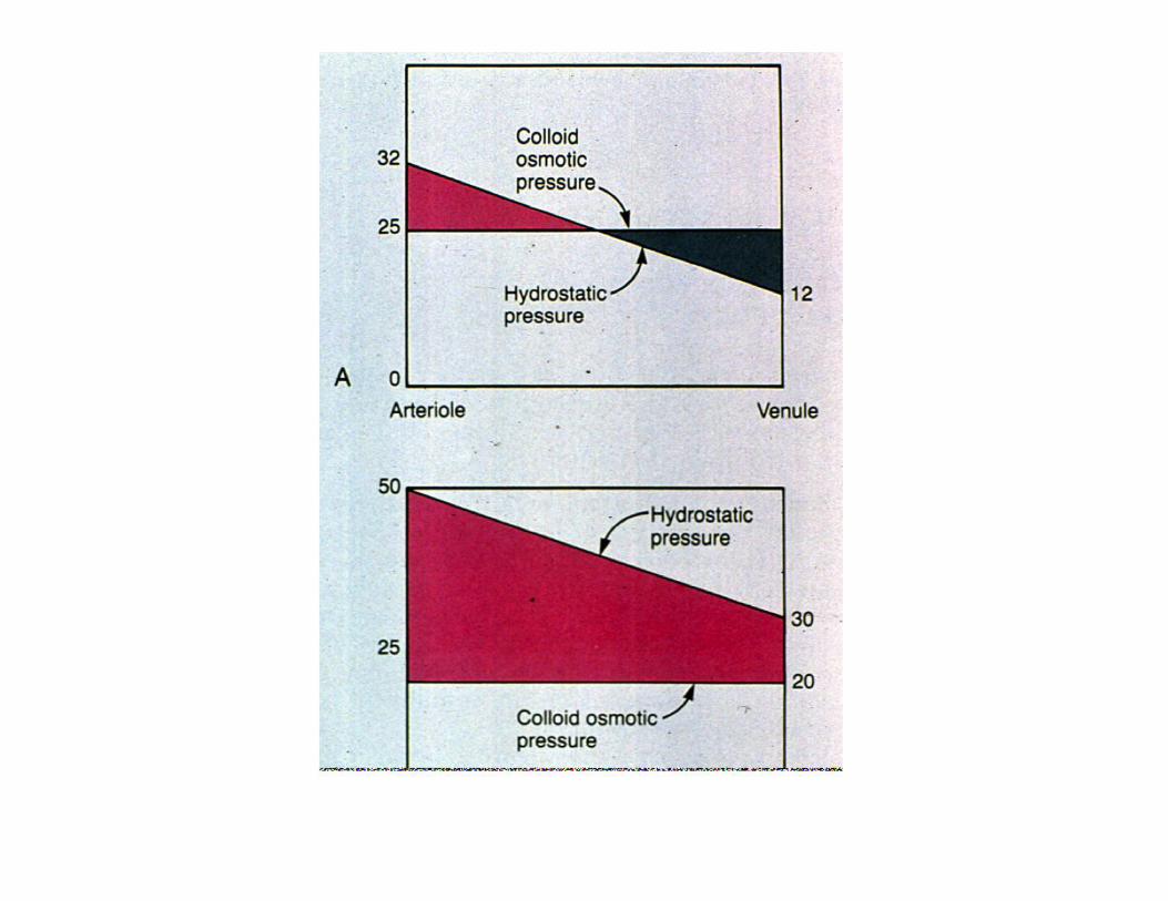
• Starling's hypothesis • In normal tissue from arteriole to
venule: Intravascular hydrostatic pressure
≅
Colloid osmotic pressure

Changes in vascular permeability (vascular leakage)
• In inflammation from arteriole to venule:
Intravascular hydrostatic pressure
Colloid osmotic pressure
+ = Edema


Lymphatics in inflammation:
• Lymphatics are responsible for draining edema.

Related definitions:
• Edema: • An excess of fluid in the interstitial
tissue or serous cavities--either a transudate or an exudate
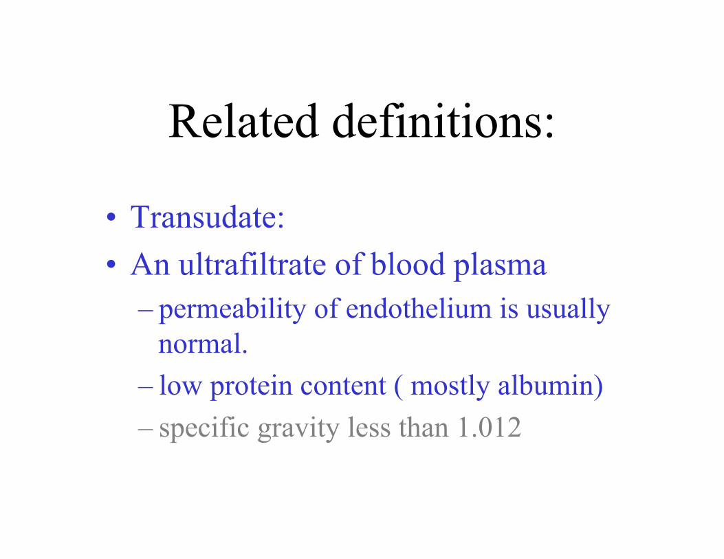
Related definitions:
• Transudate: • An ultrafiltrate of blood plasma
– permeability of endothelium is usually normal.
– low protein content ( mostly albumin) – specific gravity less than 1.012

Related definitions:
• Exudate: • A filtrate of blood plasma mixed with
inflammatory and cellular debris. – permeability of endothelium is usually
altered – high protein content – specific gravity greater than 1.020

Related definitions:
• Pus: • A purulent exudate--an
inflammatory exudate rich in leukocytes (mostly neutrophils) and parenchymal cell debris.

Changes in vascular permeability (vascular leakage) (cont)
• Mechanisms of vascular leakage – immediate transient response – immediate sustained response – delayed-prolonged response – junctional retraction – leukocyte-dependent leakage – regenerating endothelium

Leukocyte exudation • divided into 4 steps
– 1. Margination, rolling, and adhesion
– 2. Diapedesis (transmigration across the endothelium)
– 3. Migration toward a chemotactic stimulus
– 4. Phagocytosis


Phagocytosis
• 3 distinct steps – Recognition and attachment – Engulfment – Killing or degradation


Killing or degradation
• 2 mechanisms – Oxygen dependent
• Myeloperoxidase dependent (the most important!)
• Myeloperoxidase independent – Oxygen independent

Defects in leukocyte function:
• Margination and adhesion – Etoh, steroids, AR leukocyte adhesion deficiency
• Emigration toward a chemotactic stimulus – drugs – chemotaxis inhibitors
• Phagocytosis – Chronic granulomatous disease (CGD)

Chemical mediators of inflammation
• Vasoactive amines (histamine and serotonin) – Plasma proteases
• Kinin system • Complement system • Coagulation-fibrinolytic
• Arachidonic acid metabolites • via cyclooxygenase • via lipoxygenase
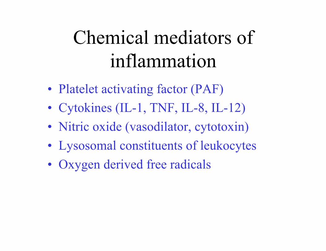
Chemical mediators of inflammation
• Platelet activating factor (PAF) • Cytokines (IL-1, TNF, IL-8, IL-12) • Nitric oxide (vasodilator, cytotoxin) • Lysosomal constituents of leukocytes • Oxygen derived free radicals

Another view of chemical mediators
• Vasodilatation (vascular flow/ caliber;
hemodynamic changes) • Prostaglandins, Nitric oxide
• Increased vascular permeability (vascular leakage) • --Vasoactive amines (histamine, serotonin) • --C3a and C5a (through liberating amines) • --Bradykinin • --Leukotrienes C4, D4, E4

Another view of chemical mediators
• Chemotaxis, leukocyte activation (leukocyte exudation)
• --C5a • --Leukotriene B4 • --Bacterial products • --Cytokines (IL-8)
• Tissue damage (a leukocyte exudation) • --Neutrophil and macrophage lysosomal enzymes • --Oxygen metabolites • --Nitric oxide

Another view of chemical mediators
• Fever – --IL-1, IL-6, TNF – --Prostaglandins
• Pain – --Prostaglandins – --Bradykinin

Two additional points:
• These systems are all interrelated. • There seems to be a very good system
of checks and balances.
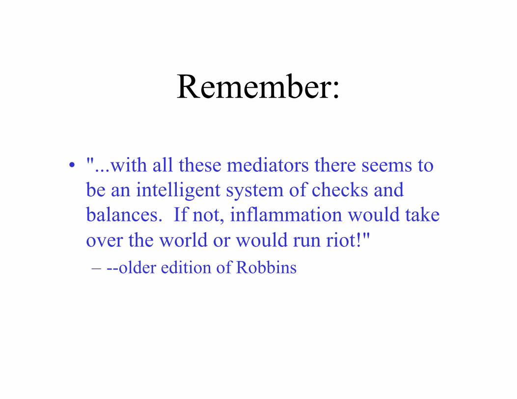
Remember:
• "...with all these mediators there seems to be an intelligent system of checks and balances. If not, inflammation would take over the world or would run riot!" – --older edition of Robbins


Acute inflammation has one of four outcomes:
• Abscess formation • Progression to chronic inflammation • Resolution--tissue goes back to normal • Repair--healing by scarring or fibrosis

Acute inflammation
Chronic inflammation
Repair Resolution
Abscess
Injury

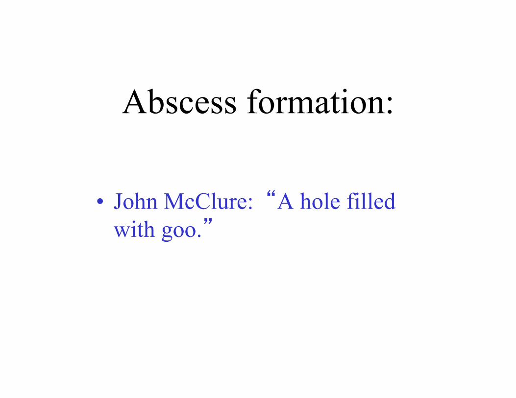
Abscess formation:
• Stedman's dictionary: "A circumscribed collection of pus appearing in an acute or chronic localized infection, and associated with tissue destruction, and frequently, swelling."-- It is usually the result of a pyogenic organism.
Abscess formation:
• John McClure: “A hole filled with goo.”


Acute inflammation
Chronic inflammation
Repair Resolution
Abscess
Injury

Chronic Inflammation
• Time course: – Greater than 48 hours (weeks, months, years)
• Cell type – Mononuclear cells (Primarily Macrophages,
Lymphocytes, Plasma cells)

Chronic inflammation
• Chronic inflammation arises in various organs in 1 of 3 ways. – Following acute inflammation – After repeated bouts of acute inflammation
(pneumonia) – Without prior acute inflammation (Tb, viruses,
silica, asbestos, rheumatoid arthritis)

Cells and mediators • Histologically chronic inflammation
includes: – Lymphocytes, plasma cells, and macrophages – Proliferation of fibroblasts and small blood
vessels – Increased connective tissue – Tissue destruction
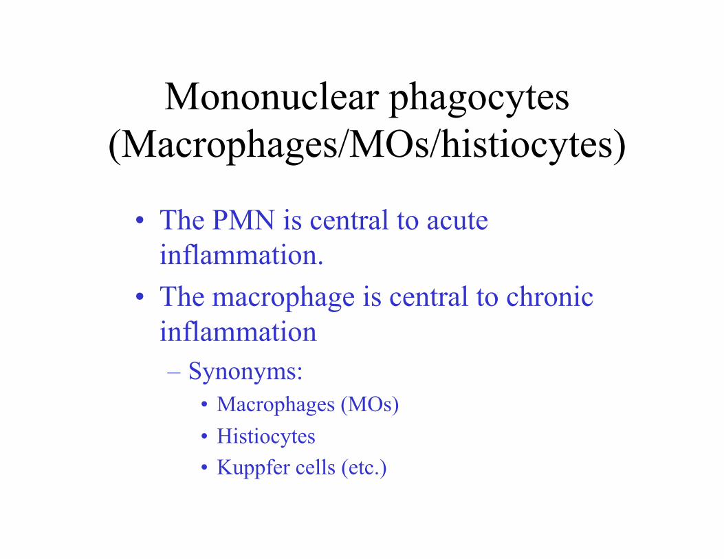
Mononuclear phagocytes (Macrophages/MOs/histiocytes)
• The PMN is central to acute inflammation.
• The macrophage is central to chronic inflammation – Synonyms:
• Macrophages (MOs) • Histiocytes • Kuppfer cells (etc.)
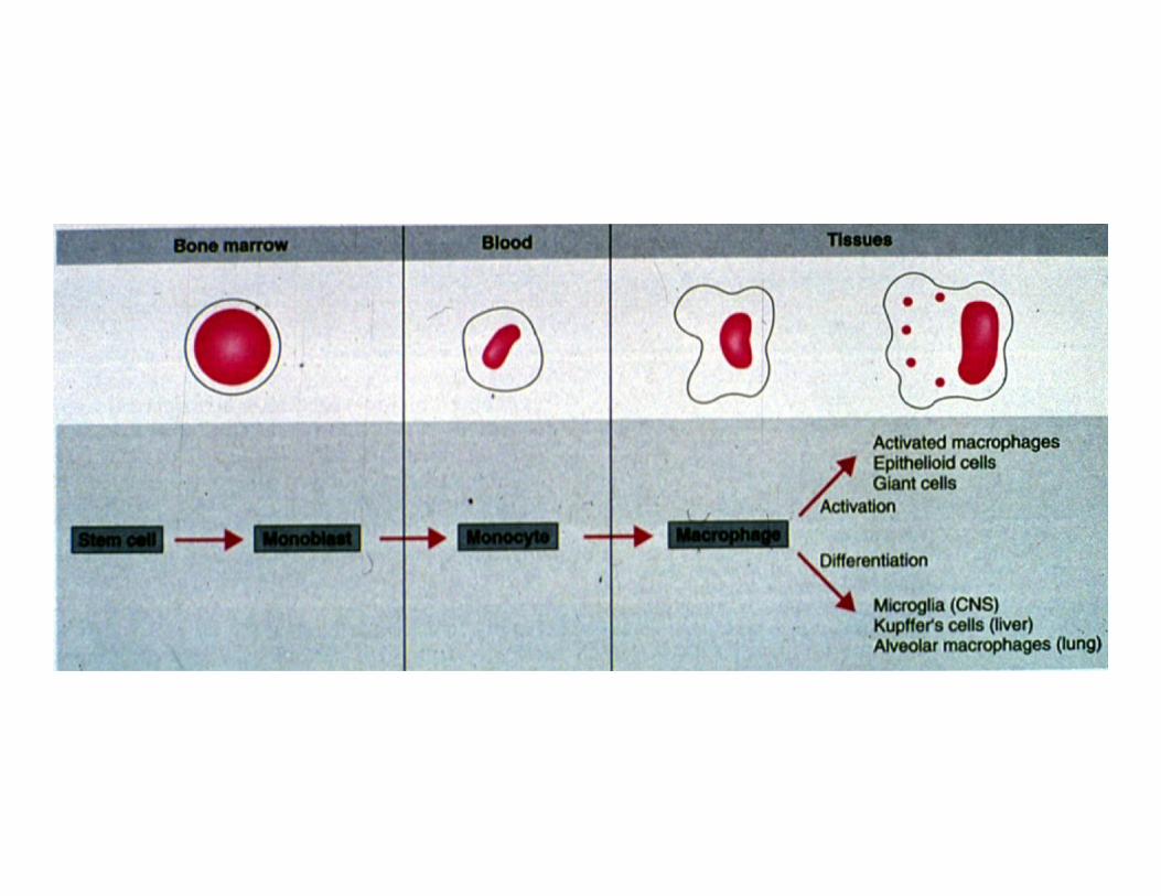

Macrophage origin:
• MOs come from the same cell line but differ depending on their microenvironment. They belong to the mononuclear phagocyte system (RES). The RES system consists of the bone marrow, peripheral blood, and tissue.

MO's share in common:
• Mobility --although slower than PMNs • Phago- and pinocytosis • Ability to become activated--especially by
lymphokines, T cells, and anything that disturbs the cell membrane—and this allows more aggressive behavior in inflammation.
• Ability to secrete large quantities of chemical mediators

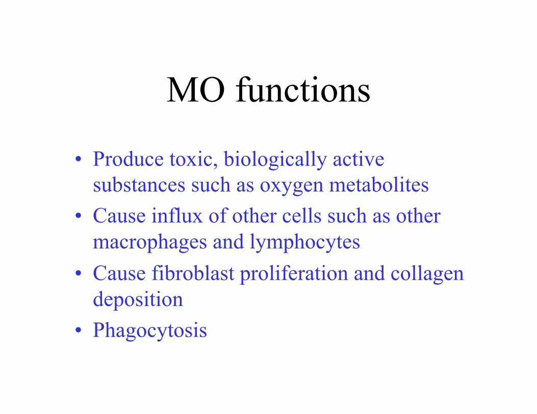
MO functions
• Produce toxic, biologically active substances such as oxygen metabolites
• Cause influx of other cells such as other macrophages and lymphocytes
• Cause fibroblast proliferation and collagen deposition
• Phagocytosis
MO time scale:
• MO’s begin emigration during acute inflammation and are the predominate cell type at 48 hours

3 ways in which MOs accumulate:
• Continued recruitment from the circulation--secondary to chemotactic factors
• Division • Prolonged survival


Other cells in chronic inflammation:
• Lymphocytes: • Plasma cells: • Eosinophils: • PMNs:


Acute inflammation
Chronic inflammation
Repair Resolution
Abscess
Injury

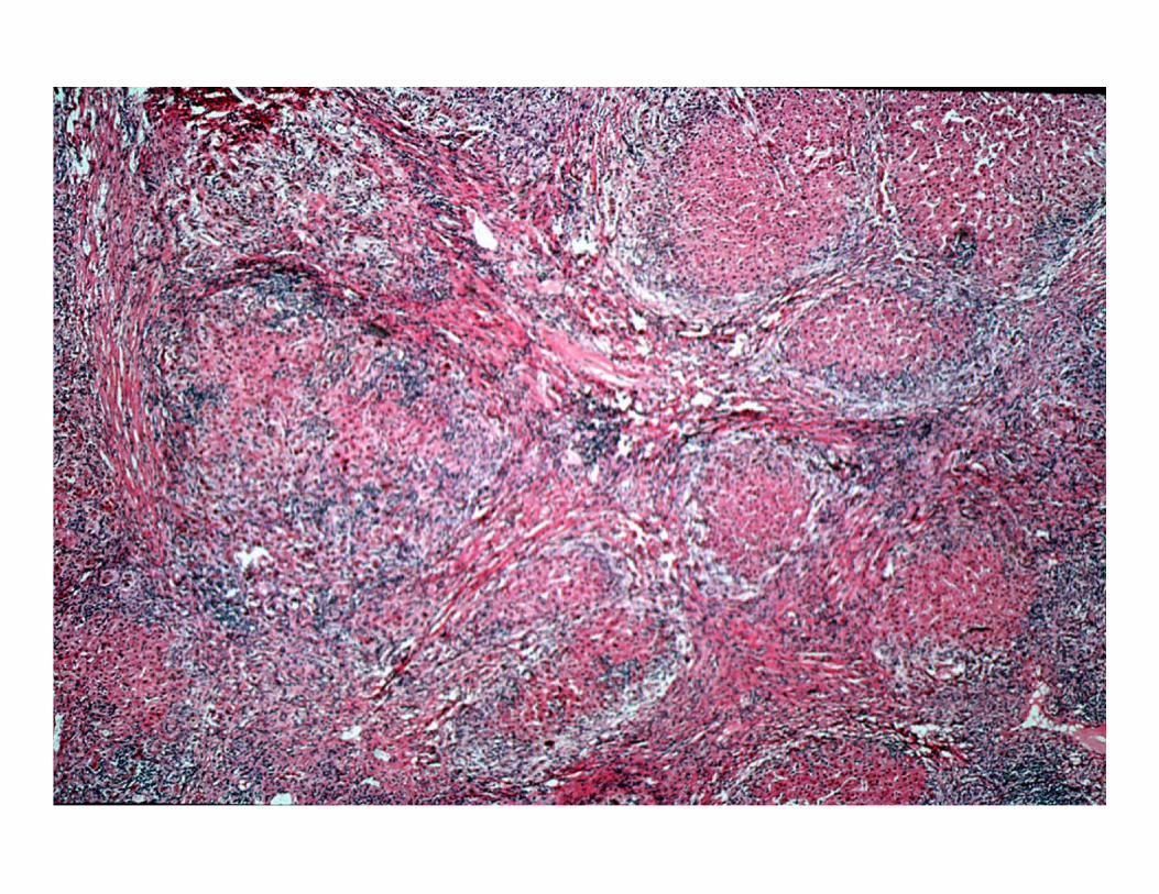
Chronic Granulomatous Inflammation (GI)
• Definition:--a type or pattern of chronic inflammation defined by the presence of granulomas which are small, 0.5 to 2 mm collections of modified "epithelioid " histiocytes/macrophages and (Langhan's) giant cells (coalesced histiocytes), usually surrounded by a rim of lymphocytes.


Granulomas occur in response to various diseases:
• Foreign body • Tuberculosis (tb) • Fungal infections • Sarcoidosis • Schitosomiasis • Leprosy

2 factors necessary for granuloma formation:
• Presence of indigestible organisms or particles (Tb, mineral oil, etc)
• Cell mediated immunity (T cells)


Outcome of chronic inflammation:
• Resolution/regeneration/restitution of normal structure
• Repair/organization/healing by connective tissue/fibrosis/scarring
• It can continue indefinitely--some disease processes are capable of continuing indefinitely such as rheumatoid arthritis..

Acute inflammation
Chronic inflammation
Repair Resolution
Abscess
Injury

Resolution
• Definition: Resolution is the return of tissue to its normal state.

Factors necessary for resolution:
• Removal of the offending agent • Regenerative ability if cells have been
destroyed • Intact stromal framework

Categorization of cells based on regenerative ability:
• Labile cells--cells which continue to proliferate throughout life (gut, skin, bone marrow)
• Stable cells--cells which retain the capacity to proliferate throughout life but usually do not unless stimulated (liver, kidney, pancreas, bone)
• Permanent cells--cells which cannot reproduce themselves after birth (neurons, cardiac and skeletal muscle)
Stromal framework:
• It is not enough to be able to regenerate. There must be an adequate stromal framework.

Acute inflammation
Chronic inflammation
Repair Resolution
Abscess
Injury

Repair
• aka organization/healing by connective tissue/fibrosis/scarring

Repair
• Similar to wound healing • Definition: Damage to both parenchymal cells
and stromal framework which results in the replacement of nonregenerated parenchymal cells by connective tissue which over time produces fibrosis and scarring.
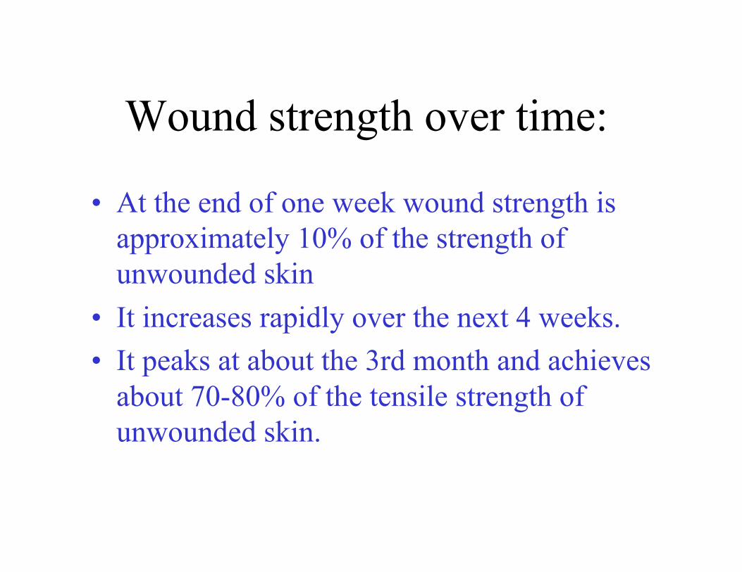
Wound strength over time:
• At the end of one week wound strength is approximately 10% of the strength of unwounded skin
• It increases rapidly over the next 4 weeks. • It peaks at about the 3rd month and achieves
about 70-80% of the tensile strength of unwounded skin.
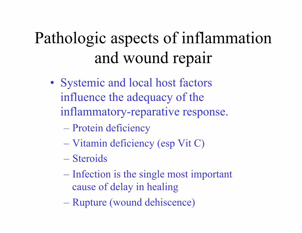
Pathologic aspects of inflammation and wound repair
• Systemic and local host factors influence the adequacy of the inflammatory-reparative response. – Protein deficiency – Vitamin deficiency (esp Vit C) – Steroids – Infection is the single most important
cause of delay in healing – Rupture (wound dehiscence)

Pathologic aspects of inflammation and wound repair
• Aberrations in growth: – Excessive amounts of collagen: keloid – Excessive amounts of granulation tissue:
proud flesh – Uncontrolled proliferation of fibroblasts:
fibromatoses


Additional definitions:
• Serous inflammation: • Marked by the outpouring of a thin fluid
that, depending on the site of injury is derived from either the blood serum or the secretions of mesothelial cells lining the peritoneal, pleural, and pericardial cavities.

Additional definitions:
• Fibrinous inflammation: • Serous fluid plus plasma proteins like
fibrinogen. Seen commonly in infections of the pleural cavity and pericardial sac.

Additional definitions:
• Suppurative or purulent inflammation: • Serous plus fibrinous plus pus (purulent
exudate). Especially common with Staph., one of several pus producing organisms. Acute appendicitis is an example.

Additional definitions:
• Ulcer: • A local defect, or excavation of the surface
of an organ or tissue, which is produced by the sloughing (shedding) of inflammatory necrotic tissue. Ulceration is defined by the presence of necrotic tissue on or near a surface.

QUESTION 1
XY presents to your clinic. He explains that last night he broke up with his girlfriend, the name of whom he had tattooed on his thumb. In the throws of his drunken misery, he burned the tattoo off with his Bic lighter. Clinical signs and symptoms of the lesion on his thumb include which of the following:

QUESTION 1
a. Rubor (redness) b. Tumor (swelling) c. Calor (heat) d. Dolor (pain) e. All of the above

QUESTION 1
a. Rubor (redness) b. Tumor (swelling) c. Calor (heat) d. Dolor (pain) e. All of the above

QUESTION 2
A week later XY again presents to your clinic. Apparently he received a lot of grief from his co-workers at the gas station about the Power Ranger’s Band-Aids you used to cover his thumb lesion, and so took them off. The wound looks worse in that it is much more erythematous and more painful to touch. Upon culture, numerous pathogenic Staphylococcus aureus organisms grow. The most important system for the killing of bacteria processed by XY’s PMNs (assuming they are normal) is which of the following:

QUESTION 2
a. Oxygen dependent, myeloperoxidase independent system
b. Oxygen dependent, myeloperoxidase dependent system
c. Oxygen independent, myeloperoxidase independent system
d. Oxygen independent, myeloperoxidase dependent system
e. AK-47 system

QUESTION 2
a. Oxygen dependent, myeloperoxidase independent system
b. Oxygen dependent, myeloperoxidase dependent system
c. Oxygen independent, myeloperoxidase independent system
d. Oxygen independent, myeloperoxidase dependent system
e. AK-47 system

QUESTION 3
Several days later XY shows up again. This time the lesion is bulging and quite warm to touch. You suspect an abscess has formed, especially because when you lance it, foul-smelling purulent debris is exuded. You take some of the debris, smear it out on a slide, and stain it. You expect to see mostly which of the following:
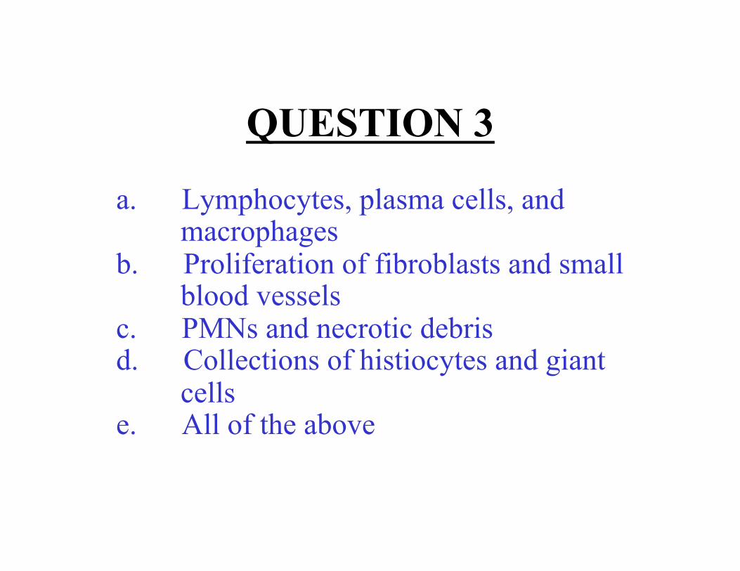
QUESTION 3
a. Lymphocytes, plasma cells, and macrophages
b. Proliferation of fibroblasts and small blood vessels
c. PMNs and necrotic debris d. Collections of histiocytes and giant
cells e. All of the above

QUESTION 3
a. Lymphocytes, plasma cells, and macrophages
b. Proliferation of fibroblasts and small blood vessels
c. PMNs and necrotic debris d. Collections of histiocytes and giant
cells e. All of the above

QUESTION 4
Upon lancing the abscess, you accidentally created an incision to large to be closed with Band-Aids and anyway, XY does not want anymore Band-Aids. Sutures are needed. You know that suturing the wound will allow it to heal by primary intention. Even if you did not suture the wound and left it open, it would heal by secondary intention. Features shared by both primary and secondary intention healing include which of the following:

QUESTION 4
a. Angiogenesis or neovascularization of new blood vessels
b. Migration and proliferation of fibroblasts c. Deposition of extracellular matrix (ECM) d. Remodeling or maturation and organization
of the fibrous tissue e. All of the above

QUESTION 4
a. Angiogenesis or neovascularization of new blood vessels
b. Migration and proliferation of fibroblasts c. Deposition of extracellular matrix (ECM) d. Remodeling or maturation and organization
of the fibrous tissue e. All of the above

QUESTION 5
While you suture up his wound, XY tells you that he is back together with his girlfriend. He wants to know if his skin will eventually be strong enough to allow for the re-tattooing of her name. You tell him the following except:
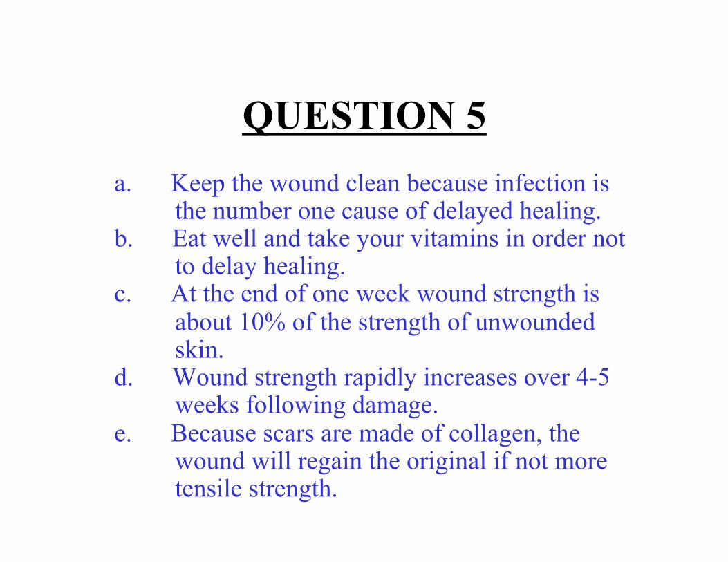
QUESTION 5 a. Keep the wound clean because infection is
the number one cause of delayed healing. b. Eat well and take your vitamins in order not
to delay healing. c. At the end of one week wound strength is
about 10% of the strength of unwounded skin.
d. Wound strength rapidly increases over 4-5 weeks following damage.
e. Because scars are made of collagen, the wound will regain the original if not more tensile strength.

QUESTION 6
If XY had used an eraser to rub out his girlfriend’s name tattooed on his thumb, and rubbed so hard that flecks of rubber from the eraser penetrated and became embedded into the underlying dermis, you would expect to see a granulomatous-type inflammatory reaction. True statements regarding granulomatous inflammation include all of the following except:

QUESTION 6
a. It is dependent on humoral mediated immunity.
b. It is considered a chronic inflammatory process.
c. It is characterized histologically by aggregates of histiocytes and giant cells.
d. It most often occurs in response to indigestible organisms or particles.
e. It progresses to repair if there is damage to stromal framework