determination of human tumor necrosis factor α by a highly sensitive enzyme immunoassay
TRANSCRIPT

Db
M*aP
R
pIsnpTiiatOwtrp(0
s
pmftToa
alhmpW
2
Biochemical and Biophysical Research Communications 289, 295–298 (2001)
doi:10.1006/bbrc.2001.5886, available online at http://www.idealibrary.com on
etermination of Human Tumor Necrosis Factor ay a Highly Sensitive Enzyme Immunoassay
ehdi Hedayati,* Razieh Yazdanparast,*,1 and Fereidoun Azizi†Institute of Biochemistry and Biophysics, University of Tehran, P.O. Box 13145-1384, Tehran, Islamic Republic of Iran;nd †Endocrine Research Center, Shaheed Beheshti University of Medical Sciences,.O. Box 19395-4743, Tehran, Islamic Republic of Iran
eceived September 14, 2001
It is generally thought that TNF-a is not producedcpdmwra
tusttmHb
saispmhiiebs
M
wGa
vS
Tumor necrosis factor a (TNF-a) is a polypeptideroduced primarily by monocytes and macrophages.t is involved in a wide variety of immune reactions. Aimple and sensitive microplate enzyme-linked immu-osorbent assay for the detection of hTNF-a in serum,lasma, and cell culture supernatants is described.he method is based on the use of horseradish perox-
dase in biotin–streptavidin amplification system whichs performed in Nunc StarWell. This system has en-bled us to achieve a sensitivity of 0.1 pg hTNF-a/ml ofhe sample. The assay is calibrated to the World Healthrganization (WHO) standard for hTNF-a (87/650). Theithin-run coefficient of variation ranged from 3.7
o 5.9 and the between-run coefficient of variationanged from 8.0 to 9.9. The results obtained by theroposed method and by a commercially available kitDRG hTNF-a ELISA) correlated well (n 5 20, r 5
.956). © 2001 Academic Press
Key Words: human TNF-a; enzyme immunoassay;treptavidin–biotin.
Tumor necrosis factor a (TNF-a) is an extremelyotent peptide cytokine which serves as an endogenousediator of inflammatory, immune and host defense
unctions. Several substances originally described forheir biological activities have been identified asNF-a, cachectin, macrophage cytotoxin (MCT), hem-rrhagic factor, macrophage cytotoxic factor (MCF),nd differentiation inducing factor (DIF).
Abbreviations used: BSA, bovine serum albumin; DIF, differenti-tion including factor; DMSO, dimethyl sulfoxide; ELISA, enzyme-inked immunosorbent assay; HRP, horseradish peroxidase;TNF-a, human tumor necrosis factor a; IL-1, interleukin-1; MCF,acrophage cytotoxic factor; MCT, macrophage cytotoxin; PBS,
hosphate-buffered saline; TMB, tetramethylbenzidine; WHO,orld Health Organization.1 To whom correspondence should be addressed. Fax: 198-21-240-
463. E-mail: [email protected].
295
onstitutively by normal cells, but needs to be inducedotently by invasive stimuli in the setting of both en-oplastic and infectious disease (1–4). In this respect,acrophages and monocytes are thought to be the cellshich contribute most to the local and systemic TNF-a
esponse to bacterial, viral and parasitic organismsnd products.Bioassays for the quantification of TNF-a, including
he cytotoxic assay on murine fibroblasts have beensed for several years. However, TNF-a and IL-1 haveeveral common biological properties which influencehe results. Therefore, these properties should beaken into account in the bioassay developmentethod. The cytotoxic assay is unaffected by IL-1.owever, it is time-consuming and might be suscepti-le to interference by other substances (5–7).Various enzyme immunoassays, with different sen-
itivities are available for detection of minutesmounts of many analytes (8–10). One of the mostmportant amplified enzyme immunoassay is biotin–treptavidin amplification system (11). Based on therinciple of this system, our group developed an ELISAethod for a faster and more sensitive quantification of
TNF-a in body fluids as well as in cell culture media,n comparison to the commercially available kits. Toncrease surface/volume ratio, Nunc C8 StarWell withight inside fins has been used. In this case, the incu-ation time can be reduced by a factor equal to thequare of the surface/volume increase factor.
ATERIALS AND METHODS
Natural and recombinant human tumor necrosis factor a (TNF-a)as purchased from Sigma Chemical Co. (Sigma-Aldrich ChemiembH, Germany). Two different kinds of anti- hTNF-a antibodiesnd other chemicals were also obtained from Sigma Co.
Coating of solid phase with anti-hTNF-a. To increase the surface/olume ratio of the test environment, the NUNC-immuno module C8tarWell was used as solid phase for fixing the anti-hTNF-a. The
0006-291X/01 $35.00Copyright © 2001 by Academic PressAll rights of reproduction in any form reserved.

cbhawmi
wp
hANmaam
ap
abd
hi1tp
bt
poo
ssa(baa
ip(
tatae
R
C
tcsgcWaa
S
tIant
H
s
TABLE 1
I
I
p
Vol. 289, No. 1, 2001 BIOCHEMICAL AND BIOPHYSICAL RESEARCH COMMUNICATIONS
oating antibody was diluted by coating buffer (0.1 M carbonate/icarbonate, pH 9.6), by a ratio of 1:100 (1 mg protein/well). Oneundred microliters of diluted anti-hTNF-a was added to each wellnd incubated overnight at room temperature. The plates were thenashed five times with washing buffer (0.2 M PBS, pH 6.8). Then 200l of the blocking buffer was added to each well. The blocking buffer
s made of 0.1 M phosphate buffer (pH 6.8) plus BSA (0.5%).The wells were incubated at room temperature for 1 h. The platesere then washed five times with the second washing buffer (PBSlus 0.05% Tween 20).
Biotinylation of anti-hTNF-a. Biotin was conjugated to anti-TNF-a by the modified method of Guesdon, Ternynck, andvramease (12–14) as follows: one hundred microliters of the biotin-hydroxy-succinimide solution in DMSO (3 mg/ml) was added to 2l of anti-hTNF-a in 0.2 M NaHCO3/KCl (15 mg/ml) and incubated
t room temperature for 10 min. Then 400 ml of NH4Cl was addednd the mixture was kept for 15 min at room temperature. Theixture was then dialyzed against PBS at 4°C for 24 h.The optimal concentrations of the biotinylated antibody for the
ssay were established using, as diluent, Tris–HCl buffer (10 mM,H 8.5) containing 10 mg BSA/milliliter.
Conjugation of HRP with streptavidin. Conjugation of HRP waschieved using the Nakane and Kawaoi method (15, 16). The dilutinguffer of the biotinylation step has been used for the titration andilution of the HRP streptavidin conjugate.
Standards. A stock solution (500 pg/ml) of the recombinantTNF-a was prepared in 50 mM phosphate buffer (pH 7.2) contain-
ng BSA(5 mg/ml) and NaCl (150 mM). Serial dilutions containing0, 5, 2.5, 1.25, 0.63, and 0.31 pg/ml of hTNF-a were prepared usinghe same phosphate buffer. The standard solution needs to be pre-ared freshly.
HRP substrate. The substrate solution should be prepared justefore use. The temperature of the substrate solution should be inhe range of 20–25°C in order to obtain optimal reproducible results.
For each plate, 12 ml of the substrate buffer (0.11 M acetate buffer,H 5.5) was mixed with 200 ml of the stock solution of TMB (6 mg/mlf DMSO) and 12 ml of H2O2 stock solution (3%). Meanwhile, 100 mlf the H2SO4 solution (1.8 M) is used as the stop reagent.
Assay procedure. Before the assay, all reagents and standardolutions are equilibrated to room temperature. Pipette 100 ml of thetandards or the samples into the appropriate wells. Cover the platend incubate it for 60 min at room temperature on a plate shaker700 rpm). The wells need to be washed five times with the washuffer (PBS with 0.05% Tween 20). Then 100 ml of the biotinylatedntibody was pipetted into each well and incubated at room temper-ture for 60 min while shaking on a plate shaker (700 rpm).The wells were washed five times with the washing buffer, follow-
ng which the HRP conjugate (100 ml) was added to each and thelate was incubated for 30 min at room temperature while shaking700 rpm) on the plate shaker.
FIG. 1. The calibration curve for the recombinant hTNF-a.
296
After the incubation time was over, the plate was washed fiveimes with the washing buffer. The HRP substrate solution was thendded to each well (100 ml per well). The plate was incubated at roomemperature for 30 min on the shaker. The process was stopped bydding 100 ml of the stop solution to each well. The optical density ofach well was measured at 450 nm.
ESULTS
alibration Graph and the Precision of the Assay
Figure 1 shows the standard curve obtained usinghe standard recombinant hTNF-a with various con-entrations ranging from 0.31 up to 10 pg/ml of the testamples. The detection limit based on the calibrationraph is about 0.1 or 0.01 pg/well. The standards werealibrated to the WHO reference 87/650. One pg/ml ofHO standard corresponds to 0.04 WHO unit. The
ssay precision are shown in Table 1 and recovery datare presented in Table 2.
pecificity of the Assay
The amounts of hTNF-a in various standard solu-ions were determined in the presence of IL-1, IL-2,L-3, IL-4, IL-6, IL-7, IL-8, and TNF-b. No interferencend cross-reactivity was detected in presence of 10g/ml of above cytokines. The results of specificity ofhe assay are presented in Table 3.
uman Serum/Plasma TNF-a
This method may be used to measure hTNF-a inerum, plasma and cell culture supernatants. Serum
Intra- and Interassay Variability (Mean 6 SD)
Sample n Mean (pg/ml) SD CV%
ntraassay A 10 8.3 0.3655 4.3B 10 2.3 0.1288 5.6C 10 0.41 0.0319 7.8
nterassay A 10 8.7 0.4524 5.2B 10 2.6 0.1976 7.6C 10 0.46 0.0446 9.7
TABLE 2
Recovery Results (pg/mL)
Sample TNF-a Added Calculated Observed Recovery %
10 2.3 12.3 11.9 96.75 2.3 7.3 7.0 95.82.5 2.3 4.8 4.6 95.81.25 2.3 3.5 3.6 101.40.62 2.3 2.9 3.1 105.80.31 2.3 2.6 2.8 107.2
Note. A known quantity of the hTNF-a has been added to eachatient’s sample.
mkogpwrctth
M
ohlT0m
S
h90
D
u
tcnmostmhe
S
((boiesafllpcohtmslbbrcrime
TABLE 3
RNIII
t
o
Vol. 289, No. 1, 2001 BIOCHEMICAL AND BIOPHYSICAL RESEARCH COMMUNICATIONS
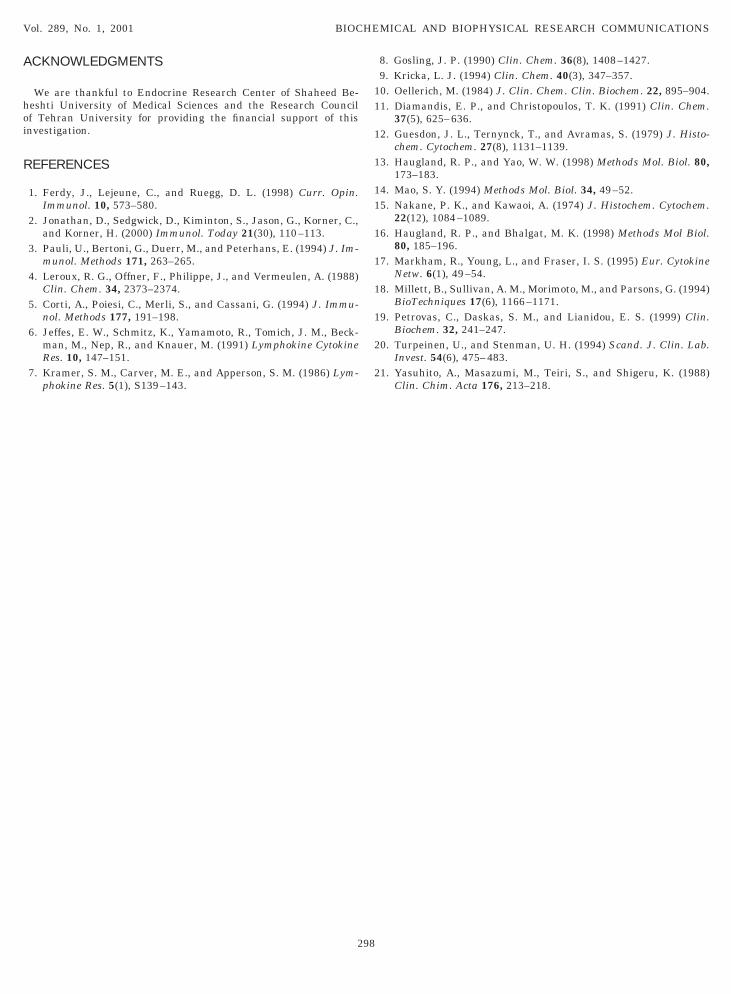
ust be removed as soon as possible from the clot andeep at 4°C. Plasma can be collected on sterile EDTAr heparin tubes and rapidly separated after centrifu-ation. The normal values of hTNF-a in sera andlasma of twenty two healthy laboratory personnelere established to be lower than 7.9 and 4.8 pg/ml,
espectively. Testing of RPMI with different lots andoncentrations of fetal bovine serum has shown thathe developed ELISA method is not disturbed by cul-ure media. Figure 2 shows the normal values ofTNF-a in sera and plasmas.
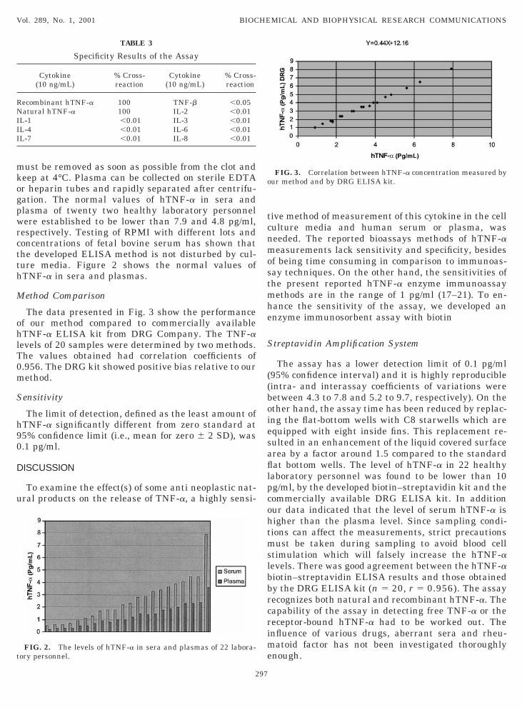
ethod Comparison
The data presented in Fig. 3 show the performancef our method compared to commercially availableTNF-a ELISA kit from DRG Company. The TNF-a
evels of 20 samples were determined by two methods.he values obtained had correlation coefficients of.956. The DRG kit showed positive bias relative to ourethod.
ensitivity
The limit of detection, defined as the least amount ofTNF-a significantly different from zero standard at5% confidence limit (i.e., mean for zero 6 2 SD), was.1 pg/ml.
ISCUSSION
To examine the effect(s) of some anti neoplastic nat-ral products on the release of TNF-a, a highly sensi-
Specificity Results of the Assay
Cytokine(10 ng/mL)
% Cross-reaction
Cytokine(10 ng/mL)
% Cross-reaction
ecombinant hTNF-a 100 TNF-b ,0.05atural hTNF-a 100 IL-2 ,0.01
L-1 ,0.01 IL-3 ,0.01L-4 ,0.01 IL-6 ,0.01L-7 ,0.01 IL-8 ,0.01
FIG. 2. The levels of hTNF-a in sera and plasmas of 22 labora-ory personnel.
297
ive method of measurement of this cytokine in the cellulture media and human serum or plasma, waseeded. The reported bioassays methods of hTNF-aeasurements lack sensitivity and specificity, besides
f being time consuming in comparison to immunoas-ay techniques. On the other hand, the sensitivities ofhe present reported hTNF-a enzyme immunoassayethods are in the range of 1 pg/ml (17–21). To en-
ance the sensitivity of the assay, we developed annzyme immunosorbent assay with biotin
treptavidin Amplification System
The assay has a lower detection limit of 0.1 pg/ml95% confidence interval) and it is highly reproducibleintra- and interassay coefficients of variations wereetween 4.3 to 7.8 and 5.2 to 9.7, respectively). On thether hand, the assay time has been reduced by replac-ng the flat-bottom wells with C8 starwells which arequipped with eight inside fins. This replacement re-ulted in an enhancement of the liquid covered surfacerea by a factor around 1.5 compared to the standardat bottom wells. The level of hTNF-a in 22 healthy
aboratory personnel was found to be lower than 10g/ml, by the developed biotin–streptavidin kit and theommercially available DRG ELISA kit. In additionur data indicated that the level of serum hTNF-a isigher than the plasma level. Since sampling condi-ions can affect the measurements, strict precautionsust be taken during sampling to avoid blood cell
timulation which will falsely increase the hTNF-aevels. There was good agreement between the hTNF-aiotin–streptavidin ELISA results and those obtainedy the DRG ELISA kit (n 5 20, r 5 0.956). The assayecognizes both natural and recombinant hTNF-a. Theapability of the assay in detecting free TNF-a or theeceptor-bound hTNF-a had to be worked out. Thenfluence of various drugs, aberrant sera and rheu-
atoid factor has not been investigated thoroughlynough.
FIG. 3. Correlation between hTNF-a concentration measured byur method and by DRG ELISA kit.
ACKNOWLEDGMENTS
hoi
R
8. Gosling, J. P. (1990) Clin. Chem. 36(8), 1408–1427.
11
1
1
11
1
1
1
1
2
2
Vol. 289, No. 1, 2001 BIOCHEMICAL AND BIOPHYSICAL RESEARCH COMMUNICATIONS
We are thankful to Endocrine Research Center of Shaheed Be-eshti University of Medical Sciences and the Research Councilf Tehran University for providing the financial support of thisnvestigation.
EFERENCES
1. Ferdy, J., Lejeune, C., and Ruegg, D. L. (1998) Curr. Opin.Immunol. 10, 573–580.
2. Jonathan, D., Sedgwick, D., Kiminton, S., Jason, G., Korner, C.,and Korner, H. (2000) Immunol. Today 21(30), 110–113.
3. Pauli, U., Bertoni, G., Duerr, M., and Peterhans, E. (1994) J. Im-munol. Methods 171, 263–265.
4. Leroux, R. G., Offner, F., Philippe, J., and Vermeulen, A. (1988)Clin. Chem. 34, 2373–2374.
5. Corti, A., Poiesi, C., Merli, S., and Cassani, G. (1994) J. Immu-nol. Methods 177, 191–198.
6. Jeffes, E. W., Schmitz, K., Yamamoto, R., Tomich, J. M., Beck-man, M., Nep, R., and Knauer, M. (1991) Lymphokine CytokineRes. 10, 147–151.
7. Kramer, S. M., Carver, M. E., and Apperson, S. M. (1986) Lym-phokine Res. 5(1), S139–143.
298
9. Kricka, L. J. (1994) Clin. Chem. 40(3), 347–357.0. Oellerich, M. (1984) J. Clin. Chem. Clin. Biochem. 22, 895–904.1. Diamandis, E. P., and Christopoulos, T. K. (1991) Clin. Chem.
37(5), 625–636.2. Guesdon, J. L., Ternynck, T., and Avramas, S. (1979) J. Histo-
chem. Cytochem. 27(8), 1131–1139.3. Haugland, R. P., and Yao, W. W. (1998) Methods Mol. Biol. 80,
173–183.4. Mao, S. Y. (1994) Methods Mol. Biol. 34, 49–52.5. Nakane, P. K., and Kawaoi, A. (1974) J. Histochem. Cytochem.
22(12), 1084–1089.6. Haugland, R. P., and Bhalgat, M. K. (1998) Methods Mol Biol.
80, 185–196.7. Markham, R., Young, L., and Fraser, I. S. (1995) Eur. Cytokine
Netw. 6(1), 49–54.8. Millett, B., Sullivan, A. M., Morimoto, M., and Parsons, G. (1994)
BioTechniques 17(6), 1166–1171.9. Petrovas, C., Daskas, S. M., and Lianidou, E. S. (1999) Clin.
Biochem. 32, 241–247.0. Turpeinen, U., and Stenman, U. H. (1994) Scand. J. Clin. Lab.
Invest. 54(6), 475–483.1. Yasuhito, A., Masazumi, M., Teiri, S., and Shigeru, K. (1988)
Clin. Chim. Acta 176, 213–218.