department of plastic and reconstructive surgery ... · diagnosi di epulide congenita gigante....
TRANSCRIPT
Congenital epulis of the newborn: difficult to diagnose but easy to treatA case of unusual size
Published online (EP) 7 February 2014 - Ann. Ital. Chir 1
Pervenuto in Redazione Aprile 2013. Accettato per la pubblicazioneSettembre 2013Correspondence to: Damiano Tambasco, MD, University Hospital A. Gemelli,Largo A. Gemelli 8, 00168 Rome, Italy (e-mail: [email protected])
Damiano Tambasco, Annalisa Seccia, Agostino Bruno, Antonio Seccia
Department of Plastic and Reconstructive Surgery, “Catholic University of the Sacred Heart,” Rome, Italy
Congenital epulis of the newborn: difficult to diagnose but easy to treat. A case of unusual size
The epulis or giant cell granuloma is a benign tumor of the connective tissue of the gingival mucosa frequent in anadvanced period of life but much more rare in its congenital form. We present the case of a female newborn, otherwise healthy, presenting with a giant swelling protruding from her mouthand originating from upper left alveolar ridge. The size of the mass has created great anxiety in parents and pediatri-cians, however this clinical presentation suggested us a diagnosis of congenital epulis with a differential diagnosis of ter-atoma. We perform surgical resection of the mass under general anesthesia, through diathermy. There wasn’t blood loss and post-operative recovery was uneventful. A definite diagnosis of giant congenital epulis was disclosed by histopathological andimmunohistochemical analysis.Although it has not been yet clarified the etiology of this tumor and the role of hormonal influences on its appearanceand development and despite have been reported cases of spontaneous regression, the treatment of choice is the early sur-gical excision.Nevertheless, particularly in the small centers, due to the rarity and large size of presentation, the diagnosis is oftendelayed or wrong, exposing so the newborn parents to useless days of waiting and anxietyIn our opinion all pediatricians and surgeons should be aware of this malformation and of its simple, safe and effec-tive surgical treatment, considering the excellent prognosis of this rare disease.
KEY WORDS: Congenita epulis, Early surgical excision, Giant cell granuloma
Introduction
The epulis or giant cell granuloma (so-called giant cellepulis) is a benign tumor of the connective tissue whoseetiology and histogenesis are still debated, localized atthe level of the gingival mucosa, which originates fromtissues that form the hanger apparatus of the teeth (peri-odontium, alveolar bone, external alveolar periosteum)
and that, in adults, can cause resorption of the alveolarprocess and the erosion of the root of the teeth.It’s very frequent in an advanced period of life, pre-dominantly in females and may arise from chronicinflammatory exuberant processes, following trauma orchronic local irritation.A congenital form (congenital epulis of the newborn) isdescribed , which is much rarer, sometimes diagnosedalready during intrauterine life 1,2.It appears as a submucosal swelling in the gingival, max-
imum size of 1-1.5 cm, without any involvement of theunderlying bone. Although is a benign lesion, it mayrecur in 5-10% of cases.Treatment consists of complete removal of the epulis andthe implant base, and in adults, in the Extraction ofTeeth Mobilized By The Tumor.
Ann. Ital. Chir.Published online (EP) 7 February 2014
pii: S2239253X1402163Xwww.annitalchir.com
READ-ONLY
COPY
PRINTIN
G PROHIB
ITED

Case report
A female newborn by caesarean section, was referred tothe department of Plastic Surgery immediately after birthfor a large mass protruding from her mouth. She wasotherwise healthy. On examination there was a giant veg-etans swelling, oval appearance (with smooth, pink sur-face, crossed by fine telangiectasias) and regular, well-defined margins. Dimensions 3,5 x 2,5 x 1,5 cm witha mucous pedicle (base planting 1 x 0,5 cm) originat-ing from upper left alveolar ridge (about 1 cm from theupper labial frenulum). It was interfering with feedingbut at that moment the mass didn’t create respiratoryproblems.This clinical presentation suggested a diagnosis of con-genital epulis in differential diagnosis with teratoma. Toexclude the presence of other concomitant malformationsin 2nd day a skull TC was performed confirming thegingival origin of the swelling and reporting moderateand uneven enhancment after administration of iodinat-ed contrast. No erosive aspects of the maxillary superi-or or other intracranial abnormalities were detected.It was therefore decided to perform early (3rd day) sur-gical resection of the mass under general anesthesia,through diathermy. There wasn’t blood loss and postop-erative recovery was uneventful.
The material was sent to histopatology and theHaematoxylin and Eosin (H&E) stain revealed sheets ofpolygonal or ovoid clusters of cells with medium-sizednucleus and nucleoli evident, granular cytoplasm, stro-ma richly vascularized with capillaries and venules, theabsence of mitosis and occasional perivascular inflam-mation and proliferation of perivascular pericytes.Immunohistochemical analysis was negative for S-100protein, keratin, desmin, CD31, CD34, CD68. A defi-nite diagnosis of giant congenital epulis was disclosed.
Discussion
Since the first description of Neumann 3 of 1871 haveso far been described only about 200 cases of congeni-tal epulis of the newborn 4. and analyzing the scientif-ic literature even recently, we find typically work on a
D. Tambasco, et al.
2 Ann. Ital. Chir - Published online (EP) 7 February 2014
Fig. 1: Appearance of the lesion at 2nd day of life. Giant vegetansswelling, oval appearance (with smooth, pink surface, crossed by finetelangiectasias) and regular, well-defined margins. Dimensions 3,5 X 2,5 X 1,5 cm.
Fig. 2: Note the mucous pedicle (base planting 1 x 0,5 cm) originating fromupper left alveolar ridge (about 1 cm from the upper labial frenulum).
Fig. 3: Post-operative view (3rd day) after surgical resection of the massunder general anesthesia, through diathermy of the implant base.
READ-ONLY
COPY
PRINTIN
G PROHIB
ITED
single case report 5-7, although there are also works thatreport ten cases 8. These epidemiological data show therarity of this benign neoplasm, the most frequently diag-nosed at birth with a female-male ratio of 8:1 9,10.The localization is prevalent in the maxilla, unique, alsorarely can have two or more localized lesions for exam-ple both at the margin of the alveolar maxilla andmandibular 11. In the case of large lesions may occurmechanical disturbances of nutrition and respiration.This lesion is well differentiated from many injuries thatoccur in childhood teratoma, dermoid cyst, angioma.lymphangioma, fibrosarcoma, leiomyoma, rhabdomyoma,etc. 12.The tumor size may vary from a few millimeters up toas much as 9 cm and 90% of cases this lesion presentssingle, interesting, therefore, only the maxilla or mandible(a ratio between the two of 3:1) 9.Although it has not been yet clarified the etiology ofthis tumor (such as the myoblastic, odontogenic, neu-rogenic, histiocytic and endocrinologic origin 13,14) andthe role of hormonal influences on its appearence anddevelopment and despite have been reported cases ofspontaneous regression 15, the treatment of choice is theearly surgical excision.Even in the presence of a non-radical resection of con-genital forms, in literature cases of local recurrence havenot been so far described.The decades of experience achieved by our center in theearly diagnosis of congenital malformations, due to thehigh number of births in our hospital and thus to aconsiderable number of infants for which pediatricsrequire our advice, led us to straightaway exclude mal-formations such as hemangiomas, lymphangiomas, fibro-mas, sarcomas (which sometimes come into differentialdiagnosis of this lesion 16 and thus significantly speedup the diagnostic and therapeutic process. Nevertheless,
particularly in the small centers, due to the rarity andlarge size of presentation, the diagnosis is often delayedor wrong, exposing so the newborn parents to uselessdays of waiting and anxietyIn our opinion all pediatricians and surgeons should beaware of this malformation and of its simple, safe andeffective of surgical treatment, considering the excellentprognosis of this rare disease.
Riassunto
L’epulide o granuloma a cellule giganti è un tumore beni-gno del tessuto connettivo la cui etiologia ed istogenesirisultano ancora dibattute. Si localizza a livello dellamucosa gengivale ed origina dai tessuti che formanol’apparato sospensore dei denti (parodonto, osso alveola-re, periostio alveolare esterno) e che, nell’adulto, puòdeterminare il riassorbimento del processo alveolare el’erosione della radice dei denti. È molto frequente inun periodo avanzato della vita, prevalentemente nel ses-so femminile, mentre è molto più rara nella sua formacongenita. In questo caso si presenta come una tumefa-zione sottomucosa, in sede gengivale delle dimensionimassime di 1-1,5 cm senza alcun interessamento dell’ossosottostante.Presentiamo il caso di una neonata oltretermine da par-to cesareo, in buone condizioni cliniche, giunta imme-diatamente dopo la nascita alla nostra osservazione peruna enorme massa sporgente dalla sua bocca, ad origi-ne dall’emiarcata dentaria superiore di sinistra. La mas-sa interferiva con l’allattamento ma al momento noncreava problemi respiratori.Questo quadro clinico suggeriva una diagnosi di epuli-de gigante congenita in diagnosi differenziale con tera-toma. È stata quindi posta indicazione ad asportazionechirurgica della massa in anestesia generale mediante dia-termobisturi, con cauterizzazione della base di impianto.Non si è registrata alcuna perdita di sangue intraopera-toria e la degenza post-operatoria è risultata priva di com-plicanze. Il materiale è stato inviato per esame istopato-logico e immunoistochimico che hanno confermato ladiagnosi di epulide congenita gigante.Nonostante non sia stata ancora chiarita l’eziopatogenesidi questo tumore e il ruolo degli influssi ormonali sul-la sua comparsa e sul suo sviluppo e sebbene siano sta-ti descritti casi di regressione spontanea, il trattamentod’elezione è l’escissione chirurgica precoce.Tuttavia, specie nei piccoli centri, per la rarità e le gros-se dimensioni di presentazione, la diagnosi è spesso ritar-data o errata, esponendo così i genitori del neonato adinutili giornate di attesa e apprensione.È importante, pertanto, che tutti i pediatri e i chirur-ghi abbiano conoscenza di tale malformazione e sianoconsapevoli della semplicità e dell’efficacia del tratta-mento chirurgico, considerata la prognosi davvero eccel-lente di questa rara patologia.
Published online (EP) 7 February 2014 - Ann. Ital. Chir 3
Congenital epulis of the newborn: difficult to diagnose but easy to treat. A case of unusual size
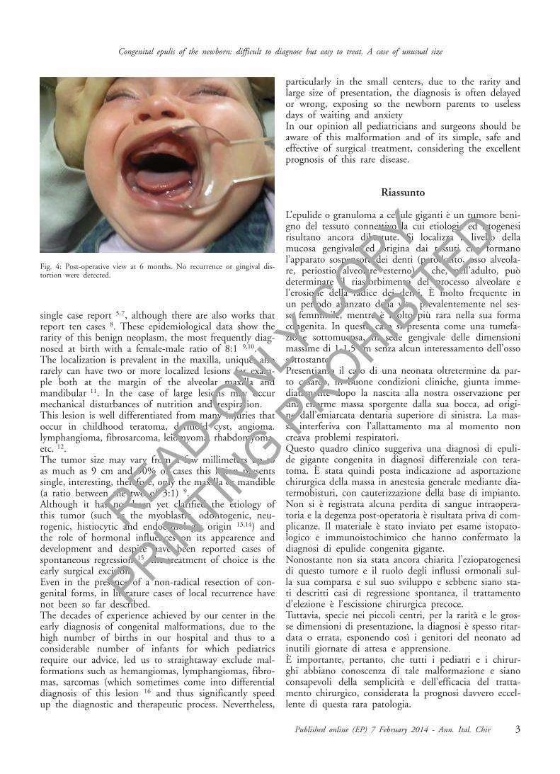
Fig. 4: Post-operative view at 6 months. No recurrence or gingival dis-tortion were detected.
READ-ONLY
COPY
PRINTIN
G PROHIB
ITED
References
1. Jiang L, Hu B, Guo Q: Prenatal sonographic diagnosis of con-genital epulis. J Clin Ultrasound, 2011; 39(4):217-20.
2. Szlachetka K, Lemcke-Berno EM, Ozcan T: Prenatal diagnosisof a rare gingival granular cell tumor of the fetal mouth. J UltrasoundMed, 2012; 31(1):125-27.
3. Neumann E: Ein fall von kongenitaler Epulis. Arch Heilkd, 1871,12:189-190.
4. Ritwik P, Brannon RB, Musselman RJ: Spontaneous regressionof congenital epulis: A case report and review of the literature. J MedCase Rep, 2010; 4:331.
5. Zuker RM, Buenechea R: Congenitak epulis: Review of the lit-erature and case report. Oral Maxillofac Surg, 1993; 51(9):1040-43.
6. Merglová V, Mukensnabl P, Andrle P: Congenital epulis. BMJCase Rep, 2012; 9:2012.
7. Bhat C, Chaugule V, Patil V, Shah K: Congenital epulis in new-born. Case report. N Y State Dent J, 2012; 78(1):50-52.
8. Childers EL, Fanburg-Smith JC: Congenital epulis of the new-born: 10 new cases of a rare oral tumor. Ann Diagn Pathol, 2011;15(3):157-61.
9. Eghbalian F, Monsef A: Congenital epulis in the newborn, reviewof the literature and a case report. J Pediatr Hematol Oncol, 2009;31(3):198-99.
10. Olson JL, Marcus JR, Zuker RM: Congenital epulis. J CraniofacSurg, 2005; 16:161-64.
11. Adeyemi BF, Oluwasola AO, Adisa AO: Congenital epulis.Indian J Dent Res, 2010; 21(2):292-94.
12. Hasanov A, Musayev J, Onal B, Rahımov C, Farzaliyev I:Gingival granular cell tumor of the newborn: A case report and reviewof literature. Turk Patoloji Derg, 2011; 27(2):161-63.
13. Nakata M, Anno K, Matsumori LT, et al.: Prenatal diagnosisof congenital epulis: A case report. Ultrasound Obstet Gynecol, 2002;20:627-29.
14. Kannan SK, Rajesh R: Congenital epulis. Congenital granular celllesion: A case report. J Indian Soc Pedod Prev Dent, 2006; 24:104-06.
15. Kadlub N, Galliani E, Oker N: Congenital epulis: Refrain fromsurgery. A case report of spontaneous regression. Arch Pediatr, 2011;18(6):657-59.
16. Bewley A, Bloom JD, Kherani S, Pawel BR: Congenital epulis.Ear Nose Throat, 2010; 89(7):299-300.
D. Tambasco, et al.
4 Ann. Ital. Chir - Published online (EP) 7 February 2014
READ-ONLY
COPY
PRINTIN
G PROHIB
ITED