deletion of dtnbp1 in mice impairs threat memory ... filein human post mortem studies, both the mrna...
TRANSCRIPT

Huang et al. Translational Psychiatry (2019) 9:132
https://doi.org/10.1038/s41398-019-0465-y Translational Psychiatry
ART ICLE Open Ac ce s s
Deletion of Dtnbp1 in mice impairs threatmemory consolidation and is associatedwith enhanced inhibitory drive in theamygdalaCathy C. Y. Huang1,2,4, Kevin J. Muszynski2, Vadim Y. Bolshakov1,3 and Darrick T. Balu 1,2
AbstractSchizophrenia is a severe and highly heritable disorder. Dystrobrevin-binding protein 1 (DTNBP1), also known asdysbindin-1, has been implicated in the pathophysiology of schizophrenia. Specifically, dysbindin-1 mRNA and proteinexpression are decreased in the brains of subjects with this disorder. Mice lacking dysbinidn-1 also display behavioralphenotypes similar to those observed in schizophrenic patients. However, it remains unknown whether deletion ofdysbindin-1 impacts functions of the amygdala, a brain region that is critical for emotional processing, which isdisrupted in patients with schizophrenia. Here, we show that dysbindin-1 is expressed in both excitatory and inhibitoryneurons of the basolateral amygdala (BLA). Deletion of dysbindin-1 in male mice (Dys−/−) impaired cued and context-dependent threat memory, without changes in measures of anxiety. The behavioral deficits observed in Dys−/− micewere associated with perturbations in the BLA, including the enhancement of GABAergic inhibition of pyramidalneurons, increased numbers of parvalbumin interneurons, and morphological abnormalities of dendritic spines onpyramidal neurons. Our findings highlight an important role for dysbindin-1 in the regulation of amygdalar functionand indicate that enhanced inhibition of BLA pyramidal neuron activity may contribute to the weakened threatmemory expression observed in Dys−/− mice.
IntroductionSchizophrenia is a severe mental illness, characterized
by positive (delusions), negative (anhedonia), and cogni-tive symptoms that affects 0.5–1% of population world-wide1. Dystrobrevin-binding protein 1 (DTNBP1), alsoknown as dysbindin-1, has been implicated in thepathophysiology of schizophrenia. In human post mortemstudies, both the mRNA and protein levels of dysbindin-1were decreased in the dorsolateral prefrontal cortex (PFC)and hippocampus of subjects with schizophrenia2–5, brainregions that are critical for memory, cognition, and
emotion. A recent study also demonstrated that humanschizophrenia subjects with genetic variants in the genethat encodes dysbindin-1 (DTNBP1) exhibited cognitivedysfunction6. Moreover, dysbindin-1 knockout mice(Dys−/−) exhibit behavioral phenotypes similar to what isobserved in schizophrenia, including reduced pre-pulseinhibition, impaired learning and memory, and decreasedsocial interaction7–13. In sum, these data suggest thatalterations in dysbindin-1 expression can contribute tothe pathophysiology of schizophrenia.Dysbinin-1 is a coiled-coil-containing protein that
serves diverse functions, including synaptic homeostasis,exocytosis, synaptic vesicle biogenesis, and dendritic spineformation14–18. Dys−/− mice exhibited impaired hippo-campal long-term potentiation19, as well as disruptedexcitatory and inhibitory synaptic transmission in the
© The Author(s) 2019OpenAccessThis article is licensedunder aCreativeCommonsAttribution 4.0 International License,whichpermits use, sharing, adaptation, distribution and reproductionin any medium or format, as long as you give appropriate credit to the original author(s) and the source, provide a link to the Creative Commons license, and indicate if
changesweremade. The images or other third partymaterial in this article are included in the article’s Creative Commons license, unless indicated otherwise in a credit line to thematerial. Ifmaterial is not included in the article’s Creative Commons license and your intended use is not permitted by statutory regulation or exceeds the permitted use, you will need to obtainpermission directly from the copyright holder. To view a copy of this license, visit http://creativecommons.org/licenses/by/4.0/.
Correspondence: Cathy C. Y. Huang ([email protected]) or Darrick T. Balu([email protected])1Department of Psychiatry, Harvard Medical School, Boston, MA, USA2Translational Psychiatry laboratory, McLean Hospital, Belmont, MA, USAFull list of author information is available at the end of the article.
1234
5678
90():,;
1234
5678
90():,;
1234567890():,;
1234
5678
90():,;
medial prefrontal cortex (mPFC)9,20,21. Additionally,abnormal dendritic morphology was found in hippo-campal neurons in Dys−/− mice in vitro15,22. Thesealterations observed in Dys−/− mice demonstrate thatdysbindin-1 may serve as a regulator of synaptic andstructural plasticity.The amygdala is located deep in the temporal lobe,
consisting of many interconnected nuclei, and it playskey roles in emotional memory and processing ofemotionally charged cues23. Clinical observations sug-gest that emotional perturbation is an important featureobserved in patients with schizophrenia. Emerging evi-dence have further suggested that amygdalar dysfunc-tion contributes to the pathophysiology ofschizophrenia 24–34. Importantly, neuroimaging studieshave shown that fear processing is impaired in patientswith schizophrenia through inhibition of amygdalaactivity33,35–37. Thus, we hypothesized that dysbindin-1may contribute to the regulation of threat memory-related behaviors by possibly regulating synaptic andneuronal functions in fear circuits in the brain, specifi-cally in the amygdala, a key structure implicated in fearlearning and memory38. In this study, we combinedbehavioral, electrophysiological, morphological andbiochemical strategies, focusing on the analysis of Dys−/
− mice, to elucidate the functional roles for dysbindin-1in neurotransmission in a fear-related neural circuit andin fear control.
Material and methodsAnimalsDys−/− mice were a generous gift from Dr. Greg C.
Carlson (University of Pennsylvania). Dys−/− mice resultedfrom a spontaneous mutation on the DBA/2J backgroundnamed Sandy39. Mice in this study were on a C57BL/B6Jbackground, obtained by breeding the Dys−/− mice onDBA/2J background with C57BL/B6J mice40. Hetero-zygous (Dys+/−) dysbindin-1 mice were backcrossed withC57BL/B6J mice to generate heterozygous dysbindin-1breeders. Female and male Dys+/− mice were bred togenerate WT and Dys−/− offspring. Adult male mice(3–5 months old) were used in this study. Animals werehoused in groups of four at 22 °C under a 12:12-h light:dark cycle with lights on at 7:00A.M. Animals were pro-vided with food and water ad libitum. Sandy Forward (5′-TCC TTG CTT CGT TCT CTG CT-3′), Sandy Reverse(5′-CTT GCC AGC CTT CGT ATT GT-3′) WT-SE3F(5′-TGA GCC ATT AGG AGA TAA GAG CA-3′), andWT-SE3R (5′-AGC TCC ACC TGC TGA ACA TT-3′)primers were used for the genotyping assays. All animalcare and experimental procedures were approved by theMcLean Hospital Institutional Animal Care and UseCommittee.
Immunoblot analysisThe amygdala samples containing the basolateral
amygdala (BLA) and the basomedial amygdala were dis-sected out (bregma –1.2 to −2.3) as previously descri-bed41. SDS–PAGE and immunoblotting using brain tissuesamples were performed and analyzed as previouslydescribed42,43. The primary antibodies used in this studywere: anti-β-actin (ab8227; Abcam), anti-Arc (sc-166461;Santa Cruz Biotechnology), anti-phospho-CaMKIIα (sc-12886; Santa Cruz Biotechnology), CaMKIIα (sc-13141;Santa Cruz Biotechnology), anti-dysbindin-1 (11132-1-AP; Proteintech), anti-PSD95 (51-6900; Thermo FisherScientific). The values obtained from Dys−/− mice weresubsequently normalized to the WT groups. Results areexpressed as a percentage of WT.
Synaptosomal plasma membrane preparationMice were decapitated and amygdala tissue from both
hemispheres was dissected out on ice. One sample con-tained tissues from three mice which were pooled toge-ther. The pooled tissues were homogenized in 500 µl ice-cold sucrose buffer (0.32M sucrose/4 mM HEPES, pH7.4)for amygdala (1000 µl for hippocampus) using a 3 ml tis-sue grinder for 20–30 strokes, and then centrifuged at800 g at 4 °C for 10min. The pellets were suspended with200 µl of the same sucrose buffer for amygdala (400 µl forhippocampus) and centrifuged again at 800 g at 4 °C for10min in order to obtain more supernatant fraction. Thecombined supernatants (S1) were centrifuged at 12,000 gat 4 °C for 15min. The crude synaptosomal fraction (P2)was suspended in 600 µl buffer (4 mM HEPES, pH 7.4;1200 µl for hippocampus) and rotated at 4 °C for 1 h. Thesamples were then centrifuged at 21,130×g at 4 °C for30min. The pellets were sonicated with 50 µl buffer foramygdala (50 mM HEPES, pH 7.4, 2 mM EGTA; 100 µlfor hippocampus) and stored at −80 °C until use. Allbuffers used for SPM preparation contained a cocktail ofphosphatase and protease inhibitors and EDTA.
ImmunofluorescenceDual antigen immunofluorescence was performed as
previously described41. Brain sections were incubatedwith primary antibody overnight at 4 °C. The primaryantibodies used in this study were: anti-dysbindin-1(11132–1-AP; Proteintech), anti-neuronal nuclei(MAB377; Millipore). Sections were then incubated withgoat-anti-rabbit Alexa 488 and goat-anti-mouse Alexa555 accordingly for 2 h at room temperature, followed byincubation with the DNA-specific fluorescent probe(DAPI) or Hoechst for 10min. Images were taken using aLeica SP8 confocal microscope or Zeiss Axio Imager.M2.All experiments were repeated three times using differentmice. For cell counting, every 6th brain section was
Huang et al. Translational Psychiatry (2019) 9:132 Page 2 of 15
selected for analysis. We analyzed 4–5 brain sections peranimal from a total of three animals.
ImmunohistochemistryThe procedure was performed as previously described41.
Brain sections were incubated with anti-parvalbumin (PV,P3088; sigma) primary antibody overnight at 4 °C. Sec-tions were then incubated with biotinylated anti-mouse(BA-2000; Vector laboratories) antibody for 2 h at roomtemperature, followed by streptavidin horseradish perox-idase (434323; Invitrogen) for 2 h at room temperature,before reaction with 3,3′-diaminobenzidine tetra-hydrochloride (D5905; Sigma). For cell counting, every6th brain section was selected. We analyzed 4–5 brainsections per animal from a total of four animals.
ElectrophysiologyAnimals were sacrificed and 250-μm coronal sections
containing the BLA were prepared using a vibratome(VT1000, Leica) in an ice-cold N-methyl-D-glucamine(NMDG)-based cutting solution. The NMDG-based cut-ting solution contained the following (in mmol/l): 2.5 KCl,20 HEPES, 1.2 NaH2PO4, 93 NMDG, 30 NaHCO3, 25glucose, 5 sodium ascorbate, 3 sodium pyruvate, 5 N-acetylcyctine, 0.5 CaCl2, 10 MgCl2, saturated with 95% O2and 5% CO2 with an osmolarity of 300–305mOsm. Sliceswere incubated in the same NMDG-based cutting solu-tion for 10min at 32 °C before transferring to HEPESsolution (in mmol/l): 92 NaCl, 2.5 KCl, 1.2 NaH2PO4, 20HEPES, 30 NaHCO3, 25 glucose, 2 CaCl2, 2 MgCl2,5 sodium ascorbate, 3 sodium pyruvate, and X5-acetylcyctine (300–305 mOsm) at 24 °C for at least 1 h,where they remained until being transferred to therecording chamber. The external solution for recordingcontained (in mmol/l): 113 NaCl, 2.5 KCl, 2.5 CaCl2, 1.2MgCl2, 1 NaH2PO4, 26 NaHCO3, 1 sodium ascorbate,3 sodium pyruvate, 20 glucose, saturated with 95% O2 and5% CO2 (300–305 mOsm). Slices were maintained at32 °C throughout all recordings.Whole-cell patch-clamp recordings were obtained using a
MultiClamp 700B (Molecular Devices) amplifier and Digi-data 1440A with Clampex10.6 software. Signals were sam-pled at 5 kHz and filtered at 1 kHz. Recordingswere performed using glass microelectrodes (2–4MΩ),which were pulled with a horizontal puller (P-97, SutterInstruments). For voltage-clamp experiments, the pipettesolution contained (in mmol/l): 119 CsMeSO4, 8 tetra-ethylammonium chloride, 15 N-2-hydroxyethylpiperazine-N-2-ethanesulfonic acid, 0.6 ethylene glycol bis-2-aminoethylether-N,N′,N′,n′-tetraacetic acid, 0.3 Na3GTP, 4 MgATP, 5QX-314.Cl, and 7 Na2CrPO4 (pH 7.2–7.3) with an osmolalityof 280mOsm. Under voltage clamp recording conditions,pyramidal neurons in the amygdala were kept at holdingpotentials specified within the description of specific
experiments. For current-clamp recording, the recordingelectrode contained (in mmol/l): 123 potassium gluconate,10 N-2-hydroxyethylpiperazine-N-2-ethanesulfonic acid, 0.2ethylene glycol bis-2-aminoethyl ether-N,N′,N″,n′-tetraaceticacid, 8 NaCl, 2 MgATP, 0.3 NaGTP (pH 7.2–7.3), with anosmolality of 270–280mOsm. Rheobase currents weremeasured under the current-clamp mode by injection of aseries of 500-ms steps at 10-pA increments. The rheobasecurrent was defined as the first current step capable ofinducing one action potential. The firing frequency of pyr-amidal neurons was calculated from the number of actionpotentials generated by 500-ms-long current injections ran-ging from 50 to 300 pA with 10-pA increments. The intrinsicmembrane properties of BLA neurons were assessed withoutblockers of ion channels or receptor antagonists (neitherglutamate or GABA receptors were blocked) in the externalmedium. To record miniature inhibitory postsynaptic cur-rents (mIPSCs), the external solution contained TTX (1 μM),as well as 6,7-dinitroquinoxaline-2,3-dione (DNQX; 50 μM)and 2-amino-5-phosphonopentanoic acid (APV; 50 μM) toblock sodium channels, α-amino-3-hydroxy-5-methyl-4-iso-xazolepropionic acid receptors (AMPARs), and N-methyl-D-aspartate receptors (NMDARs), respectively.To record electrical stimulation-induced EPSCs
(eEPSC) or IPSCs (eIPSC), the stimulating electrode waspositioned at the internal capsule (thalamic input44) andstimulation pulses were delivered at a 0.05 Hz frequency.To determine the AMPAR/NMDAR amplitude ratio,AMPAR EPSCs were recorded first at −70mV, andNMDAR EPSCs were then recorded at +40mV in thepresence of the GABAA receptor antagonist picrotoxin.To calculate the AMPAR/NMDAR EPSC amplitude ratio,the average peak amplitude of AMPAR EPSC tracesduring last 5 min of the recording (15 traces) at −70mVwere divided by the average amplitude of NMDAR-mediated component of the EPSC measured 40ms afterthe peak at +40mV. To obtain the paired-pulse ratio(PPR) estimates, we recorded evoked AMPAR EPSCs at−70mV, triggered by paired pulses with a 50-ms inter-pulse interval. The PPR was calculated by dividing thesecond EPSC amplitude by the first EPSC amplitude. Toobtain the IPSC/EPSC amplitude ratio values, we firstrecorded EPSCs at a holding potential of −70mV undervoltage-clamp condition. Then, IPSCs were recorded inthe same neuron at a holding potential of 0 mV (asdescribed in ref. 45), induced by presynaptic pulses of thesame intensity as were used to trigger EPSCs. The holdingpotential of −70mV is close to the reversal potential forGABAA receptor-mediated IPSC, whereas 0 mV is closeto the reversal potential for the AMPA receptor(AMPAR)-mediated EPSC. Therefore holding the recor-ded neuron at −70 or 0 mV sequentially, we are able torecord the isolated AMPAR EPSC or GABAA receptor-mediated IPSC, respectively. To calculate the IPSC/EPSC
Huang et al. Translational Psychiatry (2019) 9:132 Page 3 of 15

ratio, the average peak amplitude of IPSCs (15 traces) at0 mV was divided by the average amplitude of AMPAREPSCs at −70mV.
Dendritic morphological analysesAnimals were transcardially perfused with 4% paraf-
ormaldehyde/0.125% glutaraldehyde in 0.1M phosphatebuffer (pH 7.4). The BLA was visualized on a Zeiss AxioExaminer A.1 microscope under the guidance of DAPI.Pyramidal neurons in the BLA were iontophoreticallymicroinjected with Lucifer Yellow dye using a DC current(1–10 nA) for 10min (or until distal processes were filledwith dye). Imaging procedures and analysis criteria forfilled neurons were as previously described46,47. Rawz-stack images were deconvolved in AutoQuant (MediaCybernetics) and analyzed automatically using Neuron-Studio software (CNIC) for spine number and types (thin,mushroom, or others) with post-hoc manual correctionperformed blinded to genotype. Neuronal reconstructionsof dye-filled neurons were performed using Neurolucidasoftware (MBF Bioscience). For dendritic spine analysis,spine segments selected for imaging were at least 100 µmaway from cell body, and 4–6 segments per neuron weresampled. Z-stack images were acquired using a Leica SP8confocal microscope with a 63x oil lens with a zoom of3.7, NA 1.3 and step size of 0.2 µm. We included 4–6neurons/animal and 4 animals/genotype for analysis.
RNAscope in situ hybridization (ISH)Brains were flash frozen on dry ice, sectioned at 16 µm
on a HM 505 E cryostat (8243-30-1000, Global MedicalInstrumentation Inc.), and mounted directly ontomicroscope slides. RNAscope ISH was conductedaccording to the manufacturer’s instructions (AdvancedCell Diagnostics; CA). The probes used in this studyinclude: Mm-Camk2ɑ-C1 (Cat. no. 445231; target region896-1986; Accession number NM_009792.3), Mm-GAD1-C1 (Cat. no. 400951; target region 62-3113;Accession number NM_008077.4) and Mn-Dtnbp1-C2(Cat. no. 494121-C2; target region 153-1138; Accessionnumber NM_025772.4). Brain sections were imaged on aLeica SP8 confocal microscope. For cell counting, 2 brainsections per animal from a total of 3 animals wereanalyzed.
Trace-threat conditioningTrace threat conditioning was performed as described
previously43,48. For contextual and cued threat con-ditioning, the procedures were performed between 1 p.m.and 6 p.m. for 2 days. On day 1, each conditioning sessionconsisted of a 3-min acclimation period followed by fivetrials of the following structure: a 20 s tone at 90 dB fol-lowed by a 20 s trace period and then followed by a mildfoot shock (duration 2 s, amplitude 0.7 mA). Inter-trial
interval was 4 min. On day 2, mice were returned to thesame chamber and context as day 1 for 6 min. The per-centage of freezing was calculated using the first 3-minperiod of the contextual test. For the cued test, day 1followed the above paradigm; on day 2, the mice wereplaced in the same chamber that was used on day 1, butwith a different context (context B: smooth floor, plex-iglass tent, no light and peppermint odor) and acclimatedfor 3 min followed by 1 trial without shock (a 20 s tone at90 dB and a 20 s-trace period). The freezing percentagewas calculated using the 20 s-trace period for the cuedtest. All testing was performed using the Near InfraredFear Conditioning System (Med Associates, Inc.; St.Albans, VT). Freezing behavior was quantified usingVideoFreeze software.
Open field testThe open field test was conducted using Ethovision
8.5 software (Noldus Information Technology, Nether-lands). Live tracking was utilized along with center point,nose point, and tail base detection. Mice were placed in aclear plexiglass box (42 cm × 42 cm × 31 cm) with lightingset to 100 lx (center of box). Each session was 30 min.
Elevated plus maze (EPM)EPM was conducted using Ethovision 8.5 software
(Noldus Information Technology, Netherlands) for livetracking. The lights were adjusted to make both openarms yield a measurement of 30 lx on a light meter. Eachtrial was scheduled to end after 6 min. At the start of eachtrial, a mouse was placed in the center of the EPM facingone of the open arms; the initial direction that each mousefaced was counterbalanced as to avoid a bias for eitheropen arm. The amount of time spent in the open armsand closed arms was used for analysis.
Light/dark boxThe light/dark box was conducted using Ethovision
8.5 software for Live tracking. The light/dark box appa-ratus consisted of one clear bright (200 lx) chamber(28 cm × 28 cm × 31 cm) and a smaller dark (<10 lx)chamber (14 cm × 14 cm × 31 cm) with a small opening.Mice were initially placed in the opening facing the darkchamber. Each trial was run for 5 min.
Statistical analysisBiochemical data were analyzed using unpaired Stu-
dent’s t-test and one-way ANOVA followed by Tukey’spost hoc test. Behavioral data were analyzed usingunpaired t-test and two-way ANOVA with repeatedmeasures (two-way RM ANOVA). Electrophysiologicaldata were analyzed using unpaired Student’s t-test andtwo-way RM ANOVA. Morphological data were analyzedusing unpaired Student’s t-test and nested ANOVA.
Huang et al. Translational Psychiatry (2019) 9:132 Page 4 of 15
Statistical analysis was conducted using Prism 7 (Graph-pad). mEPSCs and mIPSCs were analyzed using Minianalysis software (Synaptosoft Inc.). Grubbs’ test(GraphPad Prism) was used to determine significantoutliers. Mice were randomized for all tests andrecordings.
ResultsDysbindin-1 is expressed in the BLAThe dysbindin-1 protein has three isoforms (dysbindin 1a,
1b and1c). While humans express all three isoforms, miceonly express dysbindin 1a and 1c3, which we confirmedusing brain tissue from wild-type (WT) and Dys−/− mice(Fig. 1a top). Dysbindin-1 has been found in both pre-synaptic and postsynaptic compartments of hippocampaltissue3. We therefore isolated crude synaptoneurosomefractions from the amygdala and found that dysbindin 1a ismore concentrated in the synaptic fraction than dysbindin1c (Fig. 1a bottom). Using dual-antigen immuno-fluorescence, we investigated what cell types expressdysbindin-1 in the amygdala. We first confirmed the spe-cificity of our dysbindin-1 antibody for immuno-fluorescence using brain tissue from Dys−/− mice (Fig. 1b).Dysbindin-1 protein was expressed only in neurons (neu-ronal marker NeuN) of the BLA (Fig. 1c), a subregion ofamygdala that is necessary for encoding of fear memory andits retrieval49. We found that 83% of NeuN+ cells expresseddysbindin protein in the BLA (Fig. 1c, f). We then usedsingle molecule fluorescent in situ hybridization (smFISH)to determine the cellular localization of dysbindin-1 mRNAwithin the amygdala. We found that dysbindin-1 mRNAwas expressed in 93% of excitatory neurons (calmodulinkinase II alpha-positive neurons; CaMKIIα mRNA) in theBLA (Fig. 1d, f). We further characterized the expressionpattern of dysbindin-1 in inhibitory neurons using GAD2-T2a-NLS-mCherry reporter mice and observed that 45% ofGAD2 neurons were colocalized with dysbindin-1 proteinin the BLA (Fig. 1e, f). Our results indicate that dysbindin-1mRNA, as well as protein, is expressed in both excitatoryand inhibitory neurons in the BLA.
Dys−/− mice have impaired threat memoryHaving shown that dysbindin-1 is expressed in the BLA,
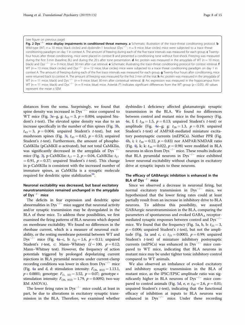
we sought to determine whether deletion of dysbindin-1 inmice impairs amygdala-dependent behavior. Pavlovianthreat conditioning is a behavioral paradigm that is used totest fear learning and memory, in which the amygdala iscritically involved 49,50. In auditory threat (fear) con-ditioning, an auditory cue (conditioned stimulus; CS) ispaired with an aversive foot-shock (unconditioned stimu-lus; US). Re-exposure to the same CS elicits a fear response(i.e. freezing). WT and Dys−/− mice were subjected to atrace threat fear conditioning (five tone-shock pairings;Fig. 2a) on day 1. Twenty-four hours after conditioning,
mice were placed in a novel context and presented withthe CS (cue retrieval). Both WT and Dys−/− mice condi-tioned equally well on day 1, as shown by similar levels offreezing (Fig. 2b, genotype: F(1, 17)= 0.45, p= 0.51; two-way RM ANOVA). However, Dys−/− mice froze lessduring the cue-dependent fear recall (Fig. 2c, t17= 2.2, p= 0.04; unpaired Student’s t-test). The behavioral deficitwas associated with reduced protein levels of activityregulated cytoskeletal protein (Arc), a plasticity-associatedprotein51,52, in the amygdala (Fig. 2d, t17= 2.1, p= 0.04;unpaired Student’s t-test). Separate cohorts of mice weresubjected to the same procedure on day 1 (Fig. 2e, f, f:genotype: F(1, 23)= 1.3, p= 0.26; two-way RM ANOVA).On day 2, mice were placed back in the same conditioningchamber to assess contextual memory. Dys−/− miceexhibited lower freezing during the context-dependentrecall test (Fig. 2g, t23=2.14, p= 0.04; Student’s t test) onday 2. We also observed lower Arc expression in theamygdala (Fig. 2h, t23= 5.4, p < 0.0001; unpaired Student’st-test) and in the hippocampus (Fig. 2i, t18= 2.6, p= 0.017;unpaired Student’s t-test) of Dys−/− mice compared toWT mice after contextual testing. Finally, we did notobserve any differences between genotypes across severalanxiety tests, including the open field test (SupplementalFig. 1a-b; time in center: t17= 0.83, p= 0.42; entries intocenter: t17= 1.3, p= 0.21; unpaired Student’s t-test), light-dark box (Supplemental Fig. 1c, t18= 0.39, p= 0.7;unpaired Student’s t-test), and EPM (Supplemental Fig. 1d,open arms: t16= 1.7, p= 0.12; closed arms: t17= 1, p=0.32; unpaired Student’s t-test). In summary, Dys−/− micedisplayed impaired threat memory recall to the tone andcontext, which was not confounded by differences inbaseline anxiety.
Dendritic spine morphology in the BLA is altered in Dys−/−
miceAs threat memory was diminished and Arc expression
was downregulated in Dys−/− mice, we hypothesized thatdendritic morphology would be altered in Dys−/− micebecause Arc is an essential mediator of activity-dependentsynaptic plasticity at the level of dendritic spines, whichcan undergo structural changes during memory encod-ing53,54. Thus, we explored whether dendritic arborizationand spine morphology were perturbed in the BLA ofDys−/− mice. Brains of naïve, WT and Dys−/− mice werefixed and pyramidal neurons in the BLA were ex vivoiontophoretically filled with Lucifer yellow (Fig. 3a) formorphological analyses55. Sholl analysis (Fig. 3b) revealedthat there was no significant difference between experi-mental groups in the number of dendritic intersections(Fig. 3c, genotype: F(1, 6)= 0.15, p= 0.71; intersection:F(24, 191)= 0.45, p= 0.84; nested ANOVA) or dendriticlength (Fig. 3d, genotype: F(1, 6)= 0.03, p= 0.87; length:F(24, 191)= 1.23, p= 0.63; nested ANOVA) at increasing
Huang et al. Translational Psychiatry (2019) 9:132 Page 5 of 15
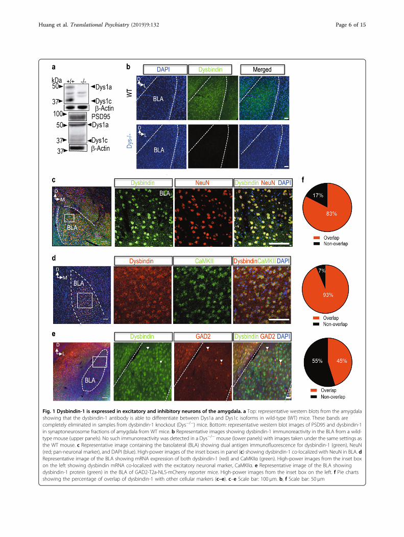
Fig. 1 Dysbindin-1 is expressed in excitatory and inhibitory neurons of the amygdala. a Top: representative western blots from the amygdalashowing that the dysbindin-1 antibody is able to differentiate between Dys1a and Dys1c isoforms in wild-type (WT) mice. These bands arecompletely eliminated in samples from dysbindin-1 knockout (Dys−/−) mice. Bottom: representative western blot images of PSD95 and dysbindin-1in synaptoneurosome fractions of amygdala from WT mice. b Representative images showing dysbindin-1 immunoreactivity in the BLA from a wild-type mouse (upper panels). No such immunoreactivity was detected in a Dys−/− mouse (lower panels) with images taken under the same settings asthe WT mouse. c Representative image containing the basolateral (BLA) showing dual antigen immunofluorescence for dysbindin-1 (green), NeuN(red; pan-neuronal marker), and DAPI (blue). High-power images of the inset boxes in panel (c) showing dysbindin-1 co-localized with NeuN in BLA. dRepresentative image of the BLA showing mRNA expression of both dysbindin-1 (red) and CaMKIIα (green). High-power images from the inset boxon the left showing dysbindin mRNA co-localized with the excitatory neuronal marker, CaMKIIα. e Representative image of the BLA showingdysbindin-1 protein (green) in the BLA of GAD2-T2a-NLS-mCherry reporter mice. High-power images from the inset box on the left. f Pie chartsshowing the percentage of overlap of dysbindin-1 with other cellular markers (c–e). c–e Scale bar: 100 µm. b, f Scale bar: 50 µm
Huang et al. Translational Psychiatry (2019) 9:132 Page 6 of 15

1 2 3 4 50
20
40
60
80
100
Trace
%of
Free
zing
WTDys-/-
CuedAmygdala
a
f
1 2 3 4 50
20
40
60
80
100
Trace
%of
Free
zing Dys-/-
WT
h
b
Day1
Tone
ShockDay2
g
0
20
40
60
80
%of
Free
zing
WT Dys-/-
*
Context A Context B
0
20
40
60
80
100
%of
Free
zing
BL 1
*
WTDys-/-
Context
x1
WT Dys-/-
Arcβ-Actin
c d
0
100
200
Arce
xpre
ssion
(%of
WT)
WT Dys-/-
*
Cued
5037
kDa
eDay1
Tone
Shock
Context A
Day2
Context A 3 min
050
100150200
Arce
xpre
ssion
(%of
WT)
****
WT Dys-/-
ContextAmygdala
WT Dys-/-
Arcβ-Actin
5037
kDa
i ContextHippocampus
Arcβ-Actin
WT Dys-/-
5037
kDa
0
50
100
150
Arc
expr
essio
n(%
ofW
T)
*
WT Dys-/-
Fig. 2 (See legend on next page.)
Huang et al. Translational Psychiatry (2019) 9:132 Page 7 of 15
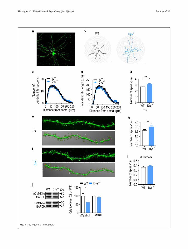
distances from the soma. Surprisingly, we found thatspine density was increased in Dys−/− mice compared toWT mice (Fig. 3e–g, g: t43= 3, p= 0.004; unpaired Stu-dent’s t-test). The elevated spine density was due to anincrease specifically in the number of thin spines (Fig. 3h,t43= 3, p= 0.004; unpaired Student’s t-test), but notmushroom spines (Fig. 3i, t43= 0.62, p= 0.53; unpairedStudent’s t-test). Furthermore, the amount of phospho-CaMKIIα (pCaMKII α-activated), but not total CaMKIIα,was significantly decreased in the amygdala of Dys−/−
mice (Fig. 3j, p-CaMKIIα: t17= 2, p= 0.04, CaMKIIα: t17= 0.91, p= 0.37; unpaired Student’s t-test). This changein p-CaMKIIα is consistent with the increased number ofimmature spines, as CaMKIIα is a synaptic moleculerequired for dendritic spine stabilization56.
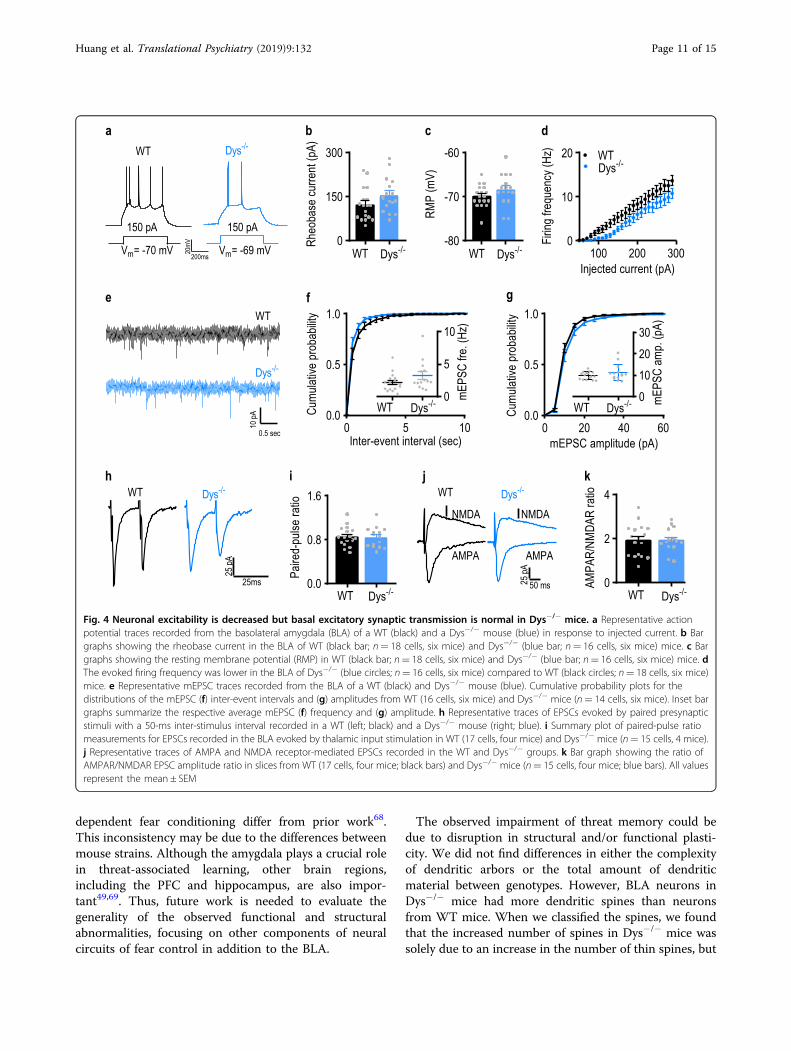
Neuronal excitability was decreased, but basal excitatoryneurotransmission remained unchanged in the amygdalaof Dys−/− miceThe deficits in fear expression and dendritic spine
abnormalities in Dys−/− mice suggest that neuronal activityand/or synaptic transmission may be dysregulated in theBLA of these mice. To address these possibilities, we firstexamined the firing patterns of BLA neurons which dependon membrane excitability. We found no differences in therheobase current, which is a measure of neuronal excit-ability, or the resting membrane potential between WT andDys−/− mice (Fig. 4a–c, b: t32= 1.6, p= 0.11; unpairedStudent’s t-test, c: Mann–Whitney U= 100, p= 0.12;Mann–Whitney test). However, the frequency of actionpotentials triggered by prolonged depolarizing currentinjections in BLA pyramidal neurons under current-clamprecording conditions was lower in slices from Dys−/− mice(Fig. 4a and d, d: stimulation intensity: F(26, 832)= 1.13.1,p < 0.0001; genotype: F(1, 32)= 3.52, p= 0.07; genotype ×stimulation intensity: F(26, 832)= 1.79, p= 0.0092; two-wayRM ANOVA).The lower firing rates in Dys−/− mice could, at least in
part, be due to alterations in excitatory synaptic trans-mission in the BLA. Therefore, we examined whether
dysbindin-1 deficiency affected glutamatergic synaptictransmission in the BLA. We found no differencesbetween control and mutant mice in the frequency (Fig.4e, f; f: t28= 1.5, p= 0.13; unpaired Student’s t-test) oramplitude (Fig. 4e–g; g: t28= 1.3, p= 0.19; unpairedStudent’s t-test) of AMPAR-mediated miniature excita-tory postsynaptic currents (mEPSCs). Neither PPR (Fig.4h, i; i: t30= 0.22, p= 0.83) nor AMPAR/NMDAR ratio(Fig. 4j, k; k: t30= 0.022, p= 0.98) were modified in BLAneurons in slices from Dys−/− mice. These results indicatethat BLA pyramidal neurons in Dys−/− mice exhibitedlower neuronal excitability without changes in excitatorydrive at synaptic inputs to these neurons.
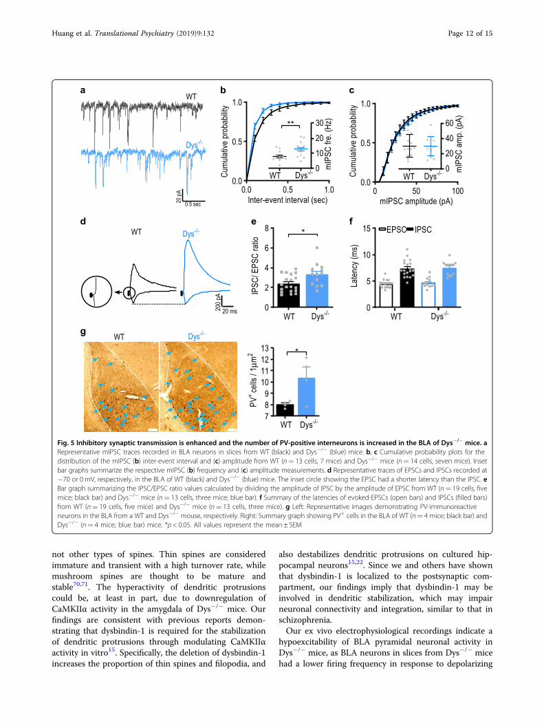
The efficacy of GABAergic inhibition is enhanced in theBLA of Dys−/− miceSince we observed a decrease in neuronal firing, but
normal excitatory transmission in Dys−/− mice, wehypothesized that the lower firing rates could at leastpartially result from an increase in inhibitory drive to BLAneurons. To address this possibility, we assayedGABAergic neurotransmission in the BLA, comparing theparameters of spontaneous and evoked GABAA receptor-mediated synaptic responses between control and Dys−/−
mice. We found that the frequency (Fig. 5a, b, b: t25= 3,p= 0.006; unpaired Student’s t-test), but not the ampli-tude (Fig. 5a and c, c: t25= 0.0003, p= 0.99; unpairedStudent’s t-test) of miniature inhibitory postsynapticcurrents (mIPSCs) was enhanced in Dys−/− mice com-pared to WT mice, indicating that BLA neurons inmutant mice may be under tighter tonic inhibitory controlcompared to WT animals.We also observed an imbalance of evoked excitatory
and inhibitory synaptic transmission in the BLA ofmutant mice, as the IPSC/EPSC amplitude ratio was sig-nificantly higher in BLA neurons of Dys−/− mice com-pared to control animals (Fig. 5d, e, e: t30= 2.6, p= 0.01;unpaired Student’s t-test), indicating that the functionalefficacy of inhibition at inputs to BLA neurons wasenhanced in Dys−/− mice. Under these recording
(see figure on previous page)Fig. 2 Dys−/− mice display impairments in conditioned threat memory. a Schematic illustration of the trace-threat conditioning protocol. bWild-type (WT; n= 10 mice; black circles) and dysbindin-1 knockout (Dys−/−; n= 9 mice; blue circles) mice were subjected to a trace threat-conditioning paradigm on day 1 in context A. The amount of freezing during each of the five trace intervals was measured for each group. c Twenty-four hours after threat conditioning, mice were placed in context B and presented a conditioning tone without foot-shock. Freezing was measuredduring the first 3 min (baseline; BL) and during the 20 s after tone presentation. d Arc protein was measured in the amygdala of WT (n= 10 mice;black) and Dys−/− (n= 9 mice; blue) 30 min after cue retrieval. e Schematic illustrating the trace-threat conditioning protocol for context retrieval. fWT (n= 13 mice; black circles) and Dys−/− (n= 12 mice; blue circles) mice were subjected to a trace threat conditioning paradigm on day 1 incontext A. The amount of freezing during each of the five trace intervals was measured for each group. g Twenty-four hours after conditioning, micewere returned back to context A. The amount of freezing was measured for the first 3 min of the trial. h Arc protein was measured in the amygdala ofWT (n= 11 mice; black) and Dys−/− (n= 9 mice; blue) 30 min after contextual retrieval. (i) Arc expression was measured in the hippocampus fromWT (n= 11 mice; black) and Dys−/− (n= 9 mice; blue) mice. Asterisk (*) indicates significant differences from the WT group (p < 0.05). All valuesrepresent the mean ± SEM
Huang et al. Translational Psychiatry (2019) 9:132 Page 8 of 15
kDa
0 50 100 150 200 250
50100150200
0
250To
tald
endr
iticlen
gth
(μm)
WTDys-/-
Distance from soma (μm)0 50 100 150 200 250
0
10
20
Num
bero
fde
ndriti
cint
erse
ction
s WTDys-/-
Distance from soma (μm)
0.00.51.01.52.02.5
Num
bero
fspin
es/μ
m **
c d
e
f
0.00.10.20.30.40.5
Num
bero
fspin
es/μ
m
0
1
2
3
4
Num
bero
fspin
es/μ
m **
h
i
WT Dys-/-
WT Dys-/-
WT Dys-/-
Thin
Mushroom
g
a bWT Dys-/-
WT
Dys-/ -
j
pCaMKIIαGAPDH
CaMKIIαGAPDH
50
50
37
37
WT Dys-/-
0
50
100
150
Relat
ivelev
el(%
ofW
T) *WT Dys-/-
pCaMKII CaMKII
Fig. 3 (See legend on next page.)
Huang et al. Translational Psychiatry (2019) 9:132 Page 9 of 15

conditions, monosynaptic glutamatergic and disynapticGABAergic evoked synaptic responses, induced by sti-mulation of the thalamic input, are recorded from thesame neuron57. As the IPSC and EPSC in a recorded BLAneuron are induced by presynaptic stimuli of an identicalintensity, the IPSC recordings are internally controlled,thus minimizing the variability potentially associated witha position of the stimulation electrode or its physicalcharacteristics. The latencies of both evoked EPSCs andIPSCs were not affected in mutant animals (Fig. 5d and f,f: EPSC: t30= 0.69, p= 0.49 between genotypes; IPSC: t30= 0.27, p= 0.79 between genotypes; unpaired Student’st-test).Notably, we found that the number of parvalbumin
immuno-reactive cells (PV+) was increased in the BLA ofDys−/− mice compared to WT mice (Fig. 5g, t6= 2.5,p= 0.04; unpaired Student’s t-test). The increased num-bers of PV+ cells in the BLA in mice lacking dysbindin-1may result in an enhancement of inhibitory drive ontoBLA neurons, which in turn could lead to decreases inneuronal activity in the BLA and subsequently impairthreat memory consolidation.
DiscussionOur findings demonstrate that the genetic ablation of
dysbindin-1, a protein expressed in both excitatory neuronsand inhibitory interneurons of the BLA, impaired the fearresponse of conditioned threat memory in mice, which wasassociated with cellular, structural, and functionalabnormalities in the amygdala. Specifically, neuronal excit-ability in the BLA was decreased in Dys−/− mice, and thelatter was associated with enhanced GABAergic synaptictransmission, an elevated inhibition/excitation ratio, and anincreased number of PV+ cells in the BLA of Dys−/− mice.These changes could be translated into reduced neuronalactivity in the BLA and subsequently reduced Arc and p-CaMKIIα protein expression in Dys−/− mice. Taken toge-ther, our results provide new insights into the functions ofdysbindin-1 in the BLA and underscore the importance of
dysbinidn-1 contribution to the mechanisms of emotionalmemory.We found dysbindin-1 mRNA and protein are expres-
sed in the BLA. Nearly all CaMKIIα-positive cellsexpressed dysbindin-1. Using several methods to visualizeboth GABA-synthesizing enzymes (GAD1 and GAD2), wefound that dysbindin-1 was located in a sub-population ofGABAergic neurons in the BLA (45%). Our results indi-cate that dysbindin-1 is widely expressed in the BLA.Future studies will investigate whether dysbindin-1 isenriched in particular subpopulations of inhibitoryamygdala neurons.We used a trace-threat-conditioning paradigm, in which
the amygdala is critically involved58–61, to evaluate thefunctional significance of dysbindin-1 deletion. In trace-threat conditioning, there is a temporal gap between theCS and US, providing a rapidly acquired model not onlyfor emotional learning and memory, also for workingmemory and attention-dependent associative learning.Both emotional processing and higher cognitive learningare disrupted in schizophrenia62,63. We found that Dys−/−
mice displayed impaired threat memory 24 h after con-ditioning in response to both the CS and conditionedcontext, which was accompanied by reduced levels of Arcexpression in the amygdala and hippocampus. Theamygdala is implicated in CS-mediated threat memoryformation and retrieval64,65. Diminished CS-induced fearresponses in Dys−/− mice indicate that dysbindin-1 mayplay a role in the amygdala gating the mechanisms ofthreat memory consolidation. While retrieval of con-textual threat conditioning is hippocampus-dependent66,the observed reduction of activity-dependent Arcexpression in the amygdala and hippocampus in con-textually conditioned Dys−/− mice is in line with a recentstudy demonstrating that ventral hippocampal projectionsto the basal amygdala are necessary for contextual threatmemory retrieval67. Our results with the context-dependent fear memory test are consistent with pre-vious findings19, whereas our data from the cue-
(see figure on previous page)Fig. 3 Dendritic spine structure of basolateral amygdala pyramidal neurons is altered in Dys−/− mice. a Representative image showing apyramidal neuron filled with Lucifer yellow in the basolateral amygdala. b Representative reconstructions of injected BLA neurons from WT and Dys−/−
mice that were used for morphologic analysis. The radius of concentric circles used for Sholl analysis was increased at 10 µm intervals from the soma. Scalebar: 100 µm. Sholl analysis was performed on reconstructed WT (black circles) and Dys−/− (blue circles) mice neurons to analyze dendritic complexity (c:intersections; d: total dendritic length at each interval). e Top: representative confocal image showing a dendritic segment of a filled BLA pyramidal WTneuron used for spine analysis. Bottom: the dendritic segment showed above was traced in 3D using reconstructions obtained from NeuronStudio toanalyze spines. Thin spine: yellow color; mushroom spine: brown color. f Top: representative confocal image showing a dendritic segment of a filled BLApyramidal Dys−/− neuron used for spine analysis. Bottom: the dendritic segment showed above was traced in 3D using reconstructions obtained fromNeuroStudio to analyze spines. Average g total spine density, h thin spine density, imushroom spine density was calculated for WT (open bars) and Dys−/− (blue bars) neurons. g–i For spine analysis, 4–6 dendritic segments per neurons were analyzed. A total of 23 neurons from 4 WT mice and 22 neuronsfrom 4 Dys−/− mice were included for all analyses. j Left: representative western blot images showing phosphorylated CaMKIIα (pCaMKIIα) and totalCaMKIIα in the amygdala from WT and Dys−/− mice. Right: levels of pCaMKIIα and CaMKIIα were measured in the amygdala from WT (open bars; n= 10mice) and Dys−/− mice (blue bars; n= 9 mice). *p < 0.05; **p < 0.01. All values represent the mean ± SEM
Huang et al. Translational Psychiatry (2019) 9:132 Page 10 of 15
dependent fear conditioning differ from prior work68.This inconsistency may be due to the differences betweenmouse strains. Although the amygdala plays a crucial rolein threat-associated learning, other brain regions,including the PFC and hippocampus, are also impor-tant49,69. Thus, future work is needed to evaluate thegenerality of the observed functional and structuralabnormalities, focusing on other components of neuralcircuits of fear control in addition to the BLA.
The observed impairment of threat memory could bedue to disruption in structural and/or functional plasti-city. We did not find differences in either the complexityof dendritic arbors or the total amount of dendriticmaterial between genotypes. However, BLA neurons inDys−/− mice had more dendritic spines than neuronsfrom WT mice. When we classified the spines, we foundthat the increased number of spines in Dys−/− mice wassolely due to an increase in the number of thin spines, but
0 5 100.0
0.5
1.0
Inter-event interval (sec)
Cum
ulativ
epr
obab
ility
0
5
10
mEPS
Cfre
.(Hz
)
WT Dys-/-
0.0
0.8
1.6
Paire
d-pu
lsera
tio
WT Dys-/-
0 20 40 600.0
0.5
1.0
mEPSC amplitude (pA)
Cum
ulativ
epr
obab
ility
0
150
300
Rheo
base
curre
nt(p
A)
WT Dys-/--80
-70
-60
RMP
(mV)
WT Dys-/- 100 200 3000
10
20
Injected current (pA)
Firing
frequ
ency
(Hz) WT
Dys-/-
a b c d
200ms
20mV
0102030
mEPS
Cam
p.(p
A)
WT Dys-/-
e g
150 pA
Vm= -70 mV
150 pA
Vm= -69 mV
WT Dys-/-
0.5 sec
10pA
WT
Dys-/-
f
h j
50 ms25pA
25ms
25pA
WT Dys-/- WT Dys-/-
NMDA NMDA
AMPA AMPA
0
2
4
AMPA
R/NM
DAR
ratio
WT Dys-/-
i k
Fig. 4 Neuronal excitability is decreased but basal excitatory synaptic transmission is normal in Dys−/− mice. a Representative actionpotential traces recorded from the basolateral amygdala (BLA) of a WT (black) and a Dys−/− mouse (blue) in response to injected current. b Bargraphs showing the rheobase current in the BLA of WT (black bar; n= 18 cells, six mice) and Dys−/− (blue bar; n= 16 cells, six mice) mice. c Bargraphs showing the resting membrane potential (RMP) in WT (black bar; n= 18 cells, six mice) and Dys−/− (blue bar; n= 16 cells, six mice) mice. dThe evoked firing frequency was lower in the BLA of Dys−/− (blue circles; n= 16 cells, six mice) compared to WT (black circles; n= 18 cells, six mice)mice. e Representative mEPSC traces recorded from the BLA of a WT (black) and Dys−/− mouse (blue). Cumulative probability plots for thedistributions of the mEPSC (f) inter-event intervals and (g) amplitudes from WT (16 cells, six mice) and Dys−/− mice (n= 14 cells, six mice). Inset bargraphs summarize the respective average mEPSC (f) frequency and (g) amplitude. h Representative traces of EPSCs evoked by paired presynapticstimuli with a 50-ms inter-stimulus interval recorded in a WT (left; black) and a Dys−/− mouse (right; blue). i Summary plot of paired-pulse ratiomeasurements for EPSCs recorded in the BLA evoked by thalamic input stimulation in WT (17 cells, four mice) and Dys−/− mice (n= 15 cells, 4 mice).j Representative traces of AMPA and NMDA receptor-mediated EPSCs recorded in the WT and Dys−/− groups. k Bar graph showing the ratio ofAMPAR/NMDAR EPSC amplitude ratio in slices from WT (17 cells, four mice; black bars) and Dys−/− mice (n= 15 cells, four mice; blue bars). All valuesrepresent the mean ± SEM
Huang et al. Translational Psychiatry (2019) 9:132 Page 11 of 15
not other types of spines. Thin spines are consideredimmature and transient with a high turnover rate, whilemushroom spines are thought to be mature andstable70,71. The hyperactivity of dendritic protrusionscould be, at least in part, due to downregulation ofCaMKIIα activity in the amygdala of Dys−/− mice. Ourfindings are consistent with previous reports demon-strating that dysbindin-1 is required for the stabilizationof dendritic protrusions through modulating CaMKIIαactivity in vitro15. Specifically, the deletion of dysbindin-1increases the proportion of thin spines and filopodia, and
also destabilizes dendritic protrusions on cultured hip-pocampal neurons15,22. Since we and others have shownthat dysbindin-1 is localized to the postsynaptic com-partment, our findings imply that dysbindin-1 may beinvolved in dendritic stabilization, which may impairneuronal connectivity and integration, similar to that inschizophrenia.Our ex vivo electrophysiological recordings indicate a
hypoexcitability of BLA pyramidal neuronal activity inDys−/− mice, as BLA neurons in slices from Dys−/− micehad a lower firing frequency in response to depolarizing
200
pA
20 ms
d
0
2
4
6
8
IPSC
/EPS
Cra
tio
WT Dys-/-
*e
0
5
10
15
Late
ncy(
ms)
WT Dys-/-
EPSC IPSC
0.5 sec20pA 0 50 100
0.0
0.5
1.0
mIPSC amplitude (pA)
Cum
ulativ
epr
obab
ility
0.0 0.5 1.00.0
0.5
1.0
Inter-event interval (sec)
Cum
ulativ
epr
obab
ility
a b c
0204060
mIP
SCam
p.(p
A)
WT Dys-/-0102030
mIPS
Cfre
.(Hz
)
**
WT Dys-/-
WT
Dys-/-
g
fWT Dys-/-
789
10111213
PV+ ce
lls/1
μm2
WT Dys-/-
* BLA
WT Dys-/-
Fig. 5 Inhibitory synaptic transmission is enhanced and the number of PV-positive interneurons is increased in the BLA of Dys−/− mice. aRepresentative mIPSC traces recorded in BLA neurons in slices from WT (black) and Dys−/− (blue) mice. b, c Cumulative probability plots for thedistribution of the mIPSC (b) inter-event interval and (c) amplitude from WT (n= 13 cells, 7 mice) and Dys−/− mice (n= 14 cells, seven mice). Insetbar graphs summarize the respective mIPSC (b) frequency and (c) amplitude measurements. d Representative traces of EPSCs and IPSCs recorded at−70 or 0 mV, respectively, in the BLA of WT (black) and Dys−/− (blue) mice. The inset circle showing the EPSC had a shorter latency than the IPSC. eBar graph summarizing the IPSC/EPSC ratio values calculated by dividing the amplitude of IPSC by the amplitude of EPSC from WT (n= 19 cells, fivemice; black bar) and Dys−/− mice (n= 13 cells, three mice; blue bar). f Summary of the latencies of evoked-EPSCs (open bars) and IPSCs (filled bars)from WT (n= 19 cells, five mice) and Dys−/− mice (n= 13 cells, three mice). g Left: Representative images demonstrating PV-immunoreactiveneurons in the BLA from a WT and Dys−/− mouse, respectively. Right: Summary graph showing PV+ cells in the BLA of WT (n= 4 mice; black bar) andDys−/− (n= 4 mice; blue bar) mice. *p < 0.05. All values represent the mean ± SEM
Huang et al. Translational Psychiatry (2019) 9:132 Page 12 of 15

pulses. Although Dys−/− mice exhibited higher dendriticspine density than WT mice, neither excitatory neuro-transmission nor AMPAR/NMDAR ratio were altered inBLA pyramidal neurons in our experiments. This incon-sistency between increased spine density without altera-tions of excitatory neurotransmission in Dys−/− mice,could be due to the increased proportion of thin, highlyunstable spines, which do not normally make functionalsynapses70. However, we found that that the frequency ofGABAA-receptor-mediated spontaneous IPSCs, but nottheir amplitude, was elevated in Dys−/− mice, which isconsistent with the role dysbindin-1 plays in regulatingGABA release9,20. The inhibition/excitation ratio wasenhanced in neurons of Dys−/− mice, indicating that theactivity of excitatory neurons could be inhibited in theseanimals. However, other mechanisms, including changesin ion conductances (e.g., due to altered levels and/orfunction of voltage-gated Na+ and/or K+ channels, and/ormodifications in slow and fast AHPs and Ih) couldpotentially also contribute to the observed decreases inneuronal excitability in mutant mice. Specifically,network-level plastic modifications in the BLA, resultingin changes in the frequency of spike-induced sEPSCs,could possibly affect neuronal excitability, thus warrant-ing further investigation. Notably, enhanced GABAergicinhibition has been shown to impair consolidation ofthreat memory72. The increase in inhibitory drive onto theBLA pyramidal neurons in Dys−/− mice could be due, inpart, to the observed increase in the number of PV+ cells,providing perisomatic inhibition of principal neurons73.Another possibility is that interneurons could becomehyperactive through the increased synaptic strength atinputs from PFC. It has been shown that pyramidalneurons in the mPFC project to interneurons within theBLA45, and deletion of dysbinidin-1 in the PFC increasesthe activity of layer 2/3 pyramidal neurons, which in turnelevates neurotransmission onto interneurons in theBLA20. The enhanced functional efficacy of inhibition inthe BLA in Dys−/− mice could contribute to impairmentsin the mechanisms of threat memory consolidation andretrieval.Our finding of enhanced GABAergic transmission in
the BLA differs from what has been reported for themPFC of Dys−/− mice, where the inhibitory drive topyramidal neurons in layer V of PFC was found to bedecreased74. The observed differences reinforce the viewthat the mutation effects may be brain region specific andbe translated into region-specific functional modifications(e.g., distinct synaptic and neuronal changes). In themPFC, impaired GABAergic transmission and fewerinhibitory synapses on pyramidal neurons of Dys−/− micewas apparently due to reduced activity-dependent secre-tion of brain-derived neurotrophic factor (BDNF) frompyramidal neurons9,20. Thus, in the BLA of Dys−/− mice,
it is possible that BDNF release is increased from excita-tory neurons, thereby enhancing BLA GABAergic trans-mission. Furthermore, BDNF deletion has been shown toreduce PV expression and cell density75. Increased BDNFin the BLA of Dys−/− mice could also explain theincreased number of PV neurons, which is in contrast towhat has been observed in the dorsolateral PFC ofpatients with schizophrenia76, a region that also exhibitsreduced BDNF expression77. There are numerous exam-ples in the literature showing that various perturbationscan either increase or decrease BDNF expressiondepending on the brain region78–81. Future work willdetermine whether increased BDNF expression and/orsecretion is responsible for the increased inhibitory drivein the BLA of Dys−/− mice.In summary, we have demonstrated that dysbindin-1 is
expressed in both excitatory neurons and inhibitoryinterneurons in the BLA, an essential part of neural cir-cuits of fear conditioning. In Dys−/− mice, an increase ininhibitory drive to principal neurons in the BLA wasassociated with a lower firing rate of BLA pyramidalneurons. The resulting reductions in neuronal activitycould explain reduced Arc expression and CaMKIIαactivation in mutant mice, observed in our studies. As theBLA is critical for fear memory encoding and, possibly,retention, the enhanced inhibitory tone in the BLA couldcontribute to the impaired threat memory observed inDys−/− mice. Our findings indicate an important role fordysbindin-1 in regulation of the amygdalar function andsuggest potential mechanisms by which dysbindin-1 maycontribute to the pathophysiology of schizophrenia.
AcknowledgementsThis research was supported by 5R00MH099252 (D.T.B.) and 5R01MH105851,5R01MH108655 (V.Y.B.). We thank Drs. Uwe Rudolph and Kerry Ressler for thegenerous use of their equipment, as well as software. We also thank Dr. TinaGruene for technical support and advice with iontophoretic microinjectionsand dendritic morphology analysis.
Author details1Department of Psychiatry, Harvard Medical School, Boston, MA, USA.2Translational Psychiatry laboratory, McLean Hospital, Belmont, MA, USA.3Cellular Neurobiology laboratory, McLean Hospital, Belmont, MA, USA.4Present address: Department of Life Sciences, National Central University,Taoyuan, Taiwan
Conflict of interestD.T.B. served as a consultant for LifeSci Capital. The remaining authors declarethat they have no conflict of interest.
Publisher’s noteSpringer Nature remains neutral with regard to jurisdictional claims inpublished maps and institutional affiliations.
Supplementary Information accompanies this paper at (https://doi.org/10.1038/s41398-019-0465-y).
Huang et al. Translational Psychiatry (2019) 9:132 Page 13 of 15

Received: 5 March 2019 Accepted: 23 March 2019
References1. Perala, J. et al. Lifetime prevalence of psychotic and bipolar I disorders in a
general population. Arch. Gen. Psychiatry 64, 19–28 (2007).2. Talbot, K. et al. Dysbindin-1 is reduced in intrinsic, glutamatergic terminals of
the hippocampal formation in schizophrenia. J. Clin. Invest. 113, 1353–1363(2004).
3. Talbot, K. et al. Synaptic dysbindin-1 reductions in schizophrenia occur in anisoform-specific manner indicating their subsynaptic location. PLoS ONE 6,e16886 (2011).
4. Tang, J. Dysbindin-1 in dorsolateral prefrontal cortex of schizophrenia cases isreduced in an isoform-specific manner unrelated to dysbindin-1 mRNAexpression. Hum. Mol. Genet. 18, 3851–3863 (2009).
5. Weickert, C. S. et al. Human dysbindin (DTNBP1) gene expression in normalbrain and in schizophrenic prefrontal cortex and midbrain. Arch. Gen. Psy-chiatry 61, 544–555 (2004).
6. Scheggia, D. et al. Variations in Dysbindin-1 are associated with cognitiveresponse to antipsychotic drug treatment. Nat. Commun. 9, 2265 (2018).
7. Cox, M. M. et al. Neurobehavioral abnormalities in the dysbindin-1 mutant,sandy, on a C57BL/6J genetic background. Genes Brain Behav. 8, 390–397(2009).
8. Papaleo, F. et al. Dysbindin-1 modulates prefrontal cortical activity andschizophrenia-like behaviors via dopamine/D2 pathways. Mol. Psychiatry 17,85–98 (2012).
9. Zhang, W. et al. BDNF rescues prefrontal dysfunction elicited by pyramidalneuron-specific DTNBP1 deletion in vivo. J. Mol. Cell Biol. 9, 117–131 (2017).
10. Bhardwaj, S. K., Ryan, R. T., Wong, T. P. & Srivastava, L. K. Loss of dysbindin-1, arisk gene for schizophrenia, leads to impaired group 1 metabotropic gluta-mate receptor function in mice. Front. Behav. Neurosci. 9, 72 (2015).
11. Feng, Y. Q. et al. Dysbindin deficiency in sandy mice causes reduction ofsnapin and displays behaviors related to schizophrenia. Schizophr. Res. 106,218–228 (2008).
12. Hattori, S. et al. Behavioral abnormalities and dopamine reductions in sdymutant mice with a deletion in Dtnbp1, a susceptibility gene for schizo-phrenia. Biochem. Biophys. Res. Commun. 373, 298–302 (2008).
13. Carr, G. V., Jenkins, K. A., Weinberger, D. R. & Papaleo, F. Loss of dysbindin-1 inmice impairs reward-based operant learning by increasing impulsive andcompulsive behavior. Behav. Brain Res. 241, 173–184 (2013).
14. Talbot K. et al. Dysbindin-1 and Its Protein Family. In: Lajtha A., Javitt D.,Kantrowitz J. (eds) Handbook of Neurochemistry and Molecular Neurobiology.Springer, Boston, MA. 107–241 (2009).
15. Jia, J. M., Hu, Z., Nordman, J. & Li, Z. The schizophrenia susceptibility genedysbindin regulates dendritic spine dynamics. J. Neurosci. 34, 13725–13736(2014).
16. Mullin, A. P. et al. Gene dosage in the dysbindin schizophrenia susceptibilitynetwork differentially affect synaptic function and plasticity. J. Neurosci. 35,325–338 (2015).
17. Wentzel, C., Delvendahl, I., Sydlik, S., Georgiev, O. & Muller, M. Dysbindin linkspresynaptic proteasome function to homeostatic recruitment of low releaseprobability vesicles. Nat. Commun. 9, 267 (2018).
18. Dickman, D. K. & Davis, G. W. The schizophrenia susceptibility gene dysbindincontrols synaptic homeostasis. Science 326, 1127–1130 (2009).
19. Glen, W. B. Jr. et al. Dysbindin-1 loss compromises NMDAR-dependentsynaptic plasticity and contextual fear conditioning. Hippocampus 24, 204–213(2014).
20. Yuan, Q. Regulation of brain-derived neurotrophic factor exocytosis andgamma-aminobutyric acidergic interneuron synapse by the schizophreniasusceptibility gene Dysbindin-1. Biol. Psychiatry 80, 312–322 (2016).
21. Karlsgodt, K. H. et al. Reduced dysbindin expression mediates N-methyl-D-aspartate receptor hypofunction and impaired working memory performance.Biol. Psychiatry 69, 28–34 (2011).
22. Ito, H. et al. Dysbindin-1, WAVE2 and Abi-1 form a complex that regulatesdendritic spine formation. Mol. Psychiatry 15, 976–986 (2010).
23. LeDoux, J. E. Emotion circuits in the brain. Annu. Rev. Neurosci. 23, 155–184(2000).
24. Rich, A. M. et al. Amygdala volume is reduced in early course schizophrenia.Psychiat. Res.-Neuroim. 250, 50–60 (2016).
25. Anticevic, A., Repovs, G. & Barch, D. M. Emotion effects on attention, amygdalaactivation, and functional connectivity in schizophrenia. Schizophr. Bull. 38,967–980 (2012).
26. Williams, L. M. et al. Dysregulation of arousal and amygdala-prefrontal systemsin paranoid schizophrenia. Am. J. Psychiatry 161, 480–489 (2004).
27. Bjorkquist, O. A., Olsen, E. K., Nelson, B. D. & Herbener, E. S. Altered amygdala-prefrontal connectivity during emotion perception in schizophrenia. Schizophr.Res. 175, 35–41 (2016).
28. Salgado-Pineda, P., Fakra, E., Delaveau, P., Hariri, A. R. & Blin, O. Differentialpatterns of initial and sustained responses in amygdala and cortical regions toemotional stimuli in schizophrenia patients and healthy participants. J. Psy-chiatry Neurosci. 35, 41–48 (2010).
29. Rahm, C. et al. Negative symptoms in schizophrenia show association withamygdala volumes and neural activation during affective processing. ActaNeuropsychiatr. 27, 213–220 (2015).
30. Pankow, A. et al. Altered amygdala activation in schizophrenia patients duringemotion processing. Schizophr. Res. 150, 101–106 (2013).
31. Chang, X. et al. RNA-seq analysis of amygdala tissue reveals characteristicexpression profiles in schizophrenia. Transl. Psychiatry 7, e1203 (2017).
32. Lawrie, S. M., Whalley, H. C., Job, D. E. & Johnstone, E. C. Structural andfunctional abnormalities of the amygdala in schizophrenia. Ann. N. Y. Acad. Sci.985, 445–460 (2003).
33. Holt, D. J. et al. Increased medial temporal lobe activation during the passiveviewing of emotional and neutral facial expressions in schizophrenia. Schi-zophr. Res. 82, 153–162 (2006).
34. Berretta, S., Pantazopoulos, H. & Lange, N. Neuron numbers and volume of theamygdala in subjects diagnosed with bipolar disorder or schizophrenia. Biol.Psychiatry 62, 884–893 (2007).
35. Das, P. et al. Functional disconnections in the direct and indirect amygdalapathways for fear processing in schizophrenia. Schizophr. Res. 90, 284–294(2007).
36. Hall, J. et al. Overactivation of fear systems to neutral faces in schizophrenia.Biol. Psychiatry 64, 70–73 (2008).
37. Holt, D. J. et al. Extinction memory is impaired in schizophrenia. Biol. Psychiatry65, 455–463 (2009).
38. Luchkina, N. V. & Bolshakov, V. Y. Mechanisms of fear learning and extinction:synaptic plasticity-fear memory connection. Psychopharmacology 236,163–182 (2019).
39. Li, W. et al. Hermansky–Pudlak syndrome type 7 (HPS-7) results from mutantdysbindin, a member of the biogenesis of lysosome-related organellescomplex 1 (BLOC-1). Nat. Genet. 35, 84–89 (2003).
40. Carlson, G. C. et al. Dysbindin-1 mutant mice implicate reduced fast-phasicinhibition as a final common disease mechanism in schizophrenia. Proc. NatlAcad. Sci. USA 108, E962–E970 (2011).
41. Balu, D. T. et al. Serine racemase and D-serine in the amygdala are dynamicallyinvolved in fear learning. Biol. Psychiatry 83, 273–283 (2018).
42. Balu, D. T. et al. Multiple risk pathways for schizophrenia converge in serineracemase knockout mice, a mouse model of NMDA receptor hypofunction.Proc. Natl Acad. Sci. USA 110, E2400–E2409 (2013).
43. Balu, D. T. et al. An mGlu5 positive allosteric modulator rescues the neuro-plasticity deficits in a genetic model of NMDA receptor hypofunction inschizophrenia. Neuropsychopharmacology 41, 2052–2061 (2016).
44. Cho, J. H. et al. Coactivation of thalamic and cortical pathways induces inputtiming-dependent plasticity in amygdala. Nat. Neurosci. 15, 113–122(2011).
45. Cho, J. H., Deisseroth, K. & Bolshakov, V. Y. Synaptic encoding of fear extinctionin mPFC-amygdala circuits. Neuron 80, 1491–1507 (2013).
46. Gruene, T. M., Roberts, E., Thomas, V., Ronzio, A. & Shansky, R. M. Sex-specificneuroanatomical correlates of fear expression in prefrontal-amygdala circuits.Biol. Psychiatry 78, 186–193 (2015).
47. Dumitriu, D., Rodriguez, A. & Morrison, J. H. High-throughput, detailed, cell-specific neuroanatomy of dendritic spines using microinjection and confocalmicroscopy. Nat. Protoc. 6, 1391–1411 (2011).
48. Balu, D. T. Serine aacemase and D-serine in the amygdala are dynamicallyinvolved in fear learning. Biol. Psychiatry 83, 273–283 (2018).
49. Maren, S. & Quirk, G. J. Neuronal signalling of fear memory. Nat. Rev. Neurosci.5, 844–852 (2004).
Huang et al. Translational Psychiatry (2019) 9:132 Page 14 of 15

50. LeDoux, J. E. Coming to terms with fear. Proc. Natl Acad. Sci. USA 111,2871–2878 (2014).
51. Bramham, C. R., Worley, P. F., Moore, M. J. & Guzowski, J. F. The immediate earlygene arc/arg3.1: regulation, mechanisms, and function. J. Neurosci. 28,11760–11767 (2008).
52. Plath, N. et al. Arc/Arg3.1 is essential for the consolidation of synaptic plasticityand memories. Neuron 52, 437–444 (2006).
53. Bramham, C. R. et al. The arc of synaptic memory. Exp. Brain Res. 200, 125–140(2010).
54. Lisman, J., Yasuda, R. & Raghavachari, S. Mechanisms of CaMKII action in long-term potentiation. Nat. Rev. Neurosci. 13, 169–182 (2012).
55. Gruene, T. et al. Activity-dependent structural plasticity after aversive experi-ences in amygdala and auditory cortex pyramidal neurons. Neuroscience 328,157–164 (2016).
56. Wilbrecht, L., Holtmaat, A., Wright, N., Fox, K. & Svoboda, K. Structural plasticityunderlies experience-dependent functional plasticity of cortical circuits. J.Neurosci. 30, 4927–4932 (2010).
57. Shin, R. M., Tsvetkov, E. & Bolshakov, V. Y. Spatiotemporal asymmetry ofassociative synaptic plasticity in fear conditioning pathways. Neuron 52,883–896 (2006).
58. Gilmartin, M. R. & Helmstetter, F. J. Trace and contextual fear conditioningrequire neural activity and NMDA receptor-dependent transmission in themedial prefrontal cortex. Learn. Mem. 17, 289–296 (2010).
59. Kwapis, J. L., Jarome, T. J., Schiff, J. C. & Helmstetter, F. J. Memory consolidationin both trace and delay fear conditioning is disrupted by intra-amygdalainfusion of the protein synthesis inhibitor anisomycin. Learn. Mem. 18,728–732 (2011).
60. Kochli, D. E., Thompson, E. C., Fricke, E. A., Postle, A. F. & Quinn, J. J. Theamygdala is critical for trace, delay, and contextual fear conditioning. Learn.Mem. 22, 92–100 (2015).
61. Guimarais, M., Gregorio, A., Cruz, A., Guyon, N. & Moita, M. A. Time determinesthe neural circuit underlying associative fear learning. Front. Behav. Neurosci. 5,89 (2011).
62. Penn, D. L., Sanna, L. J. & Roberts, D. L. Social cognition in schizophrenia: anoverview. Schizophr. Bull. 34, 408–411 (2008).
63. Anticevic, A. & Corlett, P. R. Cognition-emotion dysinteraction in schizophrenia.Front. Psychol. 3, 392 (2012).
64. Duvarci, S. & Pare, D. Amygdala microcircuits controlling learned fear. Neuron82, 966–980 (2014).
65. Tovote, P., Fadok, J. P. & Luthi, A. Neuronal circuits for fear and anxiety. Nat. Rev.Neurosci. 16, 317–331 (2015).
66. Orsini, C. A. & Maren, S. Neural and cellular mechanisms of fear and extinctionmemory formation. Neurosci. Biobehav. Rev. 36, 1773–1802 (2012).
67. Xu, C. et al. Distinct hippocampal pathways mediate dissociable roles ofcontext in memory retrieval. Cell 167, 961–972.e16 (2016).
68. Bhardwaj, S. K. et al. Behavioral characterization of dysbindin-1 deficient sandymice. Behav. Brain Res. 197, 435–441 (2009).
69. Marek, R., Strobel, C., Bredy, T. W. & Sah, P. The amygdala and medial prefrontalcortex: partners in the fear circuit. J. Physiol 591, 2381–2391 (2013).
70. Duman, C. H. & Duman, R. S. Spine synapse remodeling in the pathophy-siology and treatment of depression. Neurosci. Lett. 601, 20–29 (2015).
71. Holtmaat, A. J. et al. Transient and persistent dendritic spines in the neocortexin vivo. Neuron 45, 279–291 (2005).
72. Makkar, S. R., Zhang, S. Q. & Cranney, J. Behavioral and neural analysis of GABAin the acquisition, consolidation, reconsolidation, and extinction of fearmemory. Neuropsychopharmacology 35, 1625–1652 (2010).
73. Capogna, M. GABAergic cell type diversity in the basolateral amygdala. Curr.Opin. Neurobiol. 26, 110–116 (2014).
74. Ji, Y. Role of dysbindin in dopamine receptor trafficking and cortical GABAfunction. Proc. Natl Acad. Sci. USA 106, 19593–19598 (2009).
75. Du, X. et al. Prefrontal cortical parvalbumin and somatostatin expression andcell density increase during adolescence and are modified by BDNF and sex.Mol. Cell Neurosci. 88, 177–188 (2018).
76. Dienel, S. J. & Lewis, D. A. Alterations in cortical interneurons and cognitivefunction in schizophrenia. Neurobiol. Dis. pii: S0969-9961(18)30199-2, https://doi.org/10.1016/j.nbd.2018.06.020 (2018).
77. Ray, M. T., Shannon Weickert, C. & Webster, M. J. Decreased BDNF and TrkBmRNA expression in multiple cortical areas of patients with schizophrenia andmood disorders. Transl. Psychiatry 4, e389 (2014).
78. Dong, C. et al. Deletion of serine racemase confers D-serine-dependentresilience to chronic social defeat stress. Neurochem. Int. 116, 43–51(2018).
79. Huang, P. et al. Neural circuitry among connecting the hippocampus, pre-frontal cortex and basolateral amygdala in a mouse depression model:associations correlations between BDNF levels and BOLD—fMRI signals. BrainRes. Bull. 142, 107–115 (2018).
80. Wook Koo, J. et al. Essential role of mesolimbic brain-derived neurotrophicfactor in chronic social stress-induced depressive behaviors. Biol. Psychiatry 80,469–478 (2016).
81. Krishnan, V. et al. Molecular adaptations underlying susceptibility andresistance to social defeat in brain reward regions. Cell 131, 391–404(2007).
Huang et al. Translational Psychiatry (2019) 9:132 Page 15 of 15