dairy cattle necropsy manual - colorado state...
TRANSCRIPT

Julie A. Severidt, BS Dennis J. Madden, BS, Necropsy Laboratory Coordinator
Gary Mason, DVM, PhD, DACVP Frank Garry, DVM, MS, DACVIM Dan Gould, DVM, PhD, DACVP
Copyright © 2001, 2002 All rights reserved
Colorado State University Integrated Livestock Management
Dairy CattleNecropsy Manual

2
One of the foremost concerns of dairy producers is the health of their herd. So when an animal dies unexpectedly, it becomes imperative to know the cause of death in case it affects the rest of the herd. The ideal situation would be to have a veterinarian readily available to perform any and all diagnostic work, including a necropsy if necessary. In reality though, it may be difficult to have a veterinarian available at the optimal time to perform a necropsy, which is immediately after the animal's death. In this case, the producer may be able to do a field necropsy in order to gather information and tissue samples for his veterinarian to evaluate, and assist the producer in the treatment of other animals if needed.
It is our hope that the information in this manual will enable the dairy producer to work more closely with his/her veterinarian. It is not our intention to turn the producer into a veterinarian or diagnostician, but rather to put them in a position to collect samples correctly for a veterinarian to analyze and make a diagnosis.

3
Contacts
For information about the Integrated Livestock Management Program contact:
Dr. Frank Garry Colorado State University Veterinary Teaching Hospital Clinical Sciences 300 W. Drake Fort Collins, CO 80523 [email protected]
For help from the Diagnostic Lab at Colorado State University contact:
Mailing Address: CSU Veterinary Diagnostic Laboratory 300 West Drake Fort Collins, CO 80523
Phone: (970) 491-1281 Fax: (970) 491-0320 http://neptune.cvmbs.colostate.edu/dlab/
For tips on website improvement or to obtain a CD version contact:
Julie Severidt [email protected]

4
• What is a necropsy?• Why do a necropsy?• When is the best time to do a necropsy?• Euthanasia• Where should the necropsy be performed?• Supplies needed• Cautions/Safety
o Safetyo Types of Disinfectanto Tips on Reading Disinfectant Labels
• Prior to cutting• Necropsy check list• Basic field necropsy for a cow -
o Diagnostic samplingo How to do the necropsyo Normal Tissueso Abnormal Tissues
• Calf Necropsy• Shipping Samples• Composting
Table of Contents

5
What is a necropsy?
A necropsy, also called a post-mortem exam, is an examination of an animal after death. It is performed to obtain an accurate cause of death, and when done properly involves looking at the animal as a whole, as well as looking at each individual organ within the body. Careful examination and sampling of organs helps determine the cause of death, whether it is by disease or trauma.

6
Why should I perform a necropsy?
Sometimes you may know why an animal has died, for example, there may be evidence of severe trauma. In other cases the clinical signs of a disease may indicate what happened to the animal. Most times, however, the nature and extent of the disease process may be unknown, or the symptoms could have been caused by one of several different diseases. In these cases it may be of great benefit to examine the animal more closely.
Reasons to necropsy an animal:
• Identification of disease. • Indicate appropriate treatment of disease in a herd. • Limit future losses. • Improve understanding of disease effects on your animals. • Enhance discussion of health maintenance programs with animal health specialists.
7
When should I perform a necropsy?
Changes in tissues occur as soon as 20 minutes after an animal has expired. Since these changes may obscure the true cause of death in an animal, it is important to sample tissues as soon after death as possible for an accurate diagnosis. This is particularly true if the weather is hot, the animal was febrile (had a fever), or when disease symptoms suggest involvement of the gastrointestinal (GI) tract. For these reasons it is important that the animal be examined as soon after the time of death as possible and tissue samples should be properly collected and thoroughly chilled until examination by a veterinarian.

8
Euthanasia
" The intentional causing of a painless and easy death to a patient suffering from an incurable or painful disease"
Webster II University Dictionary 1996
It may be beneficial to euthanize an affected animal for examination, especially if there is an outbreak of disease within the herd. In this situation, it is important to select an animal that is showing clinical signs of the disease, and represents other animals in the herd that are affected with similar signs. There are many ways to euthanize an animal, however there are only a few options available to the producer that are economical, practical, legal and humane. Any euthanasia requires that the animal be rendered unconscious without distress or suffering prior to the cessation of all life functions. When deciding which procedure is right for you, the following must always be considered:
- Human Safety - Animal Welfare - Restraint - Practicality - Skill of the operator - Cost - Diagnostics - ( For example, if you must take samples of the brain, a gunshot or penetrating captive bolt might be less appropriate.)
Examples of types of euthanasia: 1. Captive bolt 2. Gunshot 3. Chemical 4. Exsanguination (bleeding out, slitting the neck veins or the main artery in the abdomen)
Captive Bolt - There are penetrating and non-penetrating captive bolt guns. With the penetrating captive bolt, there is some brain damage whereas, with the non-penetrating the animal is only stunned. However, with both types of guns the animal will still have respiratory function and have sudden movement of the limbs. Therefore, the additional use of chemicals or exsanguination must be used to cause death of an animal when using a captive bolt gun.
Proper placement of the captive bolt gun is firmly against the skull of the animal at an imaginary point between the eyes. To visualize this point, imagine two lines connecting the inner point of the eye (next to the nose) to the base of the horn, or top of the base of the ear, on the opposite side of the head. Where the two lines cross is the correct point of placement for the gun.

9
Proper restraint must be used with any captive bolt gun.
Gunshot - Like the penetrating captive bolt gun, a gunshot will cause immediate tissue damage , if done properly. A .22 caliber long rifle should be sufficient for most cows, however, a 9mm round or .22 magnum will need to be used on mature bulls. The gun needs to be positioned 2 to 10 inches away from the animal's skull. The bullet must be aimed at the same point as the captive bolt gun and perpendicular to the skull to prevent ricochet.
The use of a rifle is inexpensive and does not require close contact to the animal. However, only skillful individuals who understand the proper use of the firearm should use this method. Be sure that local law allows for use of firearms within your area and observe all rules of firearm safety.
Chemical - Intravenous sodium pentobarbital may be used to cause death in an animal, but this is a controlled substance and can only be used by a licensed veterinarian with a permit. Potassium chloride solution can also be used, but the animal must first be rendered unconscious.
Exsanguination - Cutting a major vessel in the animal will also cause death but, like chemicals, must only be used on unconscious animals. Exsanguination can be accomplished by cutting the carotid arteries in the neck or the aorta rectally. Rectal exsanguination will cause pooling of blood in the abdomen, making it difficult to perform a necropsy.
You should always check to make sure the animal is dead before performing any procedures. The best way is to monitor the animal for any breathing and heart beats for 5 minutes after euthanasia. For further information, contact your veterinarian or review "Practical Euthanasia of Cattle. Considerations for the Producer, Livestock Operator, Livestock Transporter, and Veterinarian" brochure put out by the American Association of Bovine Practitioners.
The following link should take you to this brochure. www.aabp.org/euth.pdf
10
Where should I perform a necropsy?
It is important that you keep biosecurity in mind when performing a necropsy. The best place to necropsy an animal is:
• Away from other animals, food storage areas, and workers on the property. • An area that can be easily and thoroughly disinfected. • Easily accessible for the rendering truck to enter without having to drive through animal
pens or feed areas. • Preferably it is a concrete pad, which can be cleaned fairly easily with a good
disinfectant. If you have a concrete pad, try to work in an area that is rough. Smooth concrete may pose a safety hazard once it gets wet with water and/or blood.
• If you do not have access to such a concrete pad, a dirt area would be the next best area. Like the pad, the dirt area should be away from other animals on the property and accessible for the rendering truck. Unlike concrete, the dirt area cannot be easily disinfected. For this reason it is best to have the area in direct sunlight because the heat and light will help kill many pathogens.
• For both areas it may be beneficial to put up a fence, preferably one that is buried a few feet under ground. This will help to keep out wildlife that may serve as a vector for the spread of disease.

11
Supplies Needed
Necropsy supplies
Gloves Boots Coveralls Protective glasses Boning knife - 6" Steel - for sharpening Scissors Forceps Pruning shears - AKA rib cutters or an Ax Wire cutters
(27" long heavy-duty brush cutters. Ben Meadows Company 3589 Broad St. Atlanta, GA 30341 800-241-6401)
Shipping supplies
Plastic wide mouth containers 10% buffered formalin (from a veterinarian) Sealable bag (Zip-lock bags) Permanent marker Needles Syringes

12
Cautions and Safety
• Keep a sharp knife at all times - a dull knife is a dangerous knife!
• How to sharpen a knife Sharp blades should be smooth. A steel can be used between grindings to keep your knife "sharp". The steel's true function is to smooth out any imperfections in the knife's edge created with use. To sharpen a knife with steel hold the steel in your left hand (if right handed) and the knife in your right hand. Hold the knife at a 15-degree angle, edge on the steel. Gently slide the knife toward you while moving it to the right, so you end at the knife's tip. Repeat the process with the other side of the knife. Be sure to use gentle strokes when sharpening with the steel. If used too harshly, the knife could be damaged by the steel. It will be necessary to grind your knife after every one or two necropsies performed.
• Watch where your knife is at all times. • Watch where you are standing at all times.
Be sure to place your feet under the animal's hide in order not to slip. Use caution when working on smooth concrete when it gets wet with water and/or blood.
• Always be cautious of disease. Not only should you be cautious of passing disease to other animals, but many diseases can also affect humans. Always wear protective clothing (coveralls, boots, gloves, etc..) when doing any necropsy. The use of good hygiene practices will greatly reduce the risk of infection and spread of disease.
• Clean hands, coverall, boots, and area before contact with other animals or people.
Examples of Types of disinfectants There are many disinfectants on the market, so it is important to know what each is active against. It is best to choose one that kills a wide spectrum of microorganisms. For many disinfectants, it is necessary to wash away large amounts of organic material (blood, feces, tissue, etc.…) for the chemical to work properly. Be sure that any water used is not able to contact the animal pens or feed areas.
The following are just a few examples of disinfectants you may want to utilize. All of these compounds may cause damage to your skin or eyes and may be fatal if swallowed. Handle all of these chemicals with care.
Phenolics - General disinfectant. These are active against most bacteria except for spore forming bacteria, such as Anthrax and Clostridium. Some viruses may be sensitive to these compounds.
Alkalis - Examples: Lye, Lime, and Sodium Carbonate These act against most bacteria as well as spore forming bacteria (i.e. Anthrax, Clostridium) as well as some viruses.

13
Chlorine compounds - Hypochlorites - Examples: Sodium hypochlorite and Chlorinated lime These chemicals have a wide antibacterial spectrum, but have little activity against spore forming bacteria and Mycobacterium (eg. the causitive agent of Johne's disease). They are active against viruses and protozoa as well. The activity of the chemical is greatly reduced by organic material and high pH (Alkali environments).
Chloramine - Active against most bacteria including spore forming bacteria and Mycobacterium. Can be used in the presence of a small amount of organic matter.
Quaternary Ammonium - Active against most bacteria, except Mycobacterium. Will also act against some viruses. Activity is greatly reduced by the presence of organic matter.
Chlorhexidine - Active against most bacteria and fungi, but not against spore forming bacteria or viruses. Activity greatly reduced by the presence of organic matter.
Hydrogen peroxide - Active against bacteria, spore forming bacteria and viruses.
Virkon- Active against many viruses, bacteria including some spore forming bacteria such as Clostridium, and fungi.
Tips on Reading a Disinfectant Label
There are many chemicals on the market that claim to be a disinfectant, so it is important to know what to look at on the label to find out if the chemical is the best to use.
The first thing you should do is look for an EPA registration number. This shows that the disinfectant has been approved by the Environmental Protection Agency.
Next check to see if it has been tested with hard water and in 5% plasma. This will tell you if the disinfectant will work on organic material or if you need to thoroughly clean the area prior to the use of the disinfectant.
Check to see what microorganisms the chemical is active against. The best chemicals are active against Pseudomonas, Salmonella, and Staphylococcus. These are usually labeled for hospital use. Those that are labeled for industrial use will also work well.

14
Prior to cutting
A necropsy is similar to detective work, and involves more than just looking at the inside of an animal. Try to obtain as much information about the animal as you can. This will help your veterinarian create a whole picture of the cause of death.
• Start a written record of animal age, sex, production cycle, breed, clinical signs prior to death, history of trauma or disease, etc.…
• Note where the animal died. • Does it look like the animal just laid down and died or does it appear that the animal
struggled. • Note any blood from nose, mouth, rectum, vulva, etc.… • Note if any other animals are affected, make note of their symptoms, age,
location, etc.… • Consider a feed analysis if you suspect nutritional problems. • It may be useful to take pictures of your findings to later show your veterinarian.
15
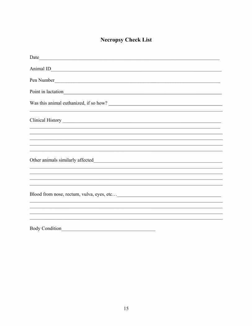
Necropsy Check List
Date_________________________________________________________________________
Animal ID_____________________________________________________________________
Pen Number___________________________________________________________________
Point in lactation________________________________________________________________
Was this animal euthanized, if so how? ______________________________________________ ______________________________________________________________________________
Clinical History ________________________________________________________________ _____________________________________________________________________________ ______________________________________________________________________________ ______________________________________________________________________________ ______________________________________________________________________________ ______________________________________________________________________________
Other animals similarly affected____________________________________________________ ______________________________________________________________________________ ______________________________________________________________________________ ______________________________________________________________________________ ______________________________________________________________________________
Blood from nose, rectum, vulva, eyes, etc…__________________________________________ ______________________________________________________________________________ ______________________________________________________________________________ ______________________________________________________________________________ ______________________________________________________________________________
Body Condition______________________________________

16
Systems Review
Oral Cavity____________________________________________________________________
Heart_________________________________________________________________________ _____________________________________________________________________________
Lungs_________________________________________________________________________ ______________________________________________________________________________
Kidneys_______________________________________________________________________ ______________________________________________________________________________
Liver_________________________________________________________________________ ______________________________________________________________________________
Intestines______________________________________________________________________ ______________________________________________________________________________ ______________________________________________________________________________
Rumen________________________________________________________________________
Reticulum_____________________________________________________________________
Omasum______________________________________________________________________
Abomasum____________________________________________________________________
Bladder_______________________________________________________________________
Uterus________________________________________________________________________ ______________________________________________________________________________
Udder_________________________________________________________________________

17
Diagnostic Sampling
What should be sampled?
• Liver, lung, kidney, intestines, forestomachs, heart, mammary gland, uterus and fetus if applicable.
• Anything that does not look normal. For example, inflamed (red, swollen) tissue or any tissue that does not have a smooth shiny surface.
• Samples taken should reflect the clinical signs the animal had. For example, if the animal had respiratory difficulty, sample the lung.
• Don't worry about taking too many samples, too much is better than too little.
How should the samples be taken?
• Taking samples for culture should be the first thing done, in order to minimize contamination.
• Use a clean knife when taking samples, especially samples submitted for culture. The best way to ensure a clean knife is to soak it, and any other instruments used, in alcohol for 5 to 10 minutes.
• If samples of lung and intestine are needed, sample the lung first in order to minimize contamination from the intestine.
• Always use a sharp knife. • If there is a lot of organic material on the sample you may wash the surface off with
water. • Refrigerate - Rapid cooling of tissue and maintaining the tissue at a cold temperature is
necessary for culture of bacteria or viruses. If you do not have access to refrigeration and the tissue cannot be submitted within 24 hours, consider placing the tissue in an ice chest surrounded with ice.
• Send each sample in separate labeled container/bag (Zip lock bags work well). • Include normal tissue adjacent to the lesion. • For tissues to be fixed, cut no more than 1 cm thick. (About the width of your little
finger.) Be sure to send in a couple of samples from each organ needed for histology. • Use 10% buffered formalin to fix the tissue for histology, tissues needed for bacterial or
viral isolation should not be placed in formalin. Contact your veterinarian to obtain the 10% buffered formalin.
• The tissue should be fixed in the formalin at a 10:1 ratio of formalin to tissue for approximately 24 hours. The formalin can then be removed for the tissue to be shipped in a plastic jar or plastic bag. Be sure to double bag the tissue to prevent leakage.
• Tissues sent in for culture need to be about 1 inch thick. When submitting intestines, you should obtain a loop of bowel approximately 6 inches long. Click on the bottom photo on this page to view a video of intestine sampling. Be sure to obtain 2 to 3 samples from each organ needed for culture.
• Specialty sampling - consult with your veterinarian. For example, if you suspect neurological disease (blind staggering, head pressing, etc.…) save the head for examination by your veterinarian or have the veterinarian perform the necropsy.

18
19
Basic Field Necropsy
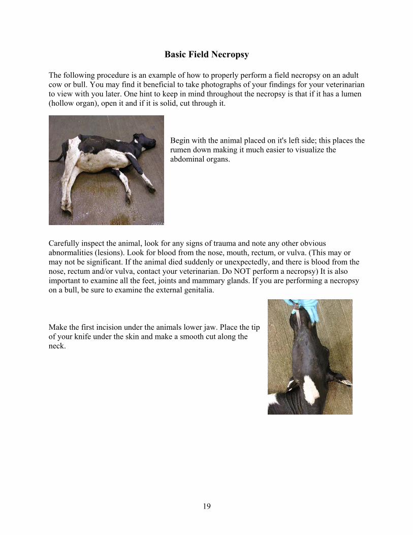
The following procedure is an example of how to properly perform a field necropsy on an adult cow or bull. You may find it beneficial to take photographs of your findings for your veterinarian to view with you later. One hint to keep in mind throughout the necropsy is that if it has a lumen (hollow organ), open it and if it is solid, cut through it.
Begin with the animal placed on it's left side; this places the rumen down making it much easier to visualize the abdominal organs.
Carefully inspect the animal, look for any signs of trauma and note any other obvious abnormalities (lesions). Look for blood from the nose, mouth, rectum, or vulva. (This may or may not be significant. If the animal died suddenly or unexpectedly, and there is blood from the nose, rectum and/or vulva, contact your veterinarian. Do NOT perform a necropsy) It is also important to examine all the feet, joints and mammary glands. If you are performing a necropsy on a bull, be sure to examine the external genitalia. Make the first incision under the animals lower jaw. Place the tip of your knife under the skin and make a smooth cut along the neck.

20
Continue the skin incision along the body, between the front limbs, and above the udder or external genitalia.
With your knife, free the skin from the body wall on the right side of the animal up to the spine, as far as possible. This is accomplished by cutting the connective tissue between the hide and the muscles of the animal. By cutting through the connective tissue you will be able to reflect the limbs without cutting through any more of the hide. Try not to make too many holes in the skin; the hide is the only part of the animal that can be salvaged by the rendering service. Also an intact hide makes it easy to close and remove the carcass after the necropsy is performed.
Reflect the right forelimb by cutting between the muscles of the shoulder and those of the body wall. There is a thin, clear to white connective tissue between the muscles. As you lift up on the forelimb you will begin to see this connective tissue. Cut this connective tissue while pulling back on the limb. You should not have to cut through the hide on the limbs. After this is done the right forelimb should be at a right angle to the rest of the body.
Now lift the hindlimb up and cut through the muscle bellies toward the hip joint. Once you reach the joint you will see that it looks like a ball and socket. There is a small ligament connecting the ball to the socket. Cut through this ligament to allow the joint to come apart. Once you have done this, the hindlimb should also lay at a right angle to the body of the animal. Again you should not have to cut through the hide on the limbs to accomplish this task.

21
Be sure to examine the mammary glands at this time. You will need to cut through all four quarters and evaluate the inner tissue.

22
Carefully make an incision in the abdomen just behind the ribs. Try not to cut too deep in order to avoid penetrating the underlying forestomach and intestines.
Reverse the knife in your hand so that the tip of the knife points toward yourself. Insert your hand and the knife handle in the abdominal incision you previously created. Cut the abdominal wall along the rib, and then continue the cut from the original point of entry towards the udder.
You should now have an abdominal flap, that when folded down exposes the abdominal contents. Later this flap will serve to keep the organs contained within the carcass during removal of the animal.
Examine the color, position and size of all of the organs. If you notice any fluid within the abdominal cavity, note the approximate amount and color of it.

23
The thick tissue covering the abdominal organs is called the Greater Omentum. This tissue acts as a "sling" for the abdominal organs.
With one hand, grab the greater omentum towards the hip joint and cut it away from the body wall of the abdominal organs.
You should now be able to observe the abdominal organs. Examine the abdominal cavity for any adhesions, discoloration (i.e. black or bright red intestine) or masses. The diaphragm lies under the last rib and separates the abdominal and chest cavities. With your knife, puncture the diaphragm while listening for air to rush into the chest cavity. Normally the chest cavity is under negative pressure, keeping the lungs inflated. After death, there should still be negative pressure in the chest cavity unless there has been trauma to the chest.
Now cut the entire right side of the diaphragm away from the rib cage. This allows for the first look into the chest cavity.

24
With your knife, cut the muscles covering the rib cage along the top of the rib cage, near where the ribs meet the spine.
With the rib (AKA bush) cutters or an ax, cut the ribs along the incision you just created in the muscle. There are 13 ribs that will need to be cut.
Lift up on the rib cage while cutting any tissue attaching the ribs at the incision site. Create a handle by cutting a hole in the muscles between the center of the ribs.
Push down on the ribs, fracturing them at the junction between bone and cartilage. This will create a tray that may be used as a cutting platform during the necropsy.

25
Now both the abdominal and chest cavities are exposed and can be examined for abnormalities. Any samples needed for culture (bacteria, virus, etc.…) should be taken now to decrease the amount of contamination of the sample.
The photo above on the right is taken from the back of the animal. The head is pointing to the top left corner of the picture.
Now focus your attention on the esophagus and trachea (windpipe). Begin by cutting between the lower jawbone, on either side of where the tongue lays. Reach in and pull out the tongue.

26
Cut the soft tissue of the upper mouth while pulling on the tongue. The photo to the left shows the cut soft tissue of the upper mouth.
You will notice some small bones (Hyoid bones)on either side of the tongue. These help suspend the larynx (voice box) and the tongue. These will need to be disarticulated (disconnected at the joint) or cut in order to completely take out the tongue with the esophagus and trachea. It may be easiest to cut these bones with small rib cutters (pruning shears).
The knife in the photo to
the left is pointing to one of the
hyoid bones.

27
You should now be able to pull out the tongue, trachea, and esophagus up to the point of the lung. You may have to cut away some tissue connecting these structures to the head and neck muscles.
The point where the trachea and the esophagus meet, at the base of the tongue, is called the larynx. From the larynx, cut the esophagus (the thin walled tube) lengthwise to the point of the lungs.
Examine the inner lining for any lesions (i.e. bruising, ulcers).
Now with a sharp knife or rib cutters, cut the larynx open and look for any lesions.

28
Cut the trachea (windpipe) lengthwise to the point of the bifurcation at lungs, and examine for any lesions.

29
The Chest
Next examine the thin sac surrounding the heart. Cut open the sac and make note of adhesions and fluid.
Normally the sac is not adhered to the heart and there is a small amount of fluid within the sac.
Make note of any large amounts of clear fluid, blood or pus in the heart sac.
The lungs should be a light pink color and "spongy" to the touch. Examine the general appearance of the right lung. Note if there appears to be areas that are darker than others or if there are adhesions or lesions on the lung.

30
Reflect the right lung by grabbing the tip of the lung closest to the diaphragm. Pull the lung toward the head, cutting any connective tissue. Now the major vessels and airways of the lung can be examined. Cut into the lung near the top of the heart. You should see some large openings close to each other once you have cut into the lung. The airway will be in the middle and is rigid. The artery is on the bottom and the vein is on the top compared to the airway. Use scissors and forceps to cut open the airways and vessels. There may be some food and/or foam in the airways. This usually occurs during the last terminal breaths, however you should note amount and color for your veterinarian. Open lung artery (vessel)
Open airway

31
The photo above shows an open lung vein (vessel).
Now make lengthwise cuts in the lung to observe the deeper tissue of the lung. Make note if you see excessive amounts of blood within the right lung or if the lung seems to "pop" when you squeeze it (like bubble wrap).
The left lung may be examined by pulling back the trachea and esophagus towards the abdomen. Lift up on the left lung and look for any adhesions or lesions on the lung. Observe the deeper tissue as you did on the right lung. Note: Blood will pool in the down lung after death. This will make the lower lung look much darker than the lung on top.
If you want to observe the vessels and airways of the left lung, it will need to be removed. This can be done by pulling on the trachea and esophagus, while cutting any tissue connecting the lungs to the body wall. The heart will be removed with the lungs.

32
The heart may now be fully examined. This can be done with or without removing it from the carcass. When it is in the carcass, you are looking at the right side of the heart. Start by finding the major vessel that enters the heart from the abdomen, this is the vena cava. With scissors, cut this vessel lengthwise, continuing into the heart. Cut to the tip of the heart, and then cut back towards the vessels that lead to the lungs at the base of the heart. You should now have a "v" cut in the right side of the heart. Examine the inside of the heart including the valves. Note any lesions, such as thickening of the muscle wall, holes, abscesses, or growths on the muscle or valves.

33

34
Be sure to look at the valves of the right heart. The photo to the bottom left shows looking into the vessel going from the right heart to the lungs. You will want to follow this vessel to look at the valve and make sure there is an opening between the valves. The photo to the bottom right shows the valves separating the atrium (top chamber) from the ventricle (bottom chamber). The valve is the shinny white structure with the web-like projections connecting it to the wall of the heart. These valves should be smooth and shinny.
Flip the heart up to reveal the left side. Make a cut from the base to the tip of the heart. Examine the inside of the heart. You should notice that the wall of the left side is two to three times thicker than that of the right side.
You will notice clotted blood in the heart and in the vessels. This will look like "jelly" and should not be attached to the wall of the heart or the vessels. Make note if there is a strong attachment of a clot to the wall, this could be a thrombus.
The photos taken of the left side of the heart while still in the cow show the heart upside down. This will be the view you will see when performing the necropsy, so keep in mind that the bottom point of the heart is pointing toward the top of the pictures. Again be sure to look at the valves on the left side of the heart.

35
36
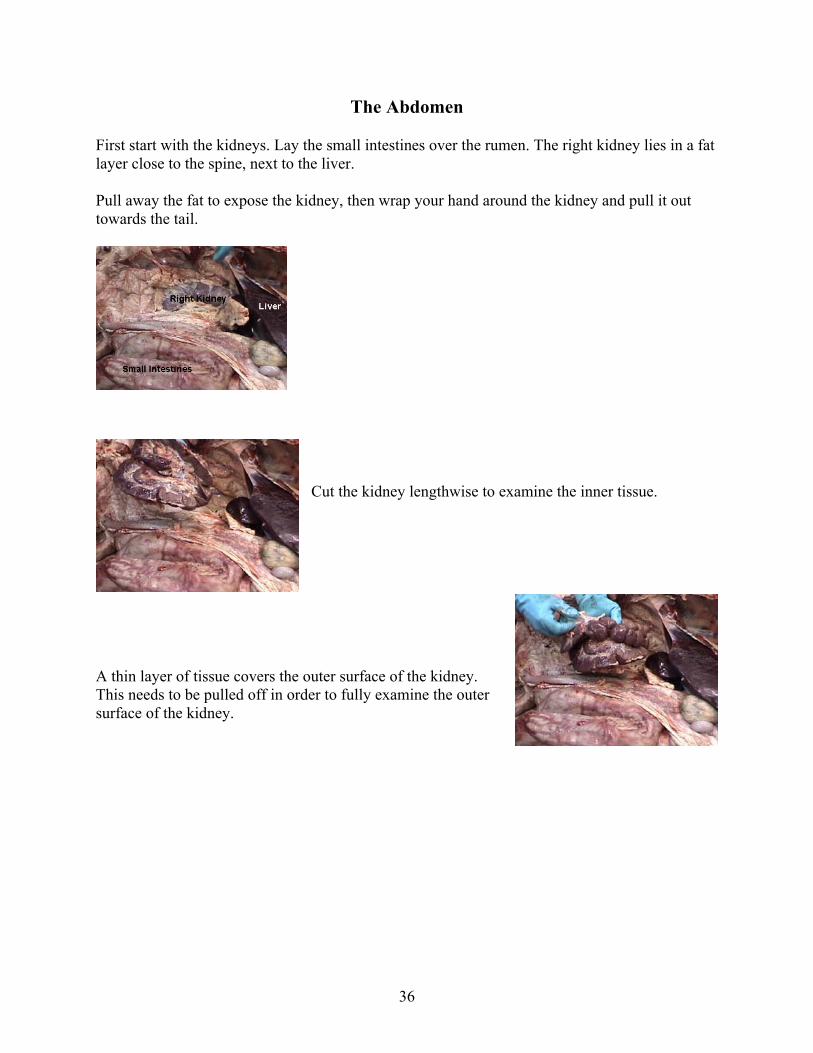
The Abdomen
First start with the kidneys. Lay the small intestines over the rumen. The right kidney lies in a fat layer close to the spine, next to the liver.
Pull away the fat to expose the kidney, then wrap your hand around the kidney and pull it out towards the tail.
Cut the kidney lengthwise to examine the inner tissue.
A thin layer of tissue covers the outer surface of the kidney. This needs to be pulled off in order to fully examine the outer surface of the kidney.

37
The left kidney is found by reflecting the entire intestine over the spine of the animal. Where all of the intestines meet, just above the rumen, is another area of fat covering the kidney. Cut through the fat to expose the left kidney and examine it in the same manner as you did the right kidney.
The following photos are close up pictures of the left kidney.

38
The urinary bladder can be found within the pelvis. If it is full of urine, now would be a good time to take a sample with a clean needle and syringe. Cut open the bladder to examine the inner surface.
Examine the reproductive tract and note if the cow was pregnant. If there is a fetus present, examine the fetus and placenta and take samples if necessary.
The spleen is found on the left side of the animal, under the rumen. Lift up on the rumen to expose and examine the spleen. Make several incisions in the spleen to examine the inner tissue. The spleen may be dark due to congestion with blood.

39
The liver is the large organ between the rumen and the diaphragm. It should have sharp edges and a smooth surface. Note if there are any lesions on the surface, along with the color and size of the liver. Enlarged livers will have rounded edges. As done with all other organs, cut into the liver to view the deeper tissue.
Located within the liver lobes is the gall bladder. This sac like organ is green in color and is typically about the size of a grapefruit in an adult cow. There are ducts associated with the gall bladder and course through the liver. Cut open the gall bladder and these ducts to look for any abnormalities. This is the site where liver flukes are found.

40
Now focus on the forestomach of the cow
.
41
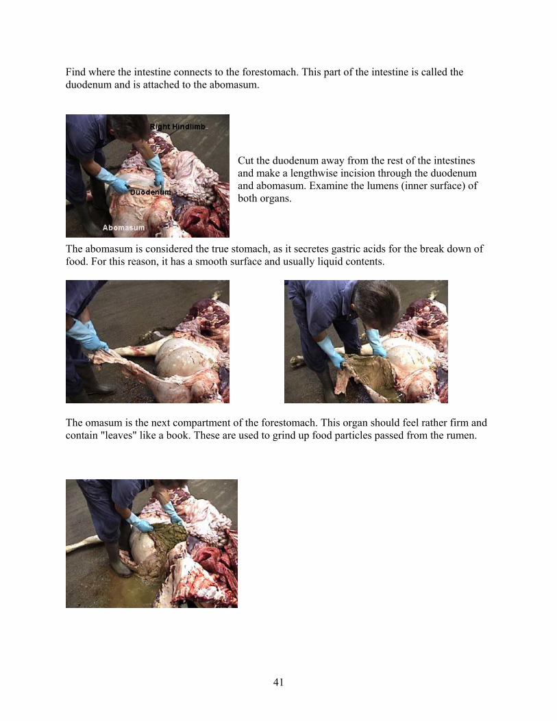
Find where the intestine connects to the forestomach. This part of the intestine is called the duodenum and is attached to the abomasum.
Cut the duodenum away from the rest of the intestines and make a lengthwise incision through the duodenum and abomasum. Examine the lumens (inner surface) of both organs.
The abomasum is considered the true stomach, as it secretes gastric acids for the break down of food. For this reason, it has a smooth surface and usually liquid contents.
The omasum is the next compartment of the forestomach. This organ should feel rather firm and contain "leaves" like a book. These are used to grind up food particles passed from the rumen.

42
The reticulum is the next compartment. This is a smaller sac that has a honey comb appearance on the inside. This will usually contain the magnet if they are used on your operation.
The largest compartment is the rumen. Its inner surface is a lot like a shag carpet, made up of thousands of papillae. These papillae should not slough off when you run your knife across them, unless the animal has been dead for more than an hour. If many of the papillae slough off and the animal has not been dead more than an hour, you may consider sampling the rumen contents for signs of a pH change, as in grain overload or other gastrointestinal upset.
Examine all compartments and make note of the appearance of the tissue as well as the contents.
The intestines should now be examined. Multiple areas of intestine should be examined for inner color and appearance. All of the intestine should have a smooth, shinny appearance.
43
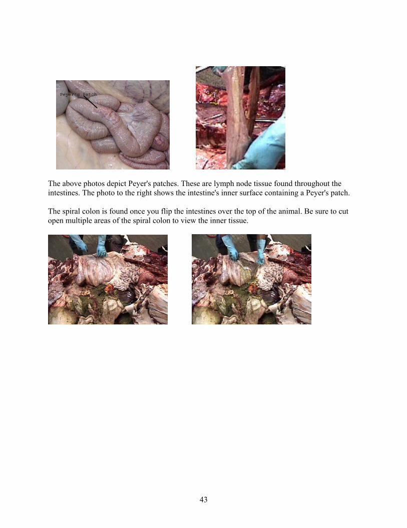
The above photos depict Peyer's patches. These are lymph node tissue found throughout the intestines. The photo to the right shows the intestine's inner surface containing a Peyer's patch.
The spiral colon is found once you flip the intestines over the top of the animal. Be sure to cut open multiple areas of the spiral colon to view the inner tissue.

44
Head Removal If the animal had any symptoms of neurologic disease, i.e. circling, blind staggers, head pressing, etc.., have your veterinarian perform the necropsy. If it is not possible for a veterinarian to perform the necropsy right away, remove the head and cool for examination by a veterinarian at a later time.
To remove the head, extend it back and cut the muscles of the neck directly behind the jaw. You should come to where the first vertebra (bones of the spine) attaches to the skull. Cut away all attachments and transect (cut) the spinal cord. The hide and the muscles above the vertebrae should now be cut, freeing the head from the body.
This drawing depicts where the hide should be cut in order to remove the head.

45
Cut through the muscles of the neck until you reach the spinal canal, as seen in the photos below.
Transect the spinal cord and cut through the connective tissue between the bones of the head and neck. Completely remove the head by cutting through the remaining muscles and the hide.
46
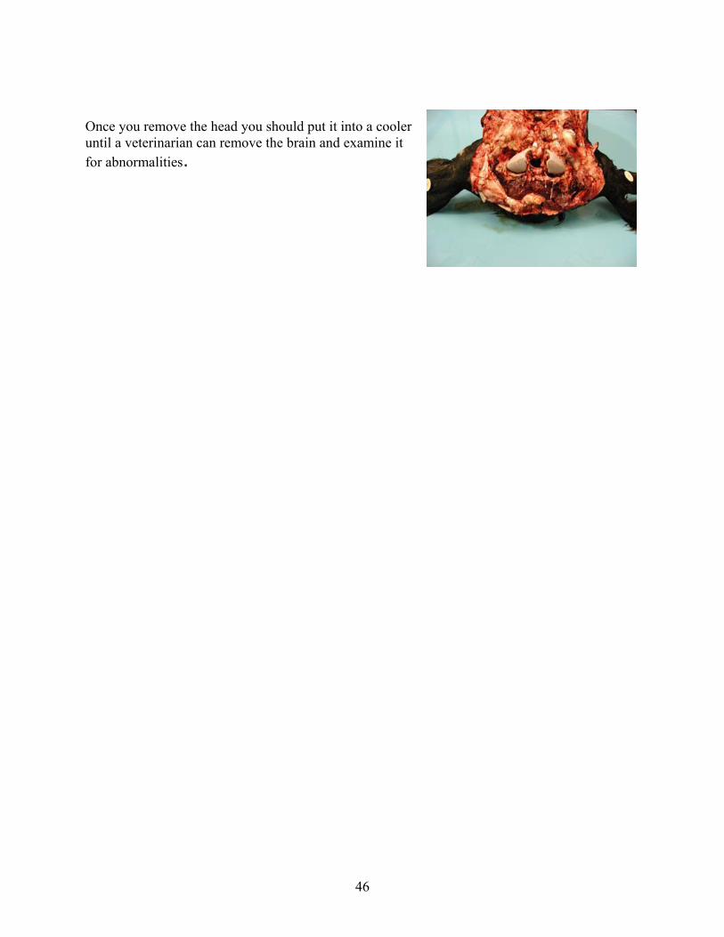
Once you remove the head you should put it into a cooler until a veterinarian can remove the brain and examine it for abnormalities.

47
Joints & Muscles
With any type of lameness, the joints should be examined. Skin the hide away from the joint of interest. Bend the joint in it's natural direction and cut through the muscles toward the middle of the joint. All joints are surrounded by a thin layer of connective tissue, cut through this to expose the inside of the joint. Carefully examine the joint fluid and the bones involved in the joint.
Normally the joint fluid is clear to slightly yellow in color and is viscous or "stringy". There should not be any blood or pus in the joint.
The bones should be smooth and white.
Different muscles should also be looked at if the animal experienced any lameness. Simply cut into the muscle and look for any abnormalities. The muscle should be the same color and texture throughout. If you notice any black areas or pale areas you may consider taking a sample for your veterinarian to look at.

48
Closing
To ready the body for removal, place all organs in the carcass and pull the hide over the body. Cut small holes in the hide, within 4 inches of the edge to decrease the amount of damage done to the hide. Tie the hide together with baling twine.
Be sure to properly clean the area where the necropsy was performed. Wash your hands, boots, and coveralls before handling other animals.

49
Normal Tissues
• Abomasum • Esophagus and Trachea • Gall Bladder • Heart • Intestine • Kidney • Liver • Lung • Omasum • Reticulum • Rumen • Spleen • Urinary Bladder • Udder • Uterus

50
Normal Abomasal Tissues
The abomasum is the only glandular part of the stomach. It contains many folds and is white to gray in color. Feed should easily wash off of the surface.

51
Normal Esophagus and Trachea
Normal Esophagus
The esophagus should have a smooth white-gray surface. It has a uniform size throughout, no areas of widening or narrowing.
Normal Larynx
The larynx should be smooth and white to gray in color.

52
Normal Trachea
The trachea is also smooth and white. The tracheal rings are not complete and should not collapse easily.
53
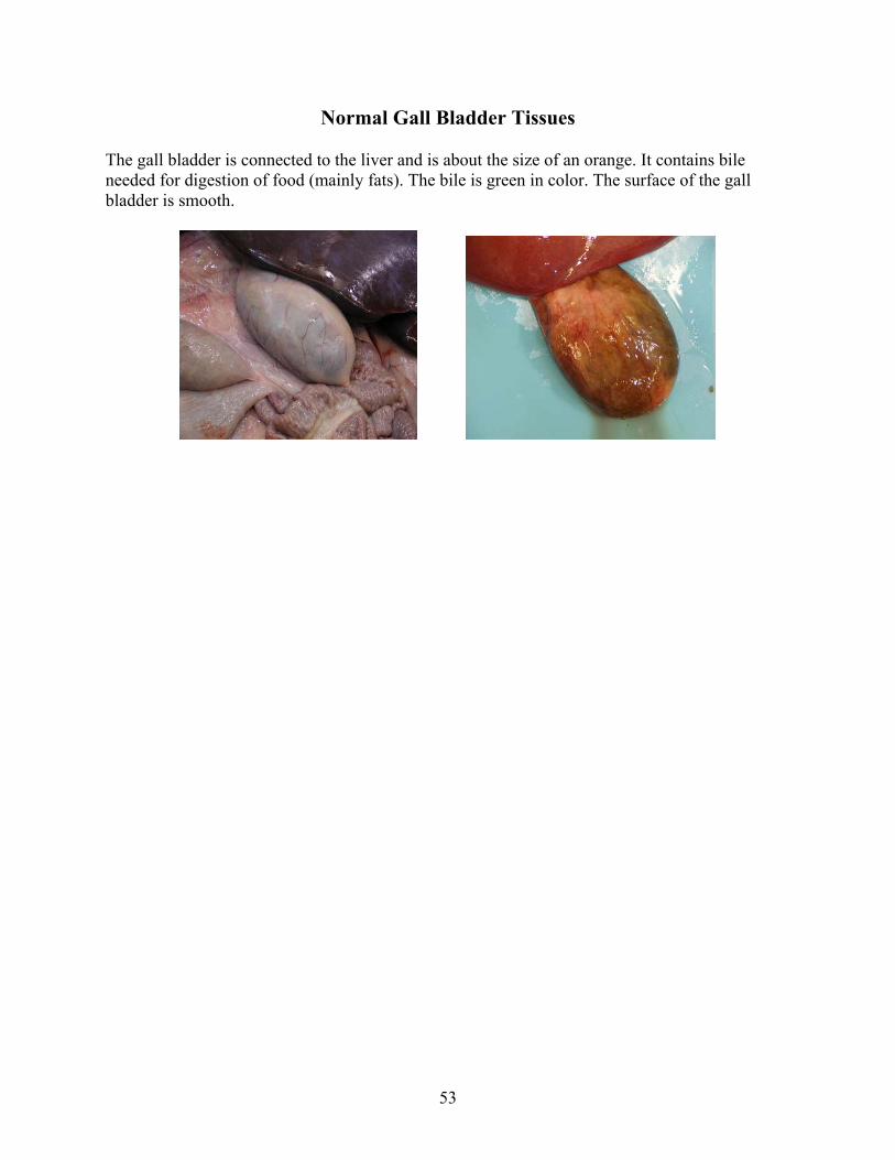
Normal Gall Bladder Tissues
The gall bladder is connected to the liver and is about the size of an orange. It contains bile needed for digestion of food (mainly fats). The bile is green in color. The surface of the gall bladder is smooth.

54
Normal Heart Tissues
The heart lays in a sac and is normally surrounded by a small amount of fluid within the sac. The heart normally has fat around the top (base) of it. The actual heart tissue is a dark red (liver color) and has a smooth surface. The inner surface should also be smooth. The left side of the heart is about 3 times the thickness of the right. The valves of the heart should be thin and connected to the wall of the heart by tendon like structures.
55
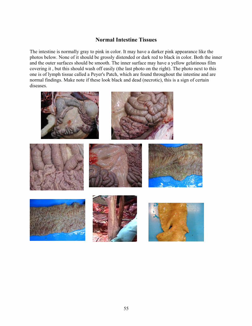
Normal Intestine Tissues
The intestine is normally gray to pink in color. It may have a darker pink appearance like the photos below. None of it should be grossly distended or dark red to black in color. Both the inner and the outer surfaces should be smooth. The inner surface may have a yellow gelatinous film covering it , but this should wash off easily (the last photo on the right). The photo next to this one is of lymph tissue called a Peyer's Patch, which are found throughout the intestine and are normal findings. Make note if these look black and dead (necrotic), this is a sign of certain diseases.

56
Normal Kidney Tissues
The kidney is normally surrounded by fat and is a lobated organ. It too has a smooth surface and may contain some red discoloration as seen in these photos due to areas of congestion. On cut surface you will notice that there seems to be two layers. The outer layer is called the cortex and the inner is called the medulla. It is important to look at each layer and to determine if any lesion affects one or the other or both of these layers.

57
Normal Liver Tissues
The liver is a smooth lobed organ that is dark red-brown in color. The gall bladder is connected to it and may be white to green in color. On cut surface the liver is uniform throughout. You may be able to observe the vessels and the bile ducts running through it. When pinched it does not easily crumble in your fingers (it is not friable), but will crush with slight pressure. The edges should be sharp, not rounded (especially the surface closest to the rumen).

58
Normal Lung Tissues
The lungs are pink to slightly gray in color. They are light and spongy feeling. The "down" lung may be congested with blood, due to gravity. There may be some areas with slight discoloration (red or purple). The area of the lung closest to the spine may look whiter than the rest of the lung.

59
60
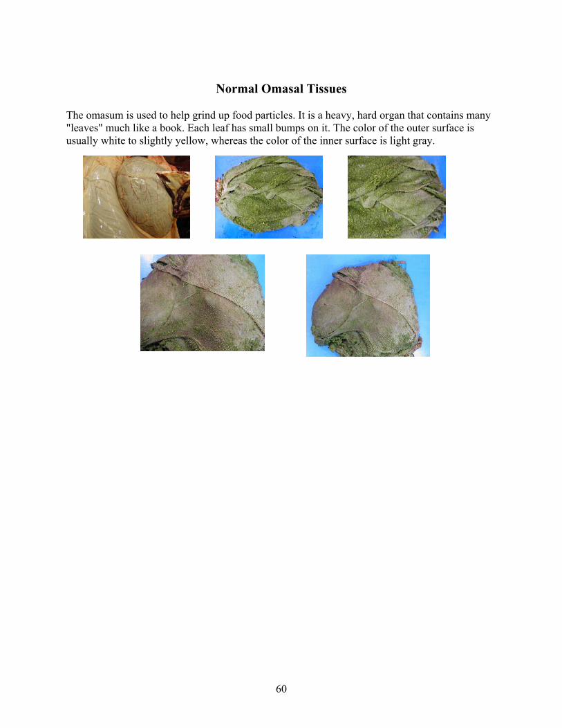
Normal Omasal Tissues
The omasum is used to help grind up food particles. It is a heavy, hard organ that contains many "leaves" much like a book. Each leaf has small bumps on it. The color of the outer surface is usually white to slightly yellow, whereas the color of the inner surface is light gray.

61
Normal Reticular Tissues
The reticulum has a "honeycomb" appearance and is also a light gray in color. The right photo shows the reticular grove to the left. This is a direct route from the esophagus to the abomasum.

62
Normal Rumen Tissues
The rumen is the largest compartment and contains a large fiber mat. It is where most of the absorption takes place. The inner surface of the rumen is like a "shag carpet". These are the papillae that are needed for absorption. You will notice small folds in the surface, but these are not as large as those found in the abomasum. The rumen also contains thick areas of tissue called pillars (the photo in the second row to the left depicts one of these pillars). These are needed to help keep the shape of the rumen.

63
Normal Splenic Tissue The spleen may differ in size and color depending on how the animal died. It may become engorged with blood if euthanasia drugs where used. It is normally a flat organ that has a gray to purple color to it. On the cut surface you may see areas of red mixed with areas of white. The red areas are red blood cells and the white areas are white blood cells.
Here the spleen is still attached to the stomach
There will normally be some blood that will ooze out of the spleen. If the spleen is left out it will start to shrink and become a darker color. Depending on how the animal died, the spleen may be enlarged and filled with blood whereas other times it may be small.
64
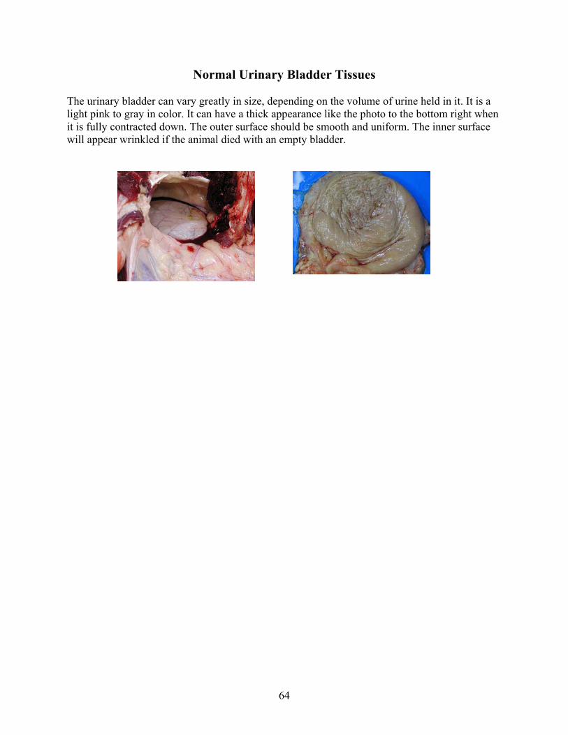
Normal Urinary Bladder Tissues
The urinary bladder can vary greatly in size, depending on the volume of urine held in it. It is a light pink to gray in color. It can have a thick appearance like the photo to the bottom right when it is fully contracted down. The outer surface should be smooth and uniform. The inner surface will appear wrinkled if the animal died with an empty bladder.

65
Normal Mammary Gland Tissue (Udder)
Normal mammary tissue is a pink-grey color and it should be spongy feeling, any hard areas may be a sign of mastitis. The milk color and consistency will depend on the stage of lactation, but should not contain any blood. The hind end of the udder has two large lymph nodes (bottom two pictures).

66
Normal Uterine Tissues
The uterus will vary greatly in size depending on the age of the animal and if she is pregnant or not. It should be pink to light gray in color and have a smooth surface.

67
Abnormal Findings
The following are a few examples of common lesions seen in cattle. This is not a complete list of lesions, therefore you should take samples of any tissue that does not appear normal.
For more pictures of abnormal findings you can visit Cornell's Dr. John M. King's Necropsy Show and Tell at http://w3.vet.cornell.edu/nst/nst.asp You will want to do a keyword search of "cattle".
Esophagus and Trachea Udder Forestomach Urinary Bladder Heart Uterus Intestine Kidney Liver Lung Muscles/Joints

68
Common Lesions of the Esophagus and Trachea
All of the photos above illustrate necrosis (dead tissue) of the larynx extending into the trachea. You may also see ulcers in this area. The above photos show foreign material or feed material that is caught in the esophagus (left photo) or the larynx (right photo). You may see some food material in the esophagus as well as the larynx and trachea due to death. Be sure to look closely at the surrounding tissue. If it looks red and inflamed or contains ulcers, it is likely that the food material was there prior to death.

69
The photos illustrate hemorrhage (bleeding) and ulceration of the esophagus.
The two photos above show examples of esophageal worms, commonly found in dairy cattle. The worms are the squiggly lines within the inner surface of the esophagus. These are incidental findings, and are of no concern.

70
Common Lesions of the Forestomach
This abomasum has hemorrhage (bleeding) and ulcers (irregular surface). You may also see air pockets in the abomasum which may be caused by bacterial organisms such as Clostridium.
This is an example of a displaced abomasum. Note that the abomasum is above the rumen when it should be under and slightly in front of the rumen.
The above photos depict an infection in the abdomen, called peritonitis. The yellow material is a fibrin and may be easily pulled off of the tissue surface depending how long the disease has been going on.

71
Be sure to look fore any foreign material in any part of the forestomach, such as this wire found in the reticulum.

72
Common Heart Lesions
Notice the black areas of the heart. This is an indication of diseased or dead tissue.
Notice the "cauliflower" lesion on the walls of the hearts in these photos. This is a thrombus (blood clot adhered to a blood vessel or the heart) and is usually found on the valves of the heart.
The hearts in these photos each have a hole that is in the septum, middle wall of the heart, connecting the two sides of the heart.
73
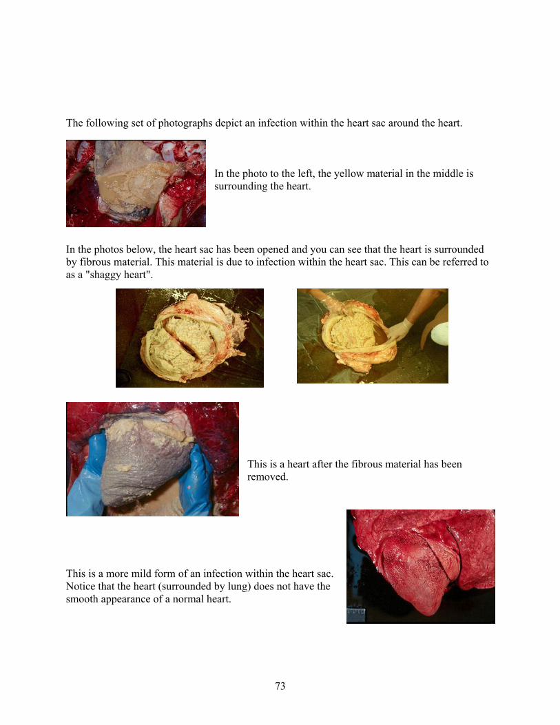
The following set of photographs depict an infection within the heart sac around the heart.
In the photo to the left, the yellow material in the middle is surrounding the heart.
In the photos below, the heart sac has been opened and you can see that the heart is surrounded by fibrous material. This material is due to infection within the heart sac. This can be referred to as a "shaggy heart".
This is a heart after the fibrous material has been removed.
This is a more mild form of an infection within the heart sac. Notice that the heart (surrounded by lung) does not have the smooth appearance of a normal heart.

74
Common Lesions of the Intestine
All of theses photos depict bowel that is full of clotted blood.
Notice the black dots in the mesentery ( tissue connecting the loops of bowel) surrounding the intestine. These are commonly found and are of no significance.

75
Most any bowel that is thick and corrugated (has many folds in it) is diseased. If you think that the bowel is thick, be sure to take a sample of it. The pieces of bowel in the above photos show ulcers in the Peyer's patches (lymph node tissue in the intestine) There may be areas of black, dead tissue with fibrous material like that depicted in the right photo.

76
Common Kidney Lesions
Notice the pale indented area on the kidney. This may be due to a decrease in oxygen or blood to the kidney causing an "infarction"
Notice how the outer layer of the kidney (cortex) looks thin and pale compared to the inner layer (medulla).
This is the outer surface of the kidney above. Here you can see that it looks scarred and small. This indicates a chronic disease of the kidney.
Notice the pale, yellow color of the kidney. The area with a red center is a cut portion of the kidney to show that it is the outer most layer (cortex) that is affected the most.

77
Both photos above are examples of a "White Spotted Kidney" You will see white spots all over the kidney surface. Be sure to cut into the kidney to see if the discoloration extends into the inner tissue.
The pale areas in this kidney are called infarcts. The red areas within the pale areas are due to hemorrhage. You will see them extend into the kidney on the cut surface.

78
Common Liver Lesions
Both of the above pictures are examples of liver abscesses. The abscesses are the yellow to white spots on the liver. Not all abscesses may be evident from the surface of the organ. It is important to cut into the organ to see if there is abnormal tissue or abscesses within the organ.
These are examples of liver flukes. These travel through the liver causing damage to the liver. You may be able to see their tracts. These are usually seen in marshy areas that have snails.
Notice how the liver looks lobated like a kidney. This is due to scaring or fibrosis from a chronic disease. This liver has a "nutmeg" appearance. White areas surrounded by red areas. This may be seen in congested livers associated with heart failure.

79
This is an example of a fractured liver. These cuts in the liver where not created by a knife, but instead where caused by blunt trauma to the liver. Knife cuts will be smooth where these are jagged. The photo to the right is a close-up of the left photo.
Note that this liver has a stripped appearance. This is an insignificant finding and is due to the animal laying on its side after death. The pressure against the rib cage forces the blood out and will cause a stripped appearance such as this.
This photo depicts a fatty liver. These livers will appear pale, have rounded edges, and may float in water.

80
Common Lung Lesions
This is an example of bronchopneumonia. Notice how the bottom-front of the lung is darker than the rest of the lung. Usually this part of the lung will be heavier than the more normal, pink lung to the left. You will need to sample the darker tissue of this lung to find out the cause of the bronchopneumonia.
This is another example of bronchopneumonia. The lungs have been removed from the animal.
This is an example of chronic pneumonia. Notice how the lung looks darker (or redder) than normal pink lung tissue. This lung may be heavier than normal lung and will not have the "spongy" feel to it.

81
Notice the diseased, darker, redder lung to the right compared to the more normal lung tissue to the left. The diseased lung is heavy compared to the light, spongy normal lung tissue.
This is a cross section of the lung above.
The photo above is an example of a lung abscess. Be sure to cut into lesions such as this. An abscess will have a liquid to "cottage cheese" like appearance in the middle. A tumor or other growth will usually be harder in the middle.
This photo is an example of pulmonary (lung) emphysema. When pinched, this lung will "pop" like bubble-wrap. Air has accumulated in areas where there is normally only tissue.

82
This is an example of pulmonary consolidation. This lung will be heavy and "wet". It is also much darker than normal lung tissue. The lung may contain blood or other types of cells due to inflammation, or other disease process. The lung will need to be sampled to determine the cause of consolidation.
These lesions are small raised areas on the surface of the lung. The lung should be cross sectioned to see if the deeper tissue is involved and to see what the inner part of the lesion looks like. Again the lung will need to be sampled to determine the cause of the lesions.
This is an example of a lungworm. These are more common in warmer climates such as the Pacific Northwest.
This is an example of a lung abscess that is connected to the rib cage. It is not uncommon to see adhesions such as this in the cow.

83
This is a lung from an aged cow. You can see an indention in the middle, this is a scar from a previous lesion (i.e. abscess) in the lung
Here you can see that the lung is adhered to the rib cage. These types of adhesions are commonly found in cattle.
Here, one of the airways in the lung has been cut open. You can see feed-like material in the airway. This could be from the animal getting feedstuff in the airways during death or could have occurred prior to death. Be sure to look closely at the surrounding tissue for any red and inflamed areas, or ulcers that may suggest that the feedstuff was there prior to death.
The above photos are of pneumonia. The red areas are the most affected and there may be some collapse of the lung in these areas.

84
This is an example of an abscess in the lung. The white circles are the abscesses. When you cut into them they will either contain a runny yellow-white material or a cheese like material.

85
Common Lesions of the Muscles and Joints
The black muscle is dead tissue.
This photo is of an open joint. The yellow material seen in the middle of the joint is due to infection. This material is thick compared to the normal joint fluid.
86
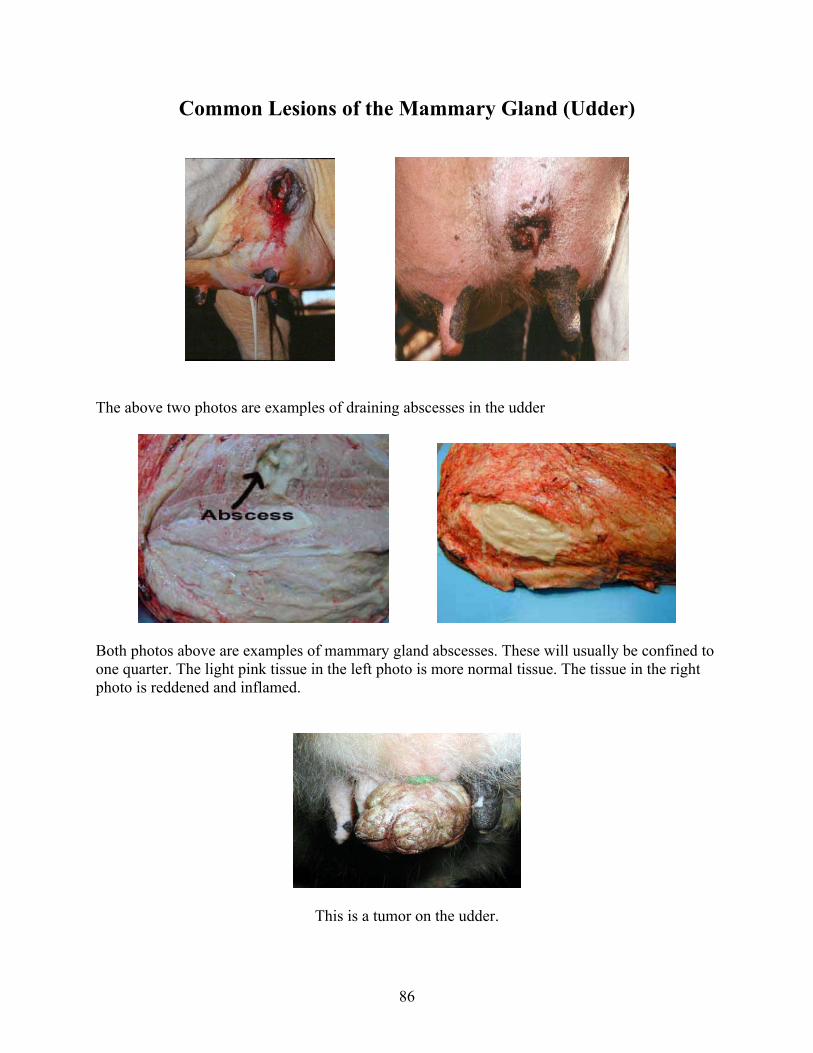
Common Lesions of the Mammary Gland (Udder)
The above two photos are examples of draining abscesses in the udder
Both photos above are examples of mammary gland abscesses. These will usually be confined to one quarter. The light pink tissue in the left photo is more normal tissue. The tissue in the right photo is reddened and inflamed.
This is a tumor on the udder.

87
These four photos are examples of Udder Rot, ulcerative lesions on the udder. This type of lesion is commonly found between the front two quarters of the udder in older cows. Lesions like this are also found between the udder and the hind legs in heifers.
The above three photos are examples of mastitis. If this type of mastitis progresses, the quarter could die and slough off.
The white area of this udder is due to infection (mastitis) and necrosis (death) of the tissue. The red tissue below is more normal mammary gland tissue.
88
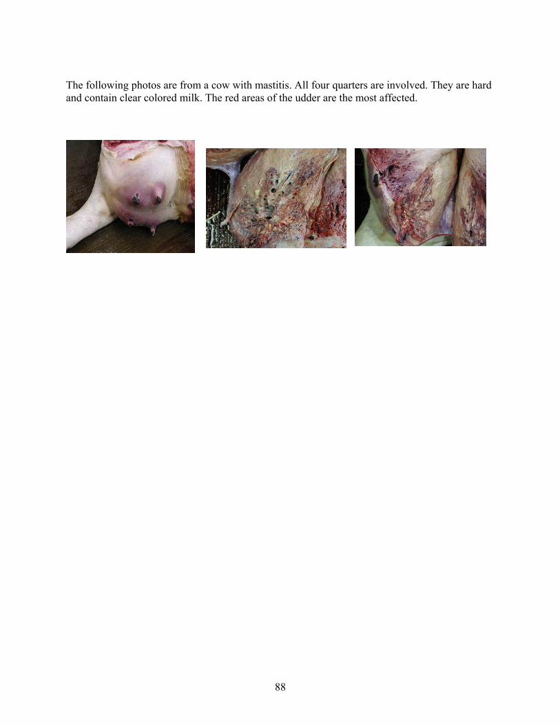
The following photos are from a cow with mastitis. All four quarters are involved. They are hard and contain clear colored milk. The red areas of the udder are the most affected.
89
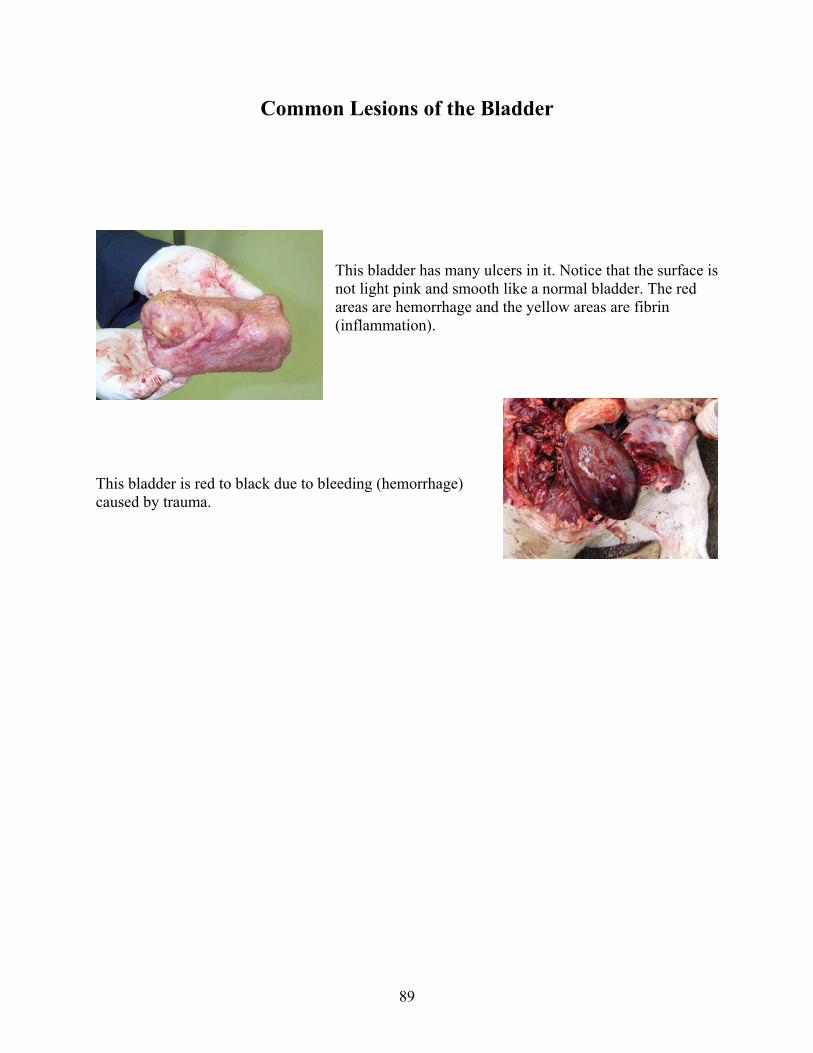
Common Lesions of the Bladder
This bladder has many ulcers in it. Notice that the surface is not light pink and smooth like a normal bladder. The red areas are hemorrhage and the yellow areas are fibrin (inflammation).
This bladder is red to black due to bleeding (hemorrhage) caused by trauma.

90
Common Lesions of the Uterus
Notice how the uterus is taking up the entire abdomen. This is seen in a condition called Hydrops. The uterus is filled with fluid. This uterus is necrotic (dead) as seen by the black tissue.
The pink tissue in this photo is normal uterine tissue. The rest of the tissue is the inner part of the uterus that is infected and inflamed.

91
Calf Necropsy
A calf is not much different to necropsy than a mature cow, but there are a few things to keep in mind when performing a calf necropsy. Usually a calf is much easier to necropsy due to it's size and the fact that there is not as much body fat. Also a calf will not have a mature, fully functional rumen. Keep in mind that when you open up the calf, the abomasum is the largest forestomach compartment. Remember that the intestine will connect to the abomasum.
Due to the types of diseases that calves commonly catch, it is important to look closely at the gastrointestinal tract, the umbilical cord, liver, lung, and the joints. If the calf is still receiving milk, there should be clotted milk in the abomasum.
When collecting tissue samples it is important to keep in mind the symptoms of disease the calf showed prior to death. If it had signs of respiratory disease you should take multiple samples of the lungs for culture and histology. With signs of gastrointestinal signs, i.e. scours, be sure to take multiple loops of intestine as well as feces.
It is important to note that the rumen in a calf is not as developed as in an adult. When you first open up the abdomen, the largest portion of the forestomach is the abomasum.
Calf urinary bladder.
It is not uncommon for the calf to have a large urinary bladder.

92
Calf liver and gall bladder (green)
Calf intestines
Be sure to cut into multiple sections of the intestine to look at the inside surface. Note any red areas and areas that have fibrous material on the inner surface of the intestine.

93
It is also important to open up the joints in a calf. Note any blood, or yellow "clumped" material in the joint.
Abortions -
Abortions are a common occurrence in dairy cattle. It is important from a herd health standpoint to try and obtain an accurate diagnosis of the cause of the abortion, however you should keep in mind that accurate diagnosis only occurs in approximately 50% of the cases. Due to the low likelihood of an accurate diagnosis and complicated sampling techniques, in general it is desirable to send the entire fetus and placenta, along with a blood sample from the dam to your veterinarian or local diagnostic lab.
94
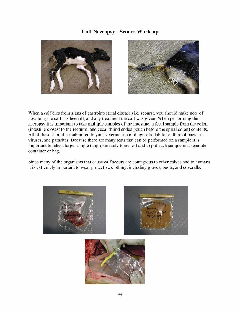
Calf Necropsy - Scours Work-up
When a calf dies from signs of gastrointestinal disease (i.e. scours), you should make note of how long the calf has been ill, and any treatment the calf was given. When performing the necropsy it is important to take multiple samples of the intestine, a fecal sample from the colon (intestine closest to the rectum), and cecal (blind ended pouch before the spiral colon) contents. All of these should be submitted to your veterinarian or diagnostic lab for culture of bacteria, viruses, and parasites. Because there are many tests that can be performed on a sample it is important to take a large sample (approximately 6 inches) and to put each sample in a separate container or bag.
Since many of the organisms that cause calf scours are contagious to other calves and to humans it is extremely important to wear protective clothing, including gloves, boots, and coveralls.

95
Collecting samples of the intestine -
When collecting a sample of the intestine you will want to cut the intestine away from the connective tissue on the one side of the intestine. The best place to take samples from is called the ileum (the end portion of the small intestine), this section of intestine is closest to the cecum and spiral colon. Once you have cut about 6 inches away from the connective tissue, take a piece of string and tie the intestine in a loop. Once it is tied cut the ends of the intestine, leaving the string with the loop of intestine being sampled.
Take at least 3 samples of intestine with this technique and place in a separate container or bag for culture. Be sure to put a piece of intestine in formalin for histology.

96
Taking a fecal sample -
When taking a fecal sample you want to obtain it from the colon, area of intestine closest to the rectum. Cut open the colon and "milk" the contents into a bag. Be sure to label the bag(s) properly with the animals ID and that it is fecal contents.
Taking a sample of cecal contents -
The cecum is a blind ended pouch prior to the spiral colon. This is a good area to obtain a sample of intestinal contents to look for some specific causes of scours. You can either tie off the cecum with a piece of string and send it in that way or cut open the cecum and send in the contents only. Be sure to label the container(s)/bag(s) with the animal ID and that it contains cecal contents.

97
Shipping Samples
• Plastic sealable containers or plastic bags (i.e. Ziplock bags). Double bag any items which may leak.
• Include absorbent material (i.e. paper towels) in the outer bag to collect any fluids that may leak.
• Send with complete history, where lesion was found, pictures of the lesions (if any were taken), and animal identification.
• If fresh tissues are submitted for culture, these should be shipped in an insulated container with sufficient ice packs to ensure that sample does not rot.
• Follow US Postal Service or any package delivery service requirements for shipping tissues. No breakable items should be shipped through the mail. Contact your local post office for regulations about shipping tissues through the mail.

98
Composting
With the increasing cost of whole animal removal, and the prohibited use of animal meat and bone meal as a feed source, producers are looking at different techniques for whole carcass disposal. One alternative is composting. Composting utilizes old feedstuff and manure to create the proper environment for microorganisms to speed up the natural decomposition process. In the right composting environment, decomposition of a mature dairy cow carcass will take approximately 6 to 8 months leaving only a few small bones, which will shatter easily when passed through a manure spreader. When done properly, composting will kill most any plant or animal pathogen, and is non-odorous.
Materials Needed
Moisture Moisture content of the composting pile is crucial. If there is too little moisture, the bacteria needed for the decomposition process will not survive. On the other hand if there is too much moisture, the pores needed for the movement of oxygen will be replaced by water. With little oxygen bacteria that produce relatively little odor are replaced by bacteria that produce highly odorous byproducts. The moisture content should be maintained between 40 and 60%. If moisture can be squeezed from a handful of composting material it is too wet and probably needs to be mixed with drier material.
Co-composting Material Certain materials can be used to reduce the attractiveness of the carcass to insects and rodents, increase movement of oxygen throughout the compost pile, and to absorb excess liquid produced by the decomposing carcass. Materials such as wood chips, ground cornstalks, straw, or old feedstuff help keep the compost porous, while smaller materials like sawdust help absorb the liquid. These materials also are a carbon source needed to sustain the microbes. You may want to use a combination of these materials to allow optimal oxygen passage while absorbing any excess liquid.
Carbon and Nitrogen The microbes in the compost need carbon and nitrogen to function properly. The optimal carbon to nitrogen (C: N) ratio is 25:1, which will keep odor to a minimum and allow the best microbial growth. As stated above good sources of carbon are also your CO-composting material, sawdust, woodchips, cornstalks, old feedstuff, and straw. Nitrogen is obtained from the manure. Due to the expense of carbon and nitrogen analysis, temperature and odor are good indicators of the C: N ratio. If there is a strong ammonia odor from the pile the ratio may be too low and more carbon is needed. On the other hand if there is no odor, the moisture levels are within normal limits, and there is slow decay there may not be enough nitrogen. In this case you may want to add more nitrogen, i.e. manure.
99
Heat Retention Heat is an important byproduct of the microbial activity. Optimal temperatures within the compost range from 110 to 150 degrees F. This high temperature promotes the growth of heat-loving bacteria, which promote rapid decay. Another benefit is that the high temperatures (131 degrees or above for at least 72 hours) kill most disease-causing microorganisms. Temperatures over 160 degrees is detrimental to the heat-loving bacteria and will retard the decaying process.
Decreasing Composting Time Reducing the carcass to smaller pieces will improve the rate of composting by increasing the ratio of surface area to volume. The carcass can be cut into parts by hand or could be processed through a grinding machine such as a manure slinger. Assure that equipment used to handle or cut dead animals is NOT used to handle animal feeds. You may consider skinning the animal. This will improve the rate of composting and the hide can be sold if there are not too many holes or defects in the hide. To find out more information about selling hides contact:
• Nation Byproducts, Denver 303-295-7551 • Southwest Hides, Scottsbluff 308-635-0060 • Fort Morgan Byproducts 970-867-5970
Facilities Needed
Site Selection The location of the compost pile should be easy to access, allow convenient handling of the carbon and nitrogen sources, away from any animals on the property, and away from any water source and neighboring residences. Proper drainage is needed to prevent pooling of any water, with an ideal slope of 1-3%. An all weather surface, such as compacted soil, asphalt, concrete, or other impermeable material, must be used for composting. This is to ensure that composting can be done year round and to prevent contamination of surface and underground water supplies. A 1-2' berm should be created around the composting site to hold any runoff, especially after a large storm. Large straw or cornstalk bales can be also placed around the pile to keep out pests and to absorb any runoff.
Construction Construction should begin by placing a plastic liner 10-12 feet wide and the length of the pile. Next, place a 1 to 1.5 foot layer of composting material (manure and carbon source) on top of the plastic. A general recommendation of a 50:50 ratio of manure to carbon should be used. Lay the carcass flat on top of the composting layer. Next, add some water. The pile should be moist but not soaked. Finally, completely cover the carcass with 8 to 12 inches of the compost mixture. Repeat layers until the pile is about 6 feet high. Place a thermometer 2.5 to 3 feet into the pile to measure the internal temperature. The core temperature should reach 145 degrees in 3 to 4 days. After about 2 weeks, the pile will be reduced in size and can be turned.
100
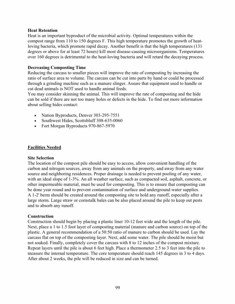
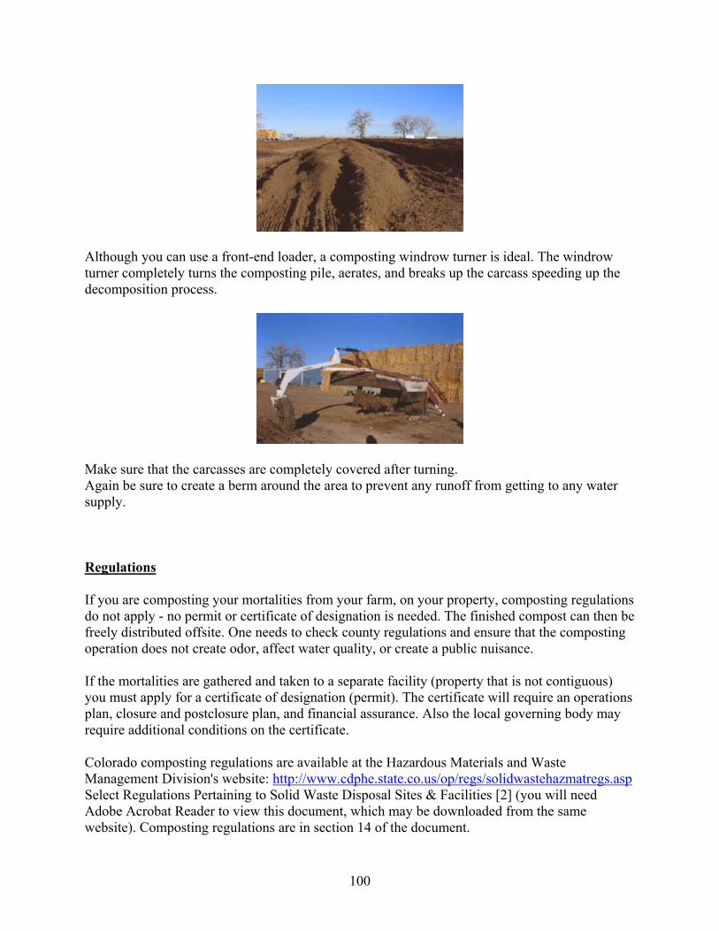
Although you can use a front-end loader, a composting windrow turner is ideal. The windrow turner completely turns the composting pile, aerates, and breaks up the carcass speeding up the decomposition process.
Make sure that the carcasses are completely covered after turning. Again be sure to create a berm around the area to prevent any runoff from getting to any water supply.
Regulations
If you are composting your mortalities from your farm, on your property, composting regulations do not apply - no permit or certificate of designation is needed. The finished compost can then be freely distributed offsite. One needs to check county regulations and ensure that the composting operation does not create odor, affect water quality, or create a public nuisance.
If the mortalities are gathered and taken to a separate facility (property that is not contiguous) you must apply for a certificate of designation (permit). The certificate will require an operations plan, closure and postclosure plan, and financial assurance. Also the local governing body may require additional conditions on the certificate.
Colorado composting regulations are available at the Hazardous Materials and Waste Management Division's website: http://www.cdphe.state.co.us/op/regs/solidwastehazmatregs.asp Select Regulations Pertaining to Solid Waste Disposal Sites & Facilities [2] (you will need Adobe Acrobat Reader to view this document, which may be downloaded from the same website). Composting regulations are in section 14 of the document.

101
Suggested Guidelines for Composting
The following are guidelines taken from the Iowa Department of Natural Resources Rules for On-Farm Carcass Composting. You should check with your local officials to find out if there are specific rules and/or permits that you need for your area.
• Dead animals should be added to the composting pile within 24 hours of death and covered with sufficient composting material.
• Composting should be done in a manner that prevents access by any animal. • Runoff and odor should be prevented.
• Dead animals should not be removed from the composting pile until all flesh, internal organs, and other soft tissues have decomposed.
• Composting needs to be done on an all weather surface of compacted soil, asphalt, concrete, or similar material that will permit accessibility during all times of the year and that will prevent ground water contamination.
• Composting must be done outside of wetlands, or the 100 year flood plain and at least 100 feet from private wells, 200 feet from public wells, 500 feet away from inhabited residences, and at least 100 feet away from any water sources.

102
Helpful Tips
• Although recycling of compost material is encouraged, be sure to add some fresh carbon material, i.e. sawdust, to the pile to create a biofilter.
• Avoid depressions on the top of the pile that could collect rainwater. The excess water may cause odor and draw in pests.
• An unused open-front barn may be perfect for composting. • Place a fence around the pile to keep out pests. • Add new mortalities immediately to the pile. Composting works best on fresh material
and not on material that is already decaying or frozen. • Carcasses should not touch one another. There should be at least one foot of composting
material surrounding the carcass on all sides. • Remember convenience. The area must be easy to access in order to turn the pile and to
add new carcasses. • Biosecurity - You should consider having your personnel work at the compost pile at the
end of the day after the other animals are handled. After you have worked with the compost be sure to use proper hygiene before interacting with other animals or people.