cutting edge: mir-223 and ebv mir-bart15 … edge: mir-223 and ebv mir-bart15 regulate the nlrp3...
TRANSCRIPT
of August 17, 2018.This information is current as
ProductionβInflammasome and IL-1miR-BART15 Regulate the NLRP3 Cutting Edge: miR-223 and EBV
O'Neill and Seth L. MastersIain B. McInnes, Wolfgang Hammerschmidt, Luke A. J.Kurowska-Stolarska, Ashleigh-Ann Rainey, Dagmar Pich, Moritz Haneklaus, Motti Gerlic, Mariola
http://www.jimmunol.org/content/189/8/3795doi: 10.4049/jimmunol.1200312September 2012;
2012; 189:3795-3799; Prepublished online 14J Immunol
MaterialSupplementary
2.DC1http://www.jimmunol.org/content/suppl/2012/09/14/jimmunol.120031
Referenceshttp://www.jimmunol.org/content/189/8/3795.full#ref-list-1
, 7 of which you can access for free at: cites 16 articlesThis article
average*
4 weeks from acceptance to publicationFast Publication! •
Every submission reviewed by practicing scientistsNo Triage! •
from submission to initial decisionRapid Reviews! 30 days* •
Submit online. ?The JIWhy
Subscriptionhttp://jimmunol.org/subscription
is online at: The Journal of ImmunologyInformation about subscribing to
Permissionshttp://www.aai.org/About/Publications/JI/copyright.htmlSubmit copyright permission requests at:
Email Alertshttp://jimmunol.org/alertsReceive free email-alerts when new articles cite this article. Sign up at:
Print ISSN: 0022-1767 Online ISSN: 1550-6606. Immunologists, Inc. All rights reserved.Copyright © 2012 by The American Association of1451 Rockville Pike, Suite 650, Rockville, MD 20852The American Association of Immunologists, Inc.,
is published twice each month byThe Journal of Immunology
by guest on August 17, 2018
http://ww
w.jim
munol.org/
Dow
nloaded from
by guest on August 17, 2018
http://ww
w.jim
munol.org/
Dow
nloaded from
Cutting Edge: miR-223 and EBV miR-BART15 Regulate theNLRP3 Inflammasome and IL-1b ProductionMoritz Haneklaus,* Motti Gerlic,† Mariola Kurowska-Stolarska,‡
Ashleigh-Ann Rainey,‡ Dagmar Pich,x Iain B. McInnes,‡ Wolfgang Hammerschmidt,x
Luke A. J. O’Neill,* and Seth L. Masters*,†
Although microRNA (miRNA) regulation of TLR sig-naling is well established, this has not yet been observedfor NLR proteins or the inflammasomes they form. Wehave now validated a highly conserved miR-223 targetsite in the NLRP3 39-untranslated region. miR-223 ex-pression decreases as monocytes differentiate into mac-rophages, whereas NLRP3 protein increases during thistime. However, overexpression of miR-223 preventsaccumulation of NLRP3 protein and inhibits IL-1bproduction from the inflammasome. Virus inhibitionof the inflammasome is an emerging theme, and wehave also identified an EBV miRNA that can target themiR-223 binding site in the NLRP3 39-untranslatedregion. Furthermore, this virus miRNA can be secretedfrom infected B cells via exosomes to inhibit the NLRP3inflammasome in noninfected cells. Therefore, we haveidentified both the first endogenous miRNA that limitsNLRP3 inflammatory capacity during myeloid cell devel-opment and also a viral miRNA that takes advantage ofthis, limiting inflammation for its own purposes. TheJournal of Immunology, 2012, 189: 3795–3799.
Many different microRNA (miRNA) are regulatedby TLR activation, and feedback to amplify ornegatively regulate that signal (1). Although TLRs
respond to specific pathogen-associated molecular patterns,NLRs are thought to monitor critical intracellular homeo-static parameters. Therefore, they can respond to a variety ofpathogens and cell stresses invoked by infection or disease.NLRP3 is the best studied NLR to date, activated in responseto toxins, uric acid crystals, amyloid, and a host of other factors(2). A conserved mechanism of activation is yet to be formal-ized; however, it is clear that many cell types require NLRP3inflammasome “priming,” such as stimulation with a TLRligand. This functions both to provide pro–IL-1b as a sub-
strate for the inflammasome and to induce NLRP3 ex-pression above a critical threshold required for activation (3).Although NLRP3 is TLR inducible, very little is knownabout what regulates the expression of NLRP3 in differenttissues and cell types, or the stability of the mRNA and pro-tein once it is generated. In this work, we investigatedNLRP3 expression and found that it can be regulated bymiR-223, which is likely to be important early in the my-eloid lineage. Furthermore, we identified a virus miRNA thattargets the miR-223 binding site in the NLRP3 39-untranslatedregion (UTR) and also inhibits inflammasome productionof IL-1b.
Materials and MethodsCell culture
Primary monocytes were isolated using anti-CD14 magnetic beads (MiltenyiBiotec) and then differentiated into macrophages by cultivation with 100 ng/ml M-CSF or 100 ng/ml GM-CSF for 7 d. Macrophages were classicallyactivated (M1) by stimulation with 100 ng/ml LPS (Alexis) and 20 ng/ml IFN-g, and alternatively activated (M2) by 20 ng/ml IL-4 for 18 h each. Mac-rophages were transfected with 25 nM control scramble or miR-223 mimicusing Dharmafect reagent 3 (Dharmacon-Thermo Scientific) and activatedwith 1 mg/ml LPS and 5 mM ATP (Sigma). Thp-1 cells were differentiatedwith 20 nM PMA (Sigma) overnight. Cells were then transfected with 100 nMsmall interfering RNA, 50 nM synthetic miRNA, or negative control precursors(Applied Biosystems) using Lipofectamine 2000 (Invitrogen). Twenty-fourhours later, transfected Thp-1 cells were primed with 100 ng/ml LPS for 3 h;then the inflammasome was activated with 1 mM ATP, 50 pM nigericin for30 min or 250 ng/ml monosodium urate (MSU; Invivogen), 100 mg/ml Alum(Brenntag Biosector), 10 mM human islet amyloid polypeptide (Sigma), 10mg/ml poly-dAdT, Salmonella (multiplicity of infection 5 10) overnight. Forthe transwell assay of EBV miRNA, B cells were added to the top of a 3-mmtranswell dish (Corning) seeded below with PMA-treated Thp-1 cells. Cellswere cultured in the same well for 24 h before the addition of 20 mMmonensin(Sigma) for 6 h with or without 100 ng/ml LPS for the final 3 h. To collectexosomes, we treated B cells with monensin for 3 h, the supernatant wasclarified by centrifugation at 500 3 g for 10 min, then 10,000 3 g for 20min, and finally exosomes were collected by centrifugation at 50,000 3 gfor 150 min. Exosomes from 15 ml supernatant were resuspended in 200 mlRPMI and added to one well of a six-well plate of PMA-differentiated Thp-1 cells for 48 h.
*Inflammation Research Group and Immunology Research Centre, School of Biochem-istry and Immunology, Trinity Biomedical Sciences Institute, Trinity College Dublin,Dublin 4, Ireland; †Inflammation Division, The Walter and Eliza Hall Institute, Mel-bourne, Victoria 3052, Australia; ‡Institute of Infection, Immunity and Inflammation,University of Glasgow, Glasgow G12 8TA, United Kingdom; and xDepartment of GeneVectors, Helmholtz Center Munich, Munich 81377, Germany
Received for publication January 30, 2012. Accepted for publication August 10, 2012.
S.L.M. was supported by a National Health and Medical Research Council OverseasBiomedical Fellowship (516783) and a Victorian Endowment for Science, Knowledgeand Innovation Fellowship. M.K.-S. was supported by an Arthritis Research UnitedKingdom Career Development Grant. A.-A.R. was supported by the Oliver Bird Foun-dation. Trinity College Dublin was supported by Science Foundation Ireland.
S.L.M., M.H., M.G., A.-A.R., D.P., and M.K.-S. designed and performed experiments,analyzed data, and wrote the manuscript; I.B.M., W.H., and L.A.J.O. provided adviceand reagents.
Address correspondence and reprint requests to Dr. Seth L. Masters, The Walter andEliza Hall Institute, 1G Royal Parade, Parkville, VIC 3052, Australia. E-mail address:[email protected]
The online version of this article contains supplemental material.
Abbreviations used in this article: miRNA, microRNA; MSU, monosodium urate; UTR,untranslated region.
Copyright� 2012 by TheAmerican Association of Immunologists, Inc. 0022-1767/12/$16.00
www.jimmunol.org/cgi/doi/10.4049/jimmunol.1200312
by guest on August 17, 2018
http://ww
w.jim
munol.org/
Dow
nloaded from
Quantitative PCR
RNA was extracted using the RNeasy Mini Kit (Qiagen), using 100% ratherthan 70% ethanol, and omitting buffer RW1 to retain miRNA. NLRP3(Hs00918082_m1) mRNA levels were determined using TaqMan Gene Ex-pression Assays (Applied Biosystems) with GAPDH as an internal control. miR-223 and RNU6B levels were determined using TaqMan miRNA assays (AppliedBiosystems).
Western blot
Cells were cultured in six-well plates, lysed directly in 70–80 ml SDS loadingbuffer, then separated by 8% Tris-glycine PAGE and blotted according tostandard protocol. Membranes were probed for NLRP3 (rabbit anti-humanNLRP3, HPA012878; Atlas Antibodies), IL-1b (goat anti-human IL-1b), andb-actin as a loading control.
Luciferase experiments
NLRP3 39-UTR luciferase construct was purchased from SwitchgearGenomics. 293T cells were transfected in 96-well plates in triplicates with 100ng 39-UTR luciferase constructs and 50 ng TK-Renilla luciferase control vectorusing Lipofectamine 2000 (Invitrogen). In addition, 50 nM pre-miRNAprecursors was cotransfected. Cells were lysed in Passive Lysis Buffer (Promega)after 24 h; then luciferase activity was measured after the addition of luciferin orcoelenterazine.
Cytokine measurement
Thp-1 cells (43 105/ml) were cultured in 96-well plates, then transfected andstimulated as indicated in triplicate. Human macrophages (2.5 3 105/ml)were cultured in 24-well plates, then transfected and stimulated as indicatedfor seven individual donors. Supernatants were collected and cytokine se-cretion was determined by ELISA for human IL-1b and TNF-a (R&DSystems).
Statistical analysis
Data are presented as mean 6 SD. Significance was determined by two-tailedunpaired t test.
ResultsNLRP3 and miR-223 expression during macrophage differentiation
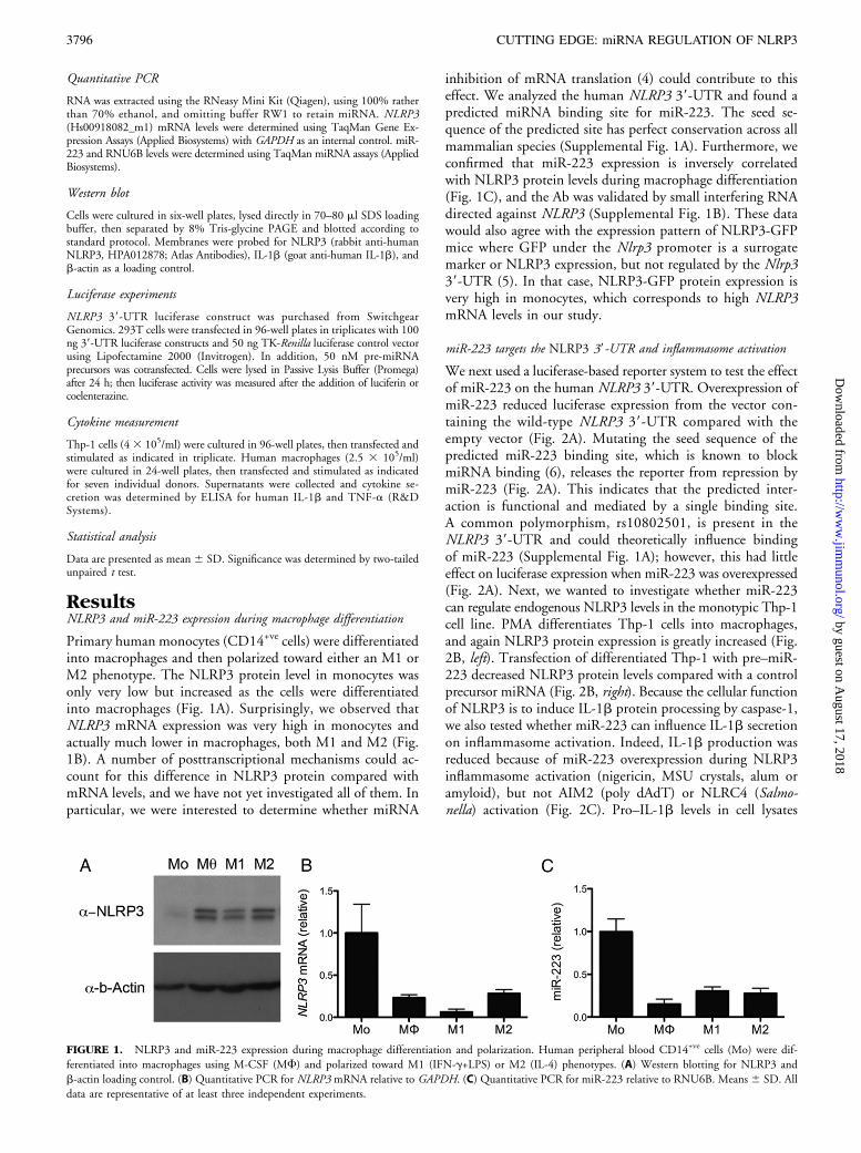
Primary human monocytes (CD14+ve cells) were differentiatedinto macrophages and then polarized toward either an M1 orM2 phenotype. The NLRP3 protein level in monocytes wasonly very low but increased as the cells were differentiatedinto macrophages (Fig. 1A). Surprisingly, we observed thatNLRP3 mRNA expression was very high in monocytes andactually much lower in macrophages, both M1 and M2 (Fig.1B). A number of posttranscriptional mechanisms could ac-count for this difference in NLRP3 protein compared withmRNA levels, and we have not yet investigated all of them. Inparticular, we were interested to determine whether miRNA
inhibition of mRNA translation (4) could contribute to thiseffect. We analyzed the human NLRP3 39-UTR and found apredicted miRNA binding site for miR-223. The seed se-quence of the predicted site has perfect conservation across allmammalian species (Supplemental Fig. 1A). Furthermore, weconfirmed that miR-223 expression is inversely correlatedwith NLRP3 protein levels during macrophage differentiation(Fig. 1C), and the Ab was validated by small interfering RNAdirected against NLRP3 (Supplemental Fig. 1B). These datawould also agree with the expression pattern of NLRP3-GFPmice where GFP under the Nlrp3 promoter is a surrogatemarker or NLRP3 expression, but not regulated by the Nlrp339-UTR (5). In that case, NLRP3-GFP protein expression isvery high in monocytes, which corresponds to high NLRP3mRNA levels in our study.
miR-223 targets the NLRP3 39-UTR and inflammasome activation
We next used a luciferase-based reporter system to test the effectof miR-223 on the human NLRP3 39-UTR. Overexpression ofmiR-223 reduced luciferase expression from the vector con-taining the wild-type NLRP3 39-UTR compared with theempty vector (Fig. 2A). Mutating the seed sequence of thepredicted miR-223 binding site, which is known to blockmiRNA binding (6), releases the reporter from repression bymiR-223 (Fig. 2A). This indicates that the predicted inter-action is functional and mediated by a single binding site.A common polymorphism, rs10802501, is present in theNLRP3 39-UTR and could theoretically influence bindingof miR-223 (Supplemental Fig. 1A); however, this had littleeffect on luciferase expression when miR-223 was overexpressed(Fig. 2A). Next, we wanted to investigate whether miR-223can regulate endogenous NLRP3 levels in the monotypic Thp-1cell line. PMA differentiates Thp-1 cells into macrophages,and again NLRP3 protein expression is greatly increased (Fig.2B, left). Transfection of differentiated Thp-1 with pre–miR-223 decreased NLRP3 protein levels compared with a controlprecursor miRNA (Fig. 2B, right). Because the cellular functionof NLRP3 is to induce IL-1b protein processing by caspase-1,we also tested whether miR-223 can influence IL-1b secretionon inflammasome activation. Indeed, IL-1b production wasreduced because of miR-223 overexpression during NLRP3inflammasome activation (nigericin, MSU crystals, alum oramyloid), but not AIM2 (poly dAdT) or NLRC4 (Salmo-nella) activation (Fig. 2C). Pro–IL-1b levels in cell lysates
FIGURE 1. NLRP3 and miR-223 expression during macrophage differentiation and polarization. Human peripheral blood CD14+ve cells (Mo) were dif-
ferentiated into macrophages using M-CSF (MF) and polarized toward M1 (IFN-g+LPS) or M2 (IL-4) phenotypes. (A) Western blotting for NLRP3 and
b-actin loading control. (B) Quantitative PCR for NLRP3 mRNA relative to GAPDH. (C) Quantitative PCR for miR-223 relative to RNU6B. Means 6 SD. All
data are representative of at least three independent experiments.
3796 CUTTING EDGE: miRNA REGULATION OF NLRP3
by guest on August 17, 2018
http://ww
w.jim
munol.org/
Dow
nloaded from
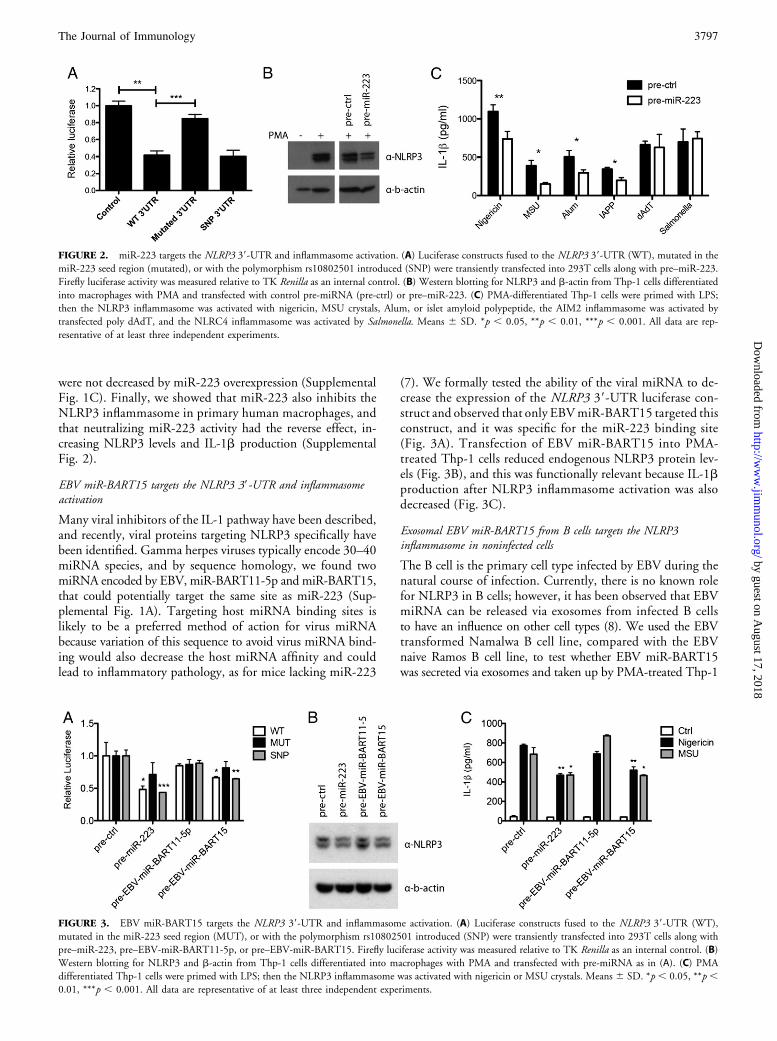
were not decreased by miR-223 overexpression (SupplementalFig. 1C). Finally, we showed that miR-223 also inhibits theNLRP3 inflammasome in primary human macrophages, andthat neutralizing miR-223 activity had the reverse effect, in-creasing NLRP3 levels and IL-1b production (SupplementalFig. 2).
EBV miR-BART15 targets the NLRP3 39-UTR and inflammasomeactivation
Many viral inhibitors of the IL-1 pathway have been described,and recently, viral proteins targeting NLRP3 specifically havebeen identified. Gamma herpes viruses typically encode 30–40miRNA species, and by sequence homology, we found twomiRNA encoded by EBV, miR-BART11-5p and miR-BART15,that could potentially target the same site as miR-223 (Sup-plemental Fig. 1A). Targeting host miRNA binding sites islikely to be a preferred method of action for virus miRNAbecause variation of this sequence to avoid virus miRNA bind-ing would also decrease the host miRNA affinity and couldlead to inflammatory pathology, as for mice lacking miR-223
(7). We formally tested the ability of the viral miRNA to de-crease the expression of the NLRP3 39-UTR luciferase con-struct and observed that only EBVmiR-BART15 targeted thisconstruct, and it was specific for the miR-223 binding site(Fig. 3A). Transfection of EBV miR-BART15 into PMA-treated Thp-1 cells reduced endogenous NLRP3 protein lev-els (Fig. 3B), and this was functionally relevant because IL-1bproduction after NLRP3 inflammasome activation was alsodecreased (Fig. 3C).
Exosomal EBV miR-BART15 from B cells targets the NLRP3inflammasome in noninfected cells
The B cell is the primary cell type infected by EBV during thenatural course of infection. Currently, there is no known rolefor NLRP3 in B cells; however, it has been observed that EBVmiRNA can be released via exosomes from infected B cellsto have an influence on other cell types (8). We used the EBVtransformed Namalwa B cell line, compared with the EBVnaive Ramos B cell line, to test whether EBV miR-BART15was secreted via exosomes and taken up by PMA-treated Thp-1
FIGURE 2. miR-223 targets the NLRP3 39-UTR and inflammasome activation. (A) Luciferase constructs fused to the NLRP3 39-UTR (WT), mutated in the
miR-223 seed region (mutated), or with the polymorphism rs10802501 introduced (SNP) were transiently transfected into 293T cells along with pre–miR-223.
Firefly luciferase activity was measured relative to TK Renilla as an internal control. (B) Western blotting for NLRP3 and b-actin from Thp-1 cells differentiated
into macrophages with PMA and transfected with control pre-miRNA (pre-ctrl) or pre–miR-223. (C) PMA-differentiated Thp-1 cells were primed with LPS;
then the NLRP3 inflammasome was activated with nigericin, MSU crystals, Alum, or islet amyloid polypeptide, the AIM2 inflammasome was activated by
transfected poly dAdT, and the NLRC4 inflammasome was activated by Salmonella. Means 6 SD. *p , 0.05, **p , 0.01, ***p , 0.001. All data are rep-
resentative of at least three independent experiments.
FIGURE 3. EBV miR-BART15 targets the NLRP3 39-UTR and inflammasome activation. (A) Luciferase constructs fused to the NLRP3 39-UTR (WT),
mutated in the miR-223 seed region (MUT), or with the polymorphism rs10802501 introduced (SNP) were transiently transfected into 293T cells along with
pre–miR-223, pre–EBV-miR-BART11-5p, or pre–EBV-miR-BART15. Firefly luciferase activity was measured relative to TK Renilla as an internal control. (B)
Western blotting for NLRP3 and b-actin from Thp-1 cells differentiated into macrophages with PMA and transfected with pre-miRNA as in (A). (C) PMA
differentiated Thp-1 cells were primed with LPS; then the NLRP3 inflammasome was activated with nigericin or MSU crystals. Means 6 SD. *p , 0.05, **p ,0.01, ***p , 0.001. All data are representative of at least three independent experiments.
The Journal of Immunology 3797
by guest on August 17, 2018
http://ww
w.jim
munol.org/
Dow
nloaded from
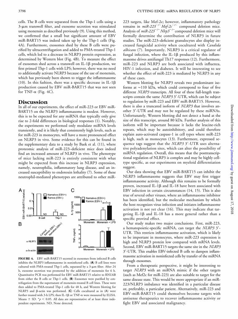
cells. The B cells were separated from the Thp-1 cells using a3-mm transwell filter, and exosome secretion was stimulatedusing monensin as described previously (9). Using this method,we confirmed that a small but significant amount of EBVmiR-BART15 was indeed taken up by the Thp-1 cells (Fig.4A). Furthermore, exosomes shed by these B cells were pu-rified by ultracentrifugation and added to PMA-treated Thp-1cells, which led to a decrease in NLRP3 protein expression, asdetermined by Western blot (Fig. 4B). To measure the effectof exosomes shed across a transwell on IL-1b production, wefirst primed Thp-1 cells with LPS; however, there was no needto additionally activate NLRP3 because of the use of monensin,which has previously been shown to trigger the inflammasome(10). In this fashion, there was a specific decrease in IL-1bproduction caused by EBV miR-BART15 that was not seenfor TNF-a (Fig. 4C).
DiscussionIn all of our experiments, the effect of miR-223 or EBV miR-BART15 on the NLRP3 inflammasome is modest. However,this is to be expected for any miRNA that typically only giverise to 2-fold differences in biological responses (1). Notably,the experiments we performed only modulate miRNA levelstransiently, and it is likely that consistently high levels, such asfor miR-223 in monocytes, will have a more pronounced effecton NLRP3 in vivo. Some evidence for this can be found inthe supplementary data to a study by Baek et al. (11), whereproteomic analysis of miR-223–deficient mice does indeedfind an increased amount of NLRP3 in vivo. The phenotypeof mice lacking miR-223 is entirely consistent with whatmight be expected from this increase in NLRP3 expression,namely, neutrophilia, inflammatory lung disease, and an in-creased susceptibility to endotoxin lethality (7). Some of theseneutrophil-mediated phenotypes are attributed to other miR-
223 targets, like Mef-2c; however, inflammatory pathologyremains in miR-2232/2Mef-2c2/2 compound deletion mice.Analysis of miR-2232/2Nlrp32/2compound deletion mice willformally determine the contribution of NLRP3 in futurestudies. The miR-223–deficient granulocytes also display in-creased fungicidal activity when cocultured with Candidaalbicans (7). Importantly, NLRP3 is a critical regulator offungal infection, where the IL-1b produced by this inflam-masome drives antifungal Th17 responses (12). Furthermore,miR-223 and NLRP3 are both associated with influenza,HIV-1 infection, and diabetes. It will be interesting to seewhether the effect of miR-223 is mediated by NLRP3 in anyof these cases.Western blotting for NLRP3 reveals two predominant iso-
forms at ∼110 kDa, which could correspond to four of fivedifferent NLRP3 transcripts. All four of these full-length tran-scripts contain the same NLRP3 39-UTR, which can be subjectto regulation by miR-223 and EBV miR-BART15. However,there is also a truncated isoform of NLRP3 that involves an-other 39-UTR and may not be regulated by these miRNAs.Unfortunately, Western blotting did not detect a band at thesize of this transcript, around 80 kDa. Further analysis of thisisoform will be important because it lacks the leucine-richrepeats, which may be autoinhibitory, and could thereforeexplain auto-activated caspase-1 in cell types where miR-223is high, such as monocytes (13). Furthermore, expressed se-quence tags suggest that the NLRP3 39-UTR uses alterna-tive polyadenylation sites, which can alter the possibility ofmiRNA regulation. Overall, this means that the posttranscrip-tional regulation of NLRP3 is complex and may be highly cell-type specific, as our experiments on myeloid differentiationsuggest.Our data showing that EBV miR-BART15 can inhibit the
NLRP3 inflammasome suggests that EBV may first triggerinflammasome activity. Although this remains to be formallyproven, increased IL-1b and IL-18 have been associated withEBV infection in certain circumstances (14, 15). This is alsotrue of several other viruses, where an inflammasome inhibitorhas been identified, but the molecular mechanism by whichthe host recognizes virus infection and initiates inflammasomeactivation is not yet clear (16). This may indicate that tar-geting IL-1b and IL-18 has a more general rather than aspecific proviral effect.Our study makes two major conclusions. First, miR-223,
a hematopoietic-specific miRNA, can target the NLRP3 39-UTR. This restricts inflammasome activation, which is likelyto be important in monocytes, where miR-223 expression ishigh and NLRP3 protein low compared with mRNA levels.Second, EBV miR-BART15 targets the same site in the NLRP339-UTR. This enables EBV-infected B cells to dampen inflam-masome activation in noninfected cells by transfer of the miRNAthrough exosomes.From a therapeutic perspective, it might be interesting to
target NLRP3 with an miRNA mimic if the other targets(such as Mef2c for miR-223) are also suitable to target for thesame disease state. This would be more appropriate if an miR-223/NLRP3 imbalance was identified in a particular diseaseor, preferably, a particular patient. Alternatively, miR-223 andEBV-miR-BART15 could themselves become targets withantisense therapeutics to recover inflammasome activity orfight EBV and associated malignancy.
FIGURE 4. EBV miR-BART15 secreted in exosomes from infected B cells
inhibits the NLRP3 inflammasome in noninfected cells. (A) B cell lines were
cultured with PMA-treated Thp-1 cells, separated by a 3-mm filter. After 24
h, exosome secretion was promoted by the addition of monensin for 6 h.
Quantitative PCR was performed for EBV miR-BART15 relative to RNU6B
from either the B cells or Thp-1 cells. (B) Exosomes were purified by cen-
trifugation from the supernatant of monensin-treated B cell lines. These were
then added to PMA-treated Thp-1 cells for 48 h, and Western blotting for
NLRP3 and b-actin was performed. (C) Cells cocultured as in (A) were
further treated with 3-h LPS; then IL-1b or TNF-a were measured by ELISA.
Means 6 SD. *p , 0.05. All data are representative of at least three inde-
pendent experiments. ND, None detected.
3798 CUTTING EDGE: miRNA REGULATION OF NLRP3
by guest on August 17, 2018
http://ww
w.jim
munol.org/
Dow
nloaded from
AcknowledgmentsWe thank Russka Shumnalieva for expert technical assistance.
DisclosuresThe authors have no financial conflicts of interest.
References1. O’Neill, L. A., F. J. Sheedy, and C. E. McCoy. 2011. MicroRNAs: the fine-tuners
of Toll-like receptor signalling. Nat. Rev. Immunol. 11: 163–175.2. Martinon, F., A. Mayor, and J. Tschopp. 2009. The inflammasomes: guardians of
the body. Annu. Rev. Immunol. 27: 229–265.3. Bauernfeind, F. G., G. Horvath, A. Stutz, E. S. Alnemri, K. MacDonald, D. Speert,
T. Fernandes-Alnemri, J. Wu, B. G. Monks, K. A. Fitzgerald, et al. 2009. Cuttingedge: NF-kappaB activating pattern recognition and cytokine receptors license NLRP3inflammasome activation by regulating NLRP3 expression. J. Immunol. 183: 787–791.
4. Mathonnet, G., M. R. Fabian, Y. V. Svitkin, A. Parsyan, L. Huck, T. Murata,S. Biffo, W. C. Merrick, E. Darzynkiewicz, R. S. Pillai, et al. 2007. MicroRNAinhibition of translation initiation in vitro by targeting the cap-binding complexeIF4F. Science 317: 1764–1767.
5. Guarda, G., M. Zenger, A. S. Yazdi, K. Schroder, I. Ferrero, P. Menu, A. Tardivel,C. Mattmann, and J. Tschopp. 2011. Differential expression of NLRP3 amonghematopoietic cells. J. Immunol. 186: 2529–2534.
6. Frankel, L. B., N. R. Christoffersen, A. Jacobsen, M. Lindow, A. Krogh, andA. H. Lund. 2008. Programmed cell death 4 (PDCD4) is an important functionaltarget of the microRNA miR-21 in breast cancer cells. J. Biol. Chem. 283: 1026–1033.
7. Johnnidis, J. B., M. H. Harris, R. T. Wheeler, S. Stehling-Sun, M. H. Lam,O. Kirak, T. R. Brummelkamp, M. D. Fleming, and F. D. Camargo. 2008. Reg-
ulation of progenitor cell proliferation and granulocyte function by microRNA-223.Nature 451: 1125–1129.
8. Pegtel, D. M., K. Cosmopoulos, D. A. Thorley-Lawson, M. A. van Eijndhoven,E. S. Hopmans, J. L. Lindenberg, T. D. de Gruijl, T. Wurdinger, andJ. M. Middeldorp. 2010. Functional delivery of viral miRNAs via exosomes. Proc.Natl. Acad. Sci. USA 107: 6328–6333.
9. Savina, A., M. Furlan, M. Vidal, and M. I. Colombo. 2003. Exosome release isregulated by a calcium-dependent mechanism in K562 cells. J. Biol. Chem. 278:20083–20090.
10. Ichinohe, T., I. K. Pang, and A. Iwasaki. 2010. Influenza virus activates inflam-masomes via its intracellular M2 ion channel. Nat. Immunol. 11: 404–410.
11. Baek, D., J. Villen, C. Shin, F. D. Camargo, S. P. Gygi, and D. P. Bartel. 2008. Theimpact of microRNAs on protein output. Nature 455: 64–71.
12. Cheng, S. C., F. L. van de Veerdonk, M. Lenardon, M. Stoffels, T. Plantinga,S. Smeekens, L. Rizzetto, L. Mukaremera, K. Preechasuth, D. Cavalieri, et al. 2011.The dectin-1/inflammasome pathway is responsible for the induction of protectiveT-helper 17 responses that discriminate between yeasts and hyphae of Candidaalbicans. J. Leukoc. Biol. 90: 357–366.
13. Netea, M. G., C. A. Nold-Petry, M. F. Nold, L. A. Joosten, B. Opitz, J. H. van derMeer, F. L. van de Veerdonk, G. Ferwerda, B. Heinhuis, I. Devesa, et al. 2009.Differential requirement for the activation of the inflammasome for processing andrelease of IL-1beta in monocytes and macrophages. Blood 113: 2324–2335.
14. Yao, L., J. Setsuda, C. Sgadari, B. Cherney, and G. Tosato. 2001. Interleukin-18expression induced by Epstein-Barr virus-infected cells. J. Leukoc. Biol. 69: 779–784.
15. Krauer, K. G., D. K. Belzer, D. Liaskou, M. Buck, S. Cross, T. Honjo, andT. Sculley. 1998. Regulation of interleukin-1beta transcription by Epstein-Barrvirus involves a number of latent proteins via their interaction with RBP. Virology252: 418–430.
16. Kanneganti, T. D. 2010. Central roles of NLRs and inflammasomes in viral in-fection. Nat. Rev. Immunol. 10: 688–698.
The Journal of Immunology 3799
by guest on August 17, 2018
http://ww
w.jim
munol.org/
Dow
nloaded from