crhr1 genotypes, neural circuits and the diathesis for ... · pdf fileoriginal article crhr1...
TRANSCRIPT

ORIGINAL ARTICLE
CRHR1 genotypes, neural circuits and the diathesis for anxiety anddepressionJ Rogers1,2, M Raveendran1, GL Fawcett1, AS Fox3,4, SE Shelton5,6, JA Oler5,6, J Cheverud7, DM Muzny1, RA Gibbs1,RJ Davidson3,4,5,6 and NH Kalin3,4,5,6
The corticotrophin-releasing hormone (CRH) system integrates the stress response and is associated with stress-relatedpsychopathology. Previous reports have identified interactions between childhood trauma and sequence variation in the CRHreceptor 1 gene (CRHR1) that increase risk for affective disorders. However, the underlying mechanisms that connect variation inCRHR1 to psychopathology are unknown. To explore potential mechanisms, we used a validated rhesus macaque model toinvestigate association between genetic variation in CRHR1, anxious temperament (AT) and brain metabolic activity. In youngrhesus monkeys, AT is analogous to the childhood risk phenotype that predicts the development of human anxiety and depressivedisorders. Regional brain metabolism was assessed with 18F-labeled fluoro-2-deoxyglucose (FDG) positron emission tomography in236 young, normally reared macaques that were also characterized for AT. We show that single nucleotide polymorphisms (SNPs)affecting exon 6 of CRHR1 influence both AT and metabolic activity in the anterior hippocampus and amygdala, components of theneural circuit underlying AT. We also find evidence for association between SNPs in CRHR1 and metabolism in the intraparietalsulcus and precuneus. These translational data suggest that genetic variation in CRHR1 affects the risk for affective disorders byinfluencing the function of the neural circuit underlying AT and that differences in gene expression or the protein sequenceinvolving exon 6 may be important. These results suggest that variation in CRHR1 may influence brain function before anychildhood adversity and may be a diathesis for the interaction between CRHR1 genotypes and childhood trauma reported to affecthuman psychopathology.
Molecular Psychiatry (2013) 18, 700–707; doi:10.1038/mp.2012.152; published online 13 November 2012
Keywords: amygdala; corticotrophin-releasing hormone; genetic association; hippocampus; non-human primate; rhesus macaque
INTRODUCTIONThe corticotrophin-releasing hormone (CRH) system mediates theadaptive physiological and behavioral responses to stress andperceived threat.1,2 CRH and related endogenous ligands exerttheir effects through either of two receptors.2 Several studiesindicate that genetic variation within the CRH receptor 1 gene(CRHR1) can affect reactivity to stress and that altered CRHR1function is associated with stress-related psychopathology,particularly anxiety and depressive disorders.2–4 Importantly,specific CRHR1 haplotypes have been associated with the risk todevelop depression following exposure to childhood trauma5–8
and with differential response to antidepressant treatment.9,10
However, despite extensive preclinical and clinical studies, thebrain mechanisms by which sequence variation in the CRHR1 geneinfluences the risk to develop stress-related psychopathologyhave not been described. Identification of these intermediatemechanisms is critical for understanding the human risk factorsassociated with the development of anxiety and depressivedisorders.
The well-validated rhesus macaque (Macaca mulatta) model11
of childhood anxious temperament (AT) provides an opportunityto characterize the effects of genetic variation in CRHR1 on
anxiety-related phenotypes, including the function of the relevantneural circuits. Children with AT are extremely shy, exhibit highlevels of behavioral inhibition when exposed to novelty or in thepresence of strangers and are at increased risk to develop anxiety,depression and comorbid substance abuse.12,13 In addition to thebehavioral phenotype, children with AT commonly displayincreased pituitary–adrenal and autonomic reactivity.12,13 Young,normally reared rhesus monkeys with high levels of trait-likeanxiety show many similarities to children with AT, includingbehavioral inhibition, or freezing in response to potential threat.11
Furthermore, as in children, the degree of AT expressed by youngmonkeys is stable, trait-like and significantly heritable.14–16 Werecently used fluoro-2-deoxyglucose-positron emissiontomography (FDG-PET) to characterize the neural circuitunderlying AT, finding that increased metabolic activity in thedorsal amygdala, central nucleus of the amygdala and anteriorhippocampus was predictive of AT, whereas activity in theintraparietal sulcus (IPS) and precuneus was negativelycorrelated with AT.17 The goal of the current study was to usethe macaque model to examine the influence of variation in theCRHR1 gene on individual differences in AT and the function of itsunderlying neural circuit.
1Human Genome Sequencing Center and Department of Molecular and Human Genetics, Baylor College of Medicine, Houston, TX, USA; 2Southwest National Primate ResearchCenter, San Antonio, TX, USA; 3Department of Psychology, University of Wisconsin, Madison, WI, USA; 4Waisman Laboratory for Brain Imaging and Behavior, University ofWisconsin, Madison, WI, USA; 5Department of Psychiatry, University of Wisconsin, Madison, WI, USA; 6HealthEmotions Research Institute, University of Wisconsin, Madison, WI,USA and 7Department of Anatomy and Neurobiology, Washington University School of Medicine, St. Louis, MO, USA. Correspondence: Dr J Rogers, Human Genome SequencingCenter, Baylor College of Medicine, One Baylor Plaza, Houston, TX 77030, USA.E-mail: [email protected] 6 April 2012; revised 29 August 2012; accepted 4 September 2012; published online 13 November 2012
Molecular Psychiatry (2013) 18, 700–707& 2013 Macmillan Publishers Limited All rights reserved 1359-4184/13
www.nature.com/mp

MATERIALS AND METHODSStudy subjects and behavioral testingTwo hundred thirty six maternal-reared rhesus macaques (114 males, 122females) ranging in age from 0.74–4.2 years (mean 2.4 years) were selectedfrom a large, multi-generation single family pedigree and subjected tobehavioral testing and FDG-PET brain imaging as previously described.17,18
AT was assessed by exposing each animal to the ‘no eye contact’ (NEC)condition of the human intruder paradigm for 30 min.11,19 In this test, theanimal was removed from its home cage and placed alone in a cage in aseparate room. An unfamiliar ‘human intruder’ then entered the testingroom and stood in profile 2.5 m from the cage, avoiding eye contact withthe animal. Previously, we developed and validated a composite measureof AT that incorporates behavioral inhibition (duration of freezing),vocalizations (frequency of ‘coo’ calling) and plasma cortisol levelsmeasured immediately after the 30 min exposure to NEC.11,15
Measurement of brain metabolic activity18F-labeled FDG (5–7 mCi) was administered intravenously to the subjectsimmediately before transfer to the test cage for NEC exposure. After the30-min NEC period, the subject was sedated with ketamine HCl (15 mg/kgintramuscular), blood was drawn for cortisol measurement and the animalwas further anesthetized with isoflurane gas for high-resolution microPETimaging (Concorde Microsystems P4 scanner, Knoxville, TN, USA). The useof FDG-PET imaging to quantify local brain metabolic activity optimallymatches uptake and kinetic parameters of radiolabeled FDG with theduration of the NEC testing paradigm. Because FDG is trapped in themetabolic cell, it provides a reliable indicator of regional brain metabolicactivity occurring during the 30 min exposure to this mildly threateningstressor.17,20,21
Quantification of regional metabolic activityBased on our previous work,17,20 we focused primarily on regions stronglyassociated with emotion and reactivity to stress or threat, thehippocampus and amygdala. Secondarily, we also investigated two brainregions exhibiting negative correlations with AT, the IPS and precuneus.For full details of imaging methods, see Oler et al.17 Briefly, each subject’sFDG-PET scan (raw spatial resolution 2 mm3; see Kalin et al.20) was alignedto their T1-weighted MRI (magnetic resonance image) using rigid-bodytransformation and then aligned to a standard space template basedon the T1-weighted MRI image using a nonlinear transformation (FSL;http://www.fmrib.ox.ac.uk/fsl/; Jenkinson et al.22). FDG-PET values werenormalized to the whole-brain average and smoothed with a 4 mm full-width half-maximum gaussian kernel. Regional gray-matter probabilityestimates were based on the T1-weighted MRI and also blurred with a4 mm gaussian kernel.23 Mean metabolism was extracted from the centralamygdala, anterior hippocampus, IPS and precuneus, regions that weremost predictive of AT, along with 95% spatial confidence intervals.17 Thesemean metabolic values were then residualized for gray-matter probabilityand standardized.
Targeted sequencing of exons to identify functional singlenucleotide polymorphisms (SNPs)We used PCR amplification and Sanger methods to sequence all 14 exonsand part of the 50 flanking region of CRHR1 in 236 study subjects(Supplementary Figure 1). The PCR primers and the assay conditions usedare available from the authors on request. This sequencing identified 103SNPs (Supplementary Table 1) with a minimum minor allele frequency of0.01. Linkage disequilibrium (LD) among SNPs was assessed as r2 usingHaploview (Broad Institute, Cambridge, MA, USA).24 After excluding SNPswith X25% missing data (n¼ 39), the polymorphisms were clustered intogroups consisting of SNPs with pairwise r2
X0.8. Identification of SNP pairsshowing high pair-wise LD is necessary because downstream tests ofassociation between individual SNPs and phenotypes can produceinaccurate (false negative) results if X2 SNPs in a single analysis are instrong LD. We found 20 of the 103 SNPs were not in high LD (r2p0.8) withany other SNP, and these singletons were included in subsequent multi-SNP analyses. We also found 10 sets of SNPs constituting high LD clusters.One SNP per cluster was included in the multi-SNP tests.
Annotation of novel SNPsWe used in-house software (Bainbridge, personal communication) to querythe Ensembl genome browser and annotate the newly discovered variants
according to putative functional classes (intronic, synonymous, non-synonymous, splice site). Among the 103 SNPs, there were 5 non-synonymous changes, 3 splice-site variants and 1 SNP that was both anon-synonymous change and a splice-site variant.
Tests of genetic associationStatistical association of individual SNPs with AT and local brain metabolicactivity was tested using pedigree-based variance components analysisimplemented in SOLAR.25 All phenotypes were adjusted for age beforeassociation tests. All tests were performed using age, sex and age� sex asco-variates, but these were not significant parameters in any analysis.These association analyses relating SNP genotypes and individualphenotypes were performed simultaneously using maximum likelihoodestimation of the correlation between known pair-wise kinship amonganimals and pair-wise phenotypic differences among them. In this way,association across all animals between a SNP genotype and a givenphenotype was adjusted for background kinship among pedigreedanimals and the heritability of the phenotype.
Because we are primarily interested in identifying potentially functionalSNPs that drive phenotypic variation, we first tested the effect of the nineannotated putative functional SNPs (amino acid changing non-synon-ymous variants and splice-site variants). Each of these nine SNPs wereevaluated for genetic association with AT, testing one SNP at a time as aco-variate, with simultaneous estimation of the effect of overall kinship onphenotypic differences. Each of the three alternative models was tested foreach SNP by coding the genotypes to accommodate a specific geneticmodel: additive effects of alleles, dominance of the major allele ordominance of the minor allele. Tests on the nine putative functional SNPsinclude some SNPs in LD with each other. The standard Bonferronicorrection assumes that these tests are independent. Considering the LDamong these SNPs, the effective number of independent SNPs in the set isseven,26 resulting in a Bonferroni-corrected family-wise 5% significancethreshold of 0.007. The three different genetic models tested are alsointer-correlated (additive model with either the dominant or recessivemodel r¼ 0.8; dominance model with recessive model r¼ 0.3) andnot independent as required by the Bonferroni procedure. Analyses ofthese correlations suggests that the effective number of independent testsfor each SNP is two,26 and the Bonferroni corrected family-wise 5%significance threshold is 0.025 for genetic models within SNPs. Correctingfor both multiple SNPs and multiple genetic models gives a 5% family-wisethreshold of 0.0036.
To assess and compare simultaneous effects of multiple SNPs, we nextconducted a series of analyses using a variance components multipleregression framework. For each phenotypic trait, we ran measuredgenotype analyses in SOLAR as maximum-likelihood variance componentsmultiple regressions with multiple SNPs run together as covariates (partialregressions). To avoid dilution of SNP effects (false negatives) resultingfrom LD among SNPs, we included only one member of each high-LDcluster (that is, one SNP from each set of two or more SNPs that exhibithigh pair-wise LD with each other, see Supplementary Figure S3 andSupplementary Table S2). Before assessing the effects of each SNP, weevaluated the overall significance of each of the three multiple regressionmodels. Only multiple regression models that were overall significant(P value o0.05) for an initial test of general heritability (h2) were then usedto evaluate the effect of each SNP. The multiple regression analysesquantifying multiple SNP effects simultaneously were conducted usingeither 7 SNPs together (the putative functional non-synonymous andsplice-site SNPs, excluding those that exhibit high LD) or 30 SNPs together(one from each of 10 identified LD clusters and 20 singletons, including theSNPs from exon 6 fixed in the model).
Predictions of protein structureWe used the online I-TASSER server27 to predict the three-dimensionalstructure of rhesus macaque CRHR1-alpha (isoform of the protein that doesnot include the amino acids encoded by exon 6) and variant DNAsequences putatively encoding altered protein sequences that weidentified in this study. The I-TASSER system was the top scoring 3Dmodeling server in the CASP series of blind protein structure predictionexperiments and bench-marking tests.27 We did not use a template for theI-TASSER analyses of the macaque protein sequences, as there wereno appropriate full-length crystallized secretin-like G-protein-coupledreceptors available. The amino-acid sequences for the seven trans-membrane core region plus the C-terminal tail containing the alternativealleles were uploaded to the I-TASSER portal, and the standard I-TASSER
CRHR1, neural circuits and risk for anxiety disordersJ Rogers et al
701
& 2013 Macmillan Publishers Limited Molecular Psychiatry (2013), 700 – 707

protocol was used to generate predicted protein structures. C-scorescalculated by I-TASSER were used to assess the quality of the proteinstructure prediction.
RESULTSTo investigate the influence of variation in the CRHR1 gene onindividual differences in AT and related phenotypes in rhesusmacaques, we sequenced all 14 exons and part of the 50 flankingregion, identifying all SNPs. We next conducted a series ofpedigree-based genetic association tests to determine whetherthat observed variation in the CRHR1 gene influences AT and/ormetabolism in the relevant neural circuits during the NECchallenge test.
Gene-wide SNP discovery in rhesus CRHR1PCR-based Sanger re-sequencing of all exons and a segment ofthe 50 putative promoter region of CRHR1 identified 103 SNPs withminor allele frequency X0.01. Of these SNPs, nine are potentiallyfunctional, being either non-synonymous variants that alterprotein amino-acid sequence or splice sites predicted to havepossible effects on alternative splicing. The remaining 94 SNPsinclude 5 synonymous, 50 intronic, 11 30 UTR SNPs and otherflanking and intergenic SNPs. We were primarily interested in theputative functional SNPs because our goal was to identify causalvariants with detectable phenotypic effects.
Separate tests of effects of functional SNPs on ATTo begin, we tested each of the nine putative functional SNPsseparately for genetic association with our composite measure ofAT. Testing one SNP at a time, we found that three functionalvariants (SNP4805, SNP5043 and SNP8689) showed significantassociation (Table 1). None of these three SNPs exhibit strong pair-wise LD with each other, so these can be considered threeindependent associations. The most robust association wasobserved with SNP5043, an amino-acid change encoded by anucleotide substitution in exon 6 (P¼ 0.0007). This result wasobtained when the effect of this SNP was modeled as a dominantsystem with the major allele (G) dominant to the minor (Figure 1).We note that this result survives conservative Bonferronicorrection for multiple testing across both SNPs and geneticmodels. Probabilities at SNP4805 (P¼ 0.012) and SNP8689(P¼ 0.017) are also significant when considered against the
threshold correcting for number of genetic models. Consideringthese tests together, and following the approach described byCheverud,26 we find that these nine SNPs constitute sevenstatistically independent tests, and the probability of observingthree of these independent tests significant at the 0.05 level underthe null hypothesis of no significant relationship between CRHR1and AT is 4.3� 10� 6. These results demonstrate that variation inCRHR1 influences AT among young rhesus macaques.
Multiple regression tests of functional SNPs on ATWe next tested association between the CRHR1 gene and AT usingmultiple regression. This model evaluates multiple SNP variantssimultaneously and accounts for multiple testing by using partialregression to estimate the effect of any given SNP within thecontext of all SNPs in the model. In this approach, SNPs exhibitinghigh pair-wise LD must be removed to avoid multicolinearity andfalse-negative results. Two pairs among the nine functional SNPsare in strong LD (r2
X0.80), so we conducted the multipleregression test using just seven of those functional SNPs. Theseven functional SNPs were first tested simultaneously against ATto determine whether the overall multiple regression model wassignificant. All three genetic models (additive allelic effect,dominant-major effects and dominant-minor effects) producedsignificant results (Table 2, Panel A). Given those significant results,we then assessed the individual significance of each of the sevenfunctional SNPs controlling for the other six SNPs. In that analysis(Table 2, Panel B), we found that three SNPs (SNP4805, SNP5043and SNP5094) produce clear evidence of independent geneticassociation. Recall that SNP5043 is in exon 6 and causes anamino-acid change in the protein. SNP5094 is also in exon 6 andcauses a different amino-acid change, while SNP4805 alters asplice site at the 30 end of exon 5, potentially altering the splicingof exon 6. Interestingly, exon 6 has evolved significantly duringprimate evolution. Its sequence is quite different in humans, apesand monkeys compared with other more distantly relatedstrepsirrhine primates and other mammals (see Figure 2 andDiscussion below).
Multiple regression tests of full SNP dataOut of 103 total SNPs identified, 20 do not exhibit pairwise LD ofr2X0.8 with any other SNP in the data set and were scored in at
least 75% of subjects. We also found 10 clusters of SNPs that show
Table 1. Results of association testing for AT, hippocampus and amygdala using functional SNPs in separate tests
SNP ID Phenotype
Anxious temperament Hippocampus Amygdala
P¼ Genetic model P¼ Genetic model P¼ Genetic model
SNP4805 0.012 D-minor 0.091 D-minor NSSNP5043 0.0007a D-major 0.016 Add. 0.009 Add.SNP5094 0.085 Add. and D-minor NS 0.008 Add. and D-minorSNP5107 NS NS NSSNP5886 NS NS NSSNP8689 0.017 D-minor NS 0.034 Add.SNP9006 NS NS NSSNP9009 NS NS NSSNP9026 NS NS 0.018 D-minor
Abbreviations: AT, anxious temperament; NS, not significant; SNP, single nucleotide polymorphism.This table presents individual tests of association for each of nine putatively functional SNPs in CRHR1 for three phenotypes of interest: AT, anteriorhippocampal metabolism or metabolism in the central nucleus of the amygdala. The model used to test significance is indicated after the P-value: additive(Add.), dominant-major (D-major) or dominant-minor (D-minor). Bolded P-values represent Pp0.05.aThe association between SNP5043 and the AT phenotype survived Bonferroni correction for multiple comparisons.
CRHR1, neural circuits and risk for anxiety disordersJ Rogers et al
702
Molecular Psychiatry (2013), 700 – 707 & 2013 Macmillan Publishers Limited
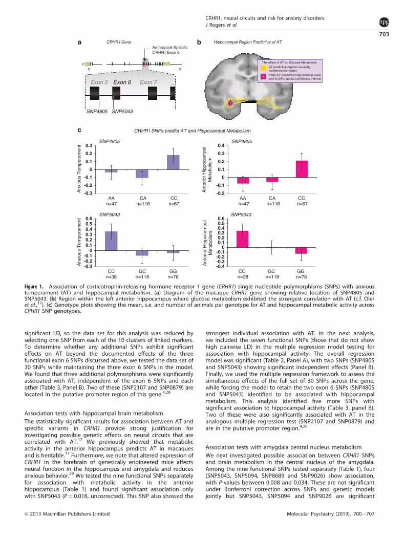
significant LD, so the data set for this analysis was reduced byselecting one SNP from each of the 10 clusters of linked markers.To determine whether any additional SNPs exhibit significanteffects on AT beyond the documented effects of the threefunctional exon 6 SNPs discussed above, we tested the data set of30 SNPs while maintaining the three exon 6 SNPs in the model.We found that three additional polymorphisms were significantlyassociated with AT, independent of the exon 6 SNPs and eachother (Table 3, Panel B). Two of these (SNP2107 and SNP0879) arelocated in the putative promoter region of this gene.4,28
Association tests with hippocampal brain metabolismThe statistically significant results for association between AT andspecific variants in CRHR1 provide strong justification forinvestigating possible genetic effects on neural circuits that arecorrelated with AT.17 We previously showed that metabolicactivity in the anterior hippocampus predicts AT in macaquesand is heritable.17 Furthermore, we note that altered expression ofCRHR1 in the forebrain of genetically engineered mice affectsneural function in the hippocampus and amygdala and reducesanxious behavior.29 We tested the nine functional SNPs separatelyfor association with metabolic activity in the anteriorhippocampus (Table 1) and found significant association onlywith SNP5043 (P¼ 0.016, uncorrected). This SNP also showed the
strongest individual association with AT. In the next analysis,we included the seven functional SNPs (those that do not showhigh pairwise LD) in the multiple regression model testing forassociation with hippocampal activity. The overall regressionmodel was significant (Table 2, Panel A), with two SNPs (SNP4805and SNP5043) showing significant independent effects (Panel B).Finally, we used the multiple regression framework to assess thesimultaneous effects of the full set of 30 SNPs across the gene,while forcing the model to retain the two exon 6 SNPs (SNP4805and SNP5043) identified to be associated with hippocampalmetabolism. This analysis identified five more SNPs withsignificant association to hippocampal activity (Table 3, panel B).Two of these were also significantly associated with AT in theanalogous multiple regression test (SNP2107 and SNP0879) andare in the putative promoter region.4,28
Association tests with amygdala central nucleus metabolismWe next investigated possible association between CRHR1 SNPsand brain metabolism in the central nucleus of the amygdala.Among the nine functional SNPs tested separately (Table 1), four(SNP5043, SNP5094, SNP8689 and SNP9026) show association,with P-values between 0.008 and 0.034. These are not significantunder Bonferroni correction across SNPs and genetic modelsjointly but SNP5043, SNP5094 and SNP9026 are significant
CRHR I Gene
5’ 3’P
SNP5043SNP4805
The effect of AT on Glucose Metabolism
Peak AT-predictive hippocampal voxeland its 95% spatial confidence interval.
AT predictive regions survivingbonferroni correction.
Anthropoid-SpecificCRHRI Exon 6
Hippocampal Region Predictive of AT
-0.2
-0.1
0
0.1
0.2
0.3
0.4
AA CA CC A
nter
ior
Hip
poca
mpa
lM
etab
olis
m
SNP4805
n=47 n=116 n=67
-0.4-0.3-0.2-0.1
00.10.20.30.40.50.6
CC GC GG
Ant
erio
r H
ippo
cam
pal
Met
abol
ism
SNP5043
n=38 n=118 n=78
-0.3
-0.2
-0.1
0
0.1
0.2
0.3
AA CA CC
Anx
ious
Tem
pera
men
t
SNP4805
n=47 n=116 n=67
-0.3-0.2-0.1
00.10.20.30.40.50.6
CC GC GG
Anx
ious
Tem
pera
men
t
SNP5043
n=38 n=118 n=78
CRHR I SNPs predict AT and Hippocampal Metabolism
Exon 6Exon 5 Exon 7 ...
Figure 1. Association of corticotrophin-releasing hormone receptor 1 gene (CRHR1) single nucleotide polymorphisms (SNPs) with anxioustemperament (AT) and hippocampal metabolism. (a) Diagram of the macaque CRHR1 gene showing relative location of SNP4805 andSNP5043. (b) Region within the left anterior hippocampus where glucose metabolism exhibited the strongest correlation with AT (c.f. Oleret al.,17). (c) Genotype plots showing the mean, s.e. and number of animals per genotype for AT and hippocampal metabolic activity acrossCRHR1 SNP genotypes.
CRHR1, neural circuits and risk for anxiety disordersJ Rogers et al
703
& 2013 Macmillan Publishers Limited Molecular Psychiatry (2013), 700 – 707
when corrected only across models. Considered together, theprobability of observing four independent tests significant at the5% level under the null hypothesis of no association between
SNPs and central amygdala metabolism is 2.4� 10� 10. Overall,these results strongly support an association between CRHR1 andamygdala activity, but it is not certain which of the four SNPscould be a false positive. The overall multiple regression testing ofCRHR1 SNPs against activity within the central amygdalagenerated no significant results under any of the three geneticmodels and thus provides no justification for evaluating individualSNPs within this regression framework. Thus, there is clearevidence for association between CRHR1 SNP genotypes andbrain metabolism in the central nucleus of the amygdala, but itremains uncertain which specific SNPs are involved.
Association tests with metabolism in the IPSWe next analyzed genetic association between the identified SNPsand metabolism in the IPS, a cortical region that participates in theintegration of sensorimotor tasks with visual inputs and informa-tion about the location and orientation of the body in space.30 Wepreviously found IPS metabolism to be significantly negativelycorrelated with AT.17 When the nine functional SNPs were testedindependently against metabolism within the IPS, one poly-morphism (SNP5043) generates a P-value of 0.0037, essentiallyreaching the joint Bonferroni threshold for genetic models andSNPs (threshold¼ 0.0036). As shown in Table 4, three other SNPs(SNP5094, SNP8689 and SNP9006) generate P-values o0.05, but40.0036. As with the amygdala test above, the probability ofobserving four SNPs with independent probabilities o0.05 underthe null hypothesis of no association between CRHR1 and IPSmetabolism is 2.4� 10� 10. When the seven functional SNPs aretested simultaneously, two (SNP5043 and SNP5094) exhibitstatistically significant association with IPS metabolism(P¼ 0.0076 and 0.0066, respectively). In the multiple regressiontest of the 30 SNPs, the multiple regression model is significant,and only one polymorphism (SNP9026) is specifically associatedwith the phenotype (P¼ 0.036). Overall, these results showthat variation in CRHR1 is also associated with local brain activityin the IPS.
Association tests with metabolism in the precuneusOur previous analyses17 also indicate that AT is negativelycorrelated with brain metabolism in the precuneus. This regionof medial parietal cortex is closely integrated with the IPS and isinvolved in a number of different functions, including integrationof information supporting purposeful visually guided bodymovements in three-dimensional space, episodic memory andvisual imagery related to self-representation.31 When the nineCRHR1 functional SNPs were tested independently against
Table 2. Results of association testing using the multiple regressionapproach with seven functional SNPs
(A) Multiple regression analysis testing overall genetic model with sevenSNPs
Phenotype
Anxious temperament Hippocampus Amygdala
P¼ P¼ P¼
Additive 0.043 0.0017 NSD-major 0.017 0.003 NSD-minor 0.032 0.0024 NS
(B) Multiple regression results for seven functional SNPs testedsimultaneously
Phenotype
Anxious Temperament Hippocampus
SNP4805 0.014 D-minor 0.017 D-minorSNP5043 0.0003 D-major 0.03 D-majorSNP5094 0.041 D-minor NSSNP5886 NS NSSNP8689 NS NSSNP9009 NS NSSNP9026 NS NS
Abbreviation: SNP, single nucleotide polymorphism.Using a multiple regression approach after taking linkage disequilibriuminto account, seven putatively functional SNPs were assessed forassociation with anxious temperament (AT), hippocampal metabolism ormetabolism in the central amygdala. (A) The significance of the full modelfor each of three alternative models (additive, dominance-major (D-major)and dominance-minor (D-minor)) was assessed for each phenotype ofinterest. (B) The partial regressions (SNPs) were assessed for thosephenotypes in which the full model was significant (AT and hippocampus).No partial regression results (SNPs) were considered for amygdalametabolism because the overall model was not statistically significant.Bolded P-values represent Pp0.05.
HumanChimpanzeeGorillaMacaqueMarmosetBushbabyMouse lemurRatDogMicrobatOpossumPlatypus
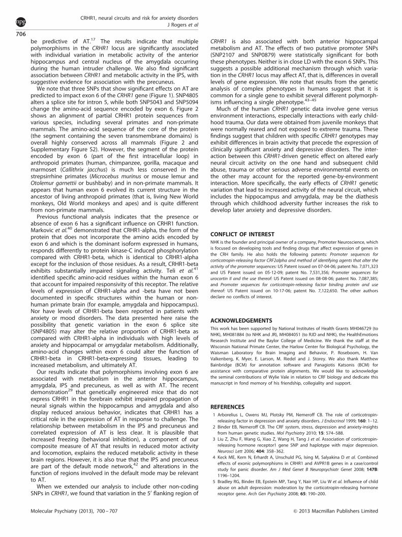
Figure 2. Corticotrophin-releasing hormone receptor 1 gene (CRHR1) protein segment encoded by exon 6 is conserved primarily inanthropoid primates. A portion of the aligned protein sequence of CRHR1 containing the segment encoded by exon 6 is illustrated for 12mammals: five anthropoid primates (human, chimpanzee, gorilla, macaque and marmoset), two more distant primates (bushbaby and mouselemur), three placental mammals (rat, dog and microbat), one marsupial (opossum) and one monotreme (platypus). The cladogram of theknown phylogenetic relationships among these species is presented on the left. The sequence encoded by part of exon 5 and all of exon 6(bounded by the red square) is shown on the right. Residues identical to the human sequence are shaded for exon 5 (light gray) and exon 6(blue). Note that while exon 5 is highly conserved across all 12 species, exon 6 is conserved primarily in anthropoid primates.
CRHR1, neural circuits and risk for anxiety disordersJ Rogers et al
704
Molecular Psychiatry (2013), 700 – 707 & 2013 Macmillan Publishers Limited

precuneus metabolism, none produced P-values o0.01, and thusnone survive joint Bonferroni correction for genetic models andSNPs. Two variants (SNP8689 and SNP9006) have P-values o0.02(Table 4), which survive Bonferroni correction for genetic modelsalone. Because four variants exhibit p-values o0.05, we onceagain conclude that the CRHR1 locus influences metabolism in thisregion, although we cannot identify specific causal SNPs withconfidence. Like our analyses of amygdala metabolism, none ofthe genetic models in the multiple regression analysis of either thesets of seven SNPs or 30 SNPs produce significant overall geneticeffects, so there is no justification for evaluating the associationbetween individual SNPs and precuneus metabolism within themultiple regression framework.
Analysis of protein structureFinally, we used the online I-TASSER software package27 to predictthe effects of selected SNPs on the three-dimensional structure ofthe macaque CRHR1 protein. These results (Supplementary FigureS4) must be considered preliminary because no validated three-dimensional crystal structure is available for any isoform of CRHR1for any species, and I-TASSER performs best when its predictionsare based on templates of known 3D structure. Nevertheless, the
results suggest that SNP5043 and SNP5094 individually affect theshape and/or orientation of the first intra-cellular loop of theprotein. Definitive conclusions await direct functional assays, butthese predictions support the idea that these two mutations doindeed alter 3D protein structure and, therefore, may influencefunction. We note that normal CRHR1 function may involve bothhomo-dimerization and hetero-dimerization.32
DISCUSSIONDespite significant progress in the quantitative genetic analysis ofthe risk to develop anxiety and depressive disorders, knowledgeof the specific genes, genetic mechanisms and physiologicalintermediates underlying this risk remains limited. Several studieshave reported associations between CRH-related genes andclinical anxiety or depression,2–4,33 as well as associationbetween CRH genes and related phenotypes in macaques.34
Several analyses report gene versus environment interactions inwhich early childhood trauma interacts with CRHR1 genotypes topredict stress-related psychopathology later in life.3,5–8,35,36
However, these data do not address the intermediateneurobiological pathways that lead from inherited geneticdifferences to altered behavioral and physiological reactivity andultimately to psychopathology. Due to fundamental similarities inthe brain structure and behavioral reactivity to stress, rhesusmonkey models are well suited for analysis of genetic effects thatalter the function of neural circuits implicated in humanpsychopathology.37–39
In this study, we identified several common SNPs in the rhesusmacaque CRHR1 gene that are independently associated with AT.Specifically, we find that SNP4805, a splice-site mutation that mayaffect the inclusion of exon 6 into the CRHR1 protein, and anothervariant (SNP5043), which alters the amino-acid sequence withinexon 6, are associated with individual differences in AT. We alsoobtained support for association between another exon 6 variant(SNP5094) and AT. To understand how variation in the CRHR1gene increases the behavioral and physiological reactivity tothreat that is characteristic of individuals with high levels of AT, weexamined brain metabolic activity in regions previously shown to
Table 3. Results of genetic association testing using 30 gene-wideSNPs in CRHR1
(A) Multiple regression analysis of overall genetic models with exon 6SNPs fixed
Phenotype
Anxious temperament Hippocampus
P¼ P¼
Additive NS 0.001D-major 0.014 0.008D-minor 0.034 0.028
(B) Multiple regression analysis testing 30 SNPs simultaneously with exon6 SNPs fixed in model
Phenotype
Anxious temperament Hippocampus
P¼ Genetic model P¼ Genetic model
SNP5886 0.043 D-minor NSSNP9009 NS 0.044 D-minorSNP2107 0.017 D-major 0.017 D-majorSNP0879 0.012 D-minor 0.021 Add., D-minorSNP4820 NS 0.048 D-minorSNP5137 NS 0.015 D-minor
Abbreviations: CRHR1, corticotrophin-releasing hormone receptor 1; SNP,single nucleotide polymorphism.After taking linkage disequilibrium into account, 30 SNPs were assessed forassociation with anxious temperament (AT) and hippocampal metabolismusing a multiple regression approach. The SNPs that showed significantassociation in the previous seven SNP analysis were held fixed in the model(SNP4805, SNP5043 and SNP5094). (A) The significance of the full model foreach of the three alternative models (additive (Add.), dominance-major (D-major) and dominance-minor (D-minor)) was assessed for AT andhippocampal metabolism. (B) For each multiple regression model thatwas significant, the partial regressions (SNPs) were assessed. When multiplemodels were significant for the same SNP, the lowest P-value is presented.Bolded P-values represent Pp0.05.
Table 4. Results of association testing for intraparietal sulcus andprecuneus using functional SNPs in separate tests
SNP ID Phenotype
Intraparietal sulcus Precuneus
P¼ Genetic model P¼ Genetic model
SNP4805 NS n.sSNP5043 0.0037 Add. 0.026 Add.SNP5094 0.012 Add. and
D-minor0.029 Add. and
D-majorSNP5107 NS NSSNP5886 NS NSSNP8689 0.029 Add. 0.015 D-minorSNP9006 0.0074 D-major 0.019 D-majorSNP9009 NS NSSNP9026 NS NS
Abbreviation: SNP, single nucleotide polymorphism.Individual tests of association for each of the nine putatively functionalSNPs in CRHR1 (corticotrophin-releasing hormone receptor 1) for intrapar-ietal sulcus and precuneus. The model used to test significance is indicatedafter the P-value: additive (Add.), dominant-major (D-major) or dominant-minor (D-minor).Bolded P-values represent Pp0.05.
CRHR1, neural circuits and risk for anxiety disordersJ Rogers et al
705
& 2013 Macmillan Publishers Limited Molecular Psychiatry (2013), 700 – 707
be predictive of AT.17 The results indicate that multiplepolymorphisms in the CRHR1 locus are significantly associatedwith individual variation in metabolic activity of the anteriorhippocampus and central nucleus of the amygdala occurringduring the human intruder challenge. We also find significantassociation between CRHR1 and metabolic activity in the IPS, withsuggestive evidence for association with the precuneus.
We note that three SNPs that show significant effects on AT arepredicted to impact exon 6 of the CRHR1 gene (Figure 1). SNP4805alters a splice site for intron 5, while both SNP5043 and SNP5094change the amino-acid sequence encoded by exon 6. Figure 2shows an alignment of partial CRHR1 protein sequences fromvarious species, including several primates and non-primatemammals. The amino-acid sequence of the core of the protein(the segment containing the seven transmembrane domains) isoverall highly conserved across all mammals (Figure 2 andSupplementary Figure S2). However, the segment of the proteinencoded by exon 6 (part of the first intracellular loop) inanthropoid primates (human, chimpanzee, gorilla, macaque andmarmoset (Callithrix jacchus) is much less conserved in thestrepsirrhine primates (Microcebus murinus or mouse lemur andOtolemur garnettii or bushbaby) and in non-primate mammals. Itappears that human exon 6 evolved its current structure in theancestor of living anthropoid primates (that is, living New Worldmonkeys, Old World monkeys and apes) and is quite differentfrom non-primate mammals.
Previous functional analysis indicates that the presence orabsence of exon 6 has a significant influence on CRHR1 function.Markovic et al.40 demonstrated that CRHR1-alpha, the form of theprotein that does not incorporate the amino acids encoded byexon 6 and which is the dominant isoform expressed in humans,responds differently to protein kinase-C induced phosphorylationcompared with CRHR1-beta, which is identical to CRHR1-alphaexcept for the inclusion of those residues. As a result, CRHR1-betaexhibits substantially impaired signaling activity. Teli et al.41
identified specific amino-acid residues within the human exon 6that account for impaired responsivity of this receptor. The relativelevels of expression of CRHR1-alpha and -beta have not beendocumented in specific structures within the human or non-human primate brain (for example, amygdala and hippocampus).Nor have levels of CRHR1-beta been reported in patients withanxiety or mood disorders. The data presented here raise thepossibility that genetic variation in the exon 6 splice site(SNP4805) may alter the relative proportion of CRHR1-beta ascompared with CRHR1-alpha in individuals with high levels ofanxiety and hippocampal or amygdalar metabolism. Additionally,amino-acid changes within exon 6 could alter the function ofCRHR1-beta in CRHR1-beta-expressing tissues, leading toincreased metabolism, and ultimately AT.
Our results indicate that polymorphisms involving exon 6 areassociated with metabolism in the anterior hippocampus,amygdala, IPS and precuneus, as well as with AT. The recentdemonstration29 that genetically engineered mice that do notexpress CRHR1 in the forebrain exhibit impaired propagation ofneural signals within the hippocampus and amygdala and alsodisplay reduced anxious behavior, indicates that CRHR1 has acritical role in the expression of AT in response to challenge. Therelationship between metabolism in the IPS and precuneus andcorrelated expression of AT is less clear. It is plausible thatincreased freezing (behavioral inhibition), a component of ourcomposite measure of AT that results in reduced motor activityand locomotion, explains the reduced metabolic activity in thesebrain regions. However, it is also true that the IPS and precuneusare part of the default mode network,42 and alterations in thefunction of regions involved in the default mode may be relevantto AT.
When we extended our analysis to include other non-codingSNPs in CRHR1, we found that variation in the 50 flanking region of
CRHR1 is also associated with both anterior hippocampalmetabolism and AT. The effects of two putative promoter SNPs(SNP2107 and SNP0879) were statistically significant for boththese phenotypes. Neither is in close LD with the exon 6 SNPs. Thissuggests a possible additional mechanism through which varia-tion in the CRHR1 locus may affect AT, that is, differences in overalllevels of gene expression. We note that results from the geneticanalysis of complex phenotypes in humans suggest that it iscommon for a single gene to exhibit several different polymorph-isms influencing a single phenotype.43–45
Much of the human CRHR1 genetic data involve gene versusenvironment interactions, especially interactions with early child-hood trauma. Our data were obtained from juvenile monkeys thatwere normally reared and not exposed to extreme trauma. Thesefindings suggest that children with specific CRHR1 genotypes mayexhibit differences in brain activity that precede the expression ofclinically significant anxiety and depressive disorders. The inter-action between this CRHR1-driven genetic effect on altered earlyneural circuit activity on the one hand and subsequent childabuse, trauma or other serious adverse environmental events onthe other may account for the reported gene-by-environmentinteraction. More specifically, the early effects of CRHR1 geneticvariation that lead to increased activity of the neural circuit, whichincludes the hippocampus and amygdala, may be the diathesisthrough which childhood adversity further increases the risk todevelop later anxiety and depressive disorders.
CONFLICT OF INTERESTNHK is the founder and principal owner of a company, Promoter Neuroscience, whichis focused on developing tools and finding drugs that affect expression of genes inthe CRH family. He also holds the following patents: Promoter sequences forcorticotropin-releasing factor CRF2alpha and method of identifying agents that alter theactivity of the promoter sequences: US Patent issued on 07-04-06; patent No. 7,071,323and US Patent issued on 05-12-09; patent No. 7,531,356; Promoter sequences forurocortin II and the use thereof: US Patent issued on 08-08-06; patent No. 7,087,385;and Promoter sequences for corticotropin-releasing factor binding protein and usethereof: US Patent issued on 10-17-06; patent No. 7,122,650. The other authorsdeclare no conflicts of interest.
ACKNOWLEDGEMENTSThis work has been supported by National Institutes of Health Grants MH046729 (toNHK), MH081884 (to NHK and JR), MH084051 (to RJD and NHK), the HealthEmotionsResearch Institute and the Baylor College of Medicine. We thank the staff at theWisconsin National Primate Center, the Harlow Center for Biological Psychology, theWaisman Laboratory for Brain Imaging and Behavior, P. Roseboom, H. VanValkenberg, K. Myer, E. Larson, M. Riedel and J. Storey. We also thank MatthewBainbridge (BCM) for annotation software and Panagiotis Katsonis (BCM) forassistance with comparative protein alignments. We would like to acknowledgethe seminal contributions of Wylie Vale in relation to CRF biology and dedicate thismanuscript in fond memory of his friendship, collegiality and support.
REFERENCES1 Arborelius L, Owens MJ, Plotsky PM, Nemeroff CB. The role of corticotropin-
releasing factor in depression and anxiety disorders. J Endocrinol 1999; 160: 1–12.2 Binder EB, Nemeroff CB. The CRF system, stress, depression and anxiety-insights
from human genetic studies. Mol Psychiatry 2010; 15: 574–588.3 Liu Z, Zhu F, Wang G, Xiao Z, Wang H, Tang J et al. Association of corticotropin-
releasing hormone receptor1 gene SNP and haplotype with major depression.Neurosci Lett 2006; 404: 358–362.
4 Keck ME, Kern N, Erhardt A, Unschuld PG, Ising M, Salyakina D et al. Combinedeffects of exonic polymorphisms in CRHR1 and AVPR1B genes in a case/controlstudy for panic disorder. Am J Med Genet B Neuropsychiatr Genet 2008; 147B:1196–1204.
5 Bradley RG, Binder EB, Epstein MP, Tang Y, Nair HP, Liu W et al. Influence of childabuse on adult depression: moderation by the corticotropin-releasing hormonereceptor gene. Arch Gen Psychiatry 2008; 65: 190–200.
CRHR1, neural circuits and risk for anxiety disordersJ Rogers et al
706
Molecular Psychiatry (2013), 700 – 707 & 2013 Macmillan Publishers Limited

6 Heim C, Bradley B, Mletzko TC, Deveau TC, Musselman DL, Nemeroff CB et al.Effect of childhood trauma on adult depression and neuroendocrine function:sex-specific moderation by CRH receptor 1 gene. Front Behav Neurosci 2009; 3: 41.
7 Polanczyk G, Caspi A, Williams B, Price TS, Danese A, Sugden K et al. Protectiveeffect of CRHR1 gene variants on the development of adult depression followingchildhood maltreatment: replication and extension. Arch Gen Psychiatry 2009; 66:978–985.
8 Tyrka AR, Price LH, Gelernter J, Schepker C, Anderson GM, Carpenter LL. Inter-action of childhood maltreatment with the corticotropin-releasing hormonereceptor gene: effects on hypothalamic-pituitary-adrenal axis reactivity. Biol Psy-chiatry 2009; 66: 681–685.
9 Licinio J, O’Kirwan F, Irizarry K, Merriman B, Thakur S, Jepson R et al. Association ofa corticotropin-releasing hormone receptor 1 haplotype and antidepressanttreatment response in Mexican-Americans. Mol Psychiatry 2004; 9: 1075–1082.
10 Liu Z, Zhu F, Wang G, Xiao Z, Tang J, Liu W et al. Association study of cortico-tropin-releasing hormone receptor1 gene polymorphisms and antidepressantresponse in major depressive disorders. Neurosci Lett 2007; 414: 155–158.
11 Kalin NH, Shelton SE. Nonhuman primate models to study anxiety, emotionregulation, and psychopathology. Ann NY Acad Sci 2003; 1008: 189–200.
12 Rapee RM, Schniering CA, Hudson JL. Anxiety disorders during childhood andadolescence: origins and treatment. Annu Rev Clin Psychol 2009; 5: 311–341.
13 Fox NA, Henderson HA, Marshall PJ, Nichols KE, Ghera MM. Behavioral inhibition:linking biology and behavior within a developmental framework. Annu Rev Psy-chol 2005; 56: 235–262.
14 Rogers J, Shelton SE, Shelledy W, Garcia R, Kalin NH. Genetic influences onbehavioral inhibition and anxiety in juvenile rhesus macaques. Genes Brain Behav2008; 7: 463–469.
15 Fox AS, Shelton SE, Oakes TR, Davidson RJ, Kalin NH. Trait-like brain activity duringadolescence predicts anxious temperament in primates. PLoS ONE 2008; 3: e2570.
16 Kalin NH, Shelton SE. Ontogeny and stability of separation and threat-induceddefensive behaviors in rhesus monkeys during the first year of life. Am J Primatol1998; 44: 125–135.
17 Oler JA, Fox AS, Shelton SE, Rogers J, Dyer TD, Davidson RJ et al. Amygdalar andhippocampal substrates of anxious temperament differ in their heritability. Nature2010; 466: 864–868.
18 Kalin NH, Shelton SE, Rickman M, Davidson RJ. Individual differences in freezing andcortisol in infant and mother rhesus monkeys. Behav Neurosci 1998; 112: 251–254.
19 Kalin NH, Shelton SE. Defensive behaviors in infant rhesus monkeys: environ-mental cues and neurochemical regulation. Science 1989; 243: 1718–1721.
20 Kalin NH, Shelton SE, Fox AS, Oakes TR, Davidson RJ. Brain regions associated withthe expression and contextual regulation of anxiety in primates. Biol Psychiatry2005; 58: 796–804.
21 Fox AS, Oakes TR, Shelton SE, Converse AK, Davidson RJ, Kalin NH. Calling for helpis independently modulated by brain systems underlying goal-directed behaviorand threat perception. Proc Nat Acad Sci USA 2005; 102: 4176–4179.
22 Jenkinson M, Bannister P, Brady M, Smith S. Improved optimization for the robustand accurate linear registration and motion correction of brain images. Neuro-Image 2002; 17: 825–841.
23 Zhang Y, Brady M, Smith S. Segmentation of brain MR images through a hiddenMarkov random field model and the expectation-maximization algorithm. IEEETrans Med Imaging 2001; 20: 45–57.
24 Barrett JC, Fry B, Maller J, Daly MJ. Haploview: analysis and visualization of LD andhaplotype maps. Bioinformatics 2005; 21: 263–265.
25 Almasy L, Blangero J. Multipoint quantitative-trait linkage analysis in generalpedigrees. Am J Hum Genet 1998; 62: 1198–1211.
26 Cheverud JM. A simple correction for multiple comparisons in interval mappinggenome scans. Heredity 2001; 87(Pt 1): 52–58.
27 Roy A, Kucukural A, Zhang Y. I-TASSER: a unified platform for automated proteinstructure and function prediction. Nat Protoc 2010; 5: 725–738.
28 Parham KL, Zervou S, Karteris E, Catalano RD, Old RW, Hillhouse EW. Promoteranalysis of human corticotropin-releasing factor (CRF) type 1 receptor and reg-ulation by CRF and urocortin. Endocrinology 2004; 145: 3971–3983.
29 Refojo D, Schweizer M, Kuehne C, Ehrenberg S, Thoeringer C, Vogl AM et al.Glutamatergic and dopaminergic neurons mediate anxiogenic and anxiolyticeffects of CRHR1. Science 2011; 333: 1903–1907.
30 Grefkes C, Fink GR. The functional organization of the intraparietal sulcus inhumans and monkeys. J Anat 2005; 207: 3–17.
31 Cavanna AE, Trimble MR. The precuneus: a review of its functional anatomy andbehavioural correlates. Brain 2006; 129(Pt 3): 564–583.
32 Murat B, Devost D, Andres M, Mion J, Boulay V, Corbani M et al. V1b and CRHR1receptor heterodimerization mediates synergistic biological actions of vaso-pressin and CRH. Mol Endocrinol 2012; 26: 502–520.
33 Smoller JW, Rosenbaum JF, Biederman J, Kennedy J, Dai D, Racette SR et al.Association of a genetic marker at the corticotropin-releasing hormone locus withbehavioral inhibition. Biol Psychiatry 2003; 54: 1376–1381.
34 Barr CS, Dvoskin RL, Gupte M, Sommer W, Sun H, Schwandt ML et al. FunctionalCRH variation increases stress-induced alcohol consumption in primates. Proc NatlAcad Sci USA 2009; 106: 14593–14598.
35 Muller MB, Zimmermann S, Sillaber I, Hagemeyer TP, Deussing JM, Timpl Pet al. Limbic corticotropin-releasing hormone receptor 1 mediates anxiety-
related behavior and hormonal adaptation to stress. Nat Neurosci 2003; 6:1100–1107.
36 Ressler KJ, Bradley B, Mercer KB, Deveau TC, Smith AK, Gillespie CF et al. Poly-morphisms in CRHR1 and the serotonin transporter loci: gene x gene x envir-onment interactions on depressive symptoms. Am J Med Genet B NeuropsychiatrGenet 153B: 812–824.
37 Barr CS, Newman TK, Becker ML, Parker CC, Champoux M, Lesch KP et al. Theutility of the non-human primate; model for studying gene by environmentinteractions in behavioral research. Genes Brain Behav 2003; 2: 336–340.
38 Barr CS, Schwandt M, Lindell SG, Chen SA, Goldman D, Suomi SJ et al. Associationof a functional polymorphism in the mu-opioid receptor gene with alcoholresponse and consumption in male rhesus macaques. Arch Gen Psychiatry 2007;64: 369–376.
39 Vallender EJ, Ruedi-Bettschen D, Miller GM, Platt DM. A pharmacogenetic modelof naltrexone-induced attenuation of alcohol consumption in rhesus monkeys.Drug Alcohol Depend 2010; 109: 252–256.
40 Markovic D, Papadopoulou N, Teli T, Randeva H, Levine MA, Hillhouse EW et al.Differential responses of corticotropin-releasing hormone receptor type 1variants to protein kinase C phosphorylation. J Pharmacol Exp Ther 2006; 319:1032–1042.
41 Teli T, Markovic D, Hewitt ME, Levine MA, Hillhouse EW, Grammatopoulos DK.Structural domains determining signalling characteristics of the CRH-receptortype 1 variant R1beta and response to PKC phosphorylation. Cell Signal 2008; 20:40–49.
42 Vincent JL, Patel GH, Fox MD, Snyder AZ, Baker JT, Van Essen DC et al. Intrinsicfunctional architecture in the anaesthetized monkey brain. Nature 2007; 447:83–86.
43 Johnson N, Fletcher O, Palles C, Rudd M, Webb E, Sellick G et al. Countingpotentially functional variants in BRCA1, BRCA2 and ATM predicts breast cancersusceptibility. Hum Mol Genet 2007; 16: 1051–1057.
44 Singleton A, Hardy J. A generalizable hypothesis for the genetic architecture ofdisease: pleomorphic risk loci. Hum Mol Genet 2011; 20: R158–R162.
45 Achkar JP, Duerr R. The expanding universe of inflammatory bowel diseasegenetics. Curr Opin Gastroenterol 2008; 24: 429–434.
Supplementary Information accompanies the paper on the Molecular Psychiatry website (http://www.nature.com/mp)
CRHR1, neural circuits and risk for anxiety disordersJ Rogers et al
707
& 2013 Macmillan Publishers Limited Molecular Psychiatry (2013), 700 – 707