craniotomy prepared by: sumi mathew. demographic data name :mr a m a age/sex : 27yrs/ male mrn no...
TRANSCRIPT
CRANIOTOMY
Prepared by: Sumi Mathew

DEMOGRAPHIC DATA
NAME :Mr A M AAGE/SEX : 27YRS/ MALE MRN NO :203915DATE OF ADMISSION :16/05/13DIAGNOSIS :ACUTE SDH, HEAD
TRAUMA&FALL FRom height
SURGERY : POSTERIOR FOSSA CRANIOTOMY+SDH EVACUATION& duraplasty
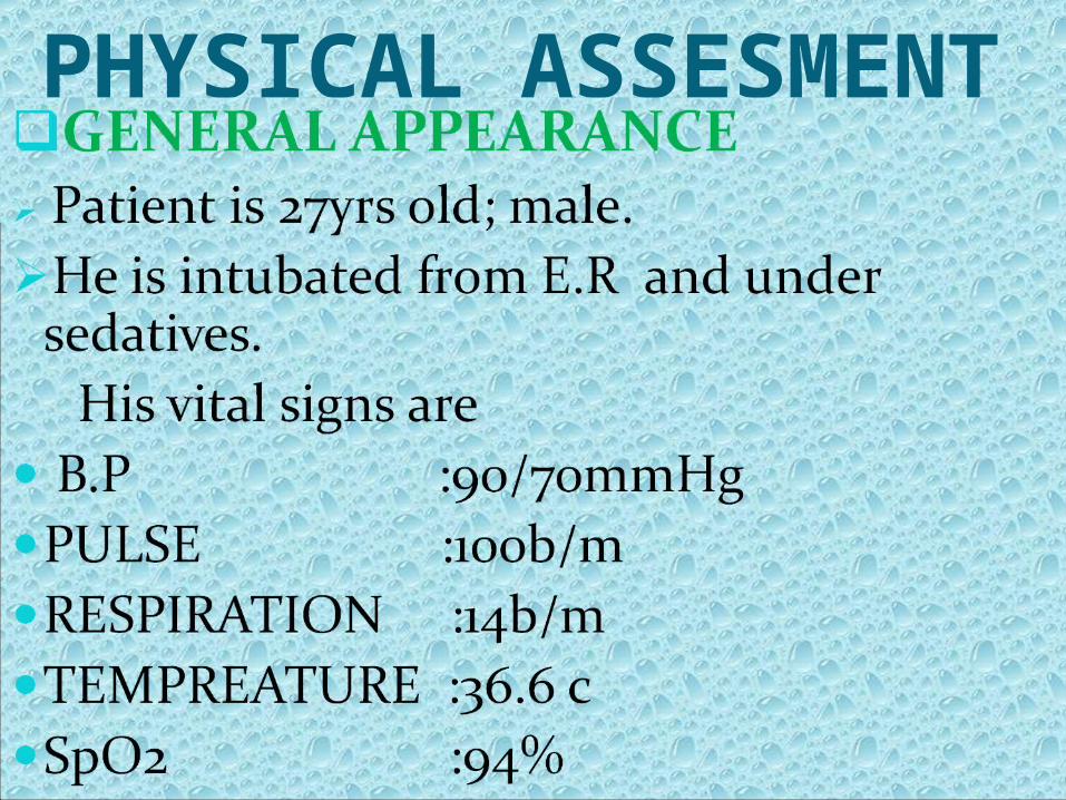
PHYSICAL ASSESMENT

LEVEL OF CONSCIOUSNESS
Patient was semiconscious on admission ;and was intubated from E.R on fully sedation .
Gcs :8/15
SKIN Fair complexion ;abrasions on back No palpable mass or lesions
HEAD
Skull slightly asymmetric Cut wound on scalp . Maxillary ,frontal and ethmoid sinuses are not tender.
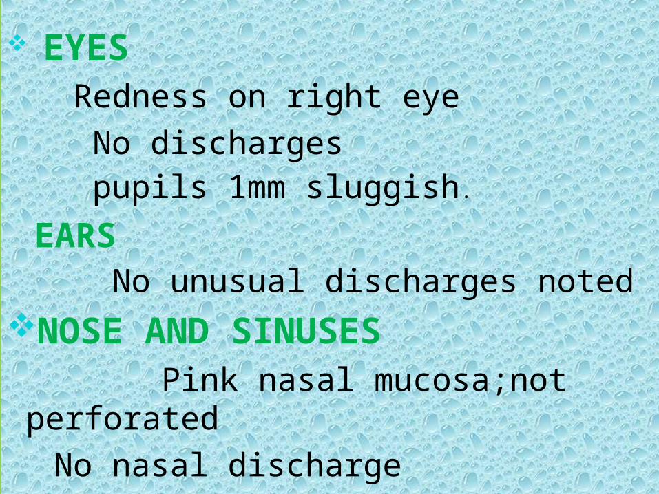
EYES Redness on right eye No discharges pupils 1mm sluggish.
EARS No unusual discharges noted
NOSE AND SINUSES Pink nasal mucosa;not perforated
No nasal discharge

MOUTH Pink and dry oral mucosa Tongue and uvula in midline position
ET tube and OGT are presentNECK AND THROAT No palpable lymph nodes
No mass and lesions seenCHEST & LUNGS Thorax is symmetric
Equal chest expansion
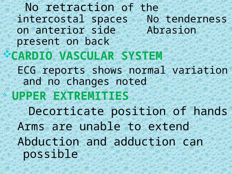
No retraction of the intercostal spaces No tenderness on anterior side Abrasion present on back
CARDIO VASCULAR SYSTEMECG reports shows normal variation and no changes noted
UPPER EXTREMITIES Decorticate position of hands Arms are unable to extendAbduction and adduction can possible

ABDOMEN Its rigid and little distention present
Bowel sounds are normalGENITO URINARY SYSTEM
No ulceration on perineal area; clean
LOWER EXTREMITIESNormal positions of tibia & fibula;legs can adbuct and adduct

PATIENT HISTORYPAST MEDICAL AND SURGICAL
HISTORY Patient has no past medical and surgical history
PRESENT MEDICAL HISTORY Patient brought to E.R H/O FALL FROM HEIGHT with loss of consciousness .He was intubated from E.R and admitted in ICU on 16/05/13 .

PRESENT SURGICAL HISTORY Patient had undergone LEFT POSTERIOR FOSSA CRANIOTOMY +EVACUATION OF SDH+DURAPLASTY on 16/05/13.
INVESTIGATIONS DONE FOR THE PATIENT
X-ray ChestCT Brain And Lumbar SpineMRI Scan of Brain
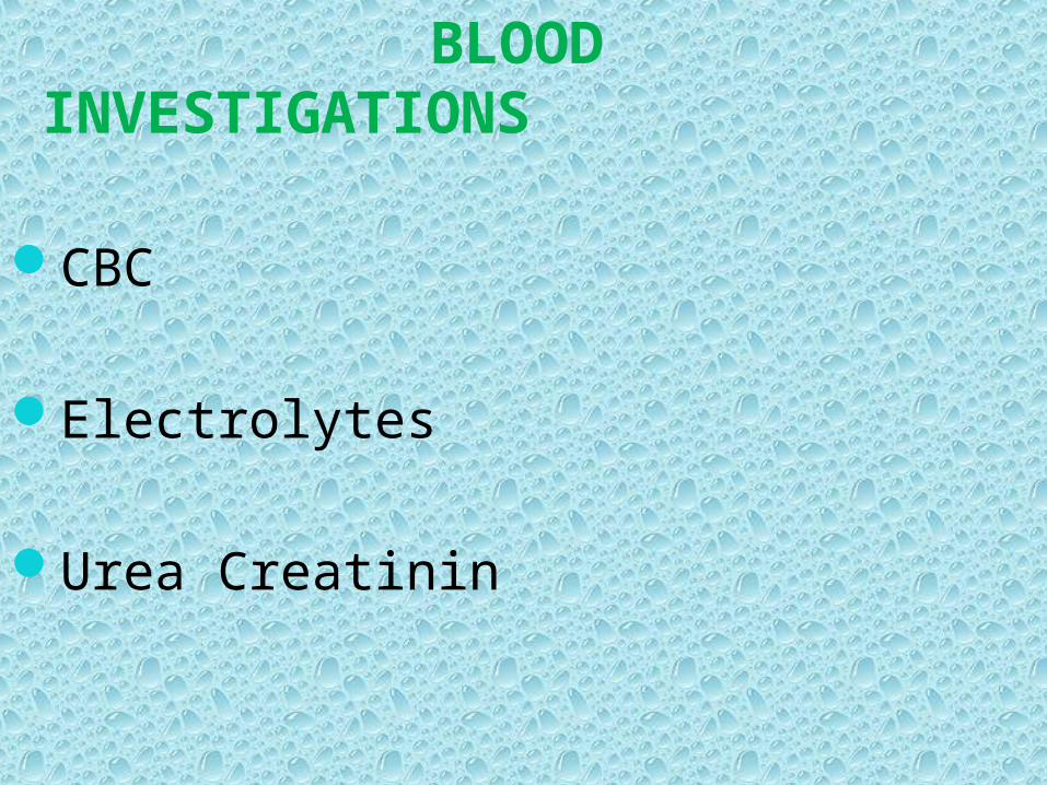
BLOOD INVESTIGATIONS
CBC
Electrolytes
Urea Creatinin

ITEMS PATIENT VALUE NORMAL VALUE
HEMATOLOGYHemoglobin(Hb) 9.5gm/dl 12-16 gm/dl
CHEMISTERY
Sodium 143 135 - 150
Potassium 3.7 3.5 - 5
ChlorideUrea
103 6.7
98 - 111 1.8 - 8.3
LAB VALUES

DRUG DOSE ROUTE ACTION
Inj Augmentin 1.2gm I.V Antibiotic
Inj Ceftriaxone 1gm I.V Antibiotic
Inj Risek 40mg I.V Histamine2-receptor antagonists
Inj Tramadol 100mg I.V Antipyretics
Inj Perfalgan 1gm I.V Analgesics
Inj Mannitol 100gm I.V Osmotic Diuretic
MEDICATION
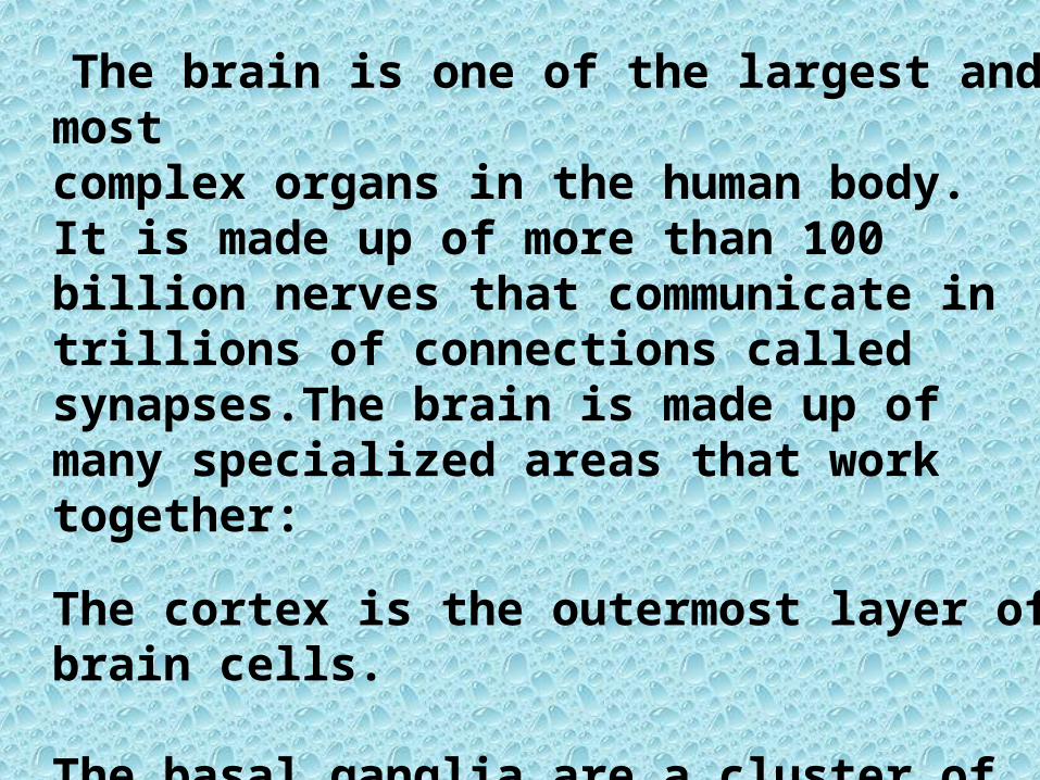
ANATOMY and physiologyOF BRAIn
The brain is one of the largest and most complex organs in the human body.It is made up of more than 100 billion nerves that communicate in trillions of connections called synapses.The brain is made up of many specialized areas that work together:
The cortex is the outermost layer of brain cells.
The basal ganglia are a cluster of structures in the center of the brain


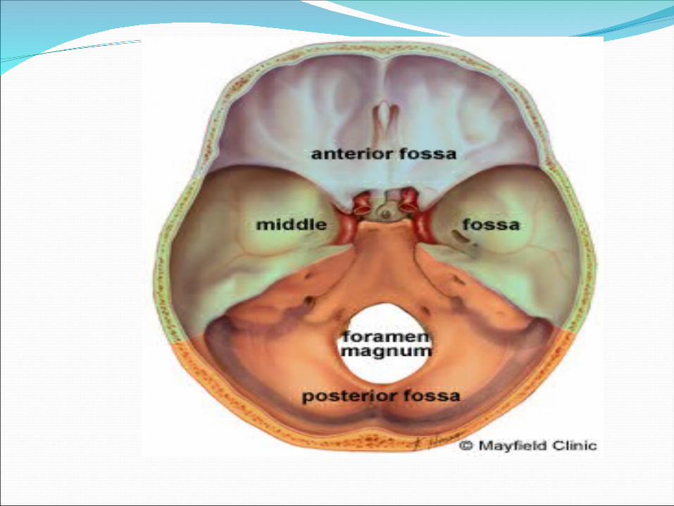
SKULL
The purpose of the bony skull is to protect the brain from injury. All the arteries, veins and nerves exit the base of the skull through holes, called foramina.
The big hole in the middle (foramen magnum) is where the spinal cord exits.
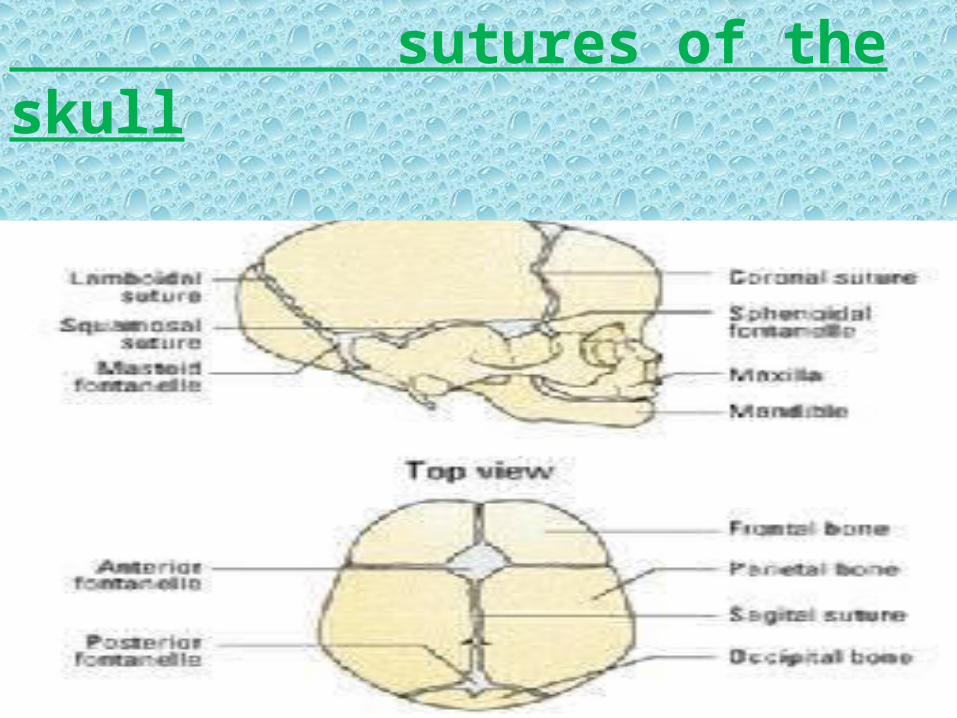
sutures of the skull

Brain

The brain is composed of three parts:
CEREBELLUM
CEREBRUM.
BRAINSTEM
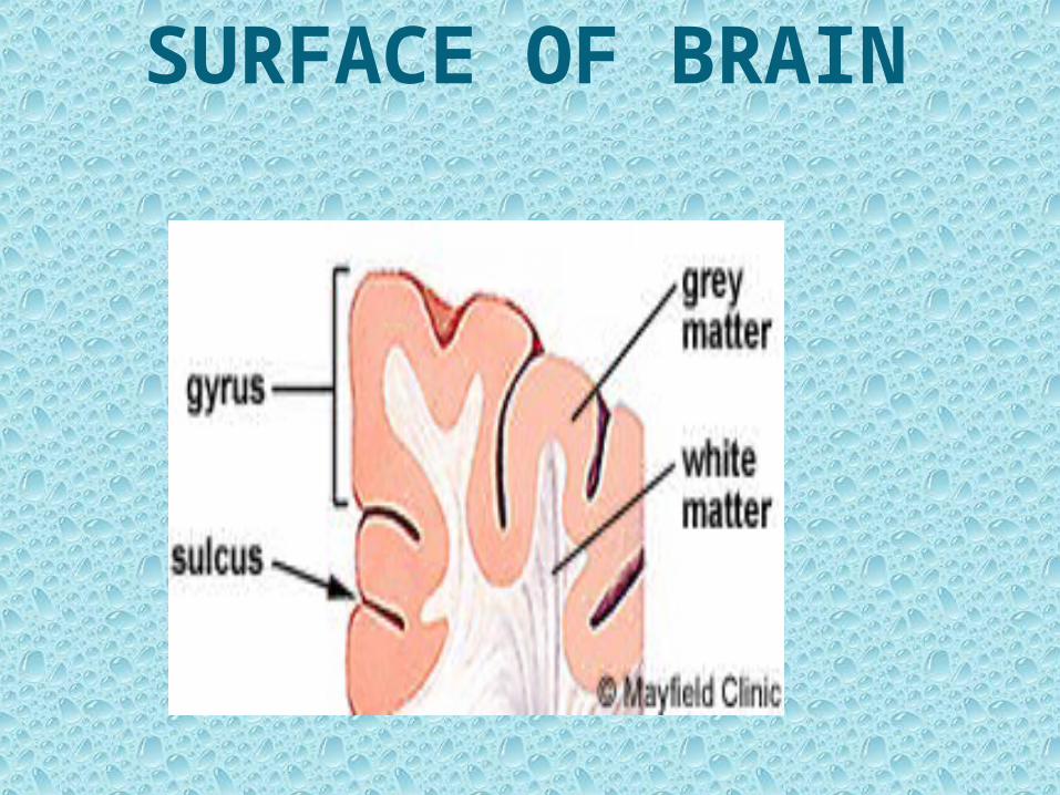
SURFACE OF BRAIN

DEEP STRUCTURES
Hypothalamus
Pituitary gland
Pineal gland
Thalamus

MENINGES The brain and spinal cord are covered and protected by three layers of tissue called meninges.
From the outermost layer inward they are: The Dura mater, Arachnoid mater, and Piamater.

Ventricles and Cerebrospinal fluid The brain has hollow fluid-filled cavities called ventricles Inside the ventricles is a ribbon-like structure called the choroid plexus that makes clear colorless cerebrospinal fluid.CSF flows within and around the brain and spinal cord to help cushion it from injury. This circulating fluid is constantly being absorbed and replenished.

Nervous system
The nervous system is divided into central and peripheral systems.
The central nervous system (CNS) is composed of the brain and
spinal cord.
The peripheral nervous system(PNS) is composed of spinal
nerves.
That branch from the spinal cord and cranial nerves that branch
from the brain.
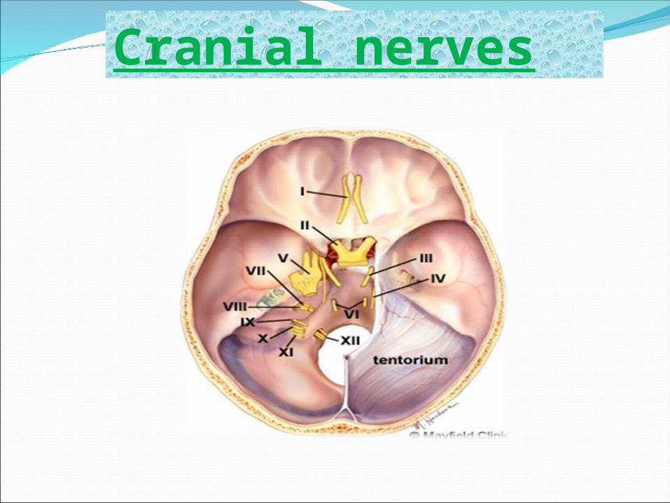
Cranial nerves
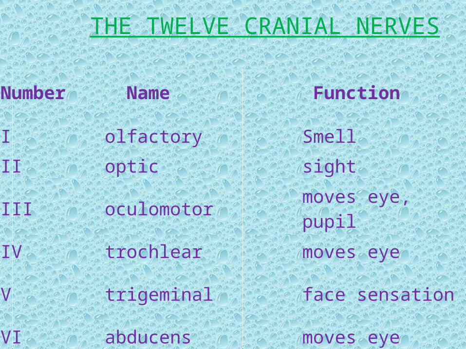
Number Name Function
I olfactory Smell
II optic sight
III oculomotor moves eye, pupil
IV trochlear moves eye
V trigeminal face sensation
VI abducens moves eye
THE TWELVE CRANIAL NERVES
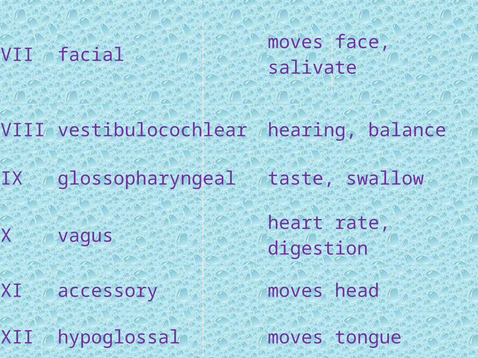
VII facial moves face, salivate
VIII vestibulocochlear hearing, balance
IX glossopharyngeal taste, swallow
X vagus heart rate, digestion
XI accessory moves head
XII hypoglossal moves tongue

Blood supply
Blood is carried to the brain by two paired arteries, the internal carotid arteries and the vertebral arteries. The internal carotid arteries supply most of the cerebrum.
The vertebral arteries supply the cerebellum, brainstem, and the underside of the cerebrum


Etiology •Head injury fall fromheight Motor vehicle collision Assault. •People with a bleeding disorder people who take blood thinners . •Elderly people are at higher risk for chronic subdural hematoma
TOPIC PRESENTATION
Subdural Hematoma
In a subdural hematoma, blood collects between the layers of tissue that surround the brain. The outermost layer is called the durra. In a subdural hematoma, bleeding occurs between the durra and the arachnoids.

ETIOLOGY• Head injury
• Fallfromheight
• Motorvehiclecollision
• Assault.
• People with a bleeding disorder
• People who take blood thinners .

Signs and Symptoms
•Headache• Confusion• Change in behavior• Dizziness• Nausea and vomiting• Lethargy or excessive drowsiness• Weakness• Apathy• Seizures• Lose of consciousness and •coma

Treatment• Burr hole trephination. A hole is drilled in the skull over the area of the subdural hematoma, and the blood is suctioned out through the hole.• Craniotomy. A larger section of the skull is removed, to allow better access to the subdural hematoma and reduce pressure. • Craniectomy. A section of the skull is removed for an extended period of time, to allow the injured brain to expand and swell without permanent damage
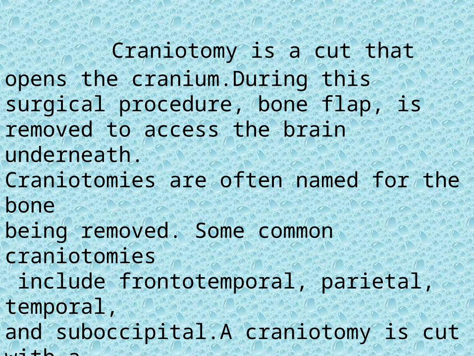
craniotomy Craniotomy is a cut that opens the cranium.During this surgical procedure, bone flap, is removed to access the brain underneath.Craniotomies are often named for the bone being removed. Some common craniotomies include frontotemporal, parietal, temporal, and suboccipital.A craniotomy is cut with a special saw called a craniotome.


STEPS OF PROCEDURE
There are 6 main steps craniotomy..
Step 1: prepare the patient
Step 2: make a skin incision.
Step 3: perform a craniotomy, open the skull
Step 4: exposure the brain
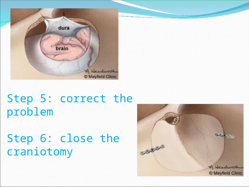
Step 5: correct the problem
Step 6: close the craniotomy
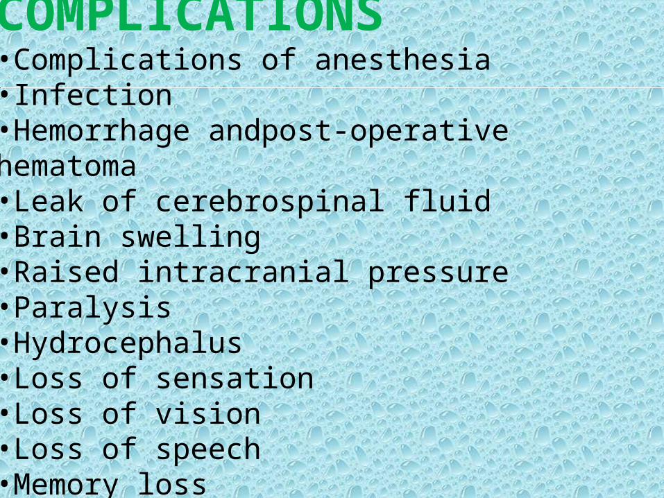
COMPLICATIONS •Complications of anesthesia •Infection•Hemorrhage andpost-operative hematoma•Leak of cerebrospinal fluid•Brain swelling•Raised intracranial pressure•Paralysis•Hydrocephalus•Loss of sensation•Loss of vision•Loss of speech•Memory loss
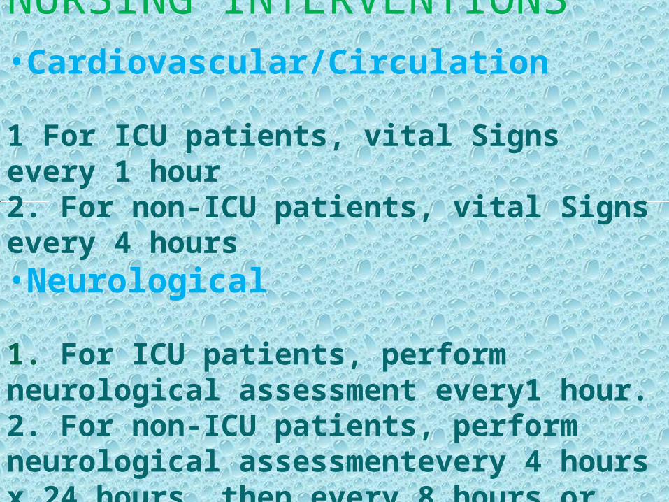
NURSING INTERVENTIONS •Cardiovascular/Circulation
1 For ICU patients, vital Signs every 1 hour2. For non-ICU patients, vital Signs every 4 hours•Neurological
1. For ICU patients, perform neurological assessment every1 hour.2. For non-ICU patients, perform neurological assessmentevery 4 hours x 24 hours, then every 8 hours or per order.

3. Assess spontaneous activity (i.e. frequent posturechanges, breathing pattern, vomiting, twitches or seizures
4.MonitorI&O per order. Fluids may be restricted to prevent fluid shift and cerebral edema.
5. Monitor for seizure activity and maintain safety 6. Evaluate patient for signs and symptoms of Increasing intracranial pressure. These include
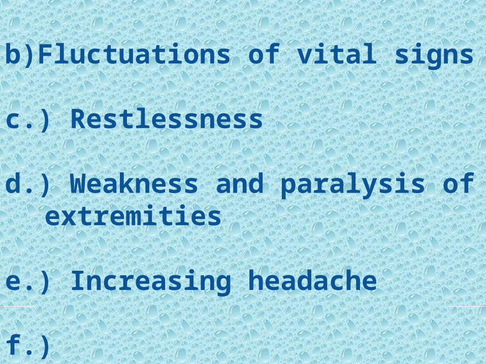
a.) Diminished response to stimuli
b)Fluctuations of vital signs
c.) Restlessness
d.) Weakness and paralysis of extremities
e.) Increasing headache
f.) Changeinvision/pupillarychanges

PRIORITIZATION OF NURSING PROBLEMS1) Altered cerebral tissue perfusion related to decreased cerebral blood flow secondary to head injury
2) Ineffective airway clearance related to accumulation of secreation and decreased LOC
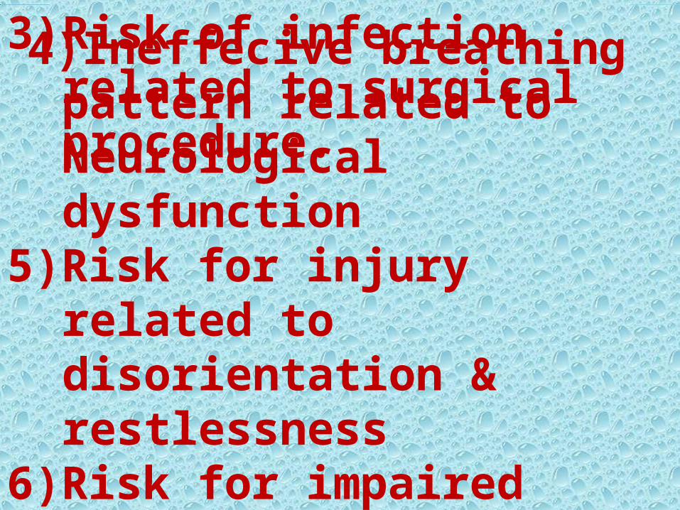
4)Ineffecive breathing pattern related to Neurological dysfunction
5)Risk for injury related to disorientation & restlessness
6)Risk for impaired skin integrity related to immobility.
3)Risk of infection related to surgical procedure.
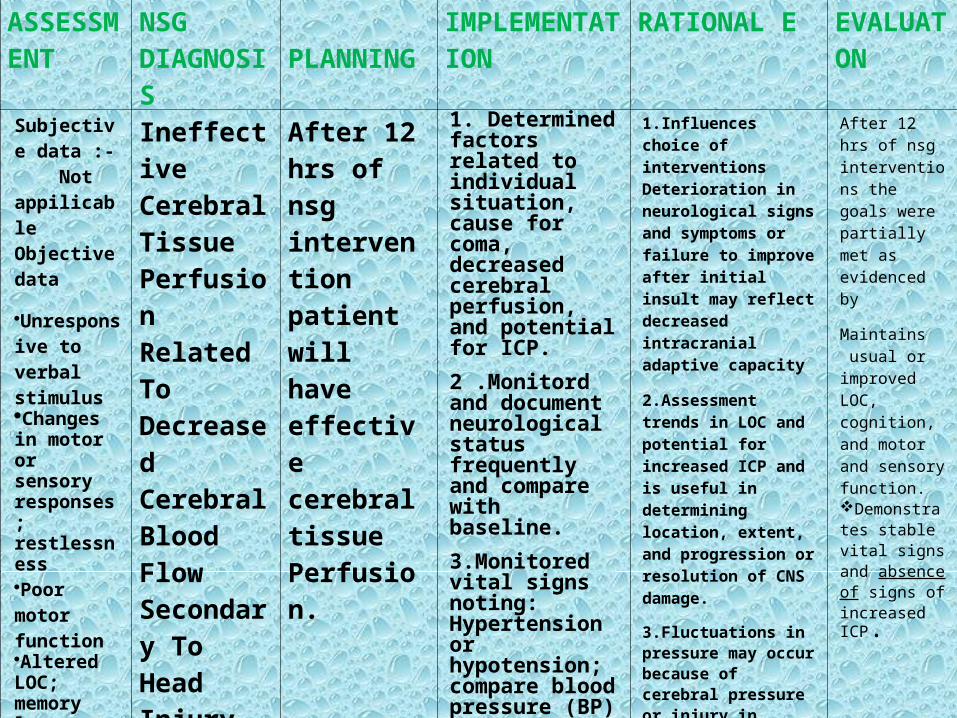
ASSESSMENT
NSG DIAGNOSIS
PLANNING IMPLEMENTATION
RATIONAL E EVALUATON
Subjective data :- Not appilicable Objective data
Unresponsive to verbal stimulusChanges in motor or sensory responses; restlessnessPoor motor functionAltered LOC; memory loss
Ineffective Cerebral Tissue Perfusion Related To Decreased Cerebral Blood Flow Secondary To Head Injury
After 12 hrs of nsg intervention patient will have effective cerebral tissue Perfusion.
1. Determined factors related to individual situation, cause for coma, decreased cerebral perfusion, and potential for ICP.
2 .Monitord and document neurological status frequently and compare with baseline.
3.Monitored vital signs noting: Hypertension or hypotension; compare blood pressure (BP) readings in both arms
1.Influences choice of interventions Deterioration in neurological signs and symptoms or failure to improve after initial insult may reflect decreased intracranial adaptive capacity
2.Assessment trends in LOC and potential for increased ICP and is useful in determining location, extent, and progression or resolution of CNS damage.
3.Fluctuations in pressure may occur because of cerebral pressure or injury in vasomotor area of the brain. Hypertension or hypotension may have been a precipitating factor.
After 12 hrs of nsg interventions the goals were partially met as evidenced by
Maintains usual or improved LOC, cognition, and motor and sensory function.Demonstrates stable vital signs and absence of signs of increased ICP.

Sensory
Languae
intellecal
And
emotioal
deficits
Changes
in vital signs
4. . Document ed changes in vision, such as reports of blurred vision and alterations in visual field or depth perception
5.Assessed higher functions, including speech, if client is alert.
6. Positioned with head slightly elevated and in neutral position.
7.Maintain bedrest, provide quiet environment, and restrict visitors or activities, as indicated. Provide rest periods between care activities, limiting duration of procedures.
4.Specific visual alterations reflect area of brain involved, indicate safety concerns, and influence choice of interventions.
5.Changes in cognition and speech content are an indicator of location and degree of cerebral involvement and may indicate increased ICP.
6. Reduces arterial pressure by promoting venous drainage and may improve cerebral circulation and perfusion
7. Continual stimulation can increase ICP. Absolute rest and quiet may be needed to prevent recurrence of bleeding, in the case of hemorrhagic stroke.
Displays no further Deterioratetion or Recurrence of deficits.

Health education
1.Instruct the patient
•Do not drive after surgery until discussed with surgeon. •Avoid sitting for long periods of time. •Do not lift anything heavier than 5 pounds.•Housework and yardwork are not permitted until the first follow-up office visit.

2. An early exercise program to gently stretch the neck and back.3. Encourage walking 4.Instruct When to Call Doctor•A temperature that exceeds 101º F •An incision that shows signs of infection. •If taking an anticonvulsant, and notice drowsiness, balance problems, or rashes. •Decreased alertness, increased drowsiness, weakness of arms or legs, increased headaches, vomiting.

CONCLUSION•Patient was intubaA case of fall from height with acute SDH was brought in ER on 16/05/13 •ted from the ER upon arrival •His GCS Was 8/15 •The patient was then shifted to OR for emergency POSTERIOR FOSSA CRANIOTOMY +SDH EVACUATION +DUROPLASTY .•Patient was shifted to ICU after surgery and was on ventillator for 10 days .•He was extubated after 10 days .

BIBILIOGRAPHY
•Wikipedia
•Lippincatt manual nursing practice 9th edition
•Mayfield clinic
•Medical-Surgical Standards Review
•Intensive Care Unit Standards