copyright by arianna kyra stefanatos 2015
TRANSCRIPT

Copyright
by
Arianna Kyra Stefanatos
2015

The Dissertation Committee for Arianna Kyra Stefanatos Certifies that this is the
approved version of the following dissertation:
Neurocognitive and Psychosocial Functions in Children with Frontal
and Temporal Lobe Epilepsy
Committee:
Catharine H. Echols, Supervisor
Nancy L. Nussbaum, Co-Supervisor
Charles J. Holahan
David M. Tucker
Jessica A. Church-Lang

Neurocognitive and Psychosocial Functions in Children with Frontal
and Temporal Lobe Epilepsy
by
Arianna Kyra Stefanatos, M.A.
Dissertation
Presented to the Faculty of the Graduate School of
The University of Texas at Austin
in Partial Fulfillment
of the Requirements
for the Degree of
Doctor of Philosophy
The University of Texas at Austin August 2015

Dedication
I would like to dedicate this manuscript to my mother and father, Melanie and
Gerry Stefanatos, my sister, Alexandra Stefanatos, and my grandmother, Gloria Barnett. I
will never be able to express how much their love and encouragement has meant to me
over the past six years. I feel so blessed every day for their continued support. Thank you
for always being my “rock”.
I also would like to dedicate this manuscript to the memory of Mr. George Reim
and Dr. Maureen Dennis. Mr. Reim emboldened me to use my voice when I was simply
content to remain quiet. Dr. Dennis’ passion for clinical research provided the inspiration
for me to pursue a course of study directed towards understanding more about the
detrimental impact of childhood disorders. I will forever be indebted to her for her
support and guidance.

v
Acknowledgements
First and foremost, I would like to express my extreme gratitude to my
supervisors, Dr. Nancy Nussbaum and Dr. Catharine Echols. Throughout the years that I
have had the opportunity to work with Dr. Nussbaum, she has provided a wealth of
support and guidance in both my clinical and research endeavors. I feel extremely
fortunate for all of the opportunities I have had under her mentorship, and I am so
grateful for everything she has done for me. Dr. Echols has graciously served as my co-
supervisor and provided me a welcoming home in the department. I will always be
incredibly thankful to her for her support and investment in my professional
development.
I would like to acknowledge and thank my other committee members, Drs.
Charles J. Holahan, David Tucker and Jessica Church-Lang, for their assistance and
feedback on this dissertation. I would like to thank Dr. Marc Lewis for his support during
the early stages of my research at UT, and Kimberly Terry, for her help with navigating
the dissertation process. I would like to thank the research assistants and psychometrists
at Austin Neuropsychology, PLLC, and Dell Children’s Medical Center for their help
with data collection and preparation. I would also like express my gratitude to the
neurologists at Dell Children’s Medical Center for classifying participants and, in
particular, to the families and children who agreed to allow their data to be used for
research purposes.

vi
Neurocognitive and Psychosocial Functions in Children with Frontal
and Temporal Lobe Epilepsy
Arianna Kyra Stefanatos, Ph.D.
The University of Texas at Austin, 2015
Supervisor: Catharine H. Echols and Nancy L. Nussbaum
A key construct at the foundation of cognitive and clinical developmental
neuropsychology is the notion that cognitive functions are localized to specific cortical
regions in the brain. Consistent with this, relatively stable cognitive and behavioral
profiles have been described for adults diagnosed with the two most common focal-onset
seizure disorders. Temporal lobe epilepsy (TLE) is primarily associated with impairments
in memory functioning (Bell & Giovagnoli, 2007) and frontal lobe epilepsy (FLE) with
impairments in executive and motor functioning (Patrikelis et al., 2009). However, the
immature brain may be particularly vulnerable to the adverse effects of recurrent seizure
activity, showing more widespread effects on cognitive processes and brain organization
compared to adults (Korman et al., 2013). Despite these observations, few studies have
directly compared performance between children with different epilepsy syndromes
utilizing broad assessments spanning multiple domains (Williams, Griebel & Dykman,
1998). Consequently, the aim of this study was to evaluate the degree and selectivity of
patterns of cognitive and psychosocial dysfunction in children with TLE and FLE.
Participants included 51 children between the ages of 6 and 16 years with

vii
intractable epilepsy who were consecutively seen for a neuropsychological evaluation
through the Dell Children’s Medical Center Comprehensive Epilepsy Program. During
this assessment, participants were administered a battery including measures of memory,
executive, motor and intellectual functioning. In addition, parents completed
questionnaires regarding their child's behavioral and psychosocial functioning. Contrary
to the selective patterns of deficits typically described in adults, both the TLE and FLE
groups demonstrated significant impairments relative to normative values on each of the
domains assessed. Moreover, no significant differences were found between the two
patient groups on any of the measures, with the exception of a task of visual memory.
These findings suggest that individuals with childhood-onset epilepsy exhibit
fairly broad patterns of cognitive compromise that do not differ significantly with frontal
lobe versus temporal lobe seizure localization. Furthermore, the range of deficits
observed would not normally be expected with analogous seizure disorders acquired in
adulthood. These results provide important insights into the organization of cognitive and
behavioral functions following early neurological insults associated with epilepsy.

viii
Table of Contents
List of Tables .................................................................................................................... xii
List of Figures .................................................................................................................. xiv
1 BACKGROUND AND SIGNIFICANCE ..................................................................1
1.1 FUNCTIONAL SPECIALIZATION IN THE BRAIN ..............................................1
1.2 EPILEPSY: A Window on Brain Function and Development…………………....…3 1.2.1 PREVALENCE .................................................................................................5 1.2.2 NEUROPHYSIOLOGY ....................................................................................6 1.2.3 NEUROPSYCHOLOGY ...................................................................................6
1.3 LOCALIZATION-RELATED EPILEPSIES ...........................................................10 1.3.1 TEMPORAL LOBE EPILEPSY .....................................................................10 1.3.1 FRONTAL LOBE EPILEPSY ........................................................................11
1.4 DOMAINS OF FUNCTIONING .............................................................................12 1.4.1 MEMORY FUNCTIONING ...........................................................................13
1.4.1.1 Memory Functioning in TLE .............................................................13 1.4.1.2 Memory Functioning in FLE ............................................................18 1.4.1.3 Comparison of Memory Functioning in FLE & TLE........................19
1.4.1 EXECUTIVE FUNCTIONING .......................................................................20 1.4.1.1 Executive Functioning in FLE ..........................................................20 1.4.1.2 Executive Functioning in TLE ..........................................................22 1.4.1.3 Comparison of Executive Functioning in FLE & TLE .....................24
1.4.1 MOTOR FUNCTIONING ...............................................................................24 1.4.1.1 Motor Functioning in FLE ................................................................24 1.4.1.2 Motor Functioning in TLE ................................................................25 1.4.1.3 Comparison of Motor Functioning in FLE & TLE ...........................26
1.4.2 INTELLECTUAL FUNCTIONING ...............................................................26 1.4.2.1 Intellectual Functioning in FLE ........................................................26

ix
1.4.2.2 Intellectual Functioning in TLE ........................................................28 1.4.2.3 Comparison of Intellectual Functioning in FLE & TLE ...................29
1.4.3 PSYCHOSOCIAL FUNCTIONING ...............................................................30 1.4.3.1 Psychosocial Functioning in TLE .....................................................30 1.4.3.2 Psychosocial Functioning in FLE .....................................................31 1.4.3.3 Comparison of Psychosocial Functioning in FLE & TLE ................32
1.5 SUMMARY OF EXISTING LITERATURE ...........................................................33
1.6 STUDY RATIONALE .............................................................................................39
2 SPECIFIC AIMS AND HYPOTHESES ..................................................................41
3 METHODOLOGY ....................................................................................................43
3.1 PARTICIPANTS ......................................................................................................43
3.2 PROCEDURE ...........................................................................................................45
3.3 INSTRUMENTS ......................................................................................................46 3.3.1 ASSESSMENT OF MEMORY .......................................................................46
3.3.1.1 Verbal Memory (List Learning) ........................................................46 3.3.1.2 Verbal Memory (Narrative) ..............................................................48 3.3.1.3 Abstract Visual Memory ...................................................................49
3.3.2 ASSESSMENT OF EXECUTIVE FUNCTIONING ......................................50 3.3.2.1 Working Memory ..............................................................................50 3.3.2.2 Behavioral Regulation & Metacognition ..........................................50 3.3.2.3 Processing Speed ..............................................................................51 3.3.2.4 Self-Monitoring .................................................................................52
3.3.3 ASSESSMENT OF MOTOR FUNCTIONING ..............................................52 3.3.3.1 Motor Coordination/Speed ...............................................................52
3.3.4 ASSESSMENT OF INTELLECTUAL FUNCTIONING ...............................53 3.3.4.1 Intelligence/General Abilities ...........................................................53
3.3.5 ASSESSMENT OF PSYCHOSOCIAL FUNCTIONING ..............................54 3.3.5.1 Internalizing/Externalizing Behavior Problems ...............................54

x
4 RESULTS ...................................................................................................................57
4.1 POWER ANALYSIS ................................................................................................57
4.2 DATA PREPARATION ...........................................................................................57
4.3 GROUP ANALYSES ...............................................................................................60 4.3.1 DEMOGRAPHICS ..........................................................................................60 4.3.2 CORRELATIONS ...........................................................................................63 4.3.3 MEMORY FUNCTIONING ...........................................................................65 4.3.4 EXECUTIVE FUNCTIONING .......................................................................67 4.3.5 MOTOR FUNCTIONING ...............................................................................69 4.3.6 INTELLECTUAL FUNCTIONING ...............................................................70 4.3.7 PSYCHOSOCIAL FUNCTIONING ...............................................................72
4.4 INDIVIDUAL ANALYSES .....................................................................................74
4.5 ADDITIONAL ANALYSES ....................................................................................75
5 DISCUSSION .............................................................................................................76
5.1 SUMMARY ..............................................................................................................76
5.2 MEMORY FUNCTIONING ....................................................................................76 5.2.1 TEMPORAL LOBE EPILEPSY .....................................................................76 5.2.2 FRONTAL LOBE EPILEPSY ........................................................................79 5.2.3 SUMMARY: FLE VS. TLE ............................................................................81
5.3 EXECUTIVE FUNCTIONING ................................................................................81 5.3.1 FRONTAL LOBE EPILEPSY ........................................................................81 5.3.2 TEMPORAL LOBE EPILEPSY .....................................................................84 5.3.1 SUMMARY: FLE VS. TLE ............................................................................86
5.4 INTELLECTUAL FUNCTIONING ........................................................................87 5.4.1 FRONTAL LOBE EPILEPSY ........................................................................87 5.4.2 TEMPORAL LOBE EPILEPSY .....................................................................89 5.4.1 SUMMARY: FLE VS. TLE ............................................................................90

xi
5.5 MOTOR FUNCTIONING ........................................................................................91 5.5.1 FRONTAL LOBE EPILEPSY ........................................................................91 5.5.2 TEMPORAL LOBE EPILEPSY .....................................................................92 5.5.1 SUMMARY: FLE VS. TLE ............................................................................92
5.6 PSYCHOSOCIAL FUNCTIONING ........................................................................93
5.7 IMPLICATIONS ......................................................................................................94
5.8 STRENGTHS AND LIMITATIONS .......................................................................99
5.9 CONCLUSION .......................................................................................................103
6 WORKS CITED .......................................................................................................106

xii
List of Tables
Table 1 Studies Comparing Children with FLE and TLE ................................................ 36
Table 2 Demographic Information of Participants ........................................................... 45
Table 3 California Verbal Learning Test - C .................................................................... 48
Table 4 TOMAL-2 Memory for Stories ........................................................................... 49
Table 5 TOMAL-2 Abstract Visual Memory ................................................................... 49
Table 6 WISC-IV Digit Span Backwards ......................................................................... 50
Table 7 BRIEF .................................................................................................................. 51
Table 8 WISC-IV Processing Speed Index ....................................................................... 52
Table 9 CVLT-C ............................................................................................................... 52
Table 10 Grooved Pegboard ............................................................................................. 53
Table 11 WISC-IV ............................................................................................................ 54
Table 12 Child Behavior Checklist ................................................................................... 56
Table 13 Neuropsychological Test Battery ....................................................................... 60
Table 14 Clinical Information of Participants .................................................................. 62
Table 15 Comparison Between Right- and Left-Hemisphere ........................................... 63
Table 16 Correlation Matrix ............................................................................................. 64
Table 17 MEMORY FUNCTIONING: Comparison to Population Norms ..................... 65
Table 18 MEMORY FUNCTIONING: Comparison Between Groups ............................ 66
Table 19 MEMORY FUNCTIONING: Proportions ........................................................ 67
Table 20 EXECUTIVE FUNCTIONING: Comparison to Population Norms ................ 68
Table 21 EXECUTIVE FUNCTIONING: C Comparison Between Groups .................... 69

xiii
Table 22 MOTOR FUNCTIONING: Comparison to Population Norms ........................ 69
Table 23 MOTOR FUNCTIONING: Comparison Between Groups ............................... 70
Table 24 INTELLECTUAL FUNCTIONING: Comparison to Population Norms ......... 71
Table 25 INTELLECTUAL FUNCTIONING: Comparison Between Groups ................ 71
Table 26 PSYCHOSOCIAL FUNCTIONING: Comparison to Population Norms ........ 72
Table 27 PSYCHOSOCIAL FUNCTIONING: Comparison Between Groups ............... 74
Table 28 Secondary Analyses ........................................................................................... 75

xiv
List of Figures
Figure 1 Factors Influencing Cognition in Epilepsy ........................................................... 7
Figure 2 MEMORY FUNCTIONING: Means ................................................................. 65
Figure 3 EXECUTIVE FUNCTIONING: Means ............................................................. 68
Figure 4 MOTOR FUNCTIONING: Means ..................................................................... 70
Figure 5 INTELLECTUAL FUNCTIONING: Means ..................................................... 70
Figure 6 PSYCHOSOCIAL FUNCTIONING: Means ..................................................... 73

1
1 BACKGROUND AND SIGNIFICANCE
1.1 FUNCTIONAL SPECIALIZATION IN THE BRAIN
Models of human brain function have traditionally conceptualized the cerebral
cortex as being composed of various discrete regions, each dedicated to specialized,
domain-specific functions (Anderson, 2010; Fodor, 1983; Fodor, 2000; Johnson, 2001).
This hypothetical framework of modular organization evolved from neuropsychological
studies involving adults with acquired circumscribed brain lesions and is based on the
supposition that it is possible to dissociate different cognitive and behavioral processes in
the brain by correlating the effects of damage to specific neural substrates with selective
patterns of cognitive and behavioral impairment (Caramazza & Coltheart, 2006; Temple,
1997). More specifically, the lesion model attributes the processing of particular types of
information to distinct areas of cortex, such that damage to those regions by injury or
disease results in the selective disruption of associated cognitive and neural processes and
conversely, the observed loss of skills is indicative of injury to that corresponding brain
region (D’Souza & Karmiloff-Smith, 2011; Fama & Sullivan, 2014; Moses & Stiles,
2002). This line of research has allowed powerful inferences to be made about the
organization of functional neural networks in adults.
Attempts have been made to apply this adult neuropsychological model to
characterizing the pattern of impairment and preserved skills of various acquired and
neurodevelopmental conditions in childhood (Karmiloff-Smith, 2013; Temple, 1997;
Thomas & Karmiloff-Smith, 2002). However, profiles derived from adult participants
with acquired neurological insults reflect neural and cognitive processes damaged within
a mature state after an extended period of normal development (D’Souza & Karmiloff-

2
Smith, 2011; Karmiloff-Smith, 2013). Consequently, it has been argued that they provide
limited information regarding the degree of specialization that exists in earlier stages of
development or about the gradual process of relative modularization that may occur
across childhood and adolescence (Dennis & Barnes, 1994; D’Souza & Karmiloff-Smith,
2011).
Early studies appeared to suggest that the brain was innately specialized from
birth (e.g., Witelson & Paille, 1973). However, emerging studies have suggested that,
despite some evidence of lateralization, cognitive and behavioral processes may not be
functionally localized to specific cortical regions early in development (see Bell &
Giovagnoli, 2007; Patrikelis, Angelakis, & Gatzonis, 2009). Instead, it has been proposed
that a process of ‘interactive specialization’ may occur by which distinct neural networks
become more specialized or segregated across ontogeny (Johnson, 2001; Johnson, 2011;
Johnson, Grossman, & Kadosh, 2009). According to this view, the immature brain is
anatomically less differentiated and more interconnected outside of regions typically
regarded as canonical network structures in the adult brain (Fair et al., 2009; Wylie et al.,
2014). Changes in the activation patterns of cortical regions occur across development as
circuits of neurons become more restricted to subserve a narrower set of specific
functions (Johnson et al., 2009; Tau & Peterson, 2010). Typical development, therefore,
appears to be characterized by a process of increasing connectivity between brain regions
subserving the same functional networks and corresponding decreases in activation
between brain regions associated with other networks (Dosenbach et al., 2010; Fair et al.,
2007; Fair et al., 2009), with the resulting endpoint being the formation of mature,
functionally specialized neural networks (Ibrahim et al., 2014).

3
However, damage or disease sustained early in childhood has been suggested to
fundamentally alter or compromise the typical trajectory of neural and behavioral
specialization and development (Dennis et al., 2014). Specifically, it has been suggested
that early brain injury may interfere with the process by which the transient projections
that support cognitive processing during early development are typically eliminated
(Nelson, 2000), resulting in atypical and more widely distributed brain networks
(Maguire, Vargha-Khadem, & Mishkin, 2001; Sutula & Pitkänen, 2002). Consequently,
attempts to generalize adult principles of functional specialization are limited given
emerging indications of alternative patterns of neural organization which can develop in
the brain following early injury (see Moses & Stiles, 2002). The findings from clinical
studies specifically examining outcomes following early focal brain injury reveal
considerable variability in the resulting profiles of deficits and apparent degree of
functional recovery, which vary as a function of cognitive domain, site of lesion, timing
of lesion onset and specific etiology (Dennis et al., 2014; Moses & Stiles, 2002).
1.2 EPILEPSY: A Window on Brain Function and Development
The study of epilepsy has been central to the conceptualization and understanding
of human brain-behavior relationships. Seminal 19th century observations attributed
specific disturbances of motor (Todd, 1849) and language (Hughlings-Jackson, 1866)
function to neural exhaustion or inhibition caused by the repeated, sustained electrical
discharges characteristic of seizures. Later, electrical cortical stimulation of conscious
patients undergoing neurosurgical resections of epileptogenic regions of the brain
resulted in important insights regarding the topographic nature of motor-sensory cortex
and the circumscribed distribution of language areas (Almeida, Martinez, & Feindel,

4
2005; Ojemann, 1979; Penfield & Roberts, 1959). Classic observations by Scoville and
Milner (1957) of anterograde amnesia following bilateral temporal lobe and hippocampal
resection for treatment of intractable epilepsy demonstrated the critical role of the
hippocampal formation and temporal lobe structures in mediating consolidation of newly
learned material in long-term memory. In addition, the use of resective surgery in
children with intractable epilepsy provided compelling demonstrations of developmental
plasticity for higher cognitive functions following removal of entire hemispheres (Basser,
1962; Dennis & Kohn, 1975; Goodman & Whitaker, 1985).
More recently, epilepsy has been proposed as an ideal disorder for understanding
brain-behavior relationships across different points in development (Matthews, 1992;
Novelly, 1992). The study of epilepsy has been instrumental in understanding the
mechanisms underlying cognitive decline in correlation with identified
pathophysiological processes (see David, Bastin, Chabardes, Minotti, & Kahane, 2010;
van Diessen et al., 2013a; van Diessen, Otte, Braun, Stam, & Jansen, 2013b). Recent
technological advances have demonstrated significant alterations in the architecture of
functional neural networks and connectivity patterns in individuals with epilepsy which
appear to emerge from pathological brain dynamics that develop following recurrent
seizure activity (Chavez, Valencia, Navarro, Latora, & Martinerie, 2010). A related body
of literature within the field of clinical neuropsychology has focused on attempting to
correlate the consequences of these pathogenic processes by examining the particular
cognitive and behavioral profiles associated with specific epilepsy syndromes (see
Helmstaedter & Kockelmann, 2006; Patrikelis, Angelakis, & Gatzonis, 2009). Given
concerns that the current function-structure mapping framework derived from adult

5
research may not be appropriate to characterize the effects of damage to the immature
brain (Moses & Stiles, 2002), establishing how recurrent seizures impact functioning in
children with epilepsy can provide important insights into the emergence of cognitive and
behavioral processes across development (Insel, 2009; Rapoport et al., 1999).
Consequently, in this study, the profiles of children with two common focal epilepsy
syndromes were compared in order to examine the impact of recurrent seizure activity on
the emergence of cognitive and behavioral functions in the developing brain.
1.2.1 PREVALENCE
Epilepsy is a chronic neurological condition characterized by recurrent
paroxysmal seizure activity (David et al., 2010; Fisher et al., 2014), which affects
approximately 2-3 million people in the United States (Hirtz et al., 2007). By most
definitions, seizures represent abnormal electrical events that are triggered by repetitive
excessive or hypersynchronous activity of neurons in the brain, sufficient enough to
result in alterations in behavior (Fisher et al., 2014; Fisher et al., 2005). While
approximately 5-10% of the population may experience at least one seizure during the
course of their lifetime (Wilden & Cohen-Gadol, 2012), only a proportion of these
individuals subsequently develop epilepsy, which requires that the seizure activity be
recurrent (Fisher et al., 2014). While epilepsy is the fourth most commonly diagnosed
neurologic disorder among adults, it is the one of the most commonly diagnosed
neurologic disorder in children and adolescents (see MacLeod & Appleton, 2007; WHO,
2012). Age-specific incidence rates have estimated that approximately 150,000 children
under the age of 16 experience a first-time, unprovoked seizure each year, and of those,
approximately 25% will subsequently receive a diagnosis of epilepsy (McAbee & Wark,

6
2000).
1.2.2 NEUROPHYSIOLOGY
The electrical basis of epilepsy became evident in the latter part of the 19th
century based on observations of pioneers in the field, such as Robert Bentley Todd
(1849), John Hughlings-Jackson (1866) and Sir William Richard Gowers (1885). Since
that time, clinical studies and animal models of epilepsy have been instrumental in
elucidating the cellular and molecular mechanisms underlying seizure activity in the
brain (see Wong, 2005). Ontogenetic changes in specific neurotransmitter systems
including glutamate and GABA occur across development and can result in cortical
imbalances in the excitatory and inhibitory influences in the brain (see David et al., 2010;
Holmes & Ben-Ari, 2001; Sanchez & Jensen, 2001; Takesian & Hensch, 2013; Wong,
2005). Although these maturational changes in the molecular and cellular systems are
critical for synapse formation and the organization of immature circuits into functional
neural networks (Sur & Rubenstein, 2005; Wong, 2005), age-specific differences in
physiology and metabolism appear to increase the susceptibility of the developing brain
to the onset of abnormal electrical activity (Ben-Ari & Holmes, 2006; Holmes & Ben-
Ari, 2001). In addition, this process of synaptic plasticity has been hypothesized to lower
the seizure threshold and promote the reoccurrence of hyperexcitable neural circuits
(Holmes & Ben-Ari, 2001; Wong, 2005), which is consistent with findings of increased
incidence rates for seizure activity in early childhood (Newton, 2012; Olafsson et al.,
2005; Wyllie, 2010).
1.2.3 NEUROPSYCHOLOGY
Around the time that the electrical basis of seizures was identified, it was also

7
recognized that prolonged or recurrent seizures could have profound, yet often specific,
impacts on cognitive function. In 1984, Herman and Whitman introduced a model for
understanding the effects of seizure activity on behavior which can be adapted to explain
its effects on cognitive function as well. Behaviors evident in individuals with epilepsy
appear to be influenced both by the brain-related factors that give rise to epilepsy and the
effects of seizures. Specifically, the resulting neuropsychological and behavioral profiles
of individuals with chronic seizure activity can be diverse, emerging from a complex
matrix of factors including the initial epileptogenic processes, the resulting brain damage
that can occur as a result of the recurrent seizures, and even as a result of the course of
treatment (Helmstaedter & Kockelmann, 2006). In addition, the role of non-brain related
factors, including social and emotional variables must also be acknowledged. The
influence of these multiple factors is summarized in the illustration in Figure 1.
Figure 1 Factors Influencing Cognition in Epilepsy
In the mature adult brain, recurrent electrical charges typically but not invariably
results in specific abnormalities in cognitive function in the absence of significant
structural damage to the brain (Hermann, Seidenberg & Bell, 2002a; Hermann et al.,

8
2002b). However, emerging evidence from histological investigations has clearly
demonstrated that recurrent seizure activity is a dynamic process which can manifest in
distinct patterns of morphological and functional changes in the developing brain (see
Ben-Ari & Holmes, 2006; Curia et al., 2014; Holmes, 2013). MRI studies have
demonstrated significant volumetric reductions in white matter and in brain connectivity
in brain regions distant from the region of primary epileptogenesis in individuals with
early-onset epilepsy (seizure onset before age 14) relative to those with late-onset seizure
activity (Hermann et al., 2002a; Hermann et al., 2002b). Moreover, this pattern of
diaschisis appears to correlate with more generalized patterns of neuropsychological
dysfunction.
Accordingly, seizure onset early in life appears to disrupt the emergence of
distinct regional functional and structural networks underlying cognition in the brain
(Ibrahim et al., 2014; Kellerman, Bonilha, Lin, & Hermann, 2015; Widjaja, Zamyadi,
Raybaud, Snead, & Smith, 2013a; Widjaja, Zamyadi, Raybaud, Snead, & Smith, 2013b;
Widjaja et al., 2015). Specifically, children demonstrate greater internetwork connectivity
and weaker intranetwork integration relative to typically developing controls (Widjaja et
al., 2015), which has been correlated with various clinical features including epilepsy
duration and cognitive outcomes (Ibrahim et al., 2014; Kellerman et al., 2015; Mankinen
et al., 2012; Widjaja et al., 2013b). These patterns of functional and structural
connectivity abnormalities have been noted to be most notable in individuals with an
early age of seizure onset (Doucet et al., 2015). Moreover, there is substantial evidence to
suggest that children with epilepsy display patterns of abnormalities which emerge
immediately following, or even in some cases, predate the onset of the seizure activity

9
(Oostrom, Smeets-Schouten, Kruitwagen, Peters, & Jennekens-Schinkel, 2003), which
likely reflect the negative effect of seizure activity in addition to the presence of
antecedent neurobiological factors associated with the underlying epileptogenesis that
may differentially influence cognition and behavior in children (Hermann & Seidenberg,
2007).
In summary, the presence of recurrent seizure activity during periods of increased
neural maturation across development is associated with critical alterations in brain
development and structure which appears to increase the vulnerability of children to
cognitive decline relative to adults (Bjornes, Stabell, Henriksen, & Loyning, 2001;
Glosser, Cole, French, Saykin, & Sperling, 1997; Hermann et al., 2002a; Hermann et al.,
2002b; Kaaden & Helmstaedter, 2009). It has been suggested that the functional
consequences of this disruption to neural networks may depend on the developmental
stage and processes that are occurring at the time of seizure onset (Ben-Ari & Holmes,
2006; Hermann et al., 2002b; Spencer-Smith & Anderson, 2009). Significant progress
has been made in the adult literature in understanding the cognitive and behavior
difficulties that are shared across epilepsy syndromes, as well as those patterns which are
unique to each (Bell & Giovagnoli, 2007; Elger, Helmstaedter & Kurthen, 2004;
Hermann, Seidenberg, Lee, Chan, & Rutecki, 2007; Lassonde, Sauerwein, Jambaqué,
Smith, & Helmstaedter, 2000; Patrikelis et al., 2009). The literature has focused largely
on the specific dynamics of functioning in adults with temporal lobe (TLE) and frontal
lobe (FLE) epilepsies due to their high prevalence (Manford, Hart, Sander, & Shorvon,
1992; Wiebe, 2000). Additionally, unlike some neurological diseases in which
generalized cognitive and behavioral impairments are observed, individuals with

10
location-specific epilepsies such as TLE and FLE have been found to have patterns of
deficits involving cognitive functions specifically mediated by the cerebral area where
the epileptogenic focus is located (see Bell & Giovagnoli, 2007; Patrikelis et al., 2009;
Risse, 2006; Riva, Saletti, Nichelli, & Bulgheroni, 2002). Specifying the effects of these
focal epilepsy syndromes on cognition and behavior in children can provide important
insights into the organization of functions in the brain and is critical in order to assist in a
better understanding of outcomes following early neurological insults in the developing
brain.
1.3 LOCALIZATION-RELATED EPILEPSIES
1.3.1 TEMPORAL LOBE EPILEPSY
Temporal lobe epilepsy (TLE) is a focal-onset syndrome characterized clinically
by the development of spontaneous seizure activity originating from structures within the
temporal lobes (Chang & Lowenstein, 2003; Engel, 1989; Zhang et al., 2002). TLE is the
most commonly diagnosed type of epilepsy, accounting for approximately 80% of all
focal cases, with typical age of onset at 6-10 years (Wiebe, 2000). Patients with TLE also
often present well-circumscribed underlying pathology and phenotypic expression
(Helmstaedter, 2001). In particular, the mesial temporal lobe structures, including the
hippocampus and the amygdala, have been demonstrated to be extremely susceptible to
epileptogenic processes (see Aroniadou-Anderjaska, Fristch, Qashu, & Braga, 2008).
The genesis and progression of recurrent seizure activity in the temporal lobe
appears to be related to the unique anatomical circuitry of the limbic structures and to
changes in the electrophysiological functioning of neurons that occur in areas of the
temporal lobe across development as a part of a natural process of neurogenesis (see

11
Kuruba, Hattiangady, & Shetty, 2009; Sharma et al., 2007). Specifically, particular
histopathological features have been identified in individuals with TLE including
abnormal loss of cells, structural changes including sprouting and cell dispersion and
gliosis (see Curia et al., 2014). Preliminary research involving animal and human models
suggests that the effect of seizures on neurogenesis in the hippocampal area may be more
evident in the immature brain than in the mature adult brain (Rao, Hattiangady, & Shetty,
2008). For example, relative to individuals with adult-onset epilepsy and typically-
developing controls, individuals with childhood-onset epilepsy have demonstrated
significant reductions in volume of hippocampal tissue (Hermann et al., 2002a; Hermann
et al., 2002b). Moreover, volumetric reduction abnormalities were noted outside of the
temporal lobe, observed in all total lobar and cerebrum measurements.
1.3.1 FRONTAL LOBE EPILEPSY
In contrast, frontal lobe epilepsy (FLE) is a localization-based disorder that is
characterized by recurring seizure activity arising from structures within the frontal lobe.
FLE has been identified as the second most common type of focal epilepsy, accounting
for approximately 20–30% of cases (Manford et al., 1992). The average age of onset of
FLE is between 4-7 years old (Sinclair, Wheatley, & Snyder, 2004). Some studies have
suggested that in children, the incidence and prevalence of extratemporal epilepsies (such
as FLE) may actually be greater than rates of temporal epilepsies (e.g., Fogarasi, Janszky,
Faveret, Pieper, & Tuxhorn, 2001). Clinical presentation of FLE has been noted to be
more heterogeneous in terms of location and nature of underlying pathology
(Helmstaedter, 2001; Jobst et al., 2000). Research appears to suggest significant
differences in seizure semiology between adult and pediatric patients (Fogarasi et al.,

12
2001).
Less is known about the mechanisms underlying epileptogenesis in individuals
with FLE (Kanemura, Sano, Tando, Sugita, & Aihara, 2012). However, the frontal lobes
undergo a protracted and well-specified course of neuroanatomical, neurophysiological
and neurochemical changes throughout adolescence and into early adulthood (Sowell,
Delis, Stiles, & Jernigan, 2001). These changes include synaptogenesis, synaptic pruning,
increases in prefrontal myelination and reorganization of synaptic connections
(Huttenlocher, 1979; Kinney, Brody, Kloman, & Gilles, 1988). During this period, the
human cerebral metabolic rate is also higher than that in adulthood, which appears to
increase its susceptibility among the cortical regions to repeated seizure activity
(Chugani, Phelps, & Mazziotta, 1987). This process is believed to play a critical role in
the development of functional neural networks, and the resulting pathogenesis of repeated
seizure activity in the immature brain appears to be retardation of prefrontal lobe growth
(Kanemura et al., 2012). Additionally, time-related factors such as epilepsy duration and
age at epilepsy onset have been demonstrated to be associated with greater progression of
structural abnormalities (Janszky et al., 2005).
1.4 DOMAINS OF FUNCTIONING
As mentioned previously, researchers have described a number of
neuropsychological trends within adult populations with TLE and FLE. Consequently,
the expected patterns of functioning of individuals with epilepsy will be discussed in the
following order of domains: Memory, Executive, Motor, Intellectual and Psychosocial
Functioning.

13
1.4.1 MEMORY FUNCTIONING
1.4.1.1 Memory Functioning in TLE
Converging evidence has widely demonstrated the involvement of the temporo-
mesial and neocortical structures such as the hippocampus and parahippocampal cortex in
memory networks in the brain (e.g., Burianova & Grady, 2007; Burianova, McIntosh, &
Grady, 2010; Corkin, Amaral, Gonzalez, Johnson, & Hyman, 1997). The earliest
demonstrations of memory deficits following focal damage to the temporal lobe were
derived from studies of the underlying pathology and surgical management of TLE
(Milner, 1970; Penfield & Milner, 1958; Scoville & Milner, 1957). Subsequent work
using functional and volumetric imaging in typically-developing individuals have widely
demonstrated activation in the temporal lobes during completion of measures of memory
functioning (e.g., Burianova & Grady, 2007; Burianova et al., 2010). However,
individuals with TLE appear to display decreased activation in the temporal lobe during
similar tasks (Bonelli et al., 2010). Smaller volume of the hippocampus has also been
found to be associated with greater impairments in learning and recall in individuals with
TLE (Baxendale, Thompson, & Paesschen, 1998; Corkin et al., 1997; Hermann et al.,
2002a; Hermann et al., 2002b; Narayanan et al., 2012; Wilkinson et al., 2012). Moreover,
these deficits can be observed in individuals without structural lesions of the temporal
lobe (Bengner et al., 2006).
Specifically, individuals with TLE appear to have significant difficulty with the
consolidation of information, which involves a process of stabilization of newly encoded
memory traces within long-term storage (Helmstaedter, Grunwald, Lehnertz, Gleissner,
& Elger, 1997; Hotting, Katz-Biletzky, Malina, & Lindenau, & Bengner, 2010). The

14
efficacy of this consolidation process has typically been demonstrated on standardized
neuropsychological tasks of delayed recall, which assess the retention of recently learned
information following a delay of approximately 20-30 minutes (Bell, Fine, Dow,
Seidenberg, & Hermann, 2005; Elliott, Isaac, & Muhlert, 2014; Mameniskiene, Jatuzis,
Kaubrys, & Budrys, 2006). On this type of measure, adults with TLE typically
demonstrate significant impairments in declarative or episodic memory (e.g., Bell, 2006;
Bell et al., 2005; Bengner et al., 2006; Exner et al., 2002; Jones-Gotman et al., 1997).
Emerging evidence has suggested that individual with temporal lobe dysfunction
can also demonstrate poor learning and retention of previously learned information on
measures of short-delay or “intermediate” recall, under conditions in which the material
to be learned exceeds working memory capacity (supraspan) or takes several minutes to
complete, when the information is not amenable to rehearsal or when a distracter task is
presented between the study phase and the recall phase (see Baddeley, Jarrold, & Vargha-
Khadem, 2011; Brady, Konkle, & Alvarez, 2011; Elliot et al., 2014; Hotting et al., 2010;
Jeneson & Squire, 2011). In fact, recall tasks involving delays greater than 10 seconds
have been demonstrated to be sensitive to damage to the temporal lobe in both animal
(Alvarez, Zola-Morgan & Squire, 1994) and human studies (Hannula, Tranel & Cohen,
2006; Hartley et al., 2007; Nichols, Kao, Verfaellie, & Gabrieli, 2006; Olsen et al., 2009;
Olson, Page, Moore, Chatterjee, & Verfaellie, 2006; Piekema et al., 2007; Rains &
Milner, 1994). Accordingly, a number of studies have also demonstrated deficits in
verbal and non-verbal memory in TLE during immediate recall conditions (Bell, 2006;
Bell et al., 2005; Bengner et al., 2006; Exner et al., 2002; Mameniskiene et al., 2006;).
Individuals with TLE also appear to demonstrate unusually rapid memory loss or

15
accelerated forgetting of information relative to healthy controls, as demonstrated by a
significant loss of details following a delay (Lah, Mohamed, Thayer, Miller, & Diamond,
2014; Mameniskiene et al., 2006; Narayanan et al., 2012; Wilkinson et al., 2012), which
appears to reflect a failure of memory consolidation (Elliot et al., 2014).
Additionally, a number of neuropsychological and neuroimaging studies have
demonstrated hemispheric specialization of memory processing in individuals with late-
onset TLE (e.g., Gleissner, Helmstaedter, & Elger, 1998; Golby et al., 2002; Hermann,
Seidenberg, Schoenfield, & Davies, 1997; Helmstaedter, Kurthern, Lux, Reuber, & Elger,
2003; Jokeit, Okujava, & Woermann, 2001; Jones-Gotman et al., 2010; Jones-Gotman et
al., 1997; Pillon et al., 1999; Powell et al., 2005). Because verbal memory processes are
typically subserved by the language-dominant hemisphere, individuals with left-sided
TLE often demonstrate deficits in verbal memory, including word list recall and story
recall (Helmstaedter & Elger, 1996; Helmstaedter et al., 1997; Jambaqué et al., 2007;
Jones-Gotman et al., 2010; Lee, Yip, & Jones-Gotman, 2002). Evidence for a link
between right-sided seizure activity and nonverbal memory in adults has been more
inconsistent (Alessio et al., 2004; Baxendale et al., 1998; Lee et al., 2002; Smith, Bigel,
& Miller, 2011), suggesting that visual-spatial memory such as visual reproduction and
facial recognition may be a more bilateral process (Baxendale & Thompson, 2010;
Saling, 2009; van Asselen et al., 2006).
Functional neuroimaging studies have confirmed age-related changes in
recruitment of regions of the temporal lobe including the hippocampus and posterior
parahippocampal gyrus for memory encoding across adolescence (Chiu, Schmithorst,
Brown, Holland, & Dunn, 2006; Ghetti, DeMaster, Yonelinas, & Bunge, 2010; Menon,

16
Boyett-Anderson, & Reiss, 2005). It has been demonstrated that an earlier age of seizure
is associated with findings of significant volumetric reduction in total cerebrum and
hippocampal tissue (Hermann et al., 2002a; Hermann et al., 2002b) and differential
patterns of activation in the temporal lobe (Sidhu et al., 2015). Accordingly, decreased
verbal and nonverbal memory efficiency has been observed in children with TLE relative
to individuals with late-onset TLE (seizure onset after age 14) (Kaaden & Helmstaedter,
2009). Additionally, standardized memory tests have largely replicated findings of
impairments on measures of immediate and delayed recall of narrative information
(Gascoigne et al., 2014; Guimaräes et al., 2007; Jambaqué et al., 1993; Jambaqué et al.,
2009; Nolan et al., 2004; Rzezak, Guimaräes, Fuentes, Guerreiro, & Valente, 2011;
Rzezak, Guimaräes, Fuentes, Guerreiro, & Valente, 2012) and verbal learning, immediate
and delayed list recall (Hernandez et al., 2003; Jambaqué et al., 1993; Nolan et al., 2004;
Rzezak et al., 2012) relative to typically-developing controls. Particularly, this appears to
be notable on tasks with high memory loads which exceed the ‘primary memory buffers’
of the neocortical working memory system due to delay length or capacity limitations
(Gabrieli, Keane, & Stebbins, 1993). Studies have suggested that early seizure onset is
associated with a significant neurodevelopmental hindrance in learning efficiency across
various recall trials (Hernandez et al., 2003), which becomes most evident across
adolescence (Helmstaedter & Elger, 2009). Additionally, it appears that children with
TLE also demonstrate increased forgetting of verbal information (Gascoigne et al., 2014)
and significant impairments in various aspects of memory for visual designs (Guimaräes
et al., 2007; Nolan et al., 2004).
However, contrary to the adult literature, the majority of available research

17
suggests that hemispheric lateralization is largely irrelevant in predicting the domain of
memory impairment in children. While some early studies appeared to demonstrate
patterns of lateralized memory impairment in children (e.g., Cohen, 1992; Fedio &
Mirsky, 1969; Jambaqué et al., 1993), these studies were limited by differences in seizure
severity, age at time of testing and sample sizes between groups. More recently, a number
of studies have demonstrated that impairments in verbal memory were comparable
between children with left- and right TLE (e.g., Bigel & Smith, 2001a; Camfield et al.,
1984; Gleissner, Helmstaedter, Schramm, & Eiger, 2002; Gonzalez, Anderson, Wood,
Mitchell & Harvey, 2007; Helmstaedter & Elger, 2009; Nolan et al., 2004).
A cross-sectional study by Helmstaedter and Elger (2009) demonstrated that
laterality effects in verbal memory are generally absent early in childhood. Specifically,
they found that significant right-left differences were evident for verbal learning only in
mid-adulthood (31-50 years old), while differences for verbal recall appeared to emerge
in late adolescence. This finding is consistent with a series of longitudinal studies which
have demonstrated that impairments in the recall of verbal information are not present
early in development in individuals with TLE (Gonzalez et al., 2007), but become
apparent across adolescence and young adulthood (Gonzalez, Mahdavi, Anderson, &
Harvey, 2012). It has also been suggested that children with TLE are vulnerable to
impairments in visual memory functioning regardless of lateralization of seizure activity
(Gonzalez et al., 2012; Helmstaedter, Pohl, & Elger, 1995), although there have been
some exceptions with facial memory (Bigel & Smith, 2001a; Gonzalez et al., 2007). The
discrepancies between studies involving pediatric and adult samples suggest that the
memory difficulties of children with TLE may be localized but not lateralized (Gonzalez

18
et al., 2007).
1.4.1.2 Memory Functioning in FLE
Classical studies did not describe memory impairment as a core feature of frontal
lobe damage (see Centeno, Thompson, Koepp, Helmstaedter, & Duncan, 2010).
Specifically, early studies of adults with frontal lobe lesions found no deficits on
measures of memory and recognition compared with typically-developing controls (e.g.,
Janowsky, Shinamura, & Squire, 1989; Kesner, Hopkins, & Fineman, 1994).
Additionally, early studies specifically examining memory functioning in adults with
FLE suggested that recall and recognition skills were intact, suggesting that memory
dysfunction is not a widespread deficit observed in this population (e.g., Delaney, Rosen,
Mattson, & Novelly, 1980; Riva et al., 2002). However, emerging evidence from
neuroimaging studies has suggested that frontal lobes may be involved in processes of
encoding and retrieving (see Blumenfeld, Parks, Yonelinas, & Ranganath, 2011; Centeno
et al., 2012; Fletcher, Shallice, & Dolan, 1998; Fletcher, Shallice, Frith, Frackowiak, &
Dolan, 1998). Accordingly, Baldo, Delis, Kramer and Shimamura (2002) found that
adults with frontal lobe lesions recalled fewer items from a list following an initial list-
learning phase and after short and long delays. Exner and colleagues (2002) also found
that adults with FLE demonstrated decreased performance on measures of immediate and
delayed verbal and nonverbal tasks relative to control participants. Centeno and
colleagues (2012) suggested that approximately 20% of adults with FLE demonstrate
impairments in memory functioning.
Furthermore, functional neuroimaging studies have demonstrated that declarative
memory formation is associated with age-related increases in activation in specific
19
regions of the prefrontal cortex (Menon et al., 2005; Ofen et al., 2007). In particular, the
emergence of more advanced memory skills including cognitive control mechanisms and
strategy use are proposed to be mediated by development of the frontal lobes (Chiu et al.,
2006), suggesting that early onset of seizure activity in the frontal regions may result in
impairments in these skills. Accordingly, a number of studies have demonstrated
impairments in aspects of encoding, free recall and retrieval on tasks of immediate and
delayed narrative memory (Nolan et al., 2004; Picard et al., 2009), verbal list learning
(Hernandez et al., 2003; Lopes, Monteiro, Fonseca, Robalo, & Simões, 2014), and
abstract-visual memory (Nolan et al., 2004) in children with FLE. Notably, most studies
have not described material-specific effects related to the lateralization of the epileptic
focus (e.g., Centeno et al., 2012; Exner et al., 2002; Nolan et al., 2004).
1.4.1.3 Comparison of Memory Functioning in FLE & TLE
When performance is directly compared between the two groups, the findings
have also been mixed. A few studies have found that individuals with TLE perform
significantly worse than individuals with FLE on measures of verbal (Breier et al., 1996;
Culhane-Shelbourne, Chapieski, Hiscock, & Glaze, 2002; Delaney et al., 1980) and
visual memory (Breier et al., 1996). However, a growing number of studies have found
no significant differences between individuals with FLE and TLE, suggesting that
memory impairments may not be unique to a particular epilepsy syndrome (Cahn-
Weiner, Wittenberg, & McDonald, 2009; Culhane-Shelbourne et al., 2002; Exner et al.,
2002; Hernandez et al., 2003; Jambaqué et al., 1993; Lendt et al., 2002; Nolan et al.,
2004; Rai et al., 2015; Sinclair et al., 2004).
20
1.4.1 EXECUTIVE FUNCTIONING
1.4.1.1 Executive Functioning in FLE
The role of the prefrontal cortex in subserving the complex behavioral and
cognitive processes associated with executive functioning is well supported by evidence
from lesion and neuroimaging studies (see Banich, 2009). Evidence linking executive
function with the frontal lobes was first demonstrated in a series of case studies described
by Milner (1962) and Luria (1966). Specifically, early case reports revealed significant
impairments in aspects of executive functioning including anticipation, planning,
execution and self-monitoring following injury to the frontal lobe due to trauma,
degenerative disorders or tumors. Subsequent work involving neuroimaging in healthy
controls have consistently revealed patterns of activation in the prefrontal cortex during
the performance of tasks involving executive function (Berman et al., 1995; Marsh et al.,
2006; Phelps, Hyder, Blamire, & Shulman, 1997). Findings of individual differences
across tasks involving neurologically intact individuals suggest that executive functioning
may involve several distinct subcomponents, including the ability to inhibit impulsive
behaviors, the ability to shift the task set guiding behavior, and the ability to update the
contents of working memory (Anderson, 2002; Banich, 2009).
Individuals with FLE appear to demonstrate insufficient activation in areas of the
frontal lobes during tasks of executive functioning (Swartz et al., 1996). Consistent with
this finding, a number of studies involving adult patients with FLE have described
impairment in processes related to various aspects of executive functioning, including
working memory (Exner et al., 2002; Helmstaedter et al., 1996) and aspects of
metacognition (Helmstaedter, Kemper, & Elger, 1996; Upton & Thompson, 1997a;

21
Upton & Thompson, 1997b). For example, working memory is the process by which
information is maintained and manipulated in mind when information is presented for
learning (Baddeley, 1992; Jeneson & Squire, 2011), through an active process involving
executive regulation and attention (Winston et al., 2013). Functional magnetic resonance
imaging (fMRI) studies have demonstrated activation patterns in the bilateral frontal lobe
during working memory tasks in typically developing individuals (Owen, McMillan,
Laird, & Bullmore, 2005). These activation patterns have been found to be reduced in
individuals with focal epilepsy (Vlooswijk et al., 2011), suggesting that working memory
is dependent on frontal lobe integrity. Helmstaedter and colleagues (1996) examined
specific patterns of neuropsychological impairment in a group of patients with FLE and
found that approximately two-thirds of the patients with FLE demonstrated impairment
on measures of executive functioning. Additionally, executive functioning deficits have
been observed in patients following resection of the frontal region (Dulay, Busch,
Chapin, Jehi, & Najm, 2013).
Boone and colleagues (1988) presented one of the earliest case studies on
childhood FLE, describing impaired performance on tasks of attention, psychomotor
speed, cognitive flexibility and planning ability. Similarly, Jambaqué and Dulac (1989)
described difficulties with processing speed in a child with frontal-onset seizure activity.
More recently, a number of studies have found that children displayed impairments on
various aspects of executive skills (Luton, Burns, & DeFilippis, 2010; Riva et al., 2002;
Riva et al., 2005; Sinclair et al., 2004). For example, children with FLE appear to
demonstrate significant difficulties with aspects of processing speed and working
memory when compared to available norms (Auclair, Jambaqué, Olivier, David, & Eric,

22
2005; Braakman et al., 2012; Bulteau et al., 2000; Hernandez et al., 2003). Difficulties in
executive functioning appear to be more significant in children with early-onset FLE
relative to late-onset FLE (Luton et al., 2010). Children with FLE have been described as
demonstrating significantly greater difficulties in aspects of metacognition and behavioral
regulation than neurotypical youth on a parent report measure of executive functioning
(Campiglia et al., 2014; Luton et al., 2010; MacAllister et al., 2012). Additionally,
individuals with early-onset FLE also appear to be more susceptible to deficits in self-
monitoring, as indicated by an increased number of intrusions on a list-learning task
(Hernandez et al., 2003; Riva et al., 2002). In general, these patterns of deficits appear to
be unrelated to hemispheric effects of seizure localization, with regards to bilateral and
unilateral foci (Culhane-Shelbourne et al., 2002; Hernandez et al., 2002). These findings
of significant difficulties in executive functioning have even been found in pediatric cases
in the absence of tumoral, gliotic or structural localised lesions (Prévost et al., 2006; Riva
et al., 2002; Riva et al., 2005).
1.4.1.2 Executive Functioning in TLE
Executive dysfunction (ED) has also been observed in a number of studies of
adults with TLE (see Stretton & Thompson, 2012; Zamarian et al., 2011). Hermann and
Seidenberg (2007) conducted a cluster analysis involving individuals with TLE and
found that approximately 29% displayed significant difficulties in memory, executive
functioning and processing speed across a series of EF measures. In contrast, it has been
generally argued that the acquisition and initial encoding of information is relatively
intact in individuals with TLE (Cave & Squire, 1992; Stretton et al., 2013), enabling them
to perform normally on measures assessing working memory. For example, individuals

23
with damage to the temporal lobe have been found to display intact working memory
performance for strings of digits, words and nonsense visual patterns and shapes
(Baddeley & Warrington, 1970; Cave & Squire, 1992; Drachman & Arbit, 1966; Milner,
1972; Wickelgren, 1968). However, emerging evidence seems to suggest that individuals
with TLE may actually display significant deficits in working memory as well (Winston
et al., 2013). While recent functional imaging studies have demonstrated activation in the
medial temporal lobe during measures of working memory in typically-developing
individuals (Axmacher et al., 2007; Cashdollar et al., 2009; Mainy et al., 2007; Schon,
Quiroz, Hasselmo & Stern, 2009), individuals with TLE appear to have progressive
deactivations in the hippocampus as working memory task demands increase (Stretton et
al., 2012; Winston et al., 2013). Accordingly, a number of studies have found that
individuals with left and right TLE demonstrate poorer performance across measures of
working memory relative to control participants (Abrahams et al., 1999; Axmacher et al.,
2007; Black et al., 2010; Owen et al., 2005; Stretton et al., 2013; Wagner, Sziklas,
Garver, & Jones-Gotman, 2009).
Some studies have replicated this finding of ED in children with TLE (e.g.,
Guimaräes et al., 2007; Rzezak et al., 2009; Rzezak et al. 2007; Rzezak et al., 2012). It
has been estimated that executive dysfunction can be observed in up to 50-84% of
children and adolescents with TLE (Igarashi et al., 2002; Rzezak et al., 2009; Rzezak et
al., 2007; W. Wang et al., 2011). In fact, earlier age of onset has been associated with
more difficulty on tasks of executive functioning in individuals with late-onset TLE
individuals with TLE (Strauss, Hunter, & Wada, 1993). For example, children with TLE
have been found to be impaired on tasks of processing speed (Hernandez et al., 2003;

24
Schmidt et al., 2015) and on a parent-report measure of executive dysfunction (Campiglia
et al., 2014), while Longo, Kerr and Smith described significant difficulties in working
memory.
1.4.1.3 Comparison of Executive Functioning in FLE & TLE
Only a few studies have directly compared FLE and TLE groups on measures of
executive function. The majority of these studies have involved only adult participants
with later-onset epilepsy (e.g., Cahn-Weiner et al., 2009; Delaney et al., 1980; Exner et
al., 2002; Helmstaedter et al., 1996; Rai et al., 2015). Some studies have suggested that
adults with FLE displayed more significant weaknesses in working memory,
psychomotor speed and attention/memory span than those with TLE (Helmstaedter et al.,
1996). However, the majority of studies have found no significant differences between
adults with FLE and TLE (Cahn-Weiner et al., 2009; Exner et al., 2002; Rai et al., 2015).
With regards to early-onset epilepsy, a few studies have demonstrated that children with
FLE display more difficulty with processing speed relative to matched peers with TLE
(Hernandez et al., 2003; Sinclair et al., 2004). Additionally, individuals with FLE were
found to demonstrate an increased number of intrusions relative to individuals with TLE
(Hernandez et al., 2003). However, other studies have failed to discriminate between
children with FLE and TLE on measures of executive functioning (Campiglia et al.,
2014; Longo et al., 2013).
1.4.1 MOTOR FUNCTIONING
1.4.1.1 Motor Functioning in FLE
The frontal lobes have widely been demonstrated to be involved in the mediation
of motor planning and coordination (Meier, Afalo, Kastner, & Graziano, 2008).

25
Recurrent seizure activity in the frontal lobe appears to result in significant changes in
motor networks in the brain, as demonstrated through neuroimaging (Woodward et al.,
2014a; Woodward et al., 2014b), direct cortical stimulation (Branco et al., 2003) and
transcranial magnetic stimulation (Labyt, Houdayer, Cassim, Bourriez, Derambure, &
Devanne, 2007). Additionally, motor symptoms are commonly observed during the ictal
period of a seizure in individuals with FLE (Woodward et al., 2014b). Accordingly, a
number of studies have examined motor coordination and sequencing skills in adults with
late-onset FLE and found significant impairments (Helmstaedter et al., 1996; Upton &
Thompson, 1996). When specifically examining these functions in children with FLE,
Hernandez and colleagues (2002) described significant difficulties on a task motor
coordination and speed. Specifically, approximately 88% of children with FLE obtained
scores that fell 1 SD or below the available norm values, while 44% performed 2 SD
below. Lendt and colleagues (2002) found that approximately 67% of children with FLE
demonstrate impairments in motor coordination. This is consistent with the results of
other studies which have also demonstrated significant difficulties with motor
coordination and speed compared to typically-developing controls (Helmstaedter et al.,
1996; Riva et al., 2002; Riva et al., 2005; Sinclair et al., 2004).
1.4.1.2 Motor Functioning in TLE
Helmstaedter and colleagues (1996) compared the performance of individuals
with TLE to normative standards and found no significant impairments in motor
sequencing. However, one study specifically examining motor functioning in children
with TLE found that approximately 38% of participants performed 1 SD below average
and 25% performed 2 SD below average when compared to available norms (Hernandez

26
et al., 2002). This is consistent with another study that additionally found that
approximately 25% of participants with TLE demonstrated impairments with motor
coordination and speed (Lendt et al., 2002).
1.4.1.3 Comparison of Motor Functioning in FLE & TLE
Helmstaedter and colleagues (1996) described more significant deficits in motor
coordination and speed in adults with FLE than with TLE. A number of other studies
have additionally found that the performance of children with FLE was significantly
inferior to that of children with TLE (Hernandez et al., 2002; Lendt et al., 2002; Sinclair
et al., 2004).
1.4.2 INTELLECTUAL FUNCTIONING
1.4.2.1 Intellectual Functioning in FLE
Most studies investigating the neural basis of intelligence have suggested that
intelligence differences emerge from a functional network primarily involving the frontal
lobe structures (See Jung & Haier, 2007). Specifically, brain imaging data obtained with
positron emission tomography (PET), functional magnetic resonance imaging (fMRI)
commonly demonstrate activation in frontal brain areas including the lateral prefrontal
cortex during completion of intellectually-demanding measures which appear to correlate
highly with performance (Duncan et al., 2000; Gray, Chabris, & Braver, 2003; Langer et
al., 2012; Pamplona, Neto, Rosset, Rogers, & Salmon, 2015; Song et al., 2008). It has
generally been suggested that intellectual functioning can be unaffected in adults with
late-onset FLE (Farrant et al., 2005; Helmstaedter et al., 1996; Milner, 1975; Upton &
Thompson, 1996). This is consistent with findings of adults with frontal lobe lesions who
also demonstrate intact performances on measures of IQ (Hebb & Pennfield, 1940;

27
Milner, 1964; Stuss, Gallup, & Alexander, 2001).
However, it has been posited that intelligence reflects a cumulative process which
undergoes rapid growth across development (Spreen, Risser, & Edgell, 1995).
Specifically, it has been demonstrated that level of intellectual functioning differs as a
function of changes in patterns of cortical growth that manifest across childhood and
adolescence (Shaw et al., 2006). This appears to be related to the structural and metabolic
reorganization of neural circuitry that occurs in the prefrontal cortex during this time
(Shaw et al., 2006). Accordingly, research has frequently demonstrated that children with
early-onset FLE demonstrate poorer performance on measures of intellectual functioning
relative to normal controls (Braakman et al., 2012; Nolan et al., 2003; Prévost, Lortie,
Nguyen, Lassonde, & Carmant, 2006; Sinclair et al., 2004). For example, Lopes and
colleagues (2013) compared performance of children with FLE to a sample of typically
developing children on a task of intellectual functioning and found that children with
FLE performed significantly poorer on the Full Scale IQ (FSIQ), Verbal Comprehension
(VCI), and Processing Speed (PSI) Indices. Specifically, they demonstrated that
approximately 28% of participants with FLE performed in the below average range and
19% performed in the borderline range. Additionally, individuals with early age of onset
of FLE have been demonstrated to have significantly greater impairment in intellectual
functioning relative to individuals with late onset FLE (Dikmen & Matthews, 1977;
Dikmen, Matthews & Harley, 1975; O’Leary et al., 1983). Consequently, it has been
suggested that FLE with onset in early development results in depressed intellectual
functioning (Braakman et al., 2012). It has been suggested this pattern might be related to
decreased connectivity within the frontal lobe (Braakman et al., 2013). Most notably,

28
individuals with FLE appear to have difficulty with aspects of ‘fluid intelligence’,
including processing speed and working memory skills (Gottlieb, Zelko, Kim, & Nordli,
2012; Lopes et al., 2013; Roca et al., 2010).
1.4.2.2 Intellectual Functioning in TLE
It has been reported that the proportion of individuals with TLE who demonstrate
impairments in intellectual functioning is relatively small, suggesting that IQ may not be
affected by the pathogenic effects of recurrent seizure activity in the temporal lobe (W.
Wang et al., 2011). Previous studies have largely suggested that intellectual functioning
can be relatively unaffected in late-onset TLE when compared to typically-developing
control participants (Aikia, Salmenpera, Partanen, & Kalviainen, 2001; Hermann et al.,
2002a; Hermann et al., 2002b; Kaaden & Helmstaedter, 2009; Seidenberg, Hermann,
Haltiner, & Wyler, 1993; Upton & Thompson, 1996). Additionally, intellectual level
appears to be stable even after resection of the temporal lobes (Williams et al., 1998).
However, a number of studies have suggested that participants with early-onset
TLE may display significantly lower performance on a measure of cognitive functioning
when compared with healthy controls (Hermann et al., 2002a; Hermann et al., 2002b;
Mataro, Junque, Vinas, & Escartin, 1998; Szabó et al., 1998) or when compared with
individuals with late-onset TLE (Cormack et al., 2007; Kaaden & Helmstaedter, 2009).
For example, Cormack and colleagues (2007) found that intellectual dysfunction was
highly prevalent in children with early-onset TLE, with approximately 57% presenting
with an IQ below 79. Guimaräes and colleagues (2007) also found that children with TLE
demonstrated significant lower intellectual functioning than control participants, although
they notably limited participation of individuals with IQ scores less than 70, resulting in

29
an overall IQ estimate that was still in the average range for their study sample. This
finding of decreased intellectual functioning is unexpected given the focal epileptogenic
processes typically associated with TLE (Hermann et al., 2002a; Hermann et al., 2002b;
Kaaden & Helmstaedter, 2009), and suggests a particular vulnerability of the developing
brain to early seizure activity (Cormack et al., 2007).
1.4.2.3 Comparison of Intellectual Functioning in FLE & TLE
A limited number of studies have attempted to directly compare the pattern of
intellectual functioning between focal epilepsy syndromes. Consistent with expectations
placed forward by the functional specialization theory, a number of studies have
demonstrated a trend for children with FLE to have lower estimates of intellectual
functioning compared to children with TLE (Hernandez et al., 2002; Lopes et al., 2013;
Nolan et al., 2003). Other studies have been more variable. For example, Exner and
colleagues (2002) found no significant differences in the IQ estimates between
individuals with FLE and TLE, with approximately 60% of FLE and 50% of TLE
participants in their sample demonstrating below average performance on a measure of
intellectual functioning. It is notable that the age of seizure onset in the group of TLE
patients was significantly lower than the FLE group, which may have contributed to the
decreased estimates of intellectual functioning demonstrated by the TLE participants.
However, other emerging studies have also suggested that when children with FLE and
TLE are directly compared, there are no significant differences in their level of cognitive
functioning (Hernandez et al., 2003; Nolan et al., 2004).

30
1.4.3 PSYCHOSOCIAL FUNCTIONING
1.4.3.1 Psychosocial Functioning in TLE
Converging evidence from both neuroimaging and case studies have
demonstrated the role of the temporal lobes in conveying susceptibility to various
internalizing behavior problems. Congenital and acquired brain damage of the temporal
lobe have been found to result in patterns of affective dysregulation, including anxiety,
and depression (e.g., Damasio, Grabowski, Frank, Galaburda, & Damasio, 1994). This is
thought to reflect dysfunction of the limbic structures, which have been implicated in the
modulation of emotional responses (Helmstaedter & Kurthen, 2001; Swinkels, Van Emde
Boas, Kuyk, Van Dyck, & Spinhoven, 2006). Neuroimaging studies in typically-
developing adults have additionally demonstrated that temporal lobes structures such as
the hippocampus and amygdala are significantly reduced in size in the context of
depression (Sheline, 2003). Consequently, it has been suggested that recurrent seizure
activity originating in the temporal lobes may disrupt the functioning of these structures
and increase the vulnerability for depression in individuals with TLE (Hecimovic et al.,
2014; Salpekar et al., 2013), given their shared pathogenic mechanisms (Pereira &
Valente, 2013). Accordingly, a number of studies have reported an increased rate of
internalizing behavior problems in individuals with late-onset TLE compared with a
normative sample (Quiske, Helmstaedter, Lux, & Elger, 2000; Perini et al., 1996; Pizzi,
Chapin, Tesar, & Busch, 2009). For example, Sanchez-Gistau and colleagues (2010)
demonstrated frequent comorbidities of anxiety and depression. It has been suggested that
the rates of internalizing behaviors in individuals with late-onset TLE may be
approximately 43-55% (Hecimovic et al., 2014; Helmstaedter & Witt, 2012; Kanner,

31
2003; Perini et al., 1996).
Research examining the behavioral profiles of children with TLE has been more
limited (Cankurtaran, Ulug, Saygi, Tiryaki, & Akalan, 2005; McLellan et al., 2005;
Pereira & Valente, 2013; Salpekar et al., 2013). However, emerging evidence appears to
suggest that internalizing behavior problems may be even more prevalent than is typically
seen in individuals with late-onset TLE. A number of studies have reported rates as high
as 80% of participants had difficulty with depressive symptoms (McLellan et al., 2005;
Pereira & Valente, 2013). It has also been suggested that children with TLE demonstrate
high frequency of other comorbid internalizing behaviors, including anxiety disorders
(Salpekar et al., 2013). Participants with temporal lobe foci also demonstrated difficulties
with social, somatic and attention problems on a parent-report measure of
emotional/behavioral functioning (Salpekar et al., 2013).
1.4.3.2 Psychosocial Functioning in FLE
Early reports suggest that frontal anomalies may also result in characteristic
patterns of emotional and behavioral functioning (Helmstaedter, 2001; Prévost et al.,
2006). For instance, in a number of case studies of children with FLE (e.g., Boone et al.,
1988; Jambaqué & Dulac, 1989; Perez, Davidoff, Despland, & Deonna, 1993), the onset
of the epilepsy was marked by sudden impairments in inattention, hyperactivity,
impulsiveness, aggressiveness and disinhibition. Emerging research has suggested that
children with FLE may be especially vulnerable to the emergence of behavioral
adjustment and self-regulation (Colonelli et al., 2012; Parisi et al., 2010). It has been
suggested that the overall prevalence of psychiatric comorbidity in children with FLE is
approximately 52% (Colonelli et al., 2012), with the prevalence of externalizing behavior

32
problems estimated at approximately 10-30%. Additionally, parent-report measures of
psychosocial functioning have indicated greater than typical difficulties with attention
(Hernandez et al., 2003; Lassonde et al., 2000; Prévost et al., 2006; Sinclair et al., 2004),
social (Lassonde et al., 2000; Sinclair et al., 2004) and thought problems (Lassonde et al.,
2000; Sinclair et al., 2004). However, Sinclair and colleagues (2004) also found that
children with FLE were rated as having elevated internalizing behavior problems.
1.4.3.3 Comparison of Psychosocial Functioning in FLE & TLE
With regards to patterns of psychological/behavioral functioning across various
epileptic syndromes, some studies have suggested that found that the most commonly
described area of difficulty reported for patients with extratemporal foci was with
attention problems (Hernandez et al., 2003; Salpekar et al., 2013), while somatic
problems and depression were increased in patients with temporal lobe seizure foci
(Salpekar et al., 2013). However, other studies have found no significant differences
between children with FLE versus TLE. For example, Sinclair and colleagues (2004)
reported that both groups demonstrate significant difficulties with internalizing behavior
function relative to normative standards. Almane, Jones, Jackson, Seidenberg and
Hermann (2014) compared rates of behavior and psychological problems among groups
of children with FLE, TLE and a normative sample. They found that the children with
epilepsy were described as significantly more impaired across several scales (including
Total Problems, Total Competence, Total Internalizing, Total Externalizing, School
Competence, Thought Problems, and Attention Problems) when compared with healthy
controls. However, when Almane and colleagues examined specificity of parent-reported
behavioral issues between epilepsy syndromes, they found no specific syndrome effects.

33
These findings suggest that the behavioral differences may be largely independent of
epilepsy syndrome in children with recurrent seizure activity.
1.5 SUMMARY OF EXISTING LITERATURE
Over the past few decades, significant progress has been made in attempting to
identify syndrome-specific neurobehavioral phenotypes associated with localization-
related epilepsies such as TLE and FLE (see Elger et al., 2004; Nolan et al., 2003). Based
on the available literature, distinct profiles have emerged, with individuals with TLE
demonstrating increased impairments in short- and long-term memory functioning
compared to typically-developing control participants, while individuals with FLE
demonstrate more significant difficulties with motor, executive and intellectual
functioning. While there has been less research attempting to identify the behavioral
correlates of FLE and TLE, it has been posited that individuals with FLE demonstrate
more significant difficulties with aspects of externalizing behavior problems while
individuals with TLE demonstrate increased impairments in internalizing behavior
problems.
However, reported studies of chronic epilepsy have been significantly biased
towards describing the cognitive and behavioral profiles of individuals with late-onset
TLE and FLE, despite the high incidence and prevalence rates of early childhood-onset
epilepsies. This is particularly problematic, given that many cases of late-onset seizure
disorders result from an identifiable injury or insult (symptomatic) such as tumors,
venous malformations and mesial temporal sclerosis (Salanova et al., 1998), while
children typically present with recurrent seizures for which there is no clear identifiable
cause (idiopathic). Consequently, the observed pattern of cognitive and behavioral

34
deficits in adulthood may be related to the underlying condition rather than the effects of
the seizure disorder itself (Culhane-Shelbourne et al., 2002).
Accordingly, risk factors for the development and severity of cognitive deficits in
children with FLE and TLE have not been clearly specified. Studies of epilepsy-related
factors, such as age at seizure onset and seizure type or frequency, have been largely
inconsistent with regards to their impact on the emergence of cognitive and behavioral
impairments. The most prevalent finding has been for age at onset of seizure activity as a
significant risk factor for poorer cognitive outcome (Berg et al., 2008; Bulteau et al.,
2000; Cormack et al., 2007; Derry et al., 2008; Exner et al., 2002; Hernandez et al., 2002;
Lopes et al., 2014; Nolan et al., 2003; Prévost et al., 2006; Riva et al., 2002; Riva et al.,
2005). Other variables have also been described as possible moderators of cognitive
decline including seizure frequency (Derry et al., 2008; Nolan et al., 2003; Riva et al.,
2002; Upton & Thompson, 1996), lateralization of the epileptic focus (Hernandez et al.,
2002; Riva et al., 2002), use of more than two anti-epileptic drugs (AEDs) (Bulteau et al.,
2000; Derry et al., 2008; Nolan et al., 2003), seizure severity (MacAllister et al., 2012;
Rzezak et al., 2009) and duration of epilepsy (Bigel & Smith, 2001a; Exner et al., 2002;
Nolan et al., 2004; Lopes et al., 2014; Riva et al., 2005; Upton & Thompson, 1996).
For example, it has also been found that the relationship between epilepsy
syndrome and intellectual ability is moderated by number of AEDS and seizure
frequency (Berg et al., 2008; Nolan et al., 2003). The effect of age of onset appears to be
the most significant predictor of reduced intellectual functioning and is present even
when factors such as seizure control, number of AEDs, duration of epilepsy and extent of
pathology are controlled (Cormack et al., 2007; Kaaden & Helmstaedter, 2009; Korman,
35
Krsek, Duchowny, Maton, Pacheco-Jacome, & Rey, 2013; Vasconcellos et al., 2001),
implying that developmental processes have an independent effect on cognitive outcome.
However, other studies have been more inconsistent regarding the association among
these factors, demonstrating no correlation between the results on various cognitive tests
and epilepsy risk factors (Braakman et al., 2012; Hernandez et al., 2002; Lopes et al.,
2014; MacAllister et al., 2012; Prévost et al., 2006; Riva et al., 2005). One of the biggest
methodological weaknesses of previous studies has been the failure to systematically
examine the impact of these aforementioned features on neurocognitive and behavioral
functioning (Hernandez et al., 2002).
Another notable limitation of the existing literature has been that most studies
have only compared performance in one domain of functioning and/or with a sample
from only one population (e.g., only TLE or only FLE), rather than demonstrating
possible disassociations by comparing performance on various domains of functioning
between TLE and FLE samples (Culhane-Shelbourne et al., 2002). Furthermore, it has
been suggested that many studies have involved the use of different selection criteria and
neuropsychological test batteries and measures that were neither age-specific nor
sufficiently sensitive to detect subtle findings or deficits in children (Centeno et al., 2010;
Cormack, Vargha-Khadem, Wood, Cross, & Baldeweg, 2012). It has also been argued
that the existing literature may be limited and more inconsistent in findings due to
relatively low sample sizes in studies of childhood-onset epilepsy (Longo et al., 2013),
particularly in studies attempting to directly compare between focal epilepsies (see Table
1). A larger sample may allow more robust statistical analysis patterns of functioning
with specific epilepsy variables such as effects of laterality and seizure localization.

36
Additionally, the existing literature has largely failed to explicitly examine the cognitive
and behavioral profiles of children with medication-resistant epilepsy, despite indications
that refractory seizures are associated with a greater degree of impairment in children
(e.g., Bjornes et al., 2001; Nolan et al., 2003).
Table 1 Studies Comparing Children with FLE and TLE
Reference TLE FLE Memory EF Motor Intell Psy Campiglia et al. (2014) 25 28 - X - - -
Culhane-Shelbourne et al. (2002) 12 15 X X - - - Hernandez et al. (2002) 8 16 - X X - - Hernandez et al. (2003) 16 16 X X - - X
Jambaqué et al. (1993) 29 12 X - - - - Lendt et al. (2002) 12 12 X X X X Longo et al. (2013) 47 19 - X - - - Nolan et al. (2003) 40 34 - - - X - Nolan et al. (2004) 32 25 X - - - - Riccio et al. (2015) 18 10 X X X
Another limitation of the existing literature regards the treatment and evaluation
of intellectual functioning. Many studies have incorporated exclusionary criteria limiting
the inclusion of participants with Full-Scale IQ scores <70 (e.g., Bailet & Turk, 2000;
Bell, 2006; Gleissner et al., 2002; Hermann et al., 2007; Hernandez et al., 2003; Lopes et
al., 2014; Luton et al., 2010). While this has typically been conducted in order to increase
the homogeneity of the sample, this practice is problematic in that this represents a
significant proportion of the typical population of children with epilepsy (Longo et al.,
2013) and renders the clinical groups less representative of the population with epilepsy
(Campiglia et al., 2014). Additionally, individuals with intellectual impairment are
commonly seen in clinical practice, and other publications have been able to assess
cognitive functioning meaningfully in these individuals (Gonzalez et al., 2007; Nolan et
al., 2004; Sinclair et al. 2004). In other studies, an IQ score within the average range has

37
been specifically used as an inclusion criterion for participation (e.g., Culhane-Shelburne
et al., 2002; Gascoigne et al., 2014; Gleissner et al., 2002; Guimaräes et al., 2007;
Hernandez et al., 2003; Lassonde et al., 2000), which raises the concern of a selection
bias in the literature. Finally, IQ has sometimes been used a covariate in studies of
epilepsy (e.g., Gascoigne et al., 2014; Nolan et al., 2004; Upton & Thompson, 1996),
despite indications that this treatment of the IQ variable is inappropriate in individuals
with neurodevelopmental or acquired conditions (Dennis et al., 2009; Hebb, 1947; Hebb,
1949; Williams & Mateer, 1992). It has been argued that this practice is problematic
given that intelligence is an outcome measure of recurrent seizure activity (Hershey et al.,
1998).
Additionally, while most of the existing studies have described full-scale IQ
estimates derived from measures in the Wechsler series, most have used previous editions
of the test or combined data across several prior test editions (e.g., Alessio et al., 2004;
Bailet & Turk, 2000; Bell, 2006; Berg et al., 2008; Blackburn et al., 2007; Cormack et al.,
2007; Cormack et al., 2012; Culhane-Shelbourne et al., 2002; Gonzalez et al., 2007;
Guimaräes et al., 2007; Hermann et al., 2007; Hernandez et al., 2002; Hernandez et al.,
2003; Lopes et al., 2014; Luton et al., 2010; Nolan et al., 2004; Riccio, Pliego, Cohen, &
Park, 2015; Riva et al., 2002; van Mil, Reijis, van Hall, & Aldenkamp, 2008). However,
it has been indicated that content and structure of the WISC-III and WISC-R is
significantly different from the WISC-IV (Sattler & Dumont, 2004; Strauss, Sherman, &
Spreen, 2006), which limits the generalizability of the findings (Sherman, Brooks, Fay-
McClymont, & MacAllister, 2012). Studies exploring differences between TLE and FLE
typically have utilized the FSIQ as an outcome measure. The FSIQ includes measures of

38
working memory and processing speed, with the rationale being that research that
suggests both working memory and processing speed are important factors that contribute
to overall intellectual functioning (e.g., Heinz-Martin, Oberauer, Wittmann, Wilhelm, &
Schulze, 2002). However, there is a significant body of evidence suggesting that
individuals with FLE have more difficulty with working memory and processing speed,
which may deflate their score on the FSIQ (Hernandez et al., 2003; Longo et al., 2013).
Consequently, it is critical that studies employ a measure of intellectual functioning that
is less dependent on working memory and processing speed in order to derive a purer
estimation of general intellectual functioning in children with TLE and FLE.
With regards to the executive functioning literature, one significant limitation has
been that few studies have incorporated both objective and subjective measures of
executive functioning. It has been argued that many laboratory tests of executive
functioning lack ecological validity and may not be appropriate for assessing frontal lobe
functions in children (Luton et al., 2010), which may explain some of the discrepancies in
the literature (Upton & Thompson, 1996). Consequently, a number of editorials and
emerging studies have highlighted the importance of incorporating both report and
laboratory measures of neuropsychological functioning (Dennis et al., 2014; Luton et al.,
2010; MacAllister et al., 2012).
Finally, a limitation and contributing factor to the discrepant findings within the
literature has been the significant variability in the measures employed to characterize
memory functioning (Williams & Haut, 1995; Yeates, Enrile, Loss, Blumenstien, &
Delis, 1995). Many of the measures described were designed for adults and may not be
developmentally appropriate for use with children and adolescents (Williams et al.,

39
2001). Consequently, it is important to explore memory functioning on a standardized
measure that is sensitive to memory functioning in children. Additionally, it has been
suggested that one limitation with previous research has been its focus on strictly
quantitative aspects of learning and memory (i.e., “what” information is remembered), to
the exclusion of more qualitative process elements (i.e., “how” information is
remembered) (Centeno et al., 2010).
1.6 STUDY RATIONALE
In summary, children appear to be particularly vulnerable to the adverse effects of
epilepsy on neurodevelopmental processes. Emerging research provides strong evidence
of a significant neurodevelopmental hindrance in individuals with an earlier onset of
seizure activity. Studying pediatric populations provide a better understanding of the
impact of the lesion and epileptogenic activity in the absence of long-lasting epilepsy or
medication exposure (Helmstaedter & Elger, 2009). Specifically, targeted studies
involving pediatric populations could help to disentangle the impact of chronic seizure
activity on cognitive, behavioral and emotional problems at different stages of brain
development (Chapieski et al., 1994). Understanding the specific cognitive and
behavioral profiles of children with focal epilepsy has particular clinical importance for
identifying appropriate recommendations and accommodations for care within the
medical, family, and school settings.
In particular, investigation of the effect of recurrent seizure activity on cognitive
and psychosocial development of children with intractable epilepsy is critical.
Specifically, the use of information from the neuropsychological assessment can also
play an important role in the pre-surgical work-up when determining eligibility for

40
resective surgery. Correct identification and removal of the epileptogenic foci in the brain
has implications for obtaining successful outcomes with regards to seizure control for
patients with medically intractable epilepsy. The results of the neuropsychological
assessment are often compared with pre-surgical neurodiagnostic findings (EEG, MRI,
MEG, and PET) to improve lateralization and localization of the epileptogenic zone
(Loddenkemper & Kotagal, 2005). It has even been argued that the pre- and post-
operative assessment of memory and executive functioning can be used as standards for
the quality and outcome control of surgical treatment (Helmstaedter & Elger, 2009).
However, the value of this approach is predicated on the idea that specific and localized
cognitive profiles can be observed based on a particular pattern of brain function (or
dysfunction). This highlights the importance of determining consistent patterns of
functioning across the different epilepsy syndromes and at various points of development.
Consequently, the purpose of the current study was to make a significant
contribution to the epilepsy literature by directly comparing cognitive and behavioral
deficits in children and adolescents with TLE and FLE using developmentally
appropriate assessment tools.

41
2 SPECIFIC AIMS AND HYPOTHESES
The first objective of this study was to determine whether children with TLE and
FLE demonstrate impairments in aspects of intellectual, motor, memory and executive
functioning. Specifically, performance on a number of neuropsychological measures of
cognitive, behavioral and emotional functioning routinely used for presurgical evaluation
was compared against standardized norms and between groups. Based on traditional
assumptions, it was hypothesized that participants with seizures originating in the
temporal lobe would demonstrate significant weaknesses in immediate and delayed
verbal narrative memory, immediate and delayed verbal list learning, immediate abstract
visual memory and an increased rate of forgetting of information relative to normative
expectations and to participants with FLE. However, it was expected that their
performance on measures of intellectual, motor and executive functioning would be intact
relative to normative expectations.
In contrast, it was hypothesized that participants with FLE would display
significant impairments in aspects of executive functioning including metacognition and
behavioral regulation on a parent-report measure relative to reference values and to
participants with TLE. Participants with FLE would also demonstrate significant deficits
on laboratory measures of processing speed and working memory and demonstrate
significantly slower performance on a task of motor coordination when compared with
normative values and participants with TLE. Finally, it was hypothesized that participants
with FLE would display lower estimates of overall intellectual functioning compared to
normative expectations and to participants with TLE. By contrast, it was expected that
their performance on measures of memory functioning would be within normal limits.

42
The second objective of the study was to examine whether children with TLE and
FLE demonstrate characteristic patterns of psychosocial functioning. Based on past
studies, it was hypothesized that participants with TLE would display significantly higher
scores on a scale of internalizing behavior problems, while participants with FLE would
demonstrate significantly higher scores on a scale of externalizing behavior problems.
Additionally, given the clinical importance of identifying individuals with scores that fall
in the “clinically significant” range of pathology, scores on specific subscales within the
internalizing and externalizing domains were examined. Specifically, it was hypothesized
that a higher proportion of participants with TLE would present with scores that fall in
the “clinical”/at-risk” range on the “Internalizing” factor scale and related subscales,
while a higher proportion of participants with FLE would present with scores that fall in
the “clinical”/at-risk” range on the “Externalizing” factor scales and related subscales.
In addition, in order to understand more about the role of age, age of seizure onset,
lateralization of seizure onset, and nature of pharmacologic treatment (monotherapy vs
polytherapy), the impact of these factors on performance was analyzed.

43
3 METHODOLOGY
3.1 PARTICIPANTS
The participants in the present study included children between the ages of 6 and
16 years with focal epilepsy who had been consecutively referred by Dell Children
Medical Center to Austin Neuropsychology, PLLC or to the Neuropsychology
Department at Pediatric Specialty Services, for a comprehensive neuropsychological
assessment while undergoing evaluation for epilepsy surgery. Deidentified data from a
database of more than 150 children was reviewed to determine whether or not they met
inclusion/exclusion criteria for this study. All children had unilateral seizure foci in the
frontal or temporal lobes documented by a board-certified neurologist with expertise in
pediatric epilepsy. Classification was based on a standard protocol that potentially
included a comprehensive neurological examination, video EEG monitoring, cortical
imaging (MRI scans), single-photon emission computed tomography (SPECT), positron
emission tomography (PET), magnetoencephalography (MEG) and a review of clinical
semiology and case history (Wagner et al., 2009). In each case, diagnostic procedures
initially began with the least invasive method (i.e., scalp EEG) and proceeded to more
invasive methods until strong evidence of seizure activity originating within the frontal or
temporal lobes was obtained in the absence of any discordant information. This approach
of diagnosis has been used in several other studies (e.g., Hershey et al., 1998; Nolan et
al., 2004; Riccio et al., 2015; Smith & Lah, 2011). Potential participants for this study
were excluded if there were indications of multiple contributory areas of seizure focus
(e.g., generalized or multilobar distributed discharges documented on previous EEG
studies). To prevent confounding of results from acquired neurological conditions known

44
to result in alterations in neuropsychological function, additional exclusion criteria
included a documented concurrent, progressive neurological or serious medical condition
other than epilepsy.
The summary demographic information for the children who met criteria for
inclusion in this study is presented in Table 2. Participants included 27 youth identified
with seizure onset from the temporal lobe (18 males, 9 females; mean age 12.48 years)
and 24 youth with seizure onset emanating from the frontal lobe (18 males, 6 females;
mean age 11.29 years). 81.5 percent of the TLE sample (n = 22) was determined to be
right-handed, while 18.5% (n =5) was left-hand dominant. 83.3% percent of the FLE
sample (n = 20) was determined to be right-handed, while 16.7% (n = 4) was left-hand
dominant. Additionally, on a report measure of family stress, the majority of parents
(50%) of children in the TLE group endorsed having a “typical” level of stress at the time
of testing, while 37.5% (n = 6) endorsed a “higher than typical” level of stress, 6.3% (n =
1) endorsed a very high level of stress and 6.3% (n = 1) endorsed very little stress. In the
FLE group, 20.8% (n = 5) of parents endorsed having a “typical” level of stress at the
time of testing, 20.8% (n = 5) endorsed a “higher than typical” level of stress, 12.5% (n =
3) endorsed a very high level of stress, 8.3% (n = 2) endorsed less than typical stress and
4.2% (n = 1) endorsed very little stress. Data from 19 families were not included in this
analysis due to failure to complete the measure. Fisher’s exact tests and independent
samples t-tests were completed which revealed no significant group differences with
respect to sex, handedness, family stress or age. The summary medical and clinical
information were all explored in follow-up analyses.

45
Table 2 Demographic Information of Participants
VARIABLE TLE (n = 27) FLE (n = 24) SIGNIFICANCE Gender, N (%) Male Female
18 (66.7%) 9 (33.3%)
18 (75%) 6 (25%)
p = .55†
Age, Mean (SD) 12.48 (3.20) 11.29 (3.44) t(49) = 1.28, p = .21‡ Handedness, N (%) Right Left
22 (81.5%) 5 (18.5%)
20 (83.3%) 4 (16.7%)
p = 1.0†
Family Stress, N (%) Very Little Stress Less than Typical Stress Typical Stress Higher than Typical Stress Very High Stress
1 (6.3%) 0 (0%) 8 (50%)
6 (37.5%) 1 (6.3%)
1 (4.2%) 2 (8.3%) 5 (20.8%) 5 (20.8%) 3 (12.5%)
p = †.56
† ��� Fisher’s exact test, ‡ Independent samples t-test
3.2 PROCEDURE
All assessment measures were administered at Austin Neuropsychology Clinic or
Pediatric Specialty Services/Dell Children’s Medical Center by an experienced
psychometrist using a comprehensive battery of standardized neuropsychological
measures with established reliability and validity. Prior to neuropsychological
assessment, informed consent was obtained for use of the clinical data for research
purposes with approval from the center’s institutional review board (IRB). The
neuropsychological evaluation included measures of intelligence, memory, executive
function and motor speed. The tests were administered and scored using standard
administration and scoring criteria. At the initial time of assessment, parents completed
questionnaires regarding their child's psychosocial/behavioral functioning. Additionally,
parents completed a background information form in order to obtain relevant
demographic information.

46
3.3 INSTRUMENTS
3.3.1 ASSESSMENT OF MEMORY
3.3.1.1 Verbal Memory (List Learning)
Verbal episodic declarative memory was assessed using the California Verbal
Learning Test – Children’s Version (CVLT-C; Delis, Kramer, Kaplan, & Ober, 1994).
The CVLT-C is a list-learning measure designed specifically for use with pediatric
populations that permits the obtainment of both quantitative (e.g., number of words
learned and recalled after delays) and qualitative (e.g., strategies, processes, and errors)
estimates of learning and memory (Williams et al., 2001). During this measure, a 16-
word list was presented repeatedly over five trials, each word belonging to 1 of 4
semantic categories. Immediately after each trial, the participant was asked to recall as
many of the words as they could in any order. Following the fifth trial, a second list
involving novel words was presented and participants were asked to recall items from
this distractor list. Then the participant was asked to recall the original list in uncued and
cued conditions. After a 20-min delay, the participant was again prompted to recall the
original list in free recall and cued recall conditions. Finally, the participant was asked to
complete a yes/no recognition test to identify items from the original list.
A measure of short-delay recall was derived based on the total number of words
the participant was able to remember from the original list immediately after they
finished recall of the distractor list (short-delay free recall condition), and a measure of
long-delayed recall was based on the number of words the participant was able to
remember from the original list following a 20 minute time delay (delayed free recall
condition). These measures have been proposed to assess temporomedial processes of

47
long-term consolidation and retrieval (Helmstaedter et al., 1997; Helmstaedter & Elger,
2009). The total number of words across the five repetitions of the original list was
included as a measure of the more neocortical aspects of learning and short-term memory
(Elger et al., 1997; Helmstaedter & Elger, 2009; Helmstaedter et al., 1997). The CVLT-C
has been administered in studies of individuals with epilepsy (e.g., Culhane-Shelburne et
al., 2002; Gascoigne et al., 2014; Hernandez et al., 2003; Riva et al., 2002; Riva et al.,
2005; Williams et al., 2001), and these variables in particular have been shown to be
sensitive to left temporal dysfunction (Aikia et al., 2001; Culhane-Shelbourne et al.,
2002; Hernandez et al., 2003; Jambaqué et al., 1993).
Additionally, loss of information between the initial apprehension trial and
delayed recall conditions was derived by comparing scaled scores from the initial recall
of the original list (trial 1) and the long delay free recall condition using a formula
developed by Wilkinson and colleagues (2012): initial apprehension span (Imm) to
delayed recall (Del) forgetting = (Imm - Del)/(Imm). Consequently, a higher score was
indicative of more accelerated forgetting over the immediate recall to delayed recall
interval. This comparison provided a more robust measure of memory decay by
correcting for potentially differing performance levels in initial apprehension span and
has been suggested to reflect temporal lobe dysfunction (Wilkinson et al., 2012). This
score was presented as a proportion of information lost (no normative data was available
for this measure).

48
Table 3 California Verbal Learning Test - C
DEPENDENT VARIABLE HYPOTHESIS Short Delay Free Recall Scaled Score TLE < FLE Long Delay Free Recall Scaled Score TLE < FLE
Trial 5 Total Recall Scaled Score TLE < FLE Forgetting Proportion Score TLE > FLE
3.3.1.2 Verbal Memory (Narrative)
TOMAL-2: Memory for Stories (Reynolds & Bigler, 1994) is a verbal memory
subtest that measures semantic and sequential recall. In contrast to the CVLT-C, in which
the test material was presented several times, the stories in this task were presented only
once, which has been suggested to yield a more direct measure of hippocampus-
dependent episodic memory (Jambaqué et al., 2007). During this task, the participant was
required to listen to two short verbal narratives read out loud by the examiner. The
specific narratives selected for each participant was dependent on age. Immediately after
the recitation of each story, the participant was prompted to repeat as many details from
the story as they could, in any order. The participant was then asked to provide details
from the story again following a 30-minute delay.
Scaled scores were derived from the Immediate and Delayed Recall conditions
based on the total number of story details recalled. Additionally, loss of information
between the immediate and delayed recall conditions was expressed as a proportion (see
formula above). Similar measures have been used to explore memory functioning in prior
studies of individuals with FLE and TLE (e.g., Exner et al., 2002; McDonald, Bauer,
Grande, Gilmore, & Roper, 2001), with story recall suggested to be particularly sensitive
to temporal lobe dysfunction due to its increased demands for verbal comprehension
(Culhane-Shelburne et al., 2002).

49
Table 4 TOMAL-2 Memory for Stories
DEPENDENT VARIABLE HYPOTHESIS Immediate Recall Scaled Score TLE < FLE
Delayed Recall Scaled Score TLE < FLE Forgetting Proportion Score TLE > FLE
3.3.1.3 Abstract Visual Memory
TOMAL: Abstract Visual Memory (Reynolds & Bigler, 1994) is a nonverbal
memory subtest that involves the immediate recognition and discrimination of geometric
images. The participant was shown a figure from a stimulus book and prompted to study
the design. After approximately 5 seconds, the examiner turned to a second page where
several abstract figures, one of which was the original target, were displayed. The
participant was then asked to indicate which figure was previously presented.
A scaled score was derived from the total number of target designs correctly
identified. Importantly, the target items in this task are abstract and hard-to-be-verbalized
(Schmitt & Decker, 2008), which has been proposed to provide a purer assessment of
nonverbal memory (Narayanan et al., 2012) and to more directly reflect right temporo-
limbic functions (Helmstaedter et al., 1995). Performance on similar delayed match-to-
sample measures have been found to be impaired in individuals with temporal lobe
epilepsy (e.g., Jeyaraj et al., 2013).
Table 5 TOMAL-2 Abstract Visual Memory
DEPENDENT VARIABLE HYPOTHESIS
Immediate Recall Scaled Score TLE < FLE

50
3.3.2 ASSESSMENT OF EXECUTIVE FUNCTIONING
3.3.2.1 Working Memory
WISC-IV Digit Span is a measure of working memory and attention and
concentration. On the Digits Backwards task, the participant was required to recall the
numbers in reverse sequence.
The number of digits recalled correctly on Digits Backward was summed together
to derive a scaled score for this measure. Similar digit recall tasks have been found to be
sensitive to the effects of increasing working memory load and appear to be subserved by
the frontal lobes (Owen, 2000). Additionally, performance on span tasks is impaired in
individuals with FLE and has been proposed to be sensitive to differences between
individuals with frontal and temporal lobe seizure onset (e.g., Helmstaedter et al., 1996;
Upton & Thompson, 1996).
Table 6 WISC-IV Digit Span Backwards
DEPENDENT VARIABLE HYPOTHESIS
Digit Span Backwards Scaled Score TLE > FLE 3.3.2.2 Behavioral Regulation & Metacognition
The Behavior Rating Inventory of Executive Function (BRIEF; Gioia, Isquith,
Guy, & Kenworthy, 2000) is a specialized questionnaire completed by parents that
assesses executive function behaviors in the school and home environment. The BRIEF is
composed of 86 items measuring 8 theoretically and statistically derived domains of
executive function: Working Memory, Inhibit, Initiate, Plan/Organize, Organization of
Materials, Monitor, Emotional Control, and Shift. Scores were derived on two composite
scales labeled the Behavioral Regulation Index (BRI) (comprised of the Inhibit, Shift, and
Emotional Control scales) and the Metacognition Index (MCI) (comprised of the Initiate,

51
Working Memory, Plan/Organize, Organization of Materials, and Monitor scales).
This measure has been demonstrated to have good basic psychometric properties
and validity (.80 to .98; Gioia et al., 2000). It has also been used within other studies of
individuals with epilepsy (e.g., MacAllister et al., 2012; Parrish et al., 2007; Pulsipher et
al., 2009; Sherman, Slick, & Eyrl, 2006; Slick, Lautzenhiser, Sherman & Eyrl, 2006) and
was demonstrated to be sensitive to frontal lobe dysfunction (e.g., Campiglia et al., 2014;
Luton et al., 2010). Performance on this measure was reverse-scored so that lower scores
indicated increased difficulty.
Table 7 BRIEF
DEPENDENT VARIABLE HYPOTHESIS
Metacognition Composite Score TLE > FLE Behavioral Regulation Composite Score TLE > FLE 3.3.2.3 Processing Speed
WISC-IV Coding assesses aspects of executive functioning including
psychomotor speed and coordination, attention, short-term memory, and cognitive
flexibility. The participant was required to copy geometric symbols paired with shapes or
numbers as quickly as possible. WISC-IV Symbol Search is a timed subtest which
required the participant to visually scan geometric symbols to determine if they matched
the stimulus symbol.
Scaled scores were derived from the WISC-IV Coding and Symbol Search tasks
that reflect the number of target stimuli correctly identified and the number of stimulus
symbols correctly matched, respectively. These measures have been demonstrated to be
more sensitive to dysfunction in children with FLE than TLE (Hernandez et al., 2003).

52
Table 8 WISC-IV Processing Speed Index
DEPENDENT VARIABLE HYPOTHESIS
Symbol Search Scaled Score TLE > FLE Coding Scaled Score TLE > FLE
3.3.2.4 Self-Monitoring
A measure of self-monitoring and impulsivity was derived using the California
Verbal Learning Test – Children (CVLT-C; Delis et al., 1994).
A scaled score was derived by summing together the total number of intrusion
errors made on all recall trials. CVLT-C process scores, including the measure of
intrusion errors, have been typically described as measuring executive functioning
(Denckla, 1996; Levin et al., 1991) and have been suggested to improve across the course
of development (Delis et al., 1994). In past research, children with FLE have been found
to make more intrusion errors than children with TLE (Hernandez et al., 2003),
suggesting that this measure is sensitive to frontal lobe dysfunction (Baldo et al., 2002;
Turner, Cipolotti, Yousry, & Shallice, 2007). Performance on this measure was reverse-
scored so that lower scores indicated increased number of intrusions.
Table 9 CVLT-C
DEPENDENT VARIABLE HYPOTHESIS Intrusions Scaled Score TLE > FLE
3.3.3 ASSESSMENT OF MOTOR FUNCTIONING
3.3.3.1 Motor Coordination/Speed
The Grooved Pegboard (Trites, 1989) is a task of psychomotor speed and fine
motor coordination in which the participant was required to place grooved pegs into slots
on a pegboard. The participant was first required to complete the entire board with their

53
dominant hand. The board was then reset and the participant completed the entire board
with their non-dominant hand.
Standardized scores for dominant hand and non-dominant hand performance were
based on time to completion. Similar measures of motor coordination have been found to
be impaired in individuals with FLE (e.g., Hermann et al., 2007; Hernandez et al., 2002;
Riva et al., 2005).
Table 10 Grooved Pegboard
DEPENDENT VARIABLE HYPOTHESIS
Dominant Hand Scaled Score TLE > FLE Non-Dominant Hand Scaled Score TLE > FLE
3.3.4 ASSESSMENT OF INTELLECTUAL FUNCTIONING
3.3.4.1 Intelligence/General Abilities
The Wechsler Intelligence Scale for Children, Fourth Edition (WISC-IV;
Wechsler, 2003) is a nationally standardized measure of intelligence for children between
the ages of 6 and 16. Various subtests were administered within four composite areas,
including Verbal Comprehension (Similarities, Vocabulary and Comprehension),
Perceptual Reasoning (Block Design, Matrix Reasoning and Picture Concepts),
Processing Speed (Cancellation and Symbol Search) and Working Memory (Digit Span,
Letter-Number Sequencing).
The WISC measure has been recommended for the clinical assessment of children
by the National Institutes of Health (NIH) National Institute of Neurological Disorders
and Stroke (NINDS) Common Data Elements for Epilepsy (Sherman et al., 2012).
Various editions of the WISC have been utilized in studies of individuals with epilepsy
(e.g., Berg et al., 2008; Blackburn et al., 2007; Cormack et al., 2007; Guimaräes et al.,

54
2007; van Mil et al., 2008). Scaled scores on each of the subtests were summed in order
to derive a Full Scale IQ (FSIQ) score which can be used as a measure of general
intellectual functioning. Children with FLE have been demonstrated to have depressed
FSIQ scores relative to typically-developing populations (Braakman et al., 2012; Nolan et
al., 2003; Prévost, Lortie, Nguyen, Lassonde, & Carmant, 2006; Sinclair et al., 2004). In
order to appreciate the full impact of FLE and TLE on cognitive functioning, all ranges of
Full-Scale IQ were included in the study.
The GAI is a composite score that is based on the three Verbal Comprehension
(Similarities, Vocabulary and Comprehension) and three Perceptual Reasoning subtests
(Block Design, Matrix Reasoning and Picture Concepts). The elimination of the
Processing Speed and Working Memory subtests from this estimate of intellectual
functioning reduces concerns about the influence of working memory and processing
speed when summarizing verbal comprehension and perceptual reasoning abilities,
respectively.
Table 11 WISC-IV
DEPENDENT VARIABLE HYPOTHESIS
General Ability Index TLE > FLE Full Scale IQ TLE > FLE
3.3.5 ASSESSMENT OF PSYCHOSOCIAL FUNCTIONING
3.3.5.1 Internalizing/Externalizing Behavior Problems
The Child Behavior Checklist (Achenbach & Rescorla, 2001) is a behavior-rating
questionnaire designed for 6- to 18-year old children. The checklist consists of a number
of descriptions of behavioral and emotional problems as observed by parents and other
caregivers. The respondent was asked to respond to 118 items by rating the child’s

55
behavior over the previous 6 months on a 3-point Likert scale (e.g., 0 = “not true”; 1 =
“somewhat or sometimes true”; 2 = “very or often true”).
The resulting profile consists of eight narrowband syndrome scales
(Anxious/Depressed, Withdrawn/Depressed, Somatic Complaints, Aggressive Behavior,
Rule-Breaking Behavior, and Attention Problems, Thought Problems, and Social
Problems), which are obtained by summing all of the ratings of the items that constitute a
scale. The CBCL also yields two broadband dimensions labeled Internalizing Problems
(subordinate scales: Anxious/Depressed, Withdrawn/Depressed, Somatic Complaints)
and Externalizing Problems (subordinate scales: Aggressive Behavior, Rule-Breaking
Behavior). Finally, an overall symptom index (Total Problems) was obtained by
summing all of the items. Scores for the CBCL consist of raw scores and converted to
standardized score based on the standardization sample provided by the CBCL scoring
program. The CBCL has been used in a growing body of literature in order to
characterize the presentation of behavioral problems in individuals with epilepsy (e.g.,
Almane et al., 2014; Salpekar et al., 2013; Sinclair et al., 2004). Performance on this
measure was reverse-scored so that lower scores were indicative of increasing behavioral
problems, with scores above 60 in the borderline range and above 70 in the clinically
elevated range. On this measure, children with TLE have typically been associated with
greater ratings of difficulties with internalizing behaviors (Salpekar et al., 2013), while
children with FLE have typically been rated as having more significant problems with
externalizing behaviors (Hernandez et al., 2003; Lassonde et al., 2000; Prévost et al.,
2006; Sinclair et al., 2004).

56
Table 12 Child Behavior Checklist
DEPENDENT VARIABLE HYPOTHESIS
Internalizing Behaviors Composite Score TLE > FLE Anxious/Depressed T-Score TLE > FLE
Withdrawn/Depressed T-Score TLE > FLE Somatic Complaints T-Score TLE > FLE
Externalizing Behaviors Composite Score TLE < FLE Aggressive Behavior T-Score TLE < FLE
Rule-Breaking Behavior T-Score TLE < FLE Attention T-Score TLE < FLE
57
4 RESULTS
4.1 POWER ANALYSIS
A power analysis, conducted using G*Power 3.1.5 (Faul, Erdfelder, Buchner, &
Lang, 2009), was conducted in order to determine the necessary sample size for this
study. Using an effect size (Pillai’s V = 0.288) based on composite scores derived from
available pilot data, the recommended sample size to obtain 81% power is approximately
50. Thus, the study was adequately powered to test the main hypotheses. A comparison
with other studies with similar goals revealed that this is one of the largest sample sizes
comparing TLE and FLE described in the literature, suggesting that previous studies may
have been significantly underpowered.
4.2 DATA PREPARATION
Raw test scores on each of the neuropsychological measures were converted to
age-appropriate standardized scores using available test standardization norms. Table 13
depicts the cognitive domains and specific abilities assessed and the test measures in this
study. Individual test scores (z-scores, standard scores) were subsequently converted to
T-scores with a mean score of 50 and a standard deviation of 10 to provide a single
metric for comparison across tests. The exception was the rate of forgetting data, which is
presented as a proportion of information lost (no normative data was available for this
subtest). Missing data ranged from approximately 1 to 5% on each of the
neuropsychological data points due to failure of the child to complete a particular
measure. Most researchers have agreed that the statistical analyses are unlikely to be
biased when the percentage of missing data is below 5% (Bennett, 2001; Peng et al.,
2006; Schafer, 1999). While other studies have simply used pairwise or listwise deletion

58
methods for missing data points, a large body of literature has suggested that these
procedures are suboptimal (see Allison, 2010; Närhi et al., 2001; Schlomer, Bauman &
Card, 2010). Expectation maximization method has been recommended as the best
practice for managing missing data points (see Närhi, Laaksonen, Hietala, Ahonen, &
Lyyti, 2001; Schlomer, Bauman, & Card, 2010). Consequently, missing values for
continuous variables were imputed using the expectation maximization method
(Tabachnick & Fidell, 2001).
All data was tabulated in Microsoft Excel® format and was subsequently
analyzed through the SPSS 22.0 computer software. Descriptive statistics were computed
in order to examine any violations of the normality of the distribution and
homoscedasticity and to ensure that statistical assumptions were met. Specifically,
measures of central tendency, distribution, and frequency were computed for each
variable across each of the diagnostic groups (TLE and FLE) in order to identify any
possible univariate outliers. Levene’s test was used to evaluate homogeneity of variance
across the two groups, and histograms and Shapiro-Wilk's test were examined to examine
departures from normality. Outliers in the cognitive test results were retained but were
winsorized by being rescored to correspond to the upper and bottom fifth and 95th
percentile scores (Tabachnick & Fidell, 2007; Wilcox, 2005). This computation corrected
for excessive distribution skewing but provided protection for clinically important
deviations from the mean.
Possible group differences on demographic and medical information were
screened using parametric (independent sample t-tests) tests for continuous variables
(age, age of onset of seizure activity, duration of seizure activity) and nonparametric tests

59
(Fisher’s exact test analysis) for categorical variables (gender, number of AEDS). In
order to determine impairment on the individual cognitive and behavioral domains within
each diagnostic group (objective 1), scores on test measures were analyzed by comparing
group means to normative standards, using one-sample t-tests (e.g., Cahn-Weiner et al.,
2009; Riva et al., 2005; Sinclair et al. 2004; Williams et al., 2001). Subscales that
assessed memory, executive functioning, internalizing and externalizing behaviors were
additionally grouped into overall composite domains. Independent samples t-tests were
conducted to determine whether children with FLE and TLE differed on these composite
domains of functioning. A significance level of p < .05 was utilized in order to interpret
these data results. Between-group differences in the various subdomains were also
explored by means of independent samples t-tests.
In an attempt to conduct a conservative assessment of these data, a 5% false
discovery rate statistical threshold was adopted to control for multiple comparisons.
While many studies have performed the Bonferroni or another study-wide error
adjustment in order to adjust for an increased number of analyses, it has been suggested
that those approaches are limited in that they do not distinguish between data- versus
hypothesis-driven testing and they require the researcher to make certain inferences
which can lead to inconsistencies in the findings (Glickman, Rao, & Schultz, 2014).
Consequently, the false discovery rate has been proposed as an approach which is more
conservative than simply comparing all p-values to an alpha level of .05, but provides
more power than the Bonferroni adjustment.
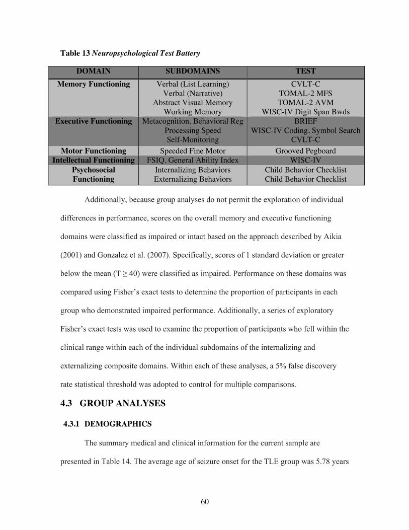
60
Table 13 Neuropsychological Test Battery
DOMAIN SUBDOMAINS TEST Memory Functioning Verbal (List Learning)
Verbal (Narrative) Abstract Visual Memory
Working Memory
CVLT-C
TOMAL-2 MFS TOMAL-2 AVM
WISC-IV Digit Span Bwds Executive Functioning Metacognition, Behavioral Reg
Processing Speed Self-Monitoring
BRIEF
WISC-IV Coding, Symbol Search CVLT-C
Motor Functioning Speeded Fine Motor Grooved Pegboard Intellectual Functioning FSIQ, General Ability Index WISC-IV
Psychosocial Functioning
Internalizing Behaviors Externalizing Behaviors
Child Behavior Checklist Child Behavior Checklist
Additionally, because group analyses do not permit the exploration of individual
differences in performance, scores on the overall memory and executive functioning
domains were classified as impaired or intact based on the approach described by Aikia
(2001) and Gonzalez et al. (2007). Specifically, scores of 1 standard deviation or greater
below the mean (T ≥ 40) were classified as impaired. Performance on these domains was
compared using Fisher’s exact tests to determine the proportion of participants in each
group who demonstrated impaired performance. Additionally, a series of exploratory
Fisher’s exact tests was used to examine the proportion of participants who fell within the
clinical range within each of the individual subdomains of the internalizing and
externalizing composite domains. Within each of these analyses, a 5% false discovery
rate statistical threshold was adopted to control for multiple comparisons.
4.3 GROUP ANALYSES
4.3.1 DEMOGRAPHICS
The summary medical and clinical information for the current sample are
presented in Table 14. The average age of seizure onset for the TLE group was 5.78 years

61
(SD = 4.88 years), while the average age for the FLE group was 7.65 years (SD = 3.94
years). The average duration of seizure activity was 6.70 years (SD = 4.79 years) for the
TLE group and 3.65 years (SD = 2.87 years) for the FLE group. Within the TLE group,
48.1% (n = 13) of participants’ seizures originated within the right hemisphere of the
brain and 51.9% (n = 14) in the left hemisphere of the brain. Within the FLE group, 50%
(n = 12) of participants’ seizures originated within the right hemisphere of the brain and
50% (n = 12) in the left hemisphere of the brain. Of the TLE group, six patients
demonstrated structural lesions on neuroimaging, whereas the remaining 18 TLE patients
exhibited no identifiable structural lesion. Lesions included tuberous sclerosis (n = 2),
tumor (n=1) cavernous malformation (n = 1) and cystic lesion (n = 2). Of the FLE group,
three patients had observable structural lesions on neuroimaging, while the remaining 25
FLE patients exhibited no identifiable structural lesion. Lesions included cortical
dysplasia (n = 2) and venous anomaly (n = 1).
Following procedures used by Bailet and Turk (2000), Bulteau and colleagues
(2000), Smith, Elliot, and Lach (2002), seizure frequency was classified on an ordinal
scale (see Table 14). Within the TLE group, 33.3% (n = 9) experienced seizures weekly
while approximately 29.6% (n = 8) experienced seizures daily, 14.8% (n = 4)
experienced seizures monthly, 7.4% (n = 2) experienced seizures quarterly and 7.4% (n =
2) experienced seizures yearly. Within the FLE group, 33.3% (n = 8) experienced
seizures occurring monthly, 33.3% (n = 8) experienced seizures daily, 12.5% (n = 3)
experienced seizures weekly, 12.5% (n = 3) experienced seizures quarterly and 4.2% (n =
1) experienced seizures yearly. 7.4% of participants with TLE (n = 2) and 4.2% of
participants with FLE (n = 1) were not experiencing seizures at the time of testing. 25.9%
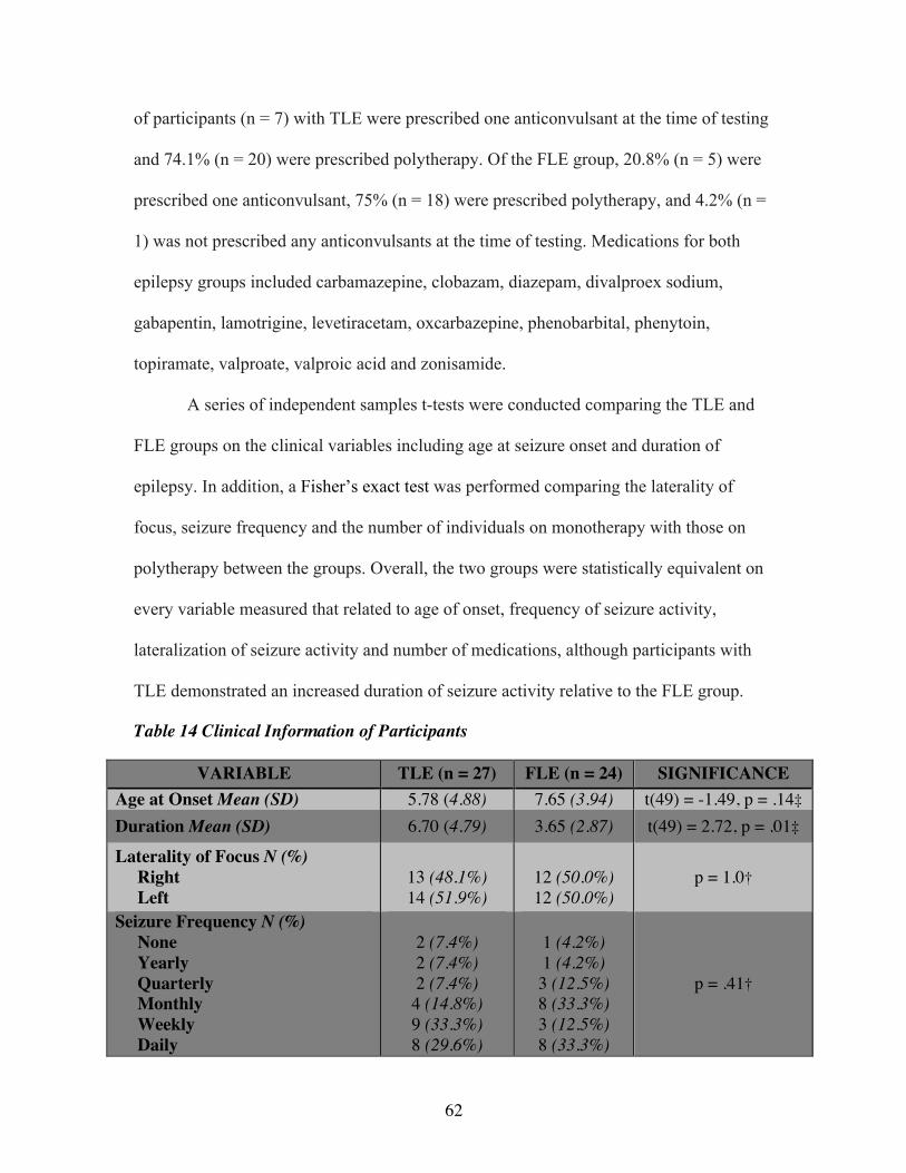
62
of participants (n = 7) with TLE were prescribed one anticonvulsant at the time of testing
and 74.1% (n = 20) were prescribed polytherapy. Of the FLE group, 20.8% (n = 5) were
prescribed one anticonvulsant, 75% (n = 18) were prescribed polytherapy, and 4.2% (n =
1) was not prescribed any anticonvulsants at the time of testing. Medications for both
epilepsy groups included carbamazepine, clobazam, diazepam, divalproex sodium,
gabapentin, lamotrigine, levetiracetam, oxcarbazepine, phenobarbital, phenytoin,
topiramate, valproate, valproic acid and zonisamide.
A series of independent samples t-tests were conducted comparing the TLE and
FLE groups on the clinical variables including age at seizure onset and duration of
epilepsy. In addition, a Fisher’s exact test was performed comparing the laterality of
focus, seizure frequency and the number of individuals on monotherapy with those on
polytherapy between the groups. Overall, the two groups were statistically equivalent on
every variable measured that related to age of onset, frequency of seizure activity,
lateralization of seizure activity and number of medications, although participants with
TLE demonstrated an increased duration of seizure activity relative to the FLE group.
Table 14 Clinical Information of Participants
VARIABLE TLE (n = 27) FLE (n = 24) SIGNIFICANCE Age at Onset Mean (SD) 5.78 (4.88) 7.65 (3.94) t(49) = -1.49, p = .14‡ Duration Mean (SD) 6.70 (4.79) 3.65 (2.87) t(49) = 2.72, p = .01‡
Laterality of Focus N (%) Right Left
13 (48.1%) 14 (51.9%)
12 (50.0%) 12 (50.0%)
p = 1.0† ���
Seizure Frequency N (%) None Yearly Quarterly Monthly Weekly Daily
2 (7.4%) 2 (7.4%) 2 (7.4%) 4 (14.8%) 9 (33.3%) 8 (29.6%)
1 (4.2%) 1 (4.2%) 3 (12.5%) 8 (33.3%) 3 (12.5%) 8 (33.3%)
p = .41† ���

63
Number of AEDs N (%) No Current Therapy Monotherapy Polytherapy
0 (0.0%) 7 (25.9%) 20 (74.1%)
1 (4.2%) 5 (20.8%) 18 (75.0%)
p = .74† ���
† ��� Fisher’s exact test, ‡ Independent samples t-test
Additionally, a series of exploratory independent sample t-tests initially were
conducted to compare participants with a left-hemisphere seizure focus with participants
with a right-lateralized seizure focus within the TLE and FLE groups to determine the
impact of laterality of seizure focus on performance on the memory tasks. As the
cognitive performances of children with right and left focus were not significantly
different (see Table 15), all right- and left-lateralized participants were grouped together
within the FLE and TLE groups.
Table 15 Comparison Between Right- and Left-Hemisphere
TLE FLE Right-Hem. vs. Left-Hem. Right-Side vs. Left-Side
CVLT-C List A Total Recall t(12) = -.86, p = .41 t(12) = -.46, p = .65 CVLT-C Immediate Free Recall t(12) = -.99, p = .34 t(12) = .19, p = .85 CVLT-C Delayed Free Recall t(12) = -1.11, p = .29 t(12) = -.36, p = .73
TOMAL-2 MFS Immediate Recall t(12) = -.98, p = .35 t(12) = -.10, p = .92 TOMAL-2 MFS Delayed Recall t(12) = -.89, p = .39 t(12) = .35, p = .73
TOMAL-2 AVM Immediate Recall t(12) = -1.34, p = .21 t(12) = -.65, p = .53
4.3.2 CORRELATIONS
A correlation matrix was generated to examine whether any of the effects of
demographics and epilepsy-related variables (including age at testing, age at onset of
seizure activity, duration of seizures, frequency of seizures and number of AEDs) were
related to neuropsychological performance within each patient group. Pearson
correlations were used for normally distributed variables, and Spearman correlations
were used for non-normally distributed variables. The correlation matrix did not reveal
any significant relationships between age at seizure onset, number of anticonvulsant

64
medications, or illness duration and performance on the cognitive and behavioral
outcome measures. However, number of prescribed AEDS was positively correlated with
increased ratings of family stress within the FLE group.
Table 16 Correlation Matrix
Age at Testing Age at Onset Duration TLE FLE TLE FLE TLE FLE CVLT-C Total Recall List A -.18 .05 -.00 .15 -.12 -.15 CVLT-C Immediate Free Recall -.29 .21 .07 .10 -.27 .12 CVLT-C Delayed Free Recall -.27 .20 -.24 .20 .06 -.03 TOMAL-2 MFS Immediate Recall -.35 .26 -.02 .18 -.22 .06 TOMAL-2 MFS Delayed Recall -.19 .16 .06 .05 -.18 .13 TOMAL-2 AVM Immediate Recall -.08 .25 .03 .30 -.08 -.11 BRIEF Metacognition .18 .06 .12 .09 .01 -.05 BRIEF Behavioral Regulation .18 .13 .12 .09 -.13 -.04 WISC-IV Digit Span Backwards .31 .28 .05 .25 .16 -.02 CVLT-C Intrusions -.13 -.14 -.22 -.18 .14 .08 WISC-IV Symbol Search .00 .22 -.02 .02 .03 .24 WISC-IV Coding -.36 .21 -.19 .22 -.05 -.05 WISC-IV GAI -.08 .01 .06 .06 -.11 -.07 WISC-IV FSIQ -.23 -.12 .04 -.21 -.24 .14 CBCL Internalizing .37 .07 .01 .14 .11 -.12 CBCL Externalizing .18 .23 .20 .10 .04 .15 Family Stress -.02† ��� -.27† ��� -.20† ��� -.18† ��� .18 .12 Seizure Frequency Number of AEDS TLE FLE TLE FLE CVLT-C Total Recall List A -.33† ��� .05† ��� .12 -.18 CVLT-C Immediate Free Recall -.08† ��� .21† ��� -.23 -.23 CVLT-C Delayed Free Recall -.25† ��� .20† ��� -.14 -.14 TOMAL-2 MFS Immediate Recall -.17† ��� .26† ��� -.23 .01 TOMAL-2 MFS Delayed Recall -.22† ��� .16† ��� -.22 .16 TOMAL-2 AVM Immediate Recall -.19† ��� .25† ��� -.13 .08 BRIEF Metacognition .12† ��� .37† ��� .11 .11 BRIEF Behavioral Regulation -.07† ��� .13† ��� -.03 -.03 WISC-IV Digit Span Backwards -.01† ��� .04† ��� -.11 -.28 CVLT-C Intrusions -.10† ��� -.14† ��� -.05 -.05 WISC-IV Symbol Search -.16† ��� .18† ��� -.10 -.10 WISC-IV Coding -.25† ��� -.02† ��� -.34 -.34 WISC-IV GAI -.30† ��� .03† ��� -.16 -.16 WISC-IV FSIQ -.21† ��� -.02† ��� -.24 -.24 CBCL Internalizing -.24† ��� -.17† ��� -.10 -.08

65
CBCL Externalizing -.00† ��� -.07† ��� .02 -.21 Family Stress -.11† ��� -.12† ��� -.07† ��� .62† ���* † ��� Spearman correlation coefficient * Correlation significant at the .05 level (2-tailed) 4.3.3 MEMORY FUNCTIONING
The first goal was to examine whether children with TLE and FLE demonstrate
significant impairments in memory functioning. Based on the literature, children with
TLE were expected to exhibit impairment in immediate and delayed verbal narrative
memory, immediate and delayed verbal list learning, immediate abstract visual memory
and an increased rate of forgetting of information relative to normative values and to
children with FLE. By contrast, it was expected that children with FLE would perform
within normal limits on measures of memory functioning.
Table 17 MEMORY FUNCTIONING: Comparison to Population Norms
MEASURE TLE MEAN (SD) FLE MEAN (SD) CVLT-C Total Recall List A 38.91 (8.11)
t(26) = -7.10, p = .000*** 39.46 (12.71)
t(23) = -4.06, p = .000*** CVLT-C Immediate Free Recall 36.37 (13.69)
t(26) = -5.17, p = .000*** 38.58 (14.23)
t(23) = -3.93, p = .000*** CVLT-C Delayed Free Recall 38.89 (14.50)
t(26) = -3.98, p = .000*** 38.03 (13.56)
t(23) = -4.32, p = .000*** TOMAL-2 MFS Immediate Recall 36.37 (13.69)
t(26) = -5.17, p = .000*** 38.58 (14.23)
t(23) = -3.93, p = .000*** TOMAL-2 MFS Delayed Recall 40.70 (10.66)
t(26) = -4.53, p = .000*** 40.22 (7.45)
t(23) = -6.43, p = .000*** TOMAL-2 AVM Immediate Recall 39.54 (10.16)
t(26) = -5.35, p = .000*** 44.98 (10.11)
t(23) = -2.43, p = .020 * Significant, p<.05, ** Significant, p<.01, *** Significant, p<.001

66
Figure 2 MEMORY FUNCTIONING: Means
As Table 17 indicates, one-sample t-tests demonstrated that children with TLE
fell significantly below the normative mean on all six of the verbal memory measures.
Additionally, overall means for the children that children with FLE fell significantly
below normative values, although after adjusting for multiple comparisons, the finding
for the FLE group on TOMAL-2 AVM was no longer significant.
Table 18 MEMORY FUNCTIONING: Comparison Between Groups
MEASURE TLE MEAN (SD) FLE MEAN (SD) Memory Composite 38.82 (10.23) 39.98 (8.68)
MEASURE TLE vs. FLE Effect Size
Memory Composite t(49) = -.43, p = .67 d = .12 CVLT-C Immediate Free Recall t(49) = -.57, p = .57 d = .16
CVLT-C Delayed Free Recall t(49) = .21, p = .84 d = .06 CVLT-C Total Recall List A t(49) = -.19, p = .85 d = .05
TOMAL-2 MFS Immediate Recall t(49) = -.66, p = .51 d = .06 TOMAL-2 MFS Delayed Recall t(49) = .19 p = .85 d = .05
TOMAL-2 AVM Immediate Recall t(49) = -1.91, p = .06 d = .54 Additionally, the various neuropsychological measures were grouped into overall

67
memory composite (see Table 18). An individual samples t-test comparing performance
between children with FLE and TLE on the overall memory composite was non-
significant. Follow-up analyses comparing performance between the groups on the
individual subtests were also non-significant, although a trend was noted for children
with TLE to demonstrate worse performance on the TOMAL-2 Abstract Visual Memory
task relative to children with FLE.
Table 19 MEMORY FUNCTIONING: Proportions
MEASURE TLE vs. FLE CVLT-C Forgetting Proportion t(44) = -.24, p = .69
TOMAL-2 MFS Forgetting Proportion t(44) = .00, p = .06 Independent samples t-tests were also used to explore differences in the
proportion of information lost at delay (see Table 19). The results of this analysis
indicated that there were no significant differences between groups with respect to loss of
information, although a trend was noted for children with TLE to demonstrate greater
forgetting on the TOMAL-2 Memory For Stories task relative to children with TLE.
4.3.4 EXECUTIVE FUNCTIONING
The second goal of this project was to examine whether there were significant
differences in executive functioning performance between participants with TLE and
FLE. It was hypothesized that children with FLE would display significant impairments
on laboratory measures of processing speed and working memory and in aspects of
executive functioning including metacognition and behavioral regulation on a parent-
report measure relative to normative standards and to participants with TLE. In contrast,
children with TLE were expected to demonstrate intact levels of executive functioning.

68
Table 20 EXECUTIVE FUNCTIONING: Comparison to Population Norms
MEASURE TLE MEAN (SD) FLE MEAN (SD) BRIEF Metacognition 38.06 (10.80)
t(26) = -5.75, p = .000*** 36.24 (9.64)
t(23) = -6.99, p = .000*** BRIEF Behavioral Regulation 40.63 (13.44)
t(26) = -3.56, p = .002** 42.67 (11.82)
t(23) = -3.04, p = .006** WISC-IV Digit Span Backwards 33.89 (6.66)
t(26) = -12.57, p = .000*** 32.53 (9.13)
t(23) = -9.37, p = .000*** CVLT-C Intrusions 45.56 (16.89)
t(26) = -1.37, p = .183 49.66 (11.66)
t(23) = -.14, p = .88 WISC-IV Symbol Search 36.19 (11.73)
t(26) = -6.12 p = .000*** 35.68 (10.74)
t(23) = -6.53, p = .000*** WISC-IV Coding 32.03 (8.72)
t(26) = -10.71, p = .000*** 30.73 (8.65)
t(23) = -10.92, p = .000*** * Significant, p<.05, ** Significant, p<.01, *** Significant, p<.001
Figure 3 EXECUTIVE FUNCTIONING: Means
Table 20 displays the group means for the six measures of executive functioning
from the subscales of the BRIEF, CVLT-C and WISC-IV. Overall means for both
children with TLE and FLE fell significantly below the normative mean on all of the
individual measures, with the exception of the CVLT-C Intrusions subscale.
0
10
20
30
40
50
60
BRIEF Beh. Regulation
BRIEF Metacognition
WISC-‐IV Digit Span BWds
WISC-‐IV Coding
WISC-‐IV Symbol Search
CVLT-‐C Intrusions
T-‐SCORE VALUES
EXECUTIVE FUNCTIONING
TLE
FLE

69
Table 21 EXECUTIVE FUNCTIONING: Comparison Between Groups
MEASURE TLE MEAN (SD) FLE MEAN (SD) Executive Composite 38.82 (10.23) 39.98 (8.68)
MEASURE TLE vs. FLE Effect Size
EF Composite t(50) = .22, p = .83 d = .12 BRIEF Metacognition t(50) = .74, p = .46 d = .18
BRIEF Behavioral Regulation t(50) = -.62, p = .54 d = .16 WISC-IV Digit Span Backwards t(50) = .24, p = .45 d = .17
CVLT-C Intrusions t(50) = -.98, p = .34 d = .28 WISC-IV Symbol Search t(50) = .09, p = 84 d = .05
WISC-IV Coding t(50) = .78, p = .44 d = .15 Additionally, the various neuropsychological measures were grouped into overall
executive functioning composite (see Table 21). No significant differences were found
between the two epilepsy groups on the overall composite measure or on any of the
individual subscales.
4.3.5 MOTOR FUNCTIONING
The third goal was to examine whether there were significant differences in motor
functioning between participants with temporal and frontal lobe epilepsy. Children with
FLE were expected to exhibit more significant impairment in motor coordination and
speed relative to normative values and to children with FLE. It was expected that children
with TLE would perform within the normal limits.
Table 22 MOTOR FUNCTIONING: Comparison to Population Norms
MEASURE TLE MEAN (SD) FLE MEAN (SD) Grooved Pegboard Dom. 28.67 (15.14)
t(26) = -7.32, p = .000*** 31.61 (15.95)
t(23) = -5.65, p = .000*** Grooved Pegboard Non-Dom. 27.76 (15.42)
t(26) = -7.49, p = .000*** 30.25 (13.98)
t(23) = -6.92, p = .000*** * Significant, p<.05, ** Significant, p<.01, *** Significant, p<.001

70
FIGURE 4 MOTOR FUNCTIONING: Means
One sample t-tests revealed that both children with FLE and TLE scored below
expected levels of functioning on tests of fine-motor coordination bilaterally when
compared to available normative data (see Table 22).
Table 23 MOTOR FUNCTIONING: Comparison Between Groups
MEASURE TLE vs. FLE Effect Size Grooved Pegboard Dom. t(49) = -.68, p = .50 d = .19
Grooved Pegboard Non-Dom. t(49) = -.60, p = .55 d = .17
Independent samples t-tests directly comparing the FLE and TLE groups were
non-significant (see Table 23).
4.3.6 INTELLECTUAL FUNCTIONING
The fourth goal was to examine whether there were significant differences in
intellectual functioning between participants with TLE and FLE. Children with FLE were
expected to exhibit more significant impairment in intellectual functioning relative to
normative standards and to children with FLE, while children with TLE were expected to
have estimates of intellectual functioning in the normal range.

71
Table 24 INTELLECTUAL FUNCTIONING: Comparison to Population Norms
MEASURE TLE MEAN (SD) FLE MEAN (SD) WISC-IV FSIQ 32.14 (10.73)
t(26) = -8.64, p = .000*** 33.95 (7.35)
t(23) = -10.68, p = .000*** WISC-IV GAI 34.86 (12.24)
t(26) = -6.42, p = .000*** 38.86 (9.25)
t(23) = -5.90, p = .000*** * Significant, p<.05, ** Significant, p<.01, *** Significant, p<.001
Figure 5 INTELLECTUAL FUNCTIONING: Means
Table 24 displays the group means for the two composite scores of intellectual
functioning from the WISC-IV. One sample t-tests revealed that overall means for both
groups of children fell significantly below the normative mean for both the FSIQ and
GAI estimates.
Table 25 INTELLECTUAL FUNCTIONING: Comparison Between Groups
MEASURE TLE vs. FLE Effect Size WISC-IV FSIQ t(49) = -.69, p = .49 d = .20 WISC-IV GAI t(49) = -1.30, p = .20 d = .37
When directly compared with one another, a series of independent samples t-tests
did not yield a significant difference between the groups for either FSIQ or GAI (see
Table 25).

72
4.3.7 PSYCHOSOCIAL FUNCTIONING
The second objective of the study was to examine the behavioral profiles of
children with FLE and TLE. It was hypothesized that participants with TLE would
display significantly higher scores on a scale of internalizing behavior problems, while
participants with FLE would demonstrate significantly higher scores on a scale of
externalizing behavior problems.
Table 26 PSYCHOSOCIAL FUNCTIONING: Comparison to Population Norms
MEASURE TLE MEAN (SD) FLE MEAN (SD) CBCL Internalizing Behaviors 41.50 (10.14)
t(26) = -4.35, p = .000*** 40.00 (9.33)
t(23) = -5.25, p = .000*** CBCL Externalizing Behaviors 45.93 (9.82)
t(26) = -2.16, p = .041 44.04 (11.83)
t(23) = -2.47, p = .022
MEASURE TLE MEAN (SD) FLE MEAN (SD) Anxious/Depressed 41.84 (8.32)
t(24) = -5.05, p = .000*** 41.50 (8.84)
t(23) = -4.71, p = .000*** Withdrawn/Depressed 40.44 (8.03)
t(24) = -5.95, p = .000*** 37.42 (7.68)
t(23) = -8.02, p = .000*** Somatic Complaints
39.52 (9.11)
t(24) = -7.05, p = .000*** 37.67 (8.57)
t(23) = -7.05, p = .000*** Social Complaints
36.04 (9.10)
t(24) = -7.67, p = .000*** 35.08 (10.45)
t(23) = -7.00, p = .000*** Thought Problems 38.60 (9.02)
t(24) = -6.32, p = .000*** 37.33 (7.96)
t(23) = -3.87, p = .000*** Aggressive Behavior 42.52 (7.81)
t(24) = -4.79, p = . 000*** 40.54 (11.96)
t(23) = -4.49, p = .000*** Rule-Breaking Behavior 45.24 (6.43)
t(24) = -3.70, p = . 001*** 43 (7.64)
t(23) = -8.98, p = .000**** Attention Problems 34.64 (12.54)
t(24) = -6.12, p = . 000*** 30.75 (10.51)
t(23) = -2.16, p = .001*** * Significant, p<.05, ** Significant, p<.01, *** Significant, p<.001

73
FIGURE 6 PSYCHOSOCIAL FUNCTIONING: Means
Within-group differences in behavioral functioning were explored using one-
sample t-test analyses (see Table 26). Both groups demonstrated significant impairment
relative to normative data in internalizing behavior problems and in the individual
subdomains. Both the FLE and TLE participants also were rated below the normative
mean in externalizing behavior problems, although after controlling for multiple
comparisons, this finding was no longer significant.
0
10
20
30
40
50
60
CBCL Internalizing CBCL Externalizing
T-‐SCORE VALUES
PSYCHOSOCIAL FUNCTIONING
TLE
FLE

74
Table 27 PSYCHOSOCIAL FUNCTIONING: Comparison Between Groups
MEASURE TLE vs. FLE Effect Size CBCL Internalizing Behaviors t(49) = .55, p = .59 d = .15 CBCL Externalizing Behaviors t(49) = .62, p = .54 d = .17
Independent samples t-tests also did not yield an overall difference between the
groups for either internalizing or externalizing behavior problems (see Table 27).
4.4 INDIVIDUAL ANALYSES
To examine individual patterns of performance across the various cognitive and
behavioral domains, performance on each of the domains was classified at intact or
impaired and compared. Performance on these composite domains was compared using
Fisher’s exact tests to determine the proportion of participants in each group who had
impaired performance in each domain. Additionally, given that the past research
involving psychosocial/behavioral outcomes of children with FLE and TLE has been
equivocal, a series of exploratory Fisher’s exact tests were used to examine the
proportion of participants who fell within the clinical range on each of the individual
subscales of the internalizing and externalizing composite domains (see Table 28). It was
hypothesized that a higher proportion of participants with TLE would present with scores
that fall in the impaired range on domains assessing hypothesized “temporal-lobe”
functions (memory functioning, internalizing behaviors), while a higher proportion of
participants with FLE would present with scores in the impaired range on domains
assessing hypothesized “frontal-lobe” functions (executive functioning, externalizing
behavior).

75
Table 28 Secondary Analyses
Children with TLE Children with FLE P Task Intact (%) Impaired (%) Intact (%) Impaired (%) -
Memory 12 (44.4%) 15 (55.6%) 11 (45.8%) 13 (54.2%) .57 EF 11 (40.7%) 16 (59.3%) 11 (45.8%) 13 (54.2%) .47
Internalizing 17 (63%) 10 (37%) 12 (50%) 12 (50%) .26 Externalizing 17 (68%) 8 (32%) 14 (58.3%) 10 (41.7%) .34
TLE FLE P
Task Intact (%) Impaired (%) Intact (%) Impaired (%) - Anxious/Depressed 14 (56%) 11 (44%) 15 (62.5%) 9 (49 %) .77
Withdrawn/Depressed 12 (48%) 13 (52%) 8 (33.3%) 16 (66.7%) .23 Somatic Complaints 13 (52%) 12 (48%) 7 (29.2%) 17 (70.8%) .09 Aggressive Behavior 16 (64%) 9 (36%) 15 (62.5%) 9 (37.5%) .57
Rule-Breaking 20 (80%) 5 (20%) 16 (66.7%) 8 (33.3%) .23 Attention Problems 8 (32%) 17 (68%) 6 (25%) 18 (75%) .41 Thought Problems 11 (44%) 14 (56%) 8 (33.3%) 16 (66.7%) .32
Social Problems 8 (32%) 17 (68%) 7 (29.2%) 17 (70.8%) .54 These analyses revealed that the frequency of significantly impaired performance
in the two patient groups was comparable, with the Fisher’s exact tests demonstrating no
group differences on any of the subscales.
4.5 ADDITIONAL ANALYSES
Due to the fact that duration of seizure activity was significantly different between
groups, the analyses were subsequently rerun using duration as a covariate. However, this
did not affect the pattern of the results. Similarly, due to concerns that performance on
the cognitive measures could be affected by level of intelligence, the analyses were also
rerun utilizing intelligence as a covariate, although this also did not significantly change
the results of the analyses.

76
5 DISCUSSION
5.1 SUMMARY
A major limitation of the extant literature on childhood-onset epilepsy is the
paucity of studies that have directly compared performance between individuals with
localization-related syndromes. Additionally, there are few reports utilizing broad,
comprehensive assessments of functioning spanning multiple cognitive domains
(Williams et al., 1998). Moreover, only a very limited number of studies have specifically
evaluated children with medically-refractory epilepsy, despite indications that
approximately 30% of patients with epilepsy have inadequate seizure control with drug
therapy (Kwan & Brodie, 2000). Consequently, this study examined the cognitive and
behavioral profiles of children with frontal or temporal lobe seizure disorders, with onset
occurring during formative periods of neurodevelopment when regions of the brain are
being sculpted by synaptic proliferation and axonal myelination, as well as subtractive
processes intended to eliminate ineffective circuitry and refine both structure and
function.
5.2 MEMORY FUNCTIONING
5.2.1 TEMPORAL LOBE EPILEPSY
The results indicated that, as expected, participants with TLE demonstrated
significant impairment relative to normative values in various aspects of memory
functioning. Story recall has been proposed to be a sensitive indicator of temporal lobe
dysfunction in adults with TLE (Frisk & Milner, 1990). Children with TLE in the current
study were found to perform in the low average range on measures of immediate and

77
delayed recall of narrative information. This is consistent with other studies which have
compared children with TLE to typically-developing controls (Gascoigne et al., 2014;
Guimaräes et al., 2007; Jambaqué et al., 1993; Jambaqué et al., 2009; Nolan et al., 2004;
Rzezak et al., 2011; Rzezak et al., 2012). It has been proposed that this characteristic
pattern of impairment on measures of narrative memory may emerge due to a failure in
the consolidation of the memory trace from short-term memory to long-term storage
(Loiseau et al., 1982, as cited in Jambaqué et al., 1993).
A similar profile was observed in the short and long delay recall conditions of the
CVLT-C. This is also consistent with previous studies utilizing verbal list-learning
paradigms in children with TLE (Hernandez et al., 2003; Jambaqué et al., 1993; Rzezak
et al., 2012). In addition, children with TLE in the present study demonstrated poor
memory efficiency during an initial learning phase on the CVLT-C, which has also been
noted in other studies (Hernandez et al., 2003; Nolan et al., 2004). It has been suggested
that this impaired initial encoding of information may result in the decreased performance
on delayed recall (Jambaqué et al., 1993).
Helmstaedter, Wietzke and Lutz (2009) demonstrated that CVLT scores
differentially load on to factors reflecting “learning efficiency” and “long-term memory,”
suggesting that this particular memory measure may be uniquely sensitive to different
aspects of epilepsy-related impairment. This finding highlights the importance of
incorporating list-learning measures into standard batteries of memory functioning. It has
been proposed that these factors may map on to anatomically-differentiable memory
subsystems. Based on surgical outcome data with adults, Helmstaedter and colleagues
(1997) suggested that processes of learning and short-delay memory may be subserved by

78
temporolateral structures, while the temporomesial structures may be functionally
specialized for processes related to long-term consolidation. The findings in the current
study suggest that both aspects can be disrupted in children with TLE. This pattern could
imply that the sample was comprised of mixed subgroups of children with temporolateral
or temporomesial pathology; however, it is often difficult to differentiate this in children
(see Strengths and Limitations). Alternatively, this pattern may indicate that memory
subsystems are not highly differentiated in children due to anomalies of network
development related to the presence of seizure activity in the developing brain.
It is also notable that the children with TLE did not display a lateralized pattern of
memory impairment on the verbal memory tasks. The literature is inconsistent with
regard to whether laterality effects are present in children with TLE. The present results
are consistent with most previous studies that have examined list-learning and story in
recall in children with TLE (e.g., Bigel & Smith, 2001a; Helmstaedter & Elger, 2009;
Nolan et al., 2004). Specifically, it has been suggested that the presence of seizures in
childhood may disrupt the development of expected patterns of hemispheric-
specialization in children.
The results of the current study also suggest that children with TLE demonstrate
significant impairment in immediate recall on a measure of abstract-visual memory, with
performance in the low average range. Previous studies have also shown significant
impairments in various aspects of memory for abstract designs (Guimaräes et al., 2007;
Jambaqué et al., 1993; Nolan et al., 2004). However, on the whole, impairments in
nonverbal memory have been less consistently described relative to verbal memory (e.g.,
Gascoigne et al., 2014; Rzezak et al., 2012).

79
A notable criticism of previous studies has been the use of visual memory
paradigms utilizing target items that are easy to verbalize (i.e., simple geometric shapes
or pictures of items), which permits dual encoding using compensatory verbal memory
strategies (Helmstaedter et al., 1995). It has been proposed that selective nonverbal
deficits in individuals with right-hemisphere TLE may only emerge when the verbal load
for a particular target surpasses the capacity of compensatory verbal memory due to its
complexity. For example, facial memory has been found to be particularly sensitive to
damage to the right hemisphere (Bigel & Smith, 2001a). Consequently, the robust
impairment in abstract-visual memory demonstrated in the TLE group in the present
study may be related to the fact that the target items on the TOMAL-2 are abstract and
hard-to-verbalize (Schmitt & Decker, 2008), which permitted a more direct and sensitive
assessment of right temporal functioning (Helmstaedter et al., 1995), or because the
ability to verbally encode visual information is less developed in children (Jambaqué et
al., 1993). The current findings suggest that children with TLE demonstrate impairments
in visual memory functioning regardless of lateralization of seizure activity
(Helmstaedter et al., 1995). This may relate to the better sensitivity of the TOMAL-2
AVM test relative to other tests such as the WRAML-2, which in some studies show a
pattern of performance in TLE that is no different from controls (Gascoigne et al., 2014).
5.2.2 FRONTAL LOBE EPILEPSY
The results of the current study also disclosed significant impairments in verbal
memory performance in children with FLE. This was evident on tasks of verbal learning
and immediate and delayed story recall relative to normative groups. When performance
between the two groups was directly compared, children with FLE could not be reliably

80
differentiated from children with TLE based on the verbal memory measures (Hernandez
et al., 2003; Jambaqué et al., 1993; Nolan et al., 2004; Sinclair et al., 2004). In contrast,
Culhane-Shelbourne and colleagues (2003) found that children with TLE demonstrated
significantly poorer performance on immediate and delayed story recall than children
with FLE. However, it is notable that participants in that study were not medically-
refractory, suggesting that increased severity of seizure activity may be associated with
more generalized patterns of impairment. However, the findings in the current study are
consistent with emerging literature suggesting that difficulties in verbal memory are not
restricted to children with TLE (Hernandez et al., 2003; Nolan et al., 2004; Picard et al.,
2009).
Performance on the abstract visual memory task was also decreased in the current
study relative to the normative value, although when the significance values were
adjusted to account for the multiple analyses, this finding was no longer statistically
significant. Findings have been more inconsistent in the literature regarding the patterns
of visual memory deficits in individuals with FLE. Specifically, some studies have
suggested that children with FLE do not display significant impairments on tasks of
picture or design memory, which may be due to the more basic visual stimuli utilized on
the WRAML-2 (Culhane-Shelbourne et al., 2002). In contrast, children with FLE have
been found to be impaired on tasks of visuospatial ordering (Nolan et al., 2004) and on
the Rey-Osterrieth Complex Figure test, although it can be argued that this finding may
have been confounded by the greater graphomotor demands of the task (Hernandez et al.,
2003). Interestingly, there was a trend towards significantly poorer performance in the
TLE group relative to the FLE group on the measure of abstract visual memory.
81
Specifically, the TLE group performance was in the low average range, while the FLE
demonstrated performance in the average range, suggesting that the TOMAL-2 Abstract
Visual Memory subtest may be more sensitive to differences in memory recall between
FLE and TLE. Additionally, no significant differences were demonstrated in the current
study between children with right- and left-sided FLE, which is consistent with a
previous study (Nolan et al., 2004).
5.2.3 SUMMARY: FLE VS. TLE
Results of the current study found similar patterns of memory functioning in
children with TLE and FLE. It has been proposed that these similar patterns of memory
impairment in children with TLE and FLE may represent a common outcome associated
with different underlying mechanisms that depend on the area of the brain impacted by
the seizure activity (McDonald, Delis, Norman, Tecoma, & Iragui-Madozi, 2005).
Specifically, it has been suggested that memory problems may arise in patients with TLE
due to a disruption in encoding or the consolidation of new information into long-term
storage, whereas memory impairments in individuals with FLE may be related to
difficulties with attention, self-monitoring and effective strategization, which in turn can
impede encoding and retrieval of information (Centeno et al., 2010; Hernandez et al.,
2003; Turner et al., 2007). This is consistent with findings that children with FLE have
been noted to have more marked difficulties with attention and strategization during
memory recall (Hernandez et al., 2003).
5.3 EXECUTIVE FUNCTIONING
5.3.1 FRONTAL LOBE EPILEPSY
As expected, participants with FLE demonstrated significant impairment across

82
most of the measures of executive functioning used in this study. As a group, the children
with FLE performed in the borderline range on a laboratory measure of processing speed.
This is consistent with observations initially put forth in a case study by Jambaqué and
Dulac (1989) and subsequently replicated in a series of studies completed by Auclair and
colleagues (2005), Braakman and colleagues (2012), Bulteau and colleagues (2000),
Hernandez and colleagues (2003) and Longo and colleagues (2013).
Participants in the FLE group also performed significantly below reference values
(borderline range) on a measure of working memory. A similar degree of impairment on
digit span tasks has been described both in adults (Rai et al., 2015; Zamarian et al., 2011)
and children with FLE (Braakman et al., 2012; Longo et al., 2013). The current findings
suggest that these subtests of the WISC-IV are sensitive to frontal lobe dysfunction,
although they are not necessarily measures that are specific to frontal lobe impairment.
Specifically, it has been suggested that other patient populations without focal
dysfunction of the frontal lobes have been found to display difficulties with the
backwards span and information processing tasks, including multiple sclerosis (Feuillet et
al., 2007) and specific language impairment (Baddeley, Gathercole, & Papagno, 1998).
Consequently, it has been suggested that performance on these tasks can be affected by
impairments in language and regulatory systems that allocate attentional resources during
complex tasks or with disruption of white matter in other areas of the brain (Jung &
Haier, 2007).
The children in the current study were also rated to be in the low average range in
behavioral regulation and the borderline range in metacognitive abilities on a parent-
report measure of executive functioning. These findings of impairment on subjective

83
measures of executive functioning are consistent with previous reports within the child
literature (Campiglia et al., 2014; Luton et al., 2010; MacAllister et al., 2011). However,
subjective rating scales are not consistently embedded into study protocols assessing
executive functioning, which is problematic given concerns that many laboratory tests of
executive functioning lack ecological validity and may not be appropriate for assessing
frontal lobe functions in children (MacAllister et al., 2012; Upton & Thompson, 1996).
For example, it has been suggested that children with later seizure onset (after age 7) may
perform better when provided with a more structured testing environment with fewer
distractions than when in their home or school settings (Luton et al., 2010).
Consequently, the results of the current study highlight the importance of assessing
executive functioning performance using both quantitative and qualitative methods.
Interestingly, children with FLE did not present with significant impairment on a
measure of self-monitoring. Baldo and colleagues (2002) found that adults with frontal
lobe lesions made more intrusions in their recall than typical controls. Additionally, two
previous studies involving children with FLE observed an increased number of intrusions
on a list-learning task relative to normative data (Hernandez et al., 2003; Riva et al.,
2002). However, it is important to note that the study completed by Hernandez and
colleagues was limited by relatively small and discrepant sample sizes (16 FLE patients
compared with 8 TLE patients). The study by Riva and colleagues was also flawed;
specifically, these investigators acknowledged that the high mean number of intrusive
errors was mainly attributable to one participant that they failed to remove from analyses.
In contrast, a subsequent study completed by Riva and colleagues in 2005 found no
significant elevations in number of intrusions in their FLE sample. The CVLT-C

84
Intrusions process index has been theoretically linked to the construct of executive
function. However, Beebe, Ris and Dietrich (2000) found that the CVLT-C learning
process indices demonstrate low correlations with other commonly accepted measures of
executive functioning. Consequently, they concluded that the CVLT-C Intrusions process
score may not represent a valid measure of executive functioning. The failure to find
patterns of impairment in the FLE group in the present study on this measure provides
additional evidence that contraindicate the use of this scale to assess frontal lobe
dysfunction.
5.3.2 TEMPORAL LOBE EPILEPSY
Participants with TLE also demonstrated significant impairment across the
various measures of executive functioning. As a group, the children with TLE performed
in the borderline range on the processing speed and working memory tasks. No
significant differences were found when children with FLE and TLE were compared
directly on these measures of executive functioning. This finding of poorer performance
on the processing speed measure is consistent with a previous studies conducted by
Hernandez and colleagues (2003) and Longo and colleagues (2013). Additionally, the
digit span task has been shown to be sensitive to working memory difficulties in groups
of adults with intractable epilepsy (Rai et al., 2015; W. Wang et al., 2011; Zamarian et
al., 2011). However, the finding of poorer performance in the TLE group in the current
study stands in contrast to three previous studies which found no significant differences
between children with TLE and a control group on a measure of working memory
(Guimaräes et al., 2007; Rzezak et al., 2012). It is notable that these previous studies did
not include children with medically-refractory epilepsy, suggesting that intractability of
85
seizure activity may be associated with increased impairment in functioning. Specifically,
the pathological activity associated with chronic seizures has been suggested to disrupt
the normal dynamics of neuronal working memory networks, leading to more significant
functional impairments (Verduzco-Flores, Ermentrout, & Bodner, 2009). Future
explorations of working memory performance in children with TLE should attempt to
compare performance between children with differing levels of epilepsy severity.
The current study also revealed significant impairments on the BRIEF parent-
report for the TLE group, with scores in the low average range on the Behavior
Regulation and Metacognition indices. These impairments could not be reliably
distinguished from the pattern of impairment observed in the FLE group. A limited
number of studies have been utilized the BRIEF to assess executive functioning in
children in heterogeneous epilepsy populations (MacAllister et al., 2012; Parrish et al.,
2007; Sherman et al., 2006). However, these studies have failed to distinguish more
specifically between patients with frontal and extra-frontal localization-related epilepsies.
Only one recent study has specifically utilized a parent-report measure to examine
executive dysfunction in children with TLE, which found that participants displayed a
similar pattern of impairment as their FLE counterparts (Campiglia et al., 2014). This
study provides additional evidence suggesting that the BRIEF is sensitive to executive
dysfunction in individuals with TLE, but that ratings may be globally similar regardless
of the area of seizure focus or the particular EF domain being examined.
Finally, similar to the findings for the FLE group, children with TLE failed to
demonstrate significant difficulties in self-monitoring on the CVLT-C Intrusions
subscale. This is consistent with one previous study which examined performance on this

86
subscale in children with TLE (Hernandez et al., 2003).
5.3.1 SUMMARY: FLE VS. TLE
Results of the present study revealed similar patterns of executive dysfunction in
children with TLE and FLE. While it has become standard practice to make attributions
of impairments on executive measures to frontal lobe dysfunction (Stuss & Alexander,
2000), research is equivocal as to whether executive functions can be reliably and
specifically localized to the frontal lobes (e.g., Miyake et al., 2000; Welsh, 2002). A
review of the existing literature has revealed significant inconsistencies, with certain
studies finding no significant deficits on tasks of executive functioning in individuals
with frontal lobe damage, and others finding that impairments in executive function can
be robust even in individuals with extrafrontal injury (see Alvarez & Emory, 2006). An
alternative view which has emerged in the literature suggests that executive functions
represent “higher-level” cognitive functions which serve to regulate distributed “lower-
level” cognitive processes and behaviors (Alvarez & Emory, 2006; Zelazo, Carter,
Reznick, & Frye, 1997). Within this conceptualization, executive functioning emerges
from interactions between diffuse anatomical and functional brain areas such as the
frontal, temporal and parietal lobes. As a result, executive function can often be impaired
with damage to the diverse areas of the cortex, although in some instances, it may be
maximal with frontal lobe damage. The results from the current study are consistent with
this view, suggesting that focal epileptiform activity within either the frontal or temporal
lobes can be associated with significant patterns of difficulties in various aspects of
executive functioning.

87
5.4 INTELLECTUAL FUNCTIONING
5.4.1 FRONTAL LOBE EPILEPSY
The results from the current analyses indicated that children with FLE performed
significantly below the average on a measure of full-scale intelligence. Specifically, the
mean Full Scale IQ estimate for the FLE group fell in the borderline range of functioning.
This finding of depressed intellectual functioning is broadly consistent with previous
studies (Braakman et al., 2012; Nolan et al., 2003; Prévost et al., 2006; Sinclair et al.,
2004). One major limitation of the pediatric literature has been that the vast majority of
published studies have prohibited the inclusion of children with full scale IQs lower than
70 in their study samples (e.g., Bell et al., 2006; Gleissner et al., 2002; Lopes et al., 2014;
Luton et al., 2010), and even more conservative clinical cut-offs (IQ > 80) have been
used in others (Bailet & Turk, 2000; Culhane-Shelbourne et al., 2002). However, it is
critical to be able to provide documentation of the full range of intellectual performance
in children with intractable epilepsy (Cormack et al., 2007). Additionally, the estimates of
intellectual functioning obtained in the previous studies have been based on
administration of outdated editions of the WISC (e.g., Braakman et al., 2012; Nolan et
al., 2003; Prévost et al., 2006; Sinclair et al., 2004). This practice is problematic, given
that content and structure of the WISC-III and WISC-R is significantly different from the
WISC-IV (Sattler & Dumont, 2004; Strauss et al., 2006), which limits the generalizability
of the findings (Sherman et al., 2012). Consequently, the present study has provided one
of the first unrestricted estimates of intellectual functioning, in a larger sample of children
than has previously been described in the literature.
It is also one of the first studies to specifically assess intellectual performance

88
using an updated version of the WISC, in a group of children that are actively being
considered for epilepsy surgery. Epilepsy has long been recognized to exert negative
effects on cognitive and behavioral development in children. Children with
“uncomplicated epilepsies” may show close to normal cognitive development over time
(Hermann, Seidenberg, & Jones, 2008; Jones, Siddharth, Gurbani, Shields, & Kaplan,
2010). However, a substantial proportion of children (~30% +) demonstrate complicated
forms of epilepsy with increased intractability to medication in which cognitive
development is significantly impacted (Ellenberg, Hertz & Nelson, 1986; Nolan et al.,
2003). Over a quarter of these individuals demonstrate IQ estimates below 80 after
approximately 10 years post-seizure onset (Berg et al. 2008). Rathouz and colleagues
(2014) followed children with new-onset epilepsy for a period of time to evaluate the
effect of epilepsy on their cognitive development. In these relatively complicated cases, it
was apparent that cognitive problems in some cases preceded onset of the epilepsy.
Following onset, epilepsy seemed to exert relatively little to mild effects on cognitive
development. By contrast, a recent longitudinal study by Van Iterson and colleagues
(2014) characterized the influence of epilepsy on intelligence as a continuous downward-
progressing function, where decline was likely to be maximal later in time.
The mean score for the General Ability Index (GAI) for the FLE group was also
significantly below the normative values. Interestingly, a closer examination of the scores
revealed that estimates of intellectual functioning derived from the GAI fell in the low
average range, while estimates based on the FSIQ fell in the borderline range, although
this difference was not statistically different.
It has been reported that working memory and processing speed demonstrate a

89
wide range of correlations with measures of intellectual functioning (see Fry & Hale,
2000). Given indications in the literature of increased difficulties with processing speed
and working memory in children with FLE (Auclair et al., 2005; Braakman et al., 2012;
Bulteau et al., 2000; Hernandez et al., 2003), it was hypothesized that inclusion of
measures of processing speed and working memory in the full scale intellectual
functioning composite could lead to a deflation in the derived estimates for individuals
with FLE. However, the results of the current study suggest that removal of these indices
does not result in meaningful change in the IQ estimate for individuals with frontal lobe
dysfunction. It has been suggested that the frontal cortex may subserve intellectual
abilities due to its role in cognitive control and flexibility (Duncan et al., 2000).
5.4.2 TEMPORAL LOBE EPILEPSY
Children with TLE also demonstrated estimates of full-scale intellectual
functioning that corresponded to the borderline range. This pattern of significantly
decreased performance relative to normative values is consistent with several prior
studies (Guimaräes et al., 2007; Cormack et al., 2007; Hermann et al., 2002a; Hermann et
al., 2002b; Mataro et al., 1998; Nolan et al., 2003). Additionally, when children with FLE
and TLE were directly compared, no significant differences in their level of FSIQ were
found (e.g., Hernandez et al., 2002; Nolan et al., 2003). Culhane-Shelbourne and
colleagues (2002) found that their TLE sample obtained a lower FSIQ relative to their
sample of FLE participants; however, it is notable that their exclusionary criteria limited
the participation of individuals with IQ scores below 70, which likely skewed their
estimate of intellectual functioning.
Results indicated that the IQ estimate from the GAI for the TLE group was also

90
significantly below the normative values. A comparison between the FSIQ and GAI
estimates for the TLE group revealed no significant differences. Additionally, no
significant differences were observed when performance on the GAI for the TLE and
FLE participants were directly compared.
5.4.1 SUMMARY: FLE VS. TLE
The findings from the present study indicate that children with TLE and FLE
perform similarly on measures of intellectual functioning. There have been considerable
efforts made in the last 20 years to identify the neural underpinnings of general
intelligence (Gläscher et al., 2010). While the most consistent findings have associated
intellectual functioning with activations in the prefrontal cortex (Duncan et al., 2000), it
has also been posited that intellectual functioning may instead involve communication
across various distributed cortical regions (Jung & Haier, 2007). One theory that has
emerged in the literature is the Parieto-Frontal Integration Theory (P-FIT), which
suggests that the neural substrates involved in general intelligence include cortical
regions in the frontal, parietal, occipital and temporal association cortices (Jung & Haier,
2007). Interestingly, a recent study conducted using voxel-based lesion symptom
mapping revealed the role of a circumscribed region of the frontal pole, in addition to a
distributed network involving various cortical regions and their white matter connections
(Gläscher et al., 2010). Consequently, the findings in the current study provide additional
evidence that dysfunction in either the frontal or temporal regions can result in significant
impairments in intellectual functioning.

91
5.5 MOTOR FUNCTIONING
5.5.1 FRONTAL LOBE EPILEPSY
As expected, children with FLE displayed significant impairments in motor
coordination and speed on the Grooved Pegboard Test (GPT). Specifically, the
participants performed in the borderline range in both the dominant and non-dominant
hand conditions. This is consistent with the results of a previous study examining motor
functioning in children with FLE (Hernandez et al., 2002). However, a series of studies
completed by Riva and colleagues (2002, 2005) failed to demonstrate significant
differences between their FLE group and normative data during conditions in which the
participants completed a peg placement task, the Purdue Pegboard task (PPBT), during
unimanual conditions. Participants with FLE were only significantly impaired relative to
reference values during an “assembly” condition in which they were required to place
pegs while alternating hands.
Hernandez and colleagues (2002) also noted more significant difficulty on the
bimanual coordination condition of the PPBT in their study of children with FLE. Based
on these observations, the researchers suggested that frontal lobe dysfunction associated
with FLE does not interfere with performance on measures of simple motor speed but
may significantly impact performance on measures of speed when more complex aspects
of motor activity and its coordination are involved (Hernandez et al., 2002; Riva et al.,
2002; Riva et al., 2005).
The PPBT was originally constructed for use with adults and utilized in industrial
and occupational assessments. Consequently, it has been suggested to be less appropriate
for assessing motor coordination in children (Y. Wang et al., 2011). While the GPT does

92
not have a comparable bimanual condition, it is generally considered to be a more
complex motor processing task due to its added requirement of sensory-motor integration
and executive planning (Merker & Podell, 2011; Roy & Square-Storer, 1994). Taken
together with trends observed in the previous studies, the results from the current study
suggest that the GPT may be more sensitive to deficits in psychomotor speed in children
with FLE than the Purdue Pegboard task. Mitrushina, Boone, Razani, and D’Eli (2005)
also noted that the GPT was sensitive to problems in FLE that were not apparent on
simple motor speed tasks such as finger tapping and grip strength.
5.5.2 TEMPORAL LOBE EPILEPSY
As a group, the children with TLE in the present study also scored below
expected levels of functioning on the GPT. Relative performance deficits were apparent
in both left and right hands when compared to available normative data. Very few studies
are available in the literature which have specifically examined motor functioning in
individuals with TLE. The present findings are, however, consistent with one study that
was completed involving adult participants with FLE (Hermann et al., 2007). The results
are also consistent with a previous study which assessed motor coordination and speed in
participants with early-onset epilepsy using the bimanual condition of the PPBT
(Hernandez et al., 2002). As in their study, comparisons between the FLE and TLE did
not reveal significant differences.
5.5.1 SUMMARY: FLE VS. TLE
The results from the present study suggest that children with TLE and FLE
demonstrate similar levels of impairment on a measure of motor coordination and speed.
Various regions have been identified in the frontal lobes that are involved in the planning

93
and execution of motor activity (Rizzolatti & Luppino, 2001). Specifically, individuals
with FLE demonstrate decreased patterns of activation in the frontal lobe of the epileptic
hemisphere during measures of motor functioning (Woodward et al., 2014a; Woodward
et al., 2014b). This correlated with poorer performance relative to typical controls. The
mechanisms contributing to the observed pattern of impairment in the TLE group are less
clear. However, there are anatomical pathways connecting the medial temporal area to
the premotor areas of the frontal cortex (Schwartz, 1994). Consequently, it is conceivable
that epileptiform activity may extend from the temporal lobe to frontal cortex through
this distributed processing pathway and may contribute to neurodevelopmental
differences that result in the difficulties seen in the TLE group during tasks of visuo-
motor processing.
5.6 PSYCHOSOCIAL FUNCTIONING
In the current study, significantly increased internalizing behaviors were noted in
both TLE and FLE groups in comparison to normative values. Additionally, both TLE
and FLE groups were rated as having increased externalizing behaviors, although after
controlling for multiple analyses, these findings were no longer significant. The TLE and
FLE groups did not differ from each other, indicating that both groups show a similar
pattern of deviation in psychosocial functioning. Additionally, a closer examination of
performance on the individual subscales comprising these broader domains revealed a
similarly elevated problem scores across all the subscales. As a group, the children with
TLE and the children with FLE also demonstrated clinical elevations on the thought
problems, somatic problems, attention problems and social adjustment problems
subscales. Taken together, the results of the present study suggest that recurrent difficult

94
to control seizure activity early in childhood is associated with broad difficulties in
psychosocial/emotional functioning.
These findings are generally consistent with the results of other investigations
utilizing the CBCL in children with localization-related epilepsy (LRE). Almane and
colleagues (2013) found similarly broad behavioral problems in children with newly
diagnosed and recent-onset focal epilepsy. As in the present study, their cohort of
children with focal epilepsies exhibited mean scores on these scales that were on average
discrepant by a standard deviation or more in comparison to normative values.
Interestingly, they found similar levels of difficulties in a cohort of children with
idiopathic generalized epilepsy (IGE), although the children with IGE showed less
impairment in social problems compared to their cohort with TLE and FLE.
5.7 IMPLICATIONS Taken together, the findings of the current study argue against the existence of
epilepsy syndrome-specific patterns of neuropsychological and behavioral dysfunction in
individuals with childhood-onset epilepsy (Hermann et al., 2002b). To be more specific,
both the TLE and FLE groups presented with significant impairment across a range of
domains including memory, executive, intellectual, motor and psychosocial functioning.
These results are unexpected given the focal epileptogenic effects typically described in
the adult literature, although they are consistent with a growing body of research
suggesting that recurrent seizure activity can have a critical impact on the course of
programmed developmental processes (Anderson, Spencer-Smith, & Wood, 2011).
Perspectives from early animal and human studies suggested that brain insults
sustained in childhood during a period of maximal brain plasticity could induce

95
significant cerebral reorganization, resulting in better functional recovery than insults
sustained later in development (Kennard, 1936; Kennard, 1940; Woods, 1980). However,
other researchers have regarded early brain injury as a ‘vulnerability’ factor, resulting in
an abnormal trajectory of development, reduced neural resources and more severe
functional deficits than would be expected following comparable injuries sustained in
later childhood or adulthood (Anderson et al., 2011; Giza & Prins, 2006; Hebb, 1947;
Hebb, 1949; Jacobs, Harvey, & Anderson, 2007). Consequently, the current study is
consistent with theoretical perspectives suggesting that damage to the immature brain can
result in a generalized pattern of cognitive and behavioral dysfunction and generally
poorer functional recovery (Anderson et al., 2011).
The observation that children with seemingly diverse areas of seizure onset can
produce similar cognitive and behavioral profiles provides additional evidence suggesting
that the presence of recurrent seizure activity during early brain maturation disrupts the
process of regional and hemispheric specialization that typically occurs across early
childhood (Williams et al., 1998). Specifically, the findings of impairments in so-called
“frontal lobe functions” in children with TLE and so-called “temporal lobe functions” in
children with FLE suggests that localizationism may not represent a complete or
sufficient account of functional organization in the brain (Stevens, 2009).
Neuroimaging studies have increasingly demonstrated that the human cerebral
cortex is organized into complex networks of neurons connecting distinct areas of the
brain that communicate with each other in a functionally specific manner (Schmidt,
Petkov, Richardson, & Terry, 2014). Over the course of ontogeny, circuits of neurons
develop greater domain-specificity through the strengthening or reorganizing of
96
frequently engaged neural connections (‘Hebbian’ connectivity) and the elimination of
extraneous synapses or connections (Stevens, 2009; Tau & Peterson, 2010). Long-
distance structural connectivity appears to emerge due to the synchronous activity of
these networks of neurons (Varela, Lachaux, Rodriguez, & Martinerie, 2001). It has been
suggested that most cognitive processes are not completely localized to a discrete brain
region, but instead are mediated by the functional integration of the widely-distributed
neuronal assemblies that comprise these neural networks (Burianova et al., 2010;
Stevens, 2009).
The resulting neural networks form the structural substrate for distributed
interactions among specialized brain systems for highly complex cognitive functions such
as memory, EF and intelligence (Bola & Sabol, 2015; Hagmann et al., 2008; Stam, 2010).
For example, participants commonly demonstrate increases in activity in the hippocampal
region across various tasks of memory functioning (Burianova & Grady, 2007; Burianova
et al., 2010). However, whole-brain analyses have additionally revealed concomitant
changes in activation in various temporal areas including the middle, superior and
inferior temporal gyri, as well as the caudate nucleus, inferior frontal gyrus, lingual
gyrus, and inferior parietal lobule, suggesting the existence of an extended common
functional network which mediates declarative memory retrieval. Similar extended
networks have been observed for executive functions, including the frontal, temporal and
parietal lobes (Alvarez & Emory, 2006; Riley, Moore, Cramer, & Lin, 2011; Zelazo et
al., 1997), and intellectual functioning, including cortical regions in the frontal, parietal,
occipital and temporal association cortices (Jung & Haier, 2007). These functional
networks appear to undergo significant reorganization across development (Boersma et

97
al., 2011), which correlates with the emergence of various cognitive skills (Langer et al.,
2012; van den Heuval & Sporns, 2009).
There is an abundance of research demonstrating that early seizure onset disrupts
development at multiple levels with respect to anatomy and biochemistry (Holmes, 2005;
Holmes & Ben-Ari, 2001). Consequently, it has been suggested that recurrent seizure
activity may result in the development of atypical and more widely distributed neural
networks compared to those seen in individuals with typically-developing brains (e.g.,
Maguire et al., 2001; Sutula & Pitkänen, 2002). Significant alterations in structural and
functional network connectivity have been identified in individuals with TLE
(Bartolomei, Bettus, Stam, & Guye, 2013; Berhardt, Hong, Bernasconi, & Bernasconi,
2013; Bonilha et al., 2007; Haneef, Lenartowicz, Yeh, Engel Jr., & Stern, 2014;
Mankinen et al., 2012) and FLE (Vaessen et al., 2012). For example, in addition to the
primary area of seizure focus, individuals with epilepsy often demonstrate abnormalities
in cortical thickness, cortical complexity and atrophy which often extend beyond the
initial area of epileptogenic focus to both ipsilateral and contralateral temporal and
extratemporal lobe regions, including subcortical areas (e.g., Bonilha, Rorden,
Castellano, & Cendes, 2005; Mueller et al., 2010) and neocortex (McDonald et al., 2008).
Additionally, neuroimaging studies have demonstrated interictal hypometabolism and
volume loss in various regions assumed to be part of this temporal-extratemporal
network, regardless of the initial area of seizure onset (e.g., Hermann al., 2002a). More
widely distributed neuroanatomic abnormalities have been associated with a greater
increase in impairments in cognition (Dabbs, Jones, Seidenberg, & Hermann, 2009; Riley
et al., 2011). Adults and children with epilepsy also appear to demonstrate abnormal
98
intrinsic brain connectivity (Haneef et al., 2014; Mankinen et al., 2012; Widjaja et al.,
2013a; Widjaja et al., 2013b), as demonstrated by the degree of coactivation of different
neural regions in the absence of processing demands (Kelly & Castellanos, 2014;
Stevens, 2009). These patterns of resting-state connectivity are similarly correlated with
significant impairments in neuropsychological functioning (Widjaja et al., 2013a;
Widjaja et al., 2013b). The findings in the current study are consistent with a growing
literature suggesting that childhood-onset seizure disorders are associated with greater
susceptibility to cognitive and behavioral impairment, which likely reflects
reorganization of functional and structural connectivity networks in the brain (Doucet et
al., 2015; Korman et al., 2013).
Network analysis and related methodologies have also suggested that these
pathologic network mechanisms in TLE and FLE may play a critical role in the genesis,
propagation and expression of seizures (see Richardson, 2010; Spencer, 2002).
Alterations in the neural networks connecting distinct areas of cortex appear to emerge
from the same mechanisms that give rise to recurrent seizure activity; specifically, the
ictal and interictal states of epilepsy are characterized by dysregulation within neural
networks and (Sloviter, 1996) and the corresponding neurotransmitter (Engelborghs et
al., 2000) systems. According to the ‘kindling’ model, the repeated pattern of excitatory
and inhibitory influences induces the neural system to become increasingly susceptible to
recurrent seizure activity (Morimoto, Fahnestock, & Racine, 2004). Studies involving the
administration of direct electrical cortical stimulation to a neural system have
demonstrated clinical manifestations and EEG discharges which often outlast the initial
electrical stimulus or appear to evolve in presentation (see David et al., 2010; Kovac,

99
Kahane, & Diehl 2015). These observations have been used to suggest that the seizure
activity has propagated along the neural connections along connected regions of a wider
neural network (Bonilha et al., 2010; Kovac et al., 2015). Diffusion weighted imaging
(DWI) has demonstrated that late-myelinating tracts are more vulnerable in epilepsy than
early myelinating tracts, particularly in tracts ipsilateral to the side of seizure onset (Lee
et al., 2013). Consequently, it has been proposed that vulnerability to seizure activity
within any one part of the network can affect functioning broadly at all other parts in the
network (Spencer, 2002), due to the propagation of the seizure activity to other areas
within the neural network. Accordingly, the findings in the current study of generalized
patterns of impairment across multiple functions could potentially be explained by
damage to cortical and subcortical networks that are still immature and atypically
interconnected at the time of onset of the seizures (Mabbott & Smith, 2003).
Additionally, this emerging body of literature also provides a compelling rationale for
why certain brain regions (e.g., temporal, frontal lobes) and cognitive functions (e.g.,
memory, executive) may be particularly vulnerable to the pathologic effects of recurrent
seizure activity, as these are still undergoing substantial reorganization across
development.
5.8 STRENGTHS AND LIMITATIONS Despite only partial support for the original hypotheses, there are a number of
strengths of this study. One is that the sample size is larger than what has typically been
reported in pediatric epilepsy studies (see Table 1). Although other studies have
compared children with FLE to those with matched controls and/or standardization
samples or compared children of various etiologies on one domain of functioning, few

100
studies have attempted to systematically compare performance on measures spanning a
range of domains (Williams et al., 1998). Performance was compared using widely used
measures of neuropsychological functioning that were age-specific and could be
implemented as part of a standard neuropsychological battery. Additionally, one of the
biggest methodological weaknesses of previous studies of children with epilepsy has
been the tendency to disregard the impact of various demographic and clinical variables.
In response to this limitation, the current study included analyses of several of these
epilepsy variables in order to rule out their potential confounding effects. The analyses
demonstrated that the two epilepsy groups were closely matched on many of the variables
that have been proposed to impact cognitive functioning, including age of onset of
seizure activity, frequency of seizures and nature of pharmacological treatment. Finally,
this study is one of the first to provide estimates of levels of memory, executive, motor,
intellectual and psychosocial functioning specifically for children with intractable
epilepsy.
However, several study limitations also need to be considered. The current
measures were administered as part of a clinical evaluation for children in candidacy for
epilepsy surgery. It was not possible to control for the possibility of brief or subclinical
ictal activity occurring during the assessment (Nolan et al., 2004; Wilkinson et al., 2012)
or differences in individual antiepileptic drug regimens (Nolan et al., 2004), which may
have adversely affected performance on the neuropsychological measures. The cross-
sectional design also prohibits understanding about whether the deficits observed in the
current study are permanent or whether the emergence of those particular skills are
merely delayed (Anderson et al., 2010). A longitudinal design would help to elucidate the

101
impact of early seizure onset on the presentation of cognitive and psychosocial functions
relative to normal developmental trajectories. Additionally, in the current study,
performance was compared to reference normative data. Future research should involve
comparing each clinical group to a healthy comparison group (Anderson et al., 2010).
However, it has been suggested that the use of normative data may actually be preferred
over a comparison group, in order to avoid comparisons to small groups with potentially
inflated IQ scores which may cause misinterpretations of the study findings (e.g.,
Ballantyne, Spilkin, Hesselink, & Trauner, 2008).
One additional limitation of the current study is regarding potential heterogeneity
in the specific area of onset of seizure activity and/or the extent and type of pathology in
the TLE and FLE groups. It is possible that this variability may have obscured possible
associations between specific areas of cortex within the temporal and frontal lobes and
particular patterns of impairment (Luton et al., 2010). For example, it has been suggested
that the orbitofrontal, mesial, and dorsolateral regions within the frontal lobes are
involved in different types of processing, with damage to these different areas each
corresponding to a unique pattern of impairment in memory, social behavior, and
executive functioning (Turner et al., 2007). Additionally, it has been suggested different
temporal structures may represent distinct functional systems subserving different aspects
of declarative memory performance (Helmstaedter & Elger, 2009), with the mesial
structures mediating long-term consolidation and retrieval of new information
(Helmstaedter et al., 1997) and the lateral structures involved in the processing of short-
term or working memory and in the storage of long-term memories (Helmstaedter et al.,
1997; Squire, 1992), or semantic memory (Patterson, Nestor, & Rogers, 2007).

102
Relatedly, while the majority of children with early-onset epilepsy are
nonlesional, pathologies associated with epilepsy can include developmental
malformations like cortical dysplasias and vascular malformations, neoplastic lesions,
neuronal loss, and structural atrophy as in hippocampal sclerosis and infectious causes
(Bigel & Smith, 2001a). In adults, lesional pathology (e.g., hippocampal sclerosis) has
been shown to result in greater memory impairment as compared with other types of
hippocampal pathology (e.g., tumors) (Helmstaedter & Elger, 2009; Helmstaedter et al.,
1997; Lah et al., 2014) or with epilepsy of unknown cause (Alessio et al., 2004;
Narayanan et al., 2012), suggesting that the extent and type of pathology may
differentially influence outcomes. Consequently, an even larger sample of participants
with more homogenous and well-specified pathology may allow for a better
understanding of these potential relationships and increase the statistical power attributed
to the findings.
However, few studies have been able to systematically explore or characterize
patient groups at that level nor has the impact of different neuropathologies on cognitive
functioning been well studied due to the challenges associated with obtaining a sizeable
group of patients with well-characterized lesions restricted to a particular area of the
brain (Riva et al., 2005). Furthermore, accurate delineation of focal onset of seizures,
particular in FLE, is often limited given the large surface of buried cortical regions in the
frontal lobes (Culhane-Shelbourne et al., 2002; Damasio, 1985). Additionally, the
majority of studies which have attempted to more clearly specify the impact of specific
patterns of pathology have not consistently revealed significant differences related to area
of pathology (i.e., mesial or lateral) or lesion type, particularly in children (e.g., Aikia et

103
al., 2001; Bigel & Smith, 2001b; Gascoigne et al., 2014; Gonzalez et al., 2007; Mabbott
& Smith, 2003; Nolan et al., 2003). Bigel and Smith (2001a) also found no significant
impact of lesion size on degree of cognitive impairment. It has been suggested that the
inclusion of children with various etiologies in the current study makes it more
representative of the general epilepsy population (Campiglia et al., 2014).
5.9 CONCLUSION Overall, the findings of patterns of generalized impairment in the current study
suggest that the classification of a “focal-onset” of seizures may be conceptually limited,
in that seizures affecting any part of a functional neural network can result in a similar
pattern of impairment. Moreover, models of seizure activity have demonstrated that
networks early in development are more likely to give rise to generalized seizure activity
across the network because they are less stable and thus more likely to propagate ictal
discharges (Terry, Benjamin, & Richardson, 2012). These findings challenge the concept
of “localization-related” or “focal” epilepsies, and also assumptions about specific focal
origins of associated cognitive deficits in epilepsy disorders (Richardson, 2010).
Interestingly, emerging from this growing body of literature, a revised approach to
categorizing seizures and types of epilepsy has recently been proposed by The
International League Against Epilepsy (ILAE) Commission on Classification and
Terminology (Berg et al., 2010). This proposition advocates transitioning away from
current conceptualizations of epilepsy as either ‘‘focal” or ‘‘generalized” onset and
instead embracing a mechanism-based classification system which conceptualizes
seizures as occurring broadly within distributed brain networks (Berg & Scheffer, 2011;
Terry et al., 2012). Accordingly, it has been proposed that the patients may be best

104
grouped according to affected networks, rather than affected regions (Carter, Shulman, &
Corbetta, 2012).
These findings also have critical implications for how the assessment of
neuropsychological functioning is incorporated within the evaluation process for
determining eligibility for surgical resection, suggesting that neurological dysfunction in
individuals with early-onset epilepsies may not be reliably differentiated on the basis of
neuropsychological profiles, depending perhaps on the features of their seizure disorder
(e.g., duration, severity). These factors can reportedly result in declining cognitive
performance and more generalized areas of impairment (Rathouz et al., 2014). This
pattern may be most prominent in children with medication-resistant epilepsy, which is
consistent with previous indications that refractory seizures often are associated with a
greater degree of impairment in children (e.g., Bjornes et al., 2001; Nolan et al., 2003).
Consequently, this study emphasizes the importance of early neuropsychological
assessment in children with intractable epilepsy in order to document level of
impairment/function, establish a baseline to track functioning and assist in treatment
recommendations.
In conclusion, while pediatric studies have provisionally adopted the function-
structure mapping framework of the adult lesion studies, the results from the current
study suggest that individuals with childhood-onset epilepsy exhibit generalized patterns
of cognitive compromise, regardless of seizure type. These results provide support for
contemporary models of the brain which propose a distributed, but integrated neural
network subserving various cognitive and behavioral development. Further, damage or
injury sustained to any brain region during development may render children vulnerable

105
to a range of deficits that would not normally be expected with analogous seizure
disorders acquired in adulthood. Consequently, these findings highlight the need for more
thoughtful and comprehensive evaluation and provision of recommendations for care for
individuals with early-onset seizures.

106
6 WORKS CITED
Abrahams, S., Morris, R. G., Polkey, C. E., Jarosz, J. M., Cox, T. C. S., Graves, M., &
Pickering, A. (1999). Hippocampal involvement in spatial and working memory:
A structural MRI analysis of patients with unilateral mesial temporal lobe
sclerosis. Brain and Cognition, 41, 39-65.
Achenbach, T. M., & Rescorla, L. A. (2001). Manual for the ASEBA School-Age Forms
& Profiles. Burlington, VT: University of Vermont, Research Center for Children,
Youth, and Families.
Aikia, M., Salmenpera, T., Partanen, K., & Kalviainen, R. (2001). Verbal memory in
newly diagnosed patients and patients with chronic left temporal lobe epilepsy.
Epilepsy and Behavior, 2(1), 20-27.
Alessio, A., Damasceno, B. P., Camargo, C. H., Kobayashi, E., Guirreiro, C. A., &
Cendes, F. (2004). Differences in memory performance and other clinical
characteristics in patients with mesial temporal lobe epilepsy with and without
hippocampal atrophy. Epilepsy and Behavior, 5(1), 22-27.
Allison, P. D. (2010) Missing data. In J. D. Wright & P. V. Marsden (Eds.), Handbook of
survey research (pp. 631-657). Bingley, UK: Emerald Group Publishing Ltd.
Almane, D., Jones, J. E., Jackson, D. C., Seidenberg, M., & Hermann, B. P. (2014). The
social competence and behavioral problem substrate of new- and recent-onset
childhood epilepsy. Epilepsy and Behavior, 31, 91-96.
Almeida, A. N., Martinez, V., & Feindel, W. (2005). The first case of invasive EEG
monitoring for the surgical treatment of epilepsy: Historical significance and
context. Epilepsia, 46(7), 1082-1085.

107
Alvarez, J. A., & Emory, E. (2006). Executive function and the frontal lobes: A meta-
analytic review. Neuropsychology Review, 16(1), 17-42.
Alvarez, P., Zola-Morgan, S., & Squire, L. R. (1994). The animal model of human
amnesia: Long-term memory impaired and short-term memory intact.
Proceedings of the National Academy of Sciences, 91, 5637-5641.
Anderson, M. L. (2010). Neural reuse: A fundamental organizational principle of the
brain. Behavioral and Brain Sciences, 33(4), 245-313.
Anderson, P. (2002). Assessment and development of executive function (EF) during
childhood. Child Neuropsychology, 8, 71–82.
Anderson, V., Spencer-Smith, M., & Wood, A. (2011). Do children really recover better?
Neurobehavioural plasticity after early brain insult. Brain, 1-25.
Aroniadou-Anderjaska, V., Fristch, B., Qashu, F., & Braga, M. F. M. (2008). Pathology
and pathophysiology of the amygdala in epileptogenesis and epilepsy. Epilepsy
Research, 78(2-3), 102-116.
Auclair, L., Jambaqué, I., Dulac, O., LaBerge, D., & Siroff, E. (2005). Deficit of
preparatory attention in children with frontal lobe epilepsy. Neuropsychologia,
43(12), 1701-1712.
Axmacher, N., Mormann, F., Fernandez, G., Cohen, M. X., Elger, C. E., & Fell, J.
(2007). Sustained neural activity patterns during working memory in the human
medial temporal lobe. The Journal of Neuroscience, 27(29), 7807-7816.���
Baddeley, A. D. (1992). Working memory. Science, 255, 556-559.
Baddeley, A. D., Gathercole, S. E., & Papagno, C. (1998). The phonological loop as a
language learning device. Psychological Review, 105, 158–173.

108
Baddeley, A. D., Jarrold, C., & Vargha-Khadem, F. (2011). Working memory and the
hippocampus. Journal of Cognitive Neuroscience, 23(12), 3855-3861.
Baddeley, A. D., & Warrington, E. K. (1970). Amnesia and the distinction between
long- and short-term memory. Journal of Verbal Learning and Verbal Behavior,
9(2), 176-189.
Bailet, L. L., & Turk, W. R. (2000). The impact of childhood epilepsy on neurocognitive
and behavioral performance: A prospective longitudinal study. Epilepsia, 41(4),
426-431.
Baldo, J. V., Delis, S., Kramer, J., & Shimamura, A. P. (2002). Memory performance on
the California Verbal Learning Test-II: Findings from patients with focal frontal
lesions. Journal of the International Neuropsychological Society, 8, 539-546.
Ballantyne, A. O., Spilkin, A. M., Hesselink, J., & Trauner, D. A. (2008). Plasticity in the
developing brain: Intellectual, language and academic functions in children with
ischaemic perinatal stroke. Brain, 131, 2975-2985.
Banich, M. T. (2009). Executive function: The search for an integrated account. Current
Directions in Psychological Science, 18(2), 89-94.
Bartolomei, F., Bettus, G., Stam, C. J., & Guye, M. (2013). Interictal network properties
in mesial temporal lobe epilepsy: A graph theoretical study from intracerebral
recordings. Clinical Neurophysiology, 124(12), 2345-2353.
Basser, L. S. (1962). Hemiplegia of early onset and the faculty of speech with special
reference to the effects of hemispherectomy. Brain, 85, 427-460.
Baxendale, S. A., & Thompson, P. J. (2010). Beyond localization: The role of traditional
neuropsychological tests in an age of imaging. Epilepsia, 51(11), 2225-2230.

109
Baxendale, S. A., Thompson, P. J., & Paesschen, W. (1998). A test of spatial memory
and its clinical utility in the pre-surgical investigation of temporal lobe epilepsy
patients. Neuropsychologia, 36(7), 591-602.
Beebe, D. W., Ris, D. M., & Dietrich, K. N. (2000). The relationship between CVLT-C
process scores and measures of executive functioning: Lack of support among
community-dwelling adolescents. Journal of Clinical and Experimental
Neuropsychology, 22(6), 779-792.
Bell, B. D. (2006). WMS-III logical memory performance after a two-week delay in
temporal lobe epilepsy and control groups.��� Journal of Clinical and Experimental
Neuropsychology, 28(8), 1435-1443.
Bell, B. D., Fine, J., Dow, C., Seidenberg, M., & Hermann, B. P. (2005). Temporal lobe
epilepsy and the selective reminding test: The conventional 30-minute delay
suffices. Psychological Assessment, 17(1), 103-109.
Bell, B. D., & Giovagnoli, A. R. (2007). Recent innovative studies of memory in
temporal lobe epilepsy. Neuropsychology Review, 17(4), 455-476.
Ben-Ari, Y., & Holmes, G. L. (2006). Effects of seizures on developmental processes in
the immature brain. The Lancet Neurology, 5(12), 1055-1063.
Bengner, T., Malina, T., Lindenau, M., Voges, B., Goebell, E., & Stodieck, S. (2006).
Face memory in MRI-positive and MRI-negative temporal lobe epilepsy.
Epilepsia, 47(11), 1904-1914.
Bennett, D. A. (2001). How can I deal with missing data in my study? Australian and
New Zealand Journal of Public Health, 25, 464–469.
Berg, A. T., Berkovic, S. F., Brodie, M. J., Buchhalter, J., Cross, J. H., Van Emde Boas,

110
W., ... Scheffer, I. E. (2010). Revised terminology and concepts for organization
of seizures and epilepsies: Report of the ILAE Commission on Classification and
Terminology, 2005–2009. Epilepsia, 51(4), 676-685.
Berg, A. T., Langfitt, J. T., Testa, F. M., Levy, S. R., DiMario, F., Westerveld, M., &
Kulas, J. (2008). Global cognitive function in children with epilepsy: A
community-based study. Epilepsia, 49(4), 608-614.
Berg, A. T., & Scheffer, I. E. (2011). New concepts in classification of the epilepsies:
Entering the 21st century. Epilepsia, 52(6), 1058-1062.
Berman, K. F., Ostrem, J. L., Randolph, C., Gold, J., Goldberg, T. E., Coppola, R., …
Weinerg, D. R. (1995). Physiological activation of a cortical network during the
performance of the Wisconsin Card Sorting Test: A positron emission
tomography study. Neuropsychologia, 33(8), 1027-1046.
Bernhardt, B. C., Hong, S., Bernasconi, A., & Bernasconi, N. (2013). Imaging structural
and functional brain networks in temporal lobe epilepsy. Frontiers in Human
Neuroscience, 7(624), 1-14.
Bigel, G. M., & Smith, M. L. (2001a). Single and dual pathologies of the temporal lobe:
Effects on cognitive functioning in children with epilepsy. Epilepsy and
Behavior, 2, 37-45.
Bigel, G. M., & Smith, M. L. (2001b). The impact of different neuropathologies on pre-
and postsurgical neuropsychological functioning in children with temporal lobe
epilepsy. Brain and Cognition, 46, 46-49.

111
Bjornes, H., Stabell, K., Henriksen, O., & Loyning, Y. (2001). The effects of refractory
epilepsy on intellectual functioning in children and adults: A longitudinal study.
Seizure, 10(4), 250-259.
Black, L. C., Schefft, B. K., Howe, S. R., Szaflarski, J. P., Yeh, H. S., & Privitera, M. D.
(2010). The effect of seizures on working memory and executive functioning
performance. Epilepsy and Behavior, 17(3), 412-419.���
Blackburn, L. B., Lee, G. P., Westerveld, M., Hempel, A., Park, Y. D., & Loring, D. W.
(2007). The Verbal IQ/Performance IQ discrepancy as a sign of seizure focus
laterality in pediatric patients with epilepsy. Epilepsy and Behavior, 10(1), 84-88.
Blumenfeld, R. S., Parks, C. M., Yonelinas, A. P., & Ranganath, C. (2011). Putting the
pieces together: The role of dorsolateral prefrontal cortex in relational memory
encoding. Journal of Cognitive Neuroscience, 23(1), 257-265.
Boersma, M., Smit, D. J., de Bie, H., Van Baal, G. C. M., Boomsma, D. I., de Geus, E. J.,
... Stam, C. J. (2011). Network analysis of resting state EEG in the developing
young brain: Structure comes with maturation. Human Brain Mapping, 32(3),
413-425.
Bola, M., & Sabel, B. A. (2015). Dynamic reorganization of brain functional networks
during cognition. NeuroImage, 114, 398-413.
Bonelli, S. B., Powell, R. H., Yogarajah, M., Samson, R. S., Symms, M. R., Thompson,
P. J., … Duncan, J. S. (2010). Imaging memory in temporal lobe epilepsy:
Predicting the effects of temporal lobe resection. Brain, 133, 1186-1199.
Bonilha, L., Edwards, J. C., Kinsman, S. L., Morgan, P. S., Fridriksson, J., Rorden, C., ...
& Halford, J. J. (2010). Extrahippocampal gray matter loss and hippocampal

112
deafferentation in patients with temporal lobe epilepsy. Epilepsia, 51(4), 519-528.
Bonilha, L., Rorden, C., Castellano, G., Cendes, F., & Li, L. M. (2005). Voxel-based
morphometry of the thalamus in patients with refractory medial temporal lobe
epilepsy. Neuroimage, 25, 1016-1021.
Bonilha, L., Rorden, C., Halford, J. J., Eckert, M., Appenzeller, S., Cendes, F., & Li, L.
M. (2007). Asymmetrical extra-hippocampal grey matter loss related to
hippocampal atrophy in patients with medial temporal lobe epilepsy. Journal of
Neurology, Neurosurgery & Psychiatry, 78(3), 286-294.
Boone, K. B., Miller, B. L., Rosenberg, L., Durazo, A., McIntyre, H., & Weil, M.
(1988). Neuropsychological and behavioral abnormalities in an adolescent with
frontal lobe seizures. Neurology, 38(4), 583-586.
Braakman, H. M. H., Ijff, D. M., Vaessen, M. J., Hall, M. H. J. A. D., Hofman, P. A. M.,
Backes, W. H., … Aldenkamp, A. P. (2012). Cognitive and behavioral findings in
children with frontal lobe epilepsy. European Journal of Paediatric Neurology,
16(6), 707-715.
Braakman, H. M. H., Vaessen, M. J., Hofman, P. A. M., Debeij-van��� Hall, M. H.J. A.,
Backes, W. H., Vles, J. S. H., … Aldenkamp, A. P. (2011). Cognitive and
behavioral complications of frontal lobe epilepsy in children: A review of the
literature. Epilepsia, 52(5), 849-856.
Braakman, H. M., Vaessen, M. J., Jansen, J. F., Debeji-van Hall, M. H., Hofman, P. A.,
… Backes, W. H. (2013). Frontal lobe connectivity and cognitive impairment in
pediatric frontal lobe epilepsy. Epilepsia, 54(3), 446-454.
113
Brady, T. F., Konkle, T., & Alvarez, G. (2011). A review of visual memory capacity:
Beyond individual items and toward structured representations.��� Journal of Vision,
11(5), 1-34.
Branco, D. M., Coelho, T. M., Branco, B. M., Schmidt, L., Calcagnotto, M. E.,
Portuguez, M., ... Da Costa, J. C. (2003). Functional variability of the human
cortical motor map: Electrical stimulation findings in perirolandic epilepsy
surgery. Journal of Clinical Neurophysiology, 20(1), 17-25.
Breier, J., Plenger, P., Wheless, J., Thomas, A. B., Brookshire, B. L., Curtis, V. L., …
Clifton, G. L. (1996). Memory tests distinguish between patients with focal
temporal and extratemporal lobe epilepsy. Epilepsia, 37(2), 165-170.
Bulteau, C., Jambaqué, I., Viguier, D., Kieffer, V., Dellatolas, G., & Dulac, O. (2000).
Epileptic syndromes, cognitive assessment and school placement: A study of 251
children. Developmental Medicine and Child Neurology, 42(5), 319-327.
Burianova, H., & Grady, C. L. (2007). Common and unique neural activations in
autobiographical, episodic and semantic recall. Journal of Cognitive
Neuroscience, 19(9), 1520-1534.
Burianova, H., McIntosh, A. R., & Grady, C. L. (2010). A common functional brain
network for autobiographical, episodic, and semantic memory retrieval.
NeuroImage, 49(1), 865-874.
Cahn-Weiner, D. A., Wittenberg, D., & McDonald, C. (2009). Everyday cognition in
temporal lobe and frontal lobe epilepsy. Epileptic Disorders, 11(3), 222-227.
Camfield, P. R., Gates, R., Ronen, G., Camfield, C., Ferguson, A., & MacDonald, G. W.
(1984). Comparison of cognitive ability, personality profile, and school success in

114
epileptic children with pure right versus left temporal lobe EEG foci. Annals of
Neurology, 15(2), 122-126.
Campiglia, M., Seegmuller, C., Le Gall, D., Fournet, N., Roulin, J. L., & Roy, A. (2014).
Assessment of everyday executive functioning in children with frontal and
temporal epilepsies. Epilepsy and Behavior, 39, 12-20.
Cankurtaran, E. S., Ulug, B., Saygi, S., Tiryaki, A., & Akalan, N. (2005). Psychiatric
morbidity, quality of life, and disability in mesial temporal lobe epilepsy patients
before and after anterior temporal lobectomy. Epilepsy and Behavior, 7(1), 116-
122.
Caramazza, A., & Coltheart, M. (2006). Cognitive neuropsychology twenty years on.
Cognitive Neuropsychology, 23(1), 3-12.
Carter, A. R., Shulman, G. L., & Corbetta, M. (2012). Why use a connectivity-based
approach to study stroke and recovery of function? NeuroImage, 62(4), 2271-
2280.
Cashdollar, N., Malecki, U., Rugg-Gunn, F.J., Duncan, J. S., Lavie, N., & Duzel, E.
(2009). Hippocampus-dependent and -independent theta-networks of active
maintenance. Proceedings of the National Academy of Sciences of the United
States of America, 106, 20493-20498.
Cave, C. B., & Squire, L. R. (1992) Intact verbal and nonverbal short-term memory
following damage to the human hippocampus. Hippocampus, 2(2), 151-163.
Centeno, M., Thompson, P. J., Koepp, M. J., Helmstaedter, C., & Duncan, J. S. (2010).
Memory in frontal lobe epilepsy. Epilepsy Research, 91, 123-132.

115
Centeno, M., Vollmar, C., O’Muircheartaigh, J., Stretton, J., Bonelli, S. B., Symms, M.
R., … Koepp, M. J. (2012). Memory in frontal lobe epilepsy: An fMRI study.
Epilepsia, 53(10), 1756-1764.
Chang, B. S., & Lowenstein, D. H. (2003). Epilepsy. New England Journal of Medicine.
349, 1257-1266.
Chapieski, L., Zelman, K., Culhane, K., Huckeba, W., Evankovich, K., & Alexander, A.
(1994). Cognitive and academic characteristics of children with newly diagnosed
epilepsy. Journal of Clinical and Experimental Neuropsychology, 16, 27.
Chavez, M., Valencia, M., Navarro, V., Latora, V., & Martinerie, J. (2010). Functional
modularity of background activities in normal and epileptic brain networks.
Physical Review Letters, 104, 118701.
Chiu, C. Y. P., Schmithorst, V. J., Brown, R. D., Holland, S. K., & Dunn, S. (2006).
Making memories: A cross-sectional investigation of episodic memory encoding
in childhood using fMRI. Developmental Neuropsychology, 29(2), 321-340.
Chugani, H. T., Phelps, M. E., & Mazziotta, J. C. (1987). Positron emission tomography
study of human brain functional development. Annals of Neurology, 22(4), 487-
497.
Cohen, M. (1992). Auditory/verbal and visual/spatial memory in children with complex
partial epilepsy of temporal lobe origin. Brain and Cognition, 20, 315-326.
Colonelli, M. C., Cross, J. H., Davies, S., D’Argenzio, L., Scott, R. C., Pickles, A., …
Heyman, I. (2012). Psychopathology in children before and after surgery for
extratemporal lobe epilepsy. Developmental Medicine and Child Neurology,
54(6), 521-526.

116
Corkin, S., Amaral, D. G., Gonzalez, R. G., Johnson, K. A., & Hyman, B. T. (1997).
H.M.’s medial temporal lobe lesion: Findings from Magnetic Resonance Imaging.
The Journal of Neuroscience, 17(10), 3964-3979.
Cormack, F., Cross, J. H., Isaacs, E., Harkness, W., Wright, I., Vargha-Khadem, F., &
Baldeweg, T. (2007). The development of intellectual abilities in pediatric
temporal lobe epilepsy. Epilepsia, 48(1), 201-204.
Cormack, F., Vargha-Khadem, F., Wood, S. J., Cross, J. H., & Baldeweg, T. (2012).
Memory in pediatric temporal lobe epilepsy: Effects of lesion type and side.
Epilepsy Research, 98(2-3), 255-259.
Culhane-Shelburne, K., Chapieski, L., Hiscock, M., & Glaze, D. (2002). Executive
functions in children with frontal and temporal lobe epilepsy. Journal of
International Neuropsychology Society, 8(5), 623-632.
Curia, G., Lucchi, C., Vinet, J., Gualtieri, F., Marinelli, C., Torsello, A., … Biagini, G.
(2014). Pathophysiogenesis of mesial temporal lobe epilepsy: Is prevention of
damage antiepileptogenic? Current Medicinal Chemistry, 21(6), 663-688.
Dabbs, K., Jones, J., Seidenberg, M., & Hermann, B. (2009). Neuroanatomical correlates
of cognitive phenotypes in temporal lobe epilepsy. Epilepsy and Behavior, 15(4),
445-451.
Damasio, A.R. (1985). The frontal lobes. In K. M. Heilman & E. Valenstein (Eds.),
Clinical neuropsychology (pp. 339–376). New York: Oxford University Press.
Damasio, H., Grabowski, T., Frank, R., Galaburda, A. M., & Damasio, A. R. (1994). The
return of Phineas Gage: Clues about the brain from the skull of a famous patient.
Science, 264(5162), 1102-1105.

117
David, O., Bastin, J., Chabardes, S., Minotti, L., & Kahane, P. (2010). Studying network
mechanisms using intracranial stimulation in epileptic patients. Frontiers in
Systems Neuroscience, 4(148), 1-10.
Delaney, R. C., Rosen, A. J., Mattson, R. H, & Novelly, R. A. (1980). Memory function
in focal epilepsy: A comparison of non-surgical, unilateral temporal lobe and
frontal lobe samples. Cortex, 16(1), 103-117.
Delis, C., Kramer, J. H., Kaplan, E., & Ober, B. A. (1994). California Verbal Learning
Test – Children’s Version. San Antonio, TX: The Psychological Corporation.
Denckla, M. B. (1996). A theory and model of executive function: A neuropsychological
perspective. In G. R. Lyon & N. A. Kranegor (Eds.), Attention, memory, and
executive function (pp. 263-278). Baltimore, MD: Paul H. Brookes Publishing.
Dennis, M., & Barnes, M. (1994). Developmental aspects of neuropsychology:
childhood. In D. Zaidel (Ed.), Neuropsychology: Handbook of perception and
cognition (pp. 219-246). New York, NY: Academic Press.
Dennis, M., Francis, D. J., Cirino, P. T., Schachar, R., Barnes, M., & Fletcher, J. M.
(2009). Why IQ is not a covariate in cognitive studies of neurodevelopmental
disorders. Journal of Neuropsychological Society, 15(3), 331-343.
Dennis, M., & Kohn, B. (1975). Comprehension of syntax in infantile hemiplegics after
cerebral hemidecortication: Left-hemisphere superiority. Brain and Language,
2(4), 472-482.
Dennis, M., Spiegler, B. J., Simic, N., Sinopoli, K. J., Wilkinson, A., Yeates, K. O., …
Fletcher, J. (2014). Functional specialization in childhood brain disorders: When,
what, how, and whom to assess. Neuropsychological Review, 24, 389-408.

118
Derry, C. P., Heron, S. E., Phillips, F., Howell, S., MacMahon, J., Phillips, H. A., …
Scheffer, I. E. (2008). Severe autosomal dominant nocturnal frontal lobe epilepsy
associated with psychiatric disorders and intellectual disability. Epilepsia, 49(12),
2125-2129.
Dikmen, S., & Matthews, C. G. (1977). Effect of major motor seizure frequency upon
cognitive-‐‑intellectual functions in adults. Epilepsia, 18(1), 21-29.
Dikmen, S., Matthews, C. G., & Harley, J. P. (1975). The effect of early versus late onset
of major motor epilepsy upon cognitive-‐‑intellectual performance. Epilepsia,
16(1), 73-81.
Dosenbach, N. U., Nardos, B., Cohen, A. L., Fair, D. A., Power, J. D., Church, J. A., …
Schlaggar, B. L. (2010). Prediction of individual brain maturity using fMRI.
Science, 329(5997), 1358-1361.
Doucet, G. E., Sharan, A., Pustina, D., Skidmore, C., Sperling, M. R., & Tracy, J. I.
(2015). Early and late age of seizure onset have a differential impact on brain
resting-state organization in temporal lobe epilepsy. Brain Topography, 28(1),
113-126.
Drachman, D. A., & Arbit, J. (1966). Memory and the hippocampal complex. II. Is
memory a multiple process? Archives of Neurology, 15(1), 52-61.
D’Souza, D., & Karmiloff-Smith, A. (2011). When modularization fails to occur: A
developmental perspective, Cognitive Neuropsychology, 28(3-4), 276-287.
Dulay, M. F., Busch, R. M., Chapin, J. S., Jehi, L., & Najm, I. (2013). Executive
functioning and depressed mood before and after unilateral frontal lobe resection
for intractable epilepsy. Neuropsychologia, 51(7), 1370-1376.

119
Duncan, J., Seitz, R.J., Kolodny, J., Bor, D., Herzog, H., Ahmed, A., … Emslie, H.
(2000). A neural basis for general intelligence. Science, 289(5478), 457-460.
Elger, C. E., Grunwald, T., Lehnertz, K., Kutas, M., Helmstaedter, C., Brockhaus, A., ...
Heinze, H. J. (1997). Human temporal lobe potentials in verbal learning and
memory processes. Neuropsychologia, 35(5), 657-667.
Elger, C. E., Helmstaedter, C., & Kurthen, M. (2004). Chronic epilepsy and cognition.
Lancet Neurology, 3(11), 663-672.
Ellenberg, J. H., Hirtz, D. G., & Nelson, K. B. (1986). Do seizures in children cause
intellectual deterioration? New England Journal of Medicine, 314(17), 1085-
1088.
Elliott, G., Isaac, C. L., & Muhlert, N. (2014). Measuring forgetting: A critical review of
accelerated long-term forgetting studies. Cortex, 54, 16-32.
Engel, J. (1989). Clinical evidence for the progressive nature of epilepsy. Epilepsy and
Research, 12, 9-20.
Engelborghs, S., D’hooge, R., & De Deyn, P. P. (2000). Pathophysiology of epilepsy.
Acta Neurologica Belgica, 100(4), 201-213.
Exner, C., Boucsein, K., Lange, C., Winter, H., Weniger, G., Steinhoff, B.J., & Irle, E.
(2002). Neuropsychological performance in frontal lobe epilepsy. Seizure, 11(1),
20-32.
Fair, D. A., Cohen, A. L., Power, J. D., Dosenbach, N. U. F., Church, J. A., Miezen, F.
M., ... Petersen, S. E. (2009). Functional brain networks develop from a “local to
distributed” organization. PLoS Computational Biology, 5(5), 1000381.

120
Fair, D. A., Dosenbach, N. U., Church, J. A., Cohen, A. L., Brehmbhatt, S., Miezen, F.
M., … Schlaggar, B. L. (2007). Development of distinct control networks through
segregation and integration. Proceedings of the National Academy of Sciences,
104(33), 13507-13512.
Fama, R., & Sullivan, E. V. (2014). Methods of association and dissociation for
establishing selective brain-behavior relations. Handbook of Clinical Neurology,
125, 175-181.
Farrant, A., Morris, R. G., Russell, T., Elwes, R., Akanuma, N., Alarcón, G., &
Koutroumanidis, M. (2005). Social cognition in frontal lobe epilepsy. Epilepsy
and Behavior, 7(3), 506-516.
Faul, F., Erdfelder, E., Buchner, A., & Lang, A. G. (2009). Statistical power analyses
using G*Power 3.1: Tests for correlation and regression analyses. Behavior
Research Methods, 41(4), 1149-1160.
Fedio, P., & Mirsky, A. F. (1969). Selective intellectual deficits in children with temporal
lobe or centrencephalic epilepsy. Neuropsychologia, 7, 287-300.
Feuillet, L., Reuter, F., Audoin, B., Malikova, I., Barrau, K., Cherif, A. A., & Pelletier, J.
(2007). Early cognitive impairment in patients with clinically isolated syndrome
suggestive of multiple sclerosis. Multiple Sclerosis, 13(1), 124-127.
Fisher, R. S., Acevedo, C., Arzimanoglou, A., Bogacz, A., Cross, H., Elger, C. E., …
Wiebe, S. (2014). A practical clinical definition of epilepsy. Epilepsia, 55(4),
475-482.
Fisher, R. S., Boas, W. V. E., Blume, W., Elger, C., Genton, P., Lee, P., & Engel, J.
(2005). Epileptic seizures and epilepsy: Definitions proposed by the International

121
League Against Epilepsy (ILAE) and the International Bureau for Epilepsy (IBE).
Epilepsia, 46(4), 470-472.
Fletcher, P. C., Shallice, T., & Dolan, R. J. (1998). The functional roles of prefrontal
cortex in episodic memory. I. Encoding. Brain, 121(7), 1239-1248.
Fletcher, P. C., Shallice, T., Frith, C. D., Frackowiak, R. S., & Dolan, R. J. (1998). The
functional roles of prefrontal cortex in episodic memory. II. Retrieval. Brain,
121(7), 1249-1256.
Fodor, J. A. (1983). The modularity of mind: An essay in faculty psychology. Cambridge,
MA: MIT Press.
Fodor, J. A. (2000). The mind doesn’t work that way: The scope and limits of
computational psychology. Cambridge, MA: MIT Press.
Fogarasi, A., Janszky, J., Faveret, E., Pieper, T., & Tuxhorn, I. (2001). A detailed
analysis of frontal lobe seizure semiology in children younger than seven years.
Epilepsia, 42(1), 80-85.
Frisk, V., & Milner, B. (1990). The role of the left hippocampal region in the acquisition
and retention of story content. Neuropsychologia, 28(4), 349-359.
Fry, A. F., & Hale, S. (1996). Processing speed, working memory, and fluid intelligence:
evidence for a developmental cascade. Psychological Science, 7, 237-241.
Fry, A. F., & Hale, S. (2000). Relationships among processing speed, working memory,
and fluid intelligence in children. Biological Psychology, 54(1), 1-34.
Gabrieli, J. D. E., Keane, M. M., & Stebbins, G. T. (1993). Reduced working memory
capacity in patients with global amnesia: Evidence for a limbic/diencephalic
contribution to working memory performance. Society for Neuroscience

122
Abstracts, 19, 1002.
Gascoigne, M. B., Smith, M. L., Barton, B., Webster, R., Gill, D., & Lah, S. (2014).
Accelerated long-term forgetting in children with temporal lobe epilepsy.
Neuropsychologia, 59, 93-102.
Ghetti, S., DeMaster, D. M., Yonelinas, A. P., & Bunge, S. A. (2010). Developmental
differences in medial temporal function during memory encoding. The Journal of
Neuroscience, 30(28), 9548-9556.
Gioia, G., Isquith, P. K., Guy, S. C., & Kenworthy, L. (2000). Test review: Behavior
Rating Inventory of Executive Function. Child Neuropsychology, 6(3), 235-238.
Giza, C. C., & Prins, M. L. (2006). Is being plastic fantastic? Mechanisms of altered
plasticity after developmental traumatic brain injury. Developmental
Neuroscience, 28(4-5), 364-379.
Gläscher, J., Rudrauf, D., Colom, R., Paul, L. K., Tranel, D., Damasio, H., & Adolphs, R.
(2010). Distributed neural system for general intelligence revealed by lesion
mapping. Proceedings of the National Academy of Sciences, 107(10), 4705-4709.
Gleissner, U., Helmstaedter, C., & Elger, C. E. (1998). Right hippocampal contribution to
visual memory: A presurgical and postsurgical study in patients with temporal
lobe epilepsy. Journal of Neurology, Neurosurgery, and Psychiatry, 65(5), 665-
669.
Gleissner, U., Helmstaedter, C., Schramm, J., & Eiger, C. (2002). Memory outcome after
selective amygdalohippocampectomy: A study in 140 patients with temporal lobe
epilepsy. Epilepsia, 43(1), 87-95.
Glickman, M. E., Rao, S. R., & Schultz, M. R. (2014). False discovery rate control is a

123
recommended alternative to Bonferroni-type adjustments in health studies.
Journal of Clinical Epidemiology, 67(8), 850-857.
Glosser, G., Cole, L. C., French, J. A., Saykin, A. J., & Sperling, M. R. (1997). Predictors
of intellectual performance in adults with intractable temporal lobe epilepsy.
Journal of the International Neuropsychological Society, 3(3), 252-259.
Golby, A. J., Poldrack, R. A., Illes, J., Chen, D., Desmond, J. E., & Gabrieli, J. D. (2002).
Memory lateralization in medial temporal lobe epilepsy assessed by functional
MRI. Epilepsia, 43(8), 855-863.
Gonzalez, L. M., Anderson, V., Wood, S. J., Mitchell, L. A., & Harvey, A. S. (2007). The
localization and ���lateralization of memory deficits in children with temporal lobe
epilepsy. Epilepsia, 48(1), 124-32.
Gonzalez, L. M., Mahdavi, N., Anderson, V. A., & Harvey, A. S. (2012). Changes in
memory function in children and young adults with temporal lobe epilepsy: A
follow-up study. Epilepsy and Behavior, 23(3), 213-219.
Goodman, R. A., & Whitaker, H. A. (1985). Hemispherectomy: A review (1928-1981)
with special reference to the linguistic abilities and disabilities of the residual
right hemisphere. In C. Best (Ed.), Hemispheric function and collaboration in the
child (pp. 121-155). San Diego, CA: Academic Press.
Gottlieb, L., Zelko, F. A., Kim, D. S., & Nordli, D. R. (2012). Cognitive proficiency in
pediatric epilepsy. Epilepsy and Behavior, 23(2), 146-151.
Gowers, W. R. (1885). Epilepsy and other chronic convulsive diseases: Their causes,
symptoms & treatment. London, UK: William Wood & Company.

124
Gray, J. R., Chabris, C. F., & Braver, T. S. (2003).��� Neural mechanisms of general fluid
intelligence. Nature of Neuroscience, 6(3), 316-322.
Guimaräes, C. A., Li, L. M., Rzezak, P., Fuentes, R. C., Montenegro, A. M., Cendes, F.,
… Guerreiro, M. M. (2007). Temporal lobe epilepsy in childhood:
Comprehensive neuropsychological assessment. Journal of Child Neurology,
22(7), 836-840.
Hagmann, P., Cammoun, L., Gigandet, X., Meuli, R., Honey, C. J., Wedeen, V. J., &
Sporns, O. (2008). Mapping the structural core of human cerebral cortex. PLoS
Biology, 6(7), e159.
Haneef, Z., Lenartowicz, A., Yeh, H. J., Levin, H. S., Engel, J., & Stern, J. M. (2014).
Functional connectivity of hippocampal networks in temporal lobe epilepsy.
Epilepsia, 55(1), 137-145.
Hannula, D. E., Tranel, D., & Cohen, N. J. (2006). The long and the short of it: Relational
memory impairments in amnesia, even at short lags. The Journal of
Neuroscience, 26(32), 8352-8359.
Hartley, T., Bird, C. M., Chan, D., Cipolotti, L., Husain, M., Vargha-Khadem, F., &
Burgess, N. (2007). The hippocampus is required for short-term topographical
memory in humans. Hippocampus, 17(1), 34.
Hebb, D. O. (1947). The effects of early experience on problem solving at maturity.
American Psychologist, 2, 306-307.
Hebb, D. O. (1949). The organization of behavior. New York, NY: McGraw-Hill.
Hebb, D. O., & Penfield, W. (1940). Human behavior after extensive bilateral removals
from the frontal lobes. Archives of Neurology and Psychiatry, 44(2), 412-438.

125
Hecimovic, H., Santos, J., Price, J. L., Sheline, Y. I., Mintun, M. A., Snyder, A. Z., …
Gilliam, F. G. (2014). Severe hippocampal atrophy is not associated with
depression in temporal lobe epilepsy. Epilepsy and Behavior, 34, 9-14.
Heinz-Martin, S., Oberauer, K., Wittmann, W. W., Wilhelm, O., & Schulze, R. (2002).
Working-memory capacity explains reasoning ability—and a little bit more.
Intelligence, 30(3), 261-288.
Helmstaedter, C. (2001). Behavioral aspects of frontal lobe epilepsy. Epilepsy and
Behavior, 2, 384-395.
Helmstaedter, C., & Elger, C. E. (1996). Cognitive consequences of two-thirds anterior
temporal lobectomy on verbal memory in 144 patients: A three-month follow-up
study. Epilepsia, 37(2), 171-180.
Helmstaedter, C., & Elger, C. E. (2009). Chronic temporal lobe epilepsy: A
neurodevelopmental or progressively dementing disease? Brain, 132(10), 2822-
2830.
Helmstaedter, C., Grunwald, T., Lehnertz, K., Gleissner, U., & Elger, C. E. (1997).
Differential involvement of left temporolateral and temporomesial structures in
verbal declarative learning and memory: Evidence from temporal lobe epilepsy.
Brain and Cognition, 35(1), 110-131.
Helmstaedter C., Kemper, B., & Elger, C. E. (1996). Neuropsychological aspects of
frontal lobe epilepsy. Neuropsychologia, 34(5), 399-406.
Helmstaedter C., & Kockelmann, E. (2006). Cognitive outcomes in patients with chronic
temporal lobe epilepsy. Epilepsia, 47(2), 96-98.

126
Helmstaedter, C., & Kurthen, M. (2001). Memory and epilepsy: Characteristics, course,
and influence of drugs and surgery. Current Opinion in Neurology, 14(2), 211-
216.
Helmstaedter, C., Kurthen, M., Lux, S., Reuber, M., & Elger, C. E. (2003). Chronic
epilepsy and cognition: A longitudinal study in temporal lobe epilepsy. Annals of
Neurology, 54(4), 425-432.
Helmstaedter, C., Pohl, C., & Elger, C. E. (1995). Relations between verbal and
nonverbal memory performance: Evidence of confounding effects particularly in
patients with right temporal lobe epilepsy. Cortex, 31(2), 345-355.
Helmstaedter, C., Wietzke, J., & Lutz, M. T. (2009). Unique and shared validity of the
“Wechsler Logical Memory Test”, the “California Verbal Learning Rest”, and the
“Verbal Learning and Memory Test” in patients with epilepsy. Epilepsy Research,
87(2), 203-212.
Helmstaedter C., & Witt, J. A. (2012). Multifactorial etiology of interictal behavior in
frontal and temporal lobe epilepsy. Epilepsia, 53(10), 1765-1773.
Hermann, B. P., & Seidenberg, M. (2007). Epilepsy and cognition. Epilepsy Current,
7(1), 1-6.
Hermann, B. P., Seidenberg, M., & Bell, B. (2002a). The neurodevelopmental impact of
childhood onset temporal lobe epilepsy on brain structure and function and the
risk of progressive cognitive effects. Progress in brain research, 135, 429-438.
Hermann, B. P., Seidenberg, M., Bell, B., Rutecki, P., Sheth, R., Ruggles, K., …
Magnotta, V. (2002b). The neurodevelopmental impact of childhood onset

127
temporal lobe epilepsy on brain structure and function. Epilepsia, 43(9), 1062-
1071.
Hermann, B., Seidenberg, M., Lee, E. J., Chan, F., & Rutecki, P. (2007). Cognitive
phenotypes in temporal lobe epilepsy. Journal of the International
Neuropsychological Society, 13(01), 12-20.
Hermann, B., Seidenberg, M., & Jones, J. (2008). The neurobehavioural comorbidities of
epilepsy: Can a natural history be developed? The Lancet Neurology, 7(2), 151-
160.
Hermann, B. P., Seidenberg, M., Schoenfeld, J., & Davies, K. (1997).
Neuropsychological characteristics of the syndrome of mesial temporal lobe
epilepsy. Annals of Neurology, 54(4), 369-376.
Hermann, B. P., & Whitman, S. (1984). Behavioral and personality correlates of
epilepsy: A review, methodological critique, and conceptual model.
Psychological Bulletin, 95(3), 451.
Hernandez, M. T., Sauerwein, H. C., Jambaqué, I., De Guise, E., Lussier, F., Lortie, A.,
… Lassonde, M. (2002). Deficits in executive functions and motor coordination in
children with frontal lobe epilepsy. Neuropsychologia, 40(4), 384-400.
Hernandez, M. T., Sauerwein, H. C., Jambaqué, I., De Guise, E., Lussier, F., Lortie, A.,
…Lassonde, M. (2003). Attention, memory, and behavioral adjustment in
children with frontal lobe epilepsy. Epilepsy and Behavior, 4(5), 522-536.
Hershey, T., Craft, S., Glauser, T. A., & Hale, S. (1998). Short-term and long-term
memory in early temporal lobe dysfunction. Neuropsychology, 12(1), 52.
128
Hirtz, D., Thurman, D. J., Gwinn-Hardy, K., Mohamed, M., Chaudhuri, A. R., &
Zalutsky, R. (2007). How common are the “common” neurologic disorders?
Neurology, 68(5), 326-337.
Holmes, G. L. (2005). Effects of seizures on brain development: Lessons from the
laboratory. Pediatric Neurology, 33(1), 1-11.
Holmes, G. L. (2013). EEG abnormalities as a biomarker for cognitive comorbidities in
pharmacoresistant epilepsy. Epilepsia, 54(2), 60-62.
Holmes, G. L., & Ben-Ari, Y. (2001). The neurobiology and consequences of epilepsy in
the developing brain. Pediatric Research, 49(3), 320-325.
Hotting, K., Katz-Biletzky, T., Malina, T., Lindenau, M., & Bengner, T. (2010). Long-
term versus short-term memory deficits for faces in temporal lobe and generalized
epilepsy patients. Journal of the International Neuropsychological Society, 16(3),
574-578.
Hughlings-Jackson, J. (1866). Clinical remarks on cases of loss of speech and of power
of expression (epileptic aphemia? aphasia? aphasia?), and on epilepsies.
Medical Times and Gazette, 442-443.
Huttenlocher, P. R. (1979). Synaptic density in human frontal cortex—Developmental
change and effects of aging. Brain Research, 163(2), 195-205.
Ibrahim, G. M., Morgan, B. R., Lee, W., Smith, M. L., Donner, E. J., Wang, F., … Snead,
O. C. (2014). Impaired development of intrinsic connectivity networks in children
with medically intractable localization-related epilepsy. Human Brain Mapping,
35(11), 5686-5700.
Igarashi, K., Oguni, H., Osawa, M., Awaya, Y., Kato, M., Mimura, M., & Kashima, H.

129
(2002). Wisconsin card sorting test in children with temporal lobe epilepsy.
Brain and Development, 24(3), 174-178.
Insel, T. R. (2009). Translating scientific opportunity into public health impact: A
strategic plan for research on mental illness. Archives of General Psychiatry,
66(2), 128-133.
Jacobs, R., Harvey, A. S., & Anderson, V. (2007). Executive function following focal
frontal lobe lesions: Impact of timing of lesion on outcome. Cortex, 43(6), 792-
805.
Jambaqué, I., Dellatolas, G., Dulac, O., Ponsot, G., & Signoret, J. L. (1993). Verbal and
visual memory impairment in children with epilepsy. Neuropsychologia, 31(12),
1321-1337.
Jambaqué, I., Dellatolas, G., Fohlen, M., Bulteau, C., Watier, L., Dorfmuller, G., …
Delalande, O. (2007). Memory functions following surgery for temporal lobe
epilepsy in children. Neuropsychologia, 45(12), 2850-2862.
Jambaqué, I., & Dulac, O. (1989). Reversible frontal syndrome and epilepsy in an 8-year-
old boy. Archives From Pediatrics, 46(7), 525-529.
Jambaqué, I., Pinabiaux, C., Dubouch, C., Fohlen, M., Bulteau, C., & Delalande, O.
(2009). Verbal emotional memory in children and adolescents with temporal lobe
epilepsy: A first study. Epilepsy and Behavior, 16(1), 69-75.
Janowsky, J. S., Shimamura, A. P., & Squire, L. R. (1989). Source memory impairment
in patients with frontal lobe lesions. Neuropsychologia, 27, 1043-1056.

130
Janszky, J., Janszky, I., Schulz, R., Hoppe, M., Behne, F., Pannek, H. W., & Ebner, A.
(2005). Temporal lobe epilepsy with hippocampal sclerosis: Predictors for long-
term surgical outcome. Brain, 128(2), 395-404.
Jeneson, A., & Squire, L. R. (2011). Working memory, long-term memory, and medial
temporal lobe function. Learning and Memory, 19(1), 15-25.
Jeyaraj, M. K., Menon, R. N., Justus, S., Alexander, A., Sarma, P. S., & Radhakrishnan,
K. (2013). A critical evaluation of the lateralizing significance of material-specific
memory deficits in patients with mesial temporal lobe epilepsy��� with hippocampal
sclerosis. Epilepsy and Behavior, 28(3), 460-466.
Jobst, B. C., Siegel, A. M., Thadani, V. M., Roberts, D. W., Rhodes, H. C., &
Williamson, P. D. (2000). Intractable seizures of frontal lobe origin: Clinical
characteristics, localizing signs and results of surgery. Epilepsia, 41(9), 1139-
1152.
Johnson, M. H. (2001). Functional brain development in humans. Nature Reviews
Neuroscience, 2(7), 475-483.
Johnson, M. H. (2011). Interactive specialization: A domain-general framework for
human functional brain development. Developmental Cognitive Neuroscience,
1(1), 7-21.
Johnson, M. H., Grossman, T., & Kadosh, K. C. (2009). Mapping functional brain
development: Building a social brain through interactive specialization.
Developmental Psychology, 45(1), 151-159.
Jokeit, H., Okujava, M., & Woermann, F. G. (2001). Memory fMRI lateralizes temporal
lobe epilepsy. Neurology, 57(10), 1786-1793.

131
Jones-Gotman, M., Smith, M. L., Risse, G. L., Westerveld, M., Swanson, S. J.,
Giovagnoli, A. R., … Piazzini, A. (2010). The contribution of neuropsychology to
diagnostic assessment in epilepsy. Epilepsy and Behavior, 18(1-2), 3-12.
Jones-Gotman, M., Zatorre, R. J., Olivier, A., Andermann, F., Cendes, F., Staunton, H.,
… Wieser, H.-G. (1997). Learning and retention of words and designs following
excision from medial or lateral temporal-lobe structures. Neuropsychologia,
35(7), 963-973.
Jung, R. E., & Haier, R. J. (2007). The parieto-frontal integration theory (P-FIT) of
intelligence: Converging neuroimaging evidence. Behavior and Brain Science,
30(2), 135-154.
Kaaden, S., & Helmstaedter, C. (2009). Age of onset of epilepsy as a determinant of
intellectual impairment. Epilepsy and Behavior, 15(2), 213-217.
Kanemura, H., Sano, F., Tando, T., Sugita, K., & Aihara, M. (2012). Repeated seizures
induce prefrontal growth disturbance in frontal lobe epilepsy. Brain and
Development, 34(3), 175-180.
Kanner, A. M. (2003). Depression in epilepsy: Prevalence, clinical semiology, pathogenic
mechanisms, and treatment. Biology and Psychiatry, 54(3), 388-398.
Karmiloff-Smith, A. (2013). Challenging the use of adult neuropsychological models for
explaining neurodevelopmental disorders: Developed versus developing brains.
The Quarterly Journal of Experimental Psychology, 66(1), 1-14.
Kellerman, T. S., Bonilha, L., Lin, J. J., & Hermann, B. P. (2015). Mapping the
landscape of cognitive development in children with epilepsy. Cortex, 66, 1-8.
Kelly, C., & Castellanos, F. X. (2014). Strengthening connections: Functional

132
connectivity and brain plasticity. Neuropsychology Review, 24(1), 63-76.
Kennard, M.A. (1936). Age and other factors in motor recovery from precentral lesions
in monkeys. American Journal of Physiology, 115, 138-146.
Kennard, M. A. (1940). Relation of age to motor impairment in man and in subhuman
primates. Archives of Neurology & Psychiatry, 44(2), 377-397.
Kesner, R. P., Hopkins, R. O., & Fineman, B. (1994). Item and order dissociation in
humans with prefrontal cortex damage. Neuropsychologia, 32(8), 881-891.
Kinney, H. C., Brody, B. A., Kloman, A. S., & Gilles, F. H. (1988). Sequence of central
nervous system myelination in human infancy. Journal of Neuropathology and
Experimental Neurology, 47(3), 217-234.
Korman, B., Krsek, P., Duchowny, M., Maton, B., Pacheco-Jacome, E., & Rey, G.
(2013). Early seizure ���onset and dysplastic lesion extent independently disrupt
cognitive networks. Neurology, 81(8), 745-751.
Kovac, S., Kahane, P., & Diehl, B. (2014). Seizures induced by direct electrical cortical
stimulation–Mechanisms and clinical considerations. Clinical Neurophysiology,
1-9.
Kuruba, R., Hattiangady, B. & Shetty, A. K. (2009). Hippocampal neurogenesis and
neural stem cells in temporal lobe epilepsy. Epilepsy and Behavior, 14(1) 65-73.
Kwan, P., & Brodie, M. J. (2000). Early identification of refractory epilepsy. The New
England Journal of Medicine, 342(5), 314-319.
Labyt, E., Houdayer, E., Cassim, F., Bourriez, J. L., Derambure, P., & Devanne, H.
(2007). Motor representation areas in epileptic patients with focal motor
seizures: A TMS study. Epilepsy Research, 75(2), 197-205.

133
Lah, S., Mohamed, A., Thayer, Z., Miller, L., & Diamond, K. (2014). Accelerated long-
term forgetting of verbal information in unilateral temporal lobe epilepsy: Is it
related to structural hippocampal abnormalities and/or incomplete learning?
Journal of Clinical and Experimental Neuropsychology, 36(2), 158-169.
Langer, N., Pedroni, A., Gianotti, L. R., Hänggi, J., Knoch, D., & Jäncke, L. (2012).
Functional brain network efficiency predicts intelligence. Human Brain
Mapping, 33(6), 1393-1406.
Lassonde, M., Sauerwein, H., Jambaqué, I., Smith, M., & Helmstaedter, C. (2000).
Neuropsychology of childhood epilepsy: Pre- and postsurgical assessment.
Epileptic Disorders, 2(1), 3-13.
Lee, C. Y., Tabesh, A., Benitez, A., Helpern, J. A., Jensen, J. H., & Bonilha, L. (2013).
Microstructural integrity of early-‐‑versus late-‐‑myelinating white matter tracts in
medial temporal lobe epilepsy. Epilepsia, 54(10), 1801-1809.
Lee, T. M., Yip, J. T. H., & Jones-Gotman, M. (2002). Memory deficits after resection
from left or right anterior temporal lobe in humans: A meta-analytic review.
Epilepsia, 43(3), 283-291.
Lendt, M., Gleissner, U., Helmstaedter, C., Sassen, R., Clusmann,
H., & Elger, C. E.
(2002). Neuropsychological outcome in children after frontal lobe epilepsy
surgery. Epilepsy and Behavior, 3(1), 51-59.
Levin, H. S., Culhane, K. A., Hartmann, J., Evankovich, K., Mattson, A. J., Harward, H.,
... & Fletcher, J. M. (1991). Developmental changes in performance on tests of
purported frontal lobe functioning. Developmental Neuropsychology, 7(3), 377-
395.

134
Loddenkemper, T., & Kotagal, P. (2005). Lateralizing signs during seizures in focal
epilepsy. Epilepsy and Behavior, 7(1), 1-17.
Longo, C. A., Kerr, E. N., & Smith, M. L. (2013). Executive functioning in children with
intractable frontal lobe or temporal lobe epilepsy. Epilepsy and Behavior, 26(1),
102-108.
Lopes, A. F., Monteiro, J. P., Fonseca, M. J., Robalo, C., & Simões, M. R. (2014).
Memory functioning in children with epilepsy: Frontal lobe epilepsy, childhood
absence epilepsy, and benign epilepsy with centrotemporal spikes. Behavioural
Neurology, 1-8.
Lopes, A. F., Simões, M. R., Monteiro, J. P., Fonseca, M. H., Martins, C., Ventosa, L., …
Robalo, C. (2013). Intellectual functioning in children with epilepsy: Frontal lobe
epilepsy, childhood absence epilepsy and benign epilepsy with centro-temporal
spikes. Seizure, 22(10), 886-892.
Luria, A. R. (1966). Higher Cortical Functions in Man. London, UK: Tavistock.
Luton, L. M., Burns, T. G., & DeFilippis, N. (2010). Frontal lobe epilepsy in children and
adolescents: A preliminary neuropsychological assessment of executive function.
Archives of Clinical Neuropsychology, 25(8), 762-709.
Mabbott, D. J., & Smith, M. L. (2003). Memory in children with temporal or extra-
temporal excisions. Neuropsychologia, 41(8), 995-1007.
MacAllister, W. S., Bender, H. A., Whitman, L., Welsh, A., Keller, S., Granader, Y., &
Sherman, E. M. (2012). Assessment of executive functioning in childhood
epilepsy: The tower of London and BRIEF. Child Neuropsychology, 18(4), 404-
415.
135
Macleod, S., & Appleton, R. E. (2007). Neurological disorders presenting mainly in
adolescence. Archives of Disease in Childhood, 92(2), 170-175.
Maguire, E. A., Vargha-Khadem, F., & Mishkin, M. (2001). The effects of bilateral
hippocampal damage on fMRI regional activations and interactions during
memory retrieval. Brain, 124(6), 1156-1170.
Mainy, N., Kahane, P., Minotti, L., Hoffmann, D., Bertrand, O., & Lachaux, J.P. (2007).
Neural correlates of consolidation in working memory. Human Brain Mapping,
28, 183-193.
Mameniskiene, R., Jatuzis, D., Kaubrys, G., & Budrys, V. (2006). The decay of memory
between delayed and long-term recall in patients with temporal lobe epilepsy.
Epilepsy and Behavior, 8(1), 278-288.
Manford, M., Hart, Y. M., Sander, J. W., & Shorvon, S. D. (1992). The national general
practice study of epilepsy. The syndromic classification of the International
League Against Epilepsy applied to epilepsy in a general population. Archives of
Neurology, 49(8), 801-808.
Mankinen, K., Jalovaara, P., Paakki, J. J., Harila, M., Rytky, S., Tervonen, O., …
Kiviniemi, V. (2012). Connectivity disruptions in resting-state functional brain
networks in children with temporal lobe epilepsy. Epilepsy Research, 100(1-2),
168-178.
Marsh, R., Zhu, H., Schultz, R. T., Quackenbush, G., Royal, J., Skudlarski, P., &
Peterson, B. S. (2006). A developmental fMRI study of self-‐‑regulatory control.
Human Brain Mapping, 27(11), 848-863.

136
Mataro, M., Junque, C., Vinas, J., & Escartin, A. (1998). Neuropsychological differences
in epileptic patients with seizures of frontal and temporal lobe origin. Applied
Neuropsychology, 5(2), 85-92.
Matthews, C. G. (1992). The neuropsychology of epilepsy: An overview. Journal of
Clinical and Experimental Neuropsychology, 14(1), 133-143.
McAbee, G. N., & Wark, J. E. (2000). A practical approach to uncomplicated seizures in
children. American Family Physician, 62(5), 1109-1116.
McDonald, C. R., Bauer, R. M., Grande, L., Gilmore, R., & Roper, S. (2001). The role of
the frontal lobes in memory: Evidence from unilateral frontal resections for relief
of intractable epilepsy. Archives of Clinical Neuropsychology, 16(6), 571-585.
McDonald, C. R., Delis, D. C., Norman, M. A., Tecoma, E. S., & Iragui-Madozi, V. I.
(2005). Is impairment in set-shifting specific to frontal-lobe dysfunction?
Evidence from patients with frontal-lobe or temporal-lobe epilepsy. Journal of
International Neuropsychology Society, 11, 477-481.
McDonald, C. R., Hagler, D. J., Ahmadi, M. E., Tecoma, E., Iragui, V., Gharapetian, L.,
... Halgren, E. (2008). Regional neocortical thinning in mesial temporal lobe
epilepsy. Epilepsia, 49(5), 794-803.
McLellan, A., Davies, S., Heyman, I., Harding, B., Harkness, W., Taylor, D., … Cross,
J. H. (2005). Psychopathology in children with epilepsy before and after temporal
lobe resection. Developmental Medicine and Child Neurology, 47(10), 666-672.
Meier, J. D., Afalo, T. N., Kastner, S., & Graziano, M. S. A. (2008). Complex
organization of human primary motor cortex: A high-resolution fMRI study.
Journal of Neurophysiology, 100(4), 1800-1812.

137
Menon, V., Boyett-Anderson, J. M., & Reiss, A. L. (2005). Maturation of medial
temporal lobe response and connectivity during memory encoding. Cognitive
Brain Research, 25(1), 379-385.
Merker, B., & Podell, K. (2011). Grooved Pegboard Test. In Encyclopedia of Clinical
Neuropsychology (pp. 1176-1178). New York, NY: Springer.
Milner, B. A. (1962). Laterality effects in audition. In V. Mountcastle (Ed.),
Interhemispheric relations and cerebral dominance (pp. 177–195). Baltimore,
MD: Johns Hopkins University Press.
Milner, B. A. (1964). Some effects of frontal lobectomy in man. In J. M. Warren & K.
Akert (Eds.), The frontal granular cortex and behavior (pp. 313-334). New
York, NY: McGraw-Hill Press.
Milner, B. A. (1970). Memory and the medial temporal regions of the brain. In P. Kh & `
D. Broadbent (Eds.), Biology of memory (pp. 29-50). New York, NY: Academic
Press.
Milner, B. A. (1972). Disorders of learning and memory after temporal lobe lesions in
man. Clinical Neuropsychology, 19, 421-466.
Milner, B. A. (1975). Psychological aspects of focal epilepsy and its neurosurgical
management. In D. P. Purpura & R. D. Walter (Eds.), Advances in Neurology (pp.
299-321). New York, NY: Oxford University Press.
Mitrushina, M., Boone, K. B., Razani, J., & D’Elia, L. F. (2005). Handbook of normative
data for neuropsychological assessment (2nd edition). New York: Oxford
University Press.
Miyake, A., Friedman, N. P., Emerson, M. J., Witzki, A. H., Howerter, A., & Wager, T.

138
D. (2000). The unity and diversity of executive functions and their contributions
to complex “frontal lobe” tasks: A latent variable analysis. Cognitive psychology,
41(1), 49-100.
Morimoto, K., Fahnestock, M., & Racine, R. J. (2004). Kindling and status epilepticus
models of epilepsy: Rewiring the brain. Progress in Neurobiology, 73(1), 1-60.
Moses, P., & Stiles, J. (2002). The lesion methodology: Contrasting views from adult and
child studies. Developmental Psychobiology, 40(3), 266-277.
Mueller, S. G., Laxer, K. D., Barakos, J., Cheong, I., Finlay, D., Garcia, P., ... Weiner, M.
W. (2010). Involvement of the thalamocortical network in TLE with and without
mesiotemporal sclerosis. Epilepsia, 51(8), 1436-1445.
Narayanan, J., Duncan, R., Greene, J., Leach, J. P., Razvi, S., McLean, J., … Evans, J. J.
(2012). Accelerated long-term forgetting in temporal lobe epilepsy: Verbal,
nonverbal and autobiographical memory. Epilepsy and Behavior, 25(4), 622-630.
Närhi, V., Laaksonen, S., Hietala, R., Ahonen, T., & Lyyti, H. (2001). Treating missing
data in a clinical neuropsychological dataset–data imputation. The Clinical
Neuropsychologist, 15(3), 380-392.
Nelson, C. A. (2000). Neural plasticity and human development: The role of early
experience in sculpting memory systems. Developmental Science, 3, 115-130.
Newton, C. R. (2012). Epilepsy in poor regions of the world. Lancet, 380(9848), 1193-
11201.
Nichols, E. A., Kao, Y. C., Verfaellie, M., & Gabrieli, J. D. (2006). Working memory
and long-‐‑term memory for faces: Evidence from fMRI and global amnesia for
involvement of the medial temporal lobes. Hippocampus, 16(7), 604-616.

139
Nolan, M. A., Redoblado, M. A., Lah, S., Sabaz, M., Lawson, J. A., Cunningham, A. M.,
… Bye, A. M. E. (2003). Intelligence in childhood epilepsy syndromes. Epilepsy
Research, 53(1-2), 139-150.
Nolan, M. A., Redoblado, M. A., Lah, S., Sabaz, M., Lawson, J. A., Cunningham, A. M.,
… Cunningham, A. M. (2004). Memory functions in childhood epilepsy
syndromes. Journal of Pediatric Child Health, 40(1-2), 20-27.
Novelly, R. A. (1992). The debt of neuropsychology to the epilepsies. American
Psychologist, 47(9), 1126-1129.
Ofen, N., Kao, Y. C., Sokol-Hessner, P., Kim, H., Whitfield-Gabrieli, S., & Gabrieli, J.
D. (2007). Development of the declarative memory system in the human brain.
Nature Neuroscience, 10(9), 1198-1205.
Ojemann, G. A. (1979). Individual variability in cortical localization of language.
Journal of Neurosurgery, 50(2), 164-169.
Olafsson E., Ludvigsson P., Gudmundsson G., Hesdorffer, D., Kjartansson, O., &
Hauser, W. A. (2005). Incidence of unprovoked seizures and epilepsy in Iceland
and assessment of the epilepsy syndrome classification: A prospective study. The
Lancet, 4(10), 627-634.
O’Leary, D. S., Lovell, M. R., Sackellares, J. C., Berent, S., Giordani, B., Seidenberg M.,
& Boll, T. J. (1983). Effects of age of onset of partial and generalized seizures on
neuropsychological performance in children. The Journal of Nervous and Mental
Disease, 171(10), 624-629.
Olsen, R. K., Nichols, E. A., Chen, J., Hunt, J. F., Glover, G. H., Gabrieli, J. D., &
Wagner, A. D. (2009). Performance-related sustained and anticipatory activity in

140
human medial temporal lobe during delayed match-to-sample. The Journal of
Neuroscience, 29(38), 11880-11890.
Olson, I. R., Page, K., Moore, K. S., Chatterjee, A., & Verfaellie, M. (2006). Working
memory for conjunctions relies on the medial temporal lobe. The Journal of
Neuroscience, 26, 4596-4601.
Oostrom, K. J., Smeets-Schouten, A., Kruitwagen, C. L., Peters, A. C., & Jennekens-
Schinkel, A. (2003). Not only a matter of epilepsy: Early problems of cognition
and behavior in children with “epilepsy only”—a prospective, longitudinal,
controlled study starting at diagnosis. Pediatrics, 112(6), 1338-1344.
Owen, A. M. (2000). The role of the lateral frontal cortex in mnemonic processing: The
contribution of functional imaging. Experimental Brain Research, 133, 33-43.
Owen, A. M., McMillan, K. M., Laird, A. R., & Bullmore, E. (2005). N-back working
memory paradigm: A meta-analysis of normative functional neuroimaging
studies. Human Brain Mapping, 25(1), 46-59.
Pamplona, O. S., Santos Neto, G. S., Rosset, S. R. E., Rogers, B. P., & Salmon, C. E. G.
(2015). Analyzing the association between functional connectivity of the brain
and intellectual performance. Frontiers in Human Neuroscience, 9(61), 1-11.
Parisi, P., Bruni, O., Villa, M. P., Verrotti, A., Miano, S., Luchetti, A., … Curatolo, P.
(2010). The relationship between sleep and epilepsy: The effect on cognitive
functioning in children. Developmental Medicine and Child Neurology, 52(9),
805-810.

141
Parrish, J., Geary, E., Jones, J., Seth, R., Hermann, B., & Seidenberg, M. (2007).
Executive functioning in a childhood epilepsy: Parent-report and cognitive
assessment. Developmental Medicine and Child Neurology, 49(6), 412-416.���
Patrikelis, P., Angelakis, E., & Gatzonis, S. (2009). Neurocognitive and behavioral
functioning in frontal lobe epilepsy: A review. Epilepsy and Behavior, 14(1), 19-
26.
Patterson, K., Nestor, P. J., & Rogers, T. T. (2007). Where do you know what you know?
The representation of semantic knowledge in the human brain. Nature Reviews
Neuroscience, 8, 976-88.
Penfield, W., & Milner, B. (1958). Memory deficit produced by bilateral lesions in the
hippocampal zone. Archives of Neurology and Psychiatry, 79(5), 475-497.
Penfield, W., & Roberts, L. (1959). Speech and brain mechanisms. Princeton, NJ:
Princeton University Press.
Peng, C. Y. J., Harwell, M., Liou, S. M., & Ehman, L. H. (2006). Advances in missing
data methods and implications for educational research. In S. Sawilowsky (Ed.),
Real data analysis (pp. 31–78). Greenwich, CT: Information Age.
Pereira, A., & Valente, K. D. (2013). Severity of depressive symptomatology and
functional impairment in children and adolescents with temporal lobe epilepsy.
Seizure, 22(9), 708-712.
Perez, E. R., Davidoff, V., Despland, P. A., & Deonna, T. (1993), Mental and behavioral
deterioration of children with epilepsy and CSWS: Acquired epileptic frontal
syndrome. Developmental Medicine & Child Neurology, 35(8), 661–674.

142
Perini, G. I., Tosin, C., Carraro, C., Bernasconi, G., Canevini, M. P., Canger, R., …
Testa, G. (1996). Interictal mood and personality disorders in temporal lobe
epilepsy and juvenile myoclonic epilepsy. Journal of Neurology, Neurosurgery,
and Psychiatry, 61(6), 601-605.
Phelps, E. A., Hyder, F., Blamire, A. M., & Shulman, R. G. (1997). FMRI of the
prefrontal cortex during overt verbal fluency. NeuroReport, 8(2), 561-565.
Picard, F., Pegna, A. J., Arntsberg, V., Lucas, N., Kaczmarek, I., Todica, O., …
Brodtkorb, E., (2009). Neuropsychological disturbances in frontal lobe epilepsy
due to mutated nicotinic receptors. Epilepsy and Behavior, 14(2), 354-359.
Piekema, C., Fernández, G., Postma, A., Hendriks, M. P., Wester, A. J., & Kessels, R. P.
(2007). Spatial and non-spatial contextual working memory in patients with
diencephalic or hippocampal dysfunction. Brain research, 1172, 103-109.
Pillon, B., Baxin, B., Deweer, B., Ehrle, N., Baulac, M., & Dubois, B. (1999). Specificity
of memory deficits after right or left temporal lobectomy. Cortex, 35(4), 561-71.
Pizzi, A. M., Chapin, J. S., Tesar, G. E., & Busch, R. M. (2009). Comparison of
personality traits in patients with frontal and temporal lobe epilepsies. Epilepsy
and Behavior, 15(2), 225-229.
Powell, H. W., Koepp, M. J., Symms, M. R., Boulby, P. A., Salek-Haddadi, A.,
Thompson, P. J., … Richardson, M. P. (2005). Material-specific lateralization of
memory encoding in the medial temporal lobe: Blocked versus event-related
design. NeuroImage, 27(1), 231-239.

143
Prévost, J., Lortie, A., Nguyen, D., Lassonde, M., & Carmant, L. (2006). Nonlesional
frontal lobe epilepsy of childhood: Clinical presentation, response to treatment
and comorbidity. Epilepsia, 47(12), 2198-2201.
Pulsipher, D. T., Seidenberg, M., Guidotti, L., Tuschscherer, V. N., Morton, J., Sheth,
R. D., & Hermann, B. (2009). Thalamofrontal circuitry and executive dysfunction
in recent-onset juvenile myoclonic epilepsy. Epilepsia, 50(5), 1210-1219.���
Quiske, A., Helmstaedter, C., Lux, S., & Elger, C. E. (2000) Depression in patients with
temporal lobe epilepsy is related to mesial temporal sclerosis. Epilepsy Research,
39(2), 121-125.
Rai, V. K., Shukla, G., Afsar, M., Poornima, S., Pandey, R. M., Rai, N., … Behari, M.
(2015). Memory, executive function and language are similarly impaired in both
temporal and extra temporal refractory epilepsy – A prospective study. Epilepsy
Research, 109, 72-80.
Rains, G. D., & Milner, B. (1994). Right-hippocampal contralateral-hand effect in the
recall of spatial location in the tactual modality. Neuropsychologia, 32(10), 1233-
1242.
Rao, M. S., Hattiangady, B., & Shetty, A. K. (2008). Status epilepticus during old age is
not associated with enhanced hippocampal neurogenesis. Hippocampus, 18(90),
931-944.
Rapoport, J. L., Giedd, J. N., Blumenthal, J., Hamburger, S., Jeffries, N., Fernandez, T.,
… Evans, A. (1999). Progressive cortical change during adolescence in
childhood-onset schizophrenia. A longitudinal magnetic resonance imaging study.
Archives of General Psychiatry, 56(7), 649-654.

144
Rathouz, P. J., Zhao, Q., Jones, J. E., Jackson, D. C., Hsu, D. A., Stafstrom, C. E., ...
Hermann, B. P. (2014). Cognitive development in children with new-onset
epilepsy. Developmental Medicine and Child Neurology, 56(7), 635-641.
Reynolds, C. R., & Bigler, E. D. (1994). Test of Memory and Learning: Examiner's
manual. Pro-ed.
Riccio, C. A., Pliego, J. A., Cohen, M. J., & Park, Y. (2014). Executive function
performance for children with epilepsy localized to the frontal or temporal
lobes. Applied Neuropsychology: Child, 1-8.
Richardson, M. P. (2010). Current themes in neuroimaging of epilepsy: Brain networks,
dynamic phenomena, and clinical relevance. Clinical Neurophysiology, 121(8),
1153-1175.
Richardson, M. P. (2012). Large scale brain models of epilepsy: Dynamics meets
connectomics. Journal of Neurology, Neurosurgery & Psychiatry, 83(12), 1238-
1248.
Riley, J. D., Moore, S., Cramer, S. C., & Lin, J. J. (2011). Caudate atrophy and impaired
frontostriatal connections are linked to executive dysfunction in temporal lobe
epilepsy. Epilepsy and Behavior, 21(1), 80-87.
Risse, G. L. (2006). Cognitive outcomes in patients with frontal lobe epilepsy. Epilepsia,
47(2), 87-89.
Riva, D., Saletti, V., Nichelli, F., & Bulgheroni, S. (2002). Neuropsychological effects of
frontal lobe epilepsy in children. Journal of Child Neurology, 17(9), 661-667.
Riva, D., Avanzini, G., Franceschetti, S., Nichelli, F., Saletti, V., Vago, C., ...
Bulgheroni, S. (2005). Unilateral frontal lobe epilepsy affects executive

145
functions in children. Neurological Sciences, 26(4), 263-270.
Rizzolatti, G., & Luppino, G. (2001). The cortical motor system. Neuron, 31(6), 889-
901.
Roca, M., Parr, A., Thompson, R., Woolgar, A., Torralva, T., Antoun, N., … Duncan, J.
(2010). Executive function and fluid intelligence after frontal lobe lesions. Brain,
133(1), 234-247.
Roy, E. A., & Square-Storer, P. A. (1994). Neuropsychology of movement sequencing
disorders and apraxia. In D. W. Zaidel (Ed.), Neuropsychology. St. Louis, MO:
Academic Press.
Rzezak, P., Fuentes, D., Guimaräes, C. A., Kuczynski, E., Li, L. M., … Valente, K. D. R.
(2009). Executive dysfunction in children and adolescents with temporal lobe
epilepsy: Is the Wisconsin Card Sorting Test enough? Epilepsy and Behavior,
15(3), 376-381.
Rzezak, P, Fuentes, D., Guimaräes, C. A., Thome-Souza, S., Kuczynski, E., Li, L. M., &
Valente, K. D. (2007). Frontal lobe dysfunction in children with temporal lobe
epilepsy. Pediatric Neurology, 37(3), 176-185.
Rzezak, P., Guimaräes, C., Fuentes, D., Guerreiro, M. M., & Valente, K. D. (2011).
Episodic and semantic memory in children with mesial temporal sclerosis.
Epilepsy and Behavior, 21(3), 242-247.
Rzezak, P., Guimaräes, A., Fuentes, D., Guerreiro, M. M., & Valente, K. D. (2012).
Memory in children with temporal lobe epilepsy is at least partially explained by
executive dysfunction. Epilepsy and Behavior, 25(4), 577-584.

146
Rzezak, P., Valente, K. D., & Duchowny, M. S. (2014). Temporal lobe epilepsy in
children: Executive and mnestic impairments. Epilepsy and Behavior, 31, 117-
122.
Salanova, V., Markand, O., Worth, R., Smith, R., Wellman, H., Hutchins, G., ...
Azzarelli, B. (1998). FDG-‐‑PET and MRI in temporal lobe epilepsy: Relationship
to febrile seizures, hippocampal sclerosis and outcome. Acta Neurologica
Scandinavica, 97(3), 146-153.
Saling, M. M. (2009). Verbal memory in mesial temporal lobe epilepsy: Beyond material
specificity. Brain, 132(3), 570-582.
Salpekar, J. A., Berl, M. M., Havens, K., Cushner-Weinstein, S., Conry, J. A. Pearl, P. L.,
… Gaillard, W. D. (2013). Psychiatric symptoms in children prior to
epilepsy surgery differ according to suspected seizure focus. Epilepsia, 54(6),
1074-1082.
Sanchez, R. M., & Jensen, F. E. (2001). Maturational aspects of epilepsy mechanisms
and consequences for the immature brain. Epilepsia, 42(5), 577-585.
Sanchez-Gistau, V., Pintor, L., Sugranyes, G., Bailles, E., Carreno, M., Donaire, A., …
Rumia, J. (2010). Prevalence of interictal psychiatric disorders in patients with
refractory temporal and extratemporal lobe epilepsy in Spain. A comparative
study. Epilepsia, 51(7), 1309-1313.
Sattler, J. M., & Dumont, R. (2004). Assessment of children: WISC-IV and WPPSI-III
supplement. San Diego, CA: Jerome M. Sattler Publisher Inc.
Schafer, J. L. (1999). Multiple imputation: A primer. Statistical Methods in Medical
Research, 8, 3–15.

147
Schlomer, G. L., Bauman, S., & Card, N. A. (2010). Best practices for missing data
management in counseling psychology. Journal of Counseling psychology, 57(1),
1-10.
Schmidt, C. S., Lassonde, M., Gagnon, L., Sauerwein, C. H., Carmant, L., Major, P., ...
Gallagher, A. (2015). Neuropsychological functioning in children with temporal
lobe epilepsy and hippocampal atrophy without mesial temporal sclerosis: A
distinct clinical entity? Epilepsy and Behavior, 44, 17-22.
Schmidt, H., Petkov, G., Richardson, M. P., & Terry, J. R. (2014). Dynamics on
networks: The role of local dynamics and global networks on the emergence of
hypersynchronous neural activity. PLOS Computational Biology, 10, e1003947.
Schmitt, A. J., & Decker, S. L. (2008). Test Review: Test of Memory and Learning:
(TOMAL-2), by C. Reynolds, & J. K. Voress. Journal of Psychoeducational
Assessment.
Schon, K., Quiroz, Y. T., Hasselmo, M. E., & Stern, C. E. (2009). Greater working
memory load results in greater medial temporal activity at retrieval. Cerebral
Cortex, 19, 2561-2571.
Schretlen, D., Pearlson, G. D., Anthony, J. C., Aylward, E. H., Augustine, A. M., Davis,
A., & Barta, P. (2000). Elucidating the contributions of processing speed,
executive ability, and frontal lobe volume to normal age-related differences in
fluid intelligence. Journal of the International Neuropsychological Society, 6(1),
52-61.
Schwartz, A. B. (1994). Distributed motor processing in cerebral cortex. Current Opinion
In Neurobiology, 4(6), 840-846.

148
Scoville, W. B., & Milner, B. (1957). Loss of recent memory after bilateral hippocampal
lesions. Journal of Neurology, Neurosurgery and Psychiatry, 20(11), 11-21.
Seidenberg, M., Hermann, B., Haltiner, A., & Wyler, A. (1993). Verbal recognition
memory performance in unilateral temporal lobe epilepsy. Brain and Language,
44(2), 191-200.
Sharma, A. K., Reams, R. Y., Jordan, W. H., Miller, M. A., Thacker, H.L., & Snyder, P.
W. (2007). Mesial temporal lobe epilepsy: Pathogenesis, induced rodent models
and lesions. Toxicologic Pathology, 35(7), 984-999.
Shaw, P., Greenstein, D., Lerch, J., Clasen, L., Lenroot, R., Gogtay, N., … Giedd, J.
(2006). Intellectual ability and cortical development in children and adolescents.
Nature, 440(7084), 676-679.
Sheline, Y. I. (2003). Neuroimaging studies of mood disorder effects on the brain.
Biological Psychiatry, 54(3), 338-352.
Sherman, E. M. S., Brooks, B. L., Fay-McClymont, T. B., & MacAllister, W. S. (2012).
Detecting epilepsy-related cognitive problems in clinically referred children with
epilepsy: Is the WISC-IV a useful tool? Epilepsia, 53(6), 1060-1066.
Sherman, E. M. S, Slick, D. J., & Eyrl, K. L. (2006). Executive dysfunction is a
significant predictor of poor quality of life in children with epilepsy. Epilepsia,
47(11), 1936-1942.
Sidhu, M. K., Stretton, J., Winston, G. P., Symms, M., Thompson, P. J., Koepp, M. J., …
Duncan, J. S. (2015). Factors affecting reorganization of memory encoding
networks in temporal lobe epilepsy. Epilepsy Research, 110, 1-9.
149
Sinclair, D. B., Wheatley, M., & Snyder, T. (2004). Frontal lobe epilepsy in childhood.
Pediatric Neurology, 30(3), 169-176.
Slick, D. J., Lautzenhiser, A., Sherman, E. M., & Eyrl, K. (2006). Frequency of scale
elevations and factor structure of the Behavior Rating Inventory of Executive
Function (BRIEF) in children and adolescents with intractable epilepsy. Child
Neuropsychology, 12(3), 181-189.
Sloviter, R. S. (1996). Hippocampal pathology and pathophysiology in temporal lobe
epilepsy. Neurologia, 11, 29-32.
Smith, M. L., Bigel, M., & Miller, L. A. (2011). Visual paired-associate learning: In
search of material-specific effects in adult patients who have undergone temporal
lobectomy. Epilepsy and Behavior, 20(2), 326-330.
Smith, M. L., Elliott, I. M., & Lach, L. (2002). Cognitive skills in children with
intractable epilepsy: Comparison of surgical and nonsurgical candidates.
Epilepsia, 43(6), 631-637.
Smith, M. L., & Lah, S. (2011). One memory system or two? The relationship between
episodic and semantic memory in children with temporal lobe epilepsy.
Neuropsychology, 25(5), 634-644.
Song, M., Zhou, Y., Li, J., Liu, Y., Tian, L., Yu, C., … Jiang, T. (2008). Brain
spontaneous functional connectivity and intelligence. NeuroImage, 41(3), 1168-
1176.
Sowell, E. R., Delis, D., Stiles, J., & Jernigan, T. L. (2001). Improved memory
functioning and frontal lobe maturation between childhood and adolescence: A

150
structural MRI study. Journal of the International Neuropsychological Society,
7(3), 312-322.
Spencer, S. S. (2002). Neural networks in human epilepsy: Evidence of and implications
for treatment. Epilepsia, 43(3), 219-227.
Spencer-Smith, M., & Anderson, V. (2009). Healthy and abnormal development of the
prefrontal cortex. Developmental Neurorehabilitation, 12(5), 279-297
Spreen, O., Risser, A. I., & Edgell, D. (1995). Developmental Neuropsychology. New
York, NY: Oxford University Press.
Squire, L. R. (1992). Memory and the hippocampus: A synthesis from findings in rats,
monkeys and humans. Psychological Review, 99(2), 195-231.
Stam, C. J. (2010). Characterization of anatomical and functional connectivity in the
brain: A complex networks perspective. International Journal of
Psychophysiology, 77(3), 186-194.
Stevens, M. C. (2009). The developmental cognitive neuroscience of functional
connectivity. Brain and Cognition, 70(1), 1-12.
Strauss, E., Hunter, M., & Wada, J. (1993). Wisconsin card sorting performance: Effects
of age of onset of damage and laterality of dysfunction. Journal of Clinical and
Experimental Neuropsychology, 15(6), 896-902.
Strauss, E., Sherman, E. M. S., & Spreen, O. (2006). A compendium of
neuropsychological tests: Administration, norms, and commentary. New York,
NY: Oxford University Press.
Stretton J., & Thompson, P. J. (2012). Frontal lobe function in temporal lobe epilepsy.
Epilepsy Research, 98(1), 1-13.

151
Stretton, J., Winston, G. P., Sidhu, M., Bonelli, S., Centeno, M., Vollmar, C., ...
Duncan, J. S. (2013). Disrupted segregation of working memory networks in
temporal lobe epilepsy. NeuroImage: Clinical, 2, 273-281.
Stretton, J., Winston, G. P., Sidhu, M., Centeno, M., Vollmar, C., Bonelli, S., …
Thompson, P. J. (2012). Neural correlates of working memory in temporal lobe
epilepsy – An fMRI study. NeuroImage, 60(3), 1696-1703.
Stuss, D. T., & Alexander, M. P. (2000). Executive functions and the frontal lobes: A
conceptual view. Psychological research, 63(3-4), 289-298.
Stuss, D. T., Gallup, J. R., & Alexander, M. P. (2001). The frontal lobes are necessary for
theory of mind. Brain, 124(2), 279-286
Sur, M., & Rubenstein, J. L. (2005). Patterning and plasticity of the cerebral cortex.
Science, 310(5749), 805-810.
Sutula, T., & Pitkänen, A. (Eds.). (2002). Do seizures damage the brain? New York, NY:
Elsevier.
Swartz, B. E., Halgren, E., Simpkins, F., Fuster, J., Mandelkern, M., Krisdakumtorn, T.,
… Blahd, W. H. (1996). Primary or working memory in frontal lobe
epilepsy: An FDG-PET study of dysfunctional zones. Neurology, 46(3), 737-747.
Swinkels, W. A., van Emde Boas, W., Kuyk, J., van Dyck, R., & Spinhoven, P. (2006).
Interictal depression, anxiety, personality traits, and psychological dissociation in
patients with temporal lobe epilepsy (TLE) and extra-TLE. Epilepsia, 47(12),
2092-2103.
Szabó, C. A., Wyllie, E., Stanford, L. D., Geckler, C., Kotagal, P., Comair, Y. G., &
Thornton, A. E. (1998). Neuropsychological effect of temporal lobe resection in

152
preadolescent children with epilepsy. Epilepsia, 39(8), 814-819.
Tabachnick, B. G., & Fidell, L. S. (1989). Using multivariate statistics (2nd edition). New
York, NY: Harper and Row.
Takesian, A. E., & Hensch, T. K. (2013). Balancing plasticity/stability across brain
development. Progress in Brain Research, 207, 3-34.
Tau, G. Z., & Peterson, B. S. (2010). Normal development of brain circuits.
Neuropsychopharmacology, 35, 147-168.
Temple, C. M. (1997). Developmental cognitive neuropsychology. Hove, UK:
Psychology Press.
Terry, J. R., Benjamin, O., & Richardson, M. P. (2012). Seizure generation: The role of
nodes and networks. Epilepsia, 53(9), 166-169.
Thomas, M. S. C., & Karmiloff-Smith, A. (2002). Are developmental disorders like cases
of adult brain damage? Implications from connectionist modeling. Behavioral and
Brain Sciences, 25, 727-788.
Todd, R. B. (1849). On the pathology and treatment of convulsive diseases. London
Medical Gazette, 8, 668.
Trites, R. (1989). Grooved Pegboard Test. Royal Ottawa Hospital.
Turner, M. S., Cipolotti, L., Yousry, T., & Shallice, T. (2007). Qualitatively different
memory impairments across frontal lobe subgroups. Neuropsychologia, 45,
1540-1552.
Upton, D., & Thompson, P. J. (1996). Epilepsy in the frontal lobes: Neuropsychological
characteristics. Journal of Epilepsy, 9(3), 215-222.

153
Upton, D., & Thompson, P. J. (1997a). Neuropsychological test performance in frontal
lobe epilepsy: The influence of aetiology, seizure type, seizure frequency and
duration of disorder. Seizure, 6(6), 443-447.
Upton, D., & Thompson, P. J. (1997b). Age at onset and neuropsychological function in
frontal lobe epilepsy. Epilepsia, 38(10), 1103-1113.
Van Asselen, M., Kessels, R. P., Neggers, S. F., Kappelle, L. J., Frijns, C. J., & Postma,
A. (2006). Brain areas involved in spatial working memory. Neuropsychologia,
44(7), 1185-1194.
Van den Heuvel, M. P., & Sporns, O. (2011). Rich-club organization of the human
connectome. The Journal of Neuroscience, 31(44), 15775-15786.
Van Diessen, E., Hanemaaijer, J. I., Otte, W. M., Zelmann, R., Jacobs, J., Jansen, F. E., ...
Zijlmans, M. (2013a). Are high frequency oscillations associated with altered
network topology in partial epilepsy? NeuroImage, 82, 564-573.
Van Diessen, E., Otte, W. M., Braun, K. P., Stam, C. J., & Jansen, F. E. (2013b).
Improved diagnosis in children with partial epilepsy using a multivariable
prediction model based on EEG network characteristics. PLoS One, 8(4), 59764.
Van Iterson, L., Zijlstra, B. J., Augustijn, P. B., van der Leij, A., & de Jong, P. F. (2014).
Duration of epilepsy and cognitive development in children: A longitudinal study.
Neuropsychology, 28(2), 212.
Van Mil, S. G., Reijs, R. P., van Hall, M. H., & Aldenkamp, A. P. (2008). The effect of
duration of epilepsy on IQ in children with CLRE: A comparison to SLRE and
IGE. Seizure, 17(4), 308-313.
Varela, F., Lachaux, J. P., Rodriguez, E., & Martinerie, J. (2001). The brainweb: Phase

154
synchronization and large-scale integration. Nature Reviews Neuroscience, 2,
229-239.
Vasconcellos, E., Wyllie, E., Sullivan, S., Stamford, L., Bulacio, J., Kotagal, P., &
Bingaman, W. (2001). Mental retardation in pediatric candidates for epilepsy
surgery: The role of early seizure onset. Epilepsia, 42(2), 268-274.
Verduzco-Flores, S., Ermentrout, B., & Bodner, M. (2009). From working memory to
epilepsy: Dynamics of facilitation and inhibition in a cortical network. Chaos: An
Interdisciplinary Journal of Nonlinear Science, 19(1), 015115.
Vlooswijk, M. C., Jansen, J. F., Jeukens, C. R., Majoie, M., Hofman, P. A., De Krom, M.
C., … Backes, W. H. (2011). Memory processes and prefrontal network
dysfunction in cryptogenic epilepsy. Epilepsia, 52(8), 1467-1475.
Wagner, D. D., Sziklas, V., Garver, K. E., & Jones-Gotman, M. (2009). Material-specific
lateralization of working memory in the medial temporal lobe. Neuropsychologia,
47(1), 112-122.
Wang, W. H., Liou, H. H., Chen, C. C., Chiu, M. J., Chen, T. F., Cheng, T. W., … Hua,
M. S. (2011). Neuropsychological performance and seizure-related risk factors in
patients with temporal lobe epilepsy: A retrospective cross-sectional study.
Epilepsy and Behavior, 22(4), 728-734.
Wang, Y. C., Magasi, S. R., Bohannon, R. W., Reuben, D. B., McCreath, H. E., Bubela,
D. J., … Rymer, W. Z. (2011). Assessing dexterity function: A comparison of
two alternatives for the NIH Toolbox. Journal of Hand Therapy, 24(4), 313-321.
Wechsler, D. (2003). Wechsler Intelligence Scale for Children. San Antonio, TX: The
Psychological Corporation.

155
Welsh, M.C. (2002). Developmental and clinical variations in executive functions. In:
D. L Molfese and V. J. Molfese (Eds.), Developmental variations in learning:
Applications to social, executive function, language, and reading skills (pp. 139–
185). Mahwah, NJ, US: Lawrence Erlbaum Associates.
WHO Epilepsy Fact Sheet (2012). Retrieved from
http://www.who.int/mediacentre/factsheets/fs999/en/.
Wickelgren, W. A. (1968). Sparing of short-term memory in an amnesic patient:
Implications for strength theory of memory. Neuropsychologia, 6(3), 235-244.
Widjaja, E., Zamyadi, M., Raybaud, C., Snead, O. C., & Smith, M. L. (2013a). Abnormal
functional network connectivity among resting-state networks in children with
frontal lobe epilepsy. American Journal of Radiology, 34(3), 2386-2392.
Widjaja, E., Zamyadi, M., Raybaud, C., Snead, O. C., & Smith, M. L. (2013b). Impaired
default mode network on resting-state fMRI in children with medically refractory
epilepsy. American Journal of Radiology, 34(3), 552–557.
Widjaja, E., Zamyadi, M., Raybaud, C., Snead, O.C., Doesburg, S. M., & Smith, M. L.
(2015). Disrupted global and regional structural networks and subnetworks in
children with localization-related epilepsy. American Journal of
Neuroradiology, 1-7.
Wiebe, S. (2000). Epidemiology of temporal lobe epilepsy. Canadian Journal of
Neurological Sciences, 27(1), 6-10.
Wilden, J. A., & Cohen-Gadol, A. A. (2012). Evaluation of first nonfebrile seizures.
American Family Physician, 86(4), 334-340.

156
Wilkinson, H., Holdstock, J. S., Baker, G., Herbert, A., Clague, F., & Downes, J. J.
(2012). Long-term accelerated forgetting of verbal and non-verbal information in
temporal lobe epilepsy. Cortex, 48(3), 317-332.
Williams, J., Griebel, M. L., & Dykman, R. A. (1998). Neuropsychological patterns in
pediatric epilepsy. Seizure, 7, 223-228.
Williams, J., & Haut, J. S. (1995). Differential performances on the WRAML in children
and adolescents diagnosed with epilepsy, head injury, and substance abuse.
Developmental Neuropsychology, 11(2), 201-213.
Williams, D., & Mateer, C. A. (1992). Developmental impact of frontal lobe injury in
middle childhood. Brain and Cognition, 20(1), 196-204.
Williams, J., Phillips, T., Griebel, M. L., Sharp, G. B., Lange, B., Edgar, T., & Simpson,
P. (2001). Patterns of memory performance in children with controlled epilepsy
on the CVLT-C. Child Neuropsychology, 7(1), 15-20.
Wilcox, R. (1998). Trimming and winsorization. In P. Armitage & T. Colton (Eds.),
Encyclopedia of biostatistics (pp. 4588-4590). New York, NY: Wiley Publishing.
Winston, G. P., Stretton, J., Sidhu, M. K., Symms, M. R., Thompson, P. J., & Duncan, J.
S. (2013). Structural correlates of impaired working memory in hippocampal
sclerosis. Epilepsia, 54(27), 1143-1153.
Witelson, S., & Paille, W. (1973). Left hemisphere specialization for language in the
newborn: Neuroanatomical evidence of ���asymmetry. Brain, 96(3), 641-646.
Wong, M. (2005). Advances in the pathophysiology of developmental epilepsies.
Seminars in Pediatric Neurology, 12(2), 72-87.
Woods, B. (1980). The restricted effects of right-hemisphere lesions after age one:

157
Wechsler test data. Neuropsychologia, 18(1), 65-70.
Woodward, K. E., Gaxiola-Valdez, I., Mainprize, D., Grossi, M., Goodyear, B. G., &
Federico, P. (2014a). Frontal lobe epilepsy alters functional connections within
the brain’s motor network: A resting-state fMRI study. Brain Connectivity, 4, 91-
99.
Woodward, K. E., Gaxiola-Valdez, I., Mainprize, D., Grossi, M., Goodyear, B. G., &
Federico, P. (2014b). Recent seizure activity alters motor organization in frontal
lobe epilepsy as revealed by task-based fMRI. Epilepsy Research, 108(8), 1286-
1298.
Wylie, K. P., Rojas, D. C., Ross, R. G., Hunter, S. K., Maharaj, K., Cornier, M. A., &
Tregelias, J. R. (2014). Reduced brain resting-state network specificity in infants
compared with adults. Neuropsychiatric Disease and Treatment, 10, 1349-1359.
Wyllie, E. (2010). Wyllie's treatment of epilepsy: Principles and practice (5th ed.).
Philadelphia, PA: Wolters Kluwer/Lippincott Williams & Wilkins.
Yeates, K. O., Enrile, B. G., Loss, N., Blumenstein, E., & Delis, D. C. (1995). Verbal
learning and memory in children with myelomeningocele. Journal of Pediatric
Psychology, 20(6), 801-815.
Zamarian, L., Trinka, E., Bonatti, E., Kuchukhidze, G., Bodner, T., Benke, T., & Delazer,
M. (2011). Executive functions in chronic mesial temporal lobes. Epilepsy
Research and Treatment, 596174.
Zelazo, P. D., Carter, A., Reznick, J. S., & Frye, D. (1997). Early development of
executive function: A problem-solving framework. Review of General
Psychology, 1, 198-226.

158
Zhang, X., Cui, S. S., Wallace, A. E., Hannesson, D. K., Schmued, L. C., Saucier, D. M.,
… Corcoran, M. E. (2002). Relations between pathology and temporal lobe
epilepsy. The Journal of Neuroscience, 22(14), 6052-6061.