controlled self-assembly of photofunctional · pdf filestructural control and insights into...
TRANSCRIPT
Controlled Self-Assembly of PhotofunctionalSupramolecular NanotubesErez Cohen,† Haim Weissman,† Iddo Pinkas,‡ Eyal Shimoni,‡ Pavel Rehak,§ Petr Kral,*,§,∥
and Boris Rybtchinski*,†
†Department of Organic Chemistry and ‡Department of Chemical Research Support, Weizmann Institute of Science, Rehovot7610001, Israel§Department of Chemistry and ∥Departments of Physics and Biopharmaceutical Sciences, University of Illinois at Chicago, Chicago,Illinois 60607, United States
*S Supporting Information
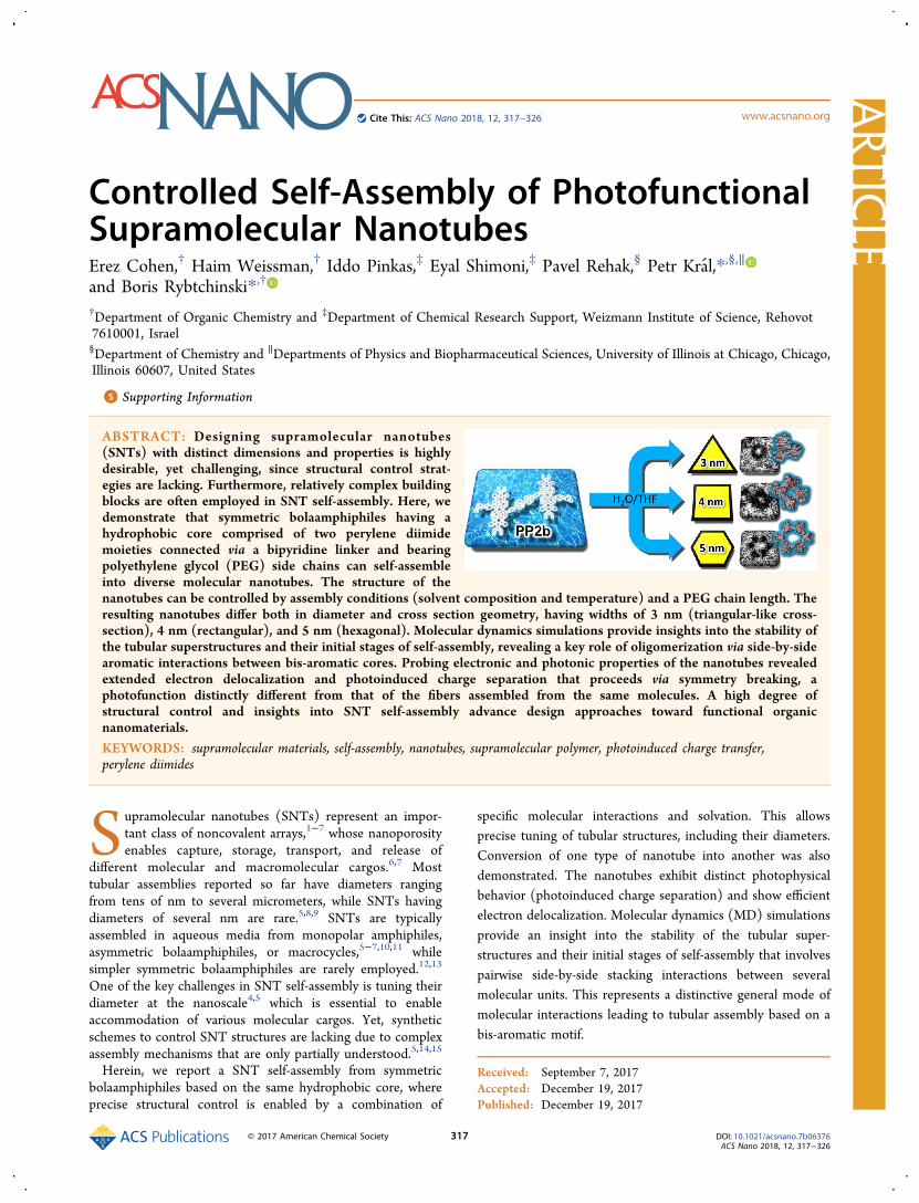
ABSTRACT: Designing supramolecular nanotubes(SNTs) with distinct dimensions and properties is highlydesirable, yet challenging, since structural control strat-egies are lacking. Furthermore, relatively complex buildingblocks are often employed in SNT self-assembly. Here, wedemonstrate that symmetric bolaamphiphiles having ahydrophobic core comprised of two perylene diimidemoieties connected via a bipyridine linker and bearingpolyethylene glycol (PEG) side chains can self-assembleinto diverse molecular nanotubes. The structure of thenanotubes can be controlled by assembly conditions (solvent composition and temperature) and a PEG chain length. Theresulting nanotubes differ both in diameter and cross section geometry, having widths of 3 nm (triangular-like cross-section), 4 nm (rectangular), and 5 nm (hexagonal). Molecular dynamics simulations provide insights into the stability ofthe tubular superstructures and their initial stages of self-assembly, revealing a key role of oligomerization via side-by-sidearomatic interactions between bis-aromatic cores. Probing electronic and photonic properties of the nanotubes revealedextended electron delocalization and photoinduced charge separation that proceeds via symmetry breaking, aphotofunction distinctly different from that of the fibers assembled from the same molecules. A high degree ofstructural control and insights into SNT self-assembly advance design approaches toward functional organicnanomaterials.KEYWORDS: supramolecular materials, self-assembly, nanotubes, supramolecular polymer, photoinduced charge transfer,perylene diimides
Supramolecular nanotubes (SNTs) represent an impor-tant class of noncovalent arrays,1−7 whose nanoporosityenables capture, storage, transport, and release of
different molecular and macromolecular cargos.6,7 Mosttubular assemblies reported so far have diameters rangingfrom tens of nm to several micrometers, while SNTs havingdiameters of several nm are rare.5,8,9 SNTs are typicallyassembled in aqueous media from monopolar amphiphiles,asymmetric bolaamphiphiles, or macrocycles,5−7,10,11 whilesimpler symmetric bolaamphiphiles are rarely employed.12,13
One of the key challenges in SNT self-assembly is tuning theirdiameter at the nanoscale4,5 which is essential to enableaccommodation of various molecular cargos. Yet, syntheticschemes to control SNT structures are lacking due to complexassembly mechanisms that are only partially understood.5,14,15
Herein, we report a SNT self-assembly from symmetricbolaamphiphiles based on the same hydrophobic core, whereprecise structural control is enabled by a combination of
specific molecular interactions and solvation. This allowsprecise tuning of tubular structures, including their diameters.Conversion of one type of nanotube into another was alsodemonstrated. The nanotubes exhibit distinct photophysicalbehavior (photoinduced charge separation) and show efficientelectron delocalization. Molecular dynamics (MD) simulationsprovide an insight into the stability of the tubular super-structures and their initial stages of self-assembly that involvespairwise side-by-side stacking interactions between severalmolecular units. This represents a distinctive general mode ofmolecular interactions leading to tubular assembly based on abis-aromatic motif.
Received: September 7, 2017Accepted: December 19, 2017Published: December 19, 2017
Artic
lewww.acsnano.orgCite This: ACS Nano 2018, 12, 317−326
© 2017 American Chemical Society 317 DOI: 10.1021/acsnano.7b06376ACS Nano 2018, 12, 317−326

RESULTS AND DISCUSSIONWe have shown that PP2b derivatives with medium-sizedPEGs self-assemble into supramolecular polymers in water/THF mixtures.16 In these systems, aromatic cores stack one ontop of the other in register, which is typical for many aromaticamphiphiles.3 Recently, it has been shown that aromaticamphiphiles can exhibit complex interaction modes, leading tounique assembly structures.17,18 Our previous work suggestedthat perylene diimide (PDI) stacking can be controlled inaqueous medium under conditions of kinetic control, leadingto nonstacked or stacked systems as a result of organiccosolvent addition/evaporation.19 We envisioned that in bis-aromatic systems connected by a linker, such as PP2b (Figure1a), interactions between aromatic cores can be attenuated to
result in side-by-side stacking (Figure 1b), leading to newsupramolecular polymerization modes. For this purpose, weinvestigated self-assembly of PEG-PP2b (PEG13 and PEG44,see Figure 1a) at a wide range of conditions. Bulky PEG44 wasexpected to partially suppress in-register stacking, while forshorter PEGs, organic cosolvent content was tuned in a broadrange in order to control stacking/hydrophobic interactions.Using this methodology, tunable tubular assemblies could beprepared that are based on side-by-side stacking interactions(Figure 1b). Assembly of diverse tubular motifs fromsymmetric building blocks (see below) contradicts thecommon SNT design concept, implying that nonsymmetricamphiphiles should be employed for SNT self-assembly.7
First, we investigated self-assembly of PP2b PEG44 bearingrelatively large PEG substituents. Cryogenic transmission
electron microscopy (cryo-TEM) imaging of PP2b PEG44in neat water revealed the formation of nanotubes with anaverage width of 4.6 ± 0.3 nm, which were hundreds of nmlong at 10−3 M concentration (Figure 2a, Figure S1) andmostly tens of nm long at 10−4 M (Figure S3). The images aretypical of tubular structures: The darker contrast results fromhigher electron density of the PP2b aromatic core stacks (tubewalls), while the lighter contrast is due to the tubeinterior.17,20−22 Highly hydrated PEGs are normally notobserved in cryo-TEM images due to their low contrast.16,23,24
Occasionally, tubular cross sections are seen in the imagingplane (for tubes that are parallel to the optical axis), revealingthe ordered hexagonal structure of the PDI core (Figure 2b,c,Figure S3). Such degree of order and complexity is not typicalfor symmetric bolamphiphiles.3 The tubes have a certainpropensity to align, typical of rigid rod-like assemblies24−26
(Figures S1 and S4). In the case of shorter SNTs formed atlower concentrations (10−4 M), small bundles of aligned tubesoccasionally orient parallel to the optical axis (Figure 2b andFigure S4), while longer tubes (micron-long, formed at higherconcentrations, 10−3 M) orient mostly perpendicularly tooptical axis, as expected based on the thickness of the vitrifiedwater layer (100−150 nm).27 Similar intertube spacings(∼14−15 nm) were observed in the aligned bundles of bothlonger and shorter tubes (Figure S1 and Figure 2b). High-resolution cryo-TEM cross-section image (dimensions of 5 ×4.6 nm, Figure 2b,c) and molecular modeling (Figure 2d,Figure S2a) suggest that three PP2b molecules are stacked ontop of another layer of three PP2b ones, leading to tubularstructure. The darker-contrast spots in the cross-section image(Figure 2c, Figures S3 and S4) correspond to PDI stacks. Thecross-section dimensions in the model (Figure 2d) fit well thedimensions observed in cryo-TEM images (Figure 2a−c). Thehexagonal tube assemblies convert into molecular fibers within7 days (Figures S5 and S6), apparently in order to optimizehydrophobic interactions, indicating that the nanotubeassembly occurs under kinetic control. The hexagonal SNTs arerelatively stable due to high kinetic barriers in aqueousmedia.3,28 The addition of THF to the aqueous solution ofnanotubes led to vesicles (40% THF, Figure S7a) or largefibrous aggregates (5% THF, Figure S7b), underscoring acritical role of a solvent in aqueous self-assembly.29 Formationof lower curvature vesicular structures upon addition of anorganic cosolvent further confirms that SNTs are formed underkinetic control.3,28 Importantly, diverse structures can beobtained from a single molecular building block.In the case of shorter PEG13, PP2b PEG13 in water:THF =
8:2 (v/v, 10−4 M) assembles into molecular fibers (Figure S8).In contrast, PP2b PEG13 in water:THF = 6:4 (v/v, 10−4 M)forms nanotubes with rectangular cross sections that have anaverage width of 3.5 ± 0.4 nm and are hundreds of nm inlength (Figure 3a,b and Figure S9). The dimensions of therectangular cross-section (3.7 × 3.2 nm) correspond well tothose in the model (3.9 × 3.3 nm, Figure 3c). Cryo-TEMimaging and molecular modeling (Figure 3c, Figure S2b)suggest that two parallel PP2b molecules are stacked on top ofanother perpendicularly oriented pair of PP2b molecules,eventually forming a nanotube with a rectangular cross-section.The width of the inner cavity in the model is 0.8 nm (thedimensions of the inner cavity observed in high-resolutioncryo-TEM images are difficult to estimate due to contrastlimitations; it appears to be <1.5 nm). Interestingly, atransformation of the rectangular tubes (4 nm in diameter,
Figure 1. (a) PP2b amphiphile building blocks, PEG lengths: n =13 or n = 44. (b) Schematic illustration of PP2b stackinginteractions: In-register (left) and side-by-side (right).
ACS Nano Article
DOI: 10.1021/acsnano.7b06376ACS Nano 2018, 12, 317−326
318

Figure S10) into larger (4.8 ± 0.4 nm diameter) and morerigid ones (almost no bending observed) was achieved byheating the assembly at 55 °C for 30 min (Figure 4 and FigureS11). The resultant tubes are stable and do not change uponfurther heating. These nanotubes were also observed by SEMimaging, revealing diameters of 6.5 ± 0.8 nm (Figure S12),where the larger diameter is consistent with the presence ofPEG chains (wrapping the SNTs) that become visible in SEMimages. The nanotubes may have hexagonal cross-section (notseen in cryo-TEM images), as they are rigid and exhibitdiameters close to the ones of the rigid hexagonal tubes basedon PP2b PEG44.To further control the SNT structures, mixing PEG13 and
PEG44 PP2b was employed. Our rational for the mixing relieson the fact that when the hydrophilic parts of a PP2bamphiphile molecule become larger, the molecule gains awedge-like shape, and one can obtain increasing curvature ofthe self-assembled structures.30 Thus, we added small amountsof PP2b PEG44 to PP2b PEG13. The samples were preparedusing co-evaporation and redispersion, wherein PP2b PEG44and PP2b PEG13 in disaggregating solvent (CHCl3) werepremixed, dried in vacuum, and then assembled as follows. Themixture was first dissolved in THF, followed by fast addition ofwater and vigorous mixing. A mixture of 5% PP2b PEG44/95% PP2b PEG13 (40% THF, 10−4 M) was found to form
nanotubes with an average width of 3.2 ± 0.2 nm and lengthsof hundreds of nm (Figure 3d, Figure S13).The observed cross-section (Figure 3d) and molecular
modeling (Figure 3f, Figure S2c) suggest a structure in whichthree PP2b molecules are stacked in a helical arrangement witha triangle-like geometry and a twisting angle of 120° relative toeach other. This structure results in a helical tube (Figure 3f)with an outer diameter of 3.1 nm and an inner one of roughly0.3 nm, in agreement with the cryo-TEM imaging. Theobserved fine structure of the nanotubes (Figure S13d)appears to be compatible with helical packing. We note thatresolution of cryo-TEM images precluded further unequivocalconfirmation of helicity and precise determination of SNTs’inner diameters. The absence of self-sorting in this system(formation of two distinct aggregate types) is probably due tostrong driving forces that result in efficient irreversibleinteractions between all moieties.31
Unlike the rectangular ones, these nanotubes are stable at 55°C (Figures S15−S17, see summary of SNTs properties inTable 1). Our attempts to further control the structure byaltering the PP2b PEG44/PP2b PEG13 ratio resulted indiverse assemblies. For instance, increasing the PP2b PEG44content to 10% leads to molecular fibers (1% THF, FigureS18a) or a dense 3D network of fibers (40% THF, FigureS18b). Likewise, the ratio of 95% PP2b PEG44/ 5% PP2b
Figure 2. Cryo-TEM images (a−c) and proposed molecular model (d) of self-assembled PP2b PEG44 nanotubes in neat water. (a) 10−3 M,aging 75 min, side view, showing tubes with outer diameter of 4.6 ± 0.3 nm (cross section 5.0 ± 0.1 nm). Inset: magnified image showingconnection of a cross-section and a tube wall. Scale bar is 5 nm. (b) 10−4 M, aging 5 h, hexagonal cross section. For zoomed out and enlargedimages see Figure S4. (c) High-resolution hexagonal cross section. The tube outer dimensions are 5.0 × 4.6 nm. Molecular model: (d)Cross-section, top view. The geometry was optimized using molecular mechanics (augmented MM2 and MM3 parameters). Forcomputational simplification, PEG chains were modeled as −O(CH2−CH2)OCH3 groups. The aromatic core in the model is given in aspace-filling representation.
ACS Nano Article
DOI: 10.1021/acsnano.7b06376ACS Nano 2018, 12, 317−326
319
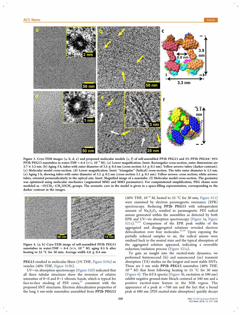
PEG13 resulted in molecular fibers (1% THF, Figure S19a) orvesicles (40% THF, Figure S19b).UV−vis absorption spectroscopy (Figure S20) indicated that
all three tubular structures show the inversion of relativeintensities of 0−0 and 0−1 vibronic bands, which is typical forface-to-face stacking of PDI cores,32 consistent with theproposed SNT structures. Electron delocalization properties ofthe long 5 nm-wide nanotubes assembled from PP2b PEG13
(40% THF, 10−4 M, heated to 55 °C for 30 min, Figure S11)were examined by electron paramagnetic resonance (EPR)spectroscopy. Reducing PP2b PEG13 with subequivalentamount of Na2S2O4 resulted in paramagnetic PDI radicalanions generated within the assemblies as detected by bothEPR and UV−vis absorption spectroscopy (Figure 5a, FigureS21a).33,34 Comparison of the EPR peak widths of theaggregated and disaggregated solutions revealed electrondelocalization over four molecules.35,36 Upon exposing thepartially reduced samples to air, the radical anions wereoxidized back to the neutral state and the typical absorption ofthe aggregated solution appeared, indicating a reversiblereduction/oxidation process (Figure S21a).To gain an insight into the excited-state dynamics, we
performed femtosecond (fs) and nanosecond (ns) transientabsorption (TA) studies on the longest and most stable SNTs.These are 5 nm wide PP2b PEG13 nanotubes (40% THF,10−4 M) that form following heating to 55 °C for 30 min(Figure 4). The fsTA spectra (Figure 5b, excitation at 590 nm)exhibit negative ground-state bleach centered at 540 nm and apositive excited-state feature in the NIR region. Theappearance of a peak at ∼760 nm and the fact that a broadpeak at 680 nm (PDI excited-state absorption) quickly decays
Figure 3. Cryo-TEM images (a, b, d, e) and proposed molecular models (c, f) of self-assembled PP2b PEG13 and 5% PP2b PEG44/ 95%PP2b PEG13 nanotubes in water:THF = 6:4 (v/v, 10−4 M). (a) Lower magnification. Inset: Rectangular cross-section, outer dimensions are3.7 × 3.2 nm. (b) Aging 3 h, tubes with outer diameter of 3.5 ± 0.4 nm (cross section 3.5 ± 0.1 nm). Yellow arrows: tubes (darker-contrast).(c) Molecular model cross-section. (d) Lower magnification. Inset: “triangular” (helical) cross-section. The tube outer diameter is 3.3 nm.(e) Aging 3 h, showing tubes with outer diameter of 3.2 ± 0.2 nm (cross section 3.1 ± 0.1 nm). Yellow arrows: cross section; white arrows:tubes, oriented permendicularly to the optical axis. Inset: Magnified image of a nanotube. (f) Molecular model cross-section. The geometrywas optimized using molecular mechanics (augmented MM2 and MM3 parameters). For computational simplification, PEG chains weremodeled as −O(CH2−CH2)OCH3 groups. The aromatic core in the model is given in a space-filling representation, corresponding to thedarker contrast in the images.
Figure 4. (a, b) Cryo-TEM image of self-assembled PP2b PEG13nanotubes in water:THF = 6:4 (v/v, 10−4 M) aging 8.5 h afterheating to 55 °C for 30 min. Average width 4.8 ± 0.4 nm.
ACS Nano Article
DOI: 10.1021/acsnano.7b06376ACS Nano 2018, 12, 317−326
320

(“shifts to the red”, Figure 5b), while the 760 nm feature has asubstantially longer lifetime (3−4 ns, Figure S21c,d), is typicalof PDI radical anion formation, occurring via symmetry-breaking charge transfer.37,38 The SNT fsTA kinetics does notshow a power dependence attributable to exciton hopping(Figure S21b),39 in contrast to segmented fibers based onPP2b PEG17, reported earlier.16 The charge transfer in thenanotubes occurs with ∼24% yield, according to kineticanalysis at 760 nm (Figure 5c, Tables S1 and S2). Theobserved photoinduced symmetry-breaking charge separa-tion40 in electron-deficient PDI-based systems is rare,37,38
since, normally, this process is not thermodynamicallyfavorable. The fast time scale of the symmetry breaking inthe nanotubes (∼0.9 ps) may be aided by highly efficientcharge delocalization over four molecules. In contrast,femtosecond TA studies of molecular fibers based on PP2bPEG13 (20% THF, 10−4 M, Figure S22) reveal a low yield ofphotoinduced charge separation (∼5% according to kineticanalysis at 750 nm, Table S3) and shorter lifetimes. Thisoccurs despite the fact that the fibers exhibit efficient electrondelocalization over eight PDI molecules (Figure S23),according to our EPR study. One possible explanation forthe observed difference in photoinduced charge transfer(PCT) could be attributed to the structural variations betweenthe assemblies, with adjacent stacked PDIs in the tubesexperiencing slightly different environments, unlike in thefibers.Further studies aimed at elucidating the PCT behavior in the
nanotubes involved the addition of an electron donor(triethylamine, TEA, 4%, v/v) to a solution of PP2b PEG13nanotubes (40% THF, 10−4 M) after heating to 55 °C for 30min (Figure 4). The fsTA spectra (Figure S24, excitation at
590 nm) exhibit different features compared with the casewithout an external electron donor. Thus, no red-shift of PDIpeaks typical of PDI symmetry breaking was observed. Asexpected, fast PCT occurs as indicated by the appearance of760 nm band. PCT state has much shorter lifetime than that inthe absence of TEA (Table S4). This underscores a differencewith regard to a symmetry breaking state. Longer lifetime ofthe latter may be due to charge delocalization within PDIstacks. Different reorganization energies (PDI and solvent)may also contribute to the observed difference. We note thatthe reported electron-deficient PDI systems showing symme-try-breaking charge transfer are small well-defined moleculararrays.37,38 Extended 1D nanostructures such as SNTs that arebuilt from a single type of robust chromophores and exhibitlonger PCT life times may enable new approaches tooptoelectronic wires, expanding the range of SNT functions.In order to gain mechanistic molecular-scale insight into the
self-assembly of different PP2b molecules and the stability ofmolecular superstructures formed, we used all-atom moleculardynamics (MD) simulations in explicit solvent to model: (i)the process of PP2b molecular self-assembly and (ii) thespatial organization of molecular/solvent components in theself-assembled superstructures (see Supporting Information).For PP2b PEG44 (Figure 6a,b, neat water), the simulationsshow the outcome for 12 monomers self-assembling for 150 nsin a water box of 5,400 nm3 (see computational details inMethods and Supporting Information). The simulationsrevealed that within 10 ns PP2b PEG44 formed a largecluster, resembling a micelle,41,42 where the hydrophobic PP2bmoieties closely interacted, while the long PEG chainssurrounded the core of the cluster. Within 22 ns, a firstPP2b dimer featuring two stacked PDIs (side-by-side arrange-
Table 1. Summary of SNTs Properties
tube composition solventcross sectiongeometry
diameter(nm) stability
PP2b PEG13 6:4 water/THF (v/v) rectangular 3.5 ± 0.4 stable at rtPP2b PEG13 (heated to 55 °C for 30min)
6:4 water/THF (v/v) probably hexagonal 4.8 ± 0.4 stable
5% PP2b PEG44/ 6:4 water/THF (v/v) triangle-like 3.2 ± 0.2 stable95% PP2b PEG13PP2b PEG44 water hexagonal 4.6 ± 0.3 kinetically stable, transform gradually to molecular
fibers
Figure 5. EPR and TA (pumped at 590 nm) of self-assembled PP2b PEG13 nanotubes in water:THF = 6:4 (v/v, 10−4 M), heated to 55 °C for30 min. (a) EPR spectra of reduced: monomer with 4% v/v Et3N (black), assembly with 0.4 equiv Na2S2O4 (red). The electrondelocalization length is four molecules. (b) Femtosecond TA spectra evolution over time, 1.5 mW. Red arrow indicates the PDI excited-statepeak decay that occurs before the PDI radical anion peak (at 760 nm) decay. (c) Femtosecond TA decay kinetics probed at four wavelengthsand their multiexponential fit, 1.5 mW. For details on decay coefficients see Table S1.
ACS Nano Article
DOI: 10.1021/acsnano.7b06376ACS Nano 2018, 12, 317−326
321

ment) formed within the core, which was followed by theformation of a trimeric chain at 25 ns. Around 70 ns, anothermonomer joined the trimer to form a jiggling linear tetramer.At 88 and 115 ns, additional dimers formed outside and withinthe cluster core, respectively. The rigid and flat bipyridine(bipy) linker was found to promote initial stacking interactions(see Supporting Information for details), eventually leading toside-by-side stacking of PDI moieties resulting in stableoligomers. The rigid nature of the linker most probably
restricts a range of possible geometries, promoting oligomercyclization that eventually leads to nanotubes.In the case of PP2b PEG13 (Figure 6c,d), the simulations
show the results for 6 PP2b PEG13 monomers after self-assembling for 266 ns in a water:THF = 8:2 (v/v) solvent in abox of 2200 nm3. These monomers with shorter PEG chainsquickly formed pairs, before forming larger clusters,surrounded by THF molecules (better solvent for hydrophobiccores). The first dimer formed at 12 ns, a second dimer formedat 27 ns, and a trimer formed from the second dimer at 66 ns.
Figure 6. MD simulations of the self-assembly of PEG-ylated PP2b monomers. PP2b molecules are vividly colored, PEG chains are shown ina finer representation, and gray structures represent THF molecules. Scale bars are 1 nm. (a, b) Two views of the main cluster core formedfrom PP2b PEG44 monomers in pure water at 150 ns. (c, d) Two views of the main cluster core formed from PP2b PEG13 monomers inwater:THF solvent at 266 ns. (e, f) Two views of the main cluster core formed from PP2b PEG13 monomers in water:THF = 6:4 (v/v) at674 ns. (g, h) MD simulations of a fragment (middle section) of a SNT pre-assembled from PEG-ylated PP2b monomers. Top and sideviews of helical superstructures of 29 PP2b PEG13 and 1 PP2b PEG44 in water:THF = 6:4 (v/v) at 20 ns. For clarity, in a side view, thesolvent molecules are not shown.
ACS Nano Article
DOI: 10.1021/acsnano.7b06376ACS Nano 2018, 12, 317−326
322

Later on, at 185 ns, this trimer formed a closed triangle. Thestructures formed were also relatively rigid. Simulations ofPP2b PEG13 (Figure 6e,f) show the outcome for 5 PP2bPEG13 monomers which self-assembled at 674 ns in thewater:THF = 6:4 (v/v) solvent in a box of 560 nm3. Here, thefirst dimer appeared only at 40 ns, a trimer at 150 ns (shown),and a second dimer only at 544 ns. Figure 6c,f also reveals aclose distribution of THF molecules around the PDI cores.These results indicate that with the increasing concentration ofa stabilizing THF solvent, the PDI dimerization via stacking isa much slower process in comparison with water:THF = 8:2system, indicating importance of specific solvation. Simulationsof the fully assembled tubes (Figure 6g,h, Figures S25 andS26) support our experimental results. They show that the 3nm (triangular-like, Figure 6g,h) and 5 nm (hexagonal) SNTsare more stable compared with a more flexible 4 nm(rectangular) tube. The latter is in accordance with theexperimentally observed transformation of rectangular to thehexagonal tubes upon heating. The high stability of thehexagonal SNTs may be due to highly ordered (crystalline-like) structures, while the triangular-like SNTs (Figure 6g,h)may be stabilized by helical arrangement.Overall, our simulations confirm that the formation of PP2b
PEG assemblies is driven by side-by-side π−π stacking/hydrophobic interactions between the PDI groups leading tosupramolecular oligomerization, which is a common process inall systems. Once the interaction has been established, itcannot be easily reversed. Importantly, pairwise noncovalentinteractions leading to side-by-side stacking appear to be a generalinitial step in oligomerization of bis-aromatic systems, reminiscentof supramolecular polymerization of molecules bearing bifunc-tional noncovalent binding motif.43 The insight into aromaticoligomerization in aqueous media and its relevance to tubularself-assembly has not been observed so far. Complete ringclosure was not observed at the explored time scales as suchnucleation events are slow and in general are difficult toobserve in MD simulations.44 Further MD studies aimed atelucidating the mechanism of tubular assembly are necessary inorder to understand the entire assembly progression.
CONCLUSIONS
In conclusion, we have shown that symmetric bolaamphiphilesare capable of assembling into a variety of nanometer-scalenanotubes. Our experimental and theoretical studies reveal thatan interplay of molecular structure and solvation specifics havea profound effect on self-assembly. This enabled tuning ofmolecular nanotube structures based on identical hydrophobiccores. Additionally, photoinduced symmetry-breaking chargetransfer was observed in SNTs, demonstrating that SNTsenable photoactive wire function. Simplification of moleculardesign, control via self-assembly conditions, and photofunctionprovide tools to create complex functional supramolecularsystems.
METHODSSolvents and reagents were purchased from commercial sources andused as received, unless stated otherwise. For all aqueous mixtures,double-distilled water was used (Barnstead NANOpure Diamondwater system). All procedures with air-sensitive compounds wereperformed under inert gas atmosphere (dried N2) using a glovebox(MBRAUN, Labmaster). Organic solvents used for these procedureswere degassed with argon and stored over molecular sieves (4 Å) inthe glovebox. Synthesis of PP2b derivatives was reported previously.45
UV−vis Absorption Measurements. Measurements werecarried out on a Cary-5000 spectrometer (Varian).
Cryo-TEM. Cryo-TEM was carried out using a Tecnai T12transmission electron microscope operating at 120 kV with a GatanOneView 4k × 4k digital camera Model 1095, or Tecnai F20transmission electron microscope operating at 200 kV with a GatanUS4000 digital camera. Samples were transferred using a transferstation to a Gatan 626 cooling holder and introduced into themicroscope. Sample preparation: 6 μL of sample solution was appliedto a 200 mesh copper grid coated with holey carbon (Pacific Grid-Tech) and was blotted at 25 °C (95% humidity), and then the gridwas immediately plunged into liquid ethane using a Leica EM-GPAuto plunger. Image analysis was done using iTEM software (v 5.2build 3554, Olympus).
Freeze-Dry cryo-SEM. A drop of 3 μL of PP2b PEG13nanotubes in 60:40 water/THF (v/v), 10−4 M aging 3 h afterheating to 55 °C for 30 min was deposited on a nitrocellulose-coated200 mesh copper EM grid, blotted with a filter paper, and plungedinto liquid ethane using a custom-made plunger (Technion, IsraelInstitute of Technology, Israel).
The grids were then transferred to a BAF 60 freeze fracture device(Leica Microsystems, Vienna, Austria) using a VCT 100 Cryo transfershuttle (Leica Microsystems) and sublimed at a temperature of −80°C and pressure of 1 × 10⁻⁶ mbar for about 1 h. The sample wastransferred to a GeminiSEM 500 scanning electron microscope (CarlZeiss AG, Oberkochen, Germany) equipped with a Cryo stage (LeicaMicrosystems). Samples were observed without or with coating with alayer of 2 nm Pt/C (coating was conducted at the BAF 60) atacceleration voltages ranging between 0.7 and 1.5 kV using an InLenssecondary electrons detector.
Molecular Modeling Computations. Molecular mechanics(MM) geometry optimizations were carried using augmented MM3parameters followed by augmented MM2 parameters with FujitsuScigress v 2.2. For computational simplification, PEG chains weremodeled as −O(CH2−CH2)OCH3 groups.
EPR. Electron paramagnetic resonance spectra were measured witha Bruker E-580 spectrometer equipped with an EN801 resonator at298 K. Aggregated solutions of deoxygenated THF/water werereacted with 0.1 and 0.02 equiv sodium dithionite, whiledisaggregated THF solution was loaded with 4% v/v triethylamine(TEA) and excited by high power LED (150W, 2 min) according to aliterature procedure.46 All samples were measured in 0.8 mm capillarytube sealed before the EPR measurements in nitrogen-filled glovebox.
Transient Absorption. The system is based on a modelockedTi:sapphire oscillator (Spectra Physics Mai Tai SP). The oscillatorproduces a train of <120 fs pulses (bandwidth ∼12 nm fwhm), with apeak wavelength at 800 nm, typically of 900 mW, corresponding to∼10 nJ per pulse. The weak oscillator pulses are amplified by achirped pulse regenerative amplifier (CPA) (Spectra Physics SpitfireACE). The pulse is first stretched to several picoseconds and then isregeneratively amplified in a Ti:sapphire cavity, pumped by a pulsedNd:YLF laser (Spectra Physics Empower 45) operating at 1 kHz.After the pulse has been amplified and recompressed, its energy isabout 5 mJ in a train of 1000 Hz pulses. An independent pump pulseis obtained by pumping an optical parametric amplifier (LightConversion, TOPAS-Prime, and NIRUVIS harmonic generator) thatproduces 120 fs pulses continuously tunable from 300 nm to 3 μm.The output power of the OPA varies between a few micro Joules totens of micro Joules (depending on the chosen wavelength) at 1000Hz. The probe beam is mechanically chopped at half the amplifierrepetition rate. The chopper (C-995 TTI) is synchronized to theSpitfire amplifier. Normally a few thousand pulse pairs (pump on/pump off) are averaged to produce a TA spectrum with a noise levelbelow 0.3 mOD. A small portion of the remaining amplified pulse isused to generate a white light continuum as a probe pulse. To thisend, the Ti:sapphire beam is focused onto a 3 mm-thick CaF2 windowby a 5 cm focal length lens, and the numerical aperture of the beam iscontrolled by an iris placed in front of the lens, which helps to obtaina stable and smooth white light continuum. In order to avoid thermaldamage on the CaF2 window and noise in the continuum white light,
ACS Nano Article
DOI: 10.1021/acsnano.7b06376ACS Nano 2018, 12, 317−326
323
it is moved in a circular motion so that each consecutive laser pulsehits a fresh spot. The system returns to the same location every fewtens of seconds. The resulting beam is passed through a Raman notchfilter to remove the remains of the fundamental beam from the probewhite light continuum. The pump and probe pulses are crossed in thesample at a small angle, while maintaining a magic angle between thepump and probe polarizations. The remains of the pump pulse areremoved by an iris, and the probe light is imaged onto an optical fiberthat brings it into an imaging interface, which focuses the light ontothe entrance slit of a Jobin Yivon Triax 180 spectrograph. The light isnormally dispersed by a 300 gr/mm grating onto a fast CCD camera(Andor Newton DU-920P-BEX2-DD, operating at 1000 spectra persecond using “crop mode”). The TA setup is controlled by NationalInstruments LabView software. A variable neutral-density filter wasemployed to adjust the pump power. The pump power intensitieswere measured using Ophir photodiode sensor power meter inproximity to the sample. The excitation densities were estimated for alaser spot of 300 μm diameter on the sample. This diameter wasmeasured by placing beam profiler (Ophir Beamstar FX33) at thesample position and determining the 4-σ (95% of the power)parameter. In the reported experiments, the pump was turned to 590nm, and the optical densities of the samples, in 1 and 2 mm opticalpath length cuvettes, were kept between 0.2 and 0.4 at the excitationwavelength. Spectral corrections and analysis were performed usingSurface Xplorer Pro (Ultrafast Systems) and Origin 9.1 (OriginLab)software.The ns−μs data were collected using the same laser as a pump
source (from oscillator to OPA), and the detection was done in anEOS subnanosecond TA spectrometer (Ultafast Systems, USA). Thespectrometer has a 100 ps instrument response and allows for themeasurement of TA between 400 and 900 nm. Data analysis was doneas mentioned before.Molecular Dynamics. MD simulations were performed with
NAMD3 package, using the CHARMM force field.47−51 Internalmolecular and dispersion parameters were taken from the CHARMM36 general force field.52 Due to difficulties of ab initio frequencycalculations of alkyne and extended aromatic groups in the PP2b core,missing parameters were approximated using the parachemsoftware.51 Ab initio calculations using Gaussian 0953 package wereperformed on the linker group between the two extended aromaticgroups, the extended aromatic groups themselves, and the terminalnoncyclical aromatic ring, using MP2/6-31g(d)//MP2/6-31g(d) levelof theory to determine partial charges of PP2b core using the ChelpGalgorithm.54 CHARMM TIP3P water model was employed forexplicit water simulations.Two types of simulations were performed: (i) self-assembly and
(ii) superstructure simulations. In the self-assembly system, each ofthe PP2b molecules were placed and rotated randomly in theirrespective environment. PEGylated chains with 44 oxygen atoms weresimulated in pure water, whereas PEGylated chains with 13 oxygenatoms were placed in water/THF mixtures. There were two differentmixtures of water and THF: (i) 60% water: 40% THF (v:v) and (ii)80% water: 20% THF (v:v). Experimental density55 of pure THF wasused to determine the number of THF molecules per system. Thevolume of each system was determined by placing the PP2bmolecules and then adding 3 nm to the maximum and minimumcoordinate values of the PP2b molecules.A total of three self-assembly systems were constructed (i) with
pure water and 12 PP2b molecules with PEGylated chains, having 44oxygen atoms, (ii) in 60% water and 40% THF with 8 PP2b withPEGylated chains, having 13 oxygen atoms, and (iii) in 80% waterand 20% THF with 6 PP2b molecules, with PEGylated chains, having13 oxygen atoms. A total of three superstructure simulations wereconstructed (i) hexagonal structure (Figure 2 of the main text) withPEGylated chains 44 oxygen atoms long in pure water, (ii) squarestructure (Figure 3a,c of the main text) in 60% water: 40% THF (v:v),(iii) square structure (actually giving rise to molecular fibers, FigureS8) in 80% water: 20% THF (v:v). Because those superstructures hadthe PEGylated chains of −O(CH2−CH2)OCH3 groups, additionalPEGylated links were added until desired length was achieved.
Water and THF were initially placed immiscibly ,and duringminimization and pre-equilibration they were allowed to diffusewithout any constrains until the solvent was homogeneous. Duringminimization (50,000 steps) and pre-equilibration (at least 3 ns), onlyatoms in the PP2b core were subjected to constraints, whereas thesolvent molecules and PEGylated chains were allowed to movewithout any constraints. During the production runs, the self-assemblymolecules had no constraints; in the superstructure simulations, onlythe linking bipyridine carbons and the nitrogen atoms on thebipyridine group, on one molecule per superstructure, wereconstrained, with 20% of value of the constraints employed duringminimization.
Simulations were done with NPT ensemble at a temperature of 310K and a pressure of 1 atm and periodic boundary conditions.Langevin dynamics were used to maintain pressure and temperature,with a damping constant of 1 ps−1, temperature bath was 310 K,hydrogen atoms were excluded from the thermal bath, piston periodwas 100 fs, piston decay was 50 fs. Excluding minimization, particlemesh Ewald56 was used with a grid spacing of 1.0. Velocity Verletintegrator was used to solve equations of motion, with a time step of 2fs. The SHAKE algorithm was used for the hydrogen atoms.Nonbonded interaction used the switching algorithm, with theswitch-on distance at 10 Å and the switch-off at 12 Å. Nonbondedpair lists were used for atoms within 13.5 Å of each other, with the listupdated every 20 steps. 1−4 nonbonded interactions were not scaled.Data and snapshots were recorded every 10 ps or every 5000 steps.The pure water simulation system lasted 200 ns, due its computa-tional expense, whereas all other systems were 300 ns or longer. Inorder to improve efficiency of the simulations, the size of the 60%water self-assembly system was first decreased by excluding onlysolvent molecules, but the number of PP2b molecules was keptconstant. Subsequently the size was further decreased from eightmolecules to only five PP2b molecules. When constructing thesuperstructure for the aqueous system, the original length of thePEGylated chains was lengthened to 44 oxygen atoms, in an extendedconformation. Then the superstructure was allowed to minimize andequilibrate in vacuum, so that the PEGylated chains would fold ontothe superstructure, thus requiring a smaller volume for the simulation.
ASSOCIATED CONTENT*S Supporting InformationThe Supporting Information is available free of charge on theACS Publications website at DOI: 10.1021/acsnano.7b06376.
Experimental details, electron microscopy images, UV−vis absorption spectra, EPR, femtosecond and nano-second TA spectra, kinetic analysis and MD (PDF)
AUTHOR INFORMATIONCorresponding Authors*E-mail: [email protected].*E-mail: [email protected] Kral: 0000-0003-2992-9027Boris Rybtchinski: 0000-0002-2071-8429NotesThe authors declare no competing financial interest.
ACKNOWLEDGMENTSThis work was supported by the Israel Science Foundation andthe Helen and Martin Kimmel Center for Molecular Design.The SEM and TEM studies were conducted at the Irving andCherna Moskowitz Center for Nano and BioNano Imaging(Weizmann Institute). P.K. acknowledges the NSF Division ofMaterials Research grant 1506886. We thank Dr. R. Carmielifor his help with EPR studies.
ACS Nano Article
DOI: 10.1021/acsnano.7b06376ACS Nano 2018, 12, 317−326
324
REFERENCES(1) Hill, J. P.; Jin, W.; Kosaka, A.; Fukushima, T.; Ichihara, H.;Shimomura, T.; Ito, K.; Hashizume, T.; Ishii, N.; Aida, T. Self-Assembled Hexa-Peri-Hexabenzocoronene Graphitic Nanotube.Science 2004, 304, 1481−1483.(2) Jin, W.; Yamamoto, Y.; Fukushima, T.; Ishii, N.; Kim, J.; Kato,K.; Takata, M.; Aida, T. Systematic Studies on Structural Parametersfor Nanotubular Assembly of Hexa-Peri-Hexabenzocoronenes. J. Am.Chem. Soc. 2008, 130, 9434−9440.(3) Krieg, E.; Bastings, M. M. C.; Besenius, P.; Rybtchinski, B.Supramolecular Polymers in Aqueous Media. Chem. Rev. 2016, 116,2414−2477.(4) Beingessner, R. L.; Fan, Y.; Fenniri, H. Molecular andSupramolecular Chemistry of Rosette Nanotubes. RSC Adv. 2016,6, 75820−75838.(5) Shimizu, T.; Masuda, M.; Minamikawa, H. SupramolecularNanotube Architectures Based on Amphiphilic Molecules. Chem. Rev.2005, 105, 1401−1444.(6) Bong, D. T.; Clark, T. D.; Granja, J. R.; Ghadiri, M. R. Self-Assembling Organic Nanotubes. Angew. Chem., Int. Ed. 2001, 40,988−1011.(7) Kameta, N.; Minamikawa, H.; Masuda, M. SupramolecularOrganic Nanotubes: How to Utilize the Inner Nanospace and theOuter Space. Soft Matter 2011, 7, 4539−4561.(8) Barclay, T. G.; Constantopoulos, K.; Matisons, J. NanotubesSelf-Assembled from Amphiphilic Molecules via Helical Intermedi-ates. Chem. Rev. 2014, 114, 10217−10291.(9) Eisele, D. M.; Arias, D. H.; Fu, X.; Bloemsma, E. A.; Steiner, C.P.; Jensen, R. A.; Rebentrost, P.; Eisele, H.; Tokmakoff, A.; Lloyd, S.;Nelson, K. A.; Nicastro, D.; Knoester, J.; Bawendi, M. G. RobustExcitons Inhabit Soft Supramolecular Nanotubes. Proc. Natl. Acad. Sci.U. S. A. 2014, 111, E3367−E3375.(10) Kim, H.-J.; Kang, S.-K.; Lee, Y.-K.; Seok, C.; Lee, J.-K.; Zin, W.-C.; Lee, M. Self-Dissociating Tubules from Helical Stacking ofNoncovalent Macrocycles. Angew. Chem., Int. Ed. 2010, 49, 8471−8475.(11) Liu, Z.; Liu, G.; Wu, Y.; Cao, D.; Sun, J.; Schneebeli, S. T.;Nassar, M. S.; Mirkin, C. A.; Stoddart, J. F. Assembly ofSupramolecular Nanotubes from Molecular Triangles and 1,2-Dihalohydrocarbons. J. Am. Chem. Soc. 2014, 136, 16651−16660.(12) Ponnuswamy, N.; Pantos, G. D.; Smulders, M. M. J.; Sanders, J.K. M. Thermodynamics of Supramolecular NaphthalenediimideNanotube Formation: The Influence of Solvents, Side Chains, andGuest Templates. J. Am. Chem. Soc. 2012, 134, 566−573.(13) Tian, Z.; Li, H.; Wang, M.; Zhang, A.; Feng, Z.-G. Vesicularand Tubular Structures Prepared from Self-Assembly of NovelAmphiphilic ABA Triblock Copolymers in Aqueous Solutions. J.Polym. Sci., Part A: Polym. Chem. 2008, 46, 1042−1050.(14) Guo, C.; Luo, Y.; Zhou, R.; Wei, G. Probing the Self-AssemblyMechanism of Diphenylalanine-Based Peptide Nanovesicles andNanotubes. ACS Nano 2012, 6, 3907−3918.(15) Valery, C.; Artzner, F.; Paternostre, M. Peptide Nanotubes:Molecular Organisations, Self-Assembly Mechanisms and Applica-tions. Soft Matter 2011, 7, 9583−9594.(16) Krieg, E.; Shirman, E.; Weissman, H.; Shimoni, E.; Wolf, S. G.;Pinkas, I.; Rybtchinski, B. Supramolecular Gel Based on a PeryleneDiimide Dye: Multiple Stimuli Responsiveness, Robustness, andPhotofunction. J. Am. Chem. Soc. 2009, 131, 14365−14373.(17) Ustinov, A.; Weissman, H.; Shirman, E.; Pinkas, I.; Zuo, X.;Rybtchinski, B. Supramolecular Polymers in Aqueous Medium:Rational Design Based on Directional Hydrophobic Interactions. J.Am. Chem. Soc. 2011, 133, 16201−16211.(18) Percec, V.; Wilson, D. A.; Leowanawat, P.; Wilson, C. J.;Hughes, A. D.; Kaucher, M. S.; Hammer, D. A.; Levine, D. H.; Kim,A. J.; Bates, F. S.; Davis, K. P.; Lodge, T. P.; Klein, M. L.; DeVane, R.H.; Aqad, E.; Rosen, B. M.; Argintaru, A. O.; Sienkowska, M. J.;Rissanen, K.; Nummelin, S.; Ropponen, J. Self-Assembly of JanusDendrimers into Uniform Dendrimersomes and Other ComplexArchitectures. Science 2010, 328, 1009−1014.
(19) Shahar, C.; Dutta, S.; Weissman, H.; Shimon, L. J. W.; Ott, H.;Rybtchinski, B. Precrystalline Aggregates Enable Control overOrganic Crystallization in Solution. Angew. Chem. 2016, 128, 187−190.(20) Cui, H.; Hodgdon, T. K.; Kaler, E. W.; Abezgauz, L.; Danino,D.; Lubovsky, M.; Talmon, Y.; Pochan, D. J. Elucidating theAssembled Structure of Amphiphiles in Solution via CryogenicTransmission Electron Microscopy. Soft Matter 2007, 3, 945−955.(21) Ziserman, L.; Lee, H.-Y.; Raghavan, S. R.; Mor, A.; Danino, D.Unraveling the Mechanism of Nanotube Formation by Chiral Self-Assembly of Amphiphiles. J. Am. Chem. Soc. 2011, 133, 2511−2517.(22) Golubkov, G.; Weissman, H.; Shirman, E.; Wolf, S. G.; Pinkas,I.; Rybtchinski, B. Economical Design in Noncovalent NanoscaleSynthesis: Diverse Photofunctional Nanostructures Based on a SingleCovalent Building Block. Angew. Chem., Int. Ed. 2009, 48, 926−930.(23) Weissman, H.; Rybtchinski, B. Noncovalent Self-Assembly inAqueous Medium: Mechanistic Insights from Time-ResolvedCryogenic Electron Microscopy. Curr. Opin. Colloid Interface Sci.2012, 17, 330−342.(24) Baram, J.; Weissman, H.; Tidhar, Y.; Pinkas, I.; Rybtchinski, B.Hydrophobic Self-Assembly Affords Robust Noncovalent PolymerIsomers. Angew. Chem., Int. Ed. 2014, 53, 4123−4126.(25) Glotzer, S. C.; Solomon, M. J. Anisotropy of Building Blocksand Their Assembly into Complex Structures. Nat. Mater. 2007, 6,557−562.(26) Barry, E.; Dogic, Z. Entropy Driven Self-Assembly ofNonamphiphilic Colloidal Membranes. Proc. Natl. Acad. Sci. U. S. A.2010, 107, 10348−10353.(27) Angert, I.; Burmester, C.; Dinges, C.; Rose, H.; Schroder, R. R.Elastic and Inelastic Scattering Cross-Sections of Amorphous Layersof Carbon and Vitrified Ice. Ultramicroscopy 1996, 63, 181−192.(28) Tidhar, Y.; Weissman, H.; Wolf, S. G.; Gulino, A.; Rybtchinski,B. Pathway-Dependent Self-Assembly of Perylene Diimide/PeptideConjugates in Aqueous Medium. Chem. - Eur. J. 2011, 17, 6068−6075.(29) Gillissen, M. A. J.; Koenigs, M. M. E.; Spiering, J. J. H.;Vekemans, J. A. J. M.; Palmans, A. R. A.; Voets, I. K.; Meijer, E. W.Triple Helix Formation in Amphiphilic Discotics: DemystifyingSolvent Effects in Supramolecular Self-Assembly. J. Am. Chem. Soc.2014, 136, 336−343.(30) Balbo Block, M. A.; Kaiser, C.; Khan, A.; Hecht, S., DiscreteOrganic Nanotubes Based on a Combination of Covalent and Non-Covalent Approaches. In Functional Molecular Nanostructures;Schluter, A. D., Ed.; Springer: Berlin, Heidelberg, 2005; pp 89−150.(31) Besenius, P. Controlling Supramolecular PolymerizationThrough Multicomponent Self-Assembly. J. Polym. Sci., Part A:Polym. Chem. 2017, 55, 34−78.(32) Wurthner, F.; Saha-Moller, C. R.; Fimmel, B.; Ogi, S.;Leowanawat, P.; Schmidt, D. Perylene Bisimide Dye Assemblies asArchetype Functional Supramolecular Materials. Chem. Rev. 2016,116, 962−1052.(33) Gosztola, D.; Niemczyk, M. P.; Svec, W.; Lukas, A. S.;Wasielewski, M. R. Excited Doublet States of ElectrochemicallyGenerated Aromatic Imide and Diimide Radical Anions. J. Phys.Chem. A 2000, 104, 6545−6551.(34) Shirman, E.; Ustinov, A.; Ben-Shitrit, N.; Weissman, H.; Iron,M. A.; Cohen, R.; Rybtchinski, B. Stable Aromatic Dianion in Water.J. Phys. Chem. B 2008, 112, 8855−8858.(35) Norris, J. R.; Uphaus, R. A.; Crespi, H. L.; Katz, J. J. ElectronSpin Resonance of Chlorophyll and the Origin of Signal I inPhotosynthesis. Proc. Natl. Acad. Sci. U. S. A. 1971, 68, 625−628.(36) Wilson, T. M.; Zeidan, T. A.; Hariharan, M.; Lewis, F. D.;Wasielewski, M. R. Electron Hopping among Cofacially StackedPerylenediimides Assembled by using DNA Hairpins. Angew. Chem.,Int. Ed. 2010, 49, 2385−2388.(37) Wu, Y.; Young, R. M.; Frasconi, M.; Schneebeli, S. T.; Spenst,P.; Gardner, D. M.; Brown, K. E.; Wurthner, F.; Stoddart, J. F.;Wasielewski, M. R. Ultrafast Photoinduced Symmetry-Breaking
ACS Nano Article
DOI: 10.1021/acsnano.7b06376ACS Nano 2018, 12, 317−326
325

Charge Separation and Electron Sharing in PerylenediimideMolecular Triangles. J. Am. Chem. Soc. 2015, 137, 13236−13239.(38) Spenst, P.; Young, R. M.; Wasielewski, M. R.; Wurthner, F.Guest and Solvent Modulated Photo-Driven Charge Separation andTriplet Generation in a Perylene Bisimide Cyclophane. Chem. Sci.2016, 7, 5428−5434.(39) Marciniak, H.; Li, X.-Q.; Wurthner, F.; Lochbrunner, S. One-Dimensional Exciton Diffusion in Perylene Bisimide Aggregates. J.Phys. Chem. A 2011, 115, 648−654.(40) Vauthey, E. Photoinduced Symmetry-Breaking ChargeSeparation. ChemPhysChem 2012, 13, 2001−2011.(41) Vukovic, L.; Madriaga, A.; Kuzmis, A.; Banerjee, A.; Tang, A.;Tao, K.; Shah, N.; Kral, P.; Onyuksel, H. Solubilization of TherapeuticAgents in Micellar Nanomedicines. Langmuir 2013, 29, 15747−15754.(42) Vukovic, L.; Khatib, F. A.; Drake, S. P.; Madriaga, A.;Brandenburg, K. S.; Kral, P.; Onyuksel, H. Structure and Dynamics ofHighly PEG-ylated Sterically Stabilized Micelles in Aqueous Media. J.Am. Chem. Soc. 2011, 133, 13481−13488.(43) Brunsveld, L.; Folmer, B. J. B.; Meijer, E. W.; Sijbesma, R. P.Supramolecular Polymers. Chem. Rev. 2001, 101, 4071.(44) Sosso, G. C.; Chen, J.; Cox, S. J.; Fitzner, M.; Pedevilla, P.; Zen,A.; Michaelides, A. Crystal Nucleation in Liquids: Open Questionsand Future Challenges in Molecular Dynamics Simulations. Chem.Rev. 2016, 116, 7078−7116.(45) Cohen, E.; Weissman, H.; Shimoni, E.; Kaplan-Ashiri, I.; Werle,K.; Wohlleben, W.; Rybtchinski, B. Robust Aqua Material: A Pressure-Resistant Self-Assembled Membrane for Water Purification. Angew.Chem., Int. Ed. 2017, 56, 2203−2207.(46) Wilson, T. M.; Tauber, M. J.; Wasielewski, M. R. Toward an n-Type Molecular Wire: Electron Hopping within Linearly LinkedPerylenediimide Oligomers. J. Am. Chem. Soc. 2009, 131, 8952−8957.(47) MacKerell, A. D., Jr; Bashford, D.; Bellott, M.; Dunbrack, R. L.,Jr; Evanseck, J. D.; Field, M. J.; Fischer, S.; Gao, J.; Guo, H.; Ha, S.;et al. All-Atom Empirical Potential for Molecular Modeling andDynamics Studies of Proteins. J. Phys. Chem. B 1998, 102, 3586−3616.(48) Vanommeslaeghe, K.; Hatcher, E.; Acharya, C.; Kundu, S.;Zhong, S.; Shim, J.; Darian, E.; Guvench, O.; Lopes, P.; Vorobyov, I.;MacKerell, A. D. CHARMM General Force Field (CGenFF): A ForceField for Drug-like Molecules Compatible with the CHARMM All-Atom Additive Biological Force Fields. J. Comput. Chem. 2010, 31,671−690.(49) Vanommeslaeghe, K.; MacKerell, A. D. Automation of theCHARMM General Force Field (CGenFF) I: Bond Perception andAtom Typing. J. Chem. Inf. Model. 2012, 52, 3144−3154.(50) Vanommeslaeghe, K.; Raman, E. P.; MacKerell, A. D.Automation of the CHARMM General Force Field (CGenFF) II:Assignment of Bonded Parameters and Partial Atomic Charges. J.Chem. Inf. Model. 2012, 52, 3155−3168.(51) Yu, W.; He, X.; Vanommeslaeghe, K.; MacKerell, A. D.Extension of the CHARMM General Force Field to Sulfonyl-Containing Compounds and its Utility in Biomolecular Simulations. J.Comput. Chem. 2012, 33, 2451−2468.(52) CHARMM General Force Field (CGenFF), https://cgenff.paramchem.org/ (accessed November 4, 2016).(53) Frisch, M. J.; Trucks, G. W.; Schlegel, H. B.; Scuseria, G. E.;Robb, M. A.; Cheeseman, J. R.; Scalmani, G.; Barone, V.; Petersson,G. A.; Nakatsuji, H.; Li, X.; Caricato, M.; Marenich, A. V.; Bloino, J.;Janesko, B. G.; Gomperts, R.; Mennucci, B.; Hratchian, H. P.; Ortiz, J.V.; Izmaylov, A. F.; Sonnenberg, J. L.; Williams-Young, D.; Ding, F.;Lipparini, F.; Egidi, F.; Goings, J.; Peng, B.; Petrone, A.; Henderson,T.; Ranasinghe, D.; Zakrzewski, V. G.; Gao, J.; Rega, N.; Zheng, G.;Liang, W.; Hada, M.; Ehara, M.; Toyota, K.; Fukuda, R.; Hasegawa, J.;Ishida, M.; Nakajima, T.; Honda, Y.; Kitao, O.; Nakai, H.; Vreven, T.;Throssell, K.; Montgomery, J. A., Jr.; Peralta, J. E.; Ogliaro, F.;Bearpark, M. J.; Heyd, J. J.; Brothers, E. N.; Kudin, K. N.; Staroverov,V. N.; Keith, T. A.; Kobayashi, R.; Normand, J.; Raghavachari, K.;Rendell, A. P.; Burant, J. C.; Iyengar, S. S.; Tomasi, J.; Cossi, M.;
Millam, J. M.; Klene, M.; Adamo, C.; Cammi, R.; Ochterski, J. W.;Martin, R. L.; Morokuma, K.; Farkas, O.; Foresman, J. B.; Fox, D. J.Gaussian 09; Gaussian, Inc.: Wallingford, CT, 2013.(54) Breneman, C. M.; Wiberg, K. B. Determining Atom-CenteredMonopoles from Molecular Electrostatic Potentials. The Need forHigh Sampling Density in Formamide Conformational Analysis. J.Comput. Chem. 1990, 11, 361−373.(55) Belandria, V.; Mohammadi, A. H.; Richon, D. VolumetricProperties of the (Tetrahydrofuran + Water) and (Tetra-n-butylammonium bromide + Water) Systems: Experimental Measurementsand Correlations. J. Chem. Thermodyn. 2009, 41, 1382−1386.(56) Darden, T.; York, D.; Pedersen, L. Particle Mesh Ewald: An N·log(N) Method for Ewald Sums in Large Systems. J. Chem. Phys.1993, 98, 10089−10092.
ACS Nano Article
DOI: 10.1021/acsnano.7b06376ACS Nano 2018, 12, 317−326
326