complications of varicella zoster virus reactivation · the combination of fa- cial palsy and...
TRANSCRIPT
Current Treatment Options in Neurology (2013) 15:439–453DOI 10.1007/s11940-013-0246-5
NEUROLOGIC MANIFESTATIONS OF SYSTEMIC DISEASE (A PRUITT, SECTION EDITOR)
Complications of VaricellaZoster Virus ReactivationMaria A. Nagel, MD1
Don Gilden, MD1,2,*
Address*,1Department of Neurology, University of Colorado School of Medicine, Aurora,CO, USA2Department of Microbiology, University of Colorado School of Medicine,12700 E. 19th AvenueBox B182, Aurora, CO 80045, USAE-mail: [email protected]
Published online: 23 June 2013* Springer Science+Business Media New York 2013
Keywords Varicella zoster virus I Complications I Herpes zoster I Cranial nerves, Zoster paresis I Pathology ITreatment I Postherpetic neuralgia I Vasculopathy I Temporal artery infection I Myelopathy IMeningoencephalitis I Cerebellitis I Ocular disorders I Zoster sine herpete I Diagnostic tests
Opinion statement
Varicella zoster virus (VZV) is an exclusively human neurotropic alphaherpesvirus.Primary infection causes varicella (chickenpox), after which virus becomes latentin ganglionic neurons along the entire neuraxis. With advancing age or immuno-suppression, cell-mediated immunity to VZV declines and virus reactivates to causezoster (shingles), which can occur anywhere on the body. Skin lesions resolvewithin 1–2 weeks, while complete cessation of pain usually takes 4–6 weeks. Zos-ter can be followed by chronic pain (postherpetic neuralgia), cranial nerve palsies,zoster paresis, meningoencephalitis, cerebellitis, myelopathy, multiple ocular dis-orders and vasculopathy that can mimic giant cell arteritis. All of the neurologicaland ocular disorders listed above may also develop without rash. Diagnosis ofVZV-induced neurological disease may require examination of cerebrospinal fluid(CSF), serum and/ or ocular fluids. In the absence of rash in a patient with neu-rological disease potentially due to VZV, CSF should be examined for VZV DNA byPCR and for anti-VZV IgG and IgM. Detection of VZV IgG antibody in CSF is supe-rior to detection of VZV DNA in CSF to diagnose vasculopathy, recurrent myelop-athy, and brainstem encephalitis. Oral antiviral drugs speed healing of rash andshorten acute pain. Immunocompromised patients require intravenous acyclovir.First-line treatments for post-herpetic neuralgia include tricyclic antidepressants,gabapentin, pregabalin, and topical lidocaine patches. VZV vasculopathy, menin-goencephalitis, and myelitis are all treated with intravenous acyclovir.
IntroductionVaricella zoster virus (VZV) is an exclusively human neu-rotropic alphaherpesvirus. Primary infection causes var-
icella (chickenpox), after which virus becomes latent inganglionic neurons along the entire neuraxis. With ad-
vancing age or immunosuppression, cell-mediated im-munity toVZVdeclines and virus reactivates to cause her-pes zoster (shingles) which is often complicated bychronic pain (postherpetic neuralgia), cranial nerve
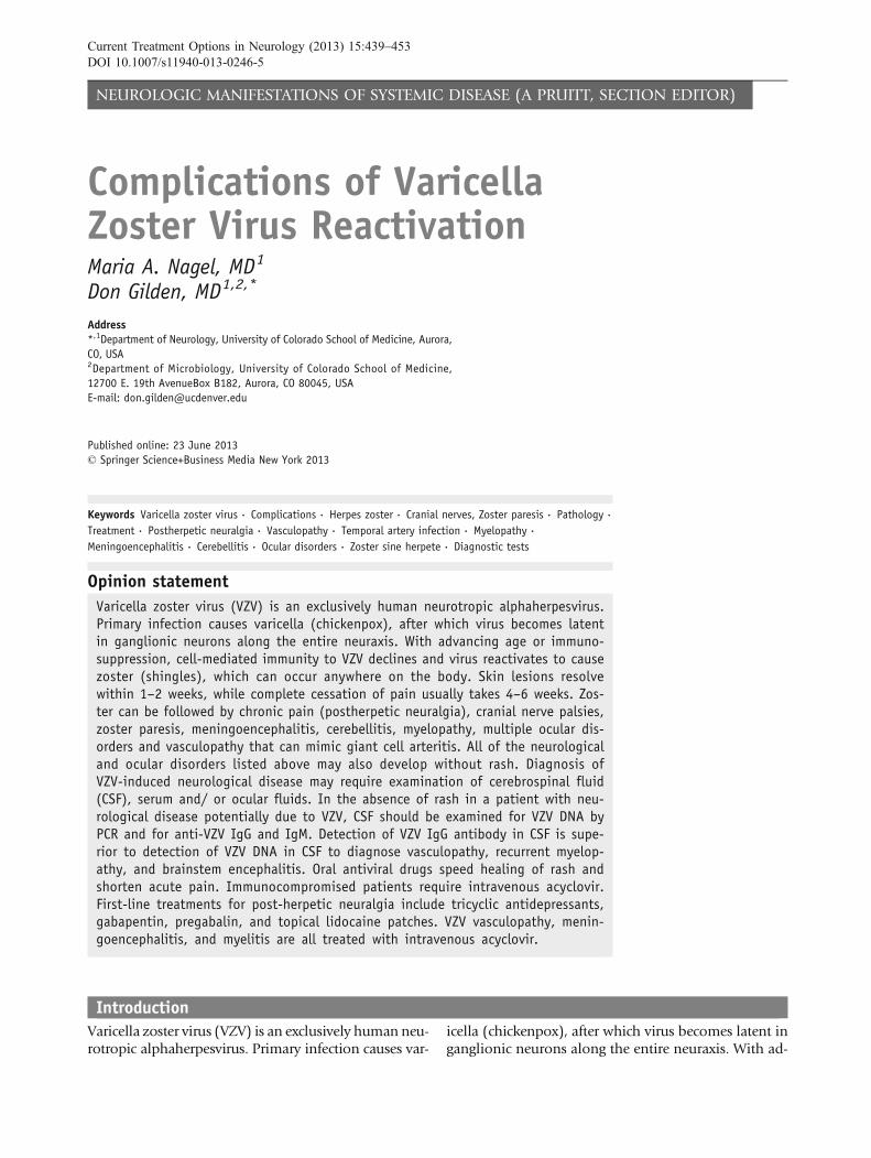
palsies, zoster paresis, vasculopathy, meningoencephali-tis, cerebellitis, myelopathy and multiple ocular disor-ders (Fig 1). VZV reactivation also produces chronicradicular pain without rash (zoster sine herpete).
Neurological complications of VZV reactivationHerpes zoster
Herpes zoster is the most common manifestation of VZV reactivation.Zoster is characterized by a vesicular eruption on an erythematous basein one to three dermatomes, accompanied by severe, lancinating radicu-lar pain; itching and unpleasant sensations (dysesthesias) produced bytouch (allodynia), as well as decreased sensation in the affected area.Rash and pain usually develop within a few days of each other, althoughpain can precede rash by weeks to months [1]. After reactivation fromcranial nerve, dorsal root or autonomic ganglia, VZV can travel peripher-ally to the corresponding dermatome; thus, zoster can affect any level ofthe neuraxis. The thoracic region is most commonly affected (9 50 %),followed by the face, cervical and lumbosacral regions.
The annual rate of zoster in the U.S. is 3.2 per 1,000 person-years [2].Zoster most frequently occurs in the elderly [3] as cell-mediated immu-nity to VZV declines. Other groups at risk for zoster are patients takingimmunosuppressive drugs (such as cancer patients, organ transplant re-cipients, and patients with autoimmune diseases and AIDS) [4]. Zosterin an otherwise healthy young person may be the first manifestation of HIV
Fig. 1. Neurological complications of VZV reactivation. Neurological complications of VZV reactivation (Reproduced from Gildenet al. [106]; copyright 2011, John Wiley & Sons; with permission).
440 NEUROLOGIC MANIFESTATIONS OF SYSTEMIC DISEASE (A PRUITT, SECTION EDITOR)
infection [5, 6]. Varicella in infancy also predisposes to zoster in early adulthood[7].
Zoster with cranial nerve involvementIn conjunction with dermatological manifestations of VZV reactivation, VZVcan reactivate from one or more cranial nerve ganglia to cause disease. Zostermay be followed by optic neuritis [8, 9] or ophthalmoplegia [9]. Herpes zos-ter ophthalmicus (HZO) is often accompanied by keratitis, which can lead toblindness. Patients with HZO and visual symptoms should have an immedi-ate slit-lamp examination by an ophthalmologist, particularly if skin lesionsextend to the nose (Hutchinson sign). Involvement of the maxillary andmandibular distribution of the trigeminal nerve can produce osteonecrosisand spontaneous tooth exfoliation [10].
Involvement of the geniculate ganglion (cranial nerve VII) causesweakness or paralysis of ipsilateral facial muscles. The combination of fa-cial palsy and vesicles in the external auditory canal or on the tympanicmembrane (zoster oticus) or on the ipsilateral anterior two-thirds of thetongue or hard palate [11] constitutes the Ramsay Hunt syndrome(RHS), which is often associated with tinnitus, hearing loss, nausea,vomiting, vertigo and nystagmus, indicating involvement of cranial nerveVIII within the bony facial canal. Facial paralysis in RHS is often more severethan in Bell’s palsy, and patients are less likely to recover completely [12].Zoster can also followed by involvement of cranial nerves IX, X, XI and XII[13, 14].
Cranial neuropathies frequently occur weeks after zoster, raising the pos-sibility that disease is due to micro-infarction of cranial nerves. Virus particlescan potentially spread transaxonally along trigeminal and other ganglionicafferent fibers to cause occlusion of small vessels supplying cranial nervesin the same manner that produces VZV vasculopathy in larger arteries (seeVZV vasculopathy below). The blood supply of cranial nerves III, IV, V1
and VI comes from the internal carotid circulation, while V2, V3 and VII,IX, X, XI and XII are supplied by the external carotid circulation [15]. It is im-portant to recognize that multiple forms of trigeminal-distribution [16, 17]and facial-distribution [18] zoster, as well as polyneuritis cranialis due toVZV [19, 20], may occur in the absence of rash.
Zoster paresisVZV reactivation from ganglia in the cervical, thoracic or lumbosacral re-gion can also cause weakness (zoster paresis).Arm weakness or diaphrag-matic paralysis [21, 22] occurs after cervical distribution zoster,abdominal muscle weakness and hernia after thoracic distribution zoster[23, 24], leg weakness after lumbar or sacral distribution zoster and uri-nary retention after sacral distribution zoster [25, 26]. Magnetic resonanceimaging (MRI) of patients with zoster paresis reveals involvement of bothanterior and posterior roots at the spinal level that correspond to the pa-tient’s clinical deficit [27]. Rarely, clinical deficit in cervical zoster paresisextends to the brachial plexus, confirmed by both electrodiagnostic test-ing and MRI [28]. In 45 patients with zoster paresis, 67 % had near-com-plete recovery [29], and in another 61 cases, 55 % had completefunctional recovery [30].
VZV Complications Nagel and Gilden 441

PathologyThe cardinal pathological features of zoster are characterized by inflammationand hemorrhagic necrosis with associated neuritis, localized leptomeningitis,unilateral segmental poliomyelitis and degeneration of related motor and sen-sory roots [31, 32]. Demyelination is seen in areas with mononuclear cell(MNC) infiltration and microglial proliferation. Intranuclear inclusions, viralantigen and herpesvirus particles have been found in acutely infected ganglia[33–35].
TreatmentAntiviral drugs, such as oral valacyclovir (1 gm three times daily for 7–10days) or acyclovir (800 mg 5 times daily for 7–10 days) speed healing of rashand shorten the duration of acute pain. Immunocompromised patients re-quire intravenous acyclovir (10–15 mg/kg every 8 hours for 10–14 days). Be-cause zoster pain may be associated with inflammation, many cliniciansadminister a short course of corticosteroids, e.g., oral prednisone, 1 mg/kgfor 5–7 days, in addition to antiviral therapy.
Postherpetic neuralgia (PHN)The most common neurological complication of zoster is PHN, defined asdermatomal-distribution pain that persists for more than 3 months after zos-ter. Age is the most important factor in predicting the development of PHN.More than 40 % of zoster patients 9 60 years of age experience chronic pain.Except for its longevity, the pain of PHN and associated allodynia are thesame as in zoster. The incidence of PHN is slightly greater in women [36]and after trigeminal distribution zoster [36–38].
The cause and pathogenesis of PHN are unknown. Two non-mutually ex-clusive theories are that: (1) excitability of ganglionic or even spinal cordneurons is altered; and (2) persistent productive virus infection exists inganglia. Analysis of ganglia from an early case of PHN of 2.5 months’ dura-tion revealed diffuse and focal infiltration by chronic inflammatory cells[39], an observation confirmed by Watson et al. [40] who found prominentcollections of lymphocytes in ganglia from a patient with PHN of 2 years’ du-ration. The inflammatory response in ganglia of these subjects raised the pos-sibility of prolonged viral infection. Further evidence that PHN may beproduced by low-level ganglionitis has come from the detection of VZVDNA and proteins in blood MNCs of many patients with PHN [41–43]and from the favorable response of some PHN patients to antiviral treatment[4, 44].
PHN is difficult to manage and no universal treatment exists. First-linetherapies include tricyclic antidepressants (TCAs), gabapentin andpregabalin, and topical lidocaine patches. Opioids, tramadol, capsaicincream and the capsaicin 8 % patch are recommended as second-line orthird-line therapies. TCAs such as amitriptyline are usually started at a doseof 10–25 mg orally at bedtime, with a maximum dose of 150–200 mg/day.Secondary amine TCAs, such as nortriptyline and despramine, can also beused due to a superior safety profile compared to the tertiary amineamitryptyline [45]. The calcium channel alpha (2)-delta ligands gabapentinand pregabalin are also used, with pregabalin providing equivalent efficacy
442 NEUROLOGIC MANIFESTATIONS OF SYSTEMIC DISEASE (A PRUITT, SECTION EDITOR)

to that of gabapentin, but at much lower doses due to its higher bioavailabil-ity and rapid absorption. Pregabalin is given at 75–150 mg orally twice dailyor 50–100 mg orally three times daily (150–300 mg/day). If minimal relief isobtained at 300 mg daily for 2 weeks, the dose can be increased to a maxi-mum of 600 mg/day in two or three divided doses. Opioids such as extend-ed-release oxycodone, morphine and methadone have shown efficacy inpatients with PHN. Tramadol is better tolerated but less effective than thesestronger opioids. The lidocaine 5 % patch has significant analgesic efficacy inpatients with PHN [46]. Capsaicin 0.075 % cream is sometimes prescribed,but the American Academy of Neurology (AAN) guidelines state that the an-algesia provided is below the threshold for a clinically important effect [47].The new capsaicin 8 % patch [48, 49], which delivers a high concentration ofcapsaicin in a single 60-minute application after application of local anes-thetic, is promising for the treatment of PHN, but its use awaits long-termsafety data. Combination therapy such as gabapentin and nortriptyline[50], morphine and gabapentin [51] or pregabalin and the lidocaine 5 %patch [52] may provide greater analgesic effects.
In patients who are refractory to non-invasive pharmacological inter-vention, botulinum toxin has successfully decreased PHN in several cases[53–55]. Epidural injection of steroids produced modest effects, but re-lief was short-lived [56]. Spinal cord stimulation has limited short-termsuccess [57].
A newer potentially promising treatment for PHN is percutaneous periph-eral nerve field stimulation. Rare reports indicate its effectiveness for refrac-tory PHN. Subjects became pain-free with minimal to no medicationneeded after ophthalmic-distribution [58], cervical-distribution [59] andthoracic-distribution [60, 61] PHN.
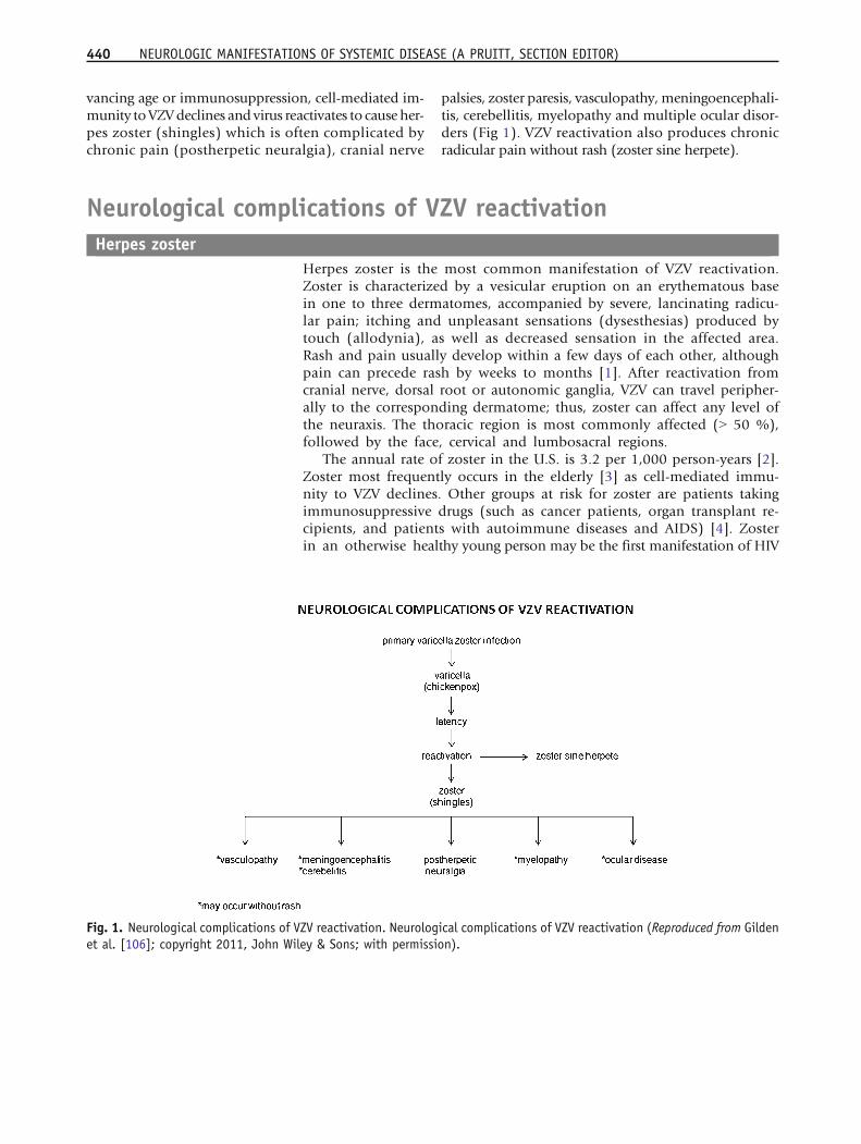
VZV vasculopathyAfter VZV reactivation from ganglia, virus can also travel centrally to infectcerebral arteries and cause ischemic and hemorrhagic stroke (VZVvasculopathy). The exact incidence of VZV vasculopathy is unknown, al-though it is a significant stroke risk factor. In adults with zoster, the risk ofstroke is increased by 30 % within the following year [62] and by 4.5-foldwhen zoster is in the ophthalmic distribution of the trigeminal nerve [63].Furthermore, up to one-third of pediatric ischemic arteriopathies are associ-ated with varicella [64]. VZV vasculopathy affects both immunocompro-mised and immunocompetent individuals and can present as headache,mental status changes and focal neurological deficits. In a study of 30 viro-logically verified cases of VZV vasculopathy [65], lesions at grey-white matterjunctions were frequently seen on MRI (Fig. 2A), and magnetic resonance an-giography (MRA) revealed focal arterial stenosis and occlusion in more thantwo-thirds of patients. Both large and small arteries were involved in 50 %patients, small arteries in 37 %, and large arteries alone in only 13 % inthe 30 subjects. Importantly: (1) up to one-third of patients did not have pre-ceding zoster rash; (2) up to one-third did not have a cerebrospinal fluid(CSF) pleocytosis; (3) detection of anti-VZV IgG antibody was superior to de-tection of VZV DNA in CSF for diagnosis; and (4) symptoms and signs oftenoccurred months after zoster [65].
VZV Complications Nagel and Gilden 443
Infarctions are mostly bland, but can also be hemorrhagic. Deep white-matter lesions often predominate and are ischemic or demyelinative,depending on the size of blood vessels involved. Infected cerebral arteriescontain multinucleated giant cells, Cowdry A inclusion bodies, herpesvirusparticles detected by electron microscopy, as well as VZV DNA and VZV an-tigen (Fig. 3). An analysis of 63 arteries from 45 normal subjects revealed noVZV DNA or VZV antigen, thus supporting the significance of VZV in arteriesof stroke patients [66]. A variety of vascular pathology has been reported,ranging from neointimal proliferation to necrosis with and without inflam-mation [67]. A recent study that examined the histological and immunohis-tochemical features of VZV-infected cerebral arteries from subjects with VZVvasculopathy revealed several distinct features: (1) the presence of VZV anti-gen in the arterial adventitia from early VZV vasculopathy and in the mediaand intima of protracted cases of VZV vasculopathy, supporting the notion ofvirus persistence in the artery and spread from the “outside-in”; (2) a thick-ened arterial intima composed of myofibroblasts and cells, most likely ofmedial smooth muscle origin; (3) a disrupted internal elastic lamina; and(4) a disrupted medial layer with a significant loss of normal smooth musclecells [68•]. The morphological changes help to explain why there is arterialocclusion and loss of vascular contractility, contributing to stroke. A fol-low-up study revealed that in VZV-infected arteries: (1) inflammatory cells,predominantly composed of T cells and macrophages, are present in adven-
Fig. 2. MRI scans of patients with varicella zoster virus (VZV) multifocal vasculopathy and myelopathy. (A) Proton-densitybrain MRI scan shows multiple areas of infarction in both hemispheres, particularly involving white matter. Arrows point togray-white matter junction lesions. (Reproduced from Gilden et al. [107]; copyright 2002, Springer; with permission.) (B) Notecervical, longitudinal, serpiginous enhancing lesions (arrows). (Reproduced from Gilden et al. [81]; copyright 1994, WoltersKluwer Health; with permission).
444 NEUROLOGIC MANIFESTATIONS OF SYSTEMIC DISEASE (A PRUITT, SECTION EDITOR)
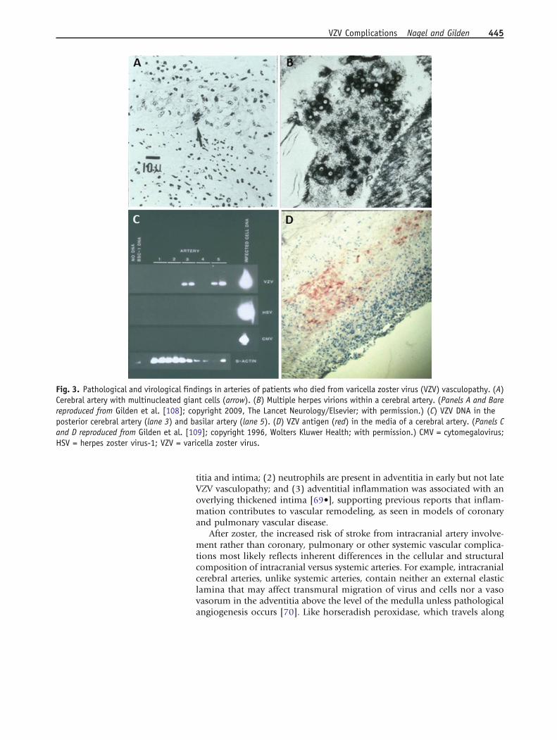
titia and intima; (2) neutrophils are present in adventitia in early but not lateVZV vasculopathy; and (3) adventitial inflammation was associated with anoverlying thickened intima [69•], supporting previous reports that inflam-mation contributes to vascular remodeling, as seen in models of coronaryand pulmonary vascular disease.
After zoster, the increased risk of stroke from intracranial artery involve-ment rather than coronary, pulmonary or other systemic vascular complica-tions most likely reflects inherent differences in the cellular and structuralcomposition of intracranial versus systemic arteries. For example, intracranialcerebral arteries, unlike systemic arteries, contain neither an external elasticlamina that may affect transmural migration of virus and cells nor a vasovasorum in the adventitia above the level of the medulla unless pathologicalangiogenesis occurs [70]. Like horseradish peroxidase, which travels along
Fig. 3. Pathological and virological findings in arteries of patients who died from varicella zoster virus (VZV) vasculopathy. (A)Cerebral artery with multinucleated giant cells (arrow). (B) Multiple herpes virions within a cerebral artery. (Panels A and Barereproduced from Gilden et al. [108]; copyright 2009, The Lancet Neurology/Elsevier; with permission.) (C) VZV DNA in theposterior cerebral artery (lane 3) and basilar artery (lane 5). (D) VZV antigen (red) in the media of a cerebral artery. (Panels Cand D reproduced from Gilden et al. [109]; copyright 1996, Wolters Kluwer Health; with permission.) CMV = cytomegalovirus;HSV = herpes zoster virus-1; VZV = varicella zoster virus.
VZV Complications Nagel and Gilden 445

trigeminal ganglionic afferent fibers to trigeminal ganglia after application tothe external surface of cerebral arteries [71, 72], reactivated VZV in gangliamay also travel along ganglionic afferent fibers to the adventitia of cerebralarteries, a notion consisted with the presence of viral antigen predominantlyin the adventitia of cerebral arteries in early VZV vasculopathy [73] and in themedia and intima of protracted cases [69•].
Multifocal VZV vasculopathy with temporal artery infectionWe have identified multifocal VZV vasculopathy in three patients with clin-ical and laboratory features mimicking giant cell arteritis (GCA). The first pa-tient was an 80-year-old man with left ophthalmic-distribution zoster whodeveloped painless left-sided loss of vision with an elevated ESR and CRP;he was diagnosed with possible GCA and underwent temporal artery biopsywhile being treated with steroids without improvement of vision. Biopsy wasGCA negative. Virological analysis revealed VZV antigen in his temporal ar-tery, after which he was treated with intravenous acyclovir and his vision im-proved [73]. A second even more remarkable case was a 75-year-old womanwithout a history of zoster, who developed left periorbital pain and loss ofvision with an elevated ESR and normal CRP; she was treated with steroidsfor presumed GCA and vision worsened; temporal artery biopsy revealedVZV antigen, and CSF analysis revealed the presence of anti-VZV IgG anti-body with reduced serum/CSF ratios of anti-VZV IgG antibody comparedto ratios for albumin and total IgG, indicative of intrathecal synthesis of an-ti-VZV IgG antibody; vision improved after antiviral treatment [74]. A thirdcase was a 54-year-old diabetic woman with no history of zoster rash whopresented with ischemic optic neuropathy and an elevated CRP [75]. The is-chemic optic neuropathy was followed by acute retinal necrosis with exten-sive venous beading characteristic of vasculopathy; PCR analysis of vitreousfluid in the same eye was positive for VZV DNA. The patient later developedjaw claudication and intermittent scalp pain; while temporal artery biopsywas GCA-negative, further study of the artery revealed VZV antigen(Fig. 4). These findings demonstrate that in patients with clinically suspectGCA, but whose temporal arteries are GCA-negative; VZV produces amultifocal vasculopathy affecting the ophthalmic and/or retinal arteries tocause vision loss, in addition to ipsilateral temporal artery infection. Treat-ment of these patients (with and without a history of zoster), with steroidsfor presumed GCA led to no improvement or to actual worsening of vision,while antiviral treatment improved vision.
Since VZV vasculopathy with temporal artery infection mimics GCA, weexamined formalin-fixed, paraffin-embedded temporal arteries obtainedfrom patients with clinically suspect GCA, but whose temporal arteries werepathologically negative. Immunohistochemical analysis revealed VZV anti-gen, but not HSV-1 antigen in five (21 %) of the 24 temporal arteries in mul-tiple regions of each artery [76••]; in contrast, none of 13 control temporalarteries obtained postmortem from subjects without symptoms or signs ofGCA contained VZV antigen. Overall, these findings indicate that in patientspresenting with clinical symptoms (particularly early visual disturbances),signs and laboratory abnormalities (particularly elevated CRP) consistentwith GCA, the diagnosis of multifocal VZV vasculopathy is an important con-
446 NEUROLOGIC MANIFESTATIONS OF SYSTEMIC DISEASE (A PRUITT, SECTION EDITOR)

sideration and warrants examination of the temporal artery not only for his-topathological features of GCA, but also for immunohistochemical evidenceof VZV antigen. Larger studies are needed to determine the full spectrum ofsymptoms, signs and laboratory abnormalities, as well as the characteristichistopathological features in temporal arteries of patients with multifocalVZV vasculopathy and temporal artery infection.
VZV meningitis, meningoencephalitis, meningoradiculitis and cerebellitisLike VZV vasculopathy, these neurological complications of VZV reactivation canoccur in the absence of zoster rash, as demonstrated by recent reports of VZVmen-ingitis [77], meningoradiculitis [78] and cerebellitis [79, 80] in which diagnosiswas confirmed by the detection of VZV DNA and anti-VZV antibody in CSF.
VZV myelopathyVZV myelopathy can present as a self-limiting, monophasic spastic paraparesis,with or without sensory features and sphincter problems. This so-calledpost-infectious myelitis usually occurs in immunocompetent patients daysto weeks after acute varicella or zoster. Its pathogenesis is unknown. TheCSF usually contains a mild mononuclear pleocytosis, with a normal orslightly elevated protein. Steroids are used to treat these patients, althoughsome improve spontaneously.
Fig. 4. Varicella zoster virus (VZV) in the temporal artery of a patient with VZV multifocal vasculopathy. (A) Positive controlcadaveric cerebral artery 14 days after VZV infection in vitro (pink color, arrows). (B) Note VZV antigen in the adventitia of thetemporal artery after staining with anti-VZV antibody (pink color, arrows), but not after staining adjacent sections with anti-HSV-1 antibody (C) or normal rabbit serum (D). Magnification 200X. (Reproduced from Mathias et al. [75]; copyright 2013,Elsevier; with permission).
VZV Complications Nagel and Gilden 447

VZV may directly invade the spinal cord or affect the spinal arteries to pro-duce myelopathy. In such instances, VZV myelopathy may present as an insid-ious, progressive and sometimes fatalmyelitis,mostly in immunocompromisedindividuals, such as patients with AIDS. MRI reveals longitudinal serpiginousenhancing lesions (Fig. 2B). Diagnosis is confirmed by the presence of VZVDNA or anti-VZV IgG or both in CSF [81]. Pathological and virological analysesof the spinal cord from fatal cases have revealed frank invasion of VZV in the pa-renchyma [67] and, in some instances, spread of virus to adjacent nerve roots[82]. Early diagnosis and aggressive treatment with intravenous acyclovir havebeen helpful, even in immunocompromised patients [83]. The benefit of ste-roids in addition to antiviral agents is unknown. Rarely, VZV myelitis recurs,even in immunocompetent patients [81]. VZVmyelitismay also occur in the ab-sence of zoster rash. VZV can also produce spinal cord infarction identified bydiffusion-weighted MRI and confirmed virologically [84]. Thus, VZVvasculopathy can cause stroke in the spinal cord as well as in the brain.
TreatmentVZV vasculopathy, meningoencephalitis and myelitis are all treated with in-travenous acyclovir, 10–15 mg/kg for 10–14 days. In immunocompromisedpatients, continued oral valacyclovir for months is sometimes necessary toprevent recurrence.
Ocular diseaseVZV infection produces acute retinal necrosis (ARN) or progressive outer ret-inal necrosis (PORN). ARN in both immunocompetent and immunocom-promised individuals presents with periorbital pain and floaters with hazyvision and loss of peripheral vision. Treatment is typically intravenous acy-clovir, steroids and aspirin followed by oral acyclovir [85]. Intravitreal injec-tions of foscarnet and oral acyclovir have also been effective. PORN presentswith painless loss of vision, floaters and constricted visual fields with resul-tant retinal detachment. Multifocal, discrete opacified lesions begin in theouter retinal layers peripherally and/or posterior pole; only late in diseaseare inner retinal layers involved. Diffuse retinal hemorrhages and whiteningwith macular involvement bilaterally are characteristic findings. VZV is themost common cause of PORN, although HSV and cytomegalovirus can alsocause this disease. Most cases are seen in AIDS patients with CD4+ T cellcounts less than 10 cells/mm3 of blood [86], as well as in otherimmunosuppressed individuals [87]. PORN may be preceded by retrobulbaroptic neuritis and aseptic meningitis [88], central retinal artery occlusion orophthalmic-distribution zoster [89] and may occur together with multifocalvasculopathy or myelitis. Treatment with intravenous acyclovir has givenpoor or inconsistent results [90] and even when acyclovir helped, VZV reti-nopathy recurred when drug was tapered or stopped. PORN patients treatedwith ganciclovir alone or in combination with foscarnet had a better final vi-sual acuity than those treated with acyclovir or foscarnet [91]. The best treat-ment for PORN in AIDS patients may be prevention with HAART [92].
Like other neurological disorders caused by VZV, ocular disease caused byVZV can also occur in the absence of rash. Multiple cases of PORN [93, 94]and a case of severe unremitting eye pain without rash were shown to be
448 NEUROLOGIC MANIFESTATIONS OF SYSTEMIC DISEASE (A PRUITT, SECTION EDITOR)

caused by VZV based on detection of VZV DNA in nasal and conjunctival sam-ples [95]. In addition, third cranial nerve palsies [96], retinal periphlebitis [97],uveitis [96], iridocyclitis [98] and disciform keratitis [99] that occurred withoutrash were confirmed virologically to be caused by VZV.
Zoster sine herpete (radicular pain in the absence of rash)Zoster sine herpete is recognized by clinicians as chronic radicular pain withoutrash causedbyVZV. Zoster sineherpetewas first described in a report ofmultiplepatients with dermatomal distribution radicular pain in areas distinct frompainwith rash in zoster [100]. The first two virologically confirmed cases of zostersine herpete were verified by detection of VZV DNA in CSF [101]. A third caseof thoracic-distribution zoster sine herpete, in which electromyography ofparaspinal muscles demonstrated frequent fibrillation potentials restricted tochronically painful thoracic root segments was confirmed by detection of VZVDNA in blood MNCs and anti-VZV IgG antibody in CSF [102]. Blumenthal etal. [103] recently described a patient with zoster sine herpete whose CSF didnot contain amplifiable VZV DNA, but did contain anti-VZV IgG with reducedserum/CSF ratios of anti-VZV IgG indicative of intrathecal synthesis. Perhaps themost compelling evidence that persistent radicular pain without rash can becaused by chronic active VZV ganglionitis came from analysis of a trigeminalganglionic mass removed from an immunocompetent adult who had experi-enced relentless trigeminal-distribution pain for more than a year; pathologicaland virological analyses of the ganglionic mass revealed active VZV ganglionitis[17]. The detection of VZV DNA and anti-VZV IgG and IgM antibody has ex-panded the spectrum of neurological disease produced by VZV in the absenceof rash to include VZV meningoencephalitis, vasculopathy, myelitis, cerebellarataxia and polyneuritis cranialis.
Diagnostic testsThe diagnosis of VZV-induced neurological disease is straightforward when thecharacteristic dermatomal distribution rash of zoster is present. When zosterrash is not present in a patient with neurological disease that can be causedby VZV (e.g., zoster sine herpete, vasculopathy, meningoencephalitis, myelopa-thy or retinal necrosis), examination of CSF and serum and ocular fluids is nec-essary. The routine CSF cell count can be helpful, since a mild lymphocyticpleocytosis is characteristically found in VZV vasculopathy, myelitis andmenin-goencephalitis. Furthermore, increased red blood cells and polymorphonuclearleukocytes are may also be seen when VZV infects the nervous system.
In the absence of rash, the CSF should be examined virologically for VZVDNA by PCR and for anti-VZV IgG and IgM. Detection of VZV DNA in CSF oranti-VZV IgM in serum or CSF is strong presumptive evidence of recent VZVinfection. If anti-VZV IgG antibody is present in CSF, the antibody indexshould be calculated to determine whether anti-VZV antibody is being pro-duced intrathecally. For molecules such as albumin and total IgG, the se-rum/CSF ratio is usually more than 100:1. A reduced ratio of anti-VZV IgGantibody compared to ratios for albumin or total IgG is seen in many neu-rological diseases produced by VZV. Importantly, many cases of VZVvasculopathy are protracted and VZV DNA is only found ~30 % of the time[65]. The detection of anti-VZV IgG antibody in CSF with intrathecal syn-
VZV Complications Nagel and Gilden 449

thesis is superior to detection of VZV DNA in CSF to diagnose VZVvasculopathy [104], recurrent myelopathy and brainstem encephalitisproduced by VZV [105].
AcknowledgmentsThis work was supported in part by National Institutes of Health grants AG006127 and AG032958 to DGand NS067070 to MAN. The authors wish to thank Marina Hoffman for editorial assistance and LoriDePriest for manuscript preparation.
Compliance with Ethics Guidelines
Conflict of InterestMaria A. Nagel and Don Gilden declare that they have no conflict of interest.
Human and Animal Rights and Informed ConsentThis article does not contain any studies with human or animal subjects performed by any of the authors.
References and Recommended ReadingPapers of particular interest, published recently, have beenhighlighted as:• Of importance•• Of major importance
1. Gilden DH, Dueland AN, Cohrs R, et al. Preherpeticneuralgia. Neurology. 1991;41:1215–8.
2. Insinga RP, Itzler RF, Pellissier JM, et al. The in-cidence of herpes zoster in a United States ad-ministrative database. J Gen Intern Med.2005;20:748–53.
3. Harnisch JP. Zoster in the elderly: clinical, immuno-logic and therapeutic considerations. J Am GeriatrSoc. 1984;32:789–93.
4. Gilden DH, Cohrs RJ, Mahalingam R. Clinical andmolecular pathogenesis of varicella virus infection.Viral Immunol. 2003;16:243–58.
5. Leppard B, Naburi AE. Herpes zoster: an earlymanifestation of HIV infection. Afr Health.1998;21:5–6.
6. Tyndall MW, Nasio J, Agoki E, et al. Herpes zoster asthe initial presentation of human immunodeficiencyvirus type 1 infection in Kenya. Clin Infect Dis.1995;21:1035–7.
7. Kakourou T, Theodoridou M, Mostrou G, et al. Her-pes zoster in children. J Am Acad Dermatol.1998;39:207–10.
8. Selbst RG, Selhorst JB, Harbison JW, et al.Parainfectious optic neuritis report and review fol-lowing varicella. Arch Neurol. 1983;40:347–50.
9. Kurimoto T, Tonari M, Ishizaki N, et al. Orbital apexsyndrome associated with herpes zosterophthalmicus. Clin Ophthalmo. 2011;5:1603–8.
10. Lambade P, Lambade D, Saha TK, et al. Maxillaryosteonecrosis and spontaneous teeth exfoliation fol-lowing herpes zoster. Oral Maxillofac Surg.2012;16:369–72.
11. Payten RJ, Dawes JDK. Herpes zoster of the head andneck. J Laryngol Otol. 1972;86:1031–55.
12. Robillard RB, Hilsinger Jr RL, Adour KK. RamsayHunt facial paralysis: clinical analyses of 185 pa-tients. Otolaryngol Head Neck Surg. 1986;95:292–7.
13. Steffen R, Selby G. ‘Atypical’ Ramsay Hunt syndrome.Med J Aust. 1972;1:227–30.
14. Asnis DS, Micic L, Giaccio D. Ramsay Hunt syn-drome presenting as a cranial polyneuropathy. Cutis.1996;57:421–4.
15. Lapresle J, Lasjaunias P. Cranial nerve ischaemic ar-terial syndromes. Brain. 1986;109:207–15.
450 NEUROLOGIC MANIFESTATIONS OF SYSTEMIC DISEASE (A PRUITT, SECTION EDITOR)
16. Easton HG. Zoster sine herpete causing acute tri-geminal neuralgia. Lancet. 1970;2:1065–6.
17. Hevner R, Vilela M, Rostomily R, et al. An unusualcause of trigeminal- distribution pain and tumour.Lancet Neurol. 2003;2:567–72.
18. Murakami S, Honda N, Mizobuchi M, et al. Rapiddiagnosis of varicella zoster virus infection in acutefacial palsy. Neurology. 1998;51:1202–5.
19. Osaki Y, Matsubayashi K, Okumiya K, et al. Poly-neuritis cranialis due to varicella-zoster virus in theabsence of rash. Neurology. 1995;45:2293–4.
20. Murata K, Miwa H, Kondo T. Polyneuritis cranialiscaused by varicella zoster virus in the absence of rash.Neurology. 2010;74:85–6.
21. Brostoff J. Diaphragmatic paralysis after herpes zos-ter. Br Med J. 1966;2:1571–2.
22. Stowasser M, Cameron J, Oliver WA. Diaphragmaticparalysis following cervical herpes zoster. Med J Aust.1990;153:555–6.
23. Tjandra J, Mansel RE. Segmental abdominal herpeszoster paresis. Aust N Z J Surg. 1986;56:807–8.
24. Molinero J, Nagore E, Obón L, et al. Metamericmotor paresis following abdominal herpes zoster.Cutis. 2002;69:143–4.
25. Izumi AK, Edwards Jr J. Herpes zoster and neurogenicbladder dysfunction. JAMA. 1973;224:1748–9.
26. Jellinek EH, Tulloch WS. Herpes zoster with dys-function of bladder and anus. Lancet. 1976;2:1219–22.
27. Umehara T, Sengoku R, Mitsumura H, et al. Findingsof segmental zoster paresis on MRI. J NeurolNeurosurg Psychiatry. 2011;82:694.
28. Choi JY, Kang CH, Kim BJ, et al. Brachial plexopathyfollowing herpes zoster infection: two cases with MRIfindings. J Neurol Sci. 2009;285:224–6.
29. Gupta SK, Helal BH, Kiely P. The prognosis in zosterparalysis. J Bone Joint Surg Br. 1969;51:593–603.
30. Thomas EJ, Howard Jr FM. Segmental zoster paresis –a disease profile. Neurology. 1972;22:459–66.
31. Head H, Campbell AW. The pathology of herpeszoster and its bearing on sensory localization. Brain.1900;23:353–523.
32. Denny-Brown D, Adams RD, Fitzgerald PJ. Patho-logic features of herpes zoster: A note on "geniculateherpes.". Arch Neurol Psychiatry. 1944;51:216–31.
33. Cheatham WJ, Dolan Jr TF, Dower JC, et al. Varicella:report on two fatal cases with necropsy, virus isola-tion, and serologic studies. Am J Pathol.1956;32:1015–35.
34. Esiri MM, Tomlinson AH. Herpes zoster: demon-stration of virus in trigeminal nerve and ganglion byimmunofluorescence and electron microscopy. JNeurol Sci. 1972;15:35–48.
35. Ghatak NR, Zimmerman HM. Spinal ganglion inherpes zoster. Arch Pathol. 1973;95:411–5.
36. Hope-Simpson RE. Postherpetic neuralgia. J R CollGen Pract. 1975;25:571–5.
37. de Moragas JM, Kierland RR. The outcome of patientswith herpes zoster. Arch Dermatol. 1957;75:193–6.
38. Rogers III RS, Tindall JP. Herpes zoster in the elderly.Postgrad Med. 1971;50:153–7.
39. Smith FP. Pathological studies of spinal nerve gangliain relation to intractable intercostal pain. SurgNeurol. 1978;10:50–3.
40. Watson CPN, Deck JH, Morshead C, et al.Postherpetic neuralgia: further post-mortem studiesof cases with and without pain. Pain. 1991;44:105–17.
41. Vafai A, Wellish M, Gilden DH. Expression of vari-cella-zoster virus in blood mononuclear cells of pa-tients with postherpetic neuralgia. Proc Natl Acad SciUSA. 1988;85:2767–70.
42. Devlin ME, Gilden DH, Mahalingam R, et al. Pe-ripheral blood mononuclear cells of the elderlycontain varicella-zoster virus DNA. J Infect Dis.1992;165:619–22.
43. Mahalingam R,Wellish M, Brucklier J, et al. Persistenceof varicella-zoster virus DNA in elderly patients withpostherpetic neuralgia. J NeuroVirol. 1995;1:130–3.
44. Terada K, Niizuma T, Kawano S, et al. Detection ofvaricella-zoster virus DNA in peripheral mononucle-ar cells from patients with Ramsay Hunt syndrome orzoster sine herpete. J Med Virol. 1998;56:359–63.
45. Attal N, Cruccu G, Baron R, et al. EFNS guidelines onthe pharmacological treatment of neuropathic pain.Eur J Neurol. 2010;17:1113–88.
46. Hans G, Sabatowski R, Binder A, et al. Efficacy andtolerability of a 5 % lidocaine medicated plaster forthe topical treatment of post-herpetic neuralgia: re-sults of a long-term study. Curr Med Res Opin.2009;25:1295–305.
47. Dubinsky RM, Kabbani H, El-Chami Z, et al. Practiceparameter: treatment of postherpetic neuralgia: anevidence-based report of the Quality Standards Sub-committee of the American Academy of Neurology.Neurology. 2004;63:959–65.
48. Backonja M, Wallace MS, Blonsky ER, et al. NGX-4010 C116 Study Group. NGX-4010, a high-con-centration capsaicin patch, for the treatment ofpostherpetic neuralgia: a randomised, double-blindstudy. Lancet Neurol. 2008;7:1106–12.
49. Backonja MM, Malan TP, Vanhove GF, et al. NGX-4010, a high-concentration capsaicin patch, for thetreatment of postherpetic neuralgia: a randomized,double-blind, controlled study with an open-labelextension. Pain Med. 2010;11:600–8.
50. Gilron I, Bailey JM, Tu D. Nortriptyline andgabapentin, alone and in combination for neuro-pathic pain: a double-blind, randomised controlledcrossover trial. Lancet. 2009;374:1252–61.
51. Gilron I, Bailey JM, Tu D, et al. Morphine,gabapentin, or their combination for neuropathicpain. N Engl J Med. 2005;352:132–1334.
52. Rehm S, Binder A, Baron R. Post-herpetic neuralgia: 5% lidocaine medicated plaster, pregabalin, or a
VZV Complications Nagel and Gilden 451

combination of both? A randomized, open, clinicaleffectiveness study. Curr Med Res Opin.2010;26:1607–19.
53. Ruiz Huete C, Bermejo PE. Botulinum toxin type A inthe treatment of neuropathic pain in a case ofpostherpetic neuralgia [in Spanish]. Neurologia.2008;23:259–62.
54. Sotiriou E, Apalla Z, Panagiotidou D, et al. Severepost-herpetic neuralgia successfully treated withbotulinum toxin A: three case reports. Acta DermVenereol. 2009;89:214–5.
55. Xiao L, Mackey S, Hui H, et al. Subcutaneous injec-tion of botulinum toxin a is beneficial inpostherpetic neuralgia. Pain Med. 2010;11:1827–33.
56. van Wijck AJ, Opstelten W, Moons KG, et al. ThePINE study of epidural steroids and local anaes-thetics to prevent postherpetic neuralgia: a random-ized controlled trial. Lancet. 2006;367:219–24.
57. Harke H, Gretenkort P, Ladleif HU, et al. Spinal cordstimulation in postherpetic neuralgia and in acuteherpes zoster pain. Anesth Analg. 2002;94:694–700.
58. Surjya PU, Shiv PR, Mishra S, et al. Successful treat-ment of an intractable postherpetic neuralgia (PHN)using peripheral nerve field stimulation (PNFS). AmJ Hosp Palliat Care. 2010;27:59–62.
59. Lynch PJ, McJunkin T, Eross E, et al. Case report:successful epiradicular peripheral nerve stimulationof the C2 dorsal root ganglion for postherpetic neu-ralgia. Neuromodulation. 2011;14:58–61.
60. Yakovlev AE, Peterson AT.: Peripheral nerve stimu-lation in treatment of intractable postherpetic neu-ralgia. Neuromodulation 2007, 373-375.
61. Kouroukli I, Neofytos D, Panaretou V, et al. Periph-eral subcutaneous stimulation for the treatment ofintractable postherpetic neuralgia: two case reportsand literature review. Pain Prac. 2009;9:225–9.
62. Kang JH, Ho JD, Chen YH, et al. Increased risk ofstroke after a herpes zoster attack: a population-based follow-up study. Stroke. 2009;40:3443–8.
63. Lin HC, Chien CW, Ho JD. Herpes zosterophthalmicus and the risk of stroke: a population-based follow-up study. Neurology. 2010;74:792–7.
64. Amlie-Lefond C, Bernard TJ, Sébire G, et al. Predic-tors of cerebral arteriopathy in children with arterialischemic stroke: results of the International PediatricStroke Study. Circulation. 2009;119:1417–23.
65. Nagel MA, Cohrs RJ, Mahalingam R, et al. The vari-cella zoster vasculopathies: clinical, CSF, imaging,and virologic features. Neurology. 2008;70:853–60.
66. Nagel MA, Choe A, Khmeleva N, et al.: Search forvaricella zoster virus and herpes simplex virus-1 innormal human cerebral arteries. J NeuroVirol.2013;19:181–85.
67. Kleinschmidt-DeMasters BK, Gilden DH. Varicella-zoster virus infections of the nervous system: clinicaland pathologic correlates. Arch Pathol Lab Med.2001;125:770–80.
68.• Nagel MA, Traktinskiy I, Azarkh Y, et al. Varicellazoster virus vasculopathy: analysis of virus-infectedarteries. Neurology. 2011;77:364–70.
Correlative analysis of viral and muscle cell markers in ce-rebral arteries from patients with VZV vasculopathy69.• Nagel MA, Traktinskiy I, Stenmark KR, et al. Varicella-
zoster virus vasculopathy: immune characteristics ofvirus-infected arteries. Neurology. 2013;80:62–8.
Analysis of the immune repertoire in arteries of patients withVZV vasculopathy70. Lee RM. Morphology of cerebral arteries. Pharmacol
Ther. 1995;66:149–73.71. Mayberg MR, Langer RS, Zervas NT, et al. Perivascular
meningeal projections from cat trigeminal ganglia:possible pathway for vascular headaches in man.Science. 1981;213:228–30.
72. Mayberg MR, Zervas NT, Moscowitz MA. Trigeminalprojections to supratentorial pial and dural bloodvessels in cats demonstrated by horseradish peroxi-dase histochemistry. J Comp Neurol. 1984;223:46–56.
73. Salazar R, Russman AN, Nagel MA, et al. VZV ische-mic optic neuropathy and subclinical temporal arteryinvolvement. Arch Neurol. 2011;68:517–20.
74. Nagel MA, Russman AN, Feit DO, et al. VZV ischemicoptic neuropathy andsubclinical temporal artery in-fection without rash. Neurology. 2013;80:220–2.
75. Mathias M, Nagel MA, Khmeleva N, et al. VZVmultifocal vasculopathy withischemic optic neurop-athy, acute retinal necrosis and temporal artery in-fection in the absence of zoster rash. J Neurol Sci.2013;325:180–2.
76.•• Nagel MA, Bennett JL, Khmeleva N, et al.: MultifocalVZV vasculopathy withtemporal artery infectionmimics giant cell arteritis. Neurology 2013, (inpress).
Exciting new study indicating that the clinical features ofmultifocal VZV vasculopathy with temporal artery infectioncan be the same as seen in classic giant cell arteritis.77. Habib AA, Gilden D, Schmid DS, et al. Varicella
zoster virus meningitis with hypoglycorrhachia in theabsence of rash and in an immunocompetent wom-an. J Neurovirol. 2009;15:206–8.
78. Gunson RN, Aitken C, Gilden D. A woman withacute headache and sacral dermatomal numbness. JClin Virol. 2011;50:191–3.
79. Moses H, Nagel MA, Gilden DH. Acute cerebellarataxia in a 41 year old woman. Lancet Neurol.2006;5:984–8.
80. Ratzka P, Schlachetzki JC, Bähr M, et al. Varicellazoster virus cerebellitis in a 66-year-old patientwithout herpes zoster. Lancet. 2006;367:182.
81. Gilden DH, Beinlich BR, Rubinstien EM, et al. Vari-cella-zoster virus myelitis: an expanding spectrum.Neurology. 1994a;44:1818–23.
82. Devinsky O, Cho ES, Petito CK, et al. Herpes zostermyelitis. Brain. 1991;114:1181–96.
452 NEUROLOGIC MANIFESTATIONS OF SYSTEMIC DISEASE (A PRUITT, SECTION EDITOR)
83. de Silva SM, Mark AS, Gilden DH, et al. Zoster my-elitis: improvement with antiviral therapy in twocases. Neurology. 1996;47:929–31.
84. Orme HT, Smith G, Nagel MA, et al. VZV spinal cordinfarction identified bydiffusion-weighted magneticresonance imaging (DWI). Neurology. 2007;69:398–400.
85. Bonfioli AA, Eller AW. Acute retinal necrosis. SeminOphthalmol. 2005;20:155–60.
86. Guex-Crosier Y, Rochat C, Herbort CP. Necrotizingherpetic retinopathies. A spectrum of herpes virus-induced diseases determined by the immune state ofthe host. Ocul Immunol Inflamm. 1997;5:259–65.
87. Lewis JM, Nagae Y, Tano Y. Progressive outer retinalnecrosis after bone marrow transplantation. Am JOphthalmol. 1996;122:892–5.
88. Franco-Paredes C, Bellehemeur T, Merchant A, et al.Aseptic meningitis and optic neuritis preceding vari-cella-zoster progressive outer retinal necrosis in apatient with AIDS. AIDS. 2002;16:1045–9.
89. Menerath JM, Gerard M, Laurichesse H, et al. Bilateralacute retinal necrosis in a patient with acquired im-munodeficiency syndrome. J Fr Ophtalmol.1995;18:625–33.
90. Johnston WH, Holland GN, Engstrom Jr RE, et al.Recurrence of presumed varicella-zoster virus reti-nopathy in patients with acquired immunodeficiencysyndrome. Am J Ophthalmol. 1993;116:42–50.
91. Moorthy RS, Weinberg DV, Teich SA, et al. Manage-ment of varicella zoster virus retinitis in AIDS. Br JOphthalmol. 1997;81:189–94.
92. Austin RB. Progressive outer retinal necrosis syn-drome: a comprehensive review of its clinicalpresentation, relationship to immune system sta-tus, and management. Clin Eye Vis Care.2000;12:119–29.
93. Friedman SM, Mames RN, Sleasman JW, et al. Acuteretinal necrosis after chickenpox in a patient withacquired immunodeficiency syndrome. ArchOphthalmol. 1993;111:1607–8.
94. Galindez OA, Sabates NR, Whitacre MM, et al. Rap-idly progressive outer retinal necrosis caused by var-icella zoster virus in a patient infected withhumanimmunodeficiency virus. Clin Infect Dis.1996;22:149–51.
95. Goon P, Wright M, Fink C. Ophthalmic zoster sineherpete. J R Soc Med. 2000;93:191–2.
96. Hon C, Au WY, Cheng VC. Ophthalmic zoster sineherpete presenting as oculomotor palsy after marrow
transplantation for acute myeloid leukemia.Haematologica. 2005;90:12. EIM04.
97. Noda Y, Nakazawa M, Takahashi D, et al. Retinalperiphlebitis as zoster sine herpete. ArchOphthalmol. 2001;119:1550–2.
98. Yamamoto S, Tada R, Shimomura Y, et al. Detectingvaricella-zoster virus DNA in iridocyclitis using po-lymerase chain reaction: a case of zoster sine herpete.Arch Ophthalmol. 1995;113:1358–9.
99. Silverstein BE, Chandler D, Neger R, et al. Disciformkeratitis: a case of herpes zoster sine herpete. Am JOphthalmol. 1997;123:254–5.
100. Lewis GW. Zoster sine herpete. Br Med. 1958;J2:418–42.
101. Gilden DH, Wright RR, Schneck SA, et al. Zostersine herpete, a clinical variant. Ann Neurol.1994;35:530–3.
102. Amlie-Lefond C, Mackin GA, Ferguson M, et al.Another case of virologically confirmed zoster sineherpete with electrophysiologic correlation. JNeurovirol. 1996;2:136–8.
103. Blumenthal DT, Shacham-Shmueli E, Bokstein F, etal. Zoster sine herpete: virological verification bydetection of anti-VZV IgG antibody in CSF. Neu-rology. 2011;76:484–5.
104. Nagel MA, Forghani B, Mahalingam R, et al. Thevalue of detecting anti-VZV IgG antibody in CSF todiagnose VZV vasculopathy. Neurology.2007;68:1069–73.
105. Haug A, Mahalingam R, Cohrs RJ, et al. Recurrentpolymorphonuclear pleocytosis with increased redblood cells caused by varicella zoster virus infectionof the central nervous system. J Neurol Sci.2010;292:85–8.
106. Gilden D, Mahalingam R, Nagel MA, PugazhenthiS, Cohrs RJ. Review: The neurobiology of varicellazoster virus infection. Neuropathol ApplNeurobiol. 2011;37:441–63.
107. Gilden DH, Mahalingam R, Cohrs RJ, et al. Theprotean manifestations of varicella-zoster virusvasculopathy. J NeuroVirol. 2002;8:75–9.
108. Gilden D, Cohrs RJ, Mahalingam R, et al. Varicellazoster virus vasculopathies: diverse clinical mani-festations, laboratory features, pathogenesis, andtreatment. Lancet Neurol. 2009;8:731–40.
109. Gilden DH, Kleinschmidt-DeMasters BK, Wellish M,et al. Varicella zoster virus, a cause of waxing andwaning vasculitis: the NEJM case 5-1995 revisited.Neurology. 1996;47:1441–6.
VZV Complications Nagel and Gilden 453