coeliac disease pathological mimics and complications
TRANSCRIPT

Coeliac Disease (CD)Pathological mimics and
complications
Dr. Shaun Walsh
Dept of Pathology
Ninewells Hospital, Dundee

Pathological mimics and complications25 minutes
• Recent challenging case
• Place of histopath. in investigating CD
• Common mimics of CD
• Refractory CD

A recent case
• 7 year old boy
• c/o Abdo pain
• Type 1 DM, TTG 52
• D2 biopsies under GA
• Villous blunting, crypt hyperplasia

Raised intraepithelial lymphocytes (IELs)
40/100 Ecs

One bx normal villi ... but increased IELs
• Loss of the decrescendopattern of IELS
• >23 IELS/20 x5tip ECs
• The stomach was also biopsied ……
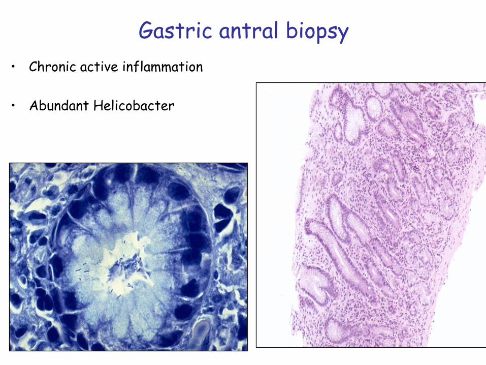
Gastric antral biopsy
• Chronic active inflammation
• Abundant Helicobacter

Report and MDT
• Features in keeping with CD present however......
• H.Pylori gastritis also present which can be associated with duodenal changes in children
• Clinical correlation and discussion at MDTM is required
• CD clinically/serologically but family very reluctant for GFD
• Decision: Treat H.pylori first and rebiopsy

6 months later ... H.Pylori eradicated
• Pt asymptomatic, TTG 15
• Second GA and endoscopy (Stomach normal)
• Path report: .... could represent CD but not specific
• Paediatricians feel he may have latent CD
• Family very unhappy, feel it is all due to H.Pylori. Patient not on GFD
• Who is right?
• How much weight should we give to biopsy findings?
Normal villi
Raised IELs

BSPGHAN 2005 guidelinesInvestigation of CD
• Clinical investigation pt on gluten
• Serology (TTG)
• Duodenal biopsy required for confirmation of dx
• Must have villous atrophy (VA) and increased IELs
• Increased IELs (Marsh 1) alone not specific
• Response to GFD to be documented

BSG Guidelines Adults 2010CD is commonest cause of enteropathy in UK by some margin
• Duodenal biopsy is the mandatory gold standard
• Serology has false negatives (IgA defic) and there is a need for certainty
• If Pt. on gluten and has
Villous atrophy (VA) and raised IELs ... confident

BSG/AGA: Qualifiers
• Site , number, orientation of biopsies
• Confident description/experienced pathologist
• ‘where possible histology should be reviewed in the clinical context ..diet, serology, symptoms
• ‘VA and increased IELs are not exclusive to CD’
• ‘ a small bowel biopsy is the current gold standard...’
Three paragraphs later
• ‘ small bowel biopsy in itself is not a perfect gold standard ...
• ‘Histologic features are not unique to coeliac disease’.
BSG AGA

Summary of all guidelines until 2011:
• Duodenal biopsies are required to investigate CD
• Villous flattening and increased IELs on duodenal biopsy nearly always means coeliac disease in the correct
CONTEXTPathologists do not diagnose CD by themselves
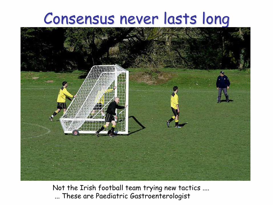
Consensus never lasts long
Not the Irish football team trying new tactics ....... These are Paediatric Gastroenterologist

Investigation of CD in children is changing
• BSPGHAN May 2012 (ESPGHAN)
• Children with clinical evidence of CD
• IgA Anti-TTG > 10 times normal
• (Pref. With EMA Ab+, HLA-DQ2,DQ8+)
• No duodenal biopsy (No GA, no delay)
• Diagnosed with probable CD
• Straight to GFD
Arch Dis Child May 2012 Vol 97; 393-394
J Pediatr Gastroent Nutr 2012 Jan 54: 136-60
J Pediatr Gastroenterol Nutr 2012 Oct 29 Epub

Investigation of CD in children is changing
Paediatricians
• Biopsy if asymptomatic
• Biopsy only when no response to GFD
• Our patient would probably not be biopsied at first!
Pathologists
• New case mix
• Typical histology will be relatively rarer
• Tricky cases: Latent CD, compliance issues, and mimics will appear more commonplace

Pathological Mimics of CD• Tropical sprue
• Bacterial overgrowth
• Blind loop syndrome
• CVID, IPEX
• AIDS enteropathy
• Graft v’s Host dse
• Malnutrition
• Zollinger Ellison
• Ischaemia
• Radiation
• MVID
• Viral enteritis
• Giardiasis
• Acid-peptic injury
• Helicobacter pylori
• Drugs/immunosuppression
• Crohns disease
• Collagenous sprue
• Derm. herpetiformis
• RCD2/Lymphoma
• Autoimmune enteritis
• Allergic gastroenteritis

RARE and not often biopsied mimics of CD
• Tropical sprue
• Bacterial overgrowth
• Blind loop syndrome
• CVID, IPEX
• AIDS enteropathy
• Graft v’s Host dse
• Malnutrition (here)
• Zollinger Ellison
• Ischaemia
• Radiation
• MVID
• Viral enteritis
• Giardiasis
• Acid-peptic injury
• Helicobacter pylori
• Drugs/immunosuppression
• Crohn’s disease
• Collagenous sprue
• Derm. herpetiformis
• RCD2/Lymphoma
• Autoimmune enteritis
• Allergic gastroenteritis

Commoner MIMICS OF CD• Tropical sprue
• Bacterial overgrowth
• Blind loop syndrome
• CVID, IPEX
• AIDS enteropathy
• Graft v’s Host dse
• Malnutrition
• Zollinger Ellison
• Ischaemia
• Radiation
• MVID
• Viral enteritis
• Giardiasis
• Acid-peptic injury
• Helicobacter pylori
• Drugs/immunosuppression
• Crohn’s disease
• Collagenous sprue
• Derm. herpetiformis
• RCD2/Lymphoma
• Autoimmune enteritis
• Allergic gastroenteritis

Self inflicted
• Biopsies must be correctly orientated
• Need 3 levels
• 3-5 reasonably parallel villi
• Tangential sections make villi look shorter

I. Acid-peptic duodenal injury
• Very common duodenal pathology
• Associated with H.Pylori infection
• Direct effect of acid/enzymes on duodenal mucosa
• Commonest in D1 (physiological?)

Six features of acid-peptic injury
1. Variable villous flattening 2. Crypt hyperplasia

Features of acid-peptic injury
3. Brunner gland hyperplasia 4. Gastric metaplasia (HP+/-)
5. Variable amounts of acute and chronic inflammation ....
6. Normal numbers of IELs
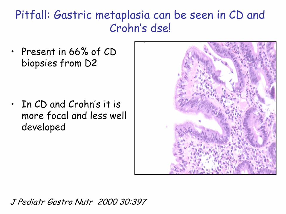
Pitfall: Gastric metaplasia can be seen in CD and Crohn’s dse!
• Present in 66% of CD biopsies from D2
• In CD and Crohn’s it is more focal and less well developed
J Pediatr Gastro Nutr 2000 30:397

Pitfall: Neutrophils can be seen in CD and Crohn’s dse.!
• 28-56% of CD biopsies
from D2
• Commoner in children
• May relate to severity of disease
Am J Clin Pathol 2012 138:42-9Am J Surg Path 2012 36:1339-45

Overlapping features of acid-peptic injury and CD
Acid-peptic injury• Villous flattening
• Crypt hyperplasia
• Gastric metaplasia
• Brunner gland hyper
• Acute inflammation
• Chronic inflammation
• Normal IEL’s
Coeliac disease• Villous flattening
• Crypt hyperplasia
• Focal gastric metaplasia
• Acute inflammation
• Chronic inflammation
• Abnormal brush border
• Raised IELs
Traditionally D2 is biopsied to avoid D1 acid-peptic injury .........

Update: D1 (bulb) bx’s for CD
• CD involves bulb in the vast majority (cranio-caudal)
• Recent studies .. 6-18% of CD missed if only D2 biopsied
• D1 biopsies should come from 9-12 o’ clock position
• D1 x2 and D2 x4 biopsies are now proposed for adults
(already std for paediatricians)
J Pediatr Gast Nutr 2008 47: 618-622
JClin pathol 2012 65:791-4
Am J Gastroenterol 2011 106: 1837-742
Gastrointest Endosc 2012 75: 1190-6

Problems with bulb biopsies:
- Acid-peptic injury
- Aware D1 villi are usually shorter than D2
- Brunner gland hyperplasia and lymphoid aggregates distort/flatten villi
- Scrappy bx’s are harder to orientate and assess (3-4 parallel villi)

What are the features of Coeliac dse in D1?
• Same as for traditonal D2 biopsies
• Raised IELs and villous flattening
• Similar IEL counts in CD bxs from D1 and D2
• May be more severe lesions in D1 than D2 Gastroent 2010 139: 112-119
Am J Gastro 2011 10:1837-742BMC Gastroenterol 2009 9:78-85

Pitfall: Lymphoid aggregates in bulb bx’s
• IELs are always increased over lymphoid aggregates
• Even in normal biopsies
• If considering counting IELs .. Count somewhere else

H.Pylori Gastritis
• May cause 2-3% of Marsh 1 lesions in Children
• Also associated with lymphocytic gastritis
Gut 1999 45:495-498J Pediat Gastro Nutr Epub 2012
Lessons
• Acid peptic injury and CD share some features
• Raised IELs most helpful for recognising CD but watch out for H.Pylori
• CD can involve the bulb
• Caution reporting CD in bulb biopsies alone
• Always demand separate pots for D1 and D2 or multiwell casettes

II. Drugs
• A growing problem
• List is getting longer
• Rarely get history/context!

42 yr old lady: Hx of diarrhoea and renal failure
Partial villous flattening and mild increase in IELs
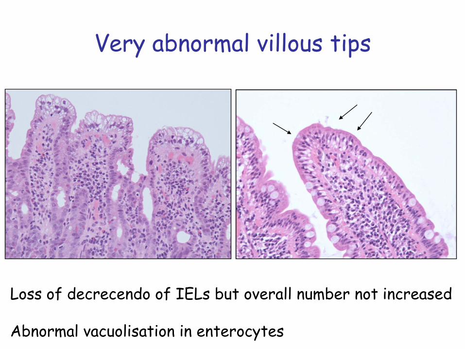
Very abnormal villous tips
Loss of decrecendo of IELs but overall number not increased
Abnormal vacuolisation in enterocytes

Context: Pt actually had a renal transplant
• And was taking

Mycophenylate
• Immunosuppressive drug ..
• Known GI toxicity
• Increasingly used transplants/vasculitis
• Also causes colonic damage

Mycophenylate clue: Apoptosis in crypts
RAREin this case!
RAREIn this case!

Other drugs can cause villous flattening and/or increased IELs
• Azothioprine (transplant setting)
• Conventional chemotherapy
• HAART
• Triparanol
• ACE inhibitor Olmesartan (Olmetec)
• Context is crucial
Mayo Clinic Proceed 2012 87:732-8

III. Immunocompromise
• AIDS enteropathy
• Hypogammaglobulinemia
• T cell disorders e.g. IPEX syndrome
• Rarely IgA deficiency
• Rare, mostly children but some adults

CVID Common variable immunodeficiency
• Abnormality throughout GI tract
• Lymphocytic gastritis
• 60% increase IELs in duod
• 80% villous blunting
• Lymphoid nodulesAm J Surg Path 2007 31: 1800-12Am J Gastroent 2003 98: 118-21

IPEXImmunodysregulation polyendocrinopathy
enteropathy X linked inheritance
• FOX P3 mutation
• Deficient Treg cells
• Numerous infections
• Can mimic CD, GVHD or produce goblet cell depletion
Modern Pathology (2009) 22, 95–102
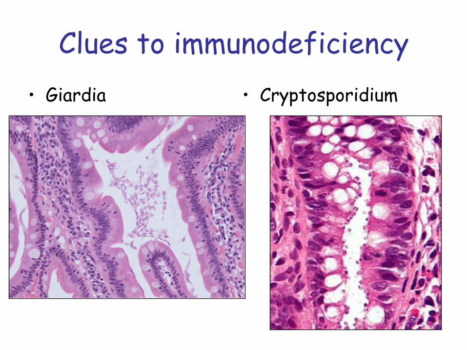
Clues to immunodeficiency
• Giardia • Cryptosporidium
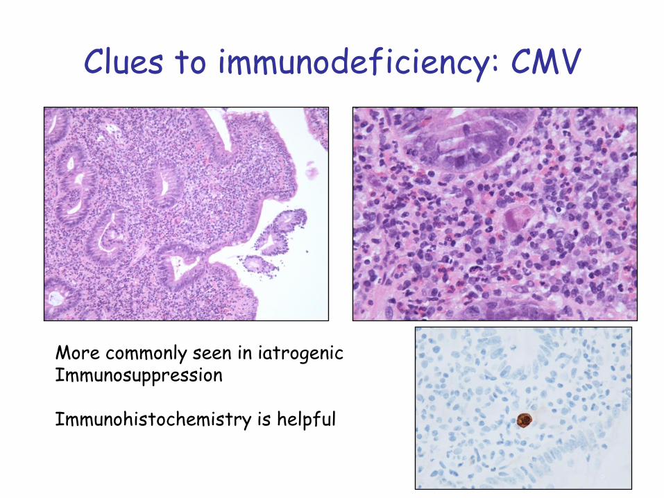
Clues to immunodeficiency: CMV
More commonly seen in iatrogenicImmunosuppression
Immunohistochemistry is helpful

Clues to immunodeficiency: MAI
ZN and PAS positive DDx is Whipples disease

When we have context path can be very useful!
7 year old girl post small bowel transplant
Distorted mucosa Something in the intervillous space
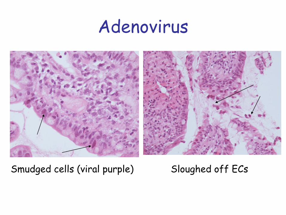
Adenovirus
Smudged cells (viral purple) Sloughed off ECs
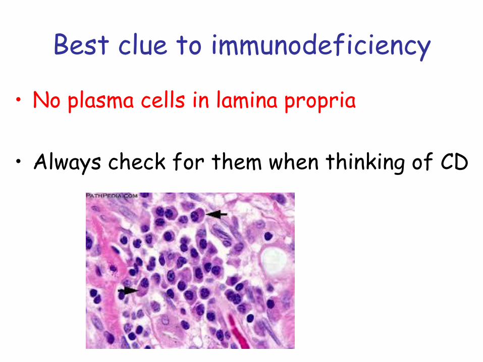
Best clue to immunodeficiency
• No plasma cells in lamina propria
• Always check for them when thinking of CD

Other infections: Tropical sprue
• Perfect mimic
• Need history of travel to endemic regions
• Biopsy features identical to CD
• Rarely biopsied

Tropical Sprue
Not just ‘tropical’
Villous flattening Raised IELs

Pathological Mimics of CD• Tropical sprue
• Bacterial overgrowth
• Blind loop syndrome
• CVID, IPEX
• AIDS enteropathy
• Graft v’s Host dse
• Malnutrition
• Zollinger Ellison
• Ischaemia
• Radiation
• MVID
• Viral enteritis
• Giardiasis
• Acid-peptic injury
• Helicobacter pylori
• Drugs/immunosuppression
• Crohns disease
• Collagenous sprue
• Derm. herpetiformis
• RCD2/Lymphoma
• Autoimmune enteritis
• Allergic gastroenteritis

Giardiasis in Immunocompetent patients
• Rarely see flat villi
Aonach Eagach ridge

IV. Autoimmune and Allergic diseases
• Crohn’s disease
• Autoimmune enteropathy
• Collagenous sprue

Crohn’s disease
• Good pathological mimic of Coeliac disease
• Can rarely coexist with CD
• TTG can be raised
• Essential to have clinical information e.g mouth ulcers, colonic disease
• Examine other biopsies from colon or stomach if available
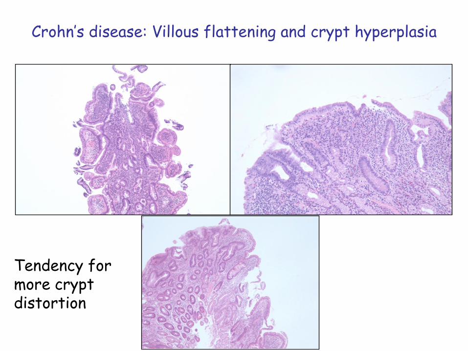
Crohn’s disease: Villous flattening and crypt hyperplasia
Tendency for more crypt distortion

Crohn’s disease: Increased IELs
Often intraepithelial component is more polymorphous than Coeliac dse.
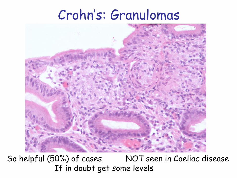
Crohn’s: Granulomas
So helpful (50%) of cases NOT seen in Coeliac diseaseIf in doubt get some levels

Autoimmune Enteropathy
• Rare: Adults and children
• Villous flattening and increased IELs
• Anti-enterocyte Ab’s
• No Coeliac autoantibodies
• Other autoimmune diseases
From: Akram S, Murray JA, Pardi DS et al. Adult Autoimmune Enteropathy: Mayo Clinic Rochester Experience. Clin Gastroenterol Hepatol 2007;5:1282–1290

Collagenous sprue
• Rare
• Deposition of subepithelial collagen
• Variable villous atrophy
• In the past associated with a poor prognosis
• Now GFD and immunosuppression effective treatment in most
AJSP 2009; 33:1440–1449Mod Pathol 2010; 23: 12-26

Collagenous Sprue: collagen entrapping immune cells
Dr Aoife Maguire ISSP meeting 2011

Allergy• Common in children
but rarely biopsied
• Clinically diagnosed/tx by exclusion diet
• Cows milk/ Eggs/Wheat
• Confusion with CD VA and Inc IEL’s
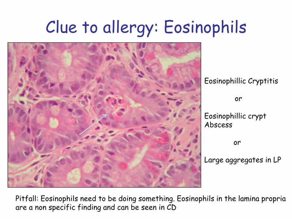
Clue to allergy: Eosinophils
Pitfall: Eosinophils need to be doing something. Eosinophils in the lamina propriaare a non specific finding and can be seen in CD
Eosinophillic Cryptitis
or
Eosinophillic crypt Abscess
or
Large aggregates in LP

How should we report CD biopsies?
• ….the findings are in keeping with CD in the correct clinical context…
• ..... in keeping with CD and serology should confirm the dx whilst the patient is on a GFD...
• ........ in keeping with CD. Clinical and serological correlation is urged.

Immunohistochemistry?
• No role in routine
diagnosis
• Not useful for counting
• Not useful for spotting mimics
CD3

Dr Goldman on CD
• ‘ The histopathologic features are non-specific in nature and can be seen in many other conditions.’
• CONTEXT

Complications of CD
• Refractory CD
• Lymphoma (GI and elsewhere)
• Carcinoma
• Liver cancer (Sweden)
• Other AI disease (thyroid)
• Malnutrition
• Others

Refractory Coeliac Disease (RCD)
• RCD may be defined as persistent or refractory symptomatic malabsorption together with villous atrophy which persists despite adherence to a gluten free diet.
• 6/12, EATL excluded
• Combined clinical, pathological and molecular approach is necessary
• A real case
Walker and Murray Histopath 2011; 59; 166-179.Ho-Yen et al Histopathology 2009; 54; 783-795.

A case of RCD
• 2006/7
• Pt had weight loss and diarrhoea
• Flat biopsy with IELs

Pathological Diagnosis of RCD• First review the previous biopsy and make
sure it is Coeliac disease and not a mimic
• Pt presented 2001
Recovered on GFD a few IELs 2002

Follow up: Pt treated with immunosupressive drugs
• Recovered for about a year
• Then fell apart
• Developed Acute abdomen
• Surgery revealed multiple perforations

Ulcerative jejeno-ileitis
Multiple discrete ulcers without tumour mass
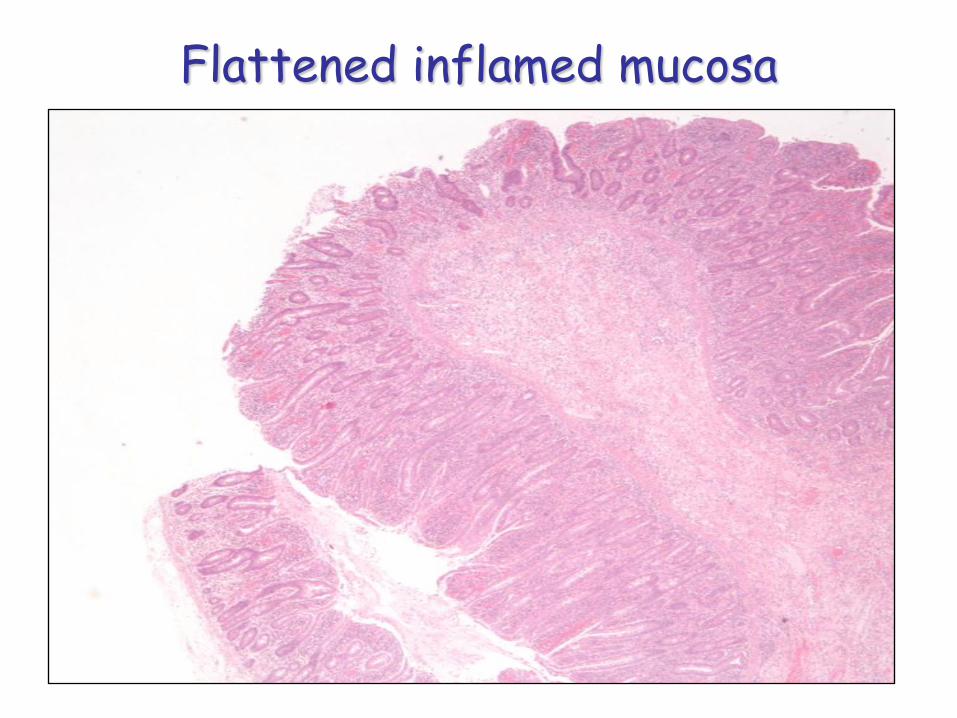
Flattened inflamed mucosa
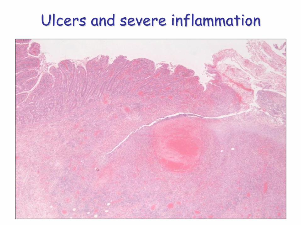
Ulcers and severe inflammation

Base of ulcers
Polyclonal by IHC and TCR

EATL: Post mortem

EATL: High grade lymphoma
MIB
CD3 +CD8 -

Biopsy features of RCD
Degree of villousflattening varies!
Almost total
Partial

Biopsy features of RCDBut increased Intraepithelial lymphocytes (IELs)
are always a feature

Recent advance : RCD split into two types on basis of finding aberrant T-cell clone
• Type 1
Polyclonal T cell IELs with a typical Coeliac immunophenotype CD3+CD8+
• Type 2
Clonal T cell IELs (TCR gamma or beta chain gene rearrangements) with loss of CD8 and cytoplasmic expression of CD3 instead of membranous localisation
Time to involve Haematopathologist

RCD Type 1
CD3 CD8
Quick and dirty alternative to double staining!

RCD Type 2
CD3 CD8
Cytoplasmic Almost entirely lost
CD3 CD8
Patient 1
Patient 2

RCD: IHC issues
• Double staining may be more sensitive
• ThresholdsRCD if >50% of IELs have abnormal phenotype
• Flow cytometryRCD if >20%

90% of IELsin normal mucosa
70% of IELsin CD
10% of IELs in normal mucosa
30% of IELsin CD
T cell receptor rearrangements
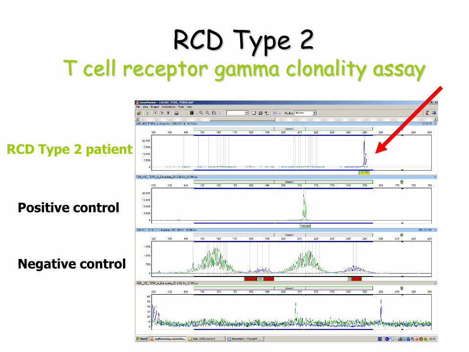
RCD Type 2 T cell receptor gamma clonality assay
RCD Type 2 patient
Positive control
Negative control

Pitfalls
• TCR studies are not perfect
• Small biopsies with small numbers of T cells can yield false positives
• Poor quality DNA can yield false negatives
• Some RCD patients may have unusual T –cell clones

Warning!• Patients who are not compliant with their GFD may
develop abnormal clones of IELs by IHC/TCR rearrangement ……
• But they can regress when back on a GFD!
• Continuously monitoring in patients suspected of RCD may be better than snapshot analysis
• Persistence of abnormal clone is most concerning
Gut 2010 59: 452-456

Importance of RCD
• Persistent Type 2 RCD at greater risk of developing Ulcerative Jejenitis and EATL (same immunophenotype)
• 5 year survival for type 2 RCD < 44%
• Change treatment: Many clinicians will treat RCD 2 patients with immunosuppression

EATL
Ind J Cancer 2011; 48:124J Gastro Oncol 2012 3: 209-225
Type I
Type II (CD3+, CD8+)
CD3m CD8 –CD7, CD103+Granzyme, Perforin+TCR clonal by PCR

RCD Pathology Conclusions
• Clinical context is VITAL
• Pt must be on a strict GFD
• Immunohistochemistry and molecular genetic analysis needed to subtype RCD
• Development of EATL almost always fatal

Thanks
• Prof D Sanders
• Dr Nigel Reynolds
• Dr Mike Bisset
• Dr Dagmar Karstner
• Dr Aoife Maguire