coagulation abnormalities in - iagh · coagulation abnormalities in ... thromboelastography (teg)...
TRANSCRIPT


Coagulation abnormalities inpatients with liver disease
H.Zojajy M.D.
&
MJ.Ehsani Ardakani M.D.
October 2015

• The clotting process is a dynamic, highly interwoven array that occurring in four phases :
Ø Initiation and formation of the platelet plug
ØPropagation of the clotting process by the coagulation cascade
ØTermination of clotting by antithrombotic control mechanisms
ØRemoval of the clot by fibrinolysis

• But in Patients with liver disease……
• We have a disturbed balance of pro-coagulant and anti-coagulant factors deviating from the normal coagulation cascade, with little in the way of "reserve”
• But why??

• 1-Increased bleeding risk :
• Decreased production of non-endothelial cell-derived coagulation factors (eg, factors II, V, VII, IX, X, XI, XIII).
• Thrombocytopenia
• altered platelet function
• abnormalities of fibrinogen
• decreased thrombin activatable fibrinolysis inhibitor (TAFI).
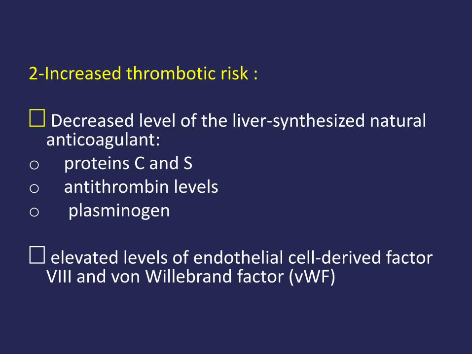
2-Increased thrombotic risk :
Ø Decreased level of the liver-synthesized natural anticoagulant:
o proteins C and So antithrombin levelso plasminogen
Ø elevated levels of endothelial cell-derived factor VIII and von Willebrand factor (vWF)

• So …..
• In liver disorders especially cirrhosis we have disruption of these opposing pathways lead to different and potentially changing hemostasis.

• Another problem in these patients is:
• The relative balance or imbalance in these
patients is not reflected in conventional indices of coagulation, such as the
prothrombin time (PT), activated partial thromboplastin time (aPTT) or International Normalized Ratio (INR) .

PT/INR ????
• hypo or hyper coagulopathy??
• Predicting the occurrence of bleeding based on factor levels or composites of factor activity such as the prothrombin time is difficult, due to the occurrence of counter forces, which favor thrombosis and the development of super-imposed conditions such as infection.
• We will talk more about this problem !


HYPOCOAGULABILITY IN CIRRHOSIS

HYPOCOAGULABILITY IN CIRRHOSIS
• Multiple factors can result in a hypocoagulablestate in patients with cirrhosis:
• 1-Decreased levels of all liver synthesized procoagulantfactors, including the vitamin K dependent clotting factors (II, VII, IX, and X) .

2 : Decreased other liver-synthesized clotting factors include:
• fibrinogen
• factors V, XI, XII
• prekallikrein, kininogen
• plasminogen.

3-Abnormalities of platelet number
Theories exist regarding the genesis of
thrombocytopenia:
• Decreased thrombopoietin (TPO) levels
• Splenic sequestration of platelets due to portal hypertension
• Auto-antibody destruction of platelets
• Bone marrow suppression due to underlying liver disease
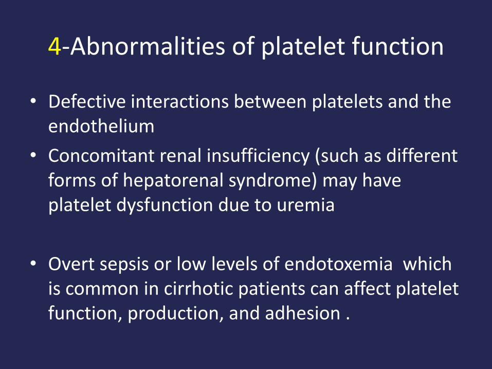
4-Abnormalities of platelet function
• Defective interactions between platelets and the endothelium
• Concomitant renal insufficiency (such as different forms of hepatorenal syndrome) may have platelet dysfunction due to uremia
• Overt sepsis or low levels of endotoxemia which is common in cirrhotic patients can affect platelet function, production, and adhesion .

Methods of measuring platelet aggregation
• Clot Signature Analyzer
• Thrombotic Status Analyzer
• Platelet Function Analyzer

5-Hyperfibrinolysis
• Evidence of systemic fibrinolysis can be detected in 30 to 46 percent of patients.
• However, clinically evident hyperfibrinolysis is less common and has been estimated to occur in 5 to 10 percent of those with decompensated cirrhosis .

• So in brief…..
• We could have HYPOCOAGULABILITY IN CIRRHOSIS due to:
• 1-Decreased levels of liver synthesized procoagulantfactors
• 2-Abnormalities of platelet number• 3-Abnormalities of platelet function• 4-Hyperfibrinolysis

• But on the other hand we have another big problem………

HYPERCOAGULABILITY

• Hypercoagulability is an increasingly recognized aspect of liver disease.
• The lack of proper measurement tools to identify those patients who are prone to develop clots, and reliance on clinical endpoints (eg, deep vein thrombosis, portal vein thrombosis) likely lead to an under-estimation of this problem.

• Awareness that the old dogma of "autoanticoagulation" represented by an elevated International Normalized Ratio (INR) in patients with cirrhosis is unfounded.
• Hypercoagulability in cirrhosis can lead to macro and micro-thrombi production, resulting in various complications.
• Several endothelium- derived procoagulantfactors are increased in cirrhosis, including factor VIII & von Willebrand factor

Macro-thromboticComplications include
• Portal vein thrombosis
• Deep vein thrombosis
• Pulmonary embolism

Micro-thrombotic complication
• Potential micro-thrombotic complications of cirrhosis are subtle
• Intra-hepatic microthrombi → localized ischemia →scarring and accelerated development of cirrhosis in a process known as parenchymal extinction → liver atrophy
• Similar process in lung microvasculature appears to play arole in a serious complication of cirrhosis knownas portopulmonary hypertension
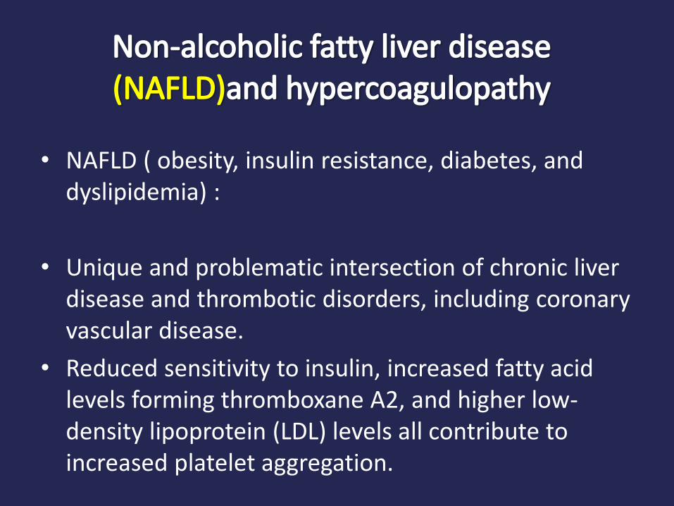
Non-alcoholic fatty liver disease (NAFLD)and hypercoagulopathy
• NAFLD ( obesity, insulin resistance, diabetes, and dyslipidemia) :
• Unique and problematic intersection of chronic liver disease and thrombotic disorders, including coronary vascular disease.
• Reduced sensitivity to insulin, increased fatty acid levels forming thromboxane A2, and higher low-density lipoprotein (LDL) levels all contribute to increased platelet aggregation.

Portal vein thrombosis
• PVT affects about 10 to 20 percent of all patients with cirrhosis
• The prevalence varies with disease severity, being much lower in Child-Pugh A
Contributing factors include:• Stasis due to portal hypertension• Development of hepatocellular cancer (HCC)• Sometimes to genetic predisposition to
hypercoagulability.
Location of PVT and its outcome
• Focal left or right branch PVT is relatively more common and often clinically silent, although its development may contribute to overall organ atrophy.
• Extrahepatic portal vein thrombosis (EPVT) is relatively less common but has increased risk of decompensation due to variceal bleeding or portosystemic shunting.

Search for what in PVT ??
• The development of any of these forms warrants a careful examination for hepatocellular carcinoma
• EPVT especially should prompt consideration of a more exhaustive search for additional factors, such as prothrombin gene mutation, factor V Leiden, or a myeloproliferative neoplasm (eg, polycythemia vera), especially if there is mesenteric extension

• In general, treatment has resulted in partial or complete recanalization in 40 to 80 percent, with minimal anticoagulation-associated bleeding.

The definition of acute/subacute thrombosis:
• Newly found absence of flow within six months of previous imaging.
• Early initiation of anticoagulation was the only factor significantly associated with recanalization
• Re-thrombosis after complete recanalization occurred in 38 percent of patients after anticoagulation was stopped.

• So ….
• We have a disease ( cirrhosis) with two distinct and different coagulation disorders .
• The question is what we should do in clinical practice ….. Its one of the challengeable entity in gastroenterology!!

Clinical aspects

Significance of type of liver disease
• Many of the hemostatic abnormalities are similar regardless of the underlying cause of liver injury. However we have some differences:
• ●Cholestatic disease ( PBC , PSC) : less pronounced effect on antihemostatic mechanisms and may be at higher risk for portal vein thrombosis. This mild hypercoagulability may be mediated by changes in platelet activity
• ●NAFLD: may confer a greater prothrombotic risk.
• ●Acute liver failure may have a lower incidence of thrombocytopenia but more severe reductions in coagulant and anticoagulant factors, compared with chronic hepatic insufficiency.

• As we mentioned above we have a COMMON CLINICAL PROBLEM …….abnormalities in routine laboratory tests of coagulation: PT,PTT….
• But these tests are very poor at predicting the risk of bleeding in individuals with liver disease because they only reflect changes in procoagulant factors .

What is more accurate testing of hemostasis ??
• Thromboelastography (TEG)
• Thromboelastometry (ROTEM)
Reflects dynamic changes in clot formation
and lysis.
• Sonorheometry

Bases of the thromboelastogram( TEG )


Thromboelastography (TEG) tracing parameters
“R” is the reaction time (the time it takes the coagulation cascade to generate thrombin and fibrin). “K” is the clot firmness. “a” (alpha) is the angle (describe the kinetics of clot formation). MA is tha maximum amplitude (describe the maximum clot strength). Ly30 is the percent clot lysis 30 minutes after the MA is reached.

Examples of qualitative TEG traces for interpretation