acquired coagulation abnormalities. acquired coagulation abnormalities - causes 1. vitamin k...
TRANSCRIPT
ACQUIRED COAGULATION ABNORMALITIES
ACQUIRED COAGULATIO
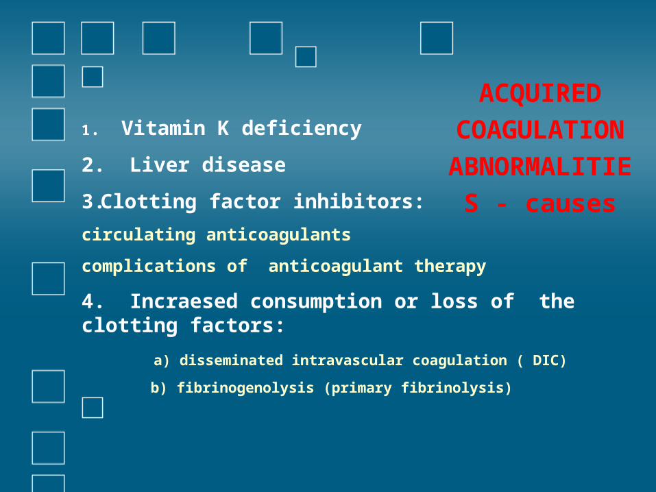
N ABNORMALITIES - causes
1. Vitamin K deficiency
2. Liver disease
3.Clotting factor inhibitors:
circulating anticoagulants
complications of anticoagulant therapy
4. Incraesed consumption or loss of the clotting factors:
a) disseminated intravascular coagulation ( DIC)
b) fibrinogenolysis (primary fibrinolysis)
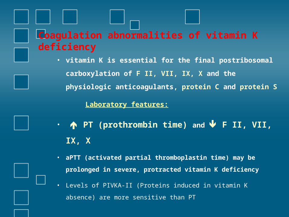
Coagulation abnormalities of vitamin K deficiency
• vitamin K is essential for the final postribosomal
carboxylation of F II, VII, IX, X and the physiologic
anticoagulants, protein C and protein S
Laboratory features:
• PT (prothrombin time) and F II, VII,
IX, X
• aPTT (activated partial thromboplastin time) may be
prolonged in severe, protracted vitamin K deficiency
• Levels of PIVKA-II (Proteins induced in vitamin K absence) are
more sensitive than PT
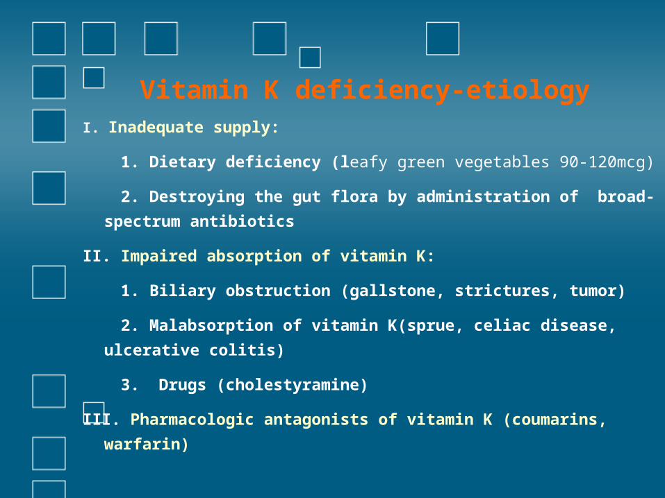
Vitamin K deficiency-etiologyI. Inadequate supply:
1. Dietary deficiency (leafy green vegetables 90-120mcg)
2. Destroying the gut flora by administration of broad-
spectrum antibiotics
II. Impaired absorption of vitamin K:
1. Biliary obstruction (gallstone, strictures, tumor)
2. Malabsorption of vitamin K(sprue, celiac disease,
ulcerative colitis)
3. Drugs (cholestyramine)
III. Pharmacologic antagonists of vitamin K (coumarins,
warfarin)
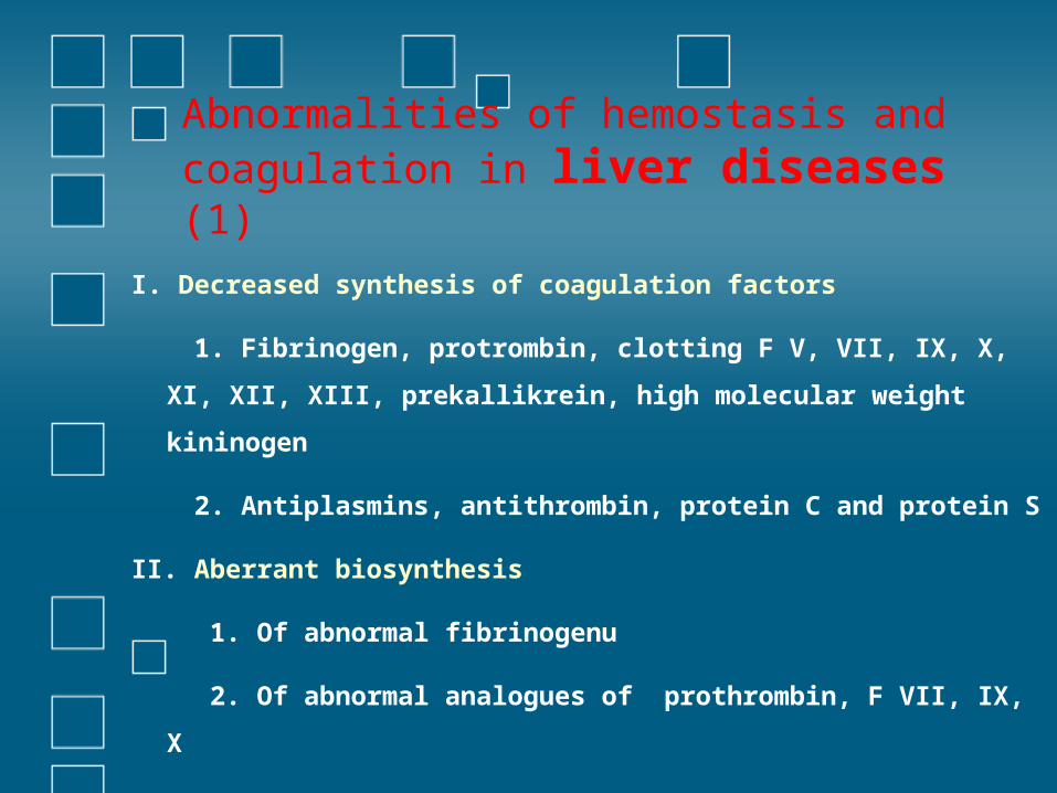
Abnormalities of hemostasis and coagulation in liver diseases (1)
I. Decreased synthesis of coagulation factors
1. Fibrinogen, protrombin, clotting F V, VII, IX, X, XI, XII,
XIII, prekallikrein, high molecular weight kininogen
2. Antiplasmins, antithrombin, protein C and protein S
II. Aberrant biosynthesis
1. Of abnormal fibrinogenu
2. Of abnormal analogues of prothrombin, F VII, IX, X
Abnormalities of hemostasis and
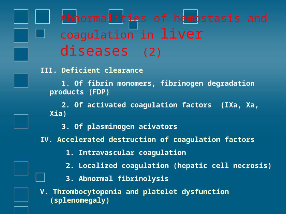
coagulation in liver diseases (2)
III. Deficient clearance
1. Of fibrin monomers, fibrinogen degradation products (FDP)
2. Of activated coagulation factors (IXa, Xa, Xia)
3. Of plasminogen acivators
IV. Accelerated destruction of coagulation factors
1. Intravascular coagulation
2. Localized coagulation (hepatic cell necrosis)
3. Abnormal fibrinolysis
V. Thrombocytopenia and platelet dysfunction (splenomegaly)
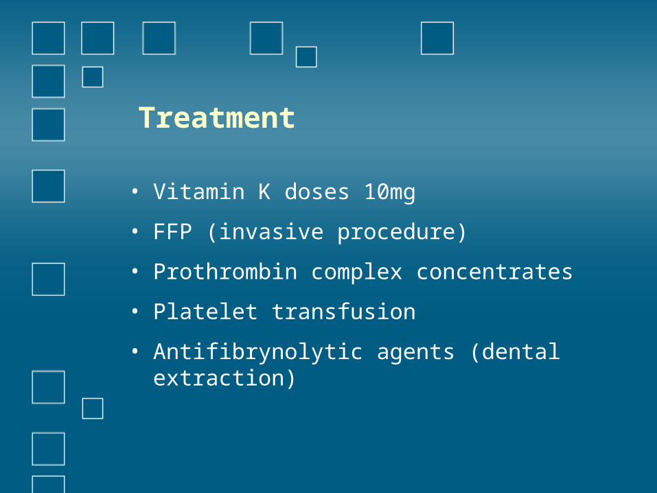
Treatment
• Vitamin K doses 10mg
• FFP (invasive procedure)
• Prothrombin complex concentrates
• Platelet transfusion
• Antifibrynolytic agents (dental extraction)
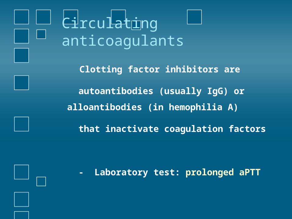
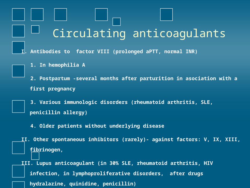
Circulating anticoagulants
Clotting factor inhibitors are
autoantibodies (usually IgG) or
alloantibodies (in hemophilia A)
that inactivate coagulation factors
- Laboratory test: prolonged aPTT
Circulating anticoagulants
I. Antibodies to factor VIII (prolonged aPTT, normal INR)
1. In hemophilia A
2. Postpartum -several months after parturition in asociation with a first
pregnancy
3. Various immunologic disorders (rheumatoid arthritis, SLE, penicillin
allergy)
4. Older patients without underlying disease
II. Other spontaneous inhibitors (rarely)- against factors: V, IX, XIII,
fibrinogen,
III. Lupus anticoagulant (in 30% SLE, rheumatoid arthritis, HIV infection, in
lymphoproliferative disorders, after drugs hydralazine, quinidine,
penicillin)
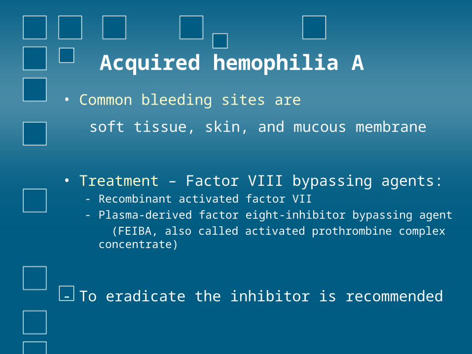
Acquired hemophilia A
• Common bleeding sites are
soft tissue, skin, and mucous membrane
• Treatment – Factor VIII bypassing agents:- Recombinant activated factor VII- Plasma-derived factor eight-inhibitor bypassing agent (FEIBA, also called activated prothrombine complex
concentrate)
- To eradicate the inhibitor is recommended
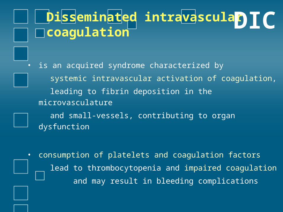
Disseminated intravascular coagulation • is an acquired syndrome characterized by
systemic intravascular activation of coagulation,
leading to fibrin deposition in the microvasculature
and small-vessels, contributing to organ dysfunction
• consumption of platelets and coagulation factors
lead to thrombocytopenia and impaired coagulation
and may result in bleeding complications
DIC
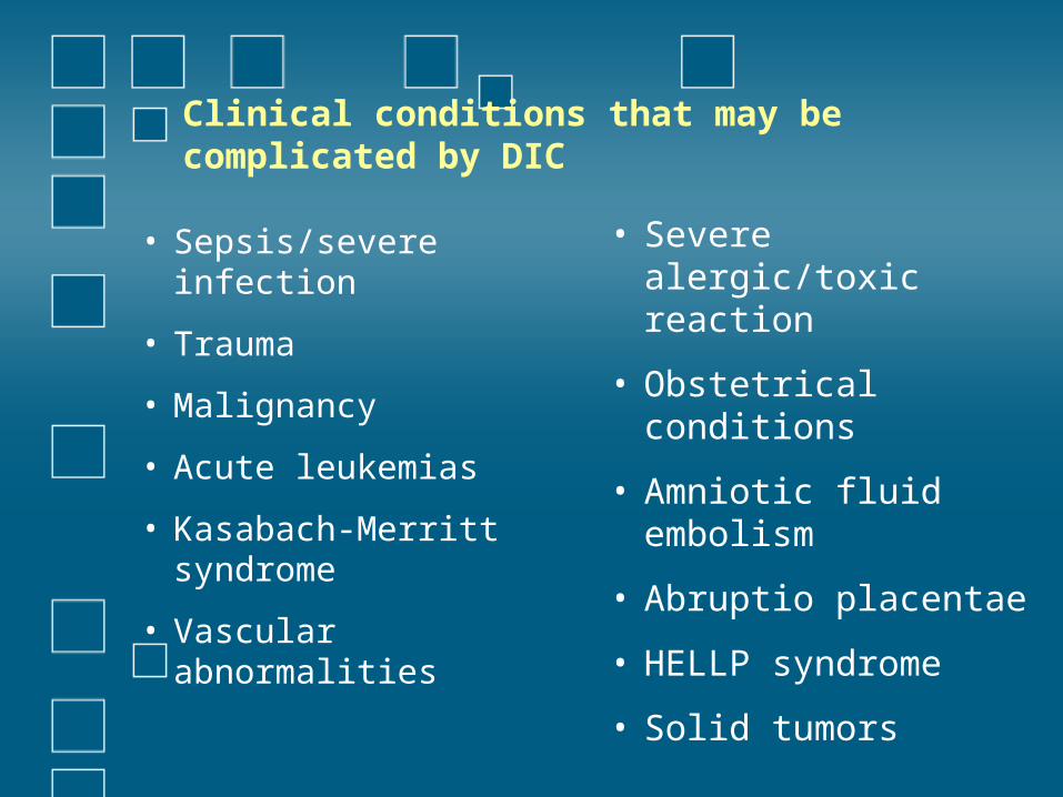
Clinical conditions that may be complicated by DIC
• Sepsis/severe infection
• Trauma
• Malignancy
• Acute leukemias
• Kasabach-Merritt syndrome
• Vascular abnormalities
• Severe alergic/toxic reaction
• Obstetrical conditions
• Amniotic fluid embolism
• Abruptio placentae
• HELLP syndrome
• Solid tumors
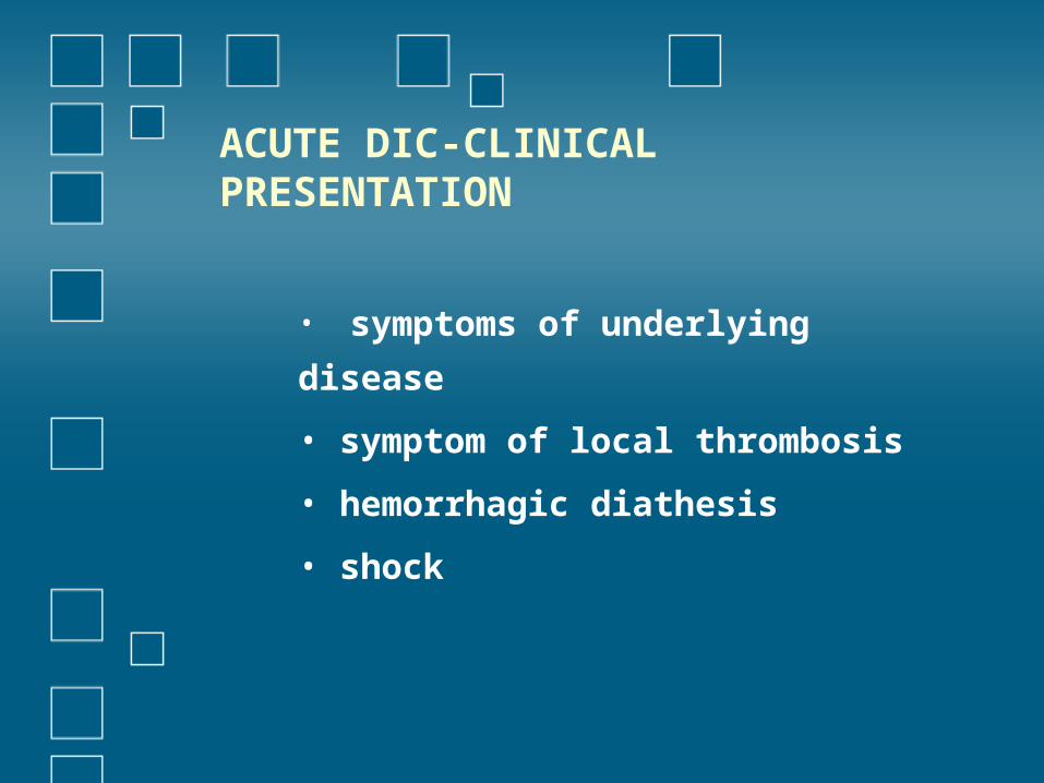
ACUTE DIC-CLINICAL PRESENTATION
• symptoms of underlying
disease
• symptom of local thrombosis
• hemorrhagic diathesis
• shock
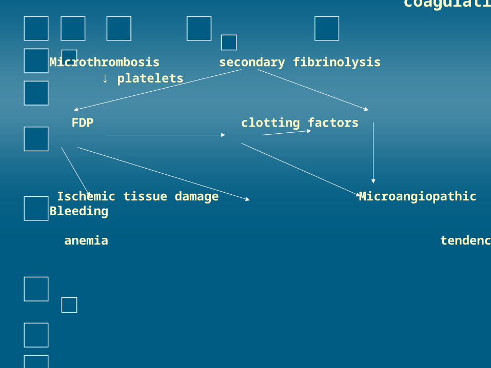
Diffuse intravascular coagulation
Microthrombosis secondary fibrinolysis ↓ platelets FDP clotting factors
Ischemic tissue damage Microangiopathic Bleeding anemia tendency
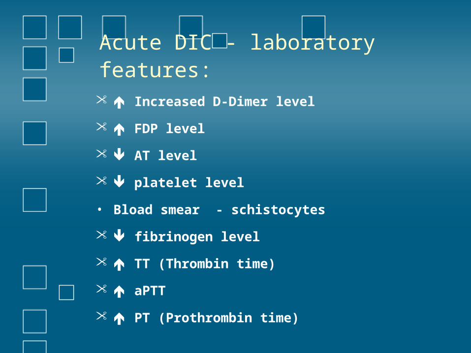
Acute DIC - laboratory features: Increased D-Dimer level
FDP level
AT level
platelet level
• Bload smear - schistocytes
fibrinogen level
TT (Thrombin time)
aPTT
PT (Prothrombin time)
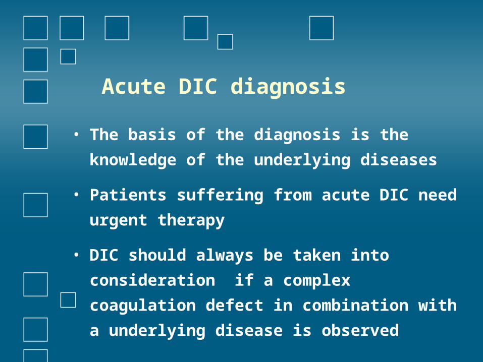
Acute DIC diagnosis
• The basis of the diagnosis is the knowledge of
the underlying diseases
• Patients suffering from acute DIC need urgent
therapy
• DIC should always be taken into consideration
if a complex coagulation defect in combination
with a underlying disease is observed
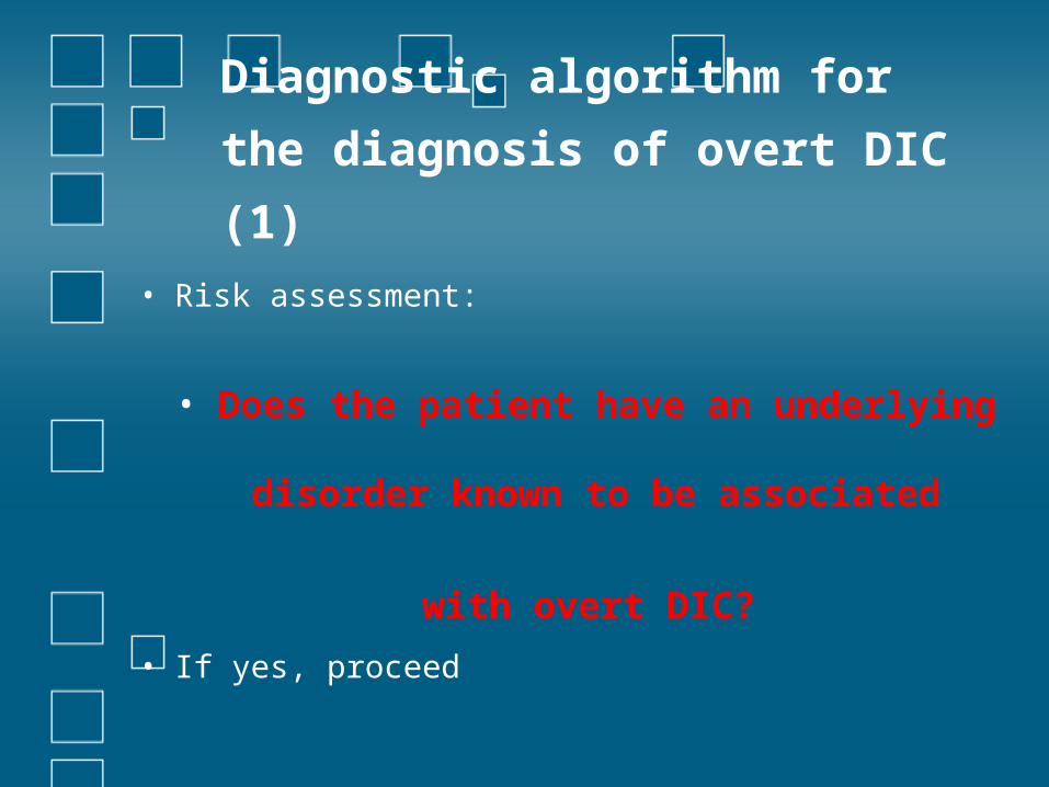
Diagnostic algorithm for
the diagnosis of overt DIC
(1)• Risk assessment:
• Does the patient have an underlying
disorder known to be associated
with overt DIC?
• If yes, proceed
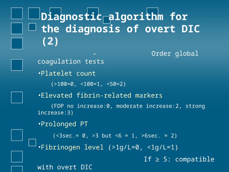
Diagnostic algorithm for the diagnosis of overt DIC (2)
- Order global coagulation tests
•Platelet count
(>100=0, <100=1, <50=2)
•Elevated fibrin-related markers
(FDP no increase:0, moderate increase:2, strong increase:3)
•Prolonged PT
(<3sec.= 0, >3 but <6 = 1, >6sec. = 2)
•Fibrinogen level (>1g/L=0, <1g/L=1)
If ≥ 5: compatible with overt DIC
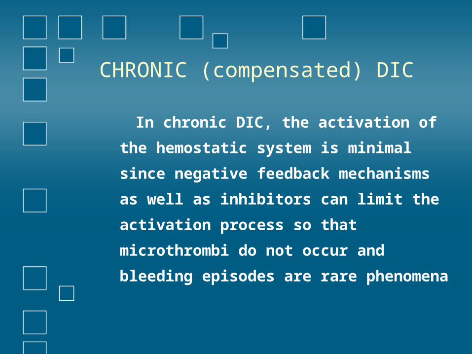
CHRONIC (compensated) DIC
In chronic DIC, the activation of the
hemostatic system is minimal since
negative feedback mechanisms as well as
inhibitors can limit the activation process
so that microthrombi do not occur and
bleeding episodes are rare phenomena
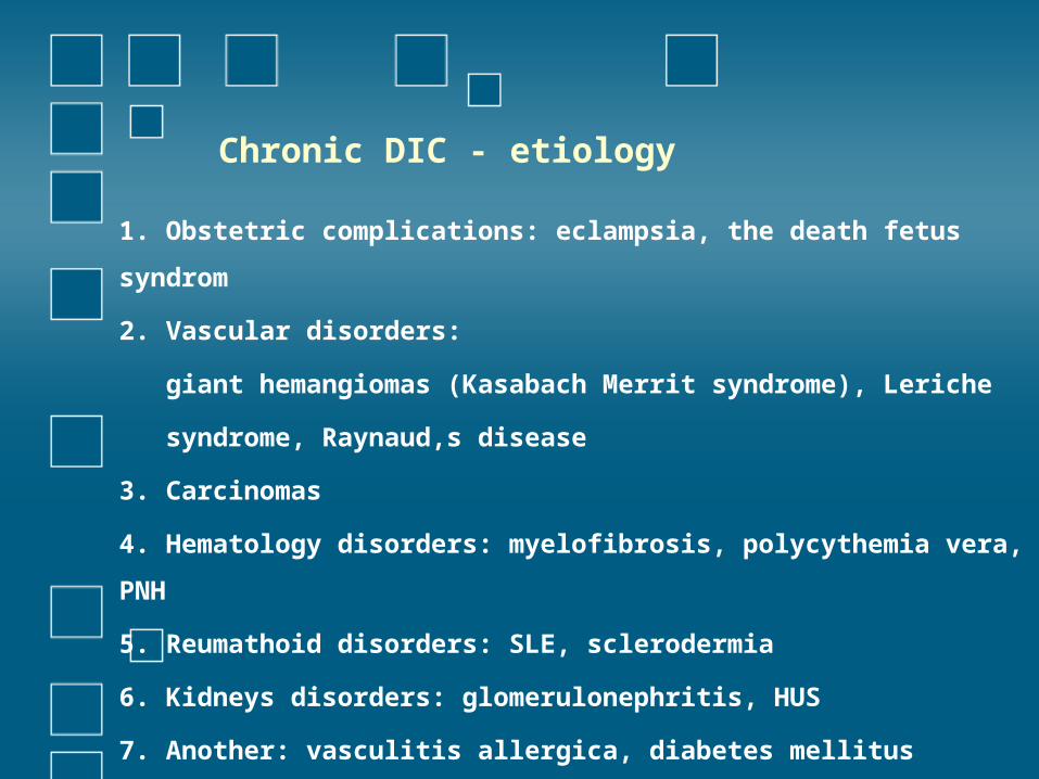
Chronic DIC - etiology
1. Obstetric complications: eclampsia, the death fetus syndrom
2. Vascular disorders:
giant hemangiomas (Kasabach Merrit syndrome), Leriche
syndrome, Raynaud,s disease
3. Carcinomas
4. Hematology disorders: myelofibrosis, polycythemia vera, PNH
5. Reumathoid disorders: SLE, sclerodermia
6. Kidneys disorders: glomerulonephritis, HUS
7. Another: vasculitis allergica, diabetes mellitus
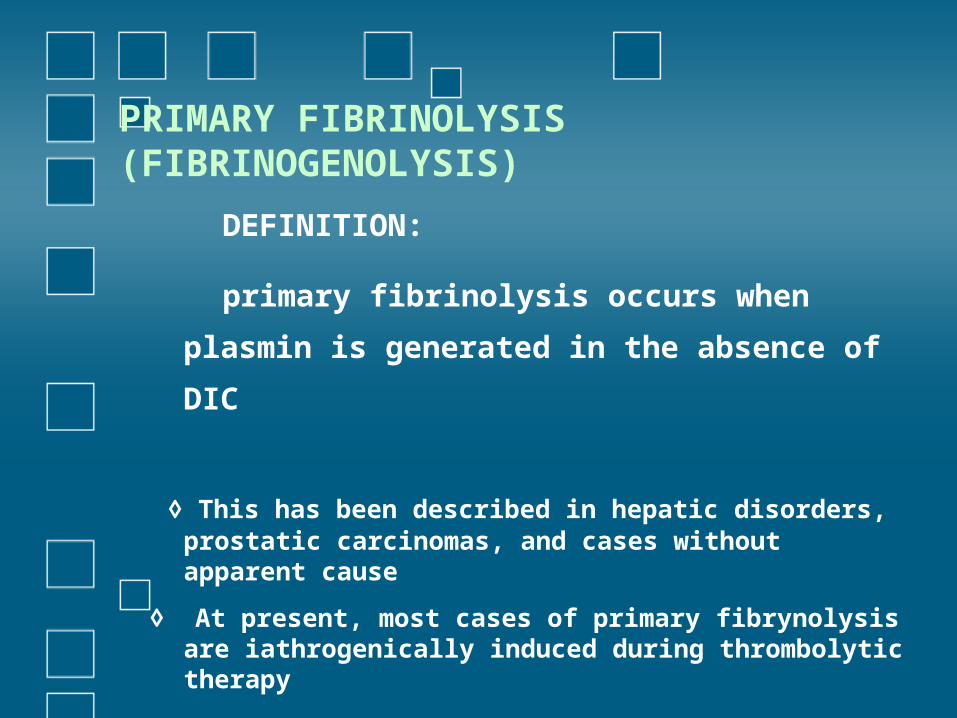
PRIMARY FIBRINOLYSIS (FIBRINOGENOLYSIS)
DEFINITION:
primary fibrinolysis occurs when
plasmin is generated in the absence of
DIC
◊ This has been described in hepatic disorders, prostatic carcinomas, and cases without apparent cause
◊ At present, most cases of primary fibrynolysis are iathrogenically induced during thrombolytic therapy
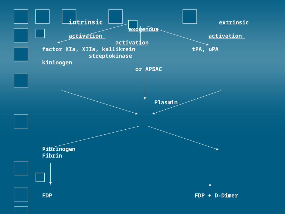
Plasminogen
intrinsic extrinsic exogenous activation activation activationfactor XIa, XIIa, kallikrein tPA, uPA streptokinasekininogen or APSAC
Plasmin
Fibrinogen Fibrin
FDP FDP + D-Dimer
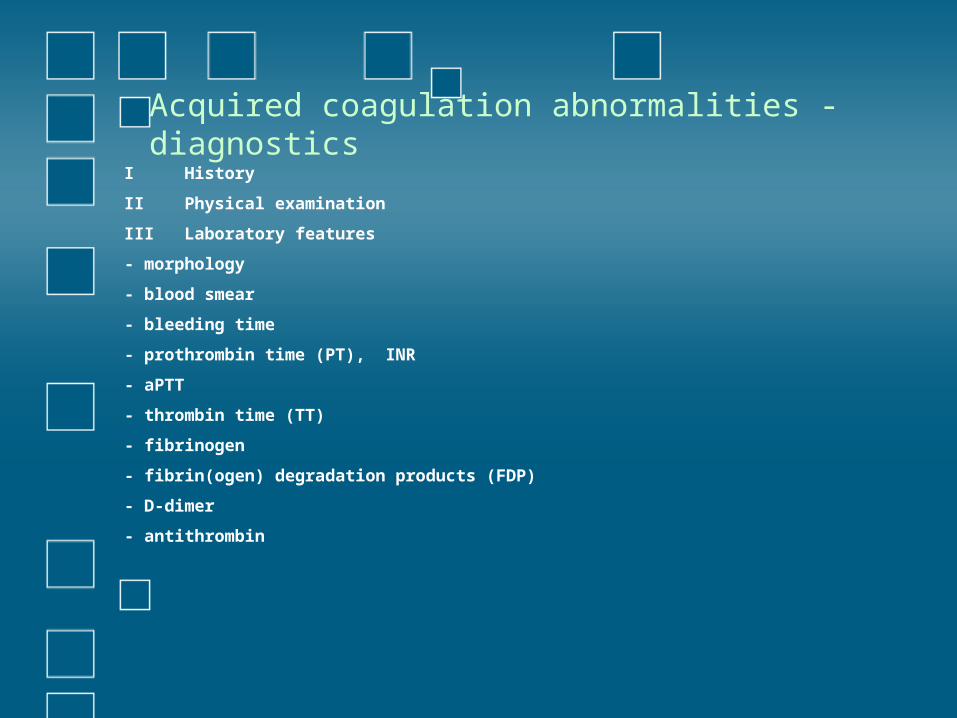
Acquired coagulation abnormalities - diagnostics
I History
II Physical examination
III Laboratory features
- morphology
- blood smear
- bleeding time
- prothrombin time (PT), INR
- aPTT
- thrombin time (TT)
- fibrinogen
- fibrin(ogen) degradation products (FDP)
- D-dimer
- antithrombin
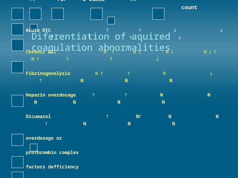
PT aPTT Platelet Fibrinogen TT FDP D-
Dimer AT
count
Acute DIC
Chronic DIC N N N N N
Fibrinogenolysis N N N N
N
Heparin overdosage N N N N
N N
Dicumarol N N N N N
N
overdosage or
prothrombin complex
factors defficiency
Diferentiation of aquired coagulation abnormalities
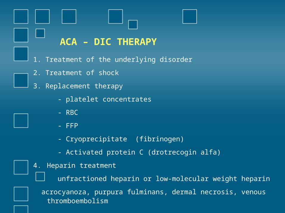
ACA – DIC THERAPY
1. Treatment of the underlying disorder
2. Treatment of shock
3. Replacement therapy
- platelet concentrates
- RBC
- FFP
- Cryoprecipitate (fibrinogen)
- Activated protein C (drotrecogin alfa)
4. Heparin treatment
unfractioned heparin or low-molecular weight heparin
acrocyanoza, purpura fulminans, dermal necrosis, venous thromboembolism
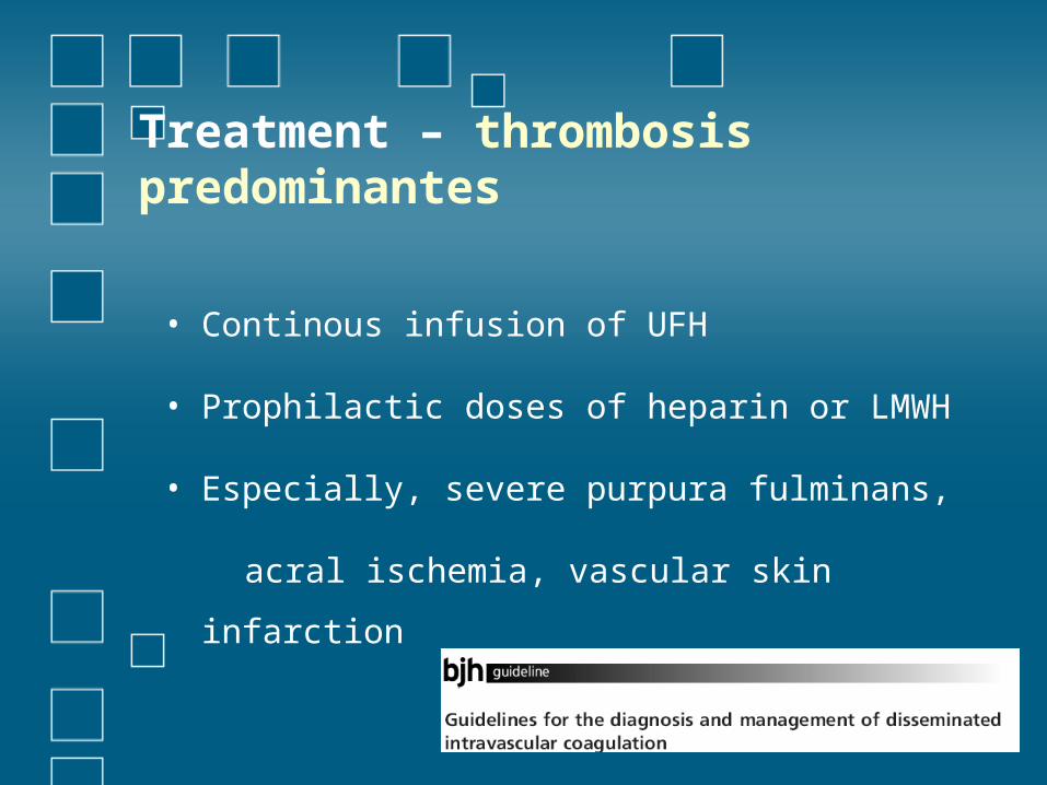
Treatment – thrombosis predominantes
• Continous infusion of UFH
• Prophilactic doses of heparin or LMWH
• Especially, severe purpura fulminans,
acral ischemia, vascular skin infarction
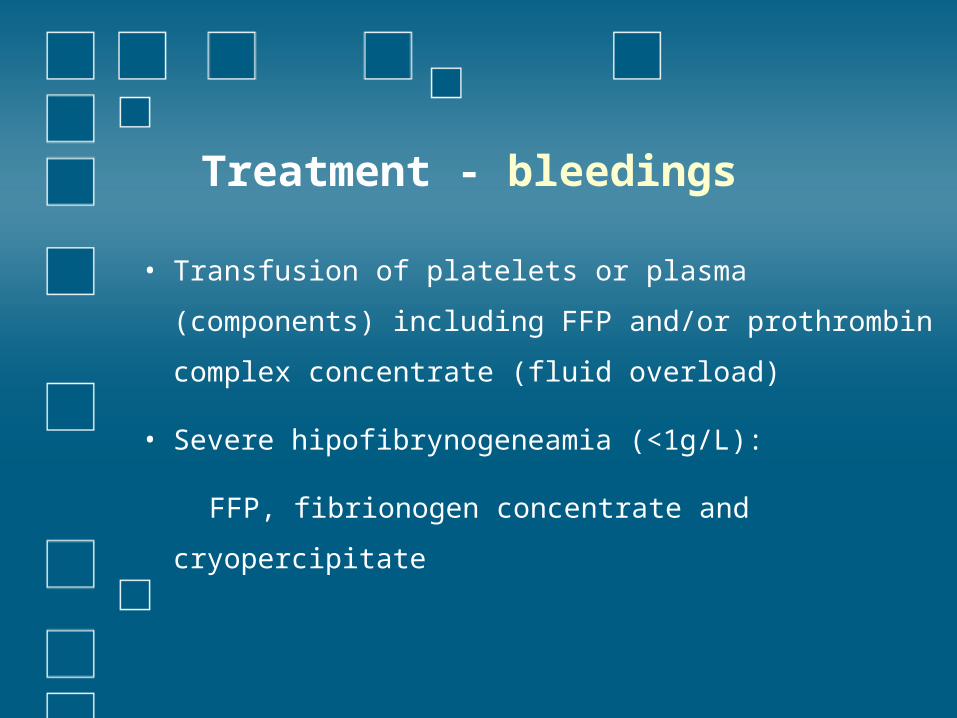
Treatment - bleedings
• Transfusion of platelets or plasma (components)
including FFP and/or prothrombin complex
concentrate (fluid overload)
• Severe hipofibrynogeneamia (<1g/L):
FFP, fibrionogen concentrate and cryopercipitate

CASE PRESENTATION