cme article brugada pattern masking anterior myocardial ... · male gender and a family history of...
TRANSCRIPT

Singapore Med J 2011; 52(9) : 647E l e c t r o c a r d i o g r a p h y S e r i e s
CME Article
Cardiology Department,National University Health System,1E Kent Ridge Road,Level 9, NUHS Tower Block,Singapore 119228
Seow SC, MBBS, MRCP, FAMSConsultant
Omar AR, MBBS, MRCP, FAMSConsultant
Hong ECT, MBBS, MRCP, FAMSConsultant
Correspondence to:Dr Seow Swee Chong Tel: (65) 6772 5286Fax: (65) 6872 2998Email: [email protected]
Brugada pattern masking anterior myocardial infarctionSeow S C, Omar A R, Hong E C T
CLINICAL PRESENTATION
A 48-year-old male smoker presented with intermittent chest pain for two days. Described as a pulling sensation that lasted for a few minutes, the most severe episode occurred within the two hours prior to presentation. An electrocardiogram (ECG) was obtained, which was consistent with Brugada Type 1 pattern (Fig. 1). There was no history of
syncope or palpitations, or a family history of sudden cardiac death. As the chest pain was atypical for coronary artery disease, the patient was placed under observation while blood samples were sent for cardiac enzymes and troponins. A second ECG obtained one hour later (Fig. 2), however, was consistent with an evolving anterior ST elevation myocardial infarction (STEMI). What is the diagnosis?
Fig. 1 ECG taken at presentation shows Brugada Type 1 Pattern.
Singapore Med J 2011; 52(9) : 648
ECG INTERPRETATION
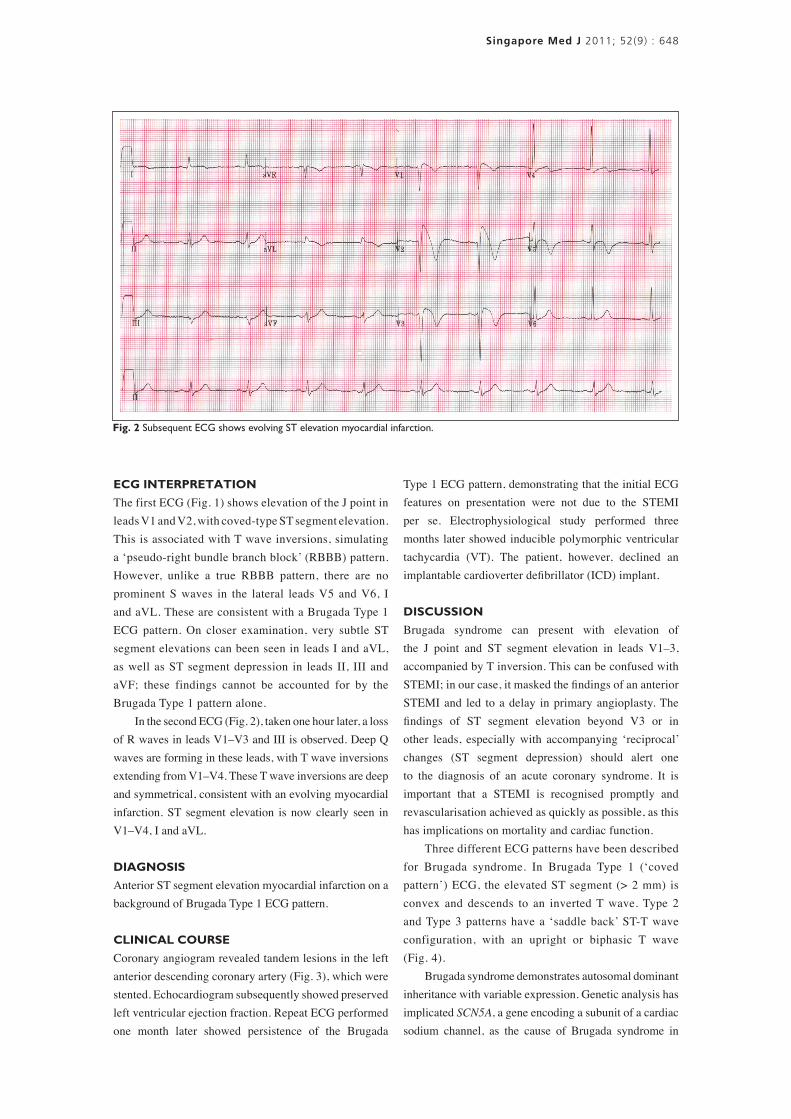
The first ECG (Fig. 1) shows elevation of the J point in leads V1 and V2, with coved-type ST segment elevation. This is associated with T wave inversions, simulating a ‘pseudo-right bundle branch block’ (RBBB) pattern. However, unlike a true RBBB pattern, there are no prominent S waves in the lateral leads V5 and V6, I and aVL. These are consistent with a Brugada Type 1 ECG pattern. On closer examination, very subtle ST segment elevations can been seen in leads I and aVL, as well as ST segment depression in leads II, III and aVF; these findings cannot be accounted for by the Brugada Type 1 pattern alone. In the second ECG (Fig. 2), taken one hour later, a loss of R waves in leads V1–V3 and III is observed. Deep Q waves are forming in these leads, with T wave inversions extending from V1–V4. These T wave inversions are deep and symmetrical, consistent with an evolving myocardial infarction. ST segment elevation is now clearly seen in V1–V4, I and aVL.
DIAGNOSIS
Anterior ST segment elevation myocardial infarction on a background of Brugada Type 1 ECG pattern.
CLINICAL COURSE
Coronary angiogram revealed tandem lesions in the left anterior descending coronary artery (Fig. 3), which were stented. Echocardiogram subsequently showed preserved left ventricular ejection fraction. Repeat ECG performed one month later showed persistence of the Brugada
Type 1 ECG pattern, demonstrating that the initial ECG features on presentation were not due to the STEMI per se. Electrophysiological study performed three months later showed inducible polymorphic ventricular tachycardia (VT). The patient, however, declined an implantable cardioverter defibrillator (ICD) implant.
DISCUSSION
Brugada syndrome can present with elevation of the J point and ST segment elevation in leads V1–3, accompanied by T inversion. This can be confused with STEMI; in our case, it masked the findings of an anterior STEMI and led to a delay in primary angioplasty. The findings of ST segment elevation beyond V3 or in other leads, especially with accompanying ‘reciprocal’ changes (ST segment depression) should alert one to the diagnosis of an acute coronary syndrome. It is important that a STEMI is recognised promptly and revascularisation achieved as quickly as possible, as this has implications on mortality and cardiac function. Three different ECG patterns have been described for Brugada syndrome. In Brugada Type 1 (‘coved pattern’) ECG, the elevated ST segment (> 2 mm) is convex and descends to an inverted T wave. Type 2 and Type 3 patterns have a ‘saddle back’ ST-T wave configuration, with an upright or biphasic T wave (Fig. 4). Brugada syndrome demonstrates autosomal dominant inheritance with variable expression. Genetic analysis has implicated SCN5A, a gene encoding a subunit of a cardiac sodium channel, as the cause of Brugada syndrome in
Fig. 2 Subsequent ECG shows evolving ST elevation myocardial infarction.

Singapore Med J 2011; 52(9) : 649
18%–30% of families.(1-4) The pathogenesis of the ST segment elevation in Brugada syndrome is not completely understood. Ventricular myocardium comprises at least three electrophysiologically distinct cell types: epicardial, endocardial and M cells. ST segment elevation is believed to be due to an alteration in the action potential in the epicardial and M cells but not in the endocardial cells.(5-7) The resulting dispersion of repolarisation across the ventricular wall, which is most pronounced in the right ventricle, results in a transmural voltage gradient that is manifested as ST segment elevation in leads V1–3. Ventricular arrhythmias may be caused by heterogeneity of the cardiac action potential across the three layers of myocardial cells as well as within the epicardium. Male gender and a family history of sudden cardiac death (SCD) are risk factors for SCD. Patients with a history of cardiac arrest and/or syncope are also at increased risk compared to asymptomatic individuals.(2,8-10) Among asymptomatic patients, the presence of a spontaneous Type 1 ECG pattern (as opposed to only after drug challenge) and inducible ventricular tachyarrhythmia on electrophysiologic testing are markers of increased risk for SCD.(11,12) At present, it is generally agreed that an ICD is indicated in all Brugada syndrome patients with a prior cardiac arrest and in patients with a spontaneous Type 1 ECG plus a history of syncope. Evidence also supports ICD implantation in patients with a history of VT that did not result in cardiac arrest.
ABSTRACT
A middle-aged male smoker presented with
atypical chest pain. Initial electrocardiogram
(ECG) showed Brugada Type 1 pattern.
Subsequent ECGs demonstrated evolving anterior
ST elevation myocardial infarction (STEMI),
consistent with the elevated cardiac enzymes.
Coronary angiogram showed significant stenoses
in the left anterior descending artery, which
were stented emergently. In retrospect, subtle
changes were noted in the initial ECG, which
could have alerted one to the STEMI. However,
the presence of a Brugada Type 1 pattern masked
the ECG changes of anterior STEMI and made the
diagnosis difficult. A discussion of the literature
surrounding Brugada syndrome is undertaken,
including its clinical features, risk stratification and
management.
Keywords: Brugada syndrome, electrocardiogram,
myocardial infarction, ST elevation
Singapore Med J 2011; 52(9): 647–651
REFERENCES1. Antzelevitch C, Brugada P, Borggrefe M, et al. Brugada syndrome:
report of the second consensus conference. Heart Rhythm 2005; 2:429-40.
2. Priori SG, Napolitano C, Gasparini M, et al. Natural history of Brugada syndrome: insights for risk stratification and management. Circulation 2002; 105:1342-7.
3. Chen Q, Kirsch GE, Zhang D, et al. Genetic basis and molecular mechanism for idiopathic ventricular fibrillation. Nature 1998; 392:293-6.
4. Priori SG, Napolitano C, Gasparini M, et al. Clinical and genetic heterogeneity of right bundle branch block and ST-segment elevation syndrome: A prospective evaluation of 52 families. Circulation 2000; 102:2509-15.
5. Yan GX, Antzelevitch C. Cellular basis for the Brugada syndrome and other mechanisms of arrhythmogenesis associated with ST-segment elevation. Circulation 1999; 100:1660-6.
Fig. 3 Coronary angiogram shows the culprit lesions (arrows). Fig. 4 Brugada ECG pattern Types 1, 2 and 3.

Singapore Med J 2011; 52(9) : 650
6. Gussak I, Antzelevitch C, Bjerregaard P, Towbin JA, Chaitman BR. The Brugada syndrome: clinical, electrophysiologic and genetic aspects. J Am Coll Cardiol 1999; 33:5-15.
7. Alings M, Wilde A. “Brugada” syndrome: clinical data and suggested pathophysiological mechanism. Circulation 1999; 99:666-73.
8. Brugada J, Brugada R, Brugada P. Determinants of sudden cardiac death in individuals with the electrocardiographic pattern of Brugada syndrome and no previous cardiac arrest. Circulation 2003; 108:3092-6.
9. Brugada J, Brugada R, Antzelevitch C, et al. Long-term follow-up of individuals with the electrocardiographic pattern of right bundle-branch block and ST-segment elevation in precordial leads V1 to V3. Circulation 2002; 105:73-8.
10. Eckardt L, Probst V, Smits JP, et al. Long-term prognosis of individuals with right precordial ST-segment-elevation Brugada syndrome. Circulation 2005; 111:257-63.
11. Brugada P, Brugada R, Brugada J. Should patients with
an asymptomatic Brugada electrocardiogram undergo pharmacological and electrophysiological testing? Circulation 2005;112:279-92; discussion 279-92.
12. Priori SG, Napolitano C. Should patients with an asymptomatic Brugada electrocardiogram undergo pharmacological and electrophysiological testing? Circulation 2005;112:279-92; discussion 279-92.
13. Zipes DP, Camm AJ, Borggrefe M, et al. ACC/AHA/ESC 2006 guidelines for management of patients with ventricular arrhythmias and the prevention of sudden cardiac death--executive summary: A report of the American College of Cardiology/American Heart Association Task Force and the European Society of Cardiology Committee for Practice Guidelines (Writing Committee to Develop Guidelines for Management of Patients with Ventricular Arrhythmias and the Prevention of Sudden Cardiac Death) Developed in collaboration with the European Heart Rhythm Association and the Heart Rhythm Society. Eur Heart J 2006; 27:2099-140.
Singapore Med J 2011; 52(9) : 651
True False
☐ ☐ ☐ ☐ ☐ ☐ ☐ ☐
☐ ☐ ☐ ☐ ☐ ☐ ☐ ☐
☐ ☐ ☐ ☐ ☐ ☐ ☐ ☐
☐ ☐ ☐ ☐ ☐ ☐ ☐ ☐
☐ ☐
☐ ☐
☐ ☐ ☐ ☐
Doctor’s particulars:Name in full: __________________________________________________________________________________
MCR number: _____________________________________ Specialty: ___________________________________
Email address: _________________________________________________________________________________
SINGAPORE MEDICAL COUNCIL CATEGORY 3B CME PROGRAMMEMultiple Choice Questions (Code SMJ 201109A)
SUBMISSION INSTRUCTIONS:(1) Log on at the SMJ website: http://www.sma.org.sg/cme/smj and select the appropriate set of questions. (2) Select your answers and provide your name, email address and MCR number. Click on “Submit answers” to submit.
RESULTS:(1) Answers will be published in the SMJ November 2011 issue. (2) The MCR numbers of successful candidates will be posted online at www.sma.org.sg/cme/smj by 24 October 2011. (3) All online submissions will receive an automatic email acknowledgment. (4) Passing mark is 60%. No mark will be deducted for incorrect answers. (5) The SMJ editorial office will submit the list of successful candidates to the Singapore Medical Council.
Deadline for submission: (September 2011 SMJ 3B CME programme): 12 noon, 17 October 2011.
Question 1. Regarding the clinical presentation of myocardial infarction:(a) It can present with atypical chest pain.(b) Index of suspicion should be high in a middle-aged man with risk factors.(c) ECG changes of ischaemia occur early on in the clinical course.(d) Rise in cardiac enzymes occur within the first 1–2 hours.
Question 2. Regarding ST elevation on ECG:(a) It can only be due to acute myocardial infarction.(b) It can also be seen in Brugada syndrome.(c) It should not extend beyond V3 in Brugada syndrome alone.(d) The association with ‘reciprocal changes’ are strongly suggestive of acute myocardial infarction.
Question 3. Regarding Brugada ECG pattern:(a) There are three patterns described.(b) Only Type 1 pattern is diagnostic and associated with sudden death.(c) Brugada ECG pattern with syncope or documented ventricular arrhythmias is an indication for ICD implant.(d) It is associated with higher risk in male patients and those with a family history of sudden death.
Question 4. Regarding acute STEMI:(a) Early revascularisation is associated with lower mortality and better clinical outcomes.(b) Myocardial reperfusion can be achieved either by mechanical means (angioplasty) or pharmacological means (thrombolysis).(c) Involvement of the left anterior descending artery usually gives rise to ST elevation in the anterior leads V1–V3.(d) It is associated with isolated subendocardial myocardial injury.
Question 5. Regarding Brugada syndrome:(a) All asymptomatic patients with any of the three different ECG patterns should undergo an electrophysiological study.(b) An ICD implant is currently the only consistently effective therapy in high-risk Brugada patients.(c) It is due to a mutation in the gene SCN5A in all cases.(d) Propensity toward ventricular arrhythmia is the result of electrical heterogeneity across the myocardial layers that may result from a loss-of-function mutation involving the fast sodium ion channels.