citologia relatório de aula prática
TRANSCRIPT

Hindawi Publishing CorporationInternational Journal of Cell BiologyVolume 2012, Article ID 219196, 8 pagesdoi:10.1155/2012/219196
Review Article
Alterations in Cell-Extracellular Matrix Interactions duringProgression of Cancers
Rajeswari Jinka,1 Renu Kapoor,2 Pavana Goury Sistla,2 T. Avinash Raj,2 and Gopal Pande2
1 Department of Biochemistry, Acharya Nagarjuna University, Guntur 522510, India2 Centre for Cellular and Molecular Biology, Council of Scientific and Industrial Research (CSIR), Uppal Road,Hyderabad 500 007, India
Correspondence should be addressed to Rajeswari Jinka, [email protected] and Gopal Pande, [email protected]
Received 3 August 2011; Revised 13 October 2011; Accepted 14 October 2011
Academic Editor: Jun Chung
Copyright © 2012 Rajeswari Jinka et al. This is an open access article distributed under the Creative Commons Attribution License,which permits unrestricted use, distribution, and reproduction in any medium, provided the original work is properly cited.
Cancer progression is a multistep process during which normal cells exhibit molecular changes that culminate into the highlymalignant and metastatic phenotype, observed in cancerous tissues. The initiation of cell transformation is generally associatedwith genetic alterations in normal cells that lead to the loss of intercellular- and/or extracellular-matrix- (ECM-) mediatedcell adhesion. Transformed cells undergo rapid multiplication and generate more modifications in adhesion and motility-related molecules which allow them to escape from the original site and acquire invasive characteristics. Integrins, which aremultifunctional adhesion receptors, and are present, on normal as well as transformed cells, assist the cells undergoing tumorprogression in creating the appropriate environment for their survival, growth, and invasion. In this paper, we have brieflydiscussed the role of ECM proteins and integrins during cancer progression and described some unique conditions whereadhesion-related changes could induce genetic mutations in anchorage-independent tumor model systems.
1. Introduction
Cancer afflicts an organ or a tissue by inducing abnormaland uncontrolled division of cells that either constitute it ormigrate to it. At the cellular level, this is caused by geneticalterations in networks that regulate cell division and celldeath. The increased rate of proliferation of transformed cellscauses further mutations in genes that regulate other cellularprocesses. For example, transformed cells eventually gain thecapacity to invade into other tissues by modulating theirown kinetic properties without losing the capacity to dividerapidly and avoid cell death, despite internal and externalperturbations.
Cancer cells adopt diverse mechanisms to cope withthe various physiological insults, such as low oxygen andmetabolic stress, that they encounter [1]. These mechanismshave been discussed in a recent review [2], and based uponthat discussion, six important hall marks of cancer cells canbe identified. These are (a) sustained proliferative signalling,(b) evasion of growth suppressors, (c) resistance to celldeath, (d) replicative immortality, (e) copious angiogenesis,and (f) active invasion and metastasis. In addition to these,
cancer cells can exhibit two other properties, that is, tumorpromoting inflammation and gene instability that assistthe cells in the transition from normal to oncogenic phe-notype [2]. Eventually transformed cells undergo somaticevolution and generate diverse populations that tend toharbor genetic and epigenetic instabilities and alterations[3]. These changes also assist the cells in adapting tothe variations in the surrounding microenvironment andeven to alter it. As a consequence of these alterations, thetumor milieu or microenvironment becomes an “enablingelement” for defining some characteristics of cancer cells. Forexample, the tumor microenvironment can induce cancercells in acquiring anoikis resistance and in selecting newsites to colonize and grow. Sometimes these cells remainunresponsive until signals generated from the ECM reachthe cell’s nucleus and they determine whether the cell wouldproceed to the next stage in cancer progression or not. Thisresponse of cancer cells to ECM-generated signals similarto the “dynamic reciprocity” proposed by Bisell for normalcells. An example of such an adaptation of cancer cells totheir microenvironment and the resultant clonal selection ofinvasive cells has been recently reported [4–6].

2 International Journal of Cell Biology
Metastatic invasion is generally the final phase of cancerprogression, and it involves formation of new blood vesselseither by neovasculogenesis, in which endothelial cell precur-sors (angioblasts) migrate to the tumor site and differentiateand assemble into primitive blood vessels, or by angiogenesisin which we observe sprouting of new blood vessels frompreexisting ones, or their longitudinal bifurcation, in thetumor [7]. The invasive tumor cells migrate through thesenewly formed blood vessels to other sites such as lung andliver brain and this leads to the death of tumor-bearingpatients or animals as the case may be.
Based on available evidence, the entire process of cancerformation can be divided into four different stages: initiation,progression, epithelial mesenchymal transition (EMT), andmetastasis (see Figure 1). At the initiation stage, a normalcell acquires oncogenic properties mainly through geneticalterations, which lead to changes in cell structure, adhesionproperties, and response to signals from ECM proteins. Inthe second stage, transformed cells respond to cues from thealtered environmental conditions and acquire properties ofadhesion-independent growth and colonization. The thirdstage is also referred to as a transitional or the EMT stage,and, in this stage, the fully transformed cells begin to exhibitmesenchymal gene expression patterns which induce themto invade into the neighbouring tissue and enter into bloodcirculation [8]. The fourth and prominent stage is metastasisin which the invasive mesenchymes like cells move fromthe primary site and colonize in a new location. This stagespreads the disease into different parts of the body andinvolves several alterations in the adhesion properties of cells.
From all earlier observations, it is clear that the celladhesion in transformed cells plays an important role inall four stages of cancer formation. This paper highlightsrecent studies done on the integrin-mediated interaction oftransformed cells with the ECM and discusses its role incancer progression.
2. ECM Components and Properties
Over the past two decades, research in the field of cancer biol-ogy has focussed extensively on the role of ECM constituentsduring cancer progression. These molecules comprise thecell’s microenvironment, and they can affect the mechanicaland biophysical properties of cells as well as that of the ECMsuch as its mechanics, geometry, and topology [9].
In some tissues, mainly of epithelial origin, ECMconstituents are present in the basement membrane thatdefines the boundaries of that tissue. In this location, theorganization of these components is different than in thematrix. In the basement membrane, we notice moleculessuch as collagens, proteoglycans, laminins, and fibronectinsassociate strongly with certain carbohydrate polymers andgenerate a membrane-like structure which facilitates theformation of a framework of cells and ECM constituents[10]. Specific domains in ECM proteins that are createdby partial gene duplication and exon shuffling during theprocess of evolution [11] play a critical role in keeping thecells attached to the ECM and the basement membrane andinitiating signalling cascades in the cells.
Inside the cells, the ECM-induced signaling pathwaysare transmitted mainly through integrin molecules that aretransmembrane multifunctional ECM receptors. Integrin-mediated signaling in association with many cofactors, forexample, cytokines, growth factors, and intracellular adaptermolecules, can significantly affect diverse cell processes suchas cell cycle progression, migration, and differentiation. Theinterplay between the biophysical properties of the cell andECM establishes a dynamic, mechanical reciprocity betweenthe cell and the ECM in which the cell’s ability to exertcontractile stresses against the extracellular environment bal-ances the elastic resistance of the ECM to that deformation[4, 9]. The ECM in association with the available growthfactors activates a sequence of reactions with a complexnetwork of proteases, sulfatases, and possibly other enzymesto liberate and activate various signalling pathways in ahighly specific and localized fashion. The maintenance ofECM homeostasis therefore involves a tight balance betweenbiosynthesis of ECM proteins, their 3D organization, cross-linking, and degradation.
3. Modulation of ECM-GeneratedSignaling in Cancer
During cancer progression, we observe significant changesin the structural and mechanical properties of ECM con-stituents. It has been reported that changes in matrixstiffness, which offers resistance to cell traction forces[12] and also influences “shape dependence” in cells, cancontribute actively to the tumor formation [13]. Dereg-ulation of cell shape and alterations in the interactionswith the ECM are considered as important hallmarks ofcancer cells. These changes in ECM homeostasis can bebrought about by the properties of tumor cells themselvesor by the secretions of other surrounding cells such asfibroblasts, macrophages, and leukocytes [14]. Integrin ECMinteractions are significantly modulated by crosstalk withseveral other signal-generating molecules, some of them arereceptor molecules on the cell surface whereas others arepresent in the cytoplasm as adaptor proteins and actin-binding proteins [15]. These signaling crosstalks in whichintegrin molecules lie at the center are very useful forthe transition of transformed cells to metastatic cells [16].Integrin-generated ECM remodelling is further controlled bythe localization and activity of proteases [17]. One exampleof such an integrin-directed cancer progression is seenin breast cancers, where adhesion-independent mammaryepithelial cells secrete laminin-5 and luminal cells secretelaminin-1. This leads to aberrant polarity in cells, causingupregulation of metalloproteinases (MMPs, such as MMP9)and induction of tumor invasion and metastasis [18–20].
4. Integrins: Its Ligands and Signalling
Integrins are heterodimeric cell-surface receptors that medi-ate adhesion to ECM and immunoglobulin superfam-ily molecules. At least 24 distinct integrin heterodimersare formed by the combination of 18 α-subunits and

International Journal of Cell Biology 3
ECM
Endothelial cells with basement membrane
(a) (b) (c)
(d) (e)
Normal cells
Tumor cells
Fibroblast
Blood vessel
Immune inflammatory cells
Pericyte
Figure 1: Various steps in tumor initiation and progression where panel (a) represents initiation of tumor by transforming normal cells,panel (b) shows the modulation of ECM proteins allowing transformed cells to multiply, panel (c) shows progression of cancer by replacingnormal cells, panel (d) represents the invasion, where cancer cells migrate into the blood stream by modulating ECM and cell adhesionmolecules, and panels (e) shows metastasis where the cancer cells are localized at different sites enabling angiogenesis.
8 β-subunits. Specific integrin heterodimers preferentiallybind to distinct ECM proteins like laminin, collagen IV,fibronectin, and so forth. The level of integrin expressionon the cell surface dictates the efficiency of cell adhesionand migration on different matrices. While some integrinsselectively recognise primarily a single ECM protein ligand(e.g., α5β1 recognises primarily fibronectin), others canbind several ligands (e.g., integrin αvβ3 binds vitronectin,fibronectin, fibrinogen, denatured or proteolysed collagen,and other matrix proteins). Several integrins recognise thetripeptide Arg-Gly-Asp (e.g., αvβ3, α5β1, αIIbβ3), whereasothers recognise alternative short peptide sequences (e.g.,integrin α4β1 recognises EILDV and REDV in alternativelyspliced CS-1 fibronectin). Inhibitors of integrin functioninclude function-blocking monoclonal antibodies, peptideantagonists, and small molecule peptide mimetics matrix[21–23].
The positioning of integrin receptors acts as a directbridge between the extracellular matrix and the internalcell cytoskeleton by transducing key intracellular signalsby associating with the clusters of kinases and adaptorproteins in focal adhesion complexes. Integrins thus act asmediators in transmitting different signals from “inside out”(intracellular to extracellular) and “outside in” (extracellularto intracellular) between ECM to cells and vice versa.
Through these pathways, ECM proteins are able to controlproapoptotic and antiapoptotic cascades by regulating theactivity of caspase 8 and caspase 3 [24–26]. ECM-integrininteractions thus determine the balance of apoptotic and cellsurvival signals and maintain the homeostasis of organs andtissues. Although integrins lack kinase activity, by inter- andintramolecular clustering, they recruit and activate kinases,such as focal adhesion kinases (FAKs) and src family kinases(sFKs) to a focal adhesion complex. In addition to scaffoldingmolecules, such as p130 CRK-associated substrate (p130CAs;also known as BCAR1), integrins also couple the ECMto actin cytoskeleton by recruiting cytoskeletal proteins,including talin, paxillin, α-actinin, tensin, and vinculin.Additionally, they form a ternary complex consisting of anintegrin-linked kinase, PINCH, and parvin to regulate manyscaffolding and signalling functions required for integrin-mediated effects on cell migration and survival [27].
5. Integrin Expression and Signalling inCancer Progression
Although anchorage-independent growth is a hallmark ofmalignant transformation, integrins expression levels andactivity are an important role in different steps of tumour

4 International Journal of Cell Biology
progression including initiation [28]. Higher expressionof α3, α5, α6, αv, β1, β4, α6β4, α9β1, αvβ5, and αvβ3integrins is directly correlated with the progression of thedisease [10]. Several epithelial cell tumors showed the alteredα6β4, α6β1, αvβ5, α2β1, and α3β1 integrin expression [29].Integrin recruitment to membrane microdomains has beenshown to be regulated by tetraspanins and crucially regulateintegrin function in tumour cells [30]. Recent studies havedemonstrated that cell signalling generated by growth factorsand oncogenes in transformed cells requires collaborationwith specific integrins, especially during tumour initiation.In tumor cells, several survival signals are upregulated uponintegrin ligation, which includes increased expression of BCl-2 or FlIP (also known as CFlAR), activation of the PI3K-AKTpathway or nuclear factor-κB (nF-κB) signaling, and/or p53inactivation [24].
Invasive cancer cells evacuate from the primary site andmigrate to the secondary site by the process of tissue invasionand cell migration. Integrin-mediated pathways involvingfocal adhesion kinase (FAK) and src family kinase (SFK)signaling play a major role in this. In order to survive inthe new location and to withstand the stressful conditions ofhypoxia, nutrient deprivation, and inflammatory mediatorsthe migratory cells increase the blood supply to themselvesby neoangiogenesis. This is achieved by increased expressionof αvβ3 and αvβ5 integrins and by deposition of provisionalmatrix proteins such as vitronectin, fibrinogen, von Wille-brand factor, osteopontin, and fibronectin in the tumourmicroenvironment. Interaction between these moleculesplays a critical role in the process of generating new bloodvessels in the newly formed tumor site [31, 32].
Integrins found on tumour-associated normal cells,such as the vascular endothelium, perivascular cells, fibrob-lasts, bone-marrow-derived cells, and platelets, also have aprofound effect in tumor progression via integrin-mediatedpathways. A summary of these has been given in Table 1 [33–47].
6. Genetic and Chromosomal Aberrations atthe Onset of Cancer
Neoplasia occurs when cells are exposed to cancer-promoting substances that cause single or multiple prema-lignant genetic/epigenetic changes which may coalesce toform a large lesion. These genetic changes may have neutral,deleterious, or advantageous effects on the proliferation ofa clone or clones of cells. Neutral or deleterious geneticchanges may result in stagnation or cell death, whereasthe cell receiving advantageous events may result in higherproliferation, recruitment of blood vessels to the developingtumors, and gain the ability to metastasize [48]. The model ofBraakhuis et al., 2004 [49], advanced this idea by suggestingthat initial genetic alterations occur in stem cells, forminga patch and expanding field of cells with the originaland subsequent genomic and or chromosomal alterations.Then, clonal selection of one or more cells within thisfield of preneoplastic cells leads to the development of acarcinoma. There is a considerable cytogenetic variability
among cells reflecting heterogeneity due to clonal evolutionwithin the original tumor [50]. Initial heterogeneity or cell-to-cell differences in cancer are due to cytoskeletal alter-ations which result in defecting chromosomal segregationand lead to karyotypic variations during mitosis, causingchromosomal aberrations, for example, NUMA1 gene at11q13, which results in multipolar spindles, leading todaughter cells that differ from each other and their mothercells [51]. Structural chromosome alterations also occurdue to deletions, translocations, isochromosomes, dicentricchromosomes, and endoduplicated chromosomes. The gainor amplification of chromosomal segments is driven by morethan one gene [52]. Structural rearrangements involving thecleavage and fusion of centromeres from participating chro-mosomes, also referred to as Robertsonian translocations,are the most frequently observed alterations. Chromosomalaberrations identified with the help of cytogenetic methodsincluding FISH, cCGH, or aCGH showed the gain of theentire long arm of chromosome 3 which amplifies theEGFR gene in SCCHN [53], 8q24 gain to amplify MYC andPTK2 in primary tumors, 11q13 amplifications to amplifycyclin D1 gene, loss of 3q14 causes deletion of fragile siteFRA3B/FHIT, necessary to protect cells from accumulationof DNA damage [48]. Aberrations mainly in chromosome13 and also involving chromosomes 6, 11, 12, and 17 areassociated with B-CLL [54].
7. Anchorage-Independent TumorModel System
The wide range of in vivo tumor models like syngeneic,human tumor xenograft, orthotopic, metastatic, and genet-ically engineered mouse models is available from the basisof the compounds selected and treatments that go intoclinical testing of patients [55]. The ability to exhibitanchorage-independent cell growth (colony-forming capac-ity in semisolid media) has been considered to be funda-mental in cancer biology because it has been connected withtumor cell aggressiveness in vivo such as tumorigenic andmetastatic potentials and also utilized as a marker for invitro transformation. Although multiple genetic factors foranchorage-independence have been identified, the molecularbasis for this capacity is still largely unknown [56, 57].During the process of in vitro tumorigenesis, various onco-genes with distinct pathways have been shown to transformanchorage-dependent cells to anchorage-independent cells[5, 57]. For example, transfer of c-Myc (a transcriptionfactor), v-Src (a tyrosine kinase), or H-Ras (a smallGTPase) into spontaneously immortalized mouse embryonicfibroblasts (MEFs) provides the cells an ability to grow inan anchorage-independent manner [56, 57]. Anchorage-independent multicellular spheroids made by Ewing tumorcell lines were more closely related to primary tumors withrespect to cell morphology, cell-cell junctions, proliferativeindex, and kinase activation [58].
However, changes in ECM and cell adhesion molecularinteraction and genetic variations were observed till thedate only with the primary or secondary tumors. We have

International Journal of Cell Biology 5
Table 1: Various integrins in association with different ligands to induce different signaling pathways in generation of tumor and metastasis.
Integrin type Interacting ECM protein Activated signaling cascade Tumor/metastasis Reference
α3β1 LamininMMP9 and oncogenic Ras,VEGF, FAK-paxillin signalingcascade
Invasion in keratinocytes,Induces angiogenesis,Human hepatoma cells
[33, 34]
α6β1 LamininUrokinase plasminogen activatorand MMP-2, PI3Kinase, Src
Tumor invasion in pancreaticcells
[35, 36]
α7β1 Laminin Rho-A signaling cascade Invasion in breast cancer [37]
α2β1, α1β1, α10β1,αIIβ1
Collagen FAK and src signalingInvasion of melanoma cells,cancer progression, and invasionof lung adenocarcinoma
[38, 39]
αvβ1, αvβ6, αvβ3Vitronectin, syndican,
thromospondin-1MMP9, urokinase signaling,MEK/Erk/NF-κB, PKCa, FAK
Metastatic breast Cancer,pancreatic, cervical, colon,lung/liver metastasis
[40–42]
α9β1 CCN3, osteopontinSrc, P130 Cas, Rac, NOSsignaling
Metastatic potential [43]
αIIb3, αvb3 Von Willebrand factorInteracts withthrombospondin-1 and inducesVEGF/FGF signaling
Breast cancer [44, 45]
α5 FibronectinFAK, ERK, PI-3 K, ILK, andnuclear factor-kappa B -
Metastatic lung and cervicalcancer
[46, 47]
αLβ2Intercellular cell adhesion
moleculesBreast cancer [1]
developed a cellular model system by using normal, adherentrat fibroblast cell lines. These cells lose their cytoskeletalorganization and specificity to fibronectin as α5β1 integrinsare constantly recycled between cytoplasm and plasma. Inthe drastic unfavorable stressful conditions, the mechanical,phenotypic, and genetic characteristics are altered/modifiedto sustain their identity [5, 26]. We observed that this cellularmodel system represents a tumor with all characteristicsof cancer described by Hanahan and Weinberg 2011 ashallmarks of cancer.
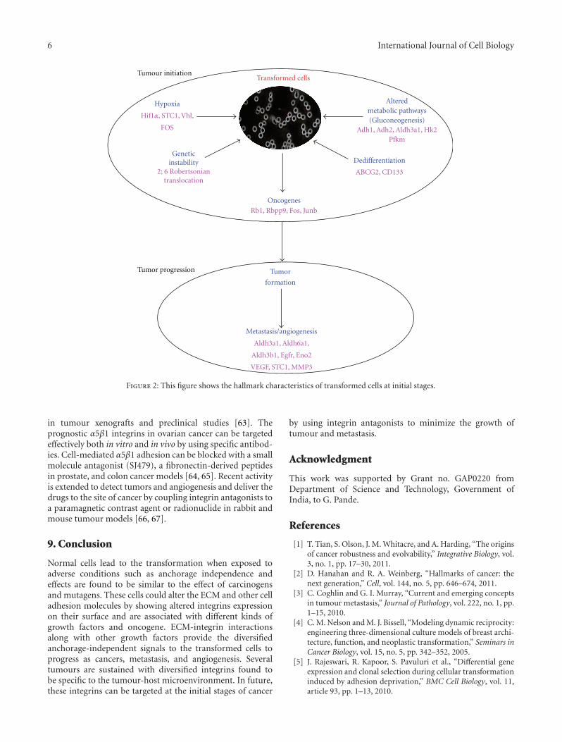
The cells during the nonadhesion process can evadefrom cell death by caspase 3 interaction with unligated α5β1integrins inducing resistance to integrin-mediated death(IMD) and also gain the ability to metastatise. Mutationalchanges mainly with 2;6 Robertsonian’s translocation andactivated Ras, FAK, and PKC provide self-sufficient growthsignals potential for uncontrollable growth of the cells(Figure 2). Upregulated Spp1, MMP3, Egfr, Rb1, Ddit3,Egln3, Vegfa, Stc1, Hif1a, MMP3, and altered pathwayslike glycolysis/gluconeogenesis and hypoxia (Pfkm, HK2,Pdk1, Adh1, aldh3a1, and Slc2a) lead the cells to invade,metastasize, and sustain angiogenesis. We observed anotherphenomenon of dedifferentiation by gaining the stem-cell-like and multidrug resistance properties by expressing Cd133and ABCG-2 when the cells are exposed to unfavorablecondition [5].
The anchorage-independent cellular model system rep-resents a multicentric tumor model system apprehendedwith genes related to tumor progression, angiogenesis, andmetastasis (Figure 2). It is very advantageous, convenient,and possible model system to study the effect of variouscancer-mediated drugs at the initial stage itself for the properdiagnosis.
8. Integrin Signalling as a Target inCancer Treatment
Several studies showed the correlation of integrin inhibitionat any point of its action will lead to the inhibition oftumor progression [24]. Therefore, integrins are focusedpharmacologically in the treatment and prevention of cancer.Antagonists of these integrins suppress cell migration andinvasion of primary and transformed cells by inducingapoptosis in primary cells could block tumor angiogenesisand metastasis. Recycling integrins present on the surface ofendothelial cells are targeted in the blood stream by exposingto the circulating drugs and agents [59]. Various antibod-ies, cyclic peptides, disintegrins, and peptidomimetics aremeant to bind the targeted integrins to prevent integrinligation. cRGD, cyclic arginine-glycine-aspartic acid; RGDK,arginine-glycine-aspartic acid-lysine; TRAIL, tumour necro-sis factor-related apoptosis-inducing ligand are being usedas antagonists integrins to hit the integrin ligand function.The function of upregulated αvβ3 integrin can be blocked byfunction-blocking monoclonal antibodies, such as LM 609[60]. The human αv integrin specific monoclonal antibodyCnTo 95, which targets both αvβ3 and αvβ5 integrinsto induce endothelial apoptosis, also had antitumour andantiangiogenic effects in xenograft tumour models [15].
Cilengitide, inhibitor of both αvβ3 and αvβ5 integrinsand volociximab, a function-blocking monoclonal antibodyagainst integrin α5β1, inhibits angiogenesis and impedestumour growth [61, 62]. αvβ3 is targeted by various ther-apeutic antibodies like LM609, vitaxin, humanized mousemonoclonal derived from LM609, CNTO 95, humanizedIgG1, c7E3, chimeric mouse human, 17E6, mouse mono-clonal antibodies to inhibit tumour growth, and angiogenesis
6 International Journal of Cell Biology
Oncogenes
Hypoxia
Geneticinstability Dedifferentiation
Tumor
formation
Metastasis/angiogenesis
Alteredmetabolic pathways(Gluconeogenesis)
Rb1, Rbpp9, Fos, Junb
2; 6 Robertsoniantranslocation
Aldh3a1, Aldh6a1,
Aldh3b1, Egfr, Eno2
VEGF, STC1, MMP3
Adh1, Adh2, Aldh3a1, Hk2Pfkm
Hif1α, STC1, Vhl,
FOS
ABCG2, CD133
Transformed cellsTumour initiation
Tumor progression
Figure 2: This figure shows the hallmark characteristics of transformed cells at initial stages.
in tumour xenografts and preclinical studies [63]. Theprognostic α5β1 integrins in ovarian cancer can be targetedeffectively both in vitro and in vivo by using specific antibod-ies. Cell-mediated α5β1 adhesion can be blocked with a smallmolecule antagonist (SJ479), a fibronectin-derived peptidesin prostate, and colon cancer models [64, 65]. Recent activityis extended to detect tumors and angiogenesis and deliver thedrugs to the site of cancer by coupling integrin antagonists toa paramagnetic contrast agent or radionuclide in rabbit andmouse tumour models [66, 67].
9. Conclusion
Normal cells lead to the transformation when exposed toadverse conditions such as anchorage independence andeffects are found to be similar to the effect of carcinogensand mutagens. These cells could alter the ECM and other celladhesion molecules by showing altered integrins expressionon their surface and are associated with different kinds ofgrowth factors and oncogene. ECM-integrin interactionsalong with other growth factors provide the diversifiedanchorage-independent signals to the transformed cells toprogress as cancers, metastasis, and angiogenesis. Severaltumours are sustained with diversified integrins found tobe specific to the tumour-host microenvironment. In future,these integrins can be targeted at the initial stages of cancer
by using integrin antagonists to minimize the growth oftumour and metastasis.
Acknowledgment
This work was supported by Grant no. GAP0220 fromDepartment of Science and Technology, Government ofIndia, to G. Pande.
References
[1] T. Tian, S. Olson, J. M. Whitacre, and A. Harding, “The originsof cancer robustness and evolvability,” Integrative Biology, vol.3, no. 1, pp. 17–30, 2011.
[2] D. Hanahan and R. A. Weinberg, “Hallmarks of cancer: thenext generation,” Cell, vol. 144, no. 5, pp. 646–674, 2011.
[3] C. Coghlin and G. I. Murray, “Current and emerging conceptsin tumour metastasis,” Journal of Pathology, vol. 222, no. 1, pp.1–15, 2010.
[4] C. M. Nelson and M. J. Bissell, “Modeling dynamic reciprocity:engineering three-dimensional culture models of breast archi-tecture, function, and neoplastic transformation,” Seminars inCancer Biology, vol. 15, no. 5, pp. 342–352, 2005.
[5] J. Rajeswari, R. Kapoor, S. Pavuluri et al., “Differential geneexpression and clonal selection during cellular transformationinduced by adhesion deprivation,” BMC Cell Biology, vol. 11,article 93, pp. 1–13, 2010.

International Journal of Cell Biology 7
[6] S. Shen, J. Fan, B. Cai et al., “Vascular endothelial growth fac-tor enhances cancer cell adhesion to microvascular endothe-lium in vivo,” Experimental Physiology, vol. 95, no. 2, pp. 369–379, 2010.
[7] E. E. Conway, “Central nervous system findings and intussus-ception: how are they related?” Pediatric Emergency Care, vol.9, no. 1, pp. 15–18, 1993.
[8] J. P. Thiery and J. P. Sleeman, “Complex networks orchestrateepithelial-mesenchymal transitions,” Nature Reviews Molecu-lar Cell Biology, vol. 7, no. 2, pp. 131–142, 2006.
[9] S. Kumar and V. M. Weaver, “Mechanics, malignancy, andmetastasis: the force journey of a tumor cell,” Cancer andMetastasis Reviews, vol. 28, no. 1-2, pp. 113–127, 2009.
[10] R. Rathinam and S. K. Alahari, “Important role of integrins inthe cancer biology,” Cancer and Metastasis Reviews, vol. 29, no.1, pp. 223–237, 2010.
[11] H. Hutter, B. E. Vogel, J. D. Plenefisch et al., “Conservationand novelty in the evolution of cell adhesion and extracellularmatrix genes,” Science, vol. 287, no. 5455, pp. 989–1010, 2000.
[12] D. E. Ingber, J. A. Madri, and J. D. Jamieson, “Role of basallamina in neoplastic disorganization of tissue architecture,”Proceedings of the National Academy of Sciences of the UnitedStates of America, vol. 78, no. 6 I, pp. 3901–3905, 1981.
[13] S. C. Wittelsberger, K. Kleene, and S. Penman, “Progressiveloss of shape-responsive metabolic controls in cells withincreasingly transformed phenotype,” Cell, vol. 24, no. 3, pp.859–866, 1981.
[14] H.-G. Zhang and W. E. Grizzle, “Exosomes and cancer: a newlydescribed pathway of immune suppression,” Clinical CancerResearch, vol. 17, no. 5, pp. 959–964, 2011.
[15] Q. Chen, C. D. Manning, H. Millar et al., “CNTO 95, afully human anti αv integrin antibody, inhibits cell signaling,migration, invasion, and spontaneous metastasis of humanbreast cancer cells,” Clinical and Experimental Metastasis, vol.25, no. 2, pp. 139–148, 2008.
[16] D. Barkan, J. E. Green, and A. F. Chambers, “Extracellularmatrix: a gatekeeper in the transition from dormancy tometastatic growth,” European Journal of Cancer, vol. 46, no. 7,pp. 1181–1188, 2010.
[17] R. K. Assoian and E. A. Klein, “Growth control by intracellulartension and extracellular stiffness,” Trends in Cell Biology, vol.18, no. 7, pp. 347–352, 2008.
[18] N. Zahir, J. N. Lakins, A. Russell et al., “Autocrine laminin-5ligates α6β4 integrin and activates RAC and NFκB to mediateanchorage-independent survival of mammary tumors,” Jour-nal of Cell Biology, vol. 163, no. 6, pp. 1397–1407, 2003.
[19] T. Gudjonsson, L. Rønnov-Jessen, R. Villadsen, F. Rank, M.J. Bissell, and O. W. Petersen, “Normal and tumor-derivedmyoepithelial cells differ in their ability to interact withluminal breast epithelial cells for polarity and basementmembrane deposition,” Journal of Cell Science, vol. 115, no. 1,pp. 39–50, 2002.
[20] A. Beliveau, J. D. Mott, A. Lo et al., “Raf-induced MMP9disrupts tissue architecture of human breast cells in three-dimensional culture and is necessary for tumor growth invivo,” Genes and Development, vol. 24, no. 24, pp. 2800–2811,2010.
[21] R. Pytela, M. D. Pierschbacher, and E. Ruoslahti, “Identifica-tion and isolation of a 140 kd cell surface glycoprotein withproperties expected of a fibronectin receptor,” Cell, vol. 40, no.1, pp. 191–198, 1985.
[22] R. O. Hynes, “Integrins: versatility, modulation, and signalingin cell adhesion,” Cell, vol. 69, no. 1, pp. 11–25, 1992.
[23] J. S. Desgrosellier and D. A. Cheresh, “Integrins in cancer:biological implications and therapeutic opportunities,” NatureReviews Cancer, vol. 10, no. 1, pp. 9–22, 2010.
[24] A. E. Aplin, A. K. Howe, and R. L. Juliano, “Cell adhesionmolecules, signal transduction and cell growth,” CurrentOpinion in Cell Biology, vol. 11, no. 6, pp. 737–744, 1999.
[25] D. G. Stupack, X. S. Puente, S. Boutsaboualoy, C. M. Storgard,and D. A. Cheresh, “Apoptosis of adherent cells by recruitmentof caspase-8 to unligated integrins,” Journal of Cell Biology, vol.155, no. 4, pp. 459–470, 2001.
[26] J. Rajeswari and G. Pande, “The significance of α5β1 integrindependent and independent actin cytoskelton organization incell transformation and survival,” Cell Biology International,vol. 26, no. 12, pp. 1043–1055, 2002.
[27] R. Zaidel-Bar and B. Geiger, “The switchable integrin adhe-some,” Journal of Cell Science, vol. 123, no. 9, pp. 1385–1388,2010.
[28] S. Han, F. R. Khuri, and J. Roman, “Fibronectin stimulatesnon-small cell lung carcinoma cell growth through activa-tion of Akt/mammalian target of rapamycin/S6 kinase andinactivation of LKB1/AMP-activated protein kinase signalpathways,” Cancer Research, vol. 66, no. 1, pp. 315–323, 2006.
[29] A. Kren, V. Baeriswyl, F. Lehembre et al., “Increased tumor celldissemination and cellular senescence in the absence of β1-integrin function,” EMBO Journal, vol. 26, no. 12, pp. 2832–2842, 2007.
[30] M. Zoller, “Tetraspanins: push and pull in suppressing andpromoting metastasis,” Nature Reviews Cancer, vol. 9, no. 1,pp. 40–55, 2009.
[31] D. Ribatti, “The contribution of Harold F. Dvorak to the studyof tumor angiogenesis and stroma generation mechanisms,”Endothelium, vol. 14, no. 3, pp. 131–135, 2007.
[32] P. C. Brooks, R. A. F. Clark, and D. A. Cheresh, “Requirementof vascular integrin αvβ3 for angiogenesis,” Science, vol. 264,no. 5158, pp. 569–571, 1994.
[33] P. C. Brooks, S. Stromblad, L. C. Sanders et al., “Localizationof matrix metalloproteinase MMP-2 to the surface of invasivecells by interaction with integrin αvβ3,” Cell, vol. 85, no. 5, pp.683–693, 1996.
[34] E. I. Deryugina, M. A. Bourdon, G. X. Luo, R. A. Reisfeld,and A. Strongin, “Matrix metalloproteinase-2 activation mod-ulates glioma cell migration,” Journal of Cell Science, vol. 110,no. 19, pp. 2473–2482, 1997.
[35] M. Kielosto, P. Nummela, K. Jarvinen, M. Yin, and E.Holtta, “Identification of integrins α6 and β7 as c-Jun- andtransformation-relevant genes in highly invasive fibrosarcomacells,” International Journal of Cancer, vol. 125, no. 5, pp. 1065–1073, 2009.
[36] Y. He, X. D. Liu, Z. Y. Chen et al., “Interaction between cancercells and stromal fibroblasts is required for activation of theuPAR-uPA-MMP-2 cascade in pancreatic cancer metastasis,”Clinical Cancer Research, vol. 13, no. 11, pp. 3115–3124, 2007.
[37] I. S. Vizirianakis, C. C. Yao, Y. Chen, B. L. Ziober, A.S. Tsiftsoglou, and R. H. Kramer, “Transfection of MCF-7 carcinoma cells with human integrin α7 cDNA promotesadhesion to laminin,” Archives of Biochemistry and Biophysics,vol. 385, no. 1, pp. 108–116, 2001.
[38] I. Staniszewska, E. M. Walsh, V. L. Rothman et al., “Effectof VP12 and viperistatin on inhibition of collagen receptors-dependent melanoma metastasis,” Cancer Biology and Ther-apy, vol. 8, no. 15, pp. 1507–1516, 2009.
[39] S. Van Slambrouck, C. Grijelmo, O. De Wever et al., “Acti-vation of the FAK-src molecular scaffolds and p130Cas-JNK
8 International Journal of Cell Biology
signaling cascades by α1-integrins during colon cancer cellinvasion,” International Journal of Oncology, vol. 31, no. 6, pp.1501–1508, 2007.
[40] M. Rolli, E. Fransvea, J. Pilch, A. Saven, and B. Felding-Habermann, “Activated integrin αvβ3 cooperates with met-alloproteinase MMP-9 in regulating migration of metastaticbreast cancer cells,” Proceedings of the National Academy ofSciences of the United States of America, vol. 100, no. 16, pp.9482–9487, 2003.
[41] J. Huang, R. Roth, J. E. Heuser, and J. E. Sadler, “Integrinalpha(v)beta(3) on human endothelial cells binds von Wille-brand factor strings under fluid shear stress,” Blood, vol. 113,no. 7, pp. 1589–1597, 2009.
[42] G. Y. Yang, K. S. Xu, Z. Q. Pan et al., “Integrin alphavbeta6mediates the potential for colon cancer cells to colonize in andmetastasize to the liver,” Cancer Science, vol. 99, no. 5, pp. 879–887, 2008.
[43] S. K. Gupta and N. E. Vlahakis, “Integrin α9β1 mediatesenhanced cell migration through nitric oxide synthase activityregulated by Src tyrosine kinase,” Journal of Cell Science, vol.122, no. 12, pp. 2043–2054, 2009.
[44] N. Gomes, C. Legrand, and F. Fauvel-Lafeve, “Shearstress induced release of von Willebrand factor andthrombospondin-1 in HUVEC extracellular matrix enhancesbreast tumour cell adhesion,” Clinical and ExperimentalMetastasis, vol. 22, no. 3, pp. 215–223, 2005.
[45] I. Gil-Bazo, V. Catalan, J. Paramo et al., “Von Willebrand factoras an intermediate between hemostasis and angiogenesis oftumor origin,” Revista de Medicina de la Universidad deNavarra, vol. 47, no. 3, pp. 22–28, 2003.
[46] J. Roman, J. D. Ritzenthaler, S. Roser-Page, X. Sun, and S. Han,“α5β1-integrin expression is essential for tumor progressionin experimental lung cancer,” American Journal of RespiratoryCell and Molecular Biology, vol. 43, no. 6, pp. 684–691, 2010.
[47] G. Maity, S. Fahreen, A. Banerji et al., “Fibronectin-integrinmediated signaling in human cervical cancer cells (SiHa),”Molecular and Cellular Biochemistry, vol. 336, no. 1-2, pp. 65–74, 2010.
[48] F. Mitelman and S. Heim, Cancer Cytogenetics, John Wiley &Sons, Chichester, UK, 2009.
[49] B. J. M. Braakhuis, C. R. Leemans, and R. H. Brakenhoff, “Agenetic progression model of oral cancer: current evidence andclinical implications,” Journal of Oral Pathology and Medicine,vol. 33, no. 6, pp. 317–322, 2004.
[50] P. L. Martin, Q. Jiao, J. Cornacoff et al., “Absence of adverseeffects in cynomolgus macaques treated with CNTO 95, afully human anti-αv integrin monoclonal antibody, despitewidespread tissue binding,” Clinical Cancer Research, vol. 11,no. 19 I, pp. 6959–6965, 2005.
[51] N. J. Quintyne, J. E. Reing, D. R. Hoffelder, S. M. Gollin,and W. S. Saunders, “Spindle multipolarity is prevented bycentrosomal clustering,” Science, vol. 307, no. 5706, pp. 127–129, 2005.
[52] X. Huang, T. E. Godfrey, W. E. Gooding, K. S. McCarty Jr.,and S. M. Gollin, “Comprehensive genome and transcriptomeanalysis of the 11q13 amplicon in human oral cancer andsynteny to the 7F5 amplicon in murine oral carcinoma,” GenesChromosomes and Cancer, vol. 45, no. 11, pp. 1058–1069, 2006.
[53] S. Kalyankrishna and J. R. Grandis, “Epidermal growth factorreceptor biology in head and neck cancer,” Journal of ClinicalOncology, vol. 24, no. 17, pp. 2666–2672, 2006.
[54] B. Jahrsdorfer, J. E. Wooldridge, S. E. Blackwell, C. M. Taylor,B. K. Link, and G. J. Weiner, “Good prognosis cytogenetics in
B-cell chronic lymphocytic leukemia is associated in vitro withlow susceptibility to apoptosis and enhanced immunogenic-ity,” Leukemia, vol. 19, no. 5, pp. 759–766, 2005.
[55] Beverly A. Teicher, Cancer Drug Discovery and Development,vol. 14, 2nd edition, 2011.
[56] M. A. Cifone and I. J. Fidler, “Correlation of patterns ofanchorage-independent growth with in vivo behavior of cellsfrom a murine fibrosarcoma,” Proceedings of the NationalAcademy of Sciences of the United States of America, vol. 77, no.2, pp. 1039–1043, 1980.
[57] D. Hanahan and R. A. Weinberg, “The hallmarks of cancer,”Cell, vol. 100, no. 1, pp. 57–70, 2000.
[58] E. R. Lawlor, C. Scheel, J. Irving, and P. H. B. Sorensen,“Anchorage-independent multi-cellular spheroids as an invitro model of growth signaling in Ewing tumors,” Oncogene,vol. 21, no. 2, pp. 307–318, 2002.
[59] A. Aparna and A. V. Judith, “Integrins in cancer,progressionand therapy,” Science & Medicine, vol. 10, no. 2, pp. 84–96,2005.
[60] P. C. Brooks, S. Stromblad, R. Klemke, D. Visscher, F. H.Sarkar, and D. A. Cheresh, “Antiintegrin αvβ3 blocks humanbreast cancer growth and angiogenesis in human skin,” Journalof Clinical Investigation, vol. 96, no. 4, pp. 1815–1822, 1995.
[61] J. W. Smith, Z. M. Ruggeri, T. J. Kunicki, and D. A.Cheresh, “Interaction of integrins αvβ3 and glycoprotein IIb-IIIa with fibrinogen. Differential peptide recognition accountsfor distinct binding sites,” Journal of Biological Chemistry, vol.265, no. 21, pp. 12267–12271, 1990.
[62] V. Bhaskar, D. Zhang, M. Fox et al., “A function blockinganti-mouse integrin α5β1 antibody inhibits angiogenesis andimpedes tumor growth in vivo,” Journal of TranslationalMedicine, vol. 5, article 61, 2007.
[63] M. Millard, S. Odde, and N. Neamati, “Integrin targetedtherapeutics,” Theranostics, vol. 1, pp. 154–188, 2011.
[64] O. Stoeltzing, W. Liu, N. Reinmuth et al., “Inhibition ofintegrin α5β1 function with a small peptide (ATN-161) pluscontinuous 5-fu infusion reduces colorectal liver metastasesand improves survival in mice,” International Journal ofCancer, vol. 104, no. 4, pp. 496–503, 2003.
[65] D. L. Livant, R. K. Brabec, K. J. Pienta et al., “Anti-invasive,antitumorigenic, and antimetastatic activities of the PHSCNsequence in prostate carcinoma,” Cancer Research, vol. 60, no.2, pp. 309–320, 2000.
[66] J. H. Kim, Y. S. Kim, K. Park et al., “Self-assembled glycolchitosan nanoparticles for the sustained and prolonged deliv-ery of antiangiogenic small peptide drugs in cancer therapy,”Biomaterials, vol. 29, no. 12, pp. 1920–1930, 2008.
[67] M. K. Yu, J. Park, Y. Y. Jeong, W. K. Moon, and S. Jon,“Integrin-targeting thermally cross-linked superparamagneticiron oxide nanoparticles for combined cancer imaging anddrug delivery,” Nanotechnology, vol. 21, no. 41, Article ID415102, 2010.