chitosan aerogel beads with microfibrillated cellulose skeleton for removal …2017)/p.521-5… ·...
TRANSCRIPT
CELLULOSE CHEMISTRY AND TECHNOLOGY
Cellulose Chem. Technol., 51 (5-6), 521-528 (2017)
CHITOSAN AEROGEL BEADS WITH MICROFIBRILLATED CELLULOSE
SKELETON FOR REMOVAL OF FORMALDEHYDE FROM INDOOR AIR
PENG WU and ZHIMING LIU
College of Material Science and Engineering, Northeast Forestry University, 26, Hexing Road,
Harbin 150040, Heilongjiang, People’s Republic of China ✉Corresponding author: Zhiming Liu, [email protected]
Received September 1, 2015
Formaldehyde is present to some degree in indoor air, and poses a considerable threat to human health. The
development of efficient and effective sorbent materials is highly necessary, in order to remove formaldehyde and
better safeguard the health of a building’s occupants. This study explores a new chitosan aerogel bead supported by
microfibrillated cellulose, found to be an efficient biosorbent for formaldehyde, with high sorption capacity. Adsorption
tests demonstrate that the sample prepared at a chitosan/MFC weight ratio of R=1 exhibits the highest specific surface
area (1351 m2/g) and maximum HCHO adsorption capacity (2.2 mmolHCHO g−1). The outstanding sorption performance
of the aerogel beads is associated with their unique physical and chemical properties, including low density, high
porosity and specific surface area, as well as abundant surface-activated amino groups. This study provides valuable
information for the fabrication of novel, environmentally friendly aerogel beads with high HCHO adsorption
performance for indoor air purification.
Keywords: chitosan aerogel beads, formaldehyde cleaning, microfibrillated cellulose
INTRODUCTION
Formaldehyde, which at room temperature is a
colorless, characteristically pungent, irritating gas,
is a rather common indoor pollutant that is emitted
from indoor furniture paint, flooring materials,
wallpaper, and cigarette smoke.1,2
At concentrations
above 0.1 ppm in an indoor environment,
formaldehyde has been shown to cause a series of
health problems related to “Sick Building
Syndrome” (SBS).3 Recent studies have also shown
a positive correlation between exposure to
formaldehyde and the development of leukemia,
particularly myeloid leukemia.4,5 To this effect,
emitted formaldehyde gas in indoor air is extremely
harmful and its removal is a crucial consideration.
Various technologies have been developed for
the purpose of removing formaldehyde from indoor
environments, including adsorption,6-9
catalytic
oxidization,10,11 and botanical filtrations.12
Considering the costs and convenience, the
adsorption method is a favorable method for
pollutant treatment and environmental purification.
The adsorptive removal of volatile aldehydes
focuses on amine-functionalized materials,
effective due to utilization of azomethine or Schiff
bases between aldehydes and targeted materials.13,14
When a Schiff base is formed, however, the
adsorbed formaldehyde is not released from the
adsorbent even heated to 60 °C for 2 h.15
Additionally, synthetic amine-functionalized
materials are generally rather expensive and may
cause secondary pollution of the environment when
they are discarded. Chitosan, a renewable and
biodegradable amino polysaccharide obtained from
chitin after deacetylation, is already widely used as
water treatment material,16-19
and has been shown to
absorb gaseous aldehydes such as formaldehyde
and acetaldehyde as well.20 The efficiency of
formaldehyde adsorption of the traditional form of
chitosan, however, is not very high,21,22
which calls
for expanding its specific surface area and
increasing its number of active amine to improve its
adsorption efficiency.
Aerogels are a class of low-density solids that
possess open three-dimensional mesoporous
structure and high specific surface area, and thus
show potential application as adsorbents, thermal
insulators, acoustic absorbers, catalysts, and
catalyst supports.23-25
Biomass aerogel materials
possess features of traditional aerogels, plus certain
additional advantages, such as renewability,

PENG WU and ZHIMING LIU
522
abundant availability, low cost, and environmental
friendliness.26,27 Their chitosan backbone, however,
is highly polar and forms strong hydrogen bonds
between adjacent chains – thus it does not easily
maintain a high specific surface area due to severe
shrinkage and deformation of the gel.20
In an effort
to remedy this, previous studies investigated
cross-linked and blended chitosan with organic
and/or inorganic compounds to successfully
improve the specific surface area of the chitosan
aerogel.28,29 The present study utilizes
environmentally friendly and highly strong
microfibrillated cellulose (MFC) as skeleton for
surface tethering of chitosan molecules, and
demonstrates that chitosan aerogel containing MFC
is an excellent candidate material for gaseous
formaldehyde removal. Moreover, the spherical
aerogel can be quite easily incorporated into air
purification systems or filters to improve overall air
quality.
EXPERIMENTAL Materials
Commercial chitosan was purchased from
Sigma-Aldrich, with a viscosity of 55 mPas and
deacetylation degree of over 96 wt%. Phyllostachys
heterocycla cv. pubescens bamboo was obtained from
Huang-gongwang Forest Park in Fuyang, Zhejiang
Province, China, and dried, ground and screened to
obtain 60-80 mesh particle size bamboo powder. All
other chemicals were of analytical grade and used
without further purification.
Investigation methods
Synthesis of aerogel beads Bamboo powder was purified by a series of chemical
treatments, as described by Okahisa30 and Abe.31
Microfibrillated cellulose (MFC) was fabricated by the
homogenization process.32,33
First, the extractives of
bamboo powder were removed using an ethanol-toluene
solution for 6 h in the Soxhlet apparatus. Afterwards, any
lignin in the sample was removed by an acidified sodium
chlorite solution at 70 ºC for 5 h. Next, hemicellulose in
the sample was treated in 5 w% potassium hydroxide
aqueous solution at room temperature for 24 h. Lastly,
the sample was filtered and rinsed with distilled water
until the residue was neutralized. After the purification
process, the as-purified cellulose was dispersed in 300
ml deionized water and homogenized using a
high-pressure fluidizer (Microfluidizer M-110,
Microfluidics Corp., USA) equipped with 87 µm-sized
chambers. Full homogenization was achieved after 12
cycles at an operating pressure of 20,000 psi. Finally, the
MFC hydrosol was obtained and diluted to
approximately 1 wt%.
Aerogel beads were synthesized using the
hand-dropping procedure, based on the pH inversion.34,35
For the synthesis of spherical aerogels at the
chitosan/MFC weight ratio R=1, 1 g chitosan was
initially dissolved in 2% v/v aqueous solution (100 mL)
of acetic acid, followed by mixing the above solution of
MFC hydrosol (100 mL) with ultrasonic dispersion for
20 min. The mixture with the pH value of 3.97 and
viscosity of 42.6 mPa·s was then dropped into 2 L of
0.1mol/L NaOH solution with a 1 mL disposable plastic
pipette, and solidified at room temperature for 12 h
before rinsing under running deionized water for 12 h.
To prevent the hydrogel networks from collapsing during
the drying process, the obtained hydrogel samples were
first exchanged with ethanol, then with t-butyl alcohol,
with several exchanges in each solvent sample. The
t-butyl alcohol-included gel was subjected to freeze
drying. The samples were also prepared at R = 0
(without chitosan), 3 (chitosan/MFC weight ratio), and ∞
(without cellulose) under the same conditions described
above. The samples prepared at R = 0, 1, 3, and ∞ were
labeled CAB-0, CAB-1, CAB-3, and CAB-∞,
respectively. For different acidity to different proportions
of the mixture during synthesis of the gel beads, there is
little impact on the pH within excessive sodium
hydroxide coagulating bath.
Characterization of aerogel beads
The surface morphology of the aerogel samples was
analyzed with a scanning electron microscope (SEM,
FEI QUATA200). The beads were coated with gold for
observation and photographed at an accelerating voltage
of 12.5 kV.
The Brunauer-Emmett-Teller (BET) specific surface
areas and porous structures of the samples were
characterized using a Micromeritics ASAP 2020 nitrogen
adsorption apparatus (USA). All samples were degassed
at 120 ºC before nitrogen adsorption measurements.
The Fourier transform infrared (FT-IR) spectra were
acquired with a Specttwn 2000 instrument (Perkin Elmer,
USA) using a diamond single reflection attenuated total
reflectance (FTIR-ATR) device. Duplicate spectra per
sample were obtained with 32 scans per spectrum at a
spectral resolution of 4 cm−1 in a wavenumber range
from 4000 to 650 cm−1
.
X-ray photoelectron spectroscopy (XPS)
measurement was performed using a Model PHI-5700
ESCA apparatus (Physical Electronics, USA). A
monochromatic Al Kα (1,486.6 eV) X-ray source and 40
eV pass energy of analyzer were used under ultra-high
vacuum conditions (5.2 × 10⁻⁹
Torr). All binding energies
(BE) were referenced with adventitious carbon (binding
energy = 284.6 eV).
Formaldehyde adsorption experiments The aerogel beads were tested for adsorption
performance in a self-designed sealed amber chamber at
room temperature, as shown in Figure 1, where the total
volume of chamber was approximately 5 L. Before
formaldehyde abatement tests, all adsorbent samples
were dried under vacuum (ca. 200 Pa) overnight at ca.

Chitosan
523
100 °C to remove physisorbed moisture and impurities,
then the prepared samples (10 mg) were wrapped in
qualitative filter paper (pore size and diameter of 15-20
µm and 15 cm, respectively) and hung in the chamber. In
order to ensure complete formaldehyde gasification, the
chamber was first fully evacuated, then injected with 8
µL of aqueous formaldehyde solution (10 wt%) by a
microsyringe. N2 gas was also introduced in order to
maintain atmospheric pressure.
Then, the system was equilibrated over time at room
temperature. Formaldehyde gas concentrations were
determined using the HPLC method.36
3.0 g/L
2,4-dinitrobenzene in acetonitrile solution (2 mL), and 2
mL acetonitrile were added into the absorption tube,
while H2 was continually fed into the chamber in order
to make the residual formaldehyde in the chamber
completely pass the 2,4-dinitrophenylhydrazine (DNPH)
sorbent and form a DNPH-formaldehyde adduct. The
extract was analyzed using an HPLC system from
Shimadzu, which consisted of C18 chemical-bonded
silica gel columns as a partitioner phase, a mobile phase
of acetonitrile and water (60:40 v/v) at a flow rate of 1
mL/min at 40 °C, and a UV detector at 360 nm. The
formaldehyde gas concentration inside the container was
determined by calibrating the peak area with
formaldehyde standard solutions.13 The adsorption
quantities of samples at 30, 60, 120, 180, and 300 min
were measured three times according to the above
method.
RESULTS AND DISCUSSION
Characterization of materials
SEM analysis The aerogel beads obtained from the
corresponding hydrogel samples with a
chitosan/MFC weight ratio of R=0 to ∞ after
freeze-drying have been assessed as successfully
prepared. As shown in Figure 2, all samples are
spherical or ellipsoidal and form similar
three-dimensional network structures with micro
and mesopores. The cellulose aerogel beads
(CAB-0), however, show small volume shrinkage
change during the gelation and drying process; their
network is formed by the aggregation and winding
of individual microfibrillated cellulose, which is
very similar to those isolated from plant material, as
indicated in a previous study.37
Conversely, for the
pure chitosan samples (CAB-∞), partial fibers seem
to have thickened, and their network has become
more intensive and non-uniform – likely due to the
high polarity of chitosan molecules and the
formation of strong hydrogen bonds between
adjacent chains.20
As for the composite samples,
results show that higher MFC concentration
lessened the volume shrinkage of the beads,
attributed to the skeletal reinforcement of MFC in
the chitosan aerogel system.
BET analysis
The N2 adsorption/desorption isotherms and
pore-size distribution curves, as shown in Figure 3,
also demonstrate that isotherms exhibit type IV
characteristics with a pronounced type H1
hysteresis loop, typical of mesoporous materials,
according to IUPAC.38 Further, the exploding
sorption capacity within a relatively high P/P0 range
reflects the presence of excessive large-diameter
mesoporosity and macroporosity of inter-fibril
voids,30
which can be expected from CAB-0 with
98.4 vol% overall porosity (calculated from
apparent density and true density of cellulose).
Among these isotherms, the CAB-1 shows the
highest adsorption volume, implying that the
introduction of MFC into chitosan aerogel does not
change the essential microstructural characteristics
of chitosan aerogel beads, but only affects the
surface area and pore space available for N2
adsorption, while reducing the aggregation of
chitosan molecular adjacent chains.
Figure 1: Experimental apparatus for the formaldehyde adsorption test

PENG WU and ZHIMING LIU
524
Figure 2: SEM images and photographs of composite aerogel beads with different chitosan/MFC weight ratio of (a)
CAB-0, (b) CAB-1, (c) CAB-3, and (d) CAB-∞ (Insets show photographs of corresponding samples)
Figure 3: Nitrogen adsorption-desorption isotherms (a) and corresponding pore size distribution curves (b) of
CAB-0, CAB-1, CAB-3 and CAB-∞ samples
Table 1
Porosity characteristics of composite aerogel beads with different chitosan/MFC weight ratios
Sample Apparent
density, mg/cm3
SBET (from micropores),
m2/g
Pore volumes
(from micropores), cm3/g
Average pore
size, nm
CAB-0 19.2±0.6 173 (12) 0.619 (0.005) 12.96
CAB-1 26.1±0.4 1351 (110) 4.486 (0.053) 12.31
CAB-3 31.0±1.2 291 (22) 1.139 (0.010) 13.37
CAB-∞ 59.6±2.1 299 (19) 1.157 (0.009) 13.39
Figure 3(b) displays the corresponding pore size
distributions of the samples. All beads exhibit a
bi-modal distribution with the first pore size peak
located in the micropore region, and the main pore
size peak located in the mesopore region. Relevant
structural parameters derived from the isotherms
are summarized in Table 1. The CAB-1 sample
shows the largest specific surface area and highest
pore volume compared to the other samples. The
specific surface area, average pore size, and pore
volume of the CAB-1 sample is 1351 m2/g, 12.31
nm, and 4.486 cm3/g, respectively. The mesopores
in CAB-1 contribute 91.8% (1241 m2/g) of the
specific surface area and 98.8% (4.433 cm3/g) of
the pore volume. This indicates that the mesopores
of the CAB-1 samples function quite effectively as
gas purifiers.
FTIR analysis FITR analysis has been also performed in order
to investigate possible chemical interaction between
MFC and chitosan in the composite aerogel beads
(Fig. 4). The FTIR spectra of pure chitosan aerogel
(CAB-∞) shows a broad band at 3445 cm−1 due to
O-H and N-H stretching, whereas the two bands at
1670 and 1600 cm−1 are attributed to C=O of amide

Chitosan
525
I and N-H of amide II.39,40
The FTIR spectrum of
MFC aerogel (CAB-0) shows characteristic bands
at 3342 cm−1
due to O-H stretching vibration, the
peaks at 2900 cm−1
for C-H stretching, and the peak
at 1630 cm−1 associated with the H–O–H stretching
vibration of absorbed water in carbohydrates,
respectively.41
All the peaks of CAB-1 are found in
the spectrum of CAB-0 (MFC) or CAB-∞ (chitosan)
without any changes in peak position, implying that
CAB-1 is simply a physical mixture of MFC and
chitosan. The results reveal the absence of any
chemical interaction between MFC and chitosan
besides breakage of hydrogen bonds during the
processes of dissolution and regeneration.
XPS analysis
Representative O 1s, N 1s and C 1s peaks of
CAB-1 and CAB-∞ are displayed in Figure 5. The
C 1s peak shows three components: at 284.8 eV,
typical of carbon only bound to carbon and
hydrogen [C-(C, H)]; near 286.2 eV, typical of
carbon forming a single bond with oxygen or
nitrogen [C-(O, N)]; and near 287.7 eV, typical of
acetal and amide [O-C-O, N-C=O].
Figure 4: FTIR spectra of composite aerogel beads with different chitosan/MFC weight ratios
Figure 5: C 1 s, O 1 s and N 1 s XPS regions in the CAB-1 and CAB-∞
PENG WU and ZHIMING LIU
526
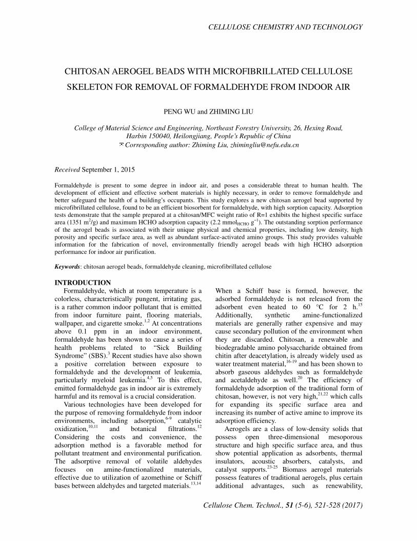
Table 2
Elemental surface composition of CAB-1 and CAB-∞
Atomic surface concentration obtained by XPS (%) Sample
C N O
CAB-1 54.75 6.84 38.41
CAB-∞ 69.38 4.25 26.38
Figure 6: Formaldehyde concentrations inside the gas chamber plotted against time elapsed
after its injection
The N 1s contribution near 399.3 eV is due to
non-protonated amine or amide.42
The major O 1s
contribution found at 532.7 ± 0.1 eV is due to
oxygen in the polysaccharide backbone [O-H],
while a small component near 531.0 ± 0.2 eV is
attributed to ether groups in the polysaccharide
backbone and amide polysaccharide backbone
[C-O-C, C=O].43
According to these peak areas, the elemental
surface compositions are calculated as listed in
Table 2. Though the chitosan content of CAB-1 is
half that of CAB-∞, CAB-1 shows higher N content
than half of CAB-∞, which is indicative of the high
mechanical strength of the MFC framework.
Chitosan molecules expose more active amino
groups to the surface of the aerogel, which is
beneficial to formaldehyde gas absorption.
Adsorption performances of formaldehyde Figure 6(a) and (b) show the results of
formaldehyde abatement testing. The gas chamber
with no adsorbent maintains the initial
formaldehyde concentration (118 ppm), showing
that no significant leakage of formaldehyde gas
occurred within the examined time period (Fig.
6(a)). In order to demonstrate the adsorption
capacity of the pre-prepared samples, the same
weight as the test sample of coconut shell activated
carbon (GAC, obtained from Beijing Walter Liyuan
Environmental Protection Technology Co. Ltd.,
with specific surface area and pore volume of
590-1500 m2/g and 0.7-1 cm3/g, respectively) has
been used as control sample under the same
conditions. The gas chamber with the GAC
adsorbent slightly decreases the formaldehyde
concentration to 100 ppm (84.7% of the starting
concentration), due to the high surface area of GAC.
The CAB-0 also slightly decreases the gas
concentration to 110 ppm, (93.2% of its initial
concentration), suggesting that formaldehyde is
partially adsorbed onto the cellulose surface. The
CAB-1 adsorbent dramatically decreases the
formaldehyde concentration to 19 ppm (16.1% of
the initial concentration) within an hour. This
clearly indicates that surface-accessible amine
groups play a crucial role in the adsorption of
formaldehyde. Compared to CAB-1, both CAB-3
and CAB-∞ adsorb formaldehyde less efficiently,
only decreasing formaldehyde concentration to 67
ppm and 77 ppm, respectively. Under the
assumption that all removed formaldehyde is
tapped into the adsorbent, the adsorption capacity, q,
is derived as listed in Table 3. Notably, the q of
CAB-1 (2.2 mmol HCHO g-1
) is the highest among all
the test samples. This supports the theory that
CAB-1 is a very successful formaldehyde
abatement material.
FTIR analysis, as shown in Figure 7, has been
adopted to further investigate formaldehyde
adsorption mechanisms of CAB-1. After adsorption,
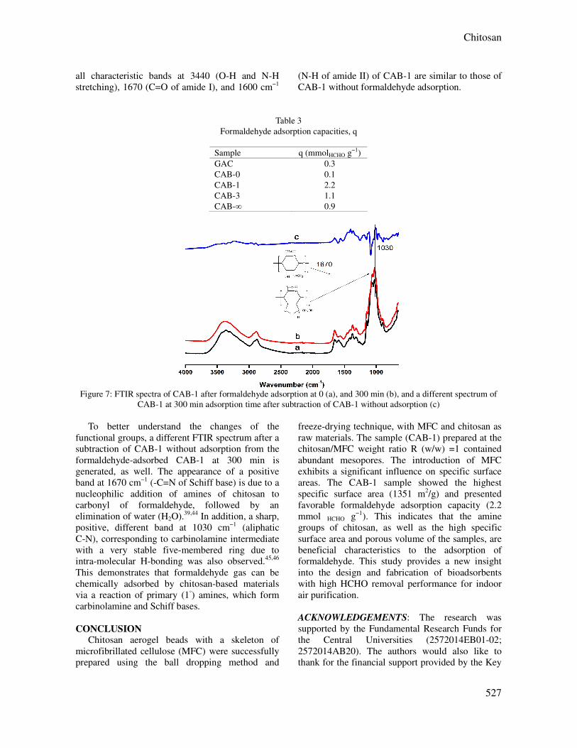
Chitosan
527
all characteristic bands at 3440 (O-H and N-H
stretching), 1670 (C=O of amide I), and 1600 cm−1
(N-H of amide II) of CAB-1 are similar to those of
CAB-1 without formaldehyde adsorption.
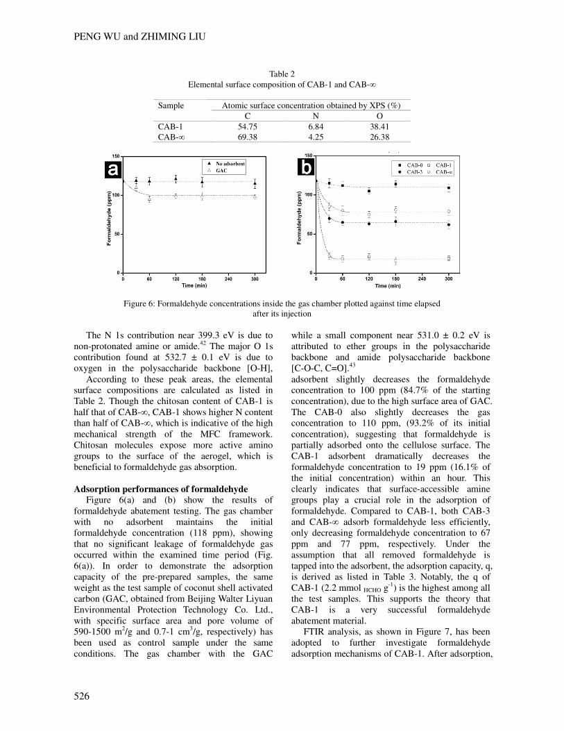
Table 3
Formaldehyde adsorption capacities, q
Sample q (mmolHCHO g−1)
GAC 0.3
CAB-0 0.1
CAB-1 2.2
CAB-3 1.1
CAB-∞ 0.9
Figure 7: FTIR spectra of CAB-1 after formaldehyde adsorption at 0 (a), and 300 min (b), and a different spectrum of
CAB-1 at 300 min adsorption time after subtraction of CAB-1 without adsorption (c)
To better understand the changes of the
functional groups, a different FTIR spectrum after a
subtraction of CAB-1 without adsorption from the
formaldehyde-adsorbed CAB-1 at 300 min is
generated, as well. The appearance of a positive
band at 1670 cm−1 (-C=N of Schiff base) is due to a
nucleophilic addition of amines of chitosan to
carbonyl of formaldehyde, followed by an
elimination of water (H2O).39,44 In addition, a sharp,
positive, different band at 1030 cm−1 (aliphatic
C-N), corresponding to carbinolamine intermediate
with a very stable five-membered ring due to
intra-molecular H-bonding was also observed.45,46
This demonstrates that formaldehyde gas can be
chemically adsorbed by chitosan-based materials
via a reaction of primary (1◦) amines, which form
carbinolamine and Schiff bases.
CONCLUSION Chitosan aerogel beads with a skeleton of
microfibrillated cellulose (MFC) were successfully
prepared using the ball dropping method and
freeze-drying technique, with MFC and chitosan as
raw materials. The sample (CAB-1) prepared at the
chitosan/MFC weight ratio R (w/w) =1 contained
abundant mesopores. The introduction of MFC
exhibits a significant influence on specific surface
areas. The CAB-1 sample showed the highest
specific surface area (1351 m2/g) and presented
favorable formaldehyde adsorption capacity (2.2
mmol HCHO g−1). This indicates that the amine
groups of chitosan, as well as the high specific
surface area and porous volume of the samples, are
beneficial characteristics to the adsorption of
formaldehyde. This study provides a new insight
into the design and fabrication of bioadsorbents
with high HCHO removal performance for indoor
air purification.
ACKNOWLEDGEMENTS: The research was
supported by the Fundamental Research Funds for
the Central Universities (2572014EB01-02;
2572014AB20). The authors would also like to
thank for the financial support provided by the Key

PENG WU and ZHIMING LIU
528
Laboratory of Wood Science and Technology,
Zhejiang Province (2014lygcz002). Special thanks
are given to Prof. Shujun Li for her test equipment
and analysis.
REFERENCES 1 H. B. An, M. J.Yu, J. M. Kim, M. Jin, J. K Jeon et al.,
Nanoscale Res. Lett., 7, 1 ( 2012). 2 W. J. Liang, J. Li, J. X. Li, T. Zhu and Y. Q. Jin, J.
Hazard. Mater., 175, 1090 (2010). 3 P. Chin, L. P. Yang and D. F. Ollis, J. Catal., 237, 29
(2006). 4 V. J. Cogliano, Y. Grosse, R. A. Baan, K. Straif, M. B.
Secretan et al., Environ. Health Persp., 113, 1205
(2005). 5 M. Hauptmann, J. H. Lubin, P. A. Stewart, R. B.
Hayes and A. Blair, Am. J. Epidemiol., 159, 1117 (2004). 6 Y. Le, D. P. Guo, B. Cheng and J. G. Yu, Appl. Surf.
Sci., 274, 110 (2013). 7 K. J. Lee, N. Shiratori, G. H. Lee, J. Miyawaki, I.
Mochida et al., Carbon, 48, 4248 (2010). 8 Z. H. Xu, J. G. Yu, G. Liu, B. Cheng, P. Zhou et al.,
Dalton T., 42, 10190 (2013). 9 S. Yamanaka, T. Oiso, Y. Kurahashi, H. Abe, K. Hara
et al., J. Nanopart. Res., 16, 1 (2014). 10 C. Y. Ma, D. H. Wang, W. J. Xue, B. J. Dou, H. L.
Wang et al., Environ. Sci. Technol., 45, 3628 (2011). 11 C. B. Zhang, H. He and K. Tanaka, Catal. Commun.,
6, 211 (2005). 12 N. Lu, J. J. Pei, Y. X. Zhao, R. Y. Qi and J. J. Liu,
Build. Environ., 57, 253 (2012). 13 A. M. Ewlad-Ahmed, M. A. Morris, S. V.
Patwardhan and L. T. Gibson, Environ. Sci. Technol., 46,
13354 (2012). 14 H. Q. Rong, Z. Y. Liu, Q. L. Wu, D. Pan and J. T.
Zheng, Cellulose, 17, 205 (2010). 15 J. J. Pei and J. S. Zhang, Chem. Eng. J., 167, 59
(2011). 16 A. I. Cocârta and E. S. Dragan, Cellulose Chem.
Technol., 48, 495 (2014). 17 W. S. Wan Ngah, L. C. Teong and M. A. K. M.
Hanafiah, Carbohyd. Polym., 83, 1446 (2011). 18 A. H. Jawad and M. A. Nawi, Carbohyd. Polym., 90,
87 (2012). 19 Y. M. Zhou, S. Y. Fu, L. L. Zhang, H. Y. Zhan and M.
V. Levit, Carbohyd. Polym., 101, 75 (2014) 20 X. H. Chang, D. R. Chen and X. L. Jiao, J. Phys.
Chem. B, 112, 7721 (2008). 21 A. El Kadib and M. Bousmina, Chem.-Eur. J., 18,
8264 (2012).
22 N. Li and R. B. Bai, Sep. Purif. Technol., 42, 237
(2005). 23 H. C. Bi, Z. Y. Yin, X. H. Cao, X. Xie, C. L. Tan et
al., Adv. Mater., 25, 5916 (2013). 24 K. H. Kim, M. Vural and M. F. Islam, Adv. Mater.,
23, 2865 (2011).
25 J. T. Korhonen, M. Kettunen, R. H. A. Ras and O.
Ikkala, ACS Appl. Mater. Inter., 3, 1813 (2011). 26 J. Li, Y. Lu, D. J. Yang, Q. F. Sun, Y. X. Liu et al.,
Biomacromolecules, 12, 1860 (2011). 27 Y. Lu, Q. F. Sun, D. J. Yang, X. L. She, X. D. Yao et
al., J. Mater. Chem., 22, 13548 (2012). 28 S. Nuasaen, P. Opaprakasit and P. Tangboriboonrat,
Carbohyd. Polym., 101, 179 (2014). 29 K. H. Yang, Y. C. Liu, C. C. Yu and B. C. Chen,
Mater. Chem. Phys., 126, 993 (2011). 30 Y. Okahisa, A. Yoshida, S. Miyaguchi and H. Yano,
Compos. Sci. Technol., 69, 1958 (2009) 31 K. Abe and H. Yano, Carbohyd. Polym., 85, 733
(2011). 32 M. Henriksson, L. A. Berglund, P. Isaksson, T.
Lindstrom and T. Nishino, Biomacromolecules, 9, 1579
(2008). 33 S. Y. Lee, S. J. Chun, I. A. Kang and J. Y. Park, J. Ind.
Eng. Chem., 15, 50 (2009). 34 A. El Kadib, K. Molvinger, T. Cacciaguerra, M.
Bousmina and D. Brunel, Micropor. Mesopor. Mat., 142,
301 (2011). 35 M. Gericke, J. Trygg and P. Fardim, Chem. Rev., 113,
4812 (2013). 36 A. Nomura and C. W. Jones, ACS Appl. Mater. Inter.,
5, 5569 (2013). 37 K. Abe and H. Yano, Cellulose, 17, 271 (2010). 38 H. C. Chien, W. Y. Cheng, Y. H. Wang and S. Y. Lu,
Adv. Funct. Mater., 22, 5038 (2012). 39 G. H. He, Z. Wang, H. Zheng, Y. H. Yin, X. Xiong et
al., Carbohyd. Polym., 90, 1614 (2012). 40 M. L. Li, J. Xu, R. H. Li, D. G. Wang, T. B. Li et al.,
J. Colloid Interf. Sci., 417, 131 (2014). 41 W. S. Chen, H. P. Yu and Y. X. Liu, Carbohyd.
Polym., 86, 453 (2011). 42 P. H. F. Pereira, H. J. C. Voorwald, M. O. H. Cioffi,
M. L. C. P. Da Silva, A. M. B. Rego et al., Cellulose, 21,
641 (2014). 43 I. F. Amaral, P. L. Granja and M. A. Barbosa, J.
Biomat. Sci.-Polym. E., 16, 1575 (2005). 44 Y. H. Zhu, H. Li, Q. Zheng, J. Q. Xu and X. X. Li,
Langmuir, 28, 7843 (2012). 45 S. Farris, J. H. Song and Q. R. Huang, J. Agric. Food
Chem., 58, 998 (2010). 46 N. Nishat, S. A. Khan, R. Rasool and S. Parveen, J.
Inorg. Organomet. P., 21, 673 (2011).