chapter 14.2: white blood cells and platelets. white blood cells (wbcs) -also called leukocytes...
TRANSCRIPT

Chapter 14.2:White Blood Cells and Platelets

White Blood Cells (WBCs)
- Also called leukocytes
- Contain a nucleus and other organelles
- No hemoglobin
- Specialize in protecting body from disease
- Two types:1) Granular2) Agranular

White Blood Cells (WBCs)
- Granulocytes: contain chemical-filled granules 1) Neutrophils2) Eosinophils3) Basophils
- Agranulocytes: no granules1) Lymphocytes: B, T, NK cels2) Monocytes: macrophages

Granulocytes
- 1st Respnoders -> Phagocytosis- 60% of WBCs- Found in pus of wounds
Neutrophils- Inflammatory reaction- Responds to allergic reactions- 1% of WBCs
Basophils- Phagocytize antigen-antibody
complexes- Mainly attack parasitic worms- 2% of WBCs
Eosinophils
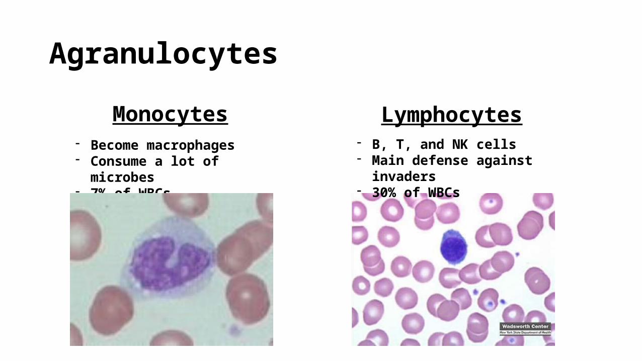
Agranulocytes
- Become macrophages- Consume a lot of microbes- 7% of WBCs
Monocytes- B, T, and NK cells- Main defense against invaders- 30% of WBCs
Lymphocytes

WBC Life Span
- About 5,000 to 10,000 WBC/µL of blood
- Life span = a few days- Phagocytosis interferes with normal metabolism- During infection, only a few hours
- Leukocytosis: an increase in the # of WBCs- WBC count used to diagnose specific illness
- Develop in red bone marrow from myeloid and lymphoid stem cells

Platelets- Pluripotent stem cells in red bone marrow -> megakaryocytes -> platelets
- About 150,000-400,000 platelets/µL blood
- Form a platelet plug to prevent blood loss

Chapter 14.3-Hemostasis

Hemostasis
- A series of responses that stops bleeding when blood vessels are injured
- Involves coagulation and clotting of blood to seal site of damage
- Prevents hemorrhage
- Three different mechanisms:1) Vascular spasm2) Platelet plug3) Blood clotting

Vascular Spasm
- Smooth muscle around blood vessel contracts immediately
- Initial response to smooth muscle damage- Detected by pain receptors- Can reduce blood loss for several hours
- Platelets accumulate and release chemicals to enhance vasoconstriction and maintain vascular spasm

Platelet Plug Formation
- Platelets in the blood contact and stick to damaged blood vessel
- Release chemicals- Activates nearby platelets and
makes them sticky- Sustain vascular spasm
- Large number of platelets form a platelet plug- Stops blood loss for minor vessel damage

Blood Clotting
- Clotting, or coagulation, is a series of chemical reactions that ultimately form fibrin threads.
- Too much = thrombosis- Too little = hemorrhage
- Released chemicals are clotting factors- Calcium ions- Enzymes made by liver- Platelet associated molecules

Three Stages of Clotting
1) Prothrombinase is formed
2) Prothrombinase converts prothrombin -> thrombin
3) Thrombin converts fibrinogen -> fibrin (threads of clot)
PROTHROMBINASE
PROTHROMBINTHROMBIN
FIBRINOGEN
Loose fibrin threadsSTRENGTHENEDFIBRIN THREADS
Fibrin threads
Red blood cell
1
2
3
Ca2+
Ca2+

Forming Prothrombinase-Two Pathways1) Extrinsic Pathway
- Occurs rapidly (seconds)- Damaged tissue release tissue factor (TF)
into blood from outside blood vessel- TF -> Prothrombinase using calcium and
clotting factors
2) Intrinsic Pathway- Much slower- Damaged cells lining blood vessel activates
clotting factors and platelets (release phospholipids)- Platelet phospholipids -> Prothrombinase
+
+
Tissue trauma Blood trauma
Damagedendothelial cellsexpose collagenfibers
TF
Clotting factorsand Ca2+
Plateletphospholipids
(a) Extrinsic pathway
(b) Intrinsic pathway
PROTHROMBINASE
Activated platelets

Three Stages of Clotting
1) Prothrombinase is formed
2) Prothrombinase converts prothrombin -> thrombin
3) Thrombin converts fibrinogen -> fibrin (threads of clot)
PROTHROMBINASE
PROTHROMBINTHROMBIN
FIBRINOGEN
Loose fibrin threadsSTRENGTHENEDFIBRIN THREADS
Fibrin threads
Red blood cell
1
2
3
Ca2+
Ca2+


Clotting in Blood Vessels
- Fatty substances accumulating on arterial walls attract platelets
- Slow blood flow allows clotting factors to accumulate
- Thrombus forms- May become dislodged and swept away in blood- Becomes an embolus