blood chapter 17. blood composition formed elements = cells (~45%) – red blood cells (rbcs,...
TRANSCRIPT

Blood
Chapter 17
Blood Composition• Formed Elements = cells (~45%)– Red Blood Cells (RBCs, Erythrocytes)– White Blood Cells (WBCs, Leukocytes)– Platelets
• Plasma = soluble materials (~55%)

Blood Composition• Percentage of blood that is formed elements –
hematocrit– Normal Values Male 42-52% Female 37-47%• Note: WBCs take up <1%, Plasma is majority (55%)

..Blood Composition• Total Blood Volume
Male = 5-6 L Female = 4-5 L

Plasma
• Straw-coloured fluid– ~91-93% water, 7% proteins, 1.5 % other solutes
• Nutrients– fats, carbohydrates (glucose), amino acids, minerals,
vitamins• Wastes – by-products of living tissue metabolism
(creatinine, lactic acid, ammonia, CO2, etc.)
Plasma
• Plasma Proteins– Albumin (54%) - osmosis and carriers– Globulins (38%)- antibodies– Fibrinogen (7%)- clotting
• Other: – oxygen– Electrolytes (Na, K, Ca, Cl, HCO3)– hormones
Plasma Proteins
• Functions:– Osmotic
• albumins pull water back into capillary by osmotic pressure
– Antibodies• globulins act to recognize foreign material
– Buffers• limit pH changes by taking up H+• normal pH of blood: 7.35-7.45 (slightly alkaline)
– Lipid Transport• Lipoproteins are complexes of fat and protein• eg. LDL’s and HDL’s (High/Low Density Lipoproteins

RBCs
• Biconcave discs, anucleate, essentially no organelles• Sickle cell – see next slide
• 5 million cells/mm3
• Normal ValuesMale 4.5-6.5 x 10¹² /LFemale 4.2-5.5 x 10¹² /L
• Why do men have more? • Does this match with hematocrit values?
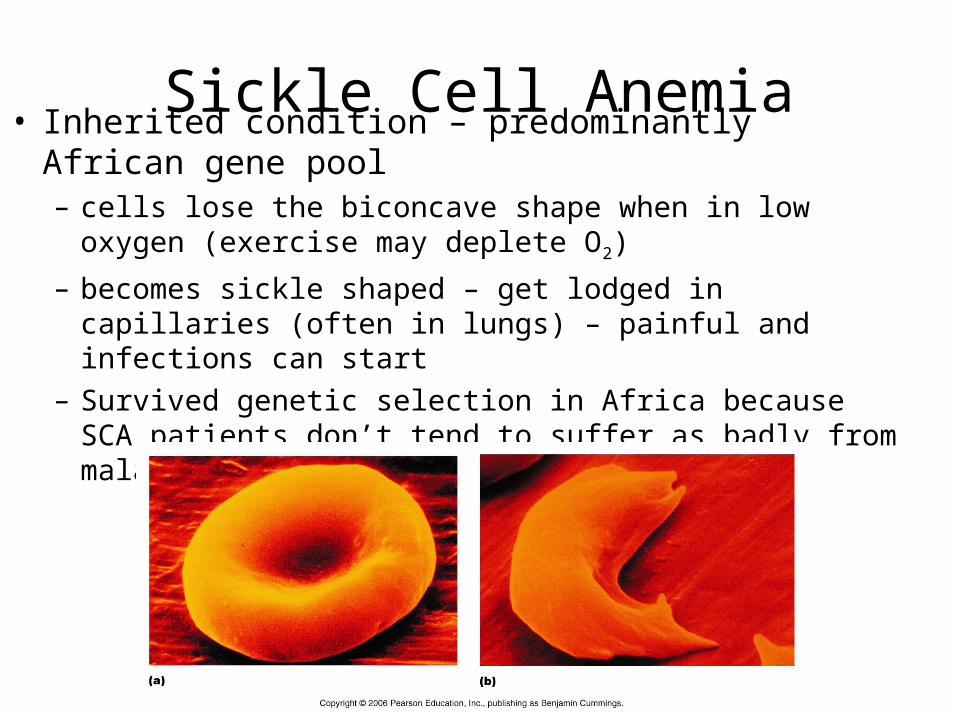
Sickle Cell Anemia• Inherited condition – predominantly African gene pool– cells lose the biconcave shape when in low oxygen (exercise
may deplete O2)
– becomes sickle shaped – get lodged in capillaries (often in lungs) – painful and infections can start
– Survived genetic selection in Africa because SCA patients don’t tend to suffer as badly from malaria

RBCs
• ~ 10,000 new cells are made every second to replace 10,000 cells that have died in that second
• Where do they get produced?– red bone marrow
• Where are they destroyed?– the spleen – recovers iron in heme, uses globin to
make amino acids – recycling at its finest
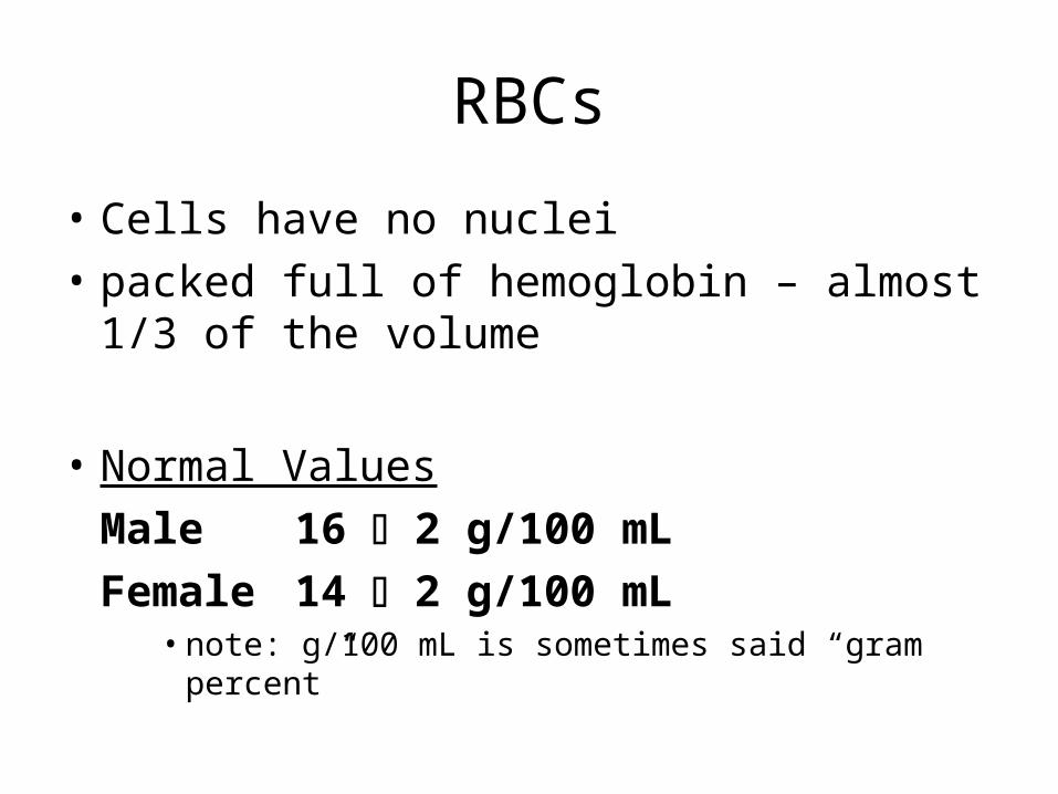
RBCs
• Cells have no nuclei• packed full of hemoglobin – almost 1/3 of the
volume
• Normal ValuesMale 16 2 g/100 mLFemale 14 2 g/100 mL
• note: g/100 mL is sometimes said “gram percent”

RBCs• All that hemoglobin (abbrev. Hb) is meant to carry oxygen• Hypoxia – tissue is starved of O2 –
– if Hb does not have enough oxygen, then hypoxia– lips and other tissues may appear blue = cyanotic
• When Hb combines with 4 molecules of O2, it’s called Oxyhemoglobin– Loads in lungs– most oxygen in blood is carried this way with some in plasma– most carbon dioxide is carried dissolved in plasma with some in Hb.
• What is Deoxyhemoglobin? Where is it formed?• What is Carbaminohemoglobin? Where is it formed?
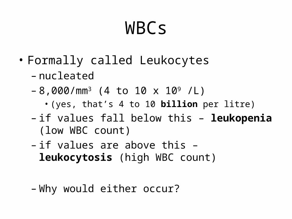
WBCs
• Formally called Leukocytes– nucleated– 8,000/mm3 (4 to 10 x 109 /L)
• (yes, that’s 4 to 10 billion per litre)
– if values fall below this – leukopenia (low WBC count)– if values are above this – leukocytosis (high WBC
count)
– Why would either occur?
WBCs
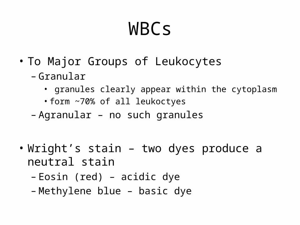
• To Major Groups of Leukocytes– Granular
• granules clearly appear within the cytoplasm• form ~70% of all leukoctyes
– Agranular – no such granules
• Wright’s stain – two dyes produce a neutral stain– Eosin (red) – acidic dye– Methylene blue – basic dye
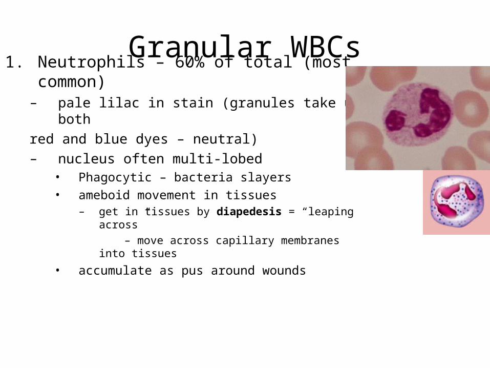
Granular WBCs1. Neutrophils – 60% of total (most common)
– pale lilac in stain (granules take up both red and blue dyes – neutral)– nucleus often multi-lobed
• Phagocytic – bacteria slayers• ameboid movement in tissues
– get in tissues by diapedesis = “leaping across” – move across capillary membranes into tissues
• accumulate as pus around wounds
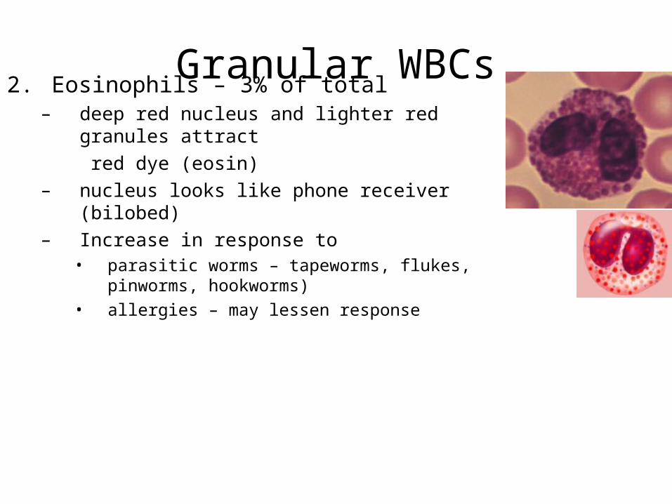
Granular WBCs2. Eosinophils – 3% of total
– deep red nucleus and lighter red granules attract red dye (eosin)
– nucleus looks like phone receiver (bilobed)– Increase in response to
• parasitic worms – tapeworms, flukes, pinworms, hookworms)• allergies – may lessen response
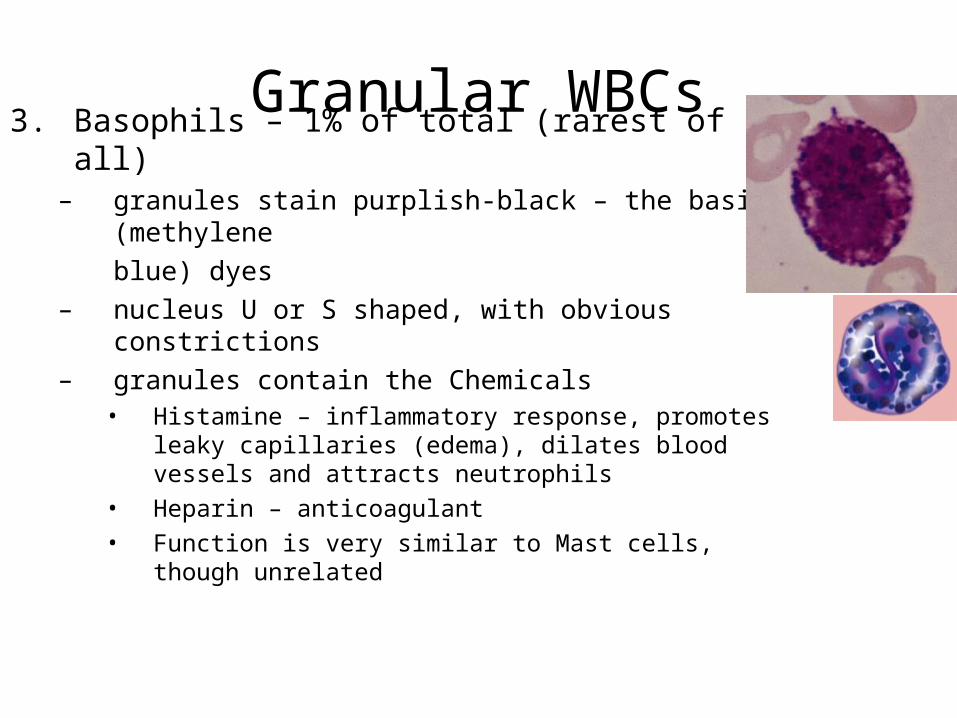
Granular WBCs3. Basophils – 1% of total (rarest of all)
– granules stain purplish-black – the basic (methyleneblue) dyes
– nucleus U or S shaped, with obvious constrictions– granules contain the Chemicals
• Histamine – inflammatory response, promotes leaky capillaries (edema), dilates blood vessels and attracts neutrophils
• Heparin – anticoagulant• Function is very similar to Mast cells, though unrelated
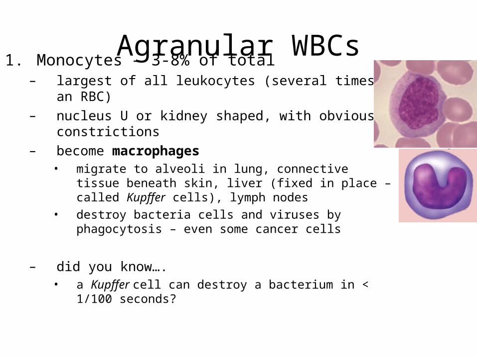
Agranular WBCs1. Monocytes – 3-8% of total
– largest of all leukocytes (several times an RBC)– nucleus U or kidney shaped, with obvious constrictions– become macrophages
• migrate to alveoli in lung, connective tissue beneath skin, liver (fixed in place – called Kupffer cells), lymph nodes
• destroy bacteria cells and viruses by phagocytosis – even some cancer cells
– did you know….• a Kupffer cell can destroy a bacterium in < 1/100 seconds?
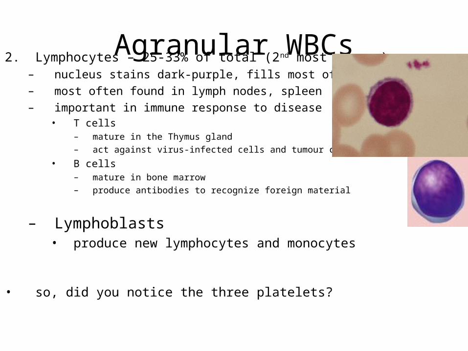
Agranular WBCs2. Lymphocytes – 25-33% of total (2nd most common)– nucleus stains dark-purple, fills most of cell– most often found in lymph nodes, spleen– important in immune response to disease
• T cells – mature in the Thymus gland– act against virus-infected cells and tumour cells
• B cells– mature in bone marrow– produce antibodies to recognize foreign material
– Lymphoblasts• produce new lymphocytes and monocytes
• so, did you notice the three platelets?
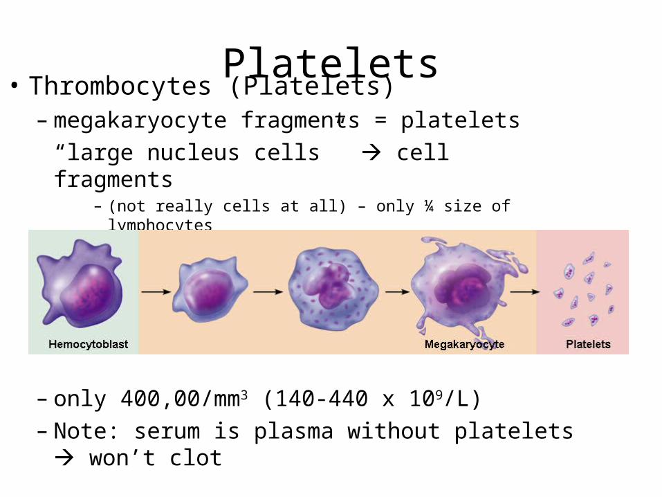
Platelets• Thrombocytes (Platelets)– megakaryocyte fragments = platelets
“large nucleus cells” cell fragments– (not really cells at all) – only ¼ size of lymphocytes
– only 400,00/mm3 (140-440 x 109/L)– Note: serum is plasma without platelets won’t
clot
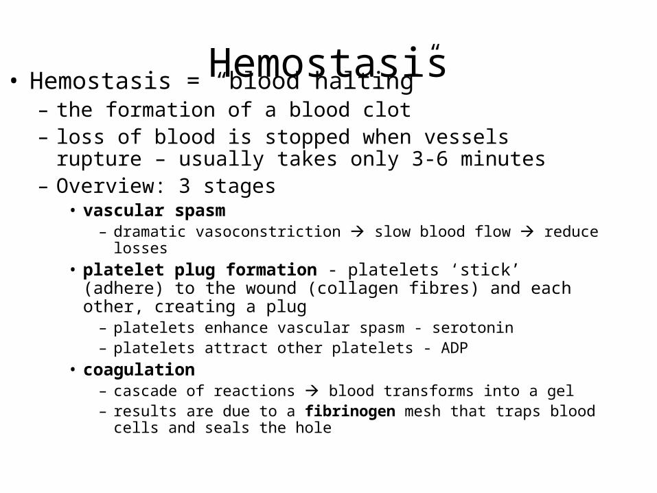
Hemostasis• Hemostasis = “blood halting” – the formation of a blood clot– loss of blood is stopped when vessels rupture – usually takes
only 3-6 minutes– Overview: 3 stages
• vascular spasm – dramatic vasoconstriction slow blood flow reduce losses
• platelet plug formation - platelets ‘stick’ (adhere) to the wound (collagen fibres) and each other, creating a plug– platelets enhance vascular spasm - serotonin – platelets attract other platelets - ADP
• coagulation – cascade of reactions blood transforms into a gel – results are due to a fibrinogen mesh that traps blood cells and seals the
hole
Hemostasis
• Platelets don’t stick to normal (undamaged) endothelium (lining of b.v.’s) or to each other – why?– negative charge of healthy tissue repels them
(platelets are also negatively charged)– Damaged tissue loses its negative charge
platelets adhere to wounds in b.v. (exposed collagen fibres)
– the Platelet Plug is formed, reducing loss of blood
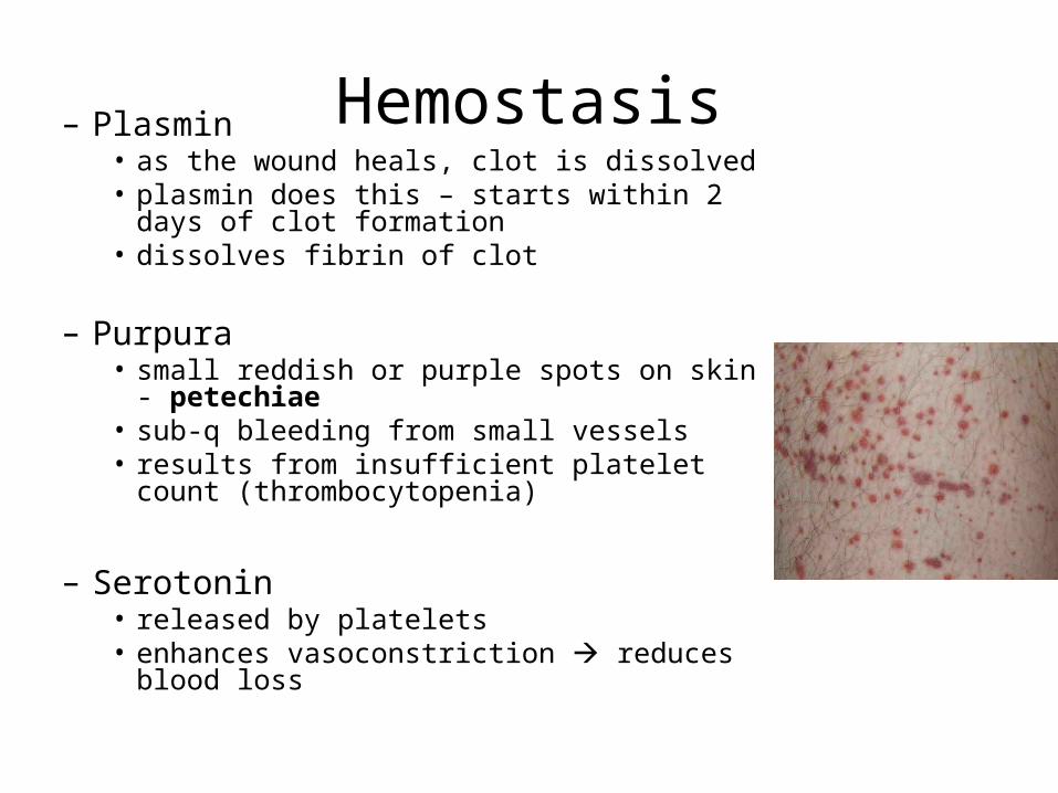
Hemostasis– Plasmin• as the wound heals, clot is dissolved• plasmin does this – starts within 2 days of clot
formation• dissolves fibrin of clot
– Purpura• small reddish or purple spots on skin - petechiae• sub-q bleeding from small vessels• results from insufficient platelet count
(thrombocytopenia)
– Serotonin• released by platelets• enhances vasoconstriction reduces blood loss
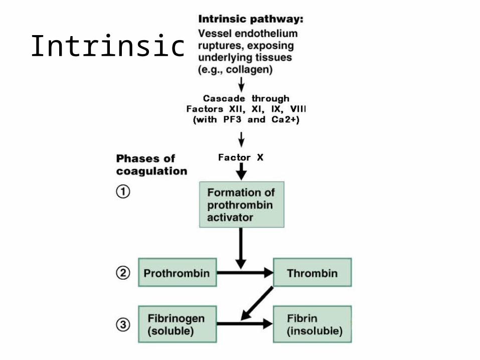
Clotting• Two pathways of clot formation
– Intrinsic – “found within”• Blood can clot on it’s own in a tube with no external stimulus
– Extrinsic – “found without”• Blood clots in response to stimuli from outside eg. chemicals released from
damaged cells/tissues• shorter (quicker) path bypasses many steps of intrinsic path
– Notes: • There are over 30 substances involved• Many (the “Factors”) are numbered I to XIII
» the numbering is NOT in the sequence in which the reaction proceeds• Calcium is ABSOLUTELY essential for clot formation• the main difference between extrinsic and intrinsic pathways is:
– extrinsic is a shorter path because of the use of Tissue thromboplastin• all other steps past that stage are shared in common
Clotting
• Extrinsic Pathway to a Clot– fastest of the two pathways (shortcut)– Tissue Thromboplastin
“thrombo” = clot “plastic” = to form
• aka Tissue Factor (TF) - produced by damaged cells • becomes Factor X (needs Ca2+)
– Factor X • which becomes Prothrombin activator (needs Ca2+)
– Prothrombin activator • converts Prothrombin (plasma protein) into Thrombin
(enzyme) (needs Ca2+)
Clotting• Extrinsic Pathway to a Clot (cont’d.)– Thrombin causes fibrinogen (plasma protein - soluble) to
polymerize (join together) into long fibrin strands (insoluble)
– Thrombin also activates factor XIII • factor XIII + Ca2+ causes fibrin strands to cross-link forming a web or
mesh
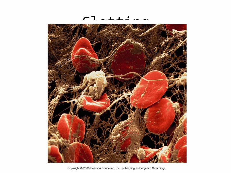
– Fibrin mesh traps formed elements (platelets, blood cells and fibres) in blood • creates a clot that seals the wound over 3-6 minutes after injury
Clotting

Extrinsic
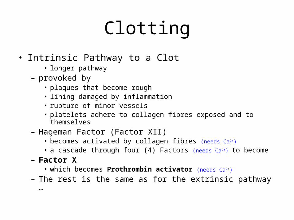
Clotting• Intrinsic Pathway to a Clot
• longer pathway – provoked by
• plaques that become rough• lining damaged by inflammation• rupture of minor vessels• platelets adhere to collagen fibres exposed and to themselves
– Hageman Factor (Factor XII)• becomes activated by collagen fibres (needs Ca2+)
• a cascade through four (4) Factors (needs Ca2+) to become– Factor X
• which becomes Prothrombin activator (needs Ca2+)
– The rest is the same as for the extrinsic pathway …
Intrinsic
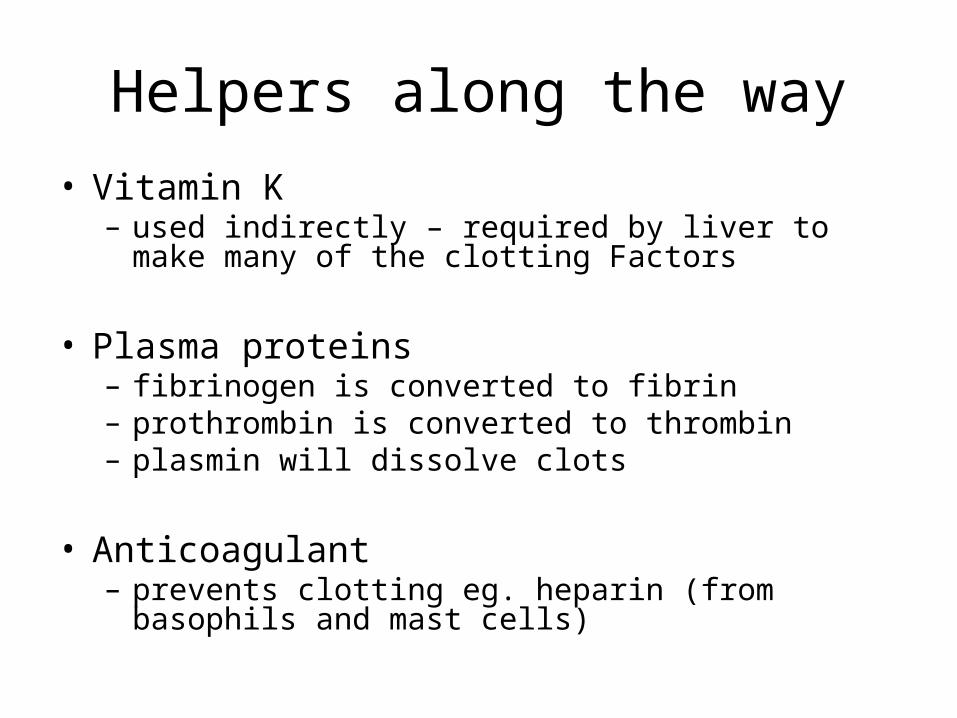
Helpers along the way• Vitamin K– used indirectly – required by liver to make many of the
clotting Factors
• Plasma proteins– fibrinogen is converted to fibrin– prothrombin is converted to thrombin– plasmin will dissolve clots
• Anticoagulant– prevents clotting eg. heparin (from basophils and mast
cells)
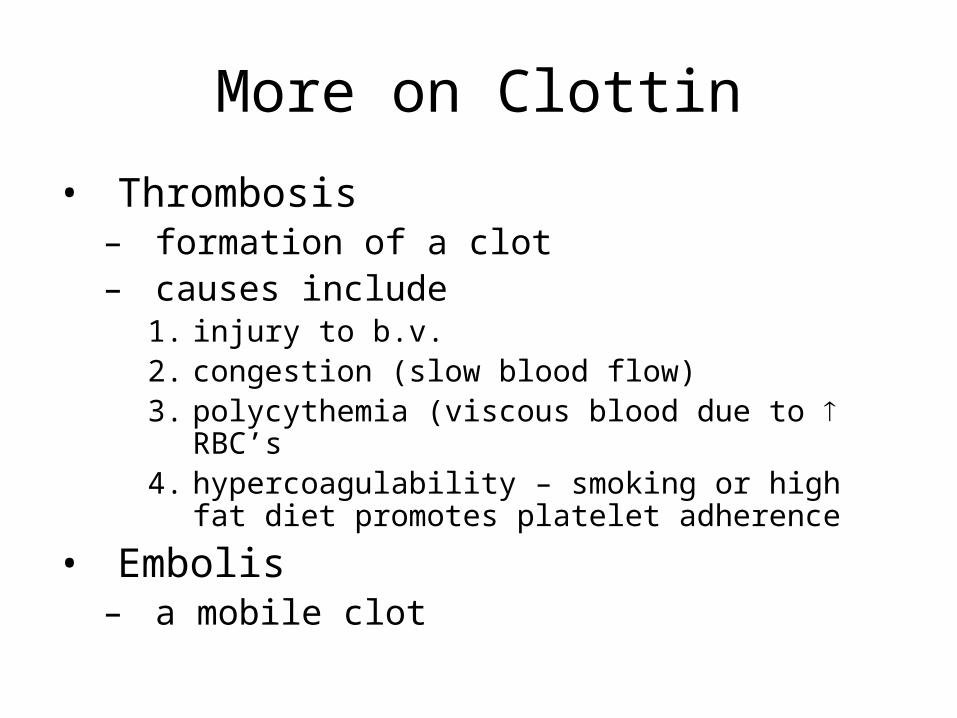
More on Clottin
• Thrombosis– formation of a clot– causes include
1. injury to b.v.2. congestion (slow blood flow)3. polycythemia (viscous blood due to RBC’s4. hypercoagulability – smoking or high fat diet
promotes platelet adherence
• Embolis– a mobile clot

What can you do?
• To hasten the formation of a clota) apply gauze (surface for platelets to adhere)b) apply heatc) suturesd) apply fibrine) apply thrombin