ch 2 prelabor rupture of the membranes · prelabor rupture of the membranes introduction prelabor...
TRANSCRIPT
1
PRELABOR RUPTURE OF THE MEMBRANES
Roberto Romero1,2,3
, Lami Yeo1,4
, Francesca Gotsch1, Eleazar Soto
1, Sonia S. Hassan
1,4, Juan
Pedro Kusanovic1 and Ray Bahado-Singh
4
1Perinatology Research Branch, NICHD/NIH/DHHS, Detroit, Michigan, USA
2Center for Molecular Obstetrics and Genetics, Wayne State University, Detroit, Michigan, USA
3Department of Epidemiology, Michigan State University, East Lansing, Michigan, USA
4Department of Obstetrics and Gynecology, Wayne State University, Detroit, Michigan, USA
This work is based on several previous chapters that were published before by the authors in
other textbooks. This work has been modified and adapted for this textbook. The original
chapters are referenced and contain a more extensive discussion of the subject. This chapter has a
clinical emphasis. The work has been primarily done by Roberto Romero, who is a government
employee, and therefore, this is not subject to copyright.
2
PRELABOR RUPTURE OF THE MEMBRANES
INTRODUCTION
Prelabor rupture of the membranes (PROM) is rupture of the chorioamniotic membranes
before the onset of labor.1 The “latency period” is the interval between PROM and the onset of
labor. There is no agreement about the length of the interval between rupture of the membranes
and the onset of labor required to diagnose PROM. This period of time has varied between 1 to
12 hours in the literature.2-9
The consequences of PROM depend on the gestational age.
Therefore, this condition has been classified as “preterm PROM” or “term PROM,” depending
upon whether the episode occurs prior to or after 37 weeks of gestation.2-12
The term “previable
PROM” has been applied to gestations in which this complication occurs before 23 weeks,12
while “preterm PROM remote from term” refers to the time frame between viability to about 32
weeks, and “PROM near term” is that which occurs between 32 and 36 weeks.12
FREQUENCY, TIMING AND SITE OF MEMBRANE RUPTURE
Frequency: Term PROM occurs in approximately 10% of patients while the frequency of
preterm PROM is 2% to 3.5%.5;13-17
Preterm PROM accounts for 30% to 40% of preterm
deliveries and, therefore, is a leading clinically identifiable cause of preterm birth and a major
contributor to perinatal morbidity and mortality.2;5;13;14;16-19
It has been estimated that in the
United States, approximately 150,000 women are diagnosed with preterm PROM every year.20
Spontaneous Rupture of Membranes in Normal Pregnancy: Figure 1 shows the
proportion of women with spontaneous rupture of membranes as a function of cervical

3
dilatation.21
Most patients rupture the chorioamniotic membranes at the end of the first stage of
labor, hence the rationale for defining PROM as rupture of membranes before the onset of labor.
The site of rupture is generally located in the most dependent part of the uterine cavity in close
proximity to the cervix.22
After invasive procedures such as fetoscopy, rupture of membranes can
occur away from the cervix. The frequency with which spontaneous rupture of membranes
occurs away from the most dependent part of the uterus (“high leak”) is unknown.
Figure 1
4
PRETERM PRELABOR RUPTURE OF MEMBRANES AS AN
OBSTETRICAL SYNDROME
The current taxonomy of disease in obstetrics is largely based on the clinical presentation
of the mother and not the mechanism responsible for disease. For example, the term “rupture of
membranes” refers to a clinical condition in which amniotic fluid leaks from the amniotic cavity
into the lower genital tract. However, the term ROM does not provide information about the
cause (e.g. infection, a vascular insult, a weakness in the structure of the membranes, trauma
caused by an invasive procedure (endoscopy), or other mechanisms of disease). We have
proposed that the classification of disease in obstetrics is at a stage in which we recognize
syndromes caused by multiple mechanisms of disease.23
The features of these obstetrical
syndromes are: 1) multiple etiologies; 2) long subclinical phase; 3) frequent fetal involvement; 4)
adaptive clinical manifestations; and 5) predisposition due to gene-environment interaction.24
Preterm PROM is one of the “great obstetrical syndromes.”23
Multiple pathologic
processes can lead to preterm PROM. The chronic nature of the pathologic process leading to
preterm PROM can be inferred from the observations that women with a short cervix (≤ 25 mm)
in the midtrimester,25
a positive fetal fibronectin (FFN) in vaginal fluid,25
bacterial vaginosis,25
and bleeding in the first and second trimester of pregnancy25-27
are at risk for the subsequent
development of preterm PROM. Fetal involvement has been demonstrated, as 10% of all fetuses
with preterm PROM have evidence of fetal bacteremia demonstrated by cordocentesis.28
We
have proposed that PROM is not the result of an accident, but rather a mechanism of host
defense in the context of intrauterine infection (or other insults), and that spontaneous rupture of
membranes occurs to facilitate the drainage of an infected cavity (intra-amniotic infection), as

5
well as to initiate labor (amniotomy is often followed by the onset of labor in term or preterm
gestation). Therefore, rupture of membranes may be considered adaptive in nature. Other
mechanisms of disease, such as chronic chorioamnionitis, in which there is infiltration of the
chorion laeve with lymphocytes but no evidence of infection represents another potential
mechanism for preterm PROM.29
The underlying mechanism of disease appears to be immune in
nature -- maternal rejection of the fetal allograft. Fetuses born to mothers whose placentas are
affected with chronic chorioamnionitis have evidence of a fetal inflammatory response
syndrome. We propose that the initiation of labor in these cases is the result of a combination of
a fetal inflammatory response and the maternal damage of chorion laeve by the lymphocytes and
natural killer cells, which are capable of inducing apoptosis of the trophoblast.29
In this context,
rupture of membranes and the initiation of labor may also be adaptive in nature because the fetus
is in a hostile environment unrelated to infection.
Finally, there is evidence that genetic factors predispose to preterm PROM.
Polymorphisms for genes coding for MMP-1,30 MMP-9,
31 MMP-8,32 and SERPINH1
33 in the
fetus have been associated with spontaneous rupture of membranes in case-control studies.
Environmental factors, such as bacterial vaginosis25;34
or a proinflammatory vaginal milieu,35
have also been associated with PROM. Evidence for a gene-environment interaction for preterm
birth has been demonstrated between bacterial vaginosis and a polymorphism for the pro-
inflammatory cytokine, TNF-α.36;37
It is also possible that gene-gene interactions are operative.
The genetic predisposition for preterm PROM is likely to result from the effect of multiple
alleles, which individually confer a small risk for preterm PROM. Epigenetic changes in MMP-1
have been found to be associated with preterm PROM.38
Collectively, the evidence reviewed

6
above supports the concept that preterm PROM is not a single condition, but one of the great
obstetrical syndromes.
Mechanisms of Disease Implicated in Preterm PROM:
1. Intra-amniotic infection/inflammation: Preterm PROM is associated with positive amniotic
fluid cultures for bacteria at the time of admission in approximately 30% of cases.39
With the use
of molecular techniques, about 50% of cases have microbial footprints in the amniotic cavity at
the time of admission.40
However, intraamniotic infection can be a primary cause of PROM or
be a consequence of the rupture of membranes. There is evidence that infection precedes preterm
PROM in a fraction of cases. In a study in which amniotic fluid cultures for microorganisms41-43
and pro-inflammatory cytokines44
were measured in the amniotic fluid of women undergoing
mid-trimester amniocentesis, some women with positive cultures or elevated biomarkers of
inflammation subsequently developed preterm PROM.41-43
2. Vascular pathology: In a study examining histologic lesions of the placenta, Arias et al.
found that women who delivered with preterm PROM fell in general terms into two subgroups:
those with acute histologic chorioamnionitis and another group with vascular lesions of the
placenta.45
Some patients have both types of lesions.46
The vascular lesions observed include:
“failure of physiologic transformation of the spiral arteries,”47;48
atherosis, fibrinoid necrosis of
the decidual vessels, and decidual vessel thrombosis consistent with decidual vasculopathy.49
Vaginal bleeding during pregnancy is a risk factor for preterm PROM.50
We have proposed that
some patients who bleed in the first or second trimester of pregnancy have a disorder of decidual
hemostasis.50
Vaginal bleeding may predispose to membrane rupture by causing a separation
between the chorioamnion and the decidua, which weakens the fetal membranes.50
Alternatively, during the formation of a retroplacental clot, thrombin is generated.51
This enzyme

7
can stimulate the production of MMP-152
and MMP-351
by decidual cells in culture media of
chorioamniotic membranes.53
These MMPs can degrade fibrillar collagen (types I and III) and
other components of the extracellular matrix of the chorioamniotic membranes54
. The
mechanisms responsible for defective decidual hemostasis during pregnancy have not been
identified. It is possible that vascular disease leading to microthrombosis in the decidua leads to
local necrosis and bleeding. In some cases, vaginal bleeding will be the only clinical
manifestation of intrauterine infection.50
This association is important because women with
preterm PROM often have clinical or sub-clinical abruptions, and histologic examination of the
membranes indicates that acute chorioamnionitis is frequent in patients with abruption.55
3. Uterine cervical pathology: Women with surgery in the lower genital tract, such as cervical
conization56;57
or previous spontaneous abortions,58
are at greater risk for preterm PROM.
Buchmayer et al. reported that a history of two or more spontaneous abortions was associated
with an odds ratio of 4.1 [95% CI: 2.2-7.8] for preterm PROM.58
We propose that some degree
of cervical insufficiency (e.g. a short cervix resulting in an inadequate mucus plug) predisposes
to ascending intrauterine infection.59;60
This hypothesis would explain the link between a short
cervix and subsequent preterm PROM.25
It is noteworthy that a cervical length ≤ 25 mm confers
an increased risk for preterm PROM before 35 weeks of gestation in nulliparas (OR 9.9), as well
as in multiparas (OR 4.2).25
4. Acquired or congenital connective tissue disorders: There is evidence that a connective
tissue disorder, which can affect the membranes (the chorioamniotic membranes are fetal tissue),
may predispose to preterm PROM. For example, patients with Ehler-Danlos syndrome are at
increased risk for preterm PROM if they carry an affected fetus.61
However, the attributable risk
of Mendelian disorders for preterm PROM is extremely low. It is likely that a genetic

8
predisposition to preterm PROM can be due to the effect of multiple genes. Indeed, a
relationship between polymorphisms in the promoter region of genes encoding for MMPs may
also predispose to membrane rupture (MMP-1,30
MMP-8,32
MMP-931
) and SERPINH1.33
polymorphisms may confer a mild to moderate risk, but this risk may be increased in patients
who have a relative deficiency of vitamin C (environmental contribution).
CLINICAL RISK FACTORS FOR MEMBRANE RUPTURE
Harger et al. reported a comprehensive analysis of risk factors associated with preterm
PROM in 341 women with preterm PROM (20 to 36 weeks) and 253 controls matched for
maternal age, gestational age, parity, type of care (private vs. clinic), and previous vaginal or
cesarean delivery.62
Three factors associated with preterm PROM were identified: 1) previous
preterm delivery; 2) vaginal bleeding during the index pregnancy; and 3) cigarette smoking.
Similar findings have been reported in a case-control study of 138 patients with preterm PROM
and 267 controls. Vaginal bleeding, smoking and low socioeconomic class were also found to be
independent risk factors for preterm PROM.63
In a large, multicenter, observational cohort study, Mercer et al. reported the risk factors
for preterm PROM (less than 35 weeks), stratified according to parity (see Table 1).25
In
conclusion, vaginal bleeding, a short cervix (≤ 25mm), a history of previous spontaneous preterm
delivery (with intact or ruptured membranes), and smoking are risk factors for preterm PROM in
the index pregnancy. A history of preterm birth with preterm PROM in a previous pregnancy
confers a high risk for recurrence (approximately 20%).56

9
CLINICAL CONSEQUENCES OF PREMATURE RUPTURE OF
THE MEMBRANES
A. Preterm Parturition: Preterm PROM is followed by the onset of labor and delivery
within a week in the majority of cases. The duration of the latency period is inversely related to
the gestational age. The lower the gestational age, the longer the latency period.64-66
Cox et al.
described the natural history of preterm PROM in 298 patients managed expectantly without the
use of steroids, tocolytics, and prophylactic antibiotics.64
Of the 267 patients who gave birth to
infants weighing ≥750 g, only 7% remained undelivered for more than 48 hours after admission.
Wilson and coworkers reported the outcome of 143 patients with preterm PROM
managed expectantly; only 18% of patients remained undelivered after 1 week of admission.65
Maternal febrile infectious morbidity (antepartum and postpartum) occurred in 10% of patients
and the neonatal death rate was 13.1%.
The most comprehensive study of the natural history of preterm PROM was reported by
Nelson et al., who evaluated the outcome following expectant management of 511 women with a
singleton gestation and preterm PROM between 20-36 weeks.66
Fifty-two percent of patients
delivered within 48 hours, while 12.9% remained undelivered after 1 week. The perinatal death
rate was 8.4%. Not surprisingly, most deaths occurred at gestational ages of less than 28 weeks
[42.7%]. Maternal infection occurred in 21.7%, and the occurrence of fetal death was strongly
associated with infection. The perinatal mortality was higher in neonates born to infected
mothers with preterm PROM before 28 weeks, than after 28 weeks [46.6% vs. 1.2%], as was
infection-related morbidity [36% vs. 19.8%].

10
B. Infection: Rupture of membranes is strongly associated with maternal or fetal infection.
Maternal infection can be expressed as clinical chorioamnionitis. However, most women who
have microbial invasion of the amniotic cavity (MIAC) do not have evidence of infection,67
such
as fever, leukocytosis, etc. Indeed, in our experience, only 20% of patients with preterm PROM
and a positive amniotic fluid culture for bacteria have clinical evidence of chorioamnionitis.68
The prevalence of positive amniotic fluid cultures in women with preterm PROM is
32.4%39
, whereas in term PROM the prevalence is 34.3%.69
However, this represents a
minimum estimate of the frequency of infection, because the frequency is dependent upon
isolation of microorganisms with standard microbiologic techniques, which underestimate the
true rate of infection. The application of molecular microbiologic techniques (cultivation
independent) has yielded a higher rate of microbial footprints (50%).40
Patients who have a
positive PCR result for microorganisms, but a negative culture, have comparable outcomes to
those who have a positive amniotic fluid culture for microorganisms.70
Genital mycoplasmas (Ureaplasma urealyticum and Mycoplasma hominis) are the most
frequent isolates from the amniotic fluid, followed by Streptococcus agalactiae, Fusobacterium
species, and Gardnerella vaginalis. Polymicrobial infection is found in 26.7% of cases28;68;71-75
and an inoculum size greater than 105 colony-forming units per mL is found in 23% of patients.
75
The most common microorganisms isolated from women with term PROM are U. urealyticum,
Peptostreptococcus, Lactobacillus, Bacteroides species, and Fusobacterium.69
Patients with intraamniotic infection are more likely to develop chorioamnionitis,
endometritis, and neonatal sepsis than patients with negative amniotic fluid cultures on
admission.28;68;71-86
The frequency of respiratory distress syndrome (RDS) is two-fold higher in
11
neonates born to women with positive amniotic fluid cultures than those born to women with
negative cultures.80
One study has examined the relationship between MIAC and the onset of preterm labor in
women with PROM.68
Patients in labor on admission had a higher rate of positive amniotic fluid
cultures than women admitted with preterm PROM but not in labor [39% vs. 26%, p=0.049].
Moreover, 75% of patients who were not in labor on admission, but subsequently went into
spontaneous labor, had a positive amniotic fluid culture around the time of the onset of labor.
Zlatnik et al. conducted a unique study in which the results of amniotic fluid culture were
not used in patient management. A higher proportion of patients with positive amniotic fluid
cultures delivered within 7 days, as compared to those with negative cultures [positive cultures:
89% vs. negative cultures: 45%, p=0.04)].75
These data support a relationship between MIAC
and the onset of preterm labor.
Can routine antibiotic administration eradicate intraamniotic infection and prevent
secondary infection? A recent study investigated the course of MIAC in 46 patients with preterm
PROM.87
All underwent amniocentesis upon admission, with an 18% prevalence of intra-
amniotic inflammation (defined as an amniotic fluid WBC count ≥100/mm3) and a 15%
prevalence of MIAC. Patients without evidence of intra-amniotic inflammation or MIAC were
treated with ampicillin and erythromycin for 7 days. Those with intra-amniotic inflammation or
MIAC were treated with ceftriaxone, clindamycin, and erythromycin for 10-14 days. At the time
of the second amniocentesis, six of the seven patients with a prior diagnosis of MIAC were again
positive for microorganisms. Of 18 patients with intra-amniotic inflammation, only three showed
no evidence of inflammation after antibiotic treatment. Of note, among patients with no evidence
of intra-amniotic inflammation or MIAC at admission, 32% developed inflammation despite
12
therapy. Five of the nine patients in question had positive amniotic fluid cultures. These
observations suggest that systemic treatment with these antibiotics may not alter the natural
course of intraamniotic infection in preterm PROM.
Evidence that fetal infection (bacteremia) is frequently present in preterm PROM was
provided by Carroll et al., who performed amniocentesis and cordocentesis at the time of
presentation with PROM.88
The frequency of positive fetal blood culture was 10.3%. The authors
found that for patients with positive amniotic fluid and fetal blood cultures, the median time to
delivery was 2 days [range: 1-5], compared with 41 days [range: 1-161] for patients with
negative cultures in both amniotic fluid and fetal blood. In the case of patients with MIAC and
negative fetal blood cultures, the median interval to delivery was 9 days [range: 1-37].88
The microorganisms isolated from septic newborns are similar to those found in the
amniotic fluid. In a study of 221 patients with preterm PROM, six cases with culture-proven
neonatal sepsis were found.68
In five of these cases, the microorganisms were the same as those
found in the amniotic fluid; in the remaining case, the amniotic fluid culture 48 hours before
delivery had been negative.68
The practical implication of this observation is that an
amniocentesis performed before delivery may provide microbiological information helpful in
guiding antibiotic choice in the newborn.
Fetuses with preterm PROM can mount a systemic inflammatory response.89
The term
“fetal inflammatory response syndrome” (FIRS) refers to an elevation in the fetal plasma
concentration of IL-6 (> 11 pg/ml) that was associated with severe neonatal morbidity89
and a
shorter cordocentesis-to-delivery interval.90
Fetal microbial invasion or other insults result in a systemic inflammatory response that
can progress toward multiple organ dysfunction, septic shock, and perhaps death in the absence

13
of timely delivery. Evidence of multisystemic involvement in cases of FIRS includes increased
concentrations of fetal plasma MMP-9,91
an enzyme involved in the digestion of type IV
collagen and in the pathophysiology of preterm PROM.92
Moreover, several fetal organs
including the hematopoietic system,93-95
adrenals,96
heart,97
brain,98;99
lungs,100;101
and skin102
are
target organs during FIRS.
Pathological examination of the umbilical cord is an easy approach to determine whether
fetal inflammation was present before birth. Funisitis and chorionic vasculitis are the
histopathologic hallmarks of FIRS.103
Funisitis is associated with endothelial activation, a key
mechanism in the development of organ damage,104
and neonates with funisitis are at increased
risk for neonatal sepsis105
and long-term handicaps, such as bronchopulmonary dysplasia
(BPD)99
and cerebral palsy.101
Indeed, newborns with funisitis are at more than a two-fold
increased risk for intraventricular hemorrhage106
and have an 11-fold risk for development of
periventricular echolucencies.107
In the context of FIRS, the combination of inflammatory
changes in the brain and fetal systemic hypotension may increase the likelihood of brain
injury.108
Collectively, these observations suggest that a subset of fetuses presenting with
preterm PROM have bacteremia and/or FIRS that may contribute to fetal organ damage.
The traditional view has been that MIAC is the consequence of membrane rupture.
However, evidence suggests that PROM may be the result of sub-clinical infection and
inflammation. Naeye and Peters reported in 1980 that patients with preterm PROM 1 to 4 hours
before the onset of labor had a higher prevalence of histologic chorioamnionitis than patients
who delivered preterm without PROM.109
Because it is unlikely that inflammation of the
chorioamniotic membranes develops in 4 hours, these data suggest that in these cases histologic
chorioamnionitis precedes rather than follows PROM. Several lines of evidence suggest that the
14
most likely cause of histologic chorioamnionitis is sub-clinical infection. Bacteria have been
recovered from 72% of placentas with histologic chorioamnionitis.110
Furthermore, we have
demonstrated a good correlation between a positive amniotic fluid culture for microorganisms
and histologic chorioamnionitis.111
MIAC can also be the consequence of PROM. The frequency of positive amniotic fluid
cultures increases with time. Indeed, 75% of patients who were quiescent on admission and
subsequently went into labor had a positive amniotic fluid culture.68
Only 25% of these patients,
however, had a positive culture on admission, and the remaining 50% became positive during the
latency period.68
These observations are consistent with those of Naeye and Peters, who showed
that the incidence of histologic chorioamnionitis increases with the duration of the latency
period.109
The authors reported that histologic chorioamnionitis (defined as the presence of 4 to 15
neutrophils in the chorionic plate) in PROM was two- to three-fold more common when rupture
of membranes occurred just before the onset of labor than when it occurred after labor began.109
This suggests that inflammation (and probably infection) is in many cases not only a
consequence of PROM, but also its cause.
C. Abruptio Placentae: Abruptio placentae occurs more frequently in patients with preterm
PROM than in those with preterm labor and intact membranes [2.29% vs. 0.86%, respectively,
RR: 3.58, 95% CI: 1.74-7.39].112
The same conclusion was reported in a systematic review113
and a population-based epidemiologic study.114
Nelson et al. proposed that leakage of fluid after
PROM may lead to a disproportion between the placental and uterine surfaces that would favor
placental separation.115
In the context of preterm PROM, the incidence of abruptio placentae
increases with the severity of oligohydramnios (12.3% for patients with a vertical pocket of 1-2

15
cm vs. 3.5% among those with a vertical pocket > 2 cm).116
However, two groups of
investigators using subjective means to estimate amniotic fluid volume could not confirm this
observation.117;118
An alternative hypothesis to explain the relationship between abruptio placentae and
preterm PROM postulates that a disorder of decidual hemostasis leads to separation of the
membranes from the decidua, with subsequent compromise of their nutritive support, weakening
of the membranes, and eventual rupture. Indeed, patients with abruptio placentae after preterm
PROM have a higher incidence of vaginal bleeding before rupture and during the latency period
than patients without abruptio placentae.
Infection/inflammation within the decidua could also facilitate premature placental
detachment. Indeed, there is an association between histologic chorioamnionitis and abruptio
placentae.55;119
The relative risk for abruption is 9.03 [95% CI: 2.80-29.15] when
chorioamnionitis is associated with preterm PROM.112
D. Pulmonary Hypoplasia: The frequency of pulmonary hypoplasia is related to
gestational age at the time of membrane rupture, and its presence increases the risk of neonatal
death and other complications such as pneumothorax and persistent pulmonary hypertension.
Three studies examined the frequency of pulmonary hypoplasia in the context of preterm
PROM. Vergani et al. conducted a prospective study of patients with PROM before 28 weeks of
gestation managed conservatively and found that the frequency of pulmonary hypoplasia was
28%.120
Gestational age at the time of PROM and presence of oligohydramnios, but not the
latency period, were independent predictors of pulmonary hypoplasia.120
Rotschild et al. studied 88 neonates born to mothers with PROM occurring before 29
weeks and a latency period of at least one week.121
The prevalence of pulmonary hypoplasia was

16
16%. Gestational age at the time of PROM, but not the duration of the latency period or the
severity of oligohydramnios, was associated with pulmonary hypoplasia. The risk of pulmonary
hypoplasia when PROM occurs at 19 weeks was 50%, whereas it was only 10% when the
membranes ruptured at 25 weeks.
Winn et al. prospectively studied 163 patients with preterm PROM from 15 to 28 weeks
of gestation.122
The incidence of pulmonary hypoplasia was 12.9%. The authors reported that
gestational age at rupture of the membranes, latency period, and either the initial or the average
amniotic fluid index (AFI) had significant influence on the development of pulmonary
hypoplasia.
The role of duration of rupture of membranes in the development of pulmonary
hypoplasia is not clear. Univariate analysis demonstrated an association between the duration of
rupture of membranes and the occurrence of pulmonary hypoplasia.120;123-125
However, there is
an inverse relationship between the duration of the latency period and the gestational age at the
time of membrane rupture. Multivariate analysis has not shown a significant effect of the
duration of PROM in the development of pulmonary hypoplasia.120
Whereas Rotschild et al.121
reported that the severity of oligohydramnios was not a factor
in the development of pulmonary hypoplasia, both Vergani et al.120
and Winn et al.122
reported
that amniotic fluid volume was an independent predictor of the development of pulmonary
hypoplasia. The discrepancy between studies may be explained by the: 1) determination of
amniotic fluid volume; 2) exclusion of patients; and 3) definition of pulmonary hypoplasia.
E. Fetal Compression Syndrome: The fetal compression syndrome was originally
described in the context of oligohydramnios and renal agenesis.126
Classically, it includes limb
position deformities and craniofacial defects that are thought to result from physical compression

17
inhibiting fetal growth and movement.127;128
Nimrod et al. reported an incidence of 12% in
women with preterm PROM, most occurring when the latency period was longer than 5
weeks.124
Blott and Greenough found that 46% of infants born after prolonged membrane
rupture (more than 4 weeks) had limb deformities.129
The median duration of rupture in the
group with deformities was 28 days, compared with 9 days in infants without deformities.
F. Fetal Growth Restriction: Preterm delivery resulting from patients with preterm labor
and intact membranes or preterm PROM has been associated with “fetal growth restriction”.
Most studies are cross-sectional and have not separated preterm PROM from preterm labor with
intact membranes.130;131
One interesting study determined the fetal growth rate for biometric
parameters in a cohort of 69 singleton pregnancies complicated with preterm PROM (24-31
weeks) and who remained undelivered for more than 14 days. The mean growth velocity of the
head and abdominal circumference was significantly lower than that of the control group [n=345
normal pregnancies]. Neonates who had either IVH, PVL or cerebral palsy had a lower growth
velocity than those not affected with these disorders.132
G. Fetal Death: The rate of fetal death in preterm PROM is approximately 1% when this
complication is diagnosed after 24 weeks and 15% if PROM occurs before.20
The etiology for
fetal death remains unknown. Fetal infection, placental abruption, fetal growth restriction,
umbilical cord proplase or accident (resulting from compression in cases of severe
oligohydramnios) have been implicated.
Fetal death, in the context of intrauterine infection, has been attributed to microbial
invasion of the fetus, where the unborn child fails to deploy an inflammatory response, which is
sufficienly intense to stimulate labor. Indeed, the frequency of histologic chorioamnionitis
(maternal inflammatory response) is 9 times more frequent than funisitis (a fetal inflammatory
18
response) in patients with stillbirth.133
In this case, in utero fetal death would represent failure of
the host response mechanisms dealing with intrauterine infection. This concept is supported by a
genetic association study which demonstrated that fetal carriage of the allele 2 of the gene
encoding for the IL-1 receptor antagonist (IL-1ra) is associated with fetal death.134
An excess of
IL-1ra in the fetal compartment may limit the ability of the fetus to deploy a pro-inflammatory
response and limit the effectiveness of the mechanisms available for host defense, including the
ability to initiate labor to exit a hostile intrauterine environment.
DIAGNOSIS
Presenting Symptoms: The most common presentation of PROM is a watery vaginal
discharge or a sudden gush of fluid from the vagina, as reported by the patient. Obtaining
information about the timing of the initial loss of vaginal fluid, color and consistency of the
discharge, and any odor may help to differentiate PROM from loss of the mucus plug in early
labor, vaginal discharge associated with infection, normal leukorrhea of pregnancy, and urinary
incontinence (sometimes present in pregnancy), as well as to determine the presence of blood or
meconium in the amniotic fluid.
Vaginal Examination: Evaluation of the patient begins with a sterile speculum examination.
Visualization of a vaginal pool or obvious leakage of fluid from the cervix into the posterior
fornix is considered evidence that PROM has occurred. Increasing intra-abdominal pressure may
assist in the visualization of this sign. If no fluid is present in the posterior fornix, the patient can
be re-examined after resting in the supine position to allow for accumulation of fluid in the
posterior fornix. Additionally, a speculum examination allows for collection of vaginal and
19
cervical cultures and amniotic fluid to assess fetal lung maturity, as well as to rule out cord
prolapse.
A sterile swab of fluid should be obtained from the posterior fornix and placed on a clean
glass slide and on a piece of nitrazine paper. Amniotic fluid, when put on a slide and allowed to
dry, will show arborization (“ferning”) under the microscope at low magnification.135
This
method has an overall accuracy of 95%.136
Rare false-positive ferning results have been
described in association with fingerprints on the slide or contamination with semen and cervical
mucus.137;138
False-negatives (5-10%) may be caused by dry swabs or by contamination with
blood.135;139;140
The slide should be evaluated after at least 10 minutes of drying to decrease the
false-negative rate.141
Should a digital examination of the cervix be performed in patients with preterm PROM?
The traditional view has been that “once an examination has been performed, the clock of
infection starts to tick”. Adoni and coworkers reported a study in which the latency period and
incidence of chorioamnionitis were evaluated in patients with preterm PROM (26-34 weeks)
who underwent a digital examination or a sterile speculum examination.142
The latency period
was longer in patients undergoing speculum examination than in those digitally examined [9.5
days ± 1.5 vs. 3.1 days ± 0.5, p<.005]. No significant difference in the incidence of
chorioamnionitis was found. Lewis and associates prospectively collected data on 271 singleton
pregnancies with preterm PROM.143
Patients who underwent a digital examination had shorter
latency periods than those following speculum examination [digital examination: 2.1 days ± 4.0
vs. speculum examination: 11.3 days ± 13.4, p<0.0001].
Sukcharoen et al. performed a retrospective study in which women with preterm PROM
had digital examinations or speculum examinations. The authors reported no differences in

20
latency periods or neonatal outcomes in the study groups. However, patients that underwent
digital examinations had a higher frequency of chorioamnionitis [digital exam: 12% vs.
speculum exam: 3.1%, p<0.05].144
Schutte and colleagues retrospectively examined the incidence of neonatal infection in
patients with PROM according to the interval between initial digital vaginal examination and
delivery.145
The incidence of neonatal infection was higher in patients examined more than 24
hours before delivery than in those whose first vaginal examination occurred less than 24 hours
before delivery [33% vs. 5%, p<0.0001].
The only justification for performing a digital examination is to determine cervical status.
In the preterm gestation this information rarely alters clinical management, but in the term
gestation the cervical state may influence decisions regarding induction. There is a strong
relationship between the results of sterile speculum examination and digital examination of the
cervix. This was demonstrated in a study in which visual speculum and digital cervical
examinations in women in labor were performed by two separate blinded examiners within 5
minutes of each other. Visual examination underestimated actual cervical dilation by only 0.6
cm [95% CI: 0.58-0.62].146-148
Biochemical Studies to Diagnose Rupture of Membranes: The biochemical
properties of the amniotic fluid are the basis to distinguish it from other fluids that can be
observed in the vagina (i.e., cervical secretions, urine and semen). The normal pH of the vagina
is 4.5 to 5.5 during gestation, and that of the amniotic fluid is 7.0 to 7.5. Nitrazine paper turns
from yellow to blue when exposed to any alkaline fluid (i.e., pH of 7.0 or more) and the use of
nitrazine paper has been reported to have an accuracy of 93.3%136
to determine the presence of
amniotic fluid in the vagina. False-positive results range from 1% to 17% and can result from
21
alkaline urine, blood, semen, vaginal discharge in cases of bacterial vaginosis, or Trichomonas
infection.149
False negatives may occur in up to 10% of cases.
Additional biochemical tests for the diagnosis of PROM include diamine oxidase (DAO)
activity,150
prolactin levels,151-153
alpha-fetoprotein (AFP; sensitivity: 94.5% and specificity:
95.4%),151;154
and insulin-like growth factor-binding protein-1 (IGFB-1).155
AFP has been
reported to be better than prolactin and more practical than DAO assay with an overall accuracy
of 98%.156;157
IGFBP-1 determinations have a sensitivity of 74.4% and specificity of 92.6%.155
Overall, the sensitivity, specificity, positive and negative predictive values of the different
diagnostic tests presented today in comparison to the nitrazine test are good.158-163
The assay for fetal fibronectin (FFN) is useful in the identification of patients at risk for
preterm and term labor and imminent delivery. However, doubts still exist about its diagnostic
value in PROM.164
Eriksen reported that for the detection of term PROM, FFN had a sensitivity
of 98.2%, but a specificity of only 26.8%.165
Although the authors proposed that the false-
positives were the result of the detection of small amounts of amniotic fluid not detected by
clinical tests (pool, Nitrazine, and ferning), an alternative explanation is that a positive FFN
detects degradation of the extracellular matrix in the fetal-maternal interface that precedes the
clinical onset of labor rather than PROM. Support for the hypothesis derives from the
observation that patients without PROM but with positive cervical FFN are more likely to deliver
within 72 hours than those with negative cervical FFN.164;166-173
In conclusion, the detection of
cervicovaginal FFN is not specific for PROM.
Transabdominal injection of dye: When the diagnosis of preterm PROM is not clear, a
transabdominal injection of dye (indigo carmine, Evans blue, fluorescein) into the amniotic
cavity may be used for confirmation.174-177
Methylene blue should not be used as it may cause
22
fetal methemoglobinemia.178-180
A tampon in the vagina can document subsequent dye leakage
in cases of PROM.
INITIAL ASSESSMENT
The initial evaluation of a patient with preterm PROM includes: 1) accurate assessment
of gestational age; 2) estimation of fetal weight and presentation; 3) evaluation of the risk of
infection; 4) determination of lung maturity; 5) assessment of fetal well-being; and 6) exclusion
of occult cord prolapse.
1. Ultrasound Examination in the Evaluation of Patients with Preterm
PROM: The initial ultrasound examination aims to: 1) assess fetal viability, biometry and
presentation; 2) quantify amniotic fluid volume; 3) rule out fetal anomalies; and 4) confirm
gestational age. The sonographic examination of fetuses with PROM may be challenging due to
the reduced amniotic fluid volume. For example sonographic estimates of fetal weight have been
shown to underestimate the birthweight.181-183
2. Diagnosis of Intrauterine Infection in Preterm PROM: Amniocentesis can be
used for the evaluation of the microbiological state of the amniotic cavity and of fetal lung
maturity in the patient with preterm PROM.184
Results of amniotic fluid analysis provide a
rational approach to the management of preterm PROM. Patients without evidence of
infection/inflammation and lung immaturity could be managed expectantly while those with
evidence of infection could be managed using algorithms tailored to the gestational age (see
management section).
One randomized clinical trial examined the value of amniocentesis in preterm PROM.185
Forty-seven patients (26 to 34 weeks of gestation with an accessible amniotic fluid pocket) were

23
randomized to amniocentesis or no amniocentesis. Indications for induction of labor included
positive Gram stain of amniotic fluid or mature fetal lungs, as determined by a lecithin-to-
sphingomyelin (L/S) ratio of more than 2.0 or positive phosphatidylglcerol (PG). Neonates born
to women who had amniocenteses had a lower incidence of “fetal stress” during labor (diagnosed
by fetal heart rate tracing) and a shorter hospital stay than those born to women who were
randomized to not have amniocenteses [“fetal distress”: 4% vs. 32%, p<0.05; hospital stay
(median): 8.5 days vs. 22 days, p<0.01]. No differences in the rate of neonatal sepsis, maternal
chorioamnionitis, or endometritis were noted between the two groups. This study had limited
power to detect differences in neonatal morbidity.
The analyses of amniotic fluid used to detect the presence of MIAC or intra-amniotic
inflammation include: 1) Gram stain; 2) a quantitative white blood cell (WBC) count; 3) glucose
concentration; and 4) microbial cultures for aerobic, anaerobic bacteria, as well as genital
mycoplasmas. Patients with a negative Gram stain (read by experienced personnel) and a high
WBC count (more than 30 cells per uL) are at a high risk of having microbial invasion with
genital mycoplasmas, which are not visible on Gram stain examination. Lower concentrations of
glucose in amniotic fluid (<10 mg/dL) can serve as an additional marker for MIAC. The results
of amniotic fluid culture may take days to be available. Therefore, most centers rely on the
determination of intra-amniotic inflammation because the outcome of preterm PROM in patients
with intra-amniotic inflammation is similar to those with MIAC proven with standard
microbiological techniques.186
Table 2 summarizes the diagnostic criteria and predictive values
of different amniotic fluid tests in detecting positive amniotic fluid cultures in patients with
preterm PROM.85;187
Amniotic fluid IL-6 performed best in detecting MIAC, as well as in
identifying patients at risk for impending preterm delivery and neonatal complications. We have

24
shown that amniotic fluid IL-6 is a sensitive test for the prospective diagnosis of acute histologic
chorioamnionitis [IL-6 of more than 17 ng per mL had a sensitivity of 79% and specificity of
100%], significant neonatal morbidity (sepsis, RDS, pneumonia, intraventricular hemorrhage,
bronchopulmonary dysplasia, and necrotizing enterocolitis), and mortality [IL-6 of more than 17
ng per mL had a sensitivity of 69% and a specificity of 79%].188
Other rapid tests reported for
the detection of MIAC include amniotic fluid catalase,79
alpha1-antitrypsin,84
limulus amebocyte
lysate test,83
and bacterial polymerase chain reaction.189
A rapid bedside test for the detection of MMP-8 in amniotic fluid has been developed.
This kit has been reported to have high accuracy in the identification of patients with MIAC and
inflammation among patients with preterm labor and intact membranes.190
Future studies may
determine the utility of this test in the identification of patients with intra-amniotic
infection/inflammation among patients with preterm PROM.
The risk of amniocentesis, when performed by experienced individuals, appears to be
extremely low. Yeast and colleagues specifically addressed this issue in 91 patients with preterm
PROM in whom amniocenteses were performed.191
A retrospective review of neonatal records
uncovered no evidence of fetal trauma with any procedure. This study also found that the
incidence of spontaneous labor in patients who underwent amniocentesis was no different from
that of patients who did not undergo amniocentesis secondary to oligohydramnios or an anterior
placenta. The authors concluded that their study failed to show that amniocentesis might induce
labor.
Assessment of Lung Maturity: Lung maturity can be assessed from the amniotic fluid
obtained by amniocentesis or from the vaginal pool. The latter has the advantage of being less
invasive and more feasible in patients with oligohydramnios. Amniotic fluid from the vaginal

25
pool can be collected in three ways: (a) from the posterior vaginal fornix by sterile speculum
examination; (b) in a clean bedpan maintained under the patient; or (c) by use of obstetric
perineal pads left in place for 12 to 24 hours to ensure saturation.192-195
The success rate in
obtaining fluid within 48 hours with these noninvasive techniques ranges from 54% to
100%.194;195
Using a pad to detect phosphatydylglycerol (PG), Esol et al. found a sensitivity of
88%, specificity of 76%, positive predictive value of 34%, and negative predictive value of
98%.196
Lewis et al. investigated the value of a rapid antibody agglutination method (Amniostat
FLM) to detect PG in vaginal pool samples.197
Thirty-six of 201 patients between 26 and 36
weeks of gestation had positive PG, and none of the infants born to these mothers developed
RDS. PG was detectable only after 30 weeks of gestation.
The reliability of lung maturity tests from amniotic fluid collected vaginally has been
challenged.198;199
This section reviews the correlation between the lecithin/sphingomyelin (L/S)
ratio and PG results in amniotic fluid obtained by amniocentesis and from the vaginal pool.
Shaver and associates compared the phospholipid profile of paired amniotic fluid samples in 28
patients with preterm PROM.192
No significant difference was found in the concentrations of
PG, phosphatidylinositol, phosphatidylethanolamine, and phosphatidylserine in amniotic fluid
obtained by the two sampling methods. The L/S ratio was higher in fluid collected transvaginally
than in fluid collected transabdominally, but this difference did not reach statistical significance.
The only phospholipid clearly increased by vaginal contamination was lysolecithin.
Dombroski et al. reported a study in which amniotic fluid was obtained by amniocentesis
in patients at term in labor.200
Thirty minutes after artificial rupture of membranes, a vaginal
sample of amniotic fluid was collected. L/S ratios obtained from amniotic fluid in the vaginal

26
pool samples were significantly lower than were those obtained by amniocentesis. However, in
22% of cases, L/S ratios were higher in the vaginal pool samples than in amniocentesis.
Several studies have examined the value of PG determinations in amniotic fluid obtained
transvaginally. Stedman et al. reported that of 25 patients with PROM between 26 and 34 weeks,
60% had positive PG and none of their neonates developed RDS (within 72 hours of the test).193
Among the newborns of the 10 patients with negative PG, four developed RDS. Similarly,
Brame and MacKenna reported no cases of neonatal RDS in 36 patients with PG found in
vaginal fluid.194
The possibility that bacterial contamination from vaginal secretions may lead to false-
positive PG results has been raised by Schumacher and associates, who reported that one patient
had PG detected in the fluid from the vaginal pool, but not in fluid retrieved by transabdominal
amniocentesis.199
The neonate developed respiratory insufficiency that was attributed to either
RDS or pneumonia (the amniotic fluid culture was positive for bacteria). These investigators
also demonstrated that bacteria might be a source of PG. Therefore, excessive bacterial
contamination may alter results of PG determinations. It would seem prudent to minimize the
interval between sample collection and assay in the hope of preventing bacterial growth in the
sample.
Three studies have reported neonatal outcome and L/S ratio results in preterm
PROM.194;195;201
In two of the studies, a mature L/S ratio was an indication for delivery.195;201
In
the third study, the presence of PG was used as an indication for delivery.194
The data are
consistent: with a mature L/S ratio, the risk of RDS is small. An L/S ratio of more than two was
found in 103 patients, and none of the neonates developed RDS.

27
The available evidence indicates that fetal lung maturity studies can be performed on
amniotic fluid obtained from the vagina, and that a mature L/S ratio or the presence of PG is
associated with a very low risk of RDS. Moreover, this noninvasive, low-risk approach allows
for serial L/S and PG determinations.
A mature phospholipid test has been demonstrated in approximately 50% of patients with
preterm PROM at gestational ages of less than 34 weeks.72;74;185
Garite and associates reported
that none of the neonates with an L/S ratio of 1.8 or greater developed RDS.74
The incidence of
this complication in neonates with an immature L/S was 33%.
Two randomized clinical trials have examined the outcome of induction of labor in
patients with a mature results. In the first trial, 47 patients with preterm PROM (less than 36
gestational weeks) and mature amniotic fluid indices were randomized to either prompt delivery
or expectant management.202
A mature test was defined as an L/S ratio above 2 or a Foam
Stability Index (FSI) of 47 or more (often from vaginal fluid). There was no difference in
perinatal mortality between the two groups. There were no cases of RDS in the expectant
management group, but two in the prompt delivery group. One newborn died from severe
hyaline membrane disease (birth weight 900 g, vaginal FSI=48), and the other neonate survived
(birth weight 1,700 g, vaginal L/S=2.0). There were no differences in the rate of neonatal sepsis
or other neonatal complications in the two groups. However, the only two cases of intracranial
hemorrhage occurred in the prompt delivery group. Maternal chorioamnionitis was more
common in the expectantly managed group than in the delivery group [38% vs. 8%, p <0.02].
Mercer et al. reported the results of a randomized clinical trial in which 93 women with
mature amniotic fluid phospholipid studies (vaginal or transabdominal amniocentesis FSI ≥ 47)
were randomized to induction of labor with oxytocin or expectant management (bed rest).203

28
Maternal chorioamnionitis was more frequent in the expectant group. However, this difference
did not reach significance. There were no significant differences in the cesarean delivery rate or
in the incidence of confirmed neonatal sepsis between the groups. Suspected sepsis was higher
in neonates born to women in the expectant group, as was antibiotic administration and septic
workups. However, neonatologists were not blinded to treatment allocation.
ASSESSMENT OF FETAL WELL-BEING: The goal of fetal evaluation is to identify
fetal infection/inflammation or a pathologic process, which increase the risk of antepartum or
neonatal death. Methods of fetal surveillance include non-stress test and the components of the
biophysical profile.
Nonstress Test (NST): The differential diagnosis of a non-reactive NST is 1) preterm
gestation; 2) infection; and 3) hypoxia. The interpretation and the significance and management
of fetal heart rate decelerations associated with umbilical cord compression due to
oligohydramnios is also a challange.
Fetuses with preterm PROM between 24 and 37 weeks have a significantly higher
incidence of reactive tracings than gestational age-matched counterparts with intact
membranes.204-206
This has been attributed to “accelerated fetal central nervous system
maturation” and umbilical vein compression with resulting fetal heart rate accelerations.207
Thus, lack of reactivity should not be ascribed to preterm gestation without further investigation.
A non-reactive NST is frequently observed in fetuses with MIAC. Three studies208-210
have found the NST to be an insensitive predictor of infection-related outcome. A major issue is
the high false positive rate (approximately 35%) of the NST for the detection of infection.
Therefore, a non-reactive NST is not sufficient to diagnose infection. Evaluation of other
29
biophysical parameters and the results of amniocentesis are recommended before delivery can be
indicated (see below).
Assessment of Amniotic Fluid Volume:
Contrary to what is generally believed, rupture of membranes is not necessarily
associated with oligohydramnios. Harding et al. noted that the amniotic fluid index (AFI) in
patients with preterm PROM remains stable after the membranes rupture, with the mean AFI on
admission being 5.9 ± 2.5 cm and on the day of delivery 5.4 ± 2.0 cm.211
Moreover, Vintzileos et
al. reported that 65.5% of patients with PROM had a vertical pocket of amniotic fluid of greater
than 2 cm, while 15.5% had a vertical pocket between 1 and 2 cm. Only 19% had a vertical
pocket of less than 1 cm.212
Several studies have examined the relationship between oligohydramnios and outcomes
in PROM. Patients with a vertical amniotic fluid pocket <1 cm have a shorter latency period and
a higher incidence of chorioamnionitis and neonatal sepsis than patients with a vertical pocket
greater than 2 cm.212
Similar findings were reported by Gonik et al.213
Women with a vertical
amniotic fluid pocket of <1 cm had a higher incidence of chorioamnionitis and endometritis than
those with an amniotic fluid pocket of >1 cm. No difference in the duration of the latency period
between the two groups was found.213
Hadi et al. reported that chorioamnionitis occurred in 26.4% of women with an amniotic
fluid pocket of less than 2 cm.214
Similarly, Lao et al.215
used a cutoff of 2 cm as the largest
pocket of amniotic fluid to define oligohydramnios, and found that the frequency of
chorioamnionitis and funisitis was higher in patients with oligohydramnios than in those without
reduced amniotic fluid volume [chorioamnionitis: 55.3% vs. 29.3%; funisitis: 44.7% vs. 16.7%].
A reduction in amniotic fluid volume was also associated with MIAC.

30
There is an association between reduced amniotic fluid volume and maternal or neonatal
infection-related morbidity and MIAC. The reason for the high rate of infection in patients with
oligohydramnios is unknown. Intra-amniotic infection may alter amniotic fluid dynamics,
leading to a reduction in fluid volume. Yoon et al. proposed that redistribution of blood flow
away from the kidneys might take place as part of the host response to microbial products, and
this may lead to olygohydramnios.216
Patients with decelerations have a lower AFI than those without decelerations [4.32 cm ±
1.67 vs. 6.47 cm ± 3.59, p<0.01].217
This observation suggests that cord compression due to
oligohydramnios may be the mechanism behind variable decelerations observed in patients with
PROM.
Preterm PROM is associated with a significant and prolonged reduction of fetal breathing
movements lasting approximately 2 weeks.218;219
This phenomenon seems to be related to
rupture of membranes per se, rather than to infection, hypoxia, or intrauterine growth restriction,
even though the precise mechanisms are unknown. Membrane rupture leads to a reduction in
intra-amniotic pressure and, thus, favors loss of lung fluid. Teleologically, a reduction in fetal
breathing may be a mechanism to protect against lung fluid loss and pulmonary hypoplasia.
Vintzileos was the first to document an association between infection and decreased fetal
breathing activity in preterm PROM.220;221
Subsequently, we confirmed these findings and
documented that women with positive amniotic fluid cultures had fewer and shorter episodes of
fetal breathing activity than women with negative amniotic fluid cultures.222
The presence of fetal breathing has a very high negative predictive value (approximately
95%) for MIAC and neonatal sepsis. However, the absence of breathing activity has a limited
positive predictive value (approximately 50%) for either of these two outcomes and, thus, it
31
cannot be used as an indication for delivery. Therefore, the presence of breathing indicates that
infection is unlikely.
Intra-amniotic infection is associated with a dramatic reduction in fetal body
movements.222
Decreased fetal motion in the context of infection may be the counterpart of the
reduction in motor behavior observed during the course of febrile illnesses in adults and children.
The biophysical profile (BPP) has been found to be helpful in the management of patients
with PROM.204;207-210;220;222-227
Vintzileos et al., using logistic regression analysis, demonstrated
that each component of the BPP contains useful information for the prediction of infection-
related morbidity (defined as maternal chorioamnionitis, possible neonatal sepsis, and proven
neonatal sepsis). In their first study, a modified BPP scoring system that incorporated placental
grading (with a maximal score of 12) was used.220
A BPP score of seven or less was much better
than any single component of the BPP in the prediction of infection-related outcome. Placental
grading was the only parameter that had no predictive value. Thus, it was excluded from
subsequent studies. The diagnostic indices of a BPP score ≤ 7 (performed 24 hours before
delivery) were: sensitivity 94%, specificity 97%, positive predictive value 95%, and negative
predictive value 97% in a population with a prevalence of infection-related outcome of 30%.
This study was observational in nature and, thus, the BPP was not used for patient management.
Subsequently, Vintzileos et al. compared the outcome of pregnancy in patients managed
with serial BPPs with two historical control groups: 1) expectant management without BPP or
amniocentesis; and 2) management with a single amniocentesis on admission.228
A BPP score ≤
7 on two examinations two hours apart was used as an indication for delivery. An abnormal
score required a non-reactive NST and absence of fetal breathing. The results of this study
indicated that patients managed with daily BPPs had a lower rate of overall neonatal sepsis
32
(suspected and culture-proven) than patients in either control group. This study did not provide
the frequency of other indices of neonatal morbidity (e.g., RDS, IVH, duration of mechanical
ventilation) in the different groups. This issue is important, since 14 patients who were delivered
because of a low BPP score showed no evidence of neonatal infection and, thus, could be
considered false positives. If intervention was not associated with an increased rate of other
neonatal complications, management with serial BPPs would seem a reasonable approach. The
investigators found that the BPP had limitations when the interval between the test and delivery
was longer than 24 hours, and that maternal infection without fetal infection was not correlated
with the results of the BPP scoring. Vintzileos et al. subsequently reported on 111 fetuses with
preterm PROM followed with daily BPPs, and found that as more of the biophysical activities
became compromised, the higher the incidence of infection-related complications.207
It is noteworthy that subsequent to this work, three studies223;224;227
reported an
association between the results of the BPP and infection-related outcomes and three others could
not confirm such an association.225;226
Our explanation for the apparent discrepancy is that
studies reporting negative results used the BPP at less-frequent testing intervals (48- to 72-hour
intervals) than the daily testing used in positive reports.
MANAGEMENT OF PATIENTS WITH PREMATURE PROM
The management of patients with premature PROM depends on the gestational age at the time of
membrane rupture (see Figure 2).

33
Figure 2
Previable PROM (see Figure 3): The major complications of previable PROM are maternal
infection, late abortion/preterm labor, low neonatal survival and a high risk of neurologic
handicap.229;230
Patients are generally offered two options: induction of labor or expectant
management. The presence of intra-amniotic inflammation/infection in amniotic fluid analysis
carries a poor prognosis because of the risk of spontaneous preterm labor as well as fetal
morbidity. Management of these patients requires an in-depth discussion involving the parents,
neonatologists and obstetricians and careful documentation in the medical record. The value of
antibiotic or corticosteroid administration in previable PROM has not been established.
However, our practice is to administer antibiotics to the patient who desires expectant

34
management. A systematic review of previable PROM indicates that the quality of the evidence
to support the management is not high.231
Leakage of amniotic fluid after second-trimester amniocentesis should be considered a
separate entity from previable PROM. It occurs in 1.2% of patients and is usually transient in
nature.232
The risk of delayed PROM in these cases is no different from that in the general
population.233
Figure 3
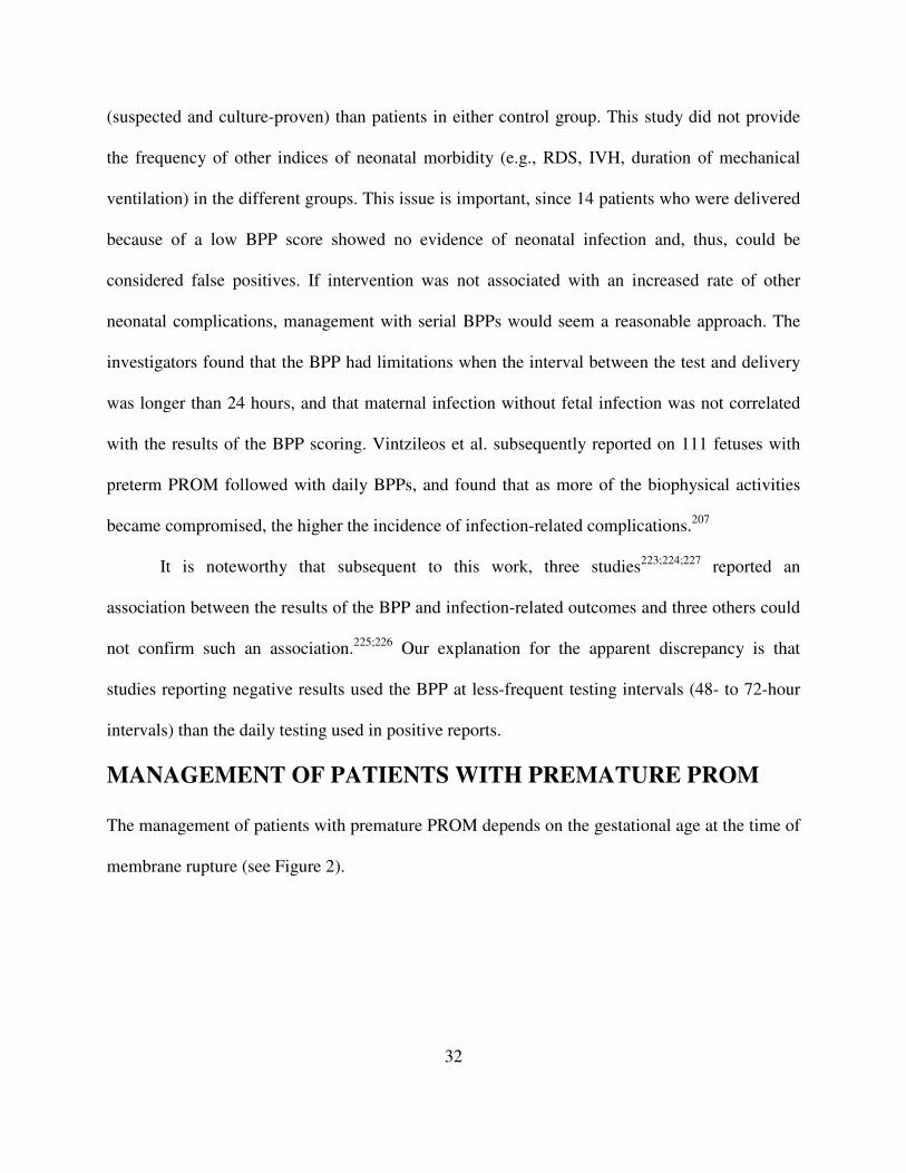
PROM Remote from Term (24-31 6/7 Weeks of Gestation) (see Figure 4): The
management goals are to 1) exclude intra-amniotic infection/inflammation; and 2) institute
expectant management in patients without documented infection/inflammation.
35
Figure 4
Intra-amniotic infection/inflammation and its management: The most accurate method for
diagnosis of intra-amniotic infection/inflammation is amniocentesis. Once intra-amniotic
infection/inflammation is identified in patients between 24-31 completed weeks of gestation, the
optimal management is a challenge: the earlier the gestational age, the more difficult the
dilemma. In patients who are close to 32 weeks of gestation, delivery would avoid continuous
exposure to microbial products and inflammatory agents and is unlikely to increase neonatal
morbidity. These patients are managed in our unit with detailed counseling, antibiotic
administration, and delivery. In contrast, in patients close to 24 weeks of gestation, the option of
parenteral administration of antibiotics is considered to eradicate intra-amniotic infection and
36
inflammation. Patients are informed that this alternative may prolong pregnancy, eradicate intra-
amniotic infection, and reduce the risk of extreme prematurity, but that it requires intensive
surveillance and repeat evaluation of the amniotic cavity to ensure eradication of microorganisms
and reduced intra-amniotic inflammation (as determined by the amniotic fluid white blood cell
count). Despite these interventions, the risk for infection and prematurity are not eliminated.
Broad coverage is recommended before the results of cultures are available and this approach
modified once the specific microorganisms involved are identified. The choice of antibiotics is
informed by the results of microbial cultures. Our choice for broad coverage antibiotics includes
azythromycin, clindamycin, and ampicillin. We have also used a combination of ceftriaxone,
clindamycin and erythromycin for 10-14 days.87
Azythromycin is included because Ureaplasma
urealyticum is the most frequent microorganism found in the amniotic cavity.67
Antibiotic treatment aimed at the eradication of intra-amniotic infection should not be
confused with the prophylactic treatment, which is now the standard of care for patients with
preterm PROM, regardless of whether the inflammatory/infection state of the amniotic fluid is
known. Thus, patients in this gestational age range, without evidence of infection and
inflammation, are given prophylactic treatment with antibiotics (ampicillin and erythromycin).
The patient in whom an amniocentesis cannot be performed is managed taking into account the
results of biophysical testing, and antibiotics and steroids are administered.
In summary, the management of PROM between 24 and 31 completed gestational weeks
is comprised of: 1) maternal and fetal inpatient surveillance in a tertiary medical center; 2)
administration of corticosteroids to accelerate fetal lung maturity;234
and 3) antibiotic
administration, which may be therapeutic or prophylactic.235;236

37
1. “Prophylactic” antibiotic administration: Antibiotic administration has now become
standard of care in patients with preterm PROM. This practice is based upon results of several
randomized clinical trials, in which antibiotic administration is associated with prolongation of
pregnancy, a reduced rate of maternal chorioamnionitis237
and a reduced frequency of neonatal
morbidity, measured as “composite neonatal outcome”.238
This approach has been often referred
to as “prophylactic” antibiotic administration. However, this may be a misnomer. One-third of
women with preterm PROM have a positive amniotic fluid culture on admission;39
furthermore,
the frequency of microbial invasion of the amniotic cavity increases as the patients are being
observed in the antepartum ward to the point that at the time of the onset of labor, 75% of
patients will have a positive amniotic fluid culture for microorganisms.68
These studies were
conducted before the administration of prophylactic antibiotics and demonstrate that
microorganisms are present at admission and that secondary infection of the amniotic cavity
occurs during expectant management. It would be inaccurate to refer to “prophylactic”
administration as therapy instituted with patients who have a proven infection (1/3 of all
patients). Anti-microbial therapy may prolong pregnancy by controlling microbial proliferation
of an existing infection and preventing secondary infection/inflammation. However, antibiotic
administration is not uniformly efficacious in eradicating microbially-proven intra-amniotic
infection.87
Several investigators have conducted randomized clinical trials to assess the potential
benefits of prophylactic antibiotic administration in patients with preterm PROM.238-251
Mercer et
al.251
reported a randomized clinical trial in which patients were allocated to receive intravenous
ampicillin (2 g every 6 hours) and erythromycin (250 mg every 6 hours for 48 hours, followed by
oral amoxicillin and erythromycin base (every 8 hours for 5 days) versus placebos. Recruitment

38
was restricted to patients with a gestational age ranging between 24 to 32 weeks. GBS carriers
were identified and treated, and tocolysis and steroids were not administered after randomization.
The primary outcome of the trial was a composite variable that included any of the following:
fetal or infant death, RDS, severe IVH, stage II or III of NEC, or sepsis within 72 hours of
birth.251
Antibiotic administration was associated with prolongation of pregnancy and a
significant reduction in the rate of RDS [RR: 0.83, 95% CI: 0.69-0.99], NEC [RR: 0.4, 95% CI:
0.17-0.95], clinical chorioamnionitis, and the composite primary outcome, which is an index of
fetal/infant morbidity and mortality [RR: 0.84, 95% CI: 0.71-0.99]. These differences were not
demonstrated in GBS carriers, an observation attributable to antibiotic administration to patients
allocated to the placebo group for this clinical indication, and thus obscuring the potential effects
of antibiotic administration.251
In the ORACLE study,238
4,826 women with preterm PROM were randomly assigned to
1) erythromycin; 2) co-amoxiclav (amoxicillin and clavulanic acid); 3) erythromycin and co-
amoxiclav; and 4) placebo. The study included patients before 36-6/7 weeks from 161 medical
centers. Tocolysis and corticosteroid administration was left to the discretion of the attending
physician. The primary outcome measure was a composite variable, which included neonatal
death, chronic lung disease or major cerebral abnormality before discharge from the hospital.
Among neonates of patients with singleton gestations allocated to erythromycin only, fewer had
the primary composite outcome than those in the placebo group [11.2% vs. 14.4%, p=0.02].
Erythromycin treatment alone significantly reduced the proportion of patients delivering within
48 hours in comparison to the placebo group. The combination of erythromycin with co-
amoxiclav significantly reduced the proportion of patients delivering within a week of
admission. Similarly, co-amoxiclav administration alone, or in combination with erythromycin,

39
significantly reduced the proportion of patients delivering within 48 hours and within 7 days
from admission, compared to the placebo group.238
The neonatal effects of erythromycin treatment included a reduction in the need for
exogenous surfactant, in neonates needing 21% O2 administration for 48 hours after delivery, as
well as a reduction of the positive neonatal blood cultures.238
Co-amoxiclav had a similar effect
on the proportion of neonates needing 21% O2 administration for 48 hours after delivery. Of
note, the rate of suspected and proven NEC was significantly higher in the group of neonates
whose mothers were treated with co-amoxiclav as a single or combined therapy. The authors
attribute their findings to the wide and non-specific effect of this broad-spectrum antibiotic that
may change the flora of the premature neonates and induce growth of pathologic bacteria that
induce NEC.238
Lovett et al. did not demonstrate an association between prophylactic antibiotic treatment
of patients with preterm PROM with co-amoxiclav and an increased incidence of NEC in
comparison to placebo.252
The studies differ in the antibiotic regimen, as well as in the
gestational age at inclusion and the number of patients. Therefore, comparison of the studies is
difficult. The recommendation of the investigators in the ORACLE I trial was to use
erythromycin and avoid using co-amoxiclav in patients with preterm PROM.238
Recently, a
systematic review by Kenyon et al. confirmed these results.253
According to Kenyon et al., the number of patients needed to treat to prevent one adverse
outcome remains high [chorioamnionitis - 10 (95% CI: 7-34); delivery within 48 hours - 9 (95%
CI: 6-20); delivery within 7 days - 7 (95% CI: 5-15); neonatal infection - 17 (95% CI: 12-50);
abnormal cerebral ultrasonography before discharge - 69 (95% CI: 35-1842)].253
It is possible

40
that the wide confidence intervals reflect the range of gestational ages of patients included in the
systematic review.
The follow-up of children to the age of seven enrolled in the ORACLE trial has
demonstrated that any antibiotic treatment (erthromycin or co-amoxiclav) did not have a
significant effect on the overall level of behavioral difficulties, on specific medical conditions on
the proportion of children achieveing each level in reading, writing, or mathematics.254
Therefore, it seems that the short-term benefits of antibiotic administration do not result in
detectable differences in outcome at the age of seven.
2. Can antibiotic treatment of women with documented intraamniotic infection alter the
natural history of preterm PROM? The traditional view has been that clinical
chorioamnionitis should be managed by immediate delivery and this view has been extended to
the management of subclinical intraamniotic infection.255
There is evidence that both of these
conditions can be treated in utero without interruption of pregnancy. Ogita and colleagues first
reported the successful treatment of established chorioamnionitis with antibiotic treatment via a
transcervical catheter.256
Subsequently, we reported that giving antibiotics to a mother with
preterm PROM at 29 weeks and an amniotic fluid culture positive for Bacteroides bivius,
Veillonella parvula, and Peptococcus without clinical signs of chorioamnionitis resulted in
eradication of MIAC.257
In a second case, we were successful at eradicating U. urealyticum from
the amniotic cavity with antibiotic treatment.258
The effects of antibiotics on the natural history of microbial invasion of the amniotic
cavity in patients in preterm PROM has been reported by Gomez et al.87
Patients who underwent
amniocentesis upon admission and those without evidence of intra-amniotic inflammation or
MIAC were treated with ampicillin and erythromycin for 7 days. In contrast, patients with intra-
41
amniotic inflammation or MIAC were treated with ceftriaxone, clindamycin and erythromycin
for 10-14 days. Patients who remained undelivered after the conclusion of the course of
antibiotics underwent a second amniocentesis. Six of seven patients who had MIAC at the time
of the first amniocentesis still had positive amniotic fluid cultures for microorganisms after a full
course of antibiotic treatment. Of the 18 patients with intra-amniotic inflammation, most (15/18)
still showed evidence of an elevated white blood cell count in amniotic fluid after antibiotic
administration. Therefore, antibiotic administration did not eradicate MIAC or intra-amniotic
inflammation. Moreover, among patients with no evidence of intra-amniotic inflammation, 32%
developed inflammation despite therapy and among those without MIAC, 55% developed a
positive amniotic fluid culture.87
These data raise important questions about the effect of
antibiotics and the nature of the invading microorganisms in preterm PROM.
3. Should corticosteroids be administrated to patients with preterm PROM remote from
term? A systematic review included 13 randomized clinical trials and demonstrated a reduction
in the incidence of RDS, IVH and NEC [RR: 0.56, 95% CI: 0.46-0.70; RR: 0.47, 95% CI: 0.31-
0.70; RR: 0.21, 95% CI: 0.05-0.82, respectively).259
A non-significant trend of reduced neonatal
mortality was observed; moreover, no increase in neonatal and fetal infection was observed.259
Steroid treatment was associated with a modest, yet significant, increase in the risk of puerperal
endometritis [RR: 2.42, 95% CI: 1.38-4.24], but no significant increase in neonatal sepsis.
Similar findings were reported in an earlier meta-analysis, which included fewer trials, by
Crowley.260;261
Clinical investigators have compared expectant management to steroid administration for
48 hours followed by delivery. However, induction of delivery immediately after steroid
administration is associated with an increased risk of RDS and, therefore, is best avoided.261
The
42
1994 National Institutes of Health Consensus Conference recommended the use of
corticosteroids in pregnancies complicated by preterm PROM with expected delivery between 24
and 30 to 32 weeks of gestation.234
This recommendation was based largely on data suggesting
that the incidence of IVH was lower in neonates exposed to corticosteroids.234
The modest
increased risk of puerperal infection is considered easy to manage. A meta-analysis comparing
the outcome of treatment with antibiotics and steroids versus antibiotics without steroids found
that steroid administration diminished the beneficial effects of antibiotics in reducing the rate of
chorioamnionitis, endometritis, neonatal sepsis, and IVH.262
4. How many courses of corticosteroids should be administered? Repeated courses of
corticosteroids have been used to enhance their effects. However, recent data have raised
questions about the safety of repeated corticosteroid administration based upon studies in
humans and animals. Guinn et al. performed a double-blind randomized controlled trial, where
women at risk for preterm delivery received one course of betamethasone or dexamethasone at
admission, and were randomly allocated for subsequent weekly courses of either betamethasone
or placebo until 34 weeks of gestation or delivery.263
There were no significant differences in
the frequency of composite neonatal morbidity (severe RDS, bronchopulmonary dysplasia,
severe IVH, periventricular leukomalacia, proven sepsis, NEC, or perinatal death) between the
study groups [weekly course: 22.5% vs. single course: 28%, p=0.16]. However, when the
analysis was stratified by gestational age, patients that delivered between 24-27 weeks who
received a single course had a higher rate of composite neonatal morbidity and severe RDS than
those in the weekly course group. An important limitation of this study is that the authors did
not control for the use of surfactant, which was more frequent in the single course group [single

43
course: 24% vs. weekly course: 15.6%, p=0.01].263
This subject is covered in detail in the
chapter on preterm labor.
5. Tocolysis:
Meta-analysis of the four randomized trials of tocolysis in preterm PROM264-267
indicates
that tocolysis does result in prolongation of pregnancy of more than 48 hours. However, no study
has shown an improvement in maternal or neonatal outcomes. Therefore, there is no evidence to
support the use of intravenous tocolysis in women with PROM.
6. Should a cervical cerclage be removed in a patient who presents with preterm PROM:
Cerclage removal has been advocated to reduce the risk of infection-related
complications,268
while leaving the cerclage in place has been recommended to prolong
pregnancy. Yeast and Garite reported the results of a case-control study in which the outcome of
patients with cervical cerclage removed after preterm PROM was compared with that of patients
with PROM of a similar gestational age.269
There was no difference in the incidence of
chorioamnionitis or other infectious complications and neonatal outcome between the two
groups. The interval between PROM and delivery was not significantly different between
patients with and without cerclage. Blickstein and associates reported similar findings after
comparing the outcome of 32 patients with cerclage and 76 without cerclage.270
In contrast,
Goldman and colleagues compared the outcome of 46 women with preterm PROM in whom the
cerclage was not removed with that of 46 women with preterm PROM without cerclage.271
Patients with a cerclage had a significantly shorter PROM-to-delivery interval and lower
gestational age at delivery than patients without the cerclage. However, the rates of
chorioamnionitis, other infection-related complications, and neonatal outcome were not different
between the two groups. Ludmir et al. evaluated the role of immediate cerclage removal in

44
preterm PROM in 30 women.272
In 20 women, the cerclage was removed immediately after the
diagnosis of ruptured membranes was made; while in 10 women, the cerclage was retained after
the membranes ruptured. Thirty-three patients with preterm PROM without cerclage served as
controls. A greater proportion of women with a cerclage left in place delivered after 48 hours
[90% vs. 50%, respectively]. However, perinatal mortality was significantly higher in infants
born to women in whom cerclage was retained in comparison to immediate removal or the
control group [70% vs. 10% vs. 18%, respectively]. Seventy-one percent of the neonatal
morbidity was attributable to sepsis. The authors did not use broad-spectrum antibiotic treatment,
which may explain this observation.272
McElrath et al. reported a randomized clinical trial conducted over 12 years comparing
removal versus non-removal of cerclage in patients with preterm PROM.273
All patients were
treated with antibiotics and corticosteroids. Patients with retained cerclage had a lower
gestational age at membrane rupture and delivered earlier than patients in which cerclage was
removed. However, there was no difference in the latency period between the groups. Neonatal
mortality was higher among patients in whom the cerclage was kept. This difference became
non-significant, when the analysis was stratified into three gestational age groups (<28, 28-30,
and >30 weeks). There were no significant differences in the rate of RDS, IVH and neonatal
sepsis between the study groups.273
In summary, the evidence indicates that patients with preterm PROM and a cerclage
could be managed by leaving the cerclage in place and maintaining close surveillance to detect
maternal and/or fetal infection. However, removal of the cerclage is a legitimate alternative.
7. Home care vs. hospital care: Practice Bulletin #80 of the American College of Obstetricians
and Gynecologists274
stated that the safety of expectant management at home has not been

45
established; although, management of selected patients at home with careful observation has
been reported. The potential disadvantages of home care include the risk of delivering a preterm
infant outside a tertiary center and the implicit delay in obstetric intervention if fetal distress or
infection occurs. The advantages of home care include decreased costs and psychological
benefits to the patient.
Two studies have compared home care with hospital care. The first, a small, randomized
trial of women with PROM at less than 37 weeks of gestation, had strict criteria for home care.275
Only patients undelivered after 72 hours (60% delivered in less than 72 hours), in cephalic
presentation, with negative amniotic fluid culture (by amniocentesis), cervical dilation of less
than 4 cm on speculum examination, and at least one amniotic fluid pocket of more than 1 cm
(22% had oligohydramnios) were eligible for participation. Follow-up included biweekly NST,
weekly ultrasound examination, and corticosteroid administration. Fifty-five patients were
randomized, 27 to home care and 28 to remain in-hospital. There was no difference in the latency
period, gestational age at delivery, chorioamnionitis [11.1% vs. 14.3%], neonatal morbidity
[3.7% vs. 7.1%], RDS [3.7% vs. 7.1%] or neonatal pneumonia [18.5% vs. 10.7%].275
The second study of home care was a retrospective study of patients with preterm PROM
between 20 and 30 weeks of gestation, in which 19 of 21 women undelivered after 7 days were
discharged home.276
All patients had “adequate or slightly diminished amniotic fluid volume.”
Eleven of 19 women delivered at term. No neonatal deaths occurred, and there was one case of
maternal and neonatal sepsis in a woman managed at home (she was infected with human
immunodeficiency virus). One case of neonatal sepsis occurred in an infant born to a woman
who was managed in-hospital and delivered preterm.

46
The data available are insufficient to recommend management of preterm PROM outside
tertiary-care centers. We believe that fetuses with preterm PROM require careful surveillance,
which is rarely available outside a hospital environment.
8. When is the appropriate time for induction? Expectant management of patients presenting
with preterm PROM remote from term is today the standard of care.20;235;236
This management
significantly reduces the neonatal complications related with prematurity;20;235;236
however, the
longer the latency period, the higher the risk for chorioamnionitis and abruption.202;277-279
Cox and Leveno performed a prospective study comparing induction versus expectant
management of patients presenting with preterm PROM between 30-34 weeks. There were no
significant differences in neonatal morbidity. However, the frequency of chorioamnionitis was
significantly higher in the expectant management group [2% vs. 15%, p=0.009]. Of note, 74% of
the expectant management group delivered within 72 hours.280
The timing of delivery is a determinant of the presence of major, but not minor,
composite neonatal morbidity. Indeed, major composite neonatal complications (RDS, IVH,
intubation, BPD, seizure, NEC, bowel perforation, retinopathy of prematurity (ROP), meningitis,
pneumonia primary pulmonary hypertension, and patent ductus arteriosus) decrease substantially
after 32 weeks of gestation.281
In contrast, minor composite neonatal outcomes
(hyperbilirubinemia, transient tachypnea of the newborn, hyper- or hypoglycemia and hyper- or
hyponatremia) decrease only after 34 weeks of gestation. Mercer et al. demonstrated that in
patients with preterm PROM, delivery after 32 weeks of gestation was not associated with a
significant increase in neonatal morbidity.203
The development of chorioamnionitis, placental
abruption or a non-reassuring fetal heart rate tracing often requires induction of labor and
delivery, regardless of gestational age.

47
PROM Near Term (32-36 Weeks of Gestation) (see Figure 5): Mercer et al.
demonstrated that there were no differences in the maternal and neonatal outcomes of expectant
management and induction of labor at this gestational age window.203
However, there was a
trend toward a higher incidence of chorioamnionitis in the expectant management group.203
Cox
and Leveno reported a higher incidence of chorioamnionitis.280
One approach to management is
to perform an amniocentesis for fetal lung maturity between 32-34 weeks and if the result is
mature, to proceed with induction of labor in order to reduce maternal morbidity.235;280;282
In
cases with negative fetal lung maturity, the management is not clear. Some physicians will
choose expectant management until 34 weeks, while others will administer corticosteroids and
induce labor 48 hours later.283
There are not sufficient data in the literature to support either
course of action. In a survey that was conducted in the USA during 2003 among members of the
Society of Maternal Fetal Medicine, 42% will induce labor in patients presenting with premature
PROM and positive lung maturity at 32 weeks.283
In cases with unknown fetal lung maturity,
58% will postpone delivery to 34 weeks of gestation.283
In summary, patients between 32 and 34 weeks need assessment of lung maturity; and, if
the profile is consistent with maturity, the patients could be offered induction of labor. In cases
without evidence of fetal lung maturity, there are not sufficient data as to the optimal
management approach. Mercer et al. proposed that women with PROM after 34 weeks should be
delivered.12
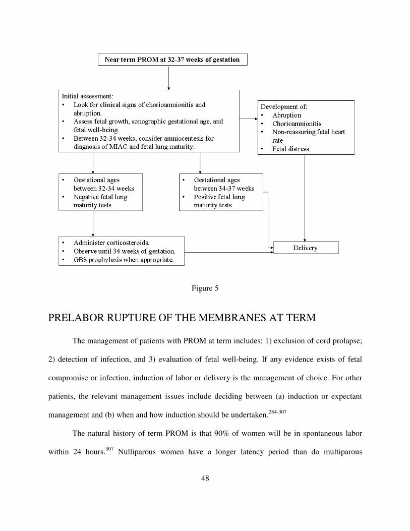
48
Figure 5
PRELABOR RUPTURE OF THE MEMBRANES AT TERM
The management of patients with PROM at term includes: 1) exclusion of cord prolapse;
2) detection of infection, and 3) evaluation of fetal well-being. If any evidence exists of fetal
compromise or infection, induction of labor or delivery is the management of choice. For other
patients, the relevant management issues include deciding between (a) induction or expectant
management and (b) when and how induction should be undertaken.284-307
The natural history of term PROM is that 90% of women will be in spontaneous labor
within 24 hours.307
Nulliparous women have a longer latency period than do multiparous

49
women.290;297;302
Patients with an unfavorable cervix at presentation and those who are not in
labor within 6 hours of rupture of membranes generally do not initiate labor spontaneously
within 24 hours and represent a management dilemma.306
The largest clinical trial in term PROM291
included 5,041 patients who were randomly
allocated to four groups: 1) immediate induction with oxytocin; 2) expectant management
followed by induction with oxytocin after 4 days; 3) induction of labor with vaginal
prostaglandin E2 (1-2 mg, followed by IV oxytocin; if not in labor in 4 hours, a second dose of
prostaglandin E2); and 4) expectant management followed by induction of labor with
prostaglandin E2, if labor had not begun within 4 days. The primary outcome of the trial was
probable neonatal infection (clinical and laboratory signs). Secondary outcomes were the need
for cesarean delivery and women’s evaluation of their treatment. The results showed no
difference in neonatal infection and cesarean delivery rate between the induction groups
(oxytocin vs. prostaglandin E2). However, the incidence of chorioamnionitis was lower in
patients allocated to induction of labor. Women’s satisfaction with their obstetric care was higher
for those allocated to induction of labor.
Mozurkewich and Wolf in their meta-analysis of 23 studies including 7,493 women,
demonstrated that induction of labor is superior to expectant management.298
The meta-analysis
compared three policies: 1) immediate induction with oxytocin; 2) induction of labor with
vaginal or endocervical prostaglandin E2 gel suppositories or tablets; and 3) expectant
management that sometimes has included delayed induction with oxytocin. The frequency of
chorioamnionitis and endometritis was significantly lower in patients undergoing immediate
induction of labor with oxytocin than in those managed expectantly [OR: 0.67, 95% CI: 0.52-
0.85 and OR: 0.71, 95% CI: 0.51-0.99, respectively]. The rate of chorioamnionitis was

50
significantly higher in patients who received vaginal prostaglandins than in those induced with
immediate administration of oxytocin but lower than that of patients in the expectant
management group [OR: 1.55, 95% CI: 1.09-2.21 and OR: 0.68, 95% CI: 0.51-0.91,
respectively]. The rates of cesarean delivery and neonatal infection were not different among the
three management schemes. Based on this data, we endorse a policy of immediate induction of
labor in patients with term PROM. Antibiotic administration is justified before cesarean delivery
for obstetric indications or for carriers of GBS.
Tan and Hannah308
performed a meta-analysis of 18 trials comparing induction of labor
with oxytocin versus expectant management in patients with PROM at or near term. Induction of
labor with oxytocin was associated with a lower risk of chorioamnionitis [OR: 0.63, 95% CI:
0.51-0.78], endometritis [OR: 0.72, 95% CI: 0.52-0.99], and neonatal infection [OR: 0.64, 95%
CI: 0.44-0.93]. Cesarean delivery rates were not statistically different. Oxytocin was associated
with a more frequent use of pain medication and internal fetal heart rate monitoring. Another
meta-analysis of 15 RCTs by Lin et al. included: 1) six studies [n=453] of misoprostol versus
placebo or expectant management; and 2) nine studies [n=1,130] of misoprostol versus oxytocin
for labor induction with term PROM.309
There were no significant differences in the frequency of
chorioamnionitis, neonatal sepsis, or caesarean delivery among study groups.
Expectant management at home of patients with PROM at term is not recommended.
This recommendation is based upon the report of Hanna et al. that home care was associated
with an increased risk of neonatal infections [OR: 1.97, 95% CI: 1.00-3.90] and cesarean
delivery in patients not colonized with GBS [OR: 1.48, 95% CI: 1.03-2.14].310
51
FIGURE LEGENDS
Figure 1. Rupture of membranes as a function of cervical dilatation and state of labor.
Percentage distribution for 517 normal spontaneous deliveries without medication or maneuvers;
vertex presentation.21
Figure 2. Initial assessment of preterm PROM. [AFI = amniotic fluid index; GBS = Group B
Streptococcus]. Modified from Mercer, B.M. 235
Figure 3. Management of previable PROM (before 24 weeks of gestation). [MIAC = microbial
invasion of the amniotic cavity]. Modified from Mercer, B.M.235
Figure 4. Management of PROM remote from term (between 24-31 6/7 weeks of gestation).
[MIAC = microbial invasion of the amniotic cavity; NICHD = National Institute of Child Health
and Human Development, NIH/DHHS]. Modified from Mercer, B.M.235
Figure 5. Management of near term PROM (between 32-37 weeks of gestation). [MIAC =
microbial invasion of the amniotic cavity; GBS = Group B Streptococcus]. Modified from
Mercer, B.M. 235

52
TABLES
Table 1. Risk factors associated with preterm PROM (at less than 35 weeks) stratified by parity.
25
Nulliparous Multiparous
Risk Factor Odds
Ratio
95% Confidence
Interval
Odds
Ratio
95% Confidence
Interval
Cervical length ≤ 25 mm 9.9 3.8-25.9 4.2 2.0-8.9
Previous preterm birth with preterm PROM -- -- 4.1 2.0-8.7
Previous preterm labor with intact membranes -- -- 2.6 1.2-5.3
Working during pregnancy 5.3 1.5-18.7 n.s. n.s.
Medical complications 4.2 1.1-16.0 n.s. n.s.
FFN (+) n.s. n.s. n.s. n.s. BV n.s. n.s. n.s. n.s. FFN (+) and absent BV n.s. n.s. 9.0 3.6-22.5
FFN (-) and present BV n.s. n.s. 2.8 1.2-6.3
FFN = fetal fibronectin; BV = bacterial vaginosis; n.s. = non-significant. Modified from Mercer et al.25
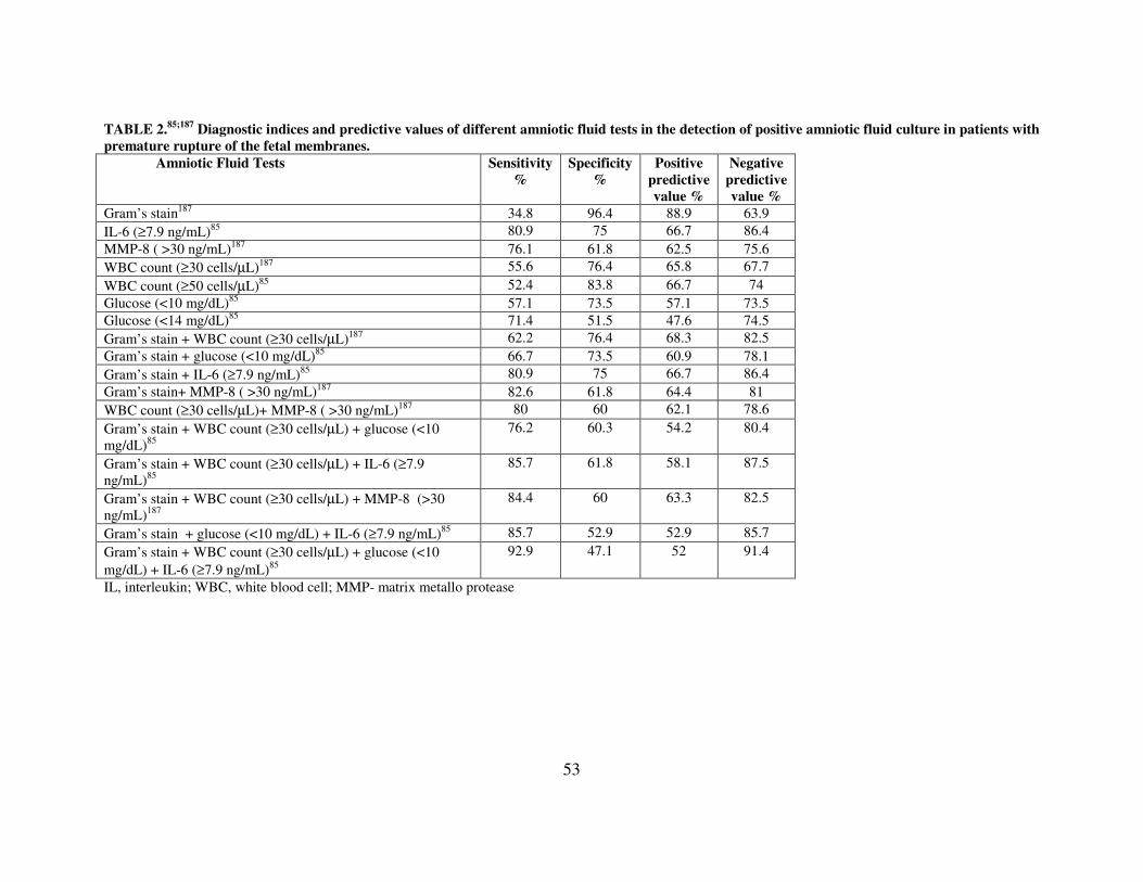
53
TABLE 2.85;187
Diagnostic indices and predictive values of different amniotic fluid tests in the detection of positive amniotic fluid culture in patients with
premature rupture of the fetal membranes.
Amniotic Fluid Tests Sensitivity
%
Specificity
%
Positive
predictive
value %
Negative
predictive
value %
Gram’s stain187
34.8 96.4 88.9 63.9
IL-6 (≥7.9 ng/mL)85
80.9 75 66.7 86.4
MMP-8 ( >30 ng/mL)187
76.1 61.8 62.5 75.6
WBC count (≥30 cells/µL)187
55.6 76.4 65.8 67.7
WBC count (≥50 cells/µL)85
52.4 83.8 66.7 74
Glucose (<10 mg/dL)85
57.1 73.5 57.1 73.5
Glucose (<14 mg/dL)85
71.4 51.5 47.6 74.5
Gram’s stain + WBC count (≥30 cells/µL)187
62.2 76.4 68.3 82.5
Gram’s stain + glucose (<10 mg/dL)85
66.7 73.5 60.9 78.1
Gram’s stain + IL-6 (≥7.9 ng/mL)85
80.9 75 66.7 86.4
Gram’s stain+ MMP-8 ( >30 ng/mL)187
82.6 61.8 64.4 81
WBC count (≥30 cells/µL)+ MMP-8 ( >30 ng/mL)187
80 60 62.1 78.6
Gram’s stain + WBC count (≥30 cells/µL) + glucose (<10
mg/dL)85
76.2 60.3 54.2 80.4
Gram’s stain + WBC count (≥30 cells/µL) + IL-6 (≥7.9
ng/mL)85
85.7 61.8 58.1 87.5
Gram’s stain + WBC count (≥30 cells/µL) + MMP-8 (>30
ng/mL)187
84.4 60 63.3 82.5
Gram’s stain + glucose (<10 mg/dL) + IL-6 (≥7.9 ng/mL)85
85.7 52.9 52.9 85.7
Gram’s stain + WBC count (≥30 cells/µL) + glucose (<10
mg/dL) + IL-6 (≥7.9 ng/mL)85
92.9 47.1 52 91.4
IL, interleukin; WBC, white blood cell; MMP- matrix metallo protease

54
Reference List
1. Keirse MJ, Ohlsson A, Treffers PE, Kanhani HHH. Prelabour rupture of the membranes
preterm. In: Chalmers I, Enkin M, Keirse MJ, editors. Effective care in pregnancy and
childbirth. Oxford: Oxford University Press; 1989. p. 666.
2. Johnson JW, Daikoku NH, Niebyl JR, Johnson TR, Jr., Khouzami VA, Witter FR.
Premature rupture of the membranes and prolonged latency. Obstet.Gynecol.
1981;57:547-56.
3. Hauth JC, Cunningham FG, Whalley PJ. Early labor initiation with oral PGE2 after
premature rupture of the membranes at term. Obstet.Gynecol. 1977;49:523-26.
4. Lange AP, Secher NJ, Nielsen FH, Pedersen GT. Stimulation of labor in cases of
premature rupture of the membranes at or near term. A consecutive randomized study of
prostaglandin E2-tablets and intravenous oxytocin. Acta Obstet.Gynecol.Scand.
1981;60:207-10.
5. LEBHERZ TB, HELLMAN LP, MADDING R, ANCTIL A, ARJE SL. DOUBLE-
BLIND STUDY OF PREMATURE RUPTURE OF THE MEMBRANES. A REPORT
OF 1,896 CASES. Am.J.Obstet.Gynecol. 1963;87:218-25.
6. Magos AL, Noble MC, Wong TY, Rodeck CH. Controlled study comparing vaginal
prostaglandin E2 pessaries with intravenous oxytocin for the stimulation of labour after
spontaneous rupture of the membranes. Br.J.Obstet.Gynaecol. 1983;90:726-31.
7. Tamsen L, Lyrenas S, Cnattingius S. Premature rupture of the membranes--intervention
or not. Gynecol.Obstet.Invest 1990;29:128-31.
8. van der WD, Venter PF. Management of term pregnancy with premature rupture of the
membranes and unfavourable cervix. S.Afr.Med.J. 1989;75:54-56.
9. Westergaard JG, Lange AP, Pedersen GT, Secher NJ. Use of oral oxytocics for
stimulation of labor in cases of premature rupture of the membranes at term. A
randomized comparative study of prostaglandin E2 tablets and demoxytocin resoriblets.
Acta Obstet.Gynecol.Scand. 1983;62:111-16.
10. Knudsen FU, Steinrud J. Septicaemia of the newborn, associated with ruptured foetal
membranes, discoloured amniotic fluid or maternal fever. Acta Paediatr.Scand.
1976;65:725-31.
11. Verber IG, Pearce JM, New LC, Hamilton PA, Davies EG. Prolonged rupture of the fetal
membranes and neonatal outcome. J.Perinat.Med. 1989;17:469-76.

55
12. Mercer BM. Preterm premature rupture of the membranes. Obstet Gynecol
2003;101:178-93.
13. Christensen KK, Christensen P, Ingemarsson I, Mardh PA, Nordenfelt E, Ripa T et al. A
study of complications in preterm deliveries after prolonged premature rupture of the
membranes. Obstet.Gynecol. 1976;48:670-77.
14. Daikoku NH, Kaltreider DF, Khouzami VA, Spence M, Johnson JW. Premature rupture
of membranes and spontaneous preterm labor: maternal endometritis risks.
Obstet.Gynecol. 1982;59:13-20.
15. Fayez JA, Hasan AA, Jonas HS, Miller GL. Management of premature rupture of the
membranes. Obstet.Gynecol. 1978;52:17-21.
16. Gunn GC, Mishell DR, Jr., Morton DG. Premature rupture of the fetal membranes. A
review. Am.J.Obstet.Gynecol. 1970;106:469-83.
17. Sachs M, Baker TH. Spontaneous premature rupture of the membranes. Am
J.Obstet.Gynecol. 1967;97:888.
18. Gibbs RS, Blanco JD. Premature rupture of the membranes. Obstet.Gynecol.
1982;60:671-79.
19. Shubert PJ, Diss E, Iams JD. Etiology of preterm premature rupture of membranes.
Obstet Gynecol Clin.North Am 1992;19:251-63.
20. Mercer BM. Preterm premature rupture of the membranes: current approaches to
evaluation and management. Obstet.Gynecol.Clin.North Am. 2005;32:411-28.
21. Schwarcz R, Belizan JM, Nieto F, Tenzer SM, Rios AM. Third progress report on the
Latin American collaborative study on the effects of late rupture of membranes on labor
and the neonate, submitted to the Director of the Pan-American Health Organization
(PAHO/WHO) and the participating groups. Montevideo:Latin American Center of
Perinatology and Human Development 1974.
22. Quintero RA, Morales WJ, Kalter CS, Allen M, Mendoza G, Angel JL et al.
Transabdominal intra-amniotic endoscopic assessment of previable premature rupture of
membranes. Am.J.Obstet.Gynecol. 1998;179:71-76.
23. Romero R. Prenatal Medicine: the child is the father of the man. Prenatal and Neonatal
Medicine 1996;1:8-11.
24. Romero R, Espinoza J, Mazor M, Chaiworapongsa T. The preterm parturition syndrome.
In: Critchley H, Bennett P, Thornton S, editors. Preterm birth. London, U.K.: RCOG
Press; 2004. p. 28-60.

56
25. Mercer BM, Goldenberg RL, Meis PJ, Moawad AH, Shellhaas C, Das A et al. The
Preterm Prediction Study: prediction of preterm premature rupture of membranes through
clinical findings and ancillary testing. The National Institute of Child Health and Human
Development Maternal-Fetal Medicine Units Network. Am J Obstet Gynecol
2000;183:738-45.
26. Weiss JL, Malone FD, Vidaver J, Ball RH, Nyberg DA, Comstock CH et al. Threatened
abortion: A risk factor for poor pregnancy outcome, a population-based screening study.
Am.J Obstet Gynecol. 2004;190:745-50.
27. Yang J, Hartmann KE, Savitz DA, Herring AH, Dole N, Olshan AF et al. Vaginal
bleeding during pregnancy and preterm birth. Am.J Epidemiol. 2004;160:118-25.
28. Carroll SG, Papaioannou S, Ntumazah IL, Philpott-Howard J, Nicolaides KH. Lower
genital tract swabs in the prediction of intrauterine infection in preterm prelabour rupture
of the membranes. Br.J.Obstet.Gynaecol. 1996;103:54-59.
29. Kim CJ, Romero R, Kusanovic JP, Yoo W, Dong Z, Topping V et al. The frequency,
clinical significance, and pathological features of chronic chorioamnionitis: a lesion
associated with spontaneous preterm birth. Mod.Pathol. 2010;23:1000-11.
30. Fujimoto T, Parry S, Urbanek M, Sammel M, Macones G, Kuivaniemi H et al. A single
nucleotide polymorphism in the matrix metalloproteinase-1 (MMP-1) promoter
influences amnion cell MMP-1 expression and risk for preterm premature rupture of the
fetal membranes. J.Biol.Chem. 2002;277:6296-302.
31. Ferrand PE, Parry S, Sammel M, Macones GA, Kuivaniemi H, Romero R et al. A
polymorphism in the matrix metalloproteinase-9 promoter is associated with increased
risk of preterm premature rupture of membranes in African Americans. Mol.Hum.Reprod
2002;8:494-501.
32. Wang H, Parry S, Macones G, Sammel MD, Ferrand PE, Kuivaniemi H et al.
Functionally significant SNP MMP8 promoter haplotypes and preterm premature rupture
of membranes (PPROM). Hum.Mol.Genet. 2004;13:2659-69.
33. Wang H, Parry S, Macones G, Sammel MD, Kuivaniemi H, Tromp G et al. A functional
SNP in the promoter of the SERPINH1 gene increases risk of preterm premature rupture
of membranes in African Americans. Proc.Natl.Acad Sci.U.S.A 2006;103:13463-67.
34. Kurki T, Sivonen A, Renkonen OV, Savia E, Ylikorkala O. Bacterial vaginosis in early
pregnancy and pregnancy outcome. Obstet Gynecol 1992;80:173-77.
35. Simhan HN, Caritis SN, Krohn MA, Hillier SL. The vaginal inflammatory milieu and the
risk of early premature preterm rupture of membranes. Am.J Obstet Gynecol.
2005;192:213-18.

57
36. Roberts AK, Monzon-Bordonaba F, Van Deerlin PG, Holder J, Macones GA, Morgan
MA et al. Association of polymorphism within the promoter of the tumor necrosis factor
alpha gene with increased risk of preterm premature rupture of the fetal membranes. Am
J Obstet Gynecol 1999;180:1297-302.
37. Macones GA, Parry S, Elkousy M, Clothier B, Ural SH, Strauss JF, III. A polymorphism
in the promoter region of TNF and bacterial vaginosis: preliminary evidence of gene-
environment interaction in the etiology of spontaneous preterm birth. Am J Obstet
Gynecol 2004;190:1504-08.
38. Wang H, Ogawa M, Wood JR, Bartolomei MS, Sammel MD, Kusanovic JP et al. Genetic
and epigenetic mechanisms combine to control MMP1 expression and its association
with preterm premature rupture of membranes. Hum.Mol.Genet. 2008;17:1087-96.
39. Goncalves LF, Chaiworapongsa T, Romero R. Intrauterine infection and prematurity.
Ment.Retard.Dev.Disabil.Res.Rev. 2002;8:3-13.
40. DiGiulio DB, Romero R, Kusanovic JP, Gomez R, Kim CJ, Seok KS et al. Prevalence
and diversity of microbes in the amniotic fluid, the fetal inflammatory response, and
pregnancy outcome in women with preterm pre-labor rupture of membranes. Am.J
Reprod.Immunol. 2010;64:38-57.
41. Cassell GH, Davis RO, Waites KB, Brown MB, Marriott PA, Stagno S et al. Isolation of
Mycoplasma hominis and Ureaplasma urealyticum from amniotic fluid at 16-20 weeks of
gestation: potential effect on outcome of pregnancy. Sex Transm.Dis. 1983;10:294-302.
42. Gray DJ, Robinson HB, Malone J, Thomson RB, Jr. Adverse outcome in pregnancy
following amniotic fluid isolation of Ureaplasma urealyticum. Prenat.Diagn.
1992;12:111-17.
43. Horowitz S, Mazor M, Romero R, Horowitz J, Glezerman M. Infection of the amniotic
cavity with Ureaplasma urealyticum in the midtrimester of pregnancy. J Reprod.Med.
1995;40:375-79.
44. Yoon BH, Oh SY, Romero R, Shim SS, Han SY, Park JS et al. An elevated amniotic
fluid matrix metalloproteinase-8 level at the time of mid-trimester genetic amniocentesis
is a risk factor for spontaneous preterm delivery. Am.J.Obstet.Gynecol. 2001;185:1162-
67.
45. Arias F, Rodriquez L, Rayne SC, Kraus FT. Maternal placental vasculopathy and
infection: two distinct subgroups among patients with preterm labor and preterm ruptured
membranes. Am.J Obstet Gynecol. 1993;168:585-91.
46. Arias F, Victoria A, Cho K, Kraus F. Placental histology and clinical characteristics of
patients with preterm premature rupture of membranes. Obstet Gynecol. 1997;89:265-71.

58
47. Romero R, Kusanovic JP, Kim CJ. Placental bed disorder in the genesis of the great
obstetrical syndrome. In: Pijnenborg R, Brosens I, Romero R, editors. Placental Bed
Disorder. Basic Science and its Translation to Obstetrics. Cambridge: Cambridge
University Press; 2010. p. 271-89.
48. Brosens I, Pijnenborg R, Vercruysse L, Romero R. The "Great Obstetrical Syndromes"
are associated with disorders of deep placentation. Am.J Obstet.Gynecol. 2011;204:193-
201.
49. Kim YM, Chaiworapongsa T, Gomez R, Bujold E, Yoon BH, Rotmensch S et al. Failure
of physiologic transformation of the spiral arteries in the placental bed in preterm
premature rupture of membranes. Am J Obstet Gynecol 2002;187:1137-42.
50. Gomez R, Romero R, Nien JK, Medina L, Carstens M, Kim YM et al. Idiopathic vaginal
bleeding during pregnancy as the only clinical manifestation of intrauterine infection. J
Matern.Fetal Neonatal Med. 2005;18:31-37.
51. Mackenzie AP, Schatz F, Krikun G, Funai EF, Kadner S, Lockwood CJ. Mechanisms of
abruption-induced premature rupture of the fetal membranes: Thrombin enhanced
decidual matrix metalloproteinase-3 (stromelysin-1) expression. Am J Obstet Gynecol
2004;191:1996-2001.
52. Rosen T, Schatz F, Kuczynski E, Lam H, Koo AB, Lockwood CJ. Thrombin-enhanced
matrix metalloproteinase-1 expression: a mechanism linking placental abruption with
premature rupture of the membranes. J Matern.Fetal Neonatal Med. 2002;11:11-17.
53. Stephenson CD, Lockwood CJ, Ma Y, Guller S. Thrombin-dependent regulation of
matrix metalloproteinase (MMP)-9 levels in human fetal membranes. J Matern.Fetal
Neonatal Med. 2005;18:17-22.
54. Curry TE, Jr., Osteen KG. The matrix metalloproteinase system: changes, regulation, and
impact throughout the ovarian and uterine reproductive cycle. Endocr.Rev. 2003;24:428-
65.
55. Darby MJ, Caritis SN, Shen-Schwarz S. Placental abruption in the preterm gestation: an
association with chorioamnionitis. Obstet Gynecol 1989;74:88-92.
56. Naeye RL. Factors that predispose to premature rupture of the fetal membranes.
Obstet.Gynecol. 1982;60:93-98.
57. Evaldson G, Lagrelius A, Winiarski J. Premature rupture of the membranes. Acta
Obstet.Gynecol.Scand. 1980;59:385-93.
58. Buchmayer SM, Sparen P, Cnattingius S. Previous pregnancy loss: risks related to
severity of preterm delivery. Am.J Obstet Gynecol. 2004;191:1225-31.

59
59. Hassan S, Romero R, Hendler I, Gomez R, Khalek N, Espinoza J et al. A sonographic
short cervix as the only clinical manifestation of intra-amniotic infection. J.Perinat.Med.
2006;34:13-19.
60. Rizzo G, Capponi A, Vlachopoulou A, Angelini E, Grassi C, Romanini C.
Ultrasonographic assessment of the uterine cervix and interleukin-8 concentrations in
cervical secretions predict intrauterine infection in patients with preterm labor and intact
membranes. Ultrasound Obstet Gynecol 1998;12:86-92.
61. Lind J, Wallenburg HC. Pregnancy and the Ehlers-Danlos syndrome: a retrospective
study in a Dutch population. Acta Obstet Gynecol Scand. 2002;81:293-300.
62. Harger JH, Hsing AW, Tuomala RE, Gibbs RS, Mead PB, Eschenbach DA et al. Risk
factors for preterm premature rupture of fetal membranes: a multicenter case-control
study. Am.J.Obstet.Gynecol. 1990;163:130-37.
63. Spinillo A, Nicola S, Piazzi G, Ghazal K, Colonna L, Baltaro F. Epidemiological
correlates of preterm premature rupture of membranes. Int.J.Gynaecol.Obstet. 1994;47:7-
15.
64. Cox SM, Williams ML, Leveno KJ. The natural history of preterm ruptured membranes:
what to expect of expectant management. Obstet.Gynecol. 1988;71:558-62.
65. Wilson JC, Levy DL, Wilds PL. Premature rupture of membranes prior to term:
consequences of nonintervention. Obstet.Gynecol. 1982;60:601-06.
66. Nelson LH, Anderson RL, O'Shea TM, Swain M. Expectant management of preterm
premature rupture of the membranes. Am.J.Obstet.Gynecol. 1994;171:350-56.
67. Romero R, Sirtori M, Oyarzun E, Avila C, Mazor M, Callahan R et al. Infection and
labor. V. Prevalence, microbiology, and clinical significance of intraamniotic infection in
women with preterm labor and intact membranes. Am.J.Obstet.Gynecol. 1989;161:817-
24.
68. Romero R, Quintero R, Oyarzun E, Wu YK, Sabo V, Mazor M et al. Intraamniotic
infection and the onset of labor in preterm premature rupture of the membranes. Am J
Obstet Gynecol 1988;159:661-66.
69. Romero R, Mazor M, Morretti R, et al. Infection and labor VII: Microbial invasion of the
amniotic cavity in spontaneous rupture of membranes at term. Am J Obstet Gynecol
1992;166:129.
70. Yoon BH, Romero R, Kim M, Kim EC, Kim T, Park JS et al. Clinical implications of
detection of Ureaplasma urealyticum in the amniotic cavity with the polymerase chain
reaction. Am.J.Obstet.Gynecol. 2000;183:1130-37.

60
71. Averbuch B, Mazor M, Shoham-Vardi I, Chaim W, Vardi H, Horowitz S et al. Intra-
uterine infection in women with preterm premature rupture of membranes: maternal and
neonatal characteristics. Eur.J Obstet Gynecol Reprod Biol 1995;62:25-29.
72. Cotton DB, Hill LM, Strassner HT, Platt LD, Ledger WJ. Use of amniocentesis in
preterm gestation with ruptured membranes. Obstet Gynecol 1984;63:38-43.
73. Coultrip LL, Grossman JH. Evaluation of rapid diagnostic tests in the detection of
microbial invasion of the amniotic cavity. Am J Obstet Gynecol 1992;167:1231-42.
74. Garite TJ, Freeman RK, Linzey EM, Braly P. The use of amniocentesis in patients with
premature rupture of membranes. Obstet Gynecol 1979;54:226-30.
75. Zlatnik FJ, Cruikshank DP, Petzold CR, Galask RP. Amniocentesis in the identification
of inapparent infection in preterm patients with premature rupture of the membranes. J
Reprod Med 1984;29:656-60.
76. Broekhuizen FF, Gilman M, Hamilton PR. Amniocentesis for gram stain and culture in
preterm premature rupture of the membranes. Obstet Gynecol 1985;66:316-21.
77. Dudley J, Malcolm G, Ellwood D. Amniocentesis in the management of preterm
premature rupture of the membranes. Aust.N.Z.J Obstet Gynaecol. 1991;31:331-36.
78. Feinstein SJ, Vintzileos AM, Lodeiro JG, Campbell WA, Weinbaum PJ, Nochimson DJ.
Amniocentesis with premature rupture of membranes. Obstet.Gynecol. 1986;68:147-52.
79. Font GE, Gauthier DW, Meyer WJ, Myles TD, Janda W, Bieniarz A. Catalase activity as
a predictor of amniotic fluid culture results in preterm labor or premature rupture of
membranes. Obstet Gynecol 1995;85:656-58.
80. Garite TJ, Freeman RK. Chorioamnionitis in the preterm gestation. Obstet Gynecol
1982;59:539-45.
81. Gauthier DW, Meyer WJ, Bieniarz A. Correlation of amniotic fluid glucose concentration
and intraamniotic infection in patients with preterm labor or premature rupture of
membranes. Am J Obstet Gynecol 1991;165:1105-10.
82. Gauthier DW, Meyer WJ. Comparison of gram stain, leukocyte esterase activity, and
amniotic fluid glucose concentration in predicting amniotic fluid culture results in
preterm premature rupture of membranes. Am J Obstet Gynecol 1992;167:1092-95.
83. Hazan Y, Mazor M, Horowitz S, Leiberman JR, Glezerman M. The diagnostic value of
amniotic fluid Gram stain examination and limulus amebocyte lysate assay in patients
with preterm birth. Acta Obstet Gynecol Scand. 1995;74:275-80.
61
84. O'Brien WF, Knuppel RA, Morales WJ, Angel JL, Torres CT. Amniotic fluid alpha 1-
antitrypsin concentration in premature rupture of the membranes. Am.J.Obstet.Gynecol.
1990;162:756-59.
85. Romero R, Yoon BH, Mazor M, Gomez R, Gonzalez R, Diamond MP et al. A
comparative study of the diagnostic performance of amniotic fluid glucose, white blood
cell count, interleukin-6, and gram stain in the detection of microbial invasion in patients
with preterm premature rupture of membranes. Am.J.Obstet.Gynecol. 1993;169:839-51.
86. Vintzileos AM, Campbell WA, Nochimson DJ, Weinbaum PJ, Escoto DT, Mirochnick
MH. Qualitative amniotic fluid volume versus amniocentesis in predicting infection in
preterm premature rupture of the membranes. Obstet.Gynecol. 1986;67:579-83.
87. Gomez R, Romero R, Nien JK, Medina L, Carstens M, Kim YM et al. Antibiotic
administration to patients with preterm premature rupture of membranes does not
eradicate intra-amniotic infection. J Matern.Fetal Neonatal Med 2007;20:167-73.
88. Carroll SG, Ville Y, Greenough A, Gamsu H, Patel B, Philpott-Howard J et al. Preterm
prelabour amniorrhexis: intrauterine infection and interval between membrane rupture
and delivery. Arch Dis.Child Fetal Neonatal Ed 1995;72:F43-F46.
89. Gomez R, Romero R, Ghezzi F, Yoon BH, Mazor M, Berry SM. The fetal inflammatory
response syndrome. Am.J.Obstet.Gynecol. 1998;179:194-202.
90. Romero R, Gomez R, Ghezzi F, Yoon BH, Mazor M, Edwin SS et al. A fetal systemic
inflammatory response is followed by the spontaneous onset of preterm parturition.
Am.J.Obstet.Gynecol. 1998;179:186-93.
91. Romero R, Chaiworapongsa T, Espinoza J, Gomez R, Yoon BH, Edwin S et al. Fetal
plasma MMP-9 concentrations are elevated in preterm premature rupture of the
membranes. Am.J Obstet Gynecol. 2002;187:1125-30.
92. Romero R, Athayde N, Gomez R, Mazor M, Yoon BH, Edwin S et al. The fetal
inflammatory response syndrom is characterized by the outpouring of a potent
extracellular matrix degrading enzyme into the fetal circulation. Am.J.Obstet.Gynecol.
1998;178:S3.
93. Berry SM, Romero R, Gomez R, Puder KS, Ghezzi F, Cotton DB et al. Premature
parturition is characterized by in utero activation of the fetal immune system.
Am.J.Obstet.Gynecol. 1995;173:1315-20.
94. Berry SM, Gomez R, Athayde N, Ghezzi F, Mazor M, Yoon BH et al. The role of
granulocyte colony stimulating factor in the neutrophilia observed in the fetal
inflammatory response syndrome. Am.J.Obstet.Gynecol. 1998;178:S202.

62
95. Gomez R, Berry S, Yoon BH, Mazor M, Athayde N, Ghezzi F et al. The hematologic
profile of the fetus with systemic inflammatory response syndrome. Am J
Obstet.Gynecol. 1998;178:S202.
96. Yoon BH, Romero R, Jun JK, Maymon E, Gomez R, Mazor M et al. An increase in fetal
plasma cortisol but not dehydroepiandrosterone sulfate is followed by the onset of
preterm labor in patients with preterm premature rupture of the membranes. Am J Obstet
Gynecol 1998;179:1107-14.
97. Romero R, Espinoza J, Goncalves LF, Gomez R, Medina L, Silva M et al. Fetal cardiac
dysfunction in preterm premature rupture of membranes. J Matern.Fetal Neonatal Med.
2004;16:146-57.
98. Yoon BH, Jun JK, Romero R, Park KH, Gomez R, Choi JH et al. Amniotic fluid
inflammatory cytokines (interleukin-6, interleukin-1beta, and tumor necrosis factor-
alpha), neonatal brain white matter lesions, and cerebral palsy. Am.J.Obstet.Gynecol.
1997;177:19-26.
99. Yoon BH, Romero R, Park JS, Kim CJ, Kim SH, Choi JH et al. Fetal exposure to an
intra-amniotic inflammation and the development of cerebral palsy at the age of three
years. Am.J.Obstet.Gynecol. 2000;182:675-81.
100. Yoon BH, Romero R, Jun JK, Park KH, Park JD, Ghezzi F et al. Amniotic fluid
cytokines (interleukin-6, tumor necrosis factor-alpha, interleukin-1 beta, and interleukin-
8) and the risk for the development of bronchopulmonary dysplasia.
Am.J.Obstet.Gynecol. 1997;177:825-30.
101. Yoon BH, Romero R, Kim KS, Park JS, Ki SH, Kim BI et al. A systemic fetal
inflammatory response and the development of bronchopulmonary dysplasia.
Am.J.Obstet.Gynecol. 1999;181:773-79.
102. Kim YM, Romero R, Chaiworapongsa T, Espinoza J, Mor G, Kim CJ. Dermatitis as a
component of the fetal inflammatory response syndrome is associated with activation of
Toll-like receptors in epidermal keratinocytes. Histopathology 2006;49:506-14.
103. Pacora P, Chaiworapongsa T, Maymon E, Kim YM, Gomez R, Yoon BH et al. Funisitis
and chorionic vasculitis: The histological counterpart of the fetal inflammatory response
syndrome. J.Matern.Fetal Neonatal Med 2002;11:18-25.
104. D'Alquen D, Kramer BW, Seidenspinner S, Marx A, Berg D, Groneck P et al. Activation
of umbilical cord endothelial cells and fetal inflammatory response in preterm infants
with chorioamnionitis and funisitis. Pediatr.Res. 2005;57:263-69.
105. Yoon BH, Romero R, Park JS, Kim M, Oh SY, Kim CJ et al. The relationship among
inflammatory lesions of the umbilical cord (funisitis), umbilical cord plasma interleukin 6
concentration, amniotic fluid infection, and neonatal sepsis. Am.J.Obstet.Gynecol.
2000;183:1124-29.

63
106. Wu YW, Colford JM, Jr. Chorioamnionitis as a risk factor for cerebral palsy: A meta-
analysis. JAMA 2000;284:1417-24.
107. Leviton A, Paneth N, Reuss ML, Susser M, Allred EN, Dammann O et al. Maternal
infection, fetal inflammatory response, and brain damage in very low birth weight
infants. Developmental Epidemiology Network Investigators. Pediatr.Res. 1999;46:566-
75.
108. Cornette L. Fetal and neonatal inflammatory response and adverse outcome. Semin.Fetal
Neonatal Med. 2004;9:459-70.
109. Naeye RL, Peters EC. Causes and consequences of premature rupture of fetal
membranes. Lancet 1980;1:192-94.
110. Pankuch GA, Appelbaum PC, Lorenz RP, Botti JJ, Schachter J, Naeye RL. Placental
microbiology and histology and the pathogenesis of chorioamnionitis. Obstet Gynecol
1984;64:802-06.
111. Romero R, Salafia CA, Athanassiadis AP, et al. The relationship between acute
inflammatory lesions of the preterm placenta and amniotic fluid mircobiology. Am J
Obstet Gynecol 1992;166:1382.
112. Ananth CV, Oyelese Y, Srinivas N, Yeo L, Vintzileos AM. Preterm premature rupture of
membranes, intrauterine infection, and oligohydramnios: risk factors for placental
abruption. Obstet Gynecol 2004;104:71-77.
113. Ananth CV, Savitz DA, Williams MA. Placental abruption and its association with
hypertension and prolonged rupture of membranes: a methodologic review and meta-
analysis. Obstet Gynecol 1996;88:309-18.
114. Ananth CV, Smulian JC, Demissie K, Vintzileos AM, Knuppel RA. Placental abruption
among singleton and twin births in the United States: risk factor profiles. Am J
Epidemiol. 2001;153:771-78.
115. Nelson DM, Stempel LE, Zuspan FP. Association of prolonged, preterm premature
rupture of the membranes and abruptio placentae. J Reprod Med 1986;31:249-53.
116. Vintzileos AM, Campbell WA, Nochimson DJ, Weinbaum PJ. Preterm premature rupture
of the membranes: a risk factor for the development of abruptio placentae.
Am.J.Obstet.Gynecol. 1987;156:1235-38.
117. Gonen R, Hannah ME, Milligan JE. Does prolonged preterm premature rupture of the
membranes predispose to abruptio placentae? Obstet Gynecol 1989;74:347-50.
118. Major CA, de Veciana M, Lewis DF, Morgan MA. Preterm premature rupture of
membranes and abruptio placentae: is there an association between these pregnancy
complications? Am J Obstet Gynecol 1995;172:672-76.

64
119. Naeye RL. Coitus and antepartum haemorrhage. Br J Obstet Gynaecol. 1981;88:765-70.
120. Vergani P, Ghidini A, Locatelli A, Cavallone M, Ciarla I, Cappellini A et al. Risk factors
for pulmonary hypoplasia in second-trimester premature rupture of membranes.
Am.J.Obstet.Gynecol. 1994;170:1359-64.
121. Rotschild A, Ling EW, Puterman ML, Farquharson D. Neonatal outcome after prolonged
preterm rupture of the membranes. Am.J.Obstet.Gynecol. 1990;162:46-52.
122. Winn HN, Chen M, Amon E, Leet TL, Shumway JB, Mostello D. Neonatal pulmonary
hypoplasia and perinatal mortality in patients with midtrimester rupture of amniotic
membranes--a critical analysis. Am J Obstet Gynecol 2000;182:1638-44.
123. Johnson A, Callan NA, Bhutani VK, Colmorgen GH, Weiner S, Bolognese RJ.
Ultrasonic ratio of fetal thoracic to abdominal circumference: an association with fetal
pulmonary hypoplasia. Am.J.Obstet.Gynecol. 1987;157:764-69.
124. Nimrod C, Varela-Gittings F, Machin G, Campbell D, Wesenberg R. The effect of very
prolonged membrane rupture on fetal development. Am.J.Obstet.Gynecol. 1984;148:540-
43.
125. Thibeault DW, Beatty EC, Jr., Hall RT, Bowen SK, O'Neill DH. Neonatal pulmonary
hypoplasia with premature rupture of fetal membranes and oligohydramnios. J.Pediatr.
1985;107:273-77.
126. Potter E. Bilateral renal agenesis. J Pediatr 1946;29:68.
127. Porter HJ. Pulmonary hypoplasia-size is not everything. Virch Arch 1998;432.
128. Wenstrom KD. Pulmonary hypoplasia and deformations related to premature rupture of
membranes. Obstet.Gynecol.Clin.North Am. 1992;19:397-408.
129. Blott M, Greenough A, Nicolaides KH, Moscoso G, Gibb D, Campbell S. Fetal breathing
movements as predictor of favourable pregnancy outcome after oligohydramnios due to
membrane rupture in second trimester. Lancet 1987;2:129-31.
130. Tamura RK, Sabbagha RE, Depp R, Vaisrub N, Dooley SL, Socol ML. Diminished
growth in fetuses born preterm after spontaneous labor or rupture of membranes. Am J
Obstet Gynecol 1984;148:1105-10.
131. Bukowski R, Gahn D, Denning J, Saade G. Impairment of growth in fetuses destined to
deliver preterm. Am J Obstet Gynecol 2001;185:463-67.
132. Spinillo A, Montanari L, Sanpaolo P, Bergante C, Chiara A, Fazzi E. Fetal growth and
infant neurodevelopmental outcome after preterm premature rupture of membranes.
Obstet Gynecol 2004;103:1286-93.
65
133. Blackwell S, Romero R, Chaiworapongsa T, Kim YM, Bujold E, Espinoza J et al.
Maternal and fetal inflammatory responses in unexplained fetal death. J.Matern.Fetal
Neonatal Med. 2003;14:151-57.
134. Gerber S, Vardhana S, Meagher-Villemure K, Vial Y, Hohlfeld P, Witkin SS.
Association between fetal interleukin-1 receptor antagonist gene polymorphism and
unexplained fetal death. Am J Obstet Gynecol 2005;193:1472-77.
135. Reece EA, Chervenak FA, Moya FR, Hobbins JC. Amniotic fluid arborization: effect of
blood, meconium, and pH alterations. Obstet Gynecol 1984;64:248-50.
136. Friedman ML, MCELIN TW. Diagnosis of ruptured fetal membranes. Clinical study and
review of the literature. Am J Obstet Gynecol 1969;104:544-50.
137. Lodeiro JG, Hsieh KA, Byers JH, Feinstein SJ. The fingerprint, a false-positive fern test.
Obstet Gynecol 1989;73:873-74.
138. McGregor JA, Johnson S. "Fig leaf" ferning and positive Nitrazine testing: semen as a
cause of misdiagnosis of premature rupture of membranes. Am J Obstet Gynecol
1985;151:1142-43.
139. Brookes C, Shand K, Jones WR. A reevaluation of the ferning test to detect ruptured
membranes. Aust.N.Z.J Obstet Gynaecol. 1986;26:260-64.
140. Rosemond RL, Lombardi SJ, Boehm FH. Ferning of amniotic fluid contaminated with
blood. Obstet Gynecol 1990;75:338-40.
141. Bennett SL, Cullen JB, Sherer DM, Woods JR, Jr. The ferning and nitrazine tests of
amniotic fluid between 12 and 41 weeks gestation. Am J Perinatol. 1993;10:101-04.
142. Adoni A, Ben Chetrit A, Zacut D, Palti Z, Hurwitz A. Prolongation of the latent period in
patients with premature rupture of the membranes by avoiding digital examination. Int.J
Gynaecol.Obstet 1990;32:19-21.
143. Lewis DF, Major CA, Towers CV, Asrat T, Harding JA, Garite TJ. Effects of digital
vaginal examinations on latency period in preterm premature rupture of membranes.
Obstet Gynecol 1992;80:630-34.
144. Sukcharoen N, Vasuratna A. Effects of digital cervical examinations on duration of
latency period, maternal and neonatal outcome in preterm premature rupture of
membranes. J Med Assoc.Thai. 1993;76:203-09.
145. Schutte MF, Treffers PE, Kloosterman GJ, Soepatmi S. Management of premature
rupture of membranes: the risk of vaginal examination to the infant. Am J Obstet
Gynecol 1983;146:395-400.

66
146. Munson LA, Graham A, Koos BJ, Valenzuela GJ. Is there a need for digital examination
in patients with spontaneous rupture of the membranes? Am J Obstet Gynecol
1985;153:562-63.
147. Brown CL, Ludwiczak MH, Blanco JD, Hirsch CE. Cervical dilation: accuracy of visual
and digital examinations. Obstet Gynecol 1993;81:215-16.
148. Morgan MA, de Veciana M, Rumney PJ, Schlinke S. Cervical assessment: visual or
digital? J Perinatol. 1996;16:103-06.
149. Smith RP. A technique for the detection of rupture of the membranes. A review and
preliminary report. Obstet Gynecol 1976;48:172-76.
150. Gahl W, Kazina TR, Furmann D, et al. Diamine oxidase in in the diagnosis of ruptured
fetal membranes. Am J Obstet Gynecol 1969;104:544.
151. Huber JF, Bischof P, Extermann P, Beguin F, Herrmann WL. Are vaginal fluid
concentrations of prolactin, alpha-fetoprotein and human placental lactogen useful for
diagnosing ruptured membranes? Br J Obstet Gynaecol. 1983;90:1183-85.
152. Koninckx PR, Trappeniers H, Van Assche FA. Prolactin concentration in vaginal fluid: a
new method for diagnosing ruptured membranes. Br J Obstet Gynaecol. 1981;88:607-10.
153. Phocas I, Sarandakou A, Kontoravdis A, Chryssicopoulos A, Zourlas PA. Vaginal fluid
prolactin: a reliable marker for the diagnosis of prematurely ruptured membranes.
Comparison with vaginal fluid alpha-fetoprotein and placental lactogen. Eur.J Obstet
Gynecol Reprod Biol 1989;31:133-41.
154. Gaucherand P, Guibaud S, Rudigoz RC, Wong A. Diagnosis of premature rupture of the
membranes by the identification of alpha-feto-protein in vaginal secretions. Acta Obstet
Gynecol Scand. 1994;73:456-59.
155. Lockwood CJ, Wein R, Chien D, Ghidini A, Alvarez M, Berkowitz RL. Fetal membrane
rupture is associated with the presence of insulin-like growth factor-binding protein-1 in
vaginal secretions. Am J Obstet Gynecol 1994;171:146-50.
156. Kishida T, Hirao A, Matsuura T, Katamine T, Yamada H, Sagawa T et al. Diagnosis of
premature rupture of membranes with an improved alpha-fetoprotein monoclonal
antibody kit. Clin.Chem. 1995;41:1500-03.
157. Kishida T, Yamada H, Negishi H, Sagawa T, Makinoda S, Fujimoto S. Diagnosis of
preterm premature rupture of the membranes using a newly developed AFP monoclonal
antibody test kit. Eur.J Obstet Gynecol Reprod Biol 1995;58:67-72.
158. Buyukbayrak EE, Turan C, Unal O, Dansuk R, Cengizoglu B. Diagnostic power of the
vaginal washing-fluid prolactin assay as an alternative method for the diagnosis of
premature rupture of membranes. J Matern.Fetal Neonatal Med. 2004;15:120-25.
67
159. Esim E, Turan C, Unal O, Dansuk R, Cengizglu B. Diagnosis of premature rupture of
membranes by identification of beta-HCG in vaginal washing fluid. Eur.J Obstet Gynecol
Reprod.Biol. 2003;107:37-40.
160. Erdemoglu E, Mungan T. Significance of detecting insulin-like growth factor binding
protein-1 in cervicovaginal secretions: comparison with nitrazine test and amniotic fluid
volume assessment. Acta Obstet Gynecol Scand. 2004;83:622-26.
161. Anai T, Tanaka Y, Hirota Y, Miyakawa I. Vaginal fluid hCG levels for detecting
premature rupture of membranes. Obstet Gynecol 1997;89:261-64.
162. Kishida T, Yamada H, Negishi H, Sagawa T, Makinoda S, Fujimoto S. Diagnosis of
premature rupture of the membranes in preterm patients, using an improved AFP kit:
comparison with ROM-check and/or nitrazine test. Eur.J Obstet Gynecol Reprod.Biol.
1996;69:77-82.
163. Atterbury JL, Groome LJ, Hoff C. Methods used to diagnose premature rupture of
membranes: a national survey of 812 obstetric nurses. Obstet Gynecol 1998;92:384-89.
164. Lockwood CJ, Senyei AE, Dische MR, Casal D, Shah KD, Thung SN et al. Fetal
fibronectin in cervical and vaginal secretions as a predictor of preterm delivery. N.Engl.J
Med 1991;325:669-74.
165. Eriksen NL, Parisi VM, Daoust S, Flamm B, Garite TJ, Cox SM. Fetal fibronectin: a
method for detecting the presence of amniotic fluid. Obstet Gynecol 1992;80:451-54.
166. Goldenberg RL, Mercer BM, Meis PJ, Copper RL, Das A, McNellis D. The preterm
prediction study: fetal fibronectin testing and spontaneous preterm birth. NICHD
Maternal Fetal Medicine Units Network. Obstet Gynecol 1996;87:643-48.
167. Greenhagen JB, Van Wagoner J, Dudley D, Hunter C, Mitchell M, Logsdon V et al.
Value of fetal fibronectin as a predictor of preterm delivery for a low-risk population. Am
J Obstet Gynecol 1996;175:1054-56.
168. Irion O, Matute S, Bischof P, Beguin F. Cervical onofetal fibronectin in patients with
false labor as a predictor of preterm delivery. Am J.Obstet.Gynecol. 1996;394:384.
169. Leeson SC, Maresh MJ, Martindale EA, Mahmood T, Muotune A, Hawkes N et al.
Detection of fetal fibronectin as a predictor of preterm delivery in high risk asymptomatic
pregnancies. Br J Obstet Gynaecol. 1996;103:48-53.
170. Malak TM, Sizmur F, Bell SC, Taylor DJ. Fetal fibronectin in cervicovaginal secretions
as a predictor of preterm birth. Br J Obstet Gynaecol. 1996;103:648-53.
171. Morrison JC, Allbert JR, McLaughlin BN, Whitworth NS, Roberts WE, Martin RW.
Oncofetal fibronectin in patients with false labor as a predictor of preterm delivery. Am J
Obstet Gynecol 1993;168:538-42.

68
172. Nageotte MP, Casal D, Senyei AE. Fetal fibronectin in patients at increased risk for
premature birth. Am J Obstet Gynecol 1994;170:20-25.
173. Rizzo G, Capponi A, Arduini D, Lorido C, Romanini C. The value of fetal fibronectin in
cervical and vaginal secretions and of ultrasonographic examination of the uterine cervix
in predicting premature delivery for patients with preterm labor and intact membranes.
Am J Obstet Gynecol 1996;175:1146-51.
174. Atlay RD, Sutherst JR. Premature rupture of the fetal membranes confirmed by intra-
amniotic injection of dye (Evans blue T-1824). Am J Obstet Gynecol 1970;108:993-94.
175. Diaz-Garzon J. Indigocarmine test of preterm rupture of membranes. Rev Columb Obstet
Gynecol 1969;20:373.
176. Fujimoto S, Kishida T, Sagawa T, Negishi H, Okuyama K, Hareyama H et al. Clinical
usefulness of the dye-injection method for diagnosing premature rupture of the
membranes in equivocal cases. J Obstet Gynaecol. 1995;21:215-20.
177. Meyer BA, Gonik B, Creasy RK. Evaluation of phenazopyridine hydrochloride as a tool
in the diagnosis of premature rupture of the membranes. Am J Perinatol. 1991;8:297-99.
178. Cowett RM, Hakanson DO, Kocon RW, Oh W. Untoward neonatal effect of
intraamniotic administration of methylene blue. Obstet Gynecol 1976;48:74S-5S.
179. Mc Enerney J, Mc Enerney L. Unfavourable neotal outcomes after intraamniotic
injection of methelene blue. Obstet Gynecol 1983;1983:351.
180. Troche BI. Methylene blue baby. N.Engl.J Med 1989;320:1756-57.
181. Divon MY, Chamberlain PF, Sipos L, Platt LD. Underestimation of fetal weight in
premature rupture of membranes. J Ultrasound Med. 1984;3:529-31.
182. Ben Haroush A, Yogev Y, Bar J, Mashiach R, Kaplan B, Hod M et al. Accuracy of
sonographically estimated fetal weight in 840 women with different pregnancy
complications prior to induction of labor. Ultrasound Obstet Gynecol. 2004;23:172-76.
183. Edwards A, Goff J, Baker L. Accuracy and modifying factors of the sonographic
estimation of fetal weight in a high-risk population. Aust.N.Z.J Obstet Gynaecol
2001;41:187-90.
184. Romero R, Goncalves LF, Ghezzi F, Gomez R, Cohen J, Mazor M. Amniocenteses. In:
Fleischer AC, Manning FA, Jeanty P, Romero R, editors. Sonography in obstetrics and
gynecology: principles and practice. Stamford, CT: Appleton & Lange; 1996.
185. Cotton DB, Gonik B, Bottoms SF. Conservative versus aggressive management of
preterm rupture of membranes. A randomized trial of amniocentesis. Am J Perinatol.
1984;1:322-24.
69
186. Shim SS, Romero R, Hong JS, Park CW, Jun JK, Kim BI et al. Clinical significance of
intra-amniotic inflammation in patients with preterm premature rupture of membranes.
Am J Obstet Gynecol 2004;191:1339-45.
187. Maymon E, Romero R, Chaiworapongsa T, Kim JC, Berman S, Gomez R et al. Value of
amniotic fluid neutrophil collagenase concentrations in preterm premature rupture of
membranes. Am J.Obstet.Gynecol. 2001;185:1143-48.
188. Yoon BH, Romero R, Kim CJ, Jun JK, Gomez R, Choi JH et al. Amniotic fluid
interleukin-6: a sensitive test for antenatal diagnosis of acute inflammatory lesions of
preterm placenta and prediction of perinatal morbidity. Am J Obstet Gynecol
1995;172:960-70.
189. Jalava J, Mantymaa ML, Ekblad U, Toivanen P, Skurnik M, Lassila O et al. Bacterial
16S rDNA polymerase chain reaction in the detection of intra-amniotic infection.
Br.J.Obstet.Gynaecol. 1996;103:664-69.
190. Nien JK, Yoon BH, Espinoza J, Kusanovic JP, Erez O, Soto E et al. A rapid MMP-8
bedside test for the detection of intra-amniotic inflammation identifies patients at risk for
imminent preterm delivery. Am.J Obstet.Gynecol. 2006;195:1025-30.
191. Yeast JD, Garite TJ, Dorchester W. The risks of amniocentesis in the management of
premature rupture of the membranes. Am J Obstet Gynecol 1984;149:505-08.
192. Shaver DC, Spinnato JA, Whybrew D, Williams WK, Anderson GD. Comparison of
phospholipids in vaginal and amniocentesis specimens of patients with premature rupture
of membranes. Am J Obstet Gynecol 1987;156:454-57.
193. Stedman CM, Crawford S, Staten E, Cherny WB. Management of preterm premature
rupture of membranes: assessing amniotic fluid in the vagina for phosphatidylglycerol.
Am J Obstet Gynecol 1981;140:34-38.
194. Brame RG, MacKenna J. Vaginal pool phospholipids in the management of premature
rupture of membranes. Am J Obstet Gynecol 1983;145:992-1000.
195. Golde SH. Use of obstetric perineal pads in collection of amniotic fluid in patients with
rupture of the membranes. Am J Obstet Gynecol 1983;146:710-12.
196. Estol PC, Poseiro JJ, Schwarcz R. Phosphatidylglycerol determination in the amniotic
fluid from a PAD placed over the vulva: a method for diagnosis of fetal lung maturity in
cases of premature ruptured membranes. J Perinat Med 1992;20:65-71.
197. Lewis DF, Towers CV, Major CA, Asrat T, Nageotte MP, Freeman RK et al. Use of
Amniostat-FLM in detecting the presence of phosphatidylglycerol in vaginal pool
samples in preterm premature rupture of membranes. Am J Obstet Gynecol
1993;169:573-76.
70
198. Gluck L, Kulovich MV, Borer RC, Jr., Keidel WN. The interpretation and significance of
the lecithin-sphingomyelin ratio in amniotic fluid. Am J Obstet Gynecol 1974;120:142-
55.
199. Schumacher RE, Parisi VM, Steady HM, Tsao FH. Bacteria causing false positive test for
phosphatidylglycerol in amniotic fluid. Am J Obstet Gynecol 1985;151:1067-68.
200. Dombroski RA, MacKenna J, Brame RG. Comparison of amniotic fluid lung maturity
profiles in paired vaginal and amniocentesis specimens. Am J Obstet Gynecol
1981;140:461-64.
201. Goldstein AS, Mangurten HH, Libretti JV, Berman AM. Lecithin/sphingomyelin ratio in
amniotic fluid obtained vaginally. Am J Obstet Gynecol 1980;138:232-33.
202. Spinnato JA, Shaver DC, Bray EM, Lipshitz J. Preterm premature rupture of the
membranes with fetal pulmonary maturity present: a prospective study. Obstet Gynecol
1987;69:196-201.
203. Mercer BM, Crocker LG, Boe NM, Sibai BM. Induction versus expectant management in
premature rupture of the membranes with mature amniotic fluid at 32 to 36 weeks: a
randomized trial. Am J Obstet Gynecol 1993;169:775-82.
204. Vintzileos AM, Feinstein SJ, Lodeiro JG, Campbell WA, Weinbaum PJ, Nochimson DJ.
Fetal biophysical profile and the effect of premature rupture of the membranes.
Obstet.Gynecol. 1986;67:818-23.
205. Vintzileos AM, Campbell WA, Nochimson DJ, Weinbaum PJ. The use of the nonstress
test in patients with premature rupture of the membranes. Am.J.Obstet.Gynecol.
1986;155:149-53.
206. Zeevi D, Sadovsky E, Younis J, Granat M, Ohel G. Antepartum fetal heart rate
characteristics in cases of premature rupture of membranes. Am J Perinatol. 1988;5:260-
63.
207. Vintzileos AM, Knuppel RA. Fetal biophysical assessment in premature rupture of the
membranes. Clin.Obstet Gynecol 1995;38:45-58.
208. Carroll SG, Papaioannou S, Nicolaides KH. Assessment of fetal activity and amniotic
fluid volume in the prediction of intrauterine infection in preterm prelabor amniorrhexis.
Am J Obstet Gynecol 1995;172:1427-35.
209. Del Valle GO, Joffe GM, Izquierdo LA, Smith JF, Gilson GJ, Curet LB. The biophysical
profile and the nonstress test: poor predictors of chorioamnionitis and fetal infection in
prolonged preterm premature rupture of membranes. Obstet Gynecol 1992;80:106-10.
210. Gauthier DW, Meyer WJ, Bieniarz A. Biophysical profile as a predictor of amniotic fluid
culture results. Obstet Gynecol 1992;80:102-05.
71
211. Harding JA, Jackson DM, Lewis DF, Major CA, Nageotte MP, Asrat T. Correlation of
amniotic fluid index and nonstress test in patients with preterm premature rupture of
membranes. Am.J Obstet.Gynecol. 1991;165:1088-94.
212. Vintzileos AM, Campbell WA, Nochimson DJ, Weinbaum PJ. Degree of
oligohydramnios and pregnancy outcome in patients with premature rupture of the
membranes. Obstet.Gynecol. 1985;66:162-67.
213. Gonik B, Bottoms SF, Cotton DB. Amniotic fluid volume as a risk factor in preterm
premature rupture of the membranes. Obstet Gynecol 1985;65:456-59.
214. Hadi HA, Hodson CA, Strickland D. Premature rupture of the membranes between 20
and 25 weeks' gestation: role of amniotic fluid volume in perinatal outcome. Am J Obstet
Gynecol 1994;170:1139-44.
215. Lao TT, Cheung VY. Expectant management of preterm prelabour rupture of
membranes--the significance of oligohydramnios at presentation. Eur.J Obstet Gynecol
Reprod Biol 1993;48:87-91.
216. Yoon BH, Kim YA, Romero R, Kim JC, Park KH, Kim MH et al. Association of
oligohydramnios in women with preterm premature rupture of membranes with an
inflammatory response in fetal, amniotic, and maternal compartments.
Am.J.Obstet.Gynecol. 1999;181:784-88.
217. Smith CV, Greenspoon J, Phelan JP, Platt LD. Clinical utility of the nonstress test in the
conservative management of women with preterm spontaneous premature rupture of the
membranes. J Reprod Med 1987;32:1-4.
218. Kivikoski AI, Amon E, Vaalamo PO, Pirhonen J, Kopta MM. Effect of third-trimester
premature rupture of membranes on fetal breathing movements: a prospective case-
control study. Am J Obstet Gynecol 1988;159:1474-77.
219. Roberts AB, Goldstein I, Romero R, Hobbins JC. Fetal breathing movements after
preterm premature rupture of membranes. Am J Obstet Gynecol 1991;164:821-25.
220. Vintzileos AM, Campbell WA, Nochimson DJ, Connolly ME, Fuenfer MM, Hoehn GJ.
The fetal biophysical profile in patients with premature rupture of the membranes--an
early predictor of fetal infection. Am.J.Obstet.Gynecol. 1985;152:510-16.
221. Vintzileos AM, Campbell WA, Nochimson DJ, Weinbaum PJ. Fetal breathing as a
predictor of infection in premature rupture of the membranes. Obstet.Gynecol.
1986;67:813-17.
222. Goldstein I, Romero R, Merrill S, Wan M, O'Connor TZ, Mazor M et al. Fetal body and
breathing movements as predictors of intraamniotic infection in preterm premature
rupture of membranes. Am J Obstet Gynecol 1988;159:363-68.
72
223. Fleming AD, Salafia CM, Vintzileos AM, Rodis JF, Campbell WA, Bantham KF. The
relationships among umbilical artery velocimetry, fetal biophysical profile, and placental
inflammation in preterm premature rupture of the membranes. Am.J.Obstet.Gynecol.
1991;164:38-41.
224. Mercer B, Moretti M, Shaver Deal. Intensive antenatal testing for women with prolonged
preterm premature rupture of the membranes. Abstract 420 Presented at 11th annual
meeting of the Society of Perinatal Obstetricians.San Francisco 1991.
225. Miller JM, Jr., Kho MS, Brown HL, Gabert HA. Clinical chorioamnionitis is not
predicted by an ultrasonic biophysical profile in patients with premature rupture of
membranes. Obstet Gynecol 1990;76:1051-54.
226. Roberts AB, Goldstein I, Romero R, Hobbins JC. Comparison of total fetal activity
measurement with the biophysical profile in predicting intra-amniotic infection in
preterm premature rupture of membranes. Ultrasound Obstet Gynecol 1991;1:36-39.
227. Roussis P, Rosemond RL, Glass C, Boehm FH. Preterm premature rupture of
membranes: detection of infection. Am J Obstet Gynecol 1991;165:1099-104.
228. Vintzileos AM, Bors-Koefoed R, Pelegano JF, Campbell WA, Rodis JF, Nochimson DJ
et al. The use of fetal biophysical profile improves pregnancy outcome in premature
rupture of the membranes. Am.J.Obstet.Gynecol. 1987;157:236-40.
229. Fanaroff AA, Hack M. Periventricular leukomalacia--prospects for prevention. N.Engl.J
Med. 1999;341:1229-31.
230. Hack M, Fanaroff AA. Outcomes of extremely immature infants--a perinatal dilemma.
N.Engl.J Med. 1993;329:1649-50.
231. Dewan H, Morris JM. A systematic review of pregnancy outcome following preterm
premature rupture of membranes at a previable gestational age. Aust.N.Z.J
Obstet.Gynaecol. 2001;41:389-94.
232. Gold RB, Goyert GL, Schwartz DB, Evans MI, Seabolt LA. Conservative management
of second-trimester post-amniocentesis fluid leakage. Obstet.Gynecol. 1989;74:745-47.
233. Hanson FW, Tennant FR, Zorn EM, Samuels S. Analysis of 2136 genetic amniocenteses:
experience of a single physician. Am.J.Obstet.Gynecol. 1985;152:436-43.
234. Effect of corticosteroids for fetal maturation on perinatal outcomes. NIH Consensus
Development Panel on the Effect of Corticosteroids for Fetal Maturation on Perinatal
Outcomes. JAMA 1995;273:413-18.
235. Mercer BM. Preterm premature rupture of the membranes: diagnosis and management.
Clin.Perinatol. 2004;31:765-82, vi.

73
236. Simhan HN, Canavan TP. Preterm premature rupture of membranes: diagnosis,
evaluation and management strategies. BJOG. 2005;112 Suppl 1:32-37.
237. Mercer BM, Arheart KL. Antimicrobial therapy in expectant management of preterm
premature rupture of the membranes. Lancet 1995;346:1271-79.
238. Kenyon SL, Taylor DJ, Tarnow-Mordi W. Broad-spectrum antibiotics for preterm,
prelabour rupture of fetal membranes: the ORACLE I randomised trial. ORACLE
Collaborative Group. Lancet 2001;357:979-88.
239. Amon E, Lewis SV, Sibai BM, Villar MA, Arheart KL. Ampicillin prophylaxis in
preterm premature rupture of the membranes: a prospective randomized study. Am J
Obstet Gynecol 1988;159:539-43.
240. Morales WJ, Angel JL, O'Brien WF, Knuppel RA. Use of ampicillin and corticosteroids
in premature rupture of membranes: a randomized study. Obstet Gynecol 1989;73:721-
26.
241. Johnston MM, Sanchez-Ramos L, Vaughn AJ, Todd MW, Benrubi GI. Antibiotic therapy
in preterm premature rupture of membranes: a randomized, prospective, double-blind
trial. Am J Obstet Gynecol 1990;163:743-47.
242. Christmas JT, Cox SM, Andrews W, Dax J, Leveno KJ, Gilstrap LC. Expectant
management of preterm ruptured membranes: effects of antimicrobial therapy. Obstet
Gynecol 1992;80:759-62.
243. McGregor JA, French JI, Seo K. Antimicrobial therapy in preterm premature rupture of
membranes: results of a prospective, double-blind, placebo-controlled trial of
erythromycin. Am J Obstet Gynecol 1991;165:632-40.
244. Kurki T, Hallman M, Zilliacus R, Teramo K, Ylikorkala O. Premature rupture of the
membranes: effect of penicillin prophylaxis and long-term outcome of the children. Am J
Perinatol. 1992;9:11-16.
245. McCaul JF, Perry KG, Jr., Moore JL, Jr., Martin RW, Bucovaz ET, Morrison JC.
Adjunctive antibiotic treatment of women with preterm rupture of membranes or preterm
labor. Int.J Gynaecol.Obstet 1992;38:19-24.
246. Mercer BM, Moretti ML, Prevost RR, Sibai BM. Erythromycin therapy in preterm
premature rupture of the membranes: a prospective, randomized trial of 220 patients. Am
J Obstet Gynecol 1992;166:794-802.
247. Owen J, Groome LJ, Hauth JC. Randomized trial of prophylactic antibiotic therapy after
preterm amnion rupture. Am J Obstet Gynecol 1993;169:976-81.

74
248. Blanco JD, Iams J, Artal R, et al. Multicentre double-blind prospective randomized trial
of ceftrizoxime versus placebo in women with preterm premature rupture of the
membranes. Am J.Obstet.Gynecol. 1993;168:378.
249. Lockwood CJ, Costigan K, Ghidini A, Wein R, Chien D, Brown BL et al. Double-blind;
placebo-controlled trial of piperacillin prophylaxis in preterm membrane rupture. Am J
Obstet Gynecol 1993;169:970-76.
250. Ernest JM, Givner LB. A prospective, randomized, placebo-controlled trial of penicillin
in preterm premature rupture of membranes. Am J Obstet Gynecol 1994;170:516-21.
251. Mercer BM, Miodovnik M, Thurnau GR, Goldenberg RL, Das AF, Ramsey RD et al.
Antibiotic therapy for reduction of infant morbidity after preterm premature rupture of
the membranes. A randomized controlled trial. National Institute of Child Health and
Human Development Maternal-Fetal Medicine Units Network. JAMA 1997;278:989-95.
252. Lovett SM, Weiss JD, Diogo MJ, Williams PT, Garite TJ. A prospective, double-blind,
randomized, controlled clinical trial of ampicillin-sulbactam for preterm premature
rupture of membranes in women receiving antenatal corticosteroid therapy. Am J Obstet
Gynecol 1997;176:1030-38.
253. Kenyon S, Boulvain M, Neilson J. Antibiotics for preterm rupture of the membranes: a
systematic review. Obstet Gynecol 2004;104:1051-57.
254. Kenyon S, Pike K, Jones DR, Brocklehurst P, Marlow N, Salt A et al. Childhood
outcomes after prescription of antibiotics to pregnant women with preterm rupture of the
membranes: 7-year follow-up of the ORACLE I trial. Lancet 2008;372:1310-18.
255. Brelje MC, Kaltreider DF. The use of vaginal antibiotics in premature rupture of the
membranes. Am J.Obstet.Gynecol. 1966;94:889.
256. Ogita S, Imanaka M, Matsumoto M, Hatanaka K. Premature rupture of the membranes
managed with a new cervical catheter. Lancet 1984;1:1330-31.
257. Romero R, Scioscia AL, Edberg SC, Hobbins JC. Use of parenteral antibiotic therapy to
eradicate bacterial colonization of amniotic fluid in premature rupture of membranes.
Obstet Gynecol 1986;67:15S-7S.
258. Romero R, Hagay Z, Nores J, Sepulveda W, Mazor M. Eradication of Ureaplasma
urealyticum from the amniotic fluid with transplacental antibiotic treatment. Am J Obstet
Gynecol 1992;166:618-20.
259. Harding JE, Pang J, Knight DB, Liggins GC. Do antenatal corticosteroids help in the
setting of preterm rupture of membranes? Am J Obstet Gynecol 2001;184:131-39.
260. Crowley P. Corticosteriods after prelabour rupture of membranes [revised May 5, 1994].
In: Keirse MJ, Renfre MJ, Neilson JP, Crowther C, editors. Pregnancy and childbirth

75
module. The Cochrane Pregnancy and Childbirth Database. The Cochrane Collaboration,
Issue 2 [Available from BMJ Publishing Group, London.]. Oxford, UK: 1995.
261. Crowley P. Corticosteroids + induction of labour after PROM preterm [revised April 22,
1993]. In: Keirse MJ, Renfre MJ, Neilson JP, Crowther C, editors. Pregnancy and
childbirth module. The Cochrane Pregnancy and Childbirth Database. The Cochrane
Collaboration, Issue 2, [Available from BMJ Publishing Group, London.]. Oxford, UK:
1995.
262. Leitich H, Egarter C, Reisenberger K, Kaider A, Berghammer P. Concomitant use of
glucocorticoids: a comparison of two metaanalyses on antibiotic treatment in preterm
premature rupture of membranes. Am J Obstet Gynecol 1998;178:899-908.
263. Guinn DA, Atkinson MW, Sullivan L, Lee M, MacGregor S, Parilla BV et al. Single vs
weekly courses of antenatal corticosteroids for women at risk of preterm delivery: A
randomized controlled trial. JAMA 2001;286:1581-87.
264. Christensen KK, Ingemarsson I, Leideman T, Solum T, Svenningsen N. Effect of
ritodrine on labor after premature rupture of the membranes. Obstet Gynecol
1980;55:187-90.
265. Garite TJ, Keegan KA, Freeman RK, Nageotte MP. A randomized trial of ritodrine
tocolysis versus expectant management in patients with premature rupture of membranes
at 25 to 30 weeks of gestation. Am J Obstet Gynecol 1987;157:388-93.
266. Matsuda Y, Ikenoue T, Hokanishi H. Premature rupture of the membranes--aggressive
versus conservative approach: effect of tocolytic and antibiotic therapy. Gynecol Obstet
Invest 1993;36:102-07.
267. Weiner CP, Renk K, Klugman M. The therapeutic efficacy and cost-effectiveness of
aggressive tocolysis for premature labor associated with premature rupture of the
membranes. Am J Obstet Gynecol 1988;159:216-22.
268. Harger JH. Comparison of success and morbidity in cervical cerclage procedures.
Obstet.Gynecol. 1980;56:543-48.
269. Yeast JD, Garite TR. The role of cervical cerclage in the management of preterm
premature rupture of the membranes. Am.J.Obstet.Gynecol. 1988;158:106-10.
270. Blickstein I, Katz Z, Lancet M, Molgilner BM. The outcome of pregnancies complicated
by preterm rupture of the membranes with and without cerclage. Int.J.Gynaecol.Obstet.
1989;28:237-42.
271. Goldman JM, Greene MF, Tuomala R, Ringer SA, Harlow BL, Crowley SM. Outcome of
expectant management in preterm premature rupture of membranes with cervical cerclage
in place. Abstract 139.Presented at the Tenth Annual Meeting of the Society of Perinatal
Obstetricians, Houston, Texas, 1990.
76
272. Ludmir J, Bader T, Chen L, Lindenbaum C, Wong G. Poor perinatal outcome associated
with retained cerclage in patients with premature rupture of membranes. Obstet.Gynecol.
1994;84:823-26.
273. McElrath TF, Norwitz ER, Lieberman ES, Heffner LJ. Perinatal outcome after preterm
premature rupture of membranes with in situ cervical cerclage. Am J Obstet Gynecol
2002;187:1147-52.
274. ACOG Practice Bulletin No. 80: premature rupture of membranes. Clinical management
guidelines for obstetrician-gynecologists. Obstet.Gynecol. 2007;109:1007-19.
275. Carlan SJ, O'Brien WF, Parsons MT, Lense JJ. Preterm premature rupture of membranes:
a randomized study of home versus hospital management. Obstet Gynecol 1993;81:61-
64.
276. Philipson EH, Hoffman DS, Hansen GO, Ingardia CJ. Preterm premature rupture of
membranes: experience with latent periods in excess of seven days. Am.J.Perinatol.
1994;11:416-19.
277. Curet LB, Rao AV, Zachman RD, Morrison JC, Burkett G, Poole WK et al. Association
between ruptured membranes, tocolytic therapy, and respiratory distress syndrome. Am J
Obstet Gynecol 1984;148:263-68.
278. Ustun C, Kokcu A, Cil E, Kandemir B. Relationship between endomyometritis and the
duration of premature membrane rupture. J Matern.Fetal Med. 1998;7:243-46.
279. McElrath TF, Allred EN, Leviton A. Prolonged latency after preterm premature rupture
of membranes: an evaluation of histologic condition and intracranial ultrasonic
abnormality in the neonate born at <28 weeks of gestation. Am J Obstet Gynecol
2003;189:794-98.
280. Cox SM, Leveno KJ. Intentional delivery versus expectant management with preterm
ruptured membranes at 30-34 weeks' gestation. Obstet Gynecol 1995;86:875-79.
281. Lieman JM, Brumfield CG, Carlo W, Ramsey PS. Preterm premature rupture of
membranes: is there an optimal gestational age for delivery? Obstet Gynecol
2005;105:12-17.
282. ACOG practice bulletin. Premature rupture of membranes. Clinical management
guidelines for obstetrician-gynecologists. Number 1, June 1998. American College of
Obstetricians and Gynecologists. Int.J Gynaecol.Obstet. 1998;63:75-84.
283. Healy AJ, Veille JC, Sciscione A, McNutt LA, Dexter SC. The timing of elective
delivery in preterm premature rupture of the membranes: a survey of members of the
Society of Maternal-Fetal Medicine. Am J Obstet Gynecol 2004;190:1479-81.
77
284. Chua S, Arulkumaran S, Yap C, Selamat N, Ratnam SS. Premature rupture of membranes
in nulliparas at term with unfavorable cervices: a double-blind randomized trial of
prostaglandin and placebo. Obstet.Gynecol. 1995;86:550-54.
285. Chung T, Rogers MS, Gordon H, Chang A. Prelabour rupture of the membranes at term
and unfavourable cervix; a randomized placebo-controlled trial on early intervention with
intravaginal prostaglandin E2 gel. Aust.N.Z.J.Obstet.Gynaecol. 1992;32:25-27.
286. Duff P, Huff RW, Gibbs RS. Management of premature rupture of membranes and
unfavorable cervix in term pregnancy. Obstet.Gynecol. 1984;63:697-702.
287. Ekman-Ordeberg G, Uldbjerg N, Ulmsten U. Comparison of intravenous oxytocin and
vaginal prostaglandin E2 gel in women with unripe cervixes and premature rupture of the
membranes. Obstet.Gynecol. 1985;66:307-10.
288. Gonen R, Samberg I, Degani S. Intracervical prostaglandin E2 for induction of labor in
patients with premature rupture of membranes and an unripe cervix. Am.J.Perinatol.
1994;11:436-38.
289. Grant JM, Serle E, Mahmood T, Sarmandal P, Conway DI. Management of prelabour
rupture of the membranes in term primigravidae: report of a randomized prospective trial.
Br.J.Obstet.Gynaecol. 1992;99:557-62.
290. Hagskog K, Nisell H, Sarman I, Westgren M. Conservative ambulatory management of
prelabor rupture of the membranes at term in nulliparous women. Acta
Obstet.Gynecol.Scand. 1994;73:765-69.
291. Hannah ME, Ohlsson A, Farine D, Hewson SA, Hodnett ED, Myhr TL et al. Induction of
labor compared with expectant management for prelabor rupture of the membranes at
term. TERMPROM Study Group. N.Engl.J Med 1996;334:1005-10.
292. Hjertberg R, Hammarstrom M, Moberger B, Nordlander E, Granstrom L. Premature
rupture of the membranes (PROM) at term in nulliparous women with a ripe cervix. A
randomized trial of 12 or 24 hours of expectant management. Acta
Obstet.Gynecol.Scand. 1996;75:48-53.
293. Ladfors L, Mattsson LA, Eriksson M, Fall O. A randomised trial of two expectant
managements of prelabour rupture of the membranes at 34 to 42 weeks.
Br.J.Obstet.Gynaecol. 1996;103:755-62.
294. Mahmood TA, Dick MJ, Smith NC, Templeton AA. Role of prostaglandin in the
management of prelabour rupture of the membranes at term. Br.J.Obstet.Gynaecol.
1992;99:112-17.
295. Mahmood TA, Dick MJ. A randomized trial of management of pre-labor rupture of
membranes at term in multiparous women using vaginal prostaglandin gel.
Obstet.Gynecol. 1995;85:71-74.

78
296. Malik N, Gittens L, Gonzalez D, Bardeguez A, Ganesh V, Apuzzio J. Clinical amnionitis
and endometritis in patients with premature rupture of membranes: endocervical
prostaglandin E2 gel versus oxytocin for induction of labor. Obstet.Gynecol.
1996;88:540-43.
297. Morales WJ, Lazar AJ. Expectant management of rupture of membranes at term.
South.Med.J. 1986;79:955-58.
298. Mozurkewich E, Wolf F. Premature rupture of membranes at term: A meta-analysis of
three management schemes. Obstet Gynecol 1997;89:1035-43.
299. Natale R, Milne JK, Campbell MK, Potts PG, Webster K, Halinda E. Management of
premature rupture of membranes at term: randomized trial. Am.J.Obstet.Gynecol.
1994;171:936-39.
300. Ngai SW, To WK, Lao T, Ho PC. Cervical priming with oral misoprostol in pre-labor
rupture of membranes at term. Obstet.Gynecol. 1996;87:923-26.
301. Ray DA, Garite TJ. Prostaglandin E2 for induction of labor in patients with premature
rupture of membranes at term. Am.J.Obstet.Gynecol. 1992;166:836-43.
302. Rydhstrom H, Ingemarsson I. No benefit from conservative management in nulliparous
women with premature rupture of the membranes (PROM) at term. A randomized study.
Acta Obstet.Gynecol.Scand. 1991;70:543-47.
303. Rymer J, Parker A. A comparison of Syntocinon infusion with prostaglandin vaginal
pessaries when spontaneous rupture of the membranes occurs without labour after 34
weeks gestation. Aust.N.Z.J.Obstet.Gynaecol. 1992;32:22-24.
304. Shalev E, Schantz A, Wahlin A, et al. Management of prelabor rupture of membranes at
term. A randomized study. Acta Obstet Gynecol Scand. 1993;72:627.
305. Shalev E, Peleg D, Eliyahu S, Nahum Z. Comparison of 12- and 72-hour expectant
management of premature rupture of membranes in term pregnancies. Obstet.Gynecol.
1995;85:766-68.
306. Wagner MV, Chin VP, Peters CJ, Drexler B, Newman LA. A comparison of early and
delayed induction of labor with spontaneous rupture of membranes at term.
Obstet.Gynecol. 1989;74:93-97.
307. Zlatnik FJ. Management of premature rupture of membranes at term.
Obstet.Gynecol.Clin.North Am. 1992;19:353-64.
308. Tan BP, Hannah ME. Oxytocin for prelabour rupture of membranes at or near term.
Cochrane.Database.Syst.Rev. 2000;CD000157.

79
309. Lin MG, Nuthalapaty FS, Carver AR, Case AS, Ramsey PS. Misoprostol for labor
induction in women with term premature rupture of membranes: a meta-analysis. Obstet
Gynecol 2005;106:593-601.
310. Hannah ME, Hodnett ED, Willan A, Foster GA, Di Cecco R, Helewa M. Prelabor rupture
of the membranes at term: expectant management at home or in hospital? The
TermPROM Study Group. Obstet Gynecol 2000;96:533-38.

80
Note
This work was conducted by members of the Perinatology Research Branch of the National
Institute of Child Health and Human Development (NICHD) of the National Institutes of Health
(NIH) and funded by the Intramural Program of NICHD/NIH.