m2 premature rupture of membranes
TRANSCRIPT
Premature rupture of membranes UE MED 518
Presented by
Dr. E. NKWABONG Pr. MBU R
Obstetricians - GynaecologistsFMBS - Yaoundé
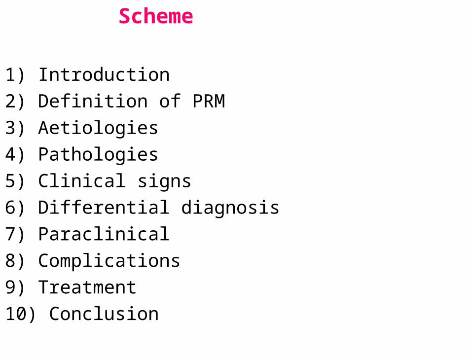
Scheme
1) Introduction 2) Definition of PRM 3) Aetiologies 4) Pathologies 5) Clinical signs6) Differential diagnosis7) Paraclinical8) Complications9) Treatment10) Conclusion
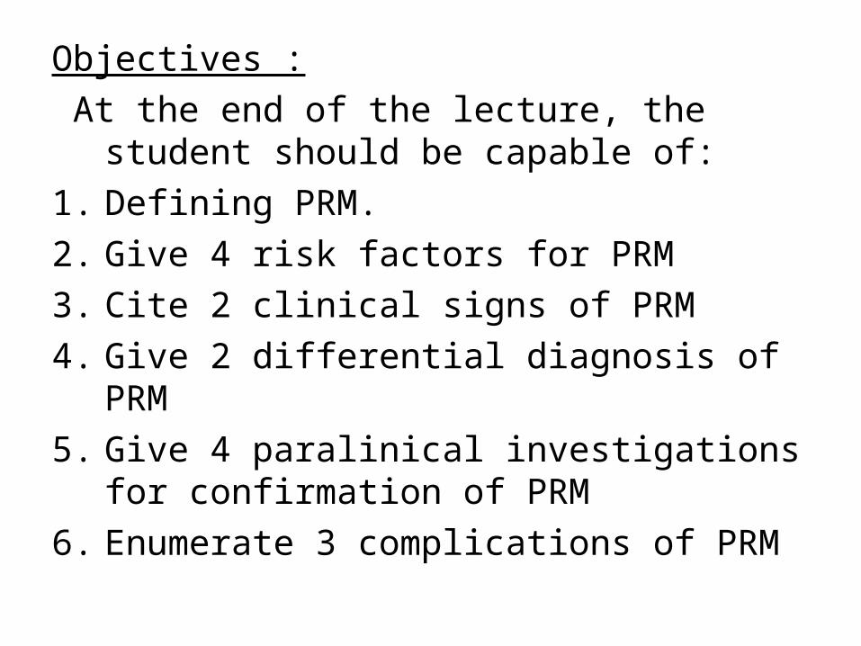
Objectives : At the end of the lecture, the student should be
capable of:1. Defining PRM.2. Give 4 risk factors for PRM3. Cite 2 clinical signs of PRM4. Give 2 differential diagnosis of PRM5. Give 4 paralinical investigations for
confirmation of PRM6. Enumerate 3 complications of PRM
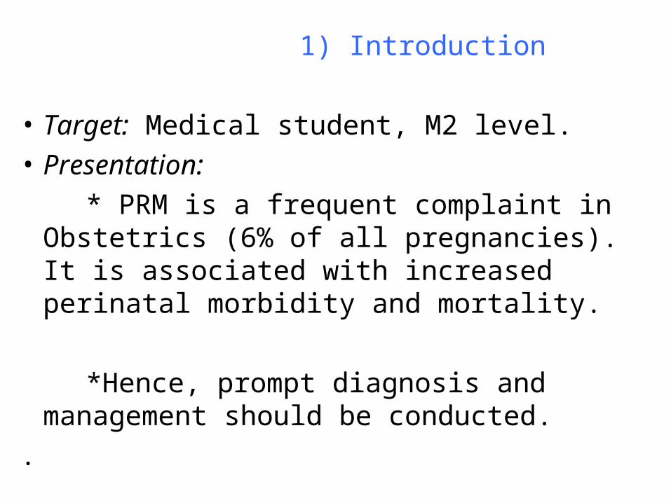
1) Introduction
• Target: Medical student, M2 level.• Presentation: * PRM is a frequent complaint in Obstetrics (6% of
all pregnancies). It is associated with increased perinatal morbidity and mortality.
*Hence, prompt diagnosis and management should be conducted.
.
2) Definition• Premature rupture of membranes (PRM) is
rupture of membranes (more than 1 hour) before unset of labour.
It can occur preterm (preterm rupture of membranes) or at term (prelabor rupture of membranes.
• If 24 h elapse between rupture of membranes and unset of labour, it is called prolonged rupture of membranes.
3) Aetiologies 3.1) Maternal causes:
• Cervicitis, vaginitis, STD• Incompetent cervix.
• Urinary tract infection• Malformed uterus (U. didelphys, U septus,…)
Maternal causes (continue)
• Undernutrition (abnormal collagen)• Grand multiparity.
• Past history of PRM (abnormal congenital collagen resistance).
3.2) Fœtal causes
• Polyhydramnios• Abnormal presentation (breech, transverse,…
• Multiple pregnancies• Chorioamnionitis.
• Placenta praevia.
3.3) Traumatic causes
4) Pathology
PRM is the most frequent cause of premature labour, cord prolapse, intra amniotic infection.
That is why there is a high perinatal mortality associated with PRM, especially when it occurs before term.
5) Clinical signs
• Vaginal flow of liquid (continuous or not).
• Speculum examination: look for fluid coming from the cervical canal.
• If there is any, do Valsalva maneuvre (increase of intra abdominal pressure)
6) Differential diagnosis• Urinary incontinence• Fissuration of membranes.
• Rupture of an amniochorial pouch• Vaginitis with increased vaginal secretion.
• Normal or premature labour (labor pain not really felt).
7) Paraclinical investigation
7.1) For diagnosis:
• Nitrazine test (brown to blue if PRM).
• Fern test (1 drop of liquid on a slide, let it dry at air, observe under microscope).
• Look for Lecithin/Sphingomyelin, phosphatidylglycerol in liquid collected from the pouch of Douglas.
Paraclinical (continue)
7.2) For treatment:
• Cervical swab (PCV)• FBC, blood parasites
• Urine culture• Ultrasound scan (amniotic fluid, gestational age,
fœtal weight)
• …
8) Complications:
• Cord prolapse• Abruptio placentae
• Chorioamnionitis (fever of 38 ° or more after PRM, leucocytosis > 16000, uterine tenderness, maternal and foetal tachycardia).
• Maternal complications are: endometritis, salpingitis, …
Complications (continue)
• Premature labour and delivery (neonatal mortality is increased: 4 times, Respiratory distress syndrom: 3 times, neonatal sepsis, intra ventricular bleeding.
• Prophylactic antibiotic seems to reduce these complications.
9) Treatment:
• The aims are to reduce the risk of infection (chorioamnionitis) and to accelerate lungs maturity if necessary before delivery.
• However, in case of chorioamnionitis, the fœtus must be delivered as soon as possible.
Treatment (continue)
• The 2 major risks are prematurity and fœtal infection.
• The treatment will depend on the gestational age and the fœtal weight.
• Before delivery, a neonatal unit and a neonatalogist should be contacted
9.1) Before 28 weeks:
• Use parenteral antibiotic for 2 days, then orally (ceftriaxone or erythromycin.
• NB: amoxicillin-clavulanic acid leads to an increased incidence of necrotizing enterocolitis).
• Before 25 weeks, there is risk of fetal lungs hypoplasia, neurological damage and limb compression deformities if PRM with oligoamnios
Treatment Before 28 weeks (continuation)
• Antibioprophylaxis reduces in newborns respiratory distress syndrome, necrotizing enterocolitis, and composite adverse outcomes.
• • The latency period is increased.
• Prolonged use of ATB increases the risk for selection of resistant bacteria.
9.2) Between 28 and 32 weeks: *ATB, *Corticotherapy (betamethasone: 12 mg IM to be
repeated after 12 to 24 hours or dexamethasone: 6 mg intramuscularly every 12 hours for four doses).
*NB: A single course of betamethasone is associated with a significantly reduced incidence of periventricular leukomalacia.
Treatment Between 28 and 32 weeks (continuation)
*No tocolysis or only for 24 to 48 h to allow lungs
maturation. Association of tocolysis and corticotherapy increases the risk of maternal pulmonary oedema .
Treatment (continuation)
9.3) Between 32 and 34 weeks: ATB, Corticosteroids (debated), if L/S is more than 1.8, then induced labour or perform C/S
9.4) After 34 weeks or fœtal weight > 2000 g: ATB, prepare for delivery.
9.5) Prevention of chorioamnionitis:
• No digital vaginal examination (only 1 is necessary when we are ready to deliver the fœtus: Bishop score, Pelvis evaluation,…). It shortens latency period.
• Change pad every 4 hours (2 for others)• No sexual intercourse
9.6) Precocious diagnosis of chorioamnionitis:
• Temperature every 6 hours (2 for others).• Appreciate the colour & odor of amniotic fluid.
• FBC, CRP.• Look for tenderness of uterine fundus
9.7) For fetal well-being: • FHR every 2 hours (or 15 min if she is in
labour)
10) Conclusion:• When premature rupture of membranes is
suspected, it must be confirmed or ruled out.
• When it is diagnosed, the treatment must be correct.
• Don’t hesitate to have the point of view of a neonatalogist if you want to deliver a premature baby.
• For neonatal better care, a neonatal unit should be available.
• Références: * Current Obstetrics & Gynaecologic diagnosis & treatment 2007 * Williams Obstetrics 2007
* Dewhurst’s textbook of Obstetrics & Gynaecology for postgraduates
• Modalités de l’évaluation: QCM, QROC, questions Rédactionnelles
• Conseils: Consulter la bibliographie, être assidu(e) pendant les stages cliniques,
• Contact: Dr Nkwabong, service de gynécologie, CHU Yaoundé, Tel. 99663843/ 77450104

Thanks for your kind attention