cell-to-cell propagation of cell death induced by excess ... · (konstam, m. a. et al., 2011 jacc...
TRANSCRIPT

Cell-to-cell propagation of cell death
induced by excess iron in cardiomyocytes
Department of Anatomy, Biochemistry, and Physiology,
Center for Cardiovascular Research
John A. Burns School of Medicine,
University of Hawaii at Manoa, Honolulu, Hawaii
Andrew Ko, BS
Nicholas Kawasaki, BS, Motoi Kobayashi MD, PhD, and Takashi Matsui,
MD, PhD; Department of Anatomy, Biochemistry, and Physiology,
JABSOM, University of Hawaii at Manoa, Honolulu, Hawaii

Heart Disease Mortality
(Benjamin, E. J. et al., 2018 Circulation 137: e257)
Coronary Heart
Disease (44%)
Stroke (17%)
Heart Failure (9%)
High Blood
Pressure (9%)
Diseases of the Arteries
(3%)
Other (18%)

LV Remodeling
(Konstam, M. A. et al., 2011 JACC Cardiovasc Imaging 4(1): 98-108)
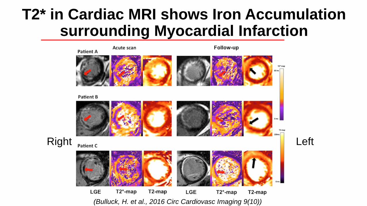
T2* in Cardiac MRI shows Iron Accumulation surrounding Myocardial Infarction
(Bulluck, H. et al., 2016 Circ Cardiovasc Imaging 9(10))
LeftRight

Stains of Hearts in Post-MI Patients

Cellular Iron Homeostasis and ROS Production
(Kobayashi, M. et al., 2018 Current Drug Targets)
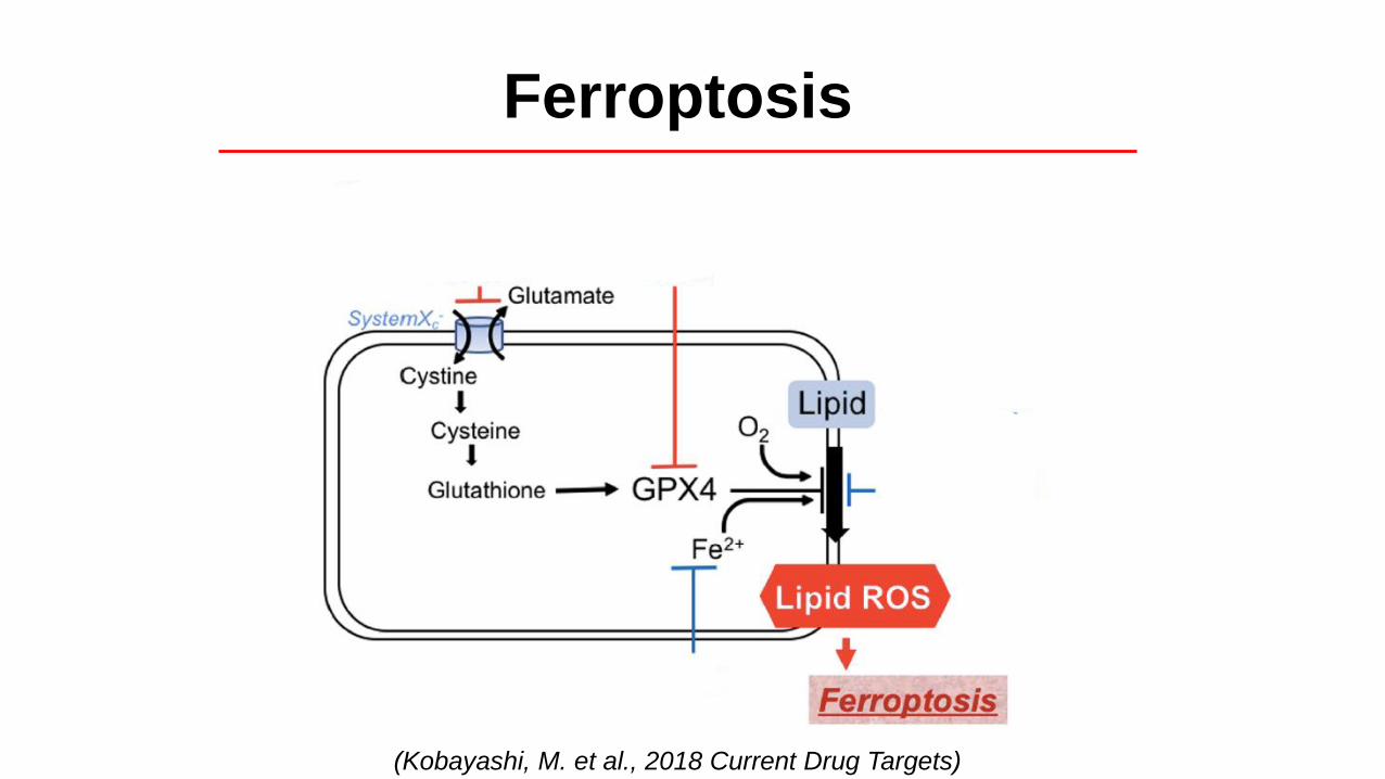
Ferroptosis
(Kobayashi, M. et al., 2018 Current Drug Targets)

Excess Iron induces Cell Death in Mouse Cardiomyocytes
(Baba Y. et al., 2018 Am J Physiol Heart Circ Physiol 314: H659-H668)
MT Iron Ferritin

Excess Iron-induced Cardiomyocyte Cell Death
(Baba Y. et al., 2018 Am J Physiol Heart Circ Physiol 314: H659-H668)

To demonstrate cell-to-cell propagation of cell death induced by excess iron in cardiomyocytes
Objective

• H9c2 cells cultured in 12-well plates containing DMEM with 10% FBS
• Negative control – DMEM
• Positive controls – DMEM with 50 ng/mL RSL-3 (ferroptosis inducer) or 20 uM H2O2 (nonspecific cell death inducer) for 3 hrs
• Experimental – DMEM with 5 mM, 10 mM, 20 mM, and 30 mM iron (III) citrate for 16 hrs
• Cell death was analyzed with Live/Dead Cell Viability Assay with fluorescent microscopy, 40X magnification
Methods

DMEM – Negative Control RSL-3 (Ferroptosis Inducer)
H2O2
(Nonspecific Inducer)

5 mM Fe (III) citrate
10 mM Fe (III) citrate
20 mM Fe (III) citrate
30 mM Fe (III) citrate
• Treatment with ferroptosis inducers resulted in contiguous or “clustered” cell death
• Treatment with excess iron induced cell death with a clustered pattern in a dose-dependent manner
Summary

• Dose-dependent clustering of cell death suggests that excess iron can produce cell-to-cell propagation of cell death
• Differences in patterns of cell death suggest specific mechanisms that contribute to cardiomyocyte cell death
• Further understanding into the differences in these mechanisms of cell death may provide insight into treatment that can prevent adverse LV remodeling
Conclusions

Dept. Anatomy, Biochemistry & Physiology
JABSOM
University of Hawai‘i at Mãnoa
Dr. Takashi MatsuiDr. Motoi KobayashiNicholas Kawasaki
Roxanne KoNorma Elizaga
Jonathan HuangAndrew PhamJoshua Freitas
Dr. Yuichi BabaHall Wu
Jonathan WooWilled Body Program
Matsui Lab
Grants: NIH grants
P30GM103341 and
P20GM113134 to T. Matsui
Acknowledgements