cell and molecular biology behrouz mahmoudi cell signaling 1
TRANSCRIPT
Cell and Molecular Biology
Behrouz Mahmoudi
Cell Signaling
1
1. Introduction
A. Definitions
B. Components involved in signaling
C. Types of signaling
2. Types of Signaling Ligands - cell-surface vs. intracellular
3. Three Major Classes of Signaling Receptors
Ion Channel-linked
G protein-coupled receptors (GPRs)
Enzyme-linked receptors
Tyrosine-Kinase Receptors
Overview
Mechanism of activation
Different ways that TKRs can be activated
TKs that are non-covalently linked with receptors
4. Second Messengers: cAMP, cGMP, IP3 and DAG, Ca+2, PIP3
5. Signaling Cascades
A. Ras GTPase
B. Adaptor proteins with SH2 and SH3 domains
C. MAP kinase pathway
D. 5 different kinases activated by different cascades
E. JAK-STAT pathway
6. Pathogen examples
A. Is signaling via entry receptors important for HIV-1 replication?
B. Signaling is involved in attachment of Plasmodium-infected RBCs to vascular endothelium 2

1. Introduction
A. Definitions
Signaling: Cell-cell communication via signals.
Signal transduction: Process of converting extracellular signals into intra-
cellular responses.
Ligand: The signaling molecule.
Receptors: Bind specific ligands. Transmit signals to intracellular targets. Different receptors can respond differently to the same ligand.
B. Components involved in signaling:
Ligands
Receptors
Intracellular Signaling Proteins
Intermediary Proteins
Enzymes
Second Messengers
Target Proteins
Inactivating Proteins 3
Overview of Signal Transduction
1. Introduction
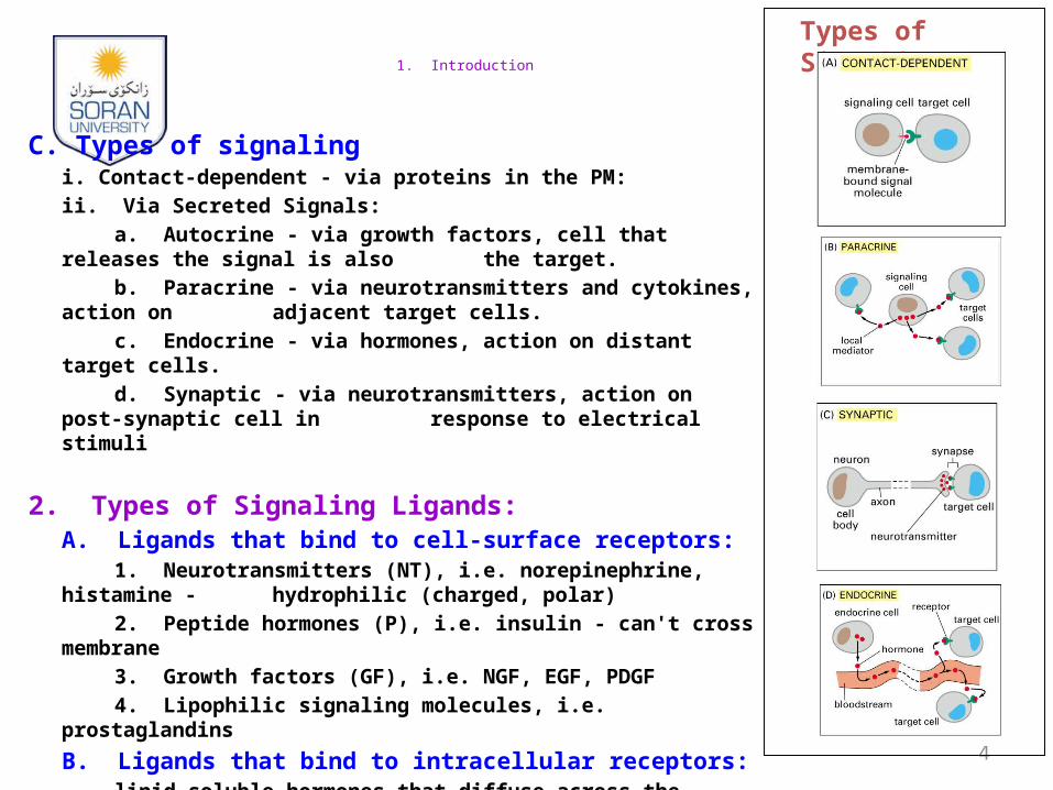
C. Types of signalingi. Contact-dependent - via proteins in the PM:
ii. Via Secreted Signals:
a. Autocrine - via growth factors, cell that releases the signal is also the target.
b. Paracrine - via neurotransmitters and cytokines, action on adjacent target cells.
c. Endocrine - via hormones, action on distant target cells.
d. Synaptic - via neurotransmitters, action on post-synaptic cell in response to electrical stimuli
2. Types of Signaling Ligands:A. Ligands that bind to cell-surface receptors:
1. Neurotransmitters (NT), i.e. norepinephrine, histamine - hydrophilic (charged, polar)
2. Peptide hormones (P), i.e. insulin - can't cross membrane
3. Growth factors (GF), i.e. NGF, EGF, PDGF
4. Lipophilic signaling molecules, i.e. prostaglandins
B. Ligands that bind to intracellular receptors: lipid soluble hormones that diffuse across the plasma
membrane and interact with receptors in the cytosol or nucleus. i.e. steroids, thyroxine, retinoic acid, nitric oxide.
4
Types of Signaling

3. Three major classes of surface receptors for signaling :
5
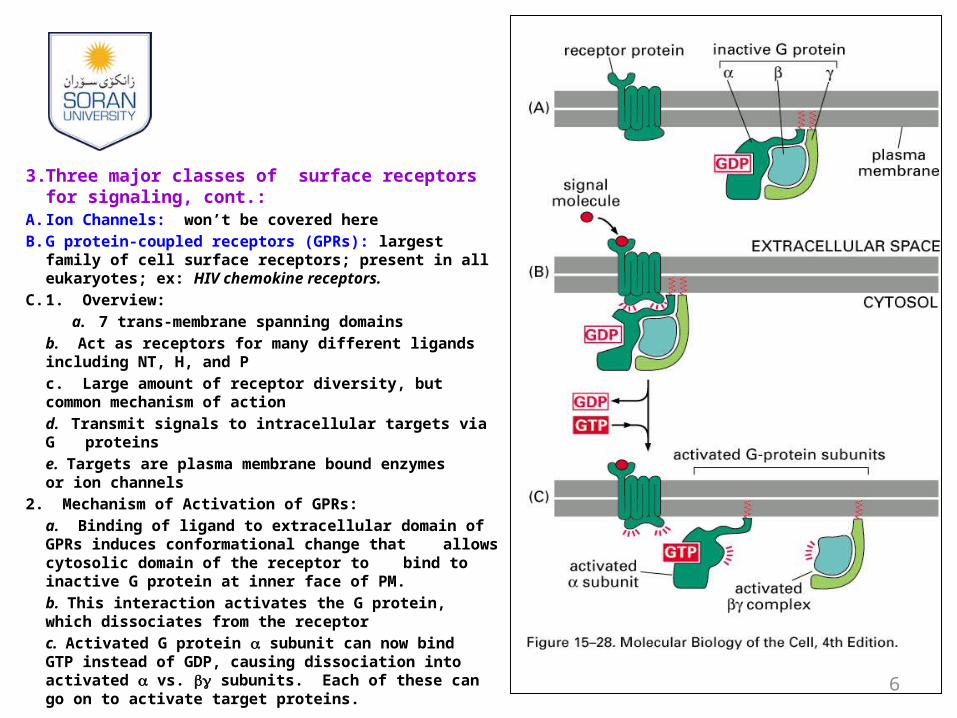
3. Three major classes of surface receptors for signaling, cont.:
A. Ion Channels: won’t be covered here
B. G protein-coupled receptors (GPRs): largest family of cell surface receptors; present in all eukaryotes; ex: HIV chemokine receptors.
C. 1. Overview:
a. 7 trans-membrane spanning domains
b. Act as receptors for many different ligands including NT, H, and P
c. Large amount of receptor diversity, but common mechanism of action
d. Transmit signals to intracellular targets via G proteins
e. Targets are plasma membrane bound enzymes or ion channels
2. Mechanism of Activation of GPRs:
a. Binding of ligand to extracellular domain of GPRs induces conformational change that allows cytosolic domain of the receptor to bind to inactive G protein at inner face of PM.
b. This interaction activates the G protein, which dissociates from the receptor
c. Activated G protein subunit can now bind GTP instead of GDP, causing dissociation into activated vs. subunits. Each of these can go on to activate target proteins.
6

Monomeric GTPase GAP binds to the GTP-bound GTPase and increases the rate of GTP hydrolysis
GEF binds to the GTPase and alters its conformation so it releases GDP
7
3. Three major classes of surface receptors for signaling, cont.:
C. Enzyme-linked receptors:1. Tyrosine kinase-linked receptors (TKRs).
A. Overview of TKRs:
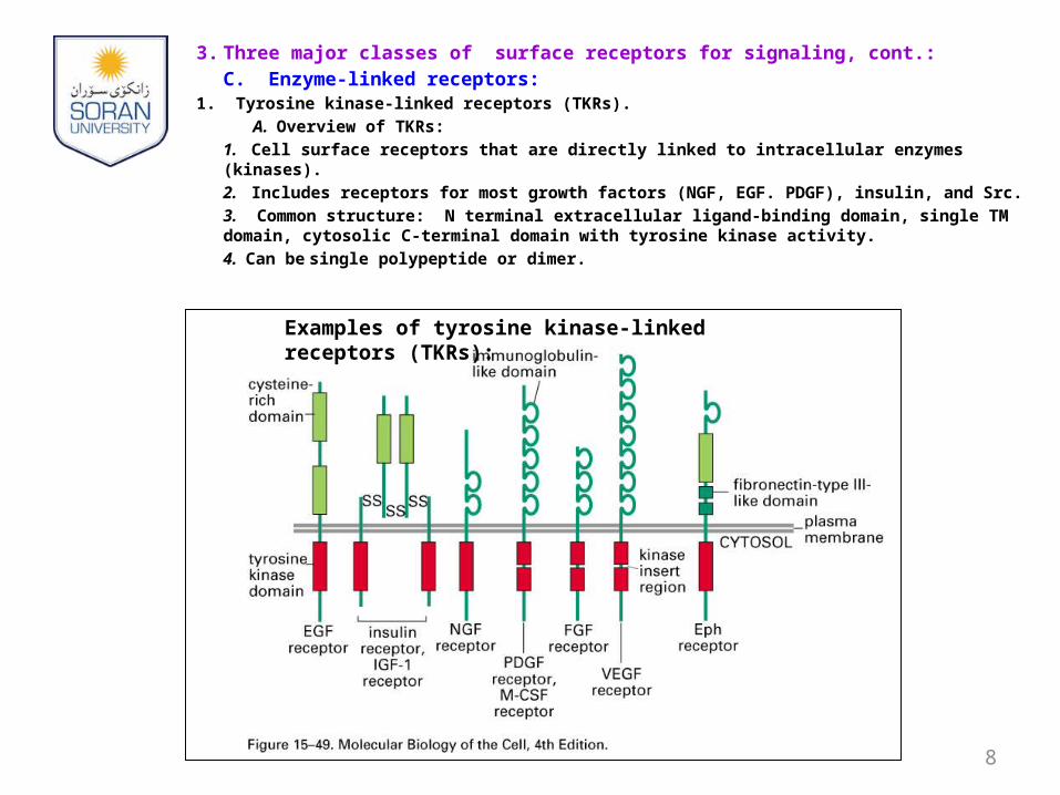
1. Cell surface receptors that are directly linked to intracellular enzymes (kinases).
2. Includes receptors for most growth factors (NGF, EGF. PDGF), insulin, and Src.
3. Common structure: N terminal extracellular ligand-binding domain, single TM domain, cytosolic C-terminal domain with tyrosine kinase activity.
4. Can be single polypeptide or dimer.
8
Examples of tyrosine kinase-linked receptors (TKRs):
3. Three major classes of surface receptors for signaling, cont.:
C. Enzyme-linked receptors, cont.:1. Tyrosine kinase-linked receptors (TKRs)
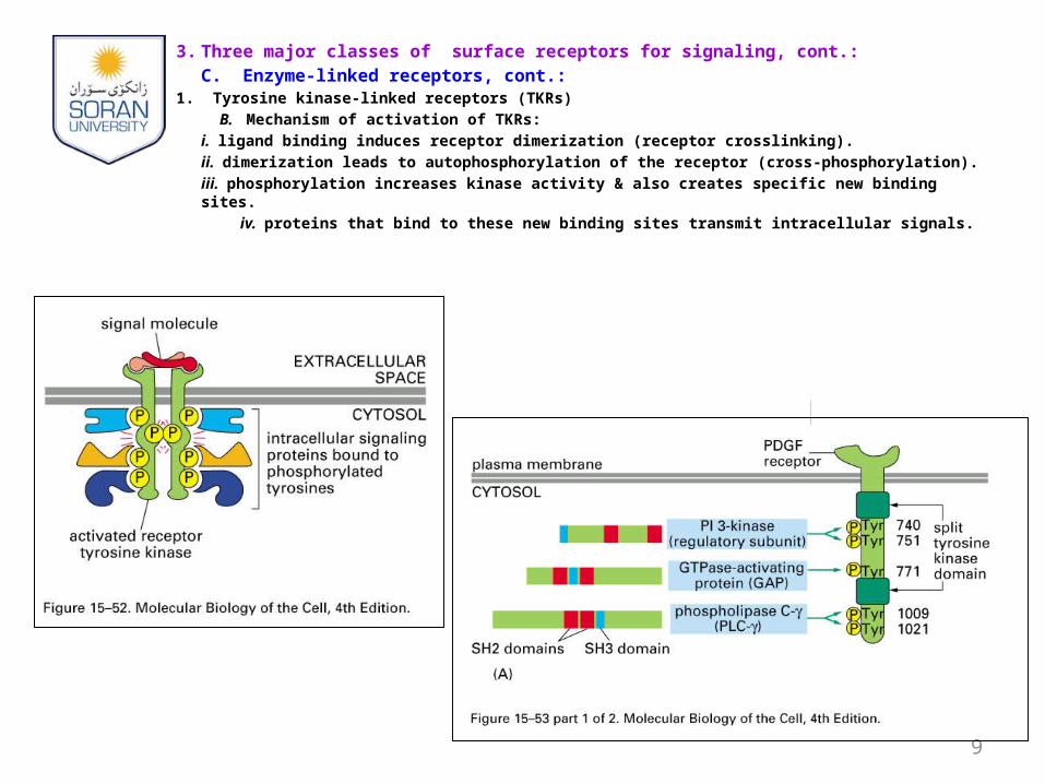
B. Mechanism of activation of TKRs:
i. ligand binding induces receptor dimerization (receptor crosslinking).
ii. dimerization leads to autophosphorylation of the receptor (cross-phosphorylation).
iii. phosphorylation increases kinase activity & also creates specific new binding sites.
iv. proteins that bind to these new binding sites transmit intracellular signals.
9
10
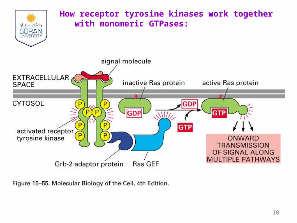
How receptor tyrosine kinases work together with monomeric GTPases:
3. Three major classes of surface receptors for signaling, cont.:
C. Enzyme-linked receptors, cont.:
1. Tyrosine kinase-linked receptors (TKRs)
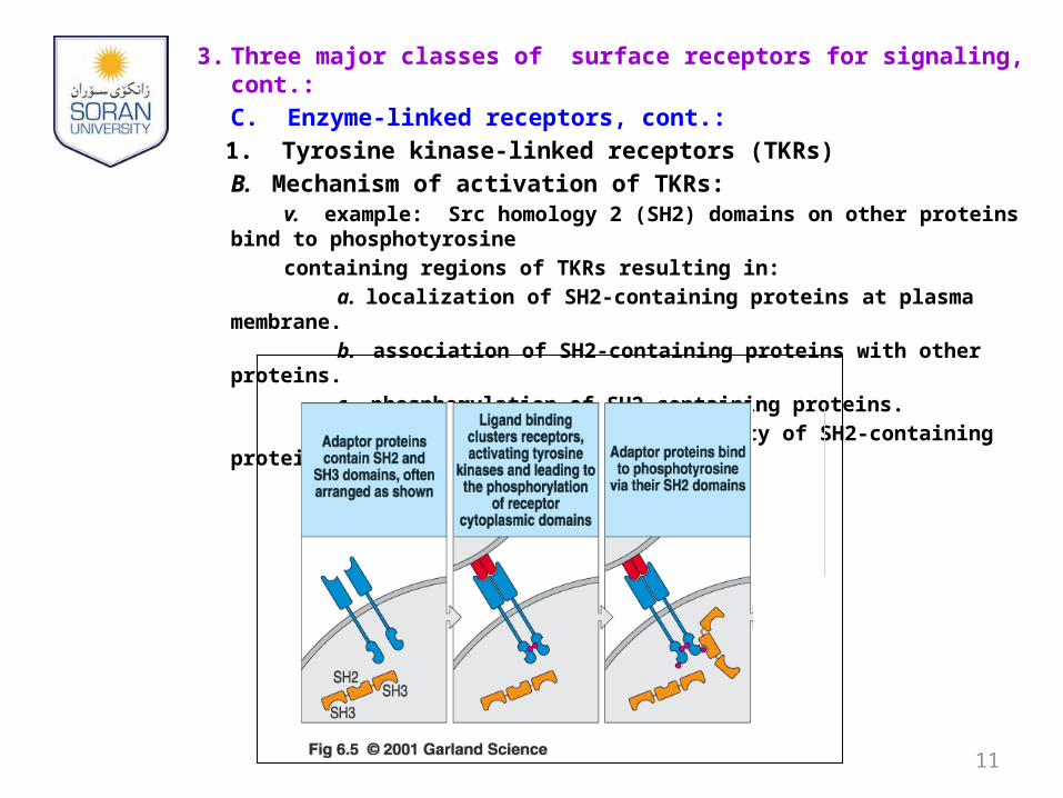
B. Mechanism of activation of TKRs:v. example: Src homology 2 (SH2) domains on other proteins bind to
phosphotyrosine
containing regions of TKRs resulting in:
a. localization of SH2-containing proteins at plasma membrane.
b. association of SH2-containing proteins with other proteins.
c. phosphorylation of SH2-containing proteins.
d. activation of enzymatic activity of SH2-containing proteins.
11
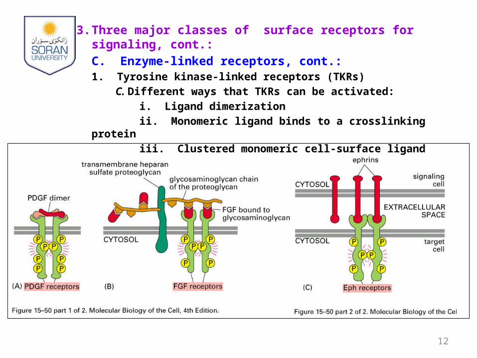
3. Three major classes of surface receptors for signaling, cont.:
C. Enzyme-linked receptors, cont.:1. Tyrosine kinase-linked receptors (TKRs)
C. Different ways that TKRs can be activated:
i. Ligand dimerization
ii. Monomeric ligand binds to a crosslinking protein
iii. Clustered monomeric cell-surface ligand
12
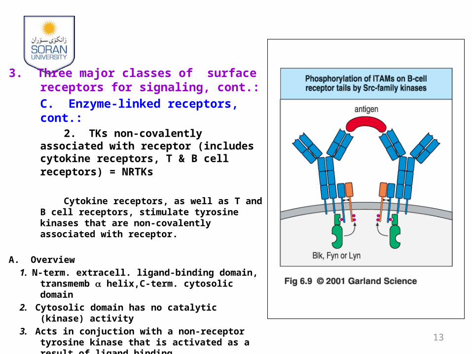
3. Three major classes of surface receptors for signaling, cont.:
C. Enzyme-linked receptors, cont.:2. TKs non-covalently associated
with receptor (includes cytokine receptors, T & B cell receptors) = NRTKs
Cytokine receptors, as well as T and B cell receptors, stimulate tyrosine kinases that are non-covalently associated with receptor.
A. Overview
1. N-term. extracell. ligand-binding domain, transmemb a helix,C-term. cytosolic domain
2. Cytosolic domain has no catalytic (kinase) activity
3. Acts in conjuction with a non-receptor tyrosine kinase that is activated as a result of ligand binding.
4. Activation is similar to that of RTKs: ligand binding causes cross phosphorylation of associated tyrosine kinases that phosphorylate the receptor, providing phosphotyrosine binding sites for recruitment of proteins with SH2 domains.
13
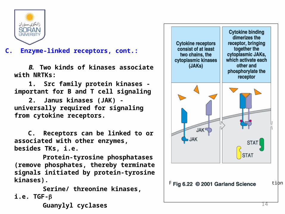
C. Enzyme-linked receptors, cont.:
B. Two kinds of kinases associate with NRTKs:
1. Src family protein kinases - important for B and T cell signaling
2. Janus kinases (JAK) - universally required for signaling from cytokine receptors.
C. Receptors can be linked to or associated with other enzymes, besides TKs, i.e.
Protein-tyrosine phosphatases (remove phosphates, thereby terminate signals initiated by protein-tyrosine kinases).
Serine/ threonine kinases, i.e. TGF-b
Guanylyl cyclases
14
From Janeway, Immunobiology, 5th edition
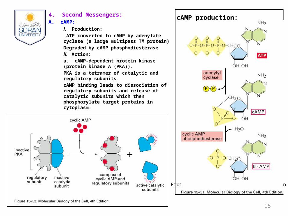
4. Second Messengers:A. cAMP:
i. Production:
ATP converted to cAMP by adenylate cyclase (a large multipass TM protein)
Degraded by cAMP phosphodiesterase
ii. Action:
a. cAMP-dependent protein kinase (protein kinase A (PKA)).
PKA is a tetramer of catalytic and regulatory subunits
cAMP binding leads to dissociation of regulatory subunits and release of catalytic subunits which then phosphorylate target proteins in cytoplasm:
15
From Janeway, Immunobiology, 5th edition
cAMP production:
4. Second Messengers, cont.:
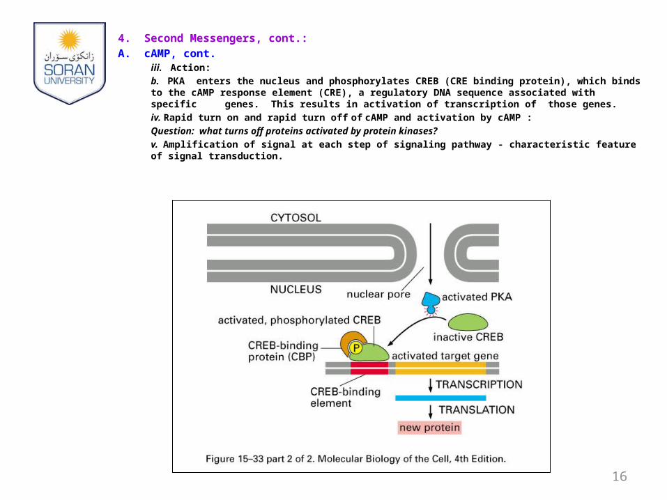
A. cAMP, cont. iii. Action:
b. PKA enters the nucleus and phosphorylates CREB (CRE binding protein), which binds to the cAMP response element (CRE), a regulatory DNA sequence associated with specific
genes. This results in activation of transcription of those genes.
iv. Rapid turn on and rapid turn off of cAMP and activation by cAMP :
Question: what turns off proteins activated by protein kinases?
v. Amplification of signal at each step of signaling pathway - characteristic feature of signal transduction.
16
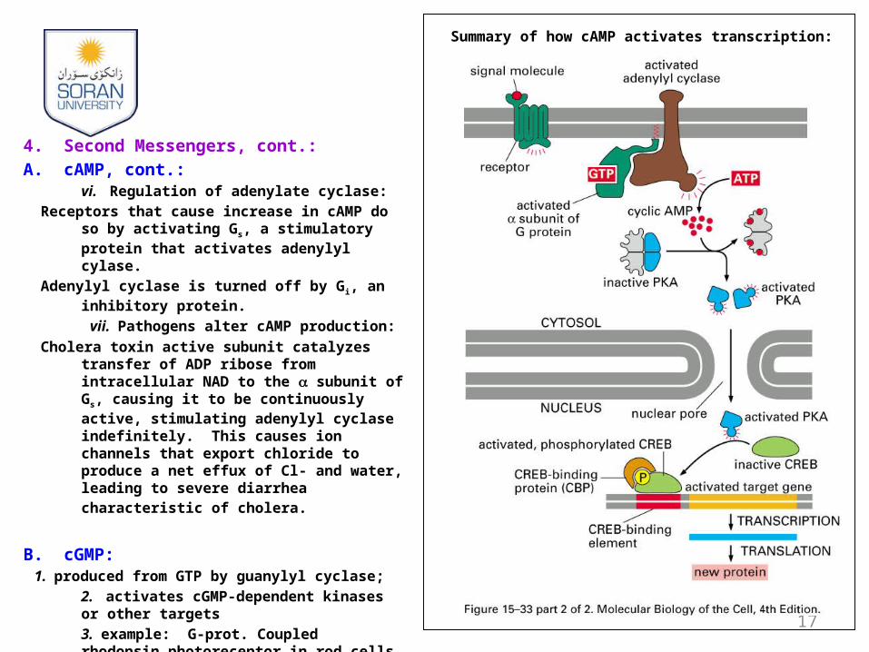
4. Second Messengers, cont.:
A. cAMP, cont.: vi. Regulation of adenylate cyclase:
Receptors that cause increase in cAMP do so by activating Gs, a stimulatory protein that activates adenylyl cylase.
Adenylyl cyclase is turned off by Gi, an inhibitory protein.
vii. Pathogens alter cAMP production:
Cholera toxin active subunit catalyzes transfer of ADP ribose from intracellular NAD to the subunit of Gs, causing it to be continuously active, stimulating adenylyl cyclase indefinitely. This causes ion channels that export chloride to produce a net effux of Cl- and water, leading to severe diarrhea characteristic of cholera.
B. cGMP:1. produced from GTP by guanylyl cyclase;
2. activates cGMP-dependent kinases or other targets
3. example: G-prot. Coupled rhodopsin photoreceptor in rod cells of retina
17
Summary of how cAMP activates transcription:
4. Second Messengers, cont.:
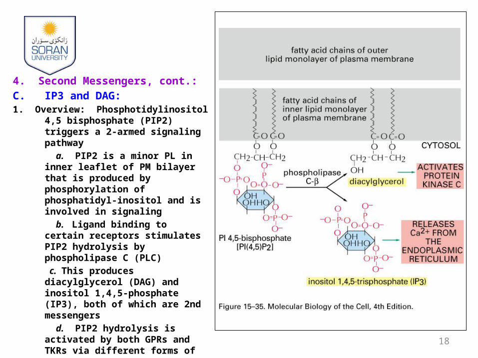
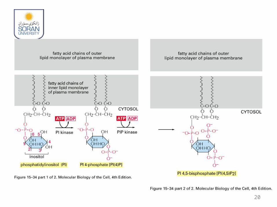
C. IP3 and DAG:1. Overview: Phosphotidylinositol 4,5
bisphosphate (PIP2) triggers a 2-armed signaling pathway
a. PIP2 is a minor PL in inner leaflet of PM bilayer that is produced by phosphorylation of phosphatidyl-inositol and is involved in signaling
b. Ligand binding to certain receptors stimulates PIP2 hydrolysis by phospholipase C (PLC)
c. This produces diacylglycerol (DAG) and inositol 1,4,5-phosphate (IP3), both of which are 2nd messengers
d. PIP2 hydrolysis is activated by both GPRs and TKRs via different forms of PLC
e. PLC-b is stimulated by Gq
proteins while PLC-g has SH2 domains that allow binding to activated tyrosine kinases 18
19
20
4. Second Messengers, cont.:
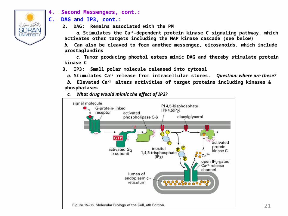
C. DAG and IP3, cont.: 2. DAG: Remains associated with the PM
a. Stimulates the Ca+2-dependent protein kinase C signaling pathway, which activates other targets including the MAP kinase cascade (see below)
b. Can also be cleaved to form another messenger, eicosanoids, which include prostaglandins
c. Tumor producing phorbol esters mimic DAG and thereby stimulate protein kinase C
3. IP3: Small polar molecule released into cytosol
a. Stimulates Ca+2 release from intracellular stores. Question: where are these?
b. Elevated Ca+2 alters activities of target proteins including kinases & phosphatases
c. What drug would mimic the effect of IP3?
21
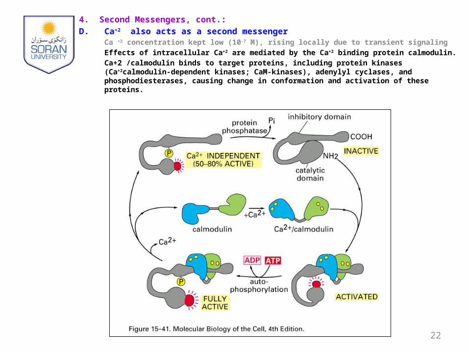
4. Second Messengers, cont.:
D. Ca+2 also acts as a second messengerCa +2 concentration kept low (10-7 M), rising locally due to transient signaling
Effects of intracellular Ca+2 are mediated by the Ca+2 binding protein calmodulin.
Ca+2 /calmodulin binds to target proteins, including protein kinases (Ca+2calmodulin-dependent kinases; CaM-kinases), adenylyl cyclases, and phosphodiesterases, causing change in conformation and activation of these proteins.
22
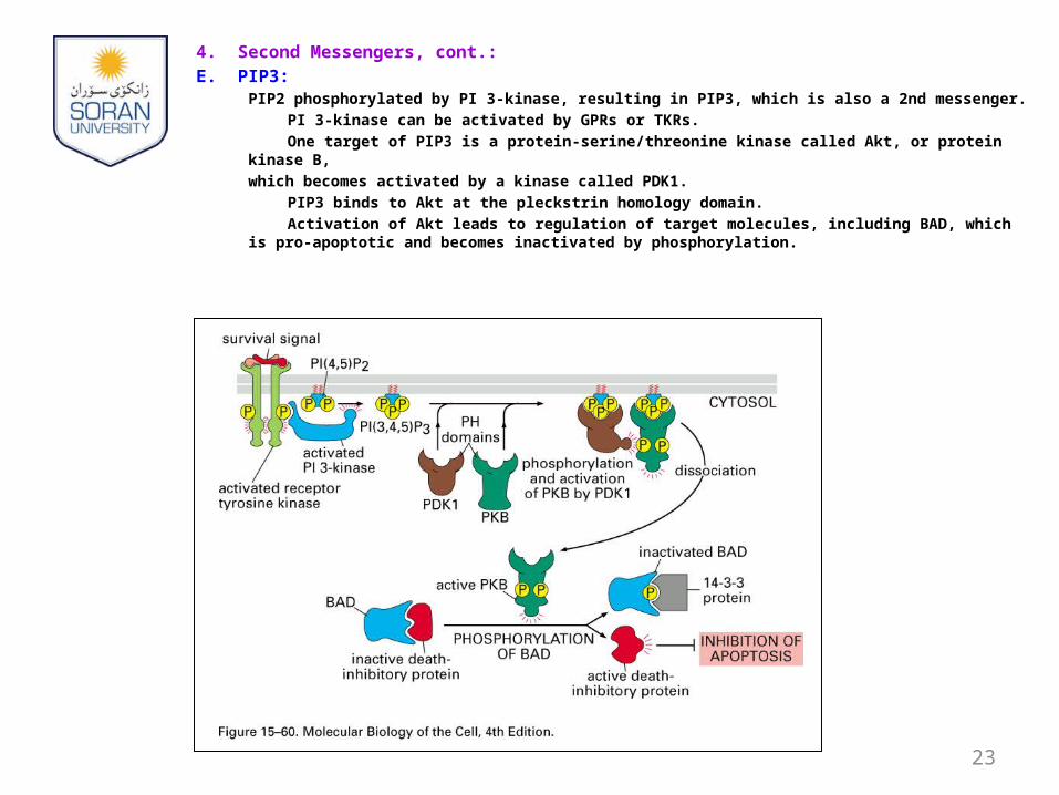
4. Second Messengers, cont.:
E. PIP3: PIP2 phosphorylated by PI 3-kinase, resulting in PIP3, which is also a 2nd messenger.
PI 3-kinase can be activated by GPRs or TKRs.
One target of PIP3 is a protein-serine/threonine kinase called Akt, or protein kinase B,
which becomes activated by a kinase called PDK1.
PIP3 binds to Akt at the pleckstrin homology domain.
Activation of Akt leads to regulation of target molecules, including BAD, which is pro-apoptotic and becomes inactivated by phosphorylation.
23
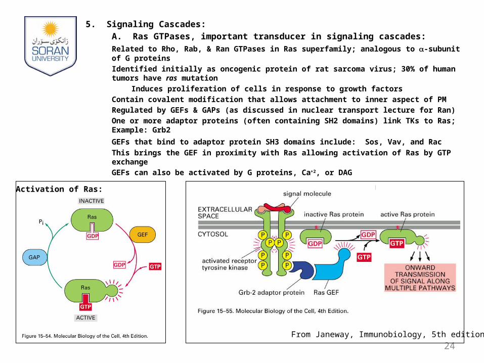
5. Signaling Cascades:A. Ras GTPases, important transducer in signaling cascades:Related to Rho, Rab, & Ran GTPases in Ras superfamily; analogous to -subunit of
G proteins
Identified initially as oncogenic protein of rat sarcoma virus; 30% of human tumors have ras mutation
Induces proliferation of cells in response to growth factors
Contain covalent modification that allows attachment to inner aspect of PM
Regulated by GEFs & GAPs (as discussed in nuclear transport lecture for Ran)
One or more adaptor proteins (often containing SH2 domains) link TKs to Ras; Example: Grb2
GEFs that bind to adaptor protein SH3 domains include: Sos, Vav, and Rac
This brings the GEF in proximity with Ras allowing activation of Ras by GTP exchange
GEFs can also be activated by G proteins, Ca+2, or DAG
24From Janeway, Immunobiology, 5th edition
Activation of Ras:
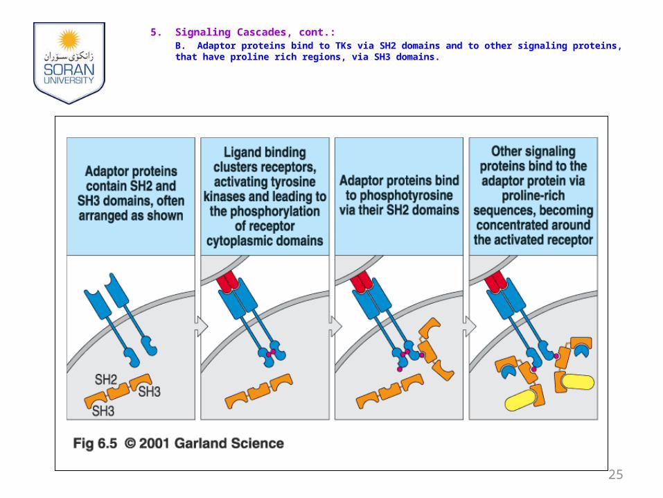
5. Signaling Cascades, cont.:B. Adaptor proteins bind to TKs via SH2 domains and to other signaling proteins, that have proline rich regions, via SH3 domains.
25
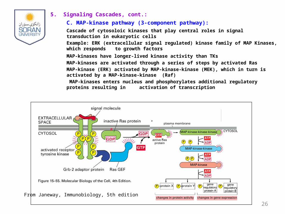
5. Signaling Cascades, cont.:
C. MAP-kinase pathway (3-component pathway):
Cascade of cytosoloic kinases that play central roles in signal transduction in eukaryotic cells
Example: ERK (extracellular signal regulated) kinase family of MAP Kinases, which responds to growth factors
MAP-kinases have longer-lived kinase activity than TKs
MAP-kinases are activated through a series of steps by activated Ras
MAP-kinase (ERK) activated by MAP-kinase-kinase (MEK), which in turn is activated by a MAP-kinase-kinase (Raf)
MAP-kinases enters nucleus and phosphorylates additional regulatory proteins resulting in activation of transcription
26
From Janeway, Immunobiology, 5th edition
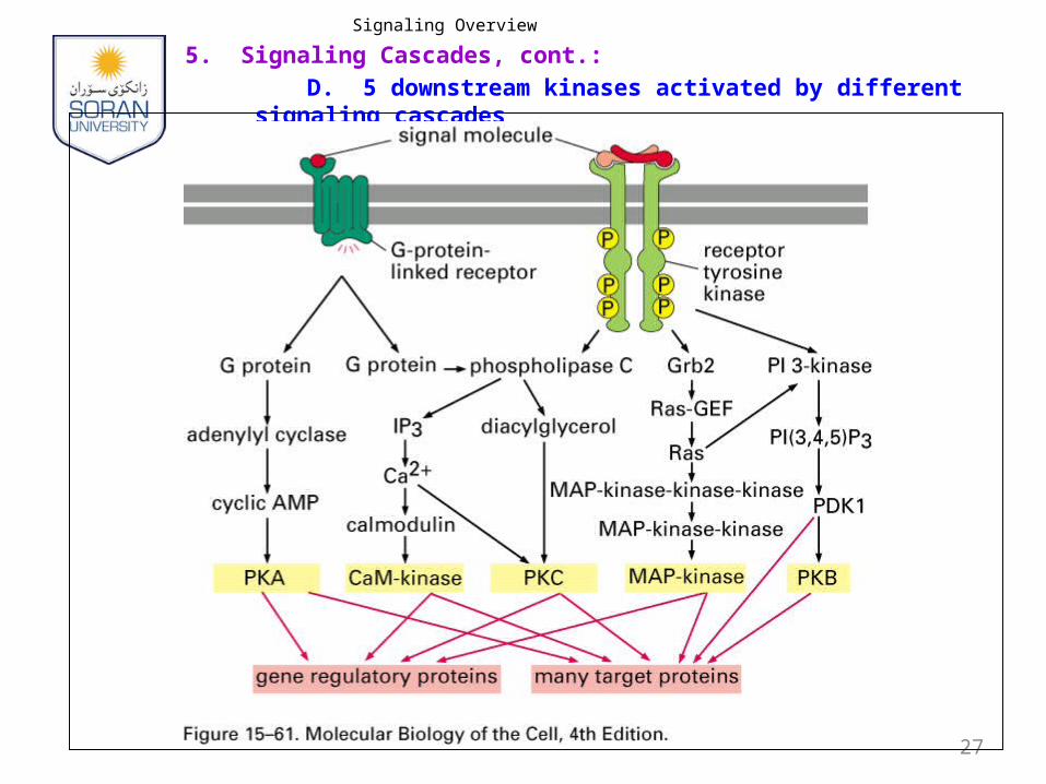
Signaling Overview
5. Signaling Cascades, cont.:
D. 5 downstream kinases activated by different signaling cascades
27
5. Signaling Cascades, cont.:
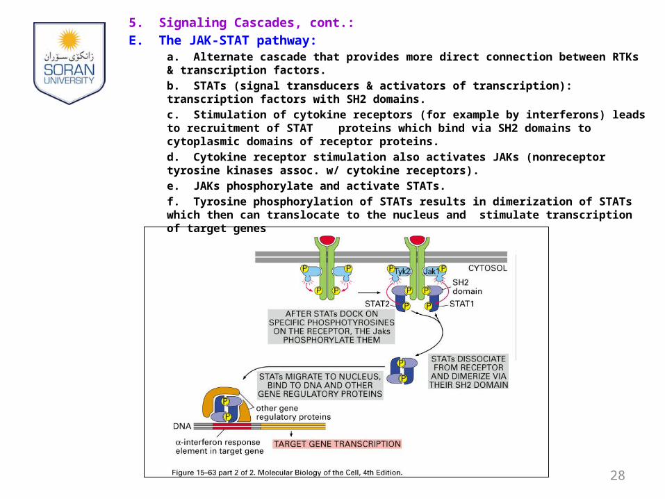
E. The JAK-STAT pathway:a. Alternate cascade that provides more direct connection between RTKs & transcription factors.
b. STATs (signal transducers & activators of transcription): transcription factors with SH2 domains.
c. Stimulation of cytokine receptors (for example by interferons) leads to recruitment of STAT proteins which bind via SH2 domains to cytoplasmic domains of receptor proteins.
d. Cytokine receptor stimulation also activates JAKs (nonreceptor tyrosine kinases assoc. w/ cytokine receptors).
e. JAKs phosphorylate and activate STATs.
f. Tyrosine phosphorylation of STATs results in dimerization of STATs which then can translocate to the nucleus and stimulate transcription of target genes
28
29
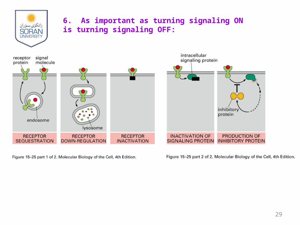
6. As important as turning signaling ON is turning signaling OFF:
7. Pathogen examples:
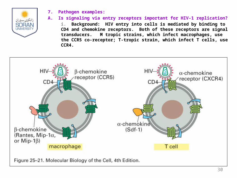
A. Is signaling via entry receptors important for HIV-1 replication?
i. Background: HIV entry into cells is mediated by binding to CD4 and chemokine receptors. Both of these receptors are signal transducers. M tropic strains, which infect macrophages, use the CCR5 co-receptor; T-tropic strain, which infect T cells, use CCR4.
30
7. Pathogen examples:
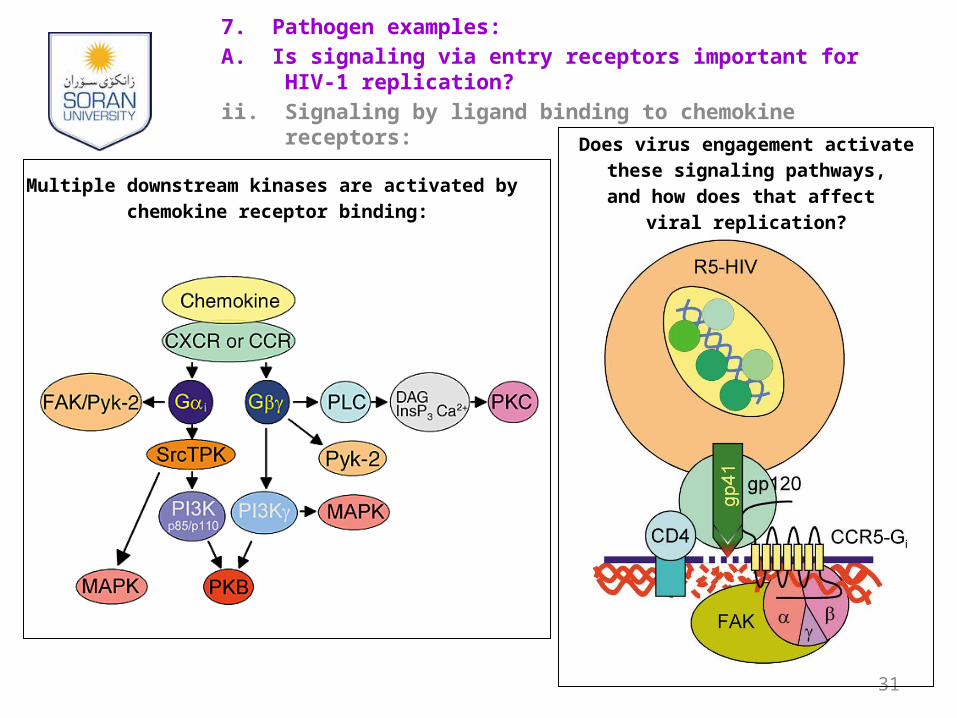
A. Is signaling via entry receptors important for HIV-1 replication?
ii. Signaling by ligand binding to chemokine receptors:
31
Multiple downstream kinases are activated by
chemokine receptor binding:
Does virus engagement activate
these signaling pathways,
and how does that affect
viral replication?
7. Pathogen examples:
A. Is signaling via entry receptors important for HIV-1 replication?
iii. Evidence that signaling is important for HIV-1 replication:1. Strains of HIV-1 that enter and replicate in macrophages
induce high levels of intracellular Ca+2 via the CCR5 receptor, while T-tropic strains that fuse with macrophages but fail to replicate in them fail to induce Ca+2 mobilization. Treatment with natural ligand chemokines, which engage the CCR5 receptor and induce signaling, overcomes this entry block (Arthos et al., J. Virol. 74: 6418, 2000 - Fauci lab).
2. Pertussis toxin, which perturbs co-receptor signaling via blockade of Ras activation, decreases infection of PBMCs with HIV-1 (Alfano et al. J. Exp. Med 190: 597, 2000).
3. PI3 kinase activity is required for infection of T cells and macrophages. PI3 kinase effectors (PKB and p70S6 kinase) are phosphorylated in response to HIV-1 infection or chemokine receptor stimulation. A PI3 kinase inhibitor inhibits infection of T cells and macrophages (Francois and Klotman, J. Virol. 77: 2539, 2003).
32
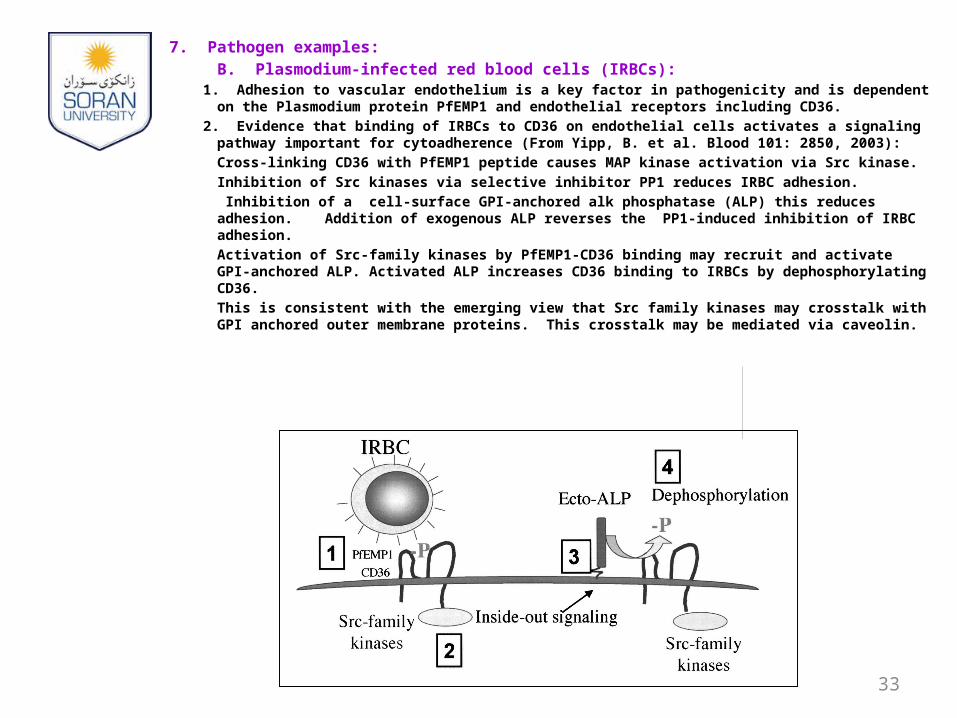
7. Pathogen examples:
B. Plasmodium-infected red blood cells (IRBCs): 1. Adhesion to vascular endothelium is a key factor in pathogenicity and is dependent on the
Plasmodium protein PfEMP1 and endothelial receptors including CD36.
2. Evidence that binding of IRBCs to CD36 on endothelial cells activates a signaling pathway important for cytoadherence (From Yipp, B. et al. Blood 101: 2850, 2003):
Cross-linking CD36 with PfEMP1 peptide causes MAP kinase activation via Src kinase.
Inhibition of Src kinases via selective inhibitor PP1 reduces IRBC adhesion.
Inhibition of a cell-surface GPI-anchored alk phosphatase (ALP) this reduces adhesion. Addition of exogenous ALP reverses the PP1-induced inhibition of IRBC adhesion.
Activation of Src-family kinases by PfEMP1-CD36 binding may recruit and activate GPI-anchored ALP. Activated ALP increases CD36 binding to IRBCs by dephosphorylating CD36.
This is consistent with the emerging view that Src family kinases may crosstalk with GPI anchored outer membrane proteins. This crosstalk may be mediated via caveolin.
33