www.soran.edu.iq physiology behrouz mahmoudi nerve system 1
TRANSCRIPT

www.soran.edu.iq 3
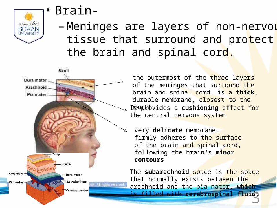
• Brain-– Meninges are layers of non-nervous tissue that
surround and protect the brain and spinal cord.
the outermost of the three layers of the meninges that surround the brain and spinal cord. is a thick, durable membrane, closest to the skull.
It provides a cushioning effect for the central nervous system
very delicate membrane. firmly adheres to the surface of the brain and spinal cord, following the brain's minor contours
The subarachnoid space is the space that normally exists between the arachnoid and the pia mater, which is filled with cerebrospinal fluid.
www.soran.edu.iq 4
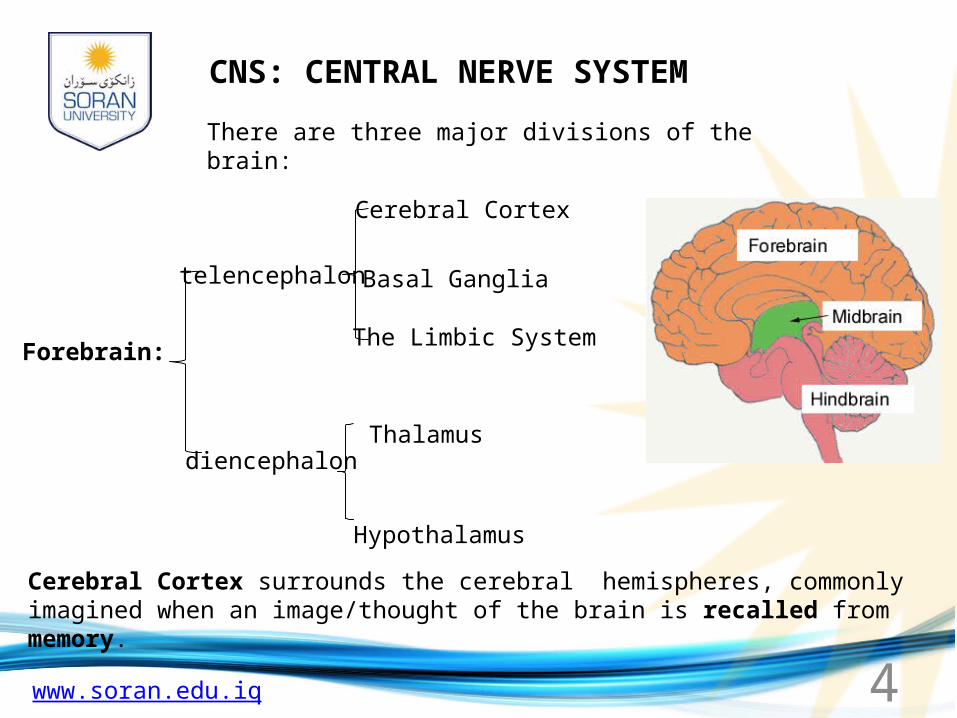
There are three major divisions of the brain:
Forebrain:
telencephalon
diencephalon
Cerebral Cortex
Basal Ganglia
The Limbic System
Thalamus
Hypothalamus
Cerebral Cortex surrounds the cerebral hemispheres, commonly imagined when an image/thought of the brain is recalled from memory.
CNS: CENTRAL NERVE SYSTEM
www.soran.edu.iq 5
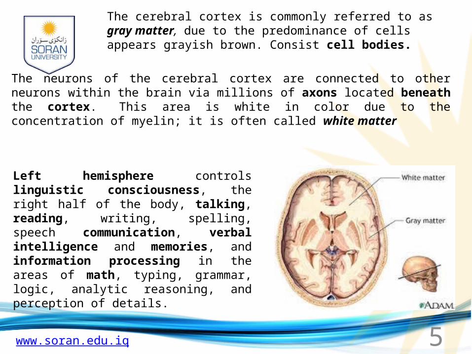
The cerebral cortex is commonly referred to as gray matter, due to the predominance of cells appears grayish brown. Consist cell bodies.
The neurons of the cerebral cortex are connected to other neurons within the brain via millions of axons located beneath the cortex. This area is white in color due to the concentration of myelin; it is often called white matter
Left hemisphere controls linguistic consciousness, the right half of the body, talking, reading, writing, spelling, speech communication, verbal intelligence and memories, and information processing in the areas of math, typing, grammar, logic, analytic reasoning, and perception of details.
www.soran.edu.iq 6
The right hemisphere is associated with 'unconscious' awareness (in the sense it is not linguistically based), perception of faces and patterns, comprehension of body language and social cues, creativity and insight, intuitive reasoning, visual-spatial processing, and holistic comprehension.
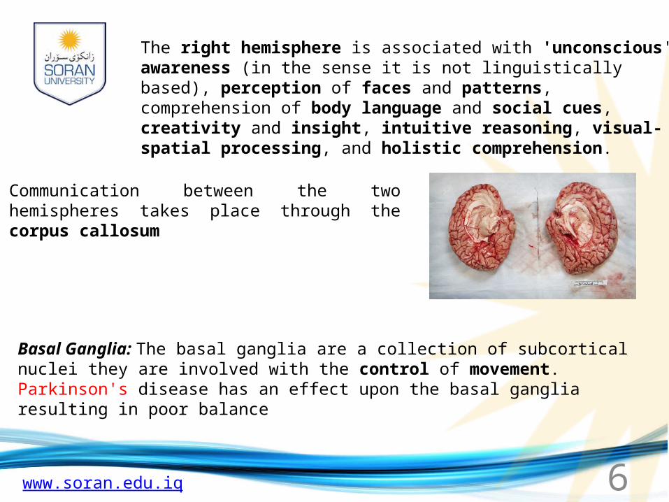
Communication between the two hemispheres takes place through the corpus callosum
Basal Ganglia: The basal ganglia are a collection of subcortical nuclei they are involved with the control of movement. Parkinson's disease has an effect upon the basal ganglia resulting in poor balance
www.soran.edu.iq 7
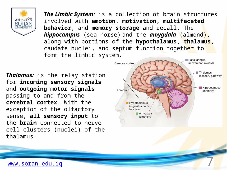
The Limbic System: is a collection of brain structures involved with emotion, motivation, multifaceted behavior, and memory storage and recall. The hippocampus (sea horse) and the amygdala (almond), along with portions of the hypothalamus, thalamus, caudate nuclei, and septum function together to form the limbic system.
Thalamus: is the relay station for incoming sensory signals and outgoing motor signals passing to and from the cerebral cortex. With the exception of the olfactory sense, all sensory input to the brain connected to nerve cell clusters (nuclei) of the thalamus.
www.soran.edu.iq 8
Hypothalamus: is comprised of distinct areas and nuclei which control vital survival behaviors and activities such as: eating, drinking, temperature regulation, sleep, emotional behavior, and sexual activity.
The autonomic nervous system and endocrine system are controlled by the hypothalamus.
Neurosecretory cells released by the hypothalamus act upon the anterior pituitary gland which then secretes its hormones.
Midbrain, The Mesencephalon:
Two primary parts comprise the midbrain: the tectum and the tegmentum.
www.soran.edu.iq 9
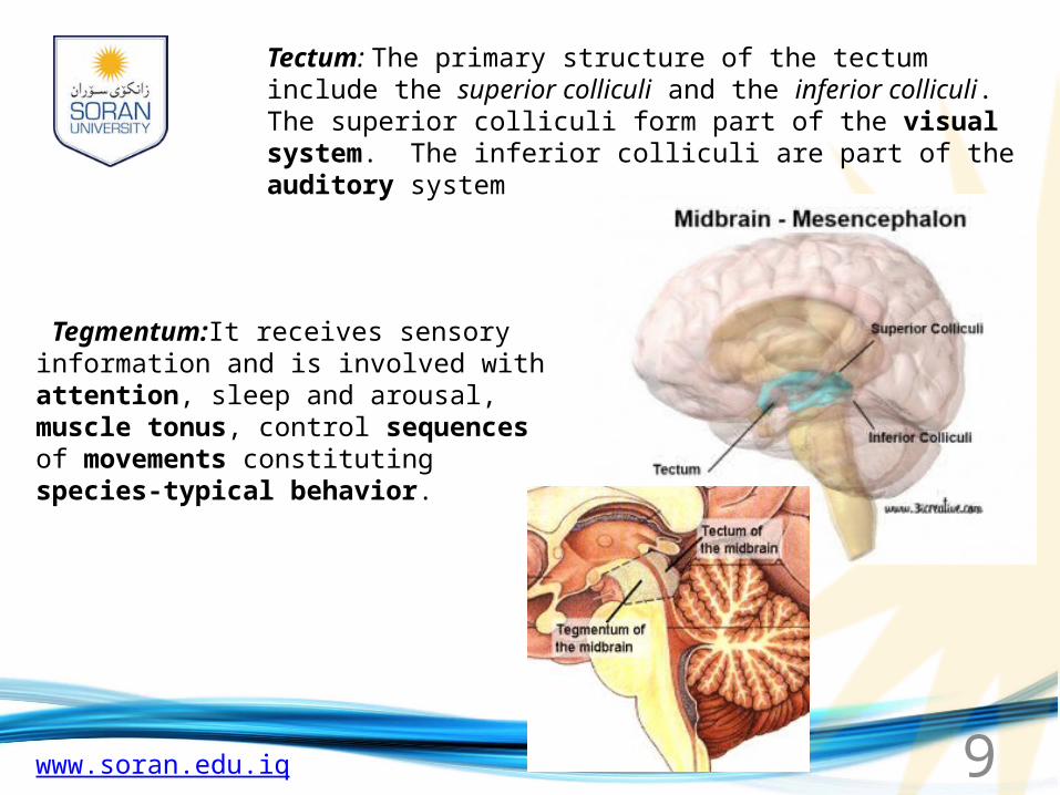
Tectum: The primary structure of the tectum include the superior colliculi and the inferior colliculi. The superior colliculi form part of the visual system. The inferior colliculi are part of the auditory system
Tegmentum:It receives sensory information and is involved with attention, sleep and arousal, muscle tonus, control sequences of movements constituting species-typical behavior.
www.soran.edu.iq 10
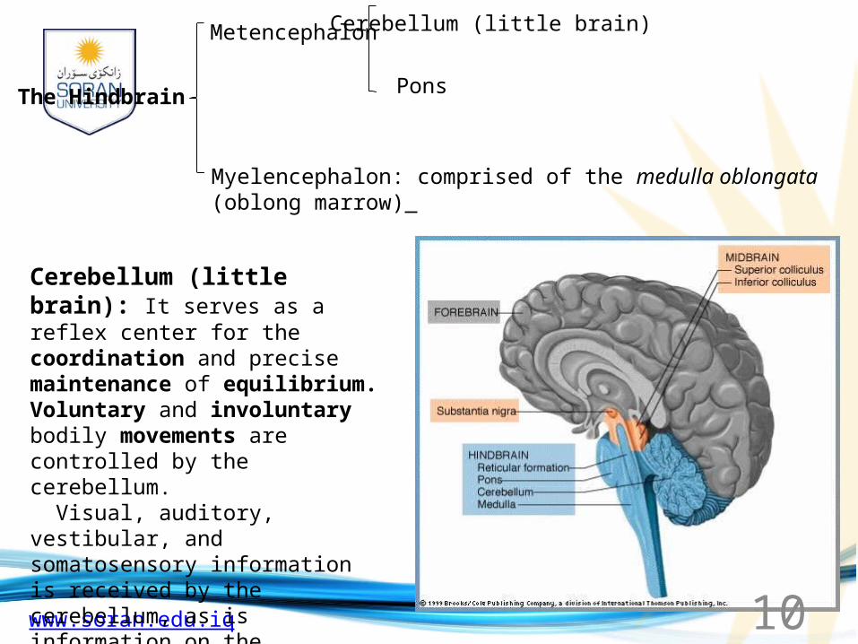
The Hindbrain
Metencephalon
Myelencephalon: comprised of the medulla oblongata (oblong marrow)
Cerebellum (little brain)
Pons
Cerebellum (little brain): It serves as a reflex center for the coordination and precise maintenance of equilibrium. Voluntary and involuntary bodily movements are controlled by the cerebellum. Visual, auditory, vestibular, and somatosensory information is received by the cerebellum, as is information on the movements of individual muscles.

www.soran.edu.iq 11
Pons: The pons contain a portion of the reticular formation as well as nuclei believed important in the role of sleep and arousal
Medulla oblongata:is the control center for cardiac, vasoconstrictor, and respiratory functions. Reflex activities, including vomiting, are controlled by this structure of the hindbrain. Appearing as a pyramid-shaped enlargement of the spinal cord, damage to this area typically results in immediate death.

www.soran.edu.iq 12
The spinal cord: is the main pathway for information connecting the brain and peripheral nervous system.
The human spinal cord is protected by the bony spinal column called vertebrae.
The spinal cord is located in the vertebral foramen and is made up of 31 segments: 8 cervical, 12 thoracic, 5 lumbar, 5 sacral and 1 coccygeal. A pair of spinal nerves leaves each segment of the spinal cord
www.soran.edu.iq 13
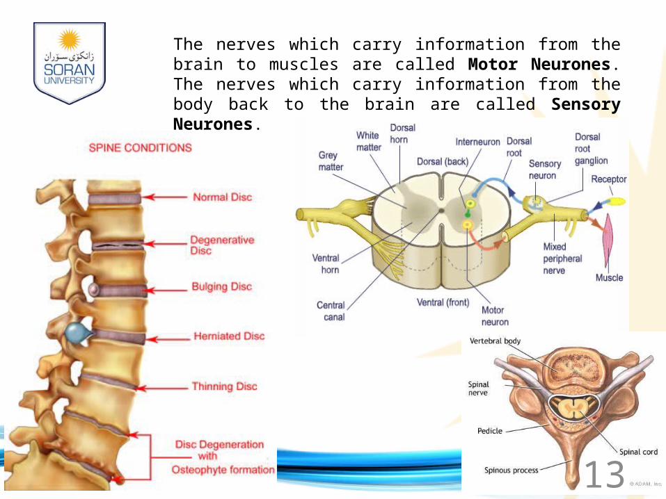
The nerves which carry information from the brain to muscles are called Motor Neurones. The nerves which carry information from the body back to the brain are called Sensory Neurones.

www.soran.edu.iq 14
The spinal cord is surrounded by a clear fluid called Cerebral Spinal Fluid (CSF), that acts as a cushion to protect the delicate nerve tissues against damage from banging against the inside of the vertebrae
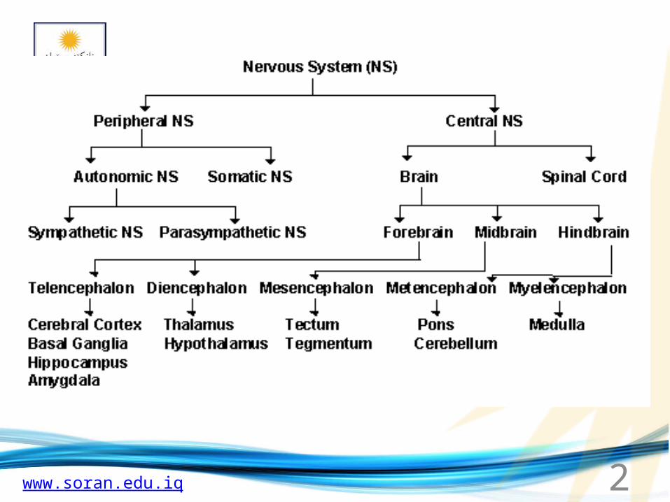
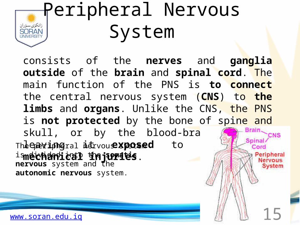
The brain and spinal cord are referred to as the Central Nervous System, whilst the nerves connecting the spinal cord to the body are referred to as the Peripheral Nervous System.
www.soran.edu.iq 15
Peripheral Nervous System
consists of the nerves and ganglia outside of the brain and spinal cord. The main function of the PNS is to connect the central nervous system (CNS) to the limbs and organs. Unlike the CNS, the PNS is not protected by the bone of spine and skull, or by the blood-brain barrier, leaving it exposed to toxins and mechanical injuries.
The peripheral nervous system is divided into the somatic nervous system and the autonomic nervous system.

www.soran.edu.iq 16
Cranial Nerves
The nuclei of cranial nerves I and II lie in the forebrain and thalamus, respectively, and are thus not considered to be true cranial nerves

www.soran.edu.iq 17
Autonomic Nervous System (ANS)
• The autonomic or involuntary nervous system is that portion of the nervous system which regulates the activity of cardiac muscle, smooth muscle, and the glands.
• The ANS has two parts:– Sympathetic– Parasympathetic
www.soran.edu.iq 18
Autonomic Nervous System
• Sympathetic – stimulates viscera– Prepares the body for emergency situations
(“fight or flight” response to stress)– Fear, emergency, physical exertion, and
embarrassment are responded to by this system– This system shifts energy and blood toward the
skeletal muscles, cardiac muscles, and respiration
www.soran.edu.iq 19
Autonomic Nervous System
• Parasympathetic – inhibits viscera– Energy conservation system– Restores body energy during rest– Responses toward digestion, elimination of waste,
and decreases heart rate
www.soran.edu.iq 20
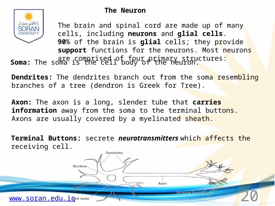
The Neuron
The brain and spinal cord are made up of many cells, including neurons and glial cells.90% of the brain is glial cells; they provide support functions for the neurons. Most neurons are comprised of four primary structures:
Soma: The soma is the cell body of the neuron.
Dendrites: The dendrites branch out from the soma resembling branches of a tree (dendron is Greek for Tree).
Axon: The axon is a long, slender tube that carries information away from the soma to the terminal buttons. Axons are usually covered by a myelinated sheath.
Terminal Buttons: secrete neurotransmitters which affects the receiving cell.

www.soran.edu.iq 21
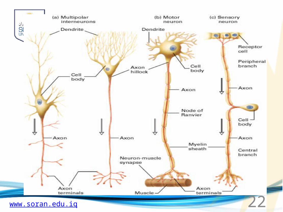
Different Types Of Neurons There are different types of neurons. They all carry electro-chemical nerve signals, but differ in structure (the number of processes, or axons, emanating from the cell body) and are found in different parts of the body. Sensory neurons or Bipolar neurons carry messages from the body's sense receptors (eyes, ears, etc.) to the Central Nervous system (CNS). These neurons have two processes. Sensory neuron account for 0.9% of all neurons. (Examples are retinal cells, olfactory epithelium cells.) Motoneurons or Multipolar neurons carry signals from the CNS to the muscles and glands. These neurons have many processes originating from the cell body. Motoneurons account for 9% of all neurons. (Examples are spinal motor neurons, pyramidal neurons, Purkinje cells.) Interneurons or Pseudopolare (Spelling) cells form all the neural wiring within the CNS. These have two axons (instead of an axon and a dendrite). One axon communicates with the spinal cord; one with either the skin or muscle. These neurons have two processes. (Examples are dorsal root ganglia cells.)
www.soran.edu.iq 23
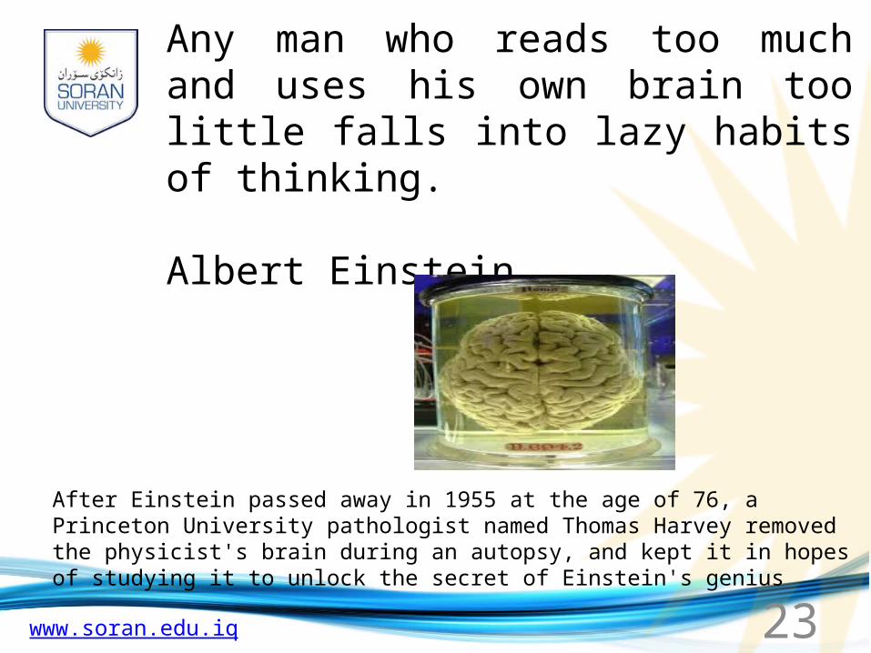
Any man who reads too much and uses his own brain too little falls into lazy habits of thinking.
Albert Einstein
After Einstein passed away in 1955 at the age of 76, a Princeton University pathologist named Thomas Harvey removed the physicist's brain during an autopsy, and kept it in hopes of studying it to unlock the secret of Einstein's genius