category a plant anatomy and physiology
TRANSCRIPT

CATEGORY A — PLANT ANATOMY AND PHYSIOLOGY
Marley Xiong, The Woodlands School
Hydrophytic and xerophytic designations are given to plants with adaptations to water-dominated or water-
scarce environments, respectively. Species within each classification share characteristics due to similar ecological
and habitat challenges rather than evolutionary homology. As such, the hydrophyte and xerophyte taxa are poly-
phyletic and may include structural disparities between members (Raven, 2003). This work looks at the adaptations
in a sampling of different hydrophytes (pleuston, elodeid) and xerophytes (succulent, woody, epiphyte, grass) with
an emphasis on the differences between the two. Given the large variety of hydrophytes and xerophytes, the fea-
tures described here are not necessarily be common to all plants within the given group, nor are the morphological
and anatomical adaptations found in nature limited to those described here.
Outer Cortex
Inner Cortex
Middle Cortex
Phloem
Xylem
Parenchyma Core
Endodermis
Pericycle
Epidermis
Lacuna
Lateral Root
Label Key:
Dermal
Ground
Vascular
Not classified
100 μm
The roots of Eichhornia crassipes, a pleuston hydrophyte, are made for floating rather than anchoring. The
middle cortex is composed of aerenchyma and lacunae (intercellular air cavities) that confer flexibility and buoyan-
cy to the root. The lacunae also provide a pathway for the rapid and energetically inexpensive exchange of gases
Figure 1: Transverse section of Eichhornia crassipes root
The section was prepared by hand, left unstained, and viewed at 100x magnification (10x objective).

(e.g. oxygen and ethylene) between the submerged root system and the above-water shoot system. Due to its unre-
stricted access to water, Eichhornia has poorly developed vascular tissues and absorptive surfaces; a single, large ele-
ment constitutes the xylem in each vascular bundle and root hairs are entirely absent from the thin-walled and uni-
layered epidermis (Pandey & Chadha, 1998).
Phalaenopsis exhibits root succulence, adapting to xeric conditions by retaining water. The cells of the cortex
are large and parenchymatous, reflecting their function in water storage (figure 2). Evaporative water loss is reduced
by the radially elongated, multi-layered epidermis and the sclerenchymatous hypodermis. The dermal tissues also
serve a protective function, reducing damage to internal tissues from high light intensities. Lignified tissues (stained
red) of the hypodermis and stele provide mechanical support to the water-heavy cortex. Phalaenopsis’s larger size and
lack of lateral root extensions reduces its surface area to volume ratio, in turn reducing the rate of water loss
through the root surface. Vascular tissues are well developed and represented by numerous bundles (Pandey &
Chadha, 1998).
Micro Differences in Root Structure
The hydrophyte's epidermis is thinner than that of the xerophyte. Lignified tissues are present in the xero-
phyte, while aerenchyma is present in the hydrophyte. Surface area to volume ratio is lower in the xerophyte.
Xylem
Epidermis
Cortex
Pericycle
Parenchyma Core
Phloem
Endodermis (with
Casparian Strip)
Hypodermis
Stele
250 μm
Figure 2: Transverse section of Phalaenopsis member aerial root
The section was prepared by hand, stained with Safranin O and Fast Green FCF, and viewed at 40x magnification
(4x objective).
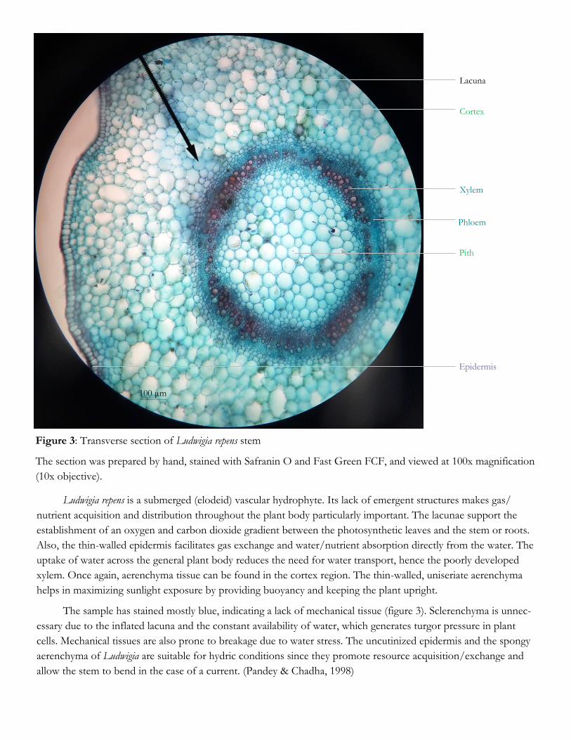
Figure 3: Transverse section of Ludwigia repens stem
The section was prepared by hand, stained with Safranin O and Fast Green FCF, and viewed at 100x magnification
(10x objective).
Xylem
Epidermis
Pith
Phloem
Lacuna
Cortex
Ludwigia repens is a submerged (elodeid) vascular hydrophyte. Its lack of emergent structures makes gas/
nutrient acquisition and distribution throughout the plant body particularly important. The lacunae support the
establishment of an oxygen and carbon dioxide gradient between the photosynthetic leaves and the stem or roots.
Also, the thin-walled epidermis facilitates gas exchange and water/nutrient absorption directly from the water. The
uptake of water across the general plant body reduces the need for water transport, hence the poorly developed
xylem. Once again, aerenchyma tissue can be found in the cortex region. The thin-walled, uniseriate aerenchyma
helps in maximizing sunlight exposure by providing buoyancy and keeping the plant upright.
The sample has stained mostly blue, indicating a lack of mechanical tissue (figure 3). Sclerenchyma is unnec-
essary due to the inflated lacuna and the constant availability of water, which generates turgor pressure in plant
cells. Mechanical tissues are also prone to breakage due to water stress. The uncutinized epidermis and the spongy
aerenchyma of Ludwigia are suitable for hydric conditions since they promote resource acquisition/exchange and
allow the stem to bend in the case of a current. (Pandey & Chadha, 1998)
100 μm

The stem of Picea is modified to reduce water loss. A thick, waxy cuticle covers the stem surface to limit
transpiration, acting as a barrier to diffusion (figure 4). The sterigmata (needle extensions) sheathe the stem and
lengthens the diffusion pathway to further reduce evaporative water loss. The epidermal and mature cork tissues
of Picea are highly lignified to resist wilting. Mechanical strengthening of the sterigmata also serves to allow photo-
synthesis during drought conditions, as the needles would otherwise wilt and cease to function. The scale-like
shape of the sterigmata decreases their surface area to volume ratio and evaporative water loss. Vascular tissues,
especially xylem, are well-developed and abundant (Schweingruber et al., 2007).
Micro Differences in Stem Structure
Cells of the xerophyte, most notably those of the cortex and vascular cylinder, are smaller than the cells of
the hydrophyte. Cell walls take up a greater portion of the xerophyte’s volume than the hydrophyte’s volume. This
can be attributed to both the xerophyte’s vulnerability to wilting and the hydrophyte’s subsistence on an excess of
water. Vascular tissues are more numerous in the xerophyte, forming a wider ring than that of the vascular tissues
in the hydrophyte. Vessels are also more compactly arranged and well-developed in the xerophyte, reflecting a
greater need to distribute water. Lignification is absent in the hydrophyte and extensive in the xerophyte. Cutin,
periderm and lateral growth are present in the xerophyte but absent in the hydrophyte.
Xylem
Epidermis
Resin Duct
Pith
Vascular Ray
Tracheid
Vascular Cambium
Phloem
Cuticle
Cork
Phelloderm
Suberized
(Mature) Cork
Cortex
Cork Cambium
Periderm
Sterigma Hypodermis
100 μm
Figure 4: Transverse section of 1-year-old Picea member stem
The section was prepared by hand, stained with Safranin O and Fast Green FCF, and viewed at 100x magnification
(10x objective).

Figure 5: Transverse section of Pistia stratiotes leaf
The section was prepared by hand, left unstained and
viewed at 100x magnification (10x objective).
Lacuna
Phloem
Xylem
Palisade
Mesophyll
Spongy
Mesophyll
Upper
Epidermis
Lower
Epidermis
Trichome
Adaxial
Epidermis
Abaxial
Epidermis
Vein Bundle
Sheath
Air Canal
Trichome
Stomata
Hydrenchyma
Chlorenchyma
Cuticle 75 μm 75 μm
Micro Differences in Leaf Structure
Pistia is a pleuston that exhibits adaptations for living at the air-water interface. The spongy mesophyll is aer-
ated by lacunae which, once again, provide buoyancy and gas exchange (figure 6). The leaf lacunae, being continu-
ous with the air spaces of the stem and root, facilitate the generation of an oxygen and carbon dioxide gradient.
Photosynthesis drives an increase in O2 partial pressure in the lacunae, while respiration drives a decrease in O2
partial pressure in the stem and roots. Diffusion then favors the roots, providing oxygen to the submerged parts
of the plant. The reverse occurs for the exchange of CO2 (Pandey & Chadha, 1998; Raven, 2003).
Tillandsia is an epiphyte of the Bromeliaceae family that has acquired xerophytic traits for living in the air.
The sheet-like structures on the leaf surface (figure 7) are actually multiseriate trichomes arranged longitudinally to
cover the leaf surface. Tillandsia relies on these trichomes to reflect light and absorb airborne water and nutrients.
A number of air canals are associated with the chlorenchyma for photosynthetic gas exchange. Hydrenchyma is
present for water storage, and a thick cuticle is present to reduce transpiration. In addition, stomata are sunken
and covered with trichomes to reduce exposure to drying winds and create a favorable microenvironment that fur-
ther decreases transpiration (Benzing, 2000).
Figure 6: Transverse section of Tillandsia leaf
The section was prepared by hand, left unstained
and viewed at 100x magnification (10x objective).

Figure 7: Transverse section of Triticum sheath, stem and leaf blade
The section was prepared by hand, left unstained, and viewed at 100x magnification (10x objective).
Chlorenchyma
Cortex
Peripheral Vascular
Bundle
Trichome
Epidermal Layers
Triticum Features of Interest
Triticum is a C3 grass and a major source of food around the world. Its leaf blade rolls up as a result of bulli-
form cell activity on the adaxial surface. Leaf rolling decreases the surface area exposed to the exterior and creates
a moist local environment. Both mechanisms serve to reduce transpiration, as wind exposure would increase evap-
oration and air humidity would decrease it. Trichomes aid in the formation of a moist microenvironment by trap-
ping air at the leaf adaxial surface. Transpiration is further reduced by the hollow, circular stem, the sheath, and
the waxy cuticle. Vascular tissues of the stem are well developed and arranged in 2 series: the peripheral and the
inner (Pandey & Chadha, 1998; Raven et al., 2003).
Cuticle
Inner Vascular
Bundle
Xylem
Phloem
Vascular Bundle
(Midvein)
Bulliform Cell
100 μm
Pistia and Tillandsia leaves also differ in morphology. Pistia is furrowed in shape and exhibits leaf pubescence.
The uniseriate trichomes help form extracellular air pockets along the leaf furrows and enhance buoyancy. Tilland-
sia is thicker and less wide, meaning a lower surface area to volume ratio. The thinner, broader leaves of Pistia max-
imize sun exposure and photosynthesis, while the smaller transpirational surface of Tillandsia promotes water reten-
tion at the expense of photosynthesis. Leaf veins are more numerous in Tillandsia. Vascular tissues are poorly de-
veloped in Pistia, consisting of phloem and a few thin-walled xylem elements.

The root systems of the submerged hydrophytes Alternanthera and Ludwigia are reduced since water and nu-
trients can be absorbed through the general plant body. Adventitious roots are present in Alternanthera (figure 8.1)
and Ludwigia (figure 8.5), and Ludwigia features primary roots used for anchorage. The leaves of the submerged
hydrophytes are thin and elongated to prevent breakage in moving water. For the same reason, the stem is thin
and noticeably bendable in Ludwigia. Both Ludwigia and Alternathera have green, photosynthetic stems, and Alter-
nanthera features inflorescences at the stem axil. Though certain hydrophytes develop emergent flowers, those seen
here are smaller and less colorful since they are pollinated through water rather than animals. Meanwhile, the
emergent portions of the floating hydrophytes are either shiny and waxy (Limnobium, Eichhornia), indicating the
presence of a cuticle, or covered in hairs (Pistia). The cuticle (figure 8.2 & 8.3) and leaf trichomes (figure 8.4) repel
water and prevent stomatal clogging. Eichhornia’s petioles are bulbous and inflated while Pistia’s leaves are fur-
rowed, both of which aid in buoyancy. The leaves of Limnobium and Eichhornia are broad to optimize sunlight ex-
posure. The roots of the pleustons are thin, fibrous and flexible for movement with water currents. Vegetative
propagation is accomplished in Limnobium through stolons (Pandey & Chadha, 1998).
Aerenchyma (Middle Cortex)
Outer Cortex
Inner Cortex
Epidermis
Endodermis
Pericycle
Pith
Xylem
Phloem
Figure 8: Frontal view of Alteranthera (1) and Ludwigia repens (5), and top view of Limnobium laevigatum (2), Eich-
hornia crassipes (3) and Pistia stratiotes (4).
Structures of interest include the root (Ro), leaf lamina (Le), flower (Fl), stolon (Sto), trichome (Tr), stem (St) and
petiole (Pe).
1
3
Ro
Le
Le
Ro
Tr
Sto
St
Pe
4
Le 2
Le
Le
Ro
5
St
Fl
Ro

1
2
3
Ro
Le
Le
Ro
Tr
Sto
Fl
Le
Sp
St
Le
Figure 9: Frontal view of Triticum (1) and Phalaenopsis (2), and surface view of an unknown succulent (3) and
Trichodiadema stelligerum (4).
Structures of interest include the root (Ro), leaf (Le), flower (Fl), stolon (Sto), trichome (Tr), stem (St) and spine (Sp).
The root system of Triticum is extensive and shallow to increase water uptake (figure 9.1). Triticum’s leaf
blades are rolled to reduce transpiration. Leaf succulence (indicated by fleshiness) can be seen in Phalaenopsis, the
unknown succulent and Trichodiadema. As well, root succulence is prominent in Phalaenopsis (figure 9.2). Certain
roots of Phalaenopsis are photosynthetic (indicated by their green color) and rounded in shape to minimize transpi-
ration through the surface. Phalaenopsis and Trichodiadema leaves are shiny due to the presence of a waxy cuticle. The
cuticle reflects light, thereby lowering leaf temperature and transpiration. Trichomes can be found on the leaf sur-
face of the unknown succulent (figure 9.3) shield it from excessive illumination and trap moisture. The spines
(modified leaves) of Trichodiadema provide shade and further lower plant temperature to reduce transpiration. The
woody growth in figure 9.4 is a means of resisting wilting and limiting transpiration. Reproduction in xerophytes is
largely vegetative (e.g. through stolon growth), however, flowering may occur in suitable conditions (Pandey &
Chadha 1998; G. Toole & E. Toole, 1999).
Macro Differences
There appears to be a trend toward more flexible and flaccid structures in the hydrophyte and more mechan-
ical tissues in the xerophyte. Unlike certain xerophytes, hydrophytes do not exhibit succulence. Many adaptations
in xerophytes are meant to reduce transpiration, while the adaptations of hydrophytes are in place to mitigate water
stress and maximize photosynthesis. Trichomes are present to trap moist air in xerophytes and to create air pock-
ets for buoyancy and stomatal opening in hydrophytes. It is clear that hydrophytes and xerophytes are well-suited
for their environments, which may be why vegetative propagation is the dominant method of reproduction.
4

Figure 2: Stomatal peel of Ludwigia repens (Lr) and Crassula ovata (Co) leaf adaxial epidermis
The section was peeled free-hand, stained with Methylene Blue and viewed at 100x magnification (10x objective).
Guard Cell
Vein Epidermis
Pavement Cell
Stomatal Complex
Stomatal Pore
Micro Differences in Leaf Epidermis
Crassula ovata has a larger stomatal aperture but lower stomatal density than Ludwigia repens. It is worth noting
that the leaves of Crassula also have a lower surface area (in relation to volume) to begin with. Stomatal density
cannot be reduced invariably, since gas exchange is required for photosynthesis. Thus, the stomatal density seen
here is most likely a function of photosynthetic requirement and how much water needs to be conserved in addi-
tion to how much water is already being conserved through other means (e.g. decreased surface area). On top of
this, Crassula employs CAM photosynthesis, which means stomata are only open during the night anyway. Mean-
while, the hydrophyte Ludwigia can afford to develop abundant stomata. The function of the stomata is, however,
unclear; stomata become clogged when immersed in water, and gas exchange occurs directly through the epider-
mis in submerged hydrophytes (). The stomata may be vestigial structures, similar to those of Potamogeton. Alt-
hough it is interesting to speculate the evolutionary reasons for epidermal anatomy, stomatal density is by no
means a definitive measure of xerophytic or hydrophytic character. Other factors such as CO2 concentration, light
intensity, photoperiod and temperature are also involved in determining stomatal density (Khan, 2001; Pandey &
Chadha, 1998).
Notes:
1. Free-hand sectioning was performed following the techniques described by Glime and Wagner, 2013.
2. Sass’s Safranin O and Fast Green FCF staining protocol was used
Pavement Cell
Guard Cell
Stomatal
Complex
Lr
Co
100 μm