case report solid pseudopapillary tumor: an invasive case ... · pdf filesolid pseudopapillary...
TRANSCRIPT
Int J Clin Exp Pathol 2015;8(7):8645-8649www.ijcep.com /ISSN:1936-2625/IJCEP0009908
Case ReportSolid pseudopapillary tumor: an invasive case report of primary ovarian origin and review of the literature
Shuqian He, Xiaoqing Yang, Ping Zhou, Yuxia Cheng, Qing Sun
Department of Pathology, Qianfoshan Hospital Affiliated to Shandong University, Jinan, Shandong, China
Received May 5, 2015; Accepted June 23, 2015; Epub July 1, 2015; Published July 15, 2015
Abstract: Solid pseudopapillary neoplasm occurring as a primary tumor outside the pancreas is a rare event. We report a case of an ovarian primary occurring with an ill-defined cystic mass in a 39-year-old woman. The morpho-logic and immunohistochemical features of the ovarian neoplasm described in this report are compatible with those of solid pseudopapillary neoplasm of the pancreas. Histologically, the tumor cells of the case we report infiltrate into the ovarian parenchyma. Because of the diagnosis is not clear before surgery, the patient had a reoccurrence two months after the operation in which laparoscopic simple ovarian cystectomy and part ovarian tissue removal, followed by the right salpingo-oophorectomy. The case herein confirms that solid pseudopapillary neoplasm of the ovary belongs to the class of low-grade malignant tumor with certain invasiveness. The diagnosis should be taken into serious consideration in order to avoid missed diagnosis and delay treatment. Through this case we have a bet-ter understanding of the biological behavior of solid pseudopapillary neoplasm of the ovary.
Keywords: Solid pseudopapillary neoplasm, ovarian neoplasm, invasiveness
Introduction
Solid pseudopapillary neoplasm (SPN) is a rare pancreatic neoplasm with indolent potential. Primary SPNs outside the pancreas are exceed-ingly rare. Several documents have described SPNs occurring ectopic pancreatic tissue, such as ovary, mesocolon and omentum [1-3]. To the present, 5 cases ectopic SPNs in ovary with benign outcome were reported by Deshpande, Cheuk and Lisa [4-6]. In this report, we present a case of SPN in ovary with uneasiness and may provide a new understanding of this tumor when arising in ovary.
Case presentation
A 39-year-old previously healthy women with once pain history in the right ovary a month ago, was admitted to hospital. Abdominal ultra-sound displayed the mass of cystic in the right ovary, 6 cm in greatest diameter, and showing high density in cystic area. The blood flow sig-nal can be seen in the periphery of the mass; an abdominal ultrasound showed normal liver, gall bladder, spleen, kidney, the uterus and pancreas were unremarkable. Laboratory tests
showed cancer antigen 125 (CA125), cancer antigen 19-9 (CA19-9), carcinoembryonic anti-gen (CEA), beta-human chorionic gonadotro-phin (β-HCG) to be within normal limits. Gyne- cologists diagnosed the ovarian cyst as benign, so laparoscopic simple ovarian cystectomy and partial ovarian tissue removal was carried out. During postoperative recovery 3 months later the ultrasonography found the cystic shadow in the right ovary and right salpingo-oophorecto-my was performed.
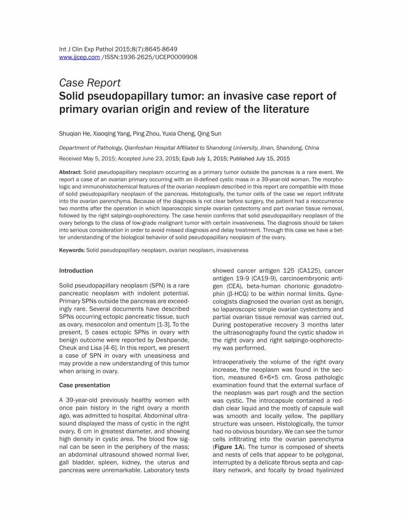
Intraoperatively the volume of the right ovary increase, the neoplasm was found in the sec-tion, measured 6×6×5 cm. Gross pathologic examination found that the external surface of the neoplasm was part rough and the section was cystic. The introcapsule contained a red-dish clear liquid and the mostly of capsule wall was smooth and locally yellow. The papillary structure was unseen. Histologically, the tumor had no obvious boundary. We can see the tumor cells infiltrating into the ovarian parenchyma (Figure 1A). The tumor is composed of sheets and nests of cells that appear to be polygonal, interrupted by a delicate fibrous septa and cap-illary network, and focally by broad hyalinized
An invasive solid pseudopapillary tumor originated in ovary
8646 Int J Clin Exp Pathol 2015;8(7):8645-8649
bands (Figure 1B). The polygonal tumor cells were monotonous and the nuclei were round to oval with finely granular chromatin. Mostly of the tumor cells displayed pale cytoplasm and lightly eosinophilic granular cytoplasm focally, occasionally neoplastic cells showed abundant foamy cytoplasm and longitudinal nuclear grooves. In some foci, intracellular and extra-
cellular eosinophilic hyaline globules were pres-ent (Figure 1C). The inner surface of the cap-sule wall showed pseudopapillary structure in some areas. The pseudopapillae consisted of a central fibrovascular core that was covered by one to multiple layers of tumor cells (Figure 1D). A small number of mitoses were evident (<1/10 HPF) and the tumor cells lacked appre-
Figure 1. A. The tumor cells embedded in the ovarian parenchyma. B. Nests of tumor cells surrounded by delicate fibrous septa. C. The polygonal tumor cells were monotonous and the nuclei were round to oval, occasional nuclear grooves, and foamy cytoplasm. The scattered intracellular and extracellular eosinophilic globules are seen. D. Pseu-dopapillary growth pattern. E. Strong and diffuse nuclear and slightly weaker cytoplasmic positivity for β-catenin. F. Strong and diffuse cytomembrance positivity for CD56.
An invasive solid pseudopapillary tumor originated in ovary
8647 Int J Clin Exp Pathol 2015;8(7):8645-8649
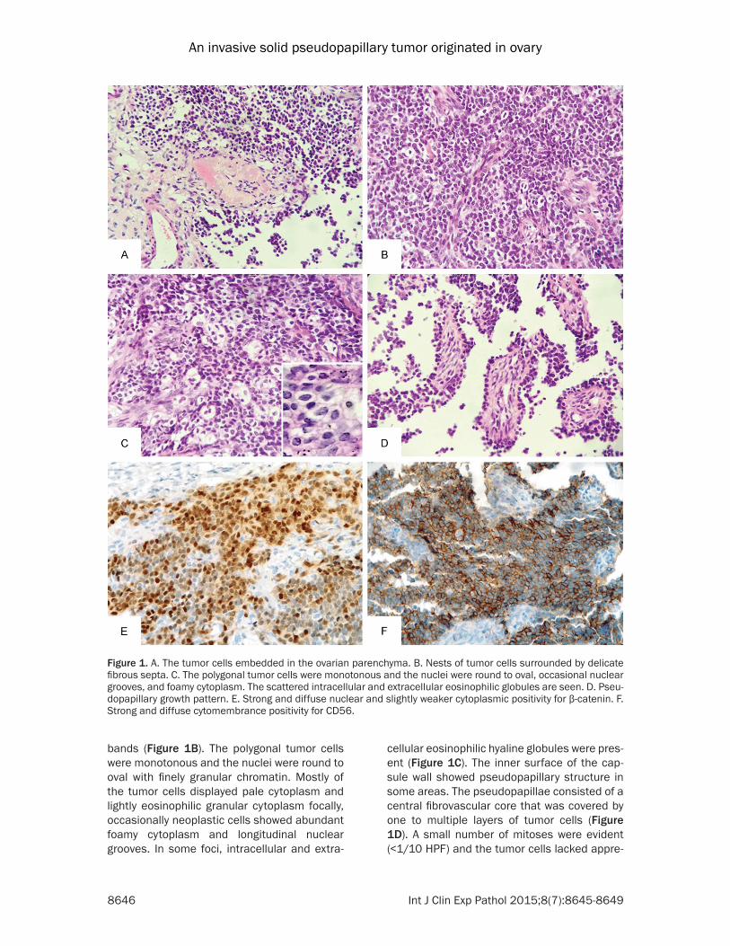
Table 1. Clinicopathologic features of reported cases of solid pseudopapillary neoplasms of the ovaryClinicopathologic index Case 1 Case 2 Case 3 Case 4 Case 5 Current caseReport Deshpande Deshpande Deshpande Wah Chewk Lisa M Shuqian HeAge (year) 17 57 21 25 48 39Premenopansal No Yes No No No NoMaximum 25.5 cm 3 cm 14 cm 16.5 cm 8.9 cm 6 cmPresentation Abdominal Mass Asymplomatic (Lesion know to be present for 7 yr) Abdominal swelling Abdominal fullness Abdominal pain Abdominal painLocation of tumor Left ovary Right ovary Left ovary Right ovary Left ovary Right ovaryGrossly examination Solid and cystic cystic Solid and cystic Solid and cystic cystic cysticTumor boundary Well-defined Well-defined Well-defined Well-defined Well-defined Ill-definedOutcome NED at 6 yr NED at 6 yr NED at 6 yr NED at 12 yr NED at 9 mo NED at 3 yrNED, no evidence of disease.
An invasive solid pseudopapillary tumor originated in ovary
8648 Int J Clin Exp Pathol 2015;8(7):8645-8649
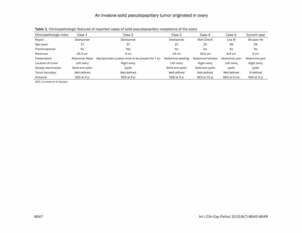
ciable nuclear atypical. Immunohistochemically all tumor cells showed both nuclear and slightly weaker cytoplasmic positivity for β-catenin (Figure 1E) and complete loss of E-cadherin staining. The tumor cells were diffusely positive for CD56 (Figure 1F), S-100, vimentin, CD99 and focally for progesterone receptor. A stain for synaptophysin and chromogranin was nega-tive. Immunostains for а-inhibin, NSE, cytokera-tin and p63 were negative. The proliferation index of Ki-67 was approximately 5%. Second postoperative pathology displays that there is residual tumor tissue in the ovary and has the same histological characteristics.
Discussion
SPNs are a rare tumor with an indolent clinical course arising predominantly from pancreas which typically presented in young females accounting for 5% of pancreatic neoplasm [7, 8]. It is reported that ovary could be another location of SPNs involved. Based on MEDLINE database search, 5 cases of SPNs of primary ovarian origin were sifted. Clinically, data show
that SPNs are variable in size (3 cm to 22.5 cm) and occurs primarily in young female (mean age: 35 yr, range: 17-57 yr). There is no intrinsic characteristic clinical presentation with ovarian origination. Morphologic features of the neo-plasm described in these reports resemble those occurring in pancreas. Tumor boundaries of the 5 cases were well-circumscribed (Table 1). As to the cases we present, in aspect of sex and age are consist with the predilection of SNPs, while the discrepancy was obvious, whose ill-defined boundary with invading to the surrounding structures different with that of the well-circumscribed previous described ones. SPNs in pancreas with infiltration and metasta-sis were reported with occurrence of 15% [7, 9], no similar statistic made relevant SPNs in ovary to date and to the best of our knowledge; this is the first case with aggressive potency reported in ovary.
Two point should take into consideration before primary ovarian SPNs were recognized. First, same malignant entity spread from pancreas should be excluded, metastasis of pancreatic
Table 2. Immunohistochemical features of reported cases of solid pseudopapillary neoplasms of the ovaryImmunohistochemical index Case 1 Case 2 Case 3 Case 4 Case 5 Current caseβ-catenin (nuclear and cytoplasmic) + + + + + +CD56 + (diffuse) + (diffuse) + (diffuse) + (diffuse) + (diffuse) + (diffuse)Synaptophysin + (focal weak) + (focal weak) NA + (focal weak) - -Chromogranin - - - - NA -Progesterone receptor + (diffuse) - - - + (focal) + (focal)CD10 + (focal) NA NA NA + (focal) NAVimentin NA NA NA NA + (diffuse) + (diffuse)S-100 NA NA NA NA NA + (diffuse)CD117 + (focal) + (focal) + (focal) NA - -E-cadherin - - - - - -а-Inhibin - - - - - -Pancytokeratin - - - - + (rare) -PLAP NA NA - NA NA NACDX-2 NA NA - NA NA NACalretinin - - - NA NA -TTF-1 NA NA NA NA - NAThyroglobulin - NA - NA - NASMA - NA NA NA - -Desmin - NA NA NA - NAEMA NA NA NA NA - -CD99 NA NA NA NA NA + (diffuse)CEA - NA NA NA NA NAKi-67 NA NA NA NA + (5%-10%) + (5%)+, positive; -, negative; NA, no IHC staining.
An invasive solid pseudopapillary tumor originated in ovary
8649 Int J Clin Exp Pathol 2015;8(7):8645-8649
SPNs to the ovary is well known [5]. As to the present case, no evidence certifies a tumor in her pancreas. Second, differential diagnosis should be made with those neoplasms that dis-play morphological similarity. Solid and pseudo-papillary region, true papillary growth pattern and cytological features in SPNs overlapping with certain ovarian tumor types, potentially raise other considerations such as sex-cord stromal tumors, neuroendocrine tumors, papil-lary epithelial neoplasm [6]. Fortunately, the differences were well described in documents and unlikely to dig a diagnostic pitfall for an experienced pathologist. In most cases the morphologic similarities are of limited extent and in some confused cases immunohisto-chemistory may be helpful. Recently, nuclear reactivity for β-catenin and lack cytoplasmic membrane staining for E-cadherin are believed to be the most robust immunohistochemistic phenotype. In present cases, tumor cells typi-cally showed well nuclear and weaker cytoplas-mic positivity for β-catenin and negative for E-cadherin, lacking of immunoreactivity for а-inhibin, chromogranin and pancytokeratin distinguishes from sex-cord stromal tumors, neuroendocrine tumors, and papillary epithelial neoplasm (Table 2).
In summary, we have presented a case of aggressive SPN with typical histological fea-tures and immunohistochemistic phenotype which may extend our understanding on bio-logical behavior of ovarian SPNs.
Acknowledgements
This study was supported by the National Natural Science Foundation of China (No. 81272420), the National Science Foundation of Shandong (No. ZR2012HM085), the Natural Scientific Foundation of Shandong (No. 2011- GSF11838), and the Scientific and Techno- logical Development Projects of Jinan City (No. 201202039). Consent: Written informed con-sent was obtained from the patient for publica-tion of this case report.
Disclosure of conflict of interest
None.
Address correspondence to: Dr. Qing Sun, Depart- ment of Pathology, Qianfoshan Hospital Affiliated to Shandong University, 16766 Jingshi Road, Jinan 250014, Shandong Province, China. Tel: (86)0531-89268155; E-mail: [email protected]
References
[1] Tornoczky T, Kalman E, Jakso P, Mehes G, Pajor L, Kajtar GG, Battyany I, Davidovics S, Sohail M and Krausz T. Solid and papillary epi-thelial neoplasm arising in heterotopic pancre-atic tissue of the mesocolon. J Clin Pathol 2001; 54: 241-245.
[2] Elorza Orue JL, Ruiz Diaz I, Tubia Landaberea J and San Vicente Leza M. [Solid and papillary tumor on ectopic pancreas in transversal me-socolon]. Rev Esp Enferm Dig 1991; 79: 429-431.
[3] Ishikawa O, Ishiguro S, Ohhigashi H, Sasaki Y, Yasuda T, Imaoka S, Iwanaga T, Nakaizumi A, Fujita M and Wada A. Solid and papillary neo-plasm arising from an ectopic pancreas in the mesocolon. Am J Gastroenterol 1990; 85: 597-601.
[4] Stoll LM, Parvataneni R, Johnson MW, Gui D, Dorigo O and Sullivan P. Solid pseudopapillary neoplasm, pancreas type, presenting as a pri-mary ovarian neoplasm. Hum Pathol 2012; 43: 1339-1343.
[5] Deshpande V, Oliva E and Young RH. Solid pseudopapillary neoplasm of the ovary: a re-port of 3 primary ovarian tumors resembling those of the pancreas. Am J Surg Pathol 2010; 34: 1514-1520.
[6] Cheuk W, Beavon I, Chui DT and Chan JK. Extrapancreatic solid pseudopapillary neo-plasm: report of a case of primary ovarian ori-gin and review of the literature. Int J Gynecol Pathol 2011; 30: 539-543.
[7] Romics L Jr, Olah A, Belagyi T, Hajdu N, Gyurus P and Ruszinko V. Solid pseudopapillary neo-plasm of the pancreas--proposed algorithms for diagnosis and surgical treatment. Langen- becks Arch Surg 2010; 395: 747-755.
[8] Santini D, Poli F and Lega S. Solid-papillary tumors of the pancreas: histopathology. JOP 2006; 7: 131-136.
[9] Yu PF, Hu ZH, Wang XB, Guo JM, Cheng XD, Zhang YL and Xu Q. Solid pseudopapillary tu-mor of the pancreas: a review of 553 cases in Chinese literature. World J Gastroenterol 2010; 16: 1209-1214.