solid pseudopapillary tumor of the pancreas in child: a case...
TRANSCRIPT
http://jsms.sch.ac.kr 29
Solid Pseudopapillary Tumor of the Pancreas in Child: A Case ReportGyo-Chang Choi
Department of Radiology, Soonchunhyang University Gumi Hospital, Soonchunhyang University College of Medicine, Gumi, Korea
Solid pseudopapillary tumor of the pancreas is a very rare form of childhood pancreatic tumor. We report the case of an 11-year-old girl having a solid pseudopapillary tumor of the pancreas presenting with left upper abdominal pain. Imaging studies showed the lesion to be an ovoid solid mass arising from the body and the tail of the pancreas. The tumor was surgically resected and was his-topathologically diagnosed as a solid pseudopapillary tumor.
Keywords: Pancreatic neoplasms; Pancreas computed tomography; Pancreas ultrasonography; Pancreas magnetic resonance
INTRODUCTION
Solid pseudopapillary tumor of the pancreas is a very rare be-nign or low-grade malignant neoplasm of childhood pancreatic tumor [1]. It is almost exclusively encountered in young women having a mean age of 26 years and has a male to female ratio of 1:9 [2,3]. Patients are often asymptomatic and the tumor is discovered incidentally on physical or radiological examination [2]. Patients may also occasionally present with an increasing abdominal mass associated with vague abdominal discomfort or may rarely pres-ent with an acute abdomen due to tumor rupture and hemoperito-neum. We report a case of solid pseudopapillary tumor of the pan-creas and describe the imaging findings of this rare tumor.
CASE REPORT
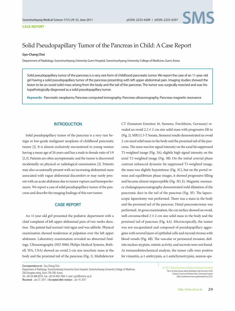
An 11-year-old girl presented the pediatric department with a chief complaint of left upper abdominal pain of two weeks dura-tion. The patient had normal vital signs and was afebrile. Physical examination showed tenderness at palpation over the left upper abdomen. Laboratory examination revealed no abnormal find-ings. Ultrasonography (HD 5000, Philips Medical Systems, Both-ell, WA, USA) showed an ovoid 2-cm size isoechoic mass at the body and the proximal tail of the pancreas (Fig. 1). Multidetector
CT (Somatom Emotion 16, Siemens, Forchheim, Germany) re-vealed an ovoid 2.2×2-cm size solid mass with progressive fill-in (Fig. 2). MRI (1.5-T Sonata, Siemens) results demonstrated an ovoid 2-cm sized solid mass in the body and the proximal tail of the pan-creas. The mass was low signal intensity on the axial fat suppressed T1-weighted image (Fig. 3A); slightly high signal intensity on the axial T2-weighted image (Fig. 3B). On the initial arterial phase contrast enhanced dynamic fat suppressed T1-weighted image, the mass was slightly hypointense (Fig. 3C), but on the portal ve-nous and equilibrium phase images, it showed progressive filling and became almost imperceptible (Fig. 3D, E). Magnetic resonan-ce cholangiopancreatography demonstrated mild dilatation of the pancreatic duct in the tail of the pancreas (Fig. 3F). The laparo-scopic laparotomy was performed. There was a mass in the body and the proximal tail of the pancreas. Distal pancreatectomy was performed. At gross examination, the cut surface showed an ovoid, well-circumscribed 2×2-cm size solid mass in the body and the proximal tail of pancreas (Fig. 4A). Microscopically, the tumor was not encapsulated and composed of pseudopapillary aggre-gates with several layers of epithelial cells and myxoid stroma with blood vessels (Fig. 4B). The vascular or perineural invasion, defi-nite nuclear atypism, mitotic activity, and necrosis were not found. At immunohistochemical analysis, the tumor cells were positive for vimentin, α-1-antitrypsin, α-1-antichymotrypsin, neuron-spe-
Soonchunhyang Medical Science 17(1):29-33, June 2011 pISSN: 2233-4289 I eISSN: 2233-4297
CASE REPORT
Correspondence to: Gyo-Chang ChoiDepartment of Radiology, Soonchunhyang University Gumi Hospital, Soonchunhyang University College of Medicine, 250 Gongdan-dong, Gumi 730-706, KoreaTel: +82-54-468-9374, Fax: +82-54-463-7504, E-mail: [email protected]: Jan 27, 2011 / Accepted after revision: Jun 14, 2011
© 2011 Soonchunhyang Medical Research InstituteThis is an Open Access article distributed under the terms of the
Creative Commons Attribution Non-Commercial License (http://creativecommons.org/licenses/by-nc/3.0/).

Choi G-C • Solid Pseudopapillary Tumor of the Pancreas in Child
Soonchunhyang Medical Science 17(1):29-3330 http://jsms.sch.ac.kr
cific enolase, chromogranin, and partly positive for progesterone receptor. The cells were negative for cytokeratin and carcinoem-bryonic antigen. These findings helped establish a diagnosis of sol-id pseudopapillary tumor of the pancreas.
DISCUSSION
Solid pseudopapillary tumors of the pancreas are very rare in children and typically diagnosed in young women, especially black or East Asian women and have a female predominance [4,5]. Syn-onyms include solid and cystic tumor, solid and papillary epithelial
neoplasm, papillary-cystic neoplasm, papillary cystic epithelial neoplasm, papillary-cystic tumor, and Franz tumor. In 1996, the World Health Organization renamed this tumor as solid pseudo-papillary tumor for the international histologic classification of tu-mor of the exocrine pancreas [6]. It is most commonly detected in-cidentally, but may occasionally present with a gradually enlarging abdominal mass or complain of vague abdominal pain or discom-fort. The abdomen is usually nontender on palpation, but obstruc-tive symptoms may occur if the tumor grows large enough to com-press adjacent organs. There are usually no abnormalities in clini-cal laboratory tests or in pancreatic cancer markers. The mass may occur in anywhere in the pancreas but is most frequently found in the head or tail. At gross examination, the mass is usually large and well encapsulated and contains varying amounts of necrosis, hem-orrhage, and cystic change [4]. At microscopic analysis, there are two distinct types of cellular arrangements: solid and papillary. The hallmark histologic pattern occurs when the tumor cells from papillary configurations composed of a fibrovascular stalk sur-rounded by several layers of epithelial cells. Solid areas containing necrosis, foamy macrophages, cholesterol granulomas, and calcifi-cations may also be seen [5]. The pathogenesis of these tumors is still controversial [7]. However solid pseudopapillary tumor shows immunohistochemical and ultrastructural evidence of both a neu-roendocrine and an acinar-ductal differentiation, suggesting that this tumor arises from a pluripotent stem cell [7,8]. Solid pseudo-papillary tumors are typically positive for vimentin, neuron-spe-cific enolase, α-1-antitrypsin, and α-1-antichymotrypsin and nega-tive for chromogranin, epithelial membrane antigen, and cytoker-
Fig. 1. Ultrasonographic transverse scan shows an ovoid isoechoic mass at the body and the proximal tail of the pancreas.
Fig. 2. (A) Contrast-enhanced arterial phase computed tomography (CT) scan demonstrates an ovoid peripherally enhanced low attenuation mass (arrows) in the body and the proximal tail of the pancreas. (B) Contrast-enhanced portal phase CT scan shows progressive fill-in of the mass (arrows).
A B

Solid Pseudopapillary Tumor of the Pancreas in Child • Choi G-C
Soonchunhyang Medical Science 17(1):29-33 http://jsms.sch.ac.kr 31
atin. In our case, the tumor cells were immunoreactive for vimen-tin, α-1-antitrypsin, α-1-antichymotrypsin, neuron-specific eno-lase, chromogranin. The above-mentioned immunohistochemical features are strongly suggestive of solid pseudopapillary tumor of the pancreas. Sex hormones may play a role in the pathogenesis of growth of solid pseudopapillary tumors. Nearly all studies demon-
strate no evidence of estrogen receptor. However progesteron re-ceptors are present in many cases [5]. In our case, the cells were positive for progesterone receptor. At sonography, the appearance of solid pseudopapillary tumor is variable and lacks correlation with gross pathology [9]. In our case, ultrasonography showed isoechoic mass. Contrast-enhanced computed tomography (CT)
Fig. 3. (A) Axial fat suppressed T1-weighted image reveals an ovoid well circumscribed hypointense mass in the body and the proximal tail of the pancreas. (B) On an axial T2-weighted image, the mass is slightly hyperintense. (C) Arterial phase contrast-enhanced dynamic fat suppressed T1-weighted image shows slightly hypoin-tense (arrows). On the portal venous (D) and equilibrium (E) phase images, the lesion (arrows) is progressive filling and becomes almost imperceptible. (F) A thick-slab coronal oblique magnetic resonance cholangiopancreatography magnetic resonance cholangiopancreatography reveals mild dilatation of the pancreatic duct in the tail of the pancreas.
A
C
E
B
D
F

Choi G-C • Solid Pseudopapillary Tumor of the Pancreas in Child
Soonchunhyang Medical Science 17(1):29-3332 http://jsms.sch.ac.kr
plays a major role in the diagnostic evaluation of neoplasms of the pancreas. However, when compared with magnetic resonance (MR) imaging, CT has inherent limitations in showing certain tis-sue characteristics, such as hemorrhage, cystic degeneration, or presence of capsule. On contrast-enhanced CT, solid pseudopapil-lary tumor typically presents as a large heterogenous mass. Our study showed relatively homogenous mass on CT. Solid pseudo-papillary tumor of the pancreas is heterogenous high or low signal intensity on T1-weighted, heterogenous high signal intensity on T2-weighted images, which reflects the complex nature of the mass. Our case is somewhat atypical due to the small size of the tu-mor, which may the lack of significant cystic change or hemor-rhage at MR imaging. On gadolinium-enhanced dynamic MR im-aging, the most common enhancement pattern of solid pseudo-papillary tumor consists of early, peripheral, and heterogenous en-hancement during the arterial phase with progressive but heterog-enous fill-in of the lesion during the portal venous and equilibrium phases [10]. Our case showed also peripheral enhancement with progressive fill-in on gadolinium-enhanced dynamic MR imag-ing, which suggests a solid pancreatic neoplasm and helps distin-guish solid pseudopapillary tumor from other pancreatic neo-plasms, such as neuroendocrine tumors, that typically enhance more than the pancreas. Solid pseudopapillary tumors posses a malignant potential risk of 5 to 10% and must therefore be resected completely and aggressively [2]. Unlikely pancreatic ductal adeno-carcinoma, complete resection of a solid pseudopapillary tumor is usually curative and patients can survive a long period after the operation. Death due to tumor growth, liver metastases, or perito-
neal seeding is rare. In children, the differential diagnosis of this lesion included neuroendocrine tumor, pancreatoblastoma. Al-though neuroendocrine tumors occur in patients who are older and do not have the female predominance observed with solid pseudopapillary tumor, neuroendocrine tumors may appear cys-tic, contain calcifications, and show areas of internal hemorrhage. Pancreatoblastoma is more aggressive than solid pseudopapillary tumor and often presents with liver metastases at the time of diag-nosis.
In conclusion, in the pediatric age group, solid pseudopapillary tumor of the pancreas is a very rare tumor. Awareness of the imag-ing findings will allow accurate diagnosis and appropriate man-agement to be undertaken.
REFERENCES
1. Choi SH, Kim SM, Oh JT, Park JY, Seo JM, Lee SK. Solid pseudopapillary tumor of the pancreas: a multicenter study of 23 pediatric cases. J Pediatr Surg 2006;41:1992-5.
2. Klöppel G, Kosmahl M. Cystic lesions and neoplasms of the pancreas. The features are becoming clearer. Pancreatology 2001;1:648-55.
3. Kaufman SL, Reddick RL, Stiegel M, Wild RE, Thomas CG Jr. Papillary cystic neoplasm of the pancreas: a curable pancreatic tumor. World J Surg 1986;10:851-9.
4. Solcia E, Capella C, Kloppel G. Tumors of the exocrine pancreas. In: Ro-sai J, Sorbin L, editors. Atlas of tumor pathology. Washinton, DC: Armed Forces Institute of Pathology; 1997. p. 31-144.
5. Lam KY, Lo CY, Fan ST. Pancreatic solid-cystic-papillary tumor: clinico-pathologic features in eight patients from Hong Kong and review of the literature. World J Surg 1999;23:1045-50.
6. Kloppel G, Solcia E, Longnecker DS, Capella C, Sobin LH. World Health Organization: institutinal histological classfication of tumors-histological
Fig. 4. (A) Photograph of the cut gross specimen shows an ovoid gray solid mass with partly brownish areas (arrows). (B) A photomicrograph shows pseudopapillae with several layers of epithelial cells and myxoid stroma with blood vessels (arrows) (H&E, × 200).
BA

Solid Pseudopapillary Tumor of the Pancreas in Child • Choi G-C
Soonchunhyang Medical Science 17(1):29-33 http://jsms.sch.ac.kr 33
typing of tumors the exocrine pancreas. 2nd ed. Berlin: Springer-Berlag; 1996.
7. Pezzi CM, Schuerch C, Erlandson RA, Deitrick J. Papillary-cystic neo-plasm of the pancreas. J Surg Oncol 1988;37:278-85.
8. Stachura J, Popiela T, Pietroń M, Tomaszewska R, Kulig J, Nowak K. Cy-tology of solid and papillary epithelial neoplasms of the pancreas: a case report. Diagn Cytopathol 1988;4:339-41.
9. Buetow PC, Buck JL, Pantongrag-Brown L, Beck KG, Ros PR, Adair CF. Solid and papillary epithelial neoplasm of the pancreas: imaging-patho-logic correlation on 56 cases. Radiology 1996;199:707-11.
10. Cantisani V, Mortele KJ, Levy A, Glickman JN, Ricci P, Passariello R, et al. MR imaging features of solid pseudopapillary tumor of the pancreas in adult and pediatric patients. AJR Am J Roentgenol 2003;181:395-401.