case report primary intramedullary spinal gliosarcoma
TRANSCRIPT

Int J Clin Exp Med 2017;10(5):8343-8349www.ijcem.com /ISSN:1940-5901/IJCEM0051088
Case ReportPrimary intramedullary spinal gliosarcoma: case report and review of the literature
Liang Wu1, Jingyi Fang2, Jun Yang1, Yulun Xu1
1Department of Neurosurgery, China National Clinical Research Center for Neurological Diseases, Beijing Tiantan Hospital, Capital Medical University, Beijing, China; 2Department of Neuropathology, Beijing Neurosurgical Institute, Capital Medical University, Beijing, China
Received February 17, 2017 Accepted March 30, 2017; Epub May 15, 2017; Published May 30, 2017
Abstract: Primary gliosarcoma (GS) in the spinal cord is extremely rare. To our knowledge, there are only two cases reported in the literature. The clinical feature, treatment, and prognosis for this rare entity have been uncertain. In this paper, we present a unique case of histopathologically proven primary intramedullary spinal GS without intra-cranial involvement. A 46-year-old male presented with a 3-month history of progressively worsening numbness and weakness of his left leg and gait instability. Spinal magnetic resonance imaging revealed an intramedullary lesion at the level T4-7 with irregularly heterogeneous enhancement. Intracranial or other bodies’ lesions were absent. The patient underwent a T4-7 laminectomy and subtotal tumor removal was achieved. Histopathological findings confirmed a GS. Postoperatively, the patient received adjuvant radiotherapy combined with concurrent and adjuvant chemotherapy with Temozolomide. However, he had tumor recurrence 5 months after surgery and experienced a progression of paraplegia with rapid enlargement of residual tumor. The patient died of severe pneumonia and respiratory failure 6 months after diagnosis. The clinical features and treatment outcome of this rare entity are dis-cussed along with a review of the relevant literature. Primary GS should be considered in the differential diagnosis of spinal cord tumors. Multidisciplinary treatment including surgical resection and adjuvant radiochemotherapy should be performed as standard protocol due to highly malignant nature, although overall prognosis of this tumor is poor.
Keywords: Gliosarcoma, intramedullary, surgical resection, radiochemotherapy, spinal cord
Introduction
Gliosarcoma (GS) is very rare and highly malig-nant tumor in the central nervous system, accounting for only 1.8-8% of astrocytic neo-plasms [1]. The tumor was firstly described in 1895 as glioblastoma (GBM) with a sarcoma-tous component and had similar clinical fea-tures with those of GBM [2]. Histopathologically, GS is typically composed of biphasic patterns of malignant glial and sarcomatous compo-nents, based on the 2007 WHO classification. GS can occur throughout the whole neuraxis and mostly affects the cerebrum, especially the temporal lobes. Primary GS in the spinal cord is extremely rare, and there are only two cases reported in the English literature [3, 4]. Here we present a unique case of histopathologically proven primary intramedullary spinal GS in a 46-year-old male patient, which may contribute to the knowledge of this unusual entity.
Case report
A 46-year-old male patient presented to our outpatient clinic with a 3-month history of pro-gressively worsening numbness and weakness of his left leg and gait instability. He denied any bowel or bladder symptoms. Neurological examination revealed that muscle power was grade 2/5 in his left leg and grade 4/5 in the right leg (classified by the Medical Research Council grading system). Somatic superficial sensation below left T2 level was reduced. Increased deep tendon reflexes and positive Babinski sign were noted in the left lower limb. Laboratory findings were within normal limits and the medical history was unremarkable.
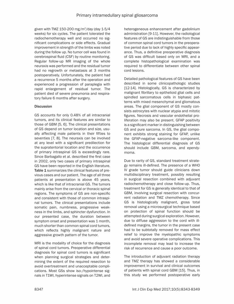
Preoperative magnetic resonance imaging (MRI) of the thoracic spine demonstrated an intramedullary lesion at the T4-7 level, measur-ing 5 mm×5 mm×56 mm in maximal dimen-

Primary intramedullary spinal gliosarcoma
8344 Int J Clin Exp Med 2017;10(5):8343-8349
sions (Figure 1). The tumor showed mixed iso-/hypointensity on T1-weighted imaging (WI) and mixed hyperintensity on T2WI. Contrast-enhanced imaging of the mass showed irregu-larly heterogeneous enhancement after gado-linium administration. Peritumoral edema and associated syringomyelia were noted. Cranial and other spinal region MRI findings were nor-mal. Moreover, bone scanning and abdominal ultrasonography showed no other tumors, which ruled out a metastatic origin for the spi-nal cord tumor. Based on the radiological fea-tures of the tumor, the lesion was preoperative-ly diagnosed as an ependymoma.
The patient underwent a T4 to T7 laminectomy for tumor resection through the posterior mid-line approach. The dura was intact but highly tense and a longitudinal incision was made on the center of it for cord exploration. The intraop-
erative findings showed that the tumor was red and greyish in color, mixed in texture, and highly vascular. The lesion had ill-defined margins with adjacent neural tissue and spread ventral-ly to the cord. Due to diffuse tumor infiltration to the cord, gross total resection is difficult based on protection of spinal functions and subtotal tumor removal was finally achieved under spinal evoked-potential monitoring. After surgery, the effect of cord compression was vanished.
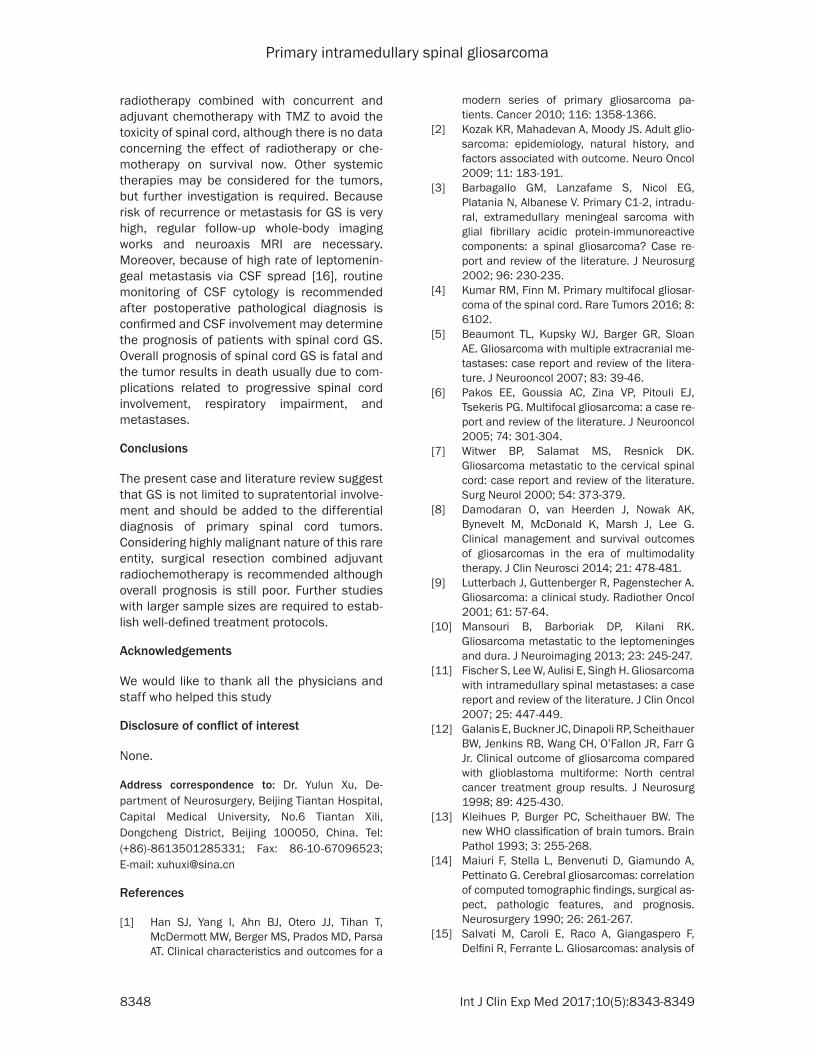
On histopathological examination, the tumor was composed of malignant fibrillary and epi-thelioid glial cells and spindled sarcomatous cells in biphasic patterns with mixed glioma-tous and mesenchymal areas (Figure 2A, 2B). The immunohistochemical examinations reveal- ed that glial fibrillary acidic protein (GFAP) had strongly positive reactivity in gliomatous areas
Figure 1. Preoperative magnetic resonance imaging showed an intramedullary lesion at the T4-7 level with mixed iso-/hypointensity on sagittal T1-weighted image (A) and mixed hyperintensity on sagittal T2-weighted image (B). Gadolinium-enhanced sagittal (C), coronal (D), and axial (E) T1-weighted images demonstrated irregularly heteroge-neous enhancement. Peritumoral edema and associated syringomyelia were observed.

Primary intramedullary spinal gliosarcoma
8345 Int J Clin Exp Med 2017;10(5):8343-8349
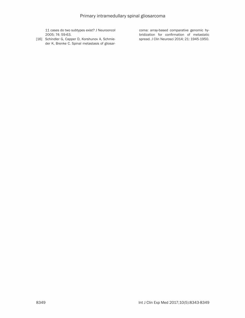
but negative reactivity in sarcomatous areas (Figure 2C). Moreover, Olig-2 and S-100 showed positivity in gliomatous areas but sparse positivity in sarcomatous areas, while vimentin showed positivity in sarcomatous areas but sparse positivity in gliomatous areas (Figure 3). Approximately 40% of cells were positive for Ki-67. All these findings were con-sistent with the diagnosis of a GS (WHO Grade IV).
Postoperative course was uneventful and sen-sory deficits were apparently relieved. Post- operative adjuvant radiotherapy combined with concurrent Temozolomide (TMZ) was strongly recommended. The interval between surgery and the start of radiochemotherapy was 18 days. The patient received focal radiotherapy 50.4 gray/28 fractions (5 fractions/week) with concurrent TMZ 75 mg/m2/day. After concur-rent treatment, adjuvant chemotherapy was
Figure 2. Photomicrograph revealed malignant fibrillary to epithelioid glial cells (A) and spindled sarcomatous cells (B) in biphasic patterns with mixed mesenchymal and gliomatous areas (hematoxylin-eosin staining, original magni-fication, ×200). The immunohistochemical examinations showed that glial fibrillary acidic protein had strongly posi-tive reactivity in glial differentiated areas but negative reactivity in sarcomatous areas (C) (original magnification, ×100). The findings were consistent with the diagnosis of a gliosarcoma (WHO Grade IV).
Figure 3. The immunohistochemical examinations demonstrated that in gliomatous areas (A-C), Olig-2 (A) and S-100 (B) showed strong positivity and vimentin (C) showed sparse positivity, while in sarcomatous areas (D-F), Olig-2 (D) and S-100 (E) showed sparse positivity and vimentin (F) showed strong positivity. (original magnification, ×100).

Primary intramedullary spinal gliosarcoma
8346 Int J Clin Exp Med 2017;10(5):8343-8349
Table 1. Summary of previously reported primary spinal gliosarcomas
Author/year Age (years), sex
Duration of illness Tumor location Presentations Treatment Outcome
Barbagallo et al., 2002 [3] 73, M 3 weeks C1-2, IDEM Neck pain; progressive hemiparesis; sensory reduction; urinary urgency and incontinence
GTR Dead of postoperative cardiopulmonary complications
Kumar et al., 2016 [4] 54, M 2 months C1-4, T6, T9, L1, IM+IDEM Progressive sensory dysfunction of legs and gait instability PR+RTX/TMZ+Avastin Dead at 9 months*
Present case 46, M 1 month T4-7, IM Numbness and weakness of left leg and gait instability STR+RTX/TMZ Dead at 6 monthsGTR: gross total removal; IDEM: intradural and extramedullary; IM: intramedullary; M: male; PR: partial removal; RTX: radiotherapy; STR: subtotal removal; TMZ: Temozolomide. *After initial diagnosis.
Primary intramedullary spinal gliosarcoma
8347 Int J Clin Exp Med 2017;10(5):8343-8349
given with TMZ 150-200 mg/m2/day (day 1-5/4 weeks) for six cycles. The patient tolerated the radiochemotherapy well and occurred no sig-nificant complications or side effects. Gradual improvement in strength of the limbs was noted during the follow up. No tumor cell was found in cerebrospinal fluid (CSF) by routine monitoring. Regular follow-up MR imaging of the whole neuraxis was performed and the residual tumor had no regrowth or metastasis at 3 months postoperatively. Unfortunately, the patient had a recurrence 5 months after the operation and experienced a progression of paraplegia with rapid enlargement of residual tumor. The patient died of severe pneumonia and respira-tory failure 6 months after surgery.
Discussion
GS accounts for only 0.48% of all intracranial tumors, and its clinical features are similar to those of GBM [5, 6]. The clinical presentations of GS depend on tumor location and size, usu-ally affecting male patients in their fifties to seventies [7, 8]. The neuraxis can be involved at any level with a significant predilection for the supratentorial location and the occurrence of primary intraspinal GS is exceedingly rare. Since Barbagallo et al. described the first case in 2002, only two cases of primary intraspinal GS have been reported in the English literature. Table 1 summarizes the clinical features of pre-vious cases and our patient. The age of all three patients at presentation is above 45 years, which is like that of intracranial GS. The tumors mainly arise from the cervical or thoracic spinal regions. The symptoms of GS are non-specific and consistent with those of common intraspi-nal tumors. The clinical presentations include somatic pain, numbness, progressive weak-ness in the limbs, and sphincter dysfunction. In our presented case, the duration between symptom onset and presentation was 1 month, much shorter than common spinal cord tumors, which reflects highly malignant nature and aggressive growth pattern of the tumor.
MRI is the modality of choice for the diagnosis of spinal cord tumors. Preoperative differential diagnosis for spinal cord tumors is significant when planning surgical strategies and deter-mining the extent of the required resection to avoid overtreatment and unacceptable compli-cations. Most GSs show iso-/hypointense sig-nals in T1WI, hyperintense signals on T2WI, and
heterogeneous enhancement after gadolinium administration [9-11]. However, the radiological features of GS are indistinguishable from those of common spinal cord tumors in the preopera-tive period due to lack of highly specific appear-ance. Thus, a definitive preoperative diagnosis of GS was difficult based only on MRI, and a complete histopathological examination was required to differentiate between other spinal cord lesions.
Detailed pathological features of GS have been described in some clinicopathologic studies [12-14]. Histologically, GS is characterized by malignant fibrillary to epithelioid glial cells and spindled sarcomatous cells in biphasic pat-terns with mixed mesenchymal and gliomatous areas. The glial component of GS mostly con-sists astrocytes with nuclear atypia and mitotic figures. Necrosis and vascular endothelial pro-liferation may also be present. GFAP positivity is a significant marker in distinguishing between GS and pure sarcoma. In GS, the glial compo-nent exhibits strong staining for GFAP, unlike the GFAP-negative sarcoma-like component. The histological differential diagnosis of GS should include GBM, sarcoma, and ependy- moma.
Due to rarity of GS, standard treatment strate-gy remains ill-defined. The presence of a WHO IV grade tumor should guide clinicians down multidisciplinary treatment, possibly resulting in surgical resection combined with adjuvant radiochemotherapy and close follow-up. Thus, treatment for GS is generally identical to that of GBM, involving surgical resection with concur-rent radiation and TMZ chemotherapy. Since GS is histologically malignant, gross total removal using a microsurgical technique based on protection of spinal function should be attempted during surgical exploration. However, due to diffuse aggression to the cord with ill-defined margins, the tumor in the present case had to be subtotally removed for mass effect relief to improve the myelopathic symptoms and avoid severe operative complications. This incomplete removal may lead to increase the risk of recurrence and cause a poor outcome.
The introduction of adjuvant radiation therapy and TMZ therapy has showed a considerable improvement in survival and clinical outcomes of patients with spinal cord GBM [15]. Thus, in this study we performed postoperative early
Primary intramedullary spinal gliosarcoma
8348 Int J Clin Exp Med 2017;10(5):8343-8349
radiotherapy combined with concurrent and adjuvant chemotherapy with TMZ to avoid the toxicity of spinal cord, although there is no data concerning the effect of radiotherapy or che-motherapy on survival now. Other systemic therapies may be considered for the tumors, but further investigation is required. Because risk of recurrence or metastasis for GS is very high, regular follow-up whole-body imaging works and neuroaxis MRI are necessary. Moreover, because of high rate of leptomenin-geal metastasis via CSF spread [16], routine monitoring of CSF cytology is recommended after postoperative pathological diagnosis is confirmed and CSF involvement may determine the prognosis of patients with spinal cord GS. Overall prognosis of spinal cord GS is fatal and the tumor results in death usually due to com-plications related to progressive spinal cord involvement, respiratory impairment, and metastases.
Conclusions
The present case and literature review suggest that GS is not limited to supratentorial involve-ment and should be added to the differential diagnosis of primary spinal cord tumors. Considering highly malignant nature of this rare entity, surgical resection combined adjuvant radiochemotherapy is recommended although overall prognosis is still poor. Further studies with larger sample sizes are required to estab-lish well-defined treatment protocols.
Acknowledgements
We would like to thank all the physicians and staff who helped this study
Disclosure of conflict of interest
None.
Address correspondence to: Dr. Yulun Xu, De- partment of Neurosurgery, Beijing Tiantan Hospital, Capital Medical University, No.6 Tiantan Xili, Dongcheng District, Beijing 100050, China. Tel: (+86)-8613501285331; Fax: 86-10-67096523; E-mail: [email protected]
References
[1] Han SJ, Yang I, Ahn BJ, Otero JJ, Tihan T, McDermott MW, Berger MS, Prados MD, Parsa AT. Clinical characteristics and outcomes for a
modern series of primary gliosarcoma pa-tients. Cancer 2010; 116: 1358-1366.
[2] Kozak KR, Mahadevan A, Moody JS. Adult glio-sarcoma: epidemiology, natural history, and factors associated with outcome. Neuro Oncol 2009; 11: 183-191.
[3] Barbagallo GM, Lanzafame S, Nicol EG, Platania N, Albanese V. Primary C1-2, intradu-ral, extramedullary meningeal sarcoma with glial fibrillary acidic protein-immunoreactive components: a spinal gliosarcoma? Case re-port and review of the literature. J Neurosurg 2002; 96: 230-235.
[4] Kumar RM, Finn M. Primary multifocal gliosar-coma of the spinal cord. Rare Tumors 2016; 8: 6102.
[5] Beaumont TL, Kupsky WJ, Barger GR, Sloan AE. Gliosarcoma with multiple extracranial me-tastases: case report and review of the litera-ture. J Neurooncol 2007; 83: 39-46.
[6] Pakos EE, Goussia AC, Zina VP, Pitouli EJ, Tsekeris PG. Multifocal gliosarcoma: a case re-port and review of the literature. J Neurooncol 2005; 74: 301-304.
[7] Witwer BP, Salamat MS, Resnick DK. Gliosarcoma metastatic to the cervical spinal cord: case report and review of the literature. Surg Neurol 2000; 54: 373-379.
[8] Damodaran O, van Heerden J, Nowak AK, Bynevelt M, McDonald K, Marsh J, Lee G. Clinical management and survival outcomes of gliosarcomas in the era of multimodality therapy. J Clin Neurosci 2014; 21: 478-481.
[9] Lutterbach J, Guttenberger R, Pagenstecher A. Gliosarcoma: a clinical study. Radiother Oncol 2001; 61: 57-64.
[10] Mansouri B, Barboriak DP, Kilani RK. Gliosarcoma metastatic to the leptomeninges and dura. J Neuroimaging 2013; 23: 245-247.
[11] Fischer S, Lee W, Aulisi E, Singh H. Gliosarcoma with intramedullary spinal metastases: a case report and review of the literature. J Clin Oncol 2007; 25: 447-449.
[12] Galanis E, Buckner JC, Dinapoli RP, Scheithauer BW, Jenkins RB, Wang CH, O’Fallon JR, Farr G Jr. Clinical outcome of gliosarcoma compared with glioblastoma multiforme: North central cancer treatment group results. J Neurosurg 1998; 89: 425-430.
[13] Kleihues P, Burger PC, Scheithauer BW. The new WHO classification of brain tumors. Brain Pathol 1993; 3: 255-268.
[14] Maiuri F, Stella L, Benvenuti D, Giamundo A, Pettinato G. Cerebral gliosarcomas: correlation of computed tomographic findings, surgical as-pect, pathologic features, and prognosis. Neurosurgery 1990; 26: 261-267.
[15] Salvati M, Caroli E, Raco A, Giangaspero F, Delfini R, Ferrante L. Gliosarcomas: analysis of
Primary intramedullary spinal gliosarcoma
8349 Int J Clin Exp Med 2017;10(5):8343-8349
11 cases do two subtypes exist? J Neurooncol 2005; 74: 59-63.
[16] Schindler G, Capper D, Korshunov A, Schmie- der K, Brenke C. Spinal metastasis of gliosar-
coma: array-based comparative genomic hy-bridization for confirmation of metastatic spread. J Clin Neurosci 2014; 21: 1945-1950.