case report peripheral t cell non-hodgkin s lymphoma
TRANSCRIPT

Case ReportPeripheral T Cell Non-Hodgkin’s Lymphoma followingTreatment of Hodgkin’s Lymphoma
Sun Hee Chang1 and Hye Ran Lee2
1Department of Pathology, Inje University Ilsan Paik Hospital, 170 Joohwa-ro, Ilsanseo-gu, Goyang 411-706, Republic of Korea2Department of Internal Medicine, Inje University Ilsan Paik Hospital, 170 Joohwa-ro, Ilsanseo-gu, Goyang 411-706, Republic of Korea
Correspondence should be addressed to Hye Ran Lee; [email protected]
Received 15 November 2014; Revised 26 December 2014; Accepted 26 December 2014
Academic Editor: Nurdan Tacyildiz
Copyright © 2015 S. H. Chang and H. R. Lee. This is an open access article distributed under the Creative Commons AttributionLicense, which permits unrestricted use, distribution, and reproduction in any medium, provided the original work is properlycited.
Previous reports have suggested that non-Hodgkin’s lymphoma (NHL) is more likely to develop in patients with Hodgkinlymphoma (HL) compared to the general population. These two can occur synchronously or metachronously. We report hereon a case of nodular sclerosis classical HL and T cell NHL that occurred in a patient metachronously. Peripheral T cell lymphoma(PTCL) of the patient was found about 2 years after treatment of classical HL. When the patient was diagnosed with HL, biopsyrevealed typical RS cells, presenting positive for CD30 and CD15 and negative for CD79a and CD3 in immunohistochemistry.And PCR analysis showed IgH gene rearrangement; however, T cell receptor gene rearrangement and Epstein-Barr virus (EBV)were not detected on PCR analysis. After 2 years of treatment of HL, colonoscopic biopsy and lymph node biopsy showed CD3positive atypical cells intermixed with small reactive lymphoid cells and plasma cells, indicating T cell lymphoma. PCR analysisdemonstrated T cell receptor gene rearrangement and did not detect EBV. Although it is rare, synchronous or metachronous HLand NHL may occur. Therefore, we may need to ensure pathological confirmation, especially in case of lymphoma that did notrespond to chemotherapy.
1. Introduction
It has been reported that the occurrence of Hodgkin’s lym-phoma (HL) and non-Hodgkin’s lymphoma (NHL) in samepatient is not rarer than expected [1]. A population basedstudy found a greater than 3-fold increased incidence ofNHL in patients previously given a diagnosis of HL [2]. Adiffuse large B-cell lymphoma following previous nodularlymphocyte predominant HL can be seen the most common,but all types of NHL andHL have been observed, including Tcell lymphoma (TCL) [3]. It can be developed synchronouslyand metachronously. Synchronous onset of HL and NHL atdifferent anatomic sites or even both histologic manifesta-tions present within the same tissue specimen referred to ascomposite lymphomas [1–3]. It is extremely rare and thoughtto be genetically identical with morphological differencesdue to different transformation events of common precursorcells [1]. Metachronously developed cases were consideredto be attributed to complications of previous chemotherapy;
however, they may be associated with immune system abnor-malities [3]. Recent studies made progress to understand thecell of origin and clonality of RS cells. Studies showed thatabout 15% to 38% of RS cells expressed lymphoid antigensCD20 and 11% to 24% of these expressed CD3. Clonalimmunoglobulin heavy chain gene rearrangements in RScells were detected in 90–95%ofHL cases. It was also revealedthat 15% to 20% of RS cells expressing T cells antigens retainclonal TCR gene rearrangements [3–5].
We report a case of PTCL that seemed to developmetachronously in the patient with HL and review of liter-atures for histogenetic relationship between two lymphomas.
2. The Case Report
A 64-year-old female patient came to the hospital com-plaining of abrupt weight loss of 10 kg in 2 months. Shebecame 40 kg from 50 kg. She also complained of abdominalpain and bloating that has been annoying her for several
Hindawi Publishing CorporationCase Reports in Oncological MedicineVolume 2015, Article ID 438385, 5 pageshttp://dx.doi.org/10.1155/2015/438385
2 Case Reports in Oncological Medicine
(a) (b)
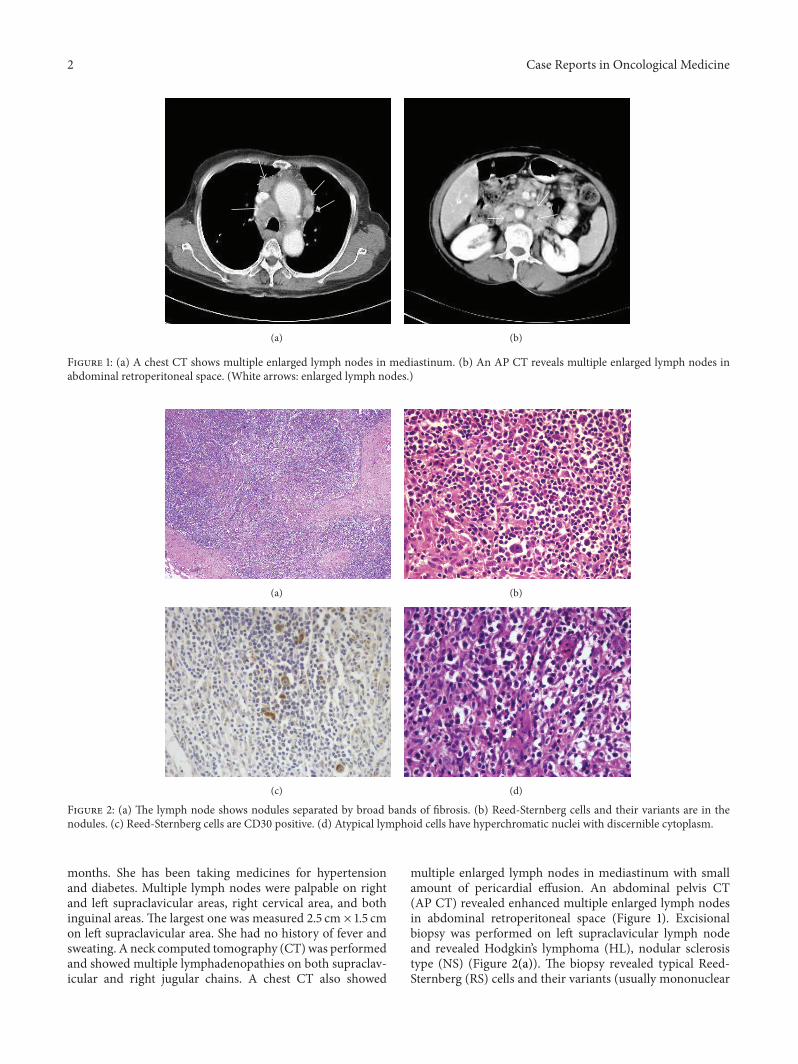
Figure 1: (a) A chest CT shows multiple enlarged lymph nodes in mediastinum. (b) An AP CT reveals multiple enlarged lymph nodes inabdominal retroperitoneal space. (White arrows: enlarged lymph nodes.)
(a) (b)
(c) (d)
Figure 2: (a) The lymph node shows nodules separated by broad bands of fibrosis. (b) Reed-Sternberg cells and their variants are in thenodules. (c) Reed-Sternberg cells are CD30 positive. (d) Atypical lymphoid cells have hyperchromatic nuclei with discernible cytoplasm.
months. She has been taking medicines for hypertensionand diabetes. Multiple lymph nodes were palpable on rightand left supraclavicular areas, right cervical area, and bothinguinal areas. The largest one was measured 2.5 cm × 1.5 cmon left supraclavicular area. She had no history of fever andsweating. A neck computed tomography (CT)was performedand showed multiple lymphadenopathies on both supraclav-icular and right jugular chains. A chest CT also showed
multiple enlarged lymph nodes in mediastinum with smallamount of pericardial effusion. An abdominal pelvis CT(AP CT) revealed enhanced multiple enlarged lymph nodesin abdominal retroperitoneal space (Figure 1). Excisionalbiopsy was performed on left supraclavicular lymph nodeand revealed Hodgkin’s lymphoma (HL), nodular sclerosistype (NS) (Figure 2(a)). The biopsy revealed typical Reed-Sternberg (RS) cells and their variants (usually mononuclear
Case Reports in Oncological Medicine 3
(a) (b)
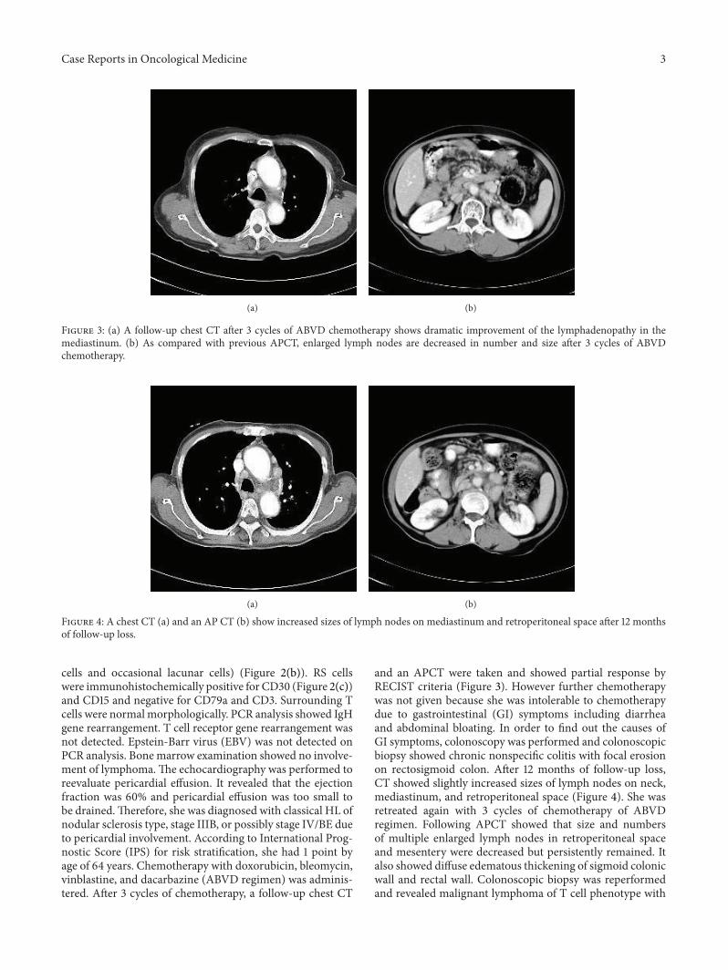
Figure 3: (a) A follow-up chest CT after 3 cycles of ABVD chemotherapy shows dramatic improvement of the lymphadenopathy in themediastinum. (b) As compared with previous APCT, enlarged lymph nodes are decreased in number and size after 3 cycles of ABVDchemotherapy.
(a) (b)
Figure 4: A chest CT (a) and an AP CT (b) show increased sizes of lymph nodes on mediastinum and retroperitoneal space after 12 monthsof follow-up loss.
cells and occasional lacunar cells) (Figure 2(b)). RS cellswere immunohistochemically positive for CD30 (Figure 2(c))and CD15 and negative for CD79a and CD3. Surrounding Tcells were normalmorphologically. PCR analysis showed IgHgene rearrangement. T cell receptor gene rearrangement wasnot detected. Epstein-Barr virus (EBV) was not detected onPCR analysis. Bonemarrow examination showed no involve-ment of lymphoma.The echocardiography was performed toreevaluate pericardial effusion. It revealed that the ejectionfraction was 60% and pericardial effusion was too small tobe drained.Therefore, she was diagnosed with classical HL ofnodular sclerosis type, stage IIIB, or possibly stage IV/BE dueto pericardial involvement. According to International Prog-nostic Score (IPS) for risk stratification, she had 1 point byage of 64 years. Chemotherapy with doxorubicin, bleomycin,vinblastine, and dacarbazine (ABVD regimen) was adminis-tered. After 3 cycles of chemotherapy, a follow-up chest CT
and an APCT were taken and showed partial response byRECIST criteria (Figure 3). However further chemotherapywas not given because she was intolerable to chemotherapydue to gastrointestinal (GI) symptoms including diarrheaand abdominal bloating. In order to find out the causes ofGI symptoms, colonoscopy was performed and colonoscopicbiopsy showed chronic nonspecific colitis with focal erosionon rectosigmoid colon. After 12 months of follow-up loss,CT showed slightly increased sizes of lymph nodes on neck,mediastinum, and retroperitoneal space (Figure 4). She wasretreated again with 3 cycles of chemotherapy of ABVDregimen. Following APCT showed that size and numbersof multiple enlarged lymph nodes in retroperitoneal spaceand mesentery were decreased but persistently remained. Italso showed diffuse edematous thickening of sigmoid colonicwall and rectal wall. Colonoscopic biopsy was reperformedand revealed malignant lymphoma of T cell phenotype with

4 Case Reports in Oncological Medicine
active ulcer alongwith necrosis on rectumand sigmoid colon,which was consistent with peripheral T cell lymphoma (TCL)involving rectum and sigmoid colon. In order to confirmperipheral TCL, enlarged right inguinal lymph node wasexcised for biopsy. Biopsy specimen of inguinal lymph noderevealed effacement of nodal architecture by an atypical lym-phoid infiltrate. The infiltrates consisted of medium to largesized lymphoid cells. The atypical cells showed hyperchro-matic nuclei and discernible cytoplasm (Figure 2(d)). Someatypical cells had open chromatin and nuclear convolution,but nuclear pleomorphism was not evident. These atypicalcells were intermixed with small reactive lymphoid cellsand plasma cells. The atypical cells were positive for CD3,indicating T cell lymphoma. PCR analysis demonstrated Tcell receptor gene rearrangement. EBV was not detected onPCR analysis. She received 3 cycles of chemotherapy withetoposide containing regimen. PTCL was regressed partiallyafter 3 cycles of chemotherapy, but she refused to receivemorechemotherapy. She died of disease progression 6months later.
3. Discussion
There are several explanations for the development of HL andsubsequent TCL such as therapy induced, immunodeficiencyrelated, and tumor biological relations [1, 3, 6]. TCLoccurringabout 2 years after chemotherapy of HL and showing moreaggressive clinical courses might be considered to be treat-ment induced [3, 6, 7].The persistent immune dysregulationsare known to be associated with HL [8]. Brown et al.described a case of HL and anaplastic large cell lymphoma(ALCL) inwhich a faint T cell receptor (TCR) gene rearrange-ment was detected in the initial HL. It suggested that thisrearrangement had arisen from an oligoclonal population ofreactive T cells because the RS cells were positive for CD20[3]. The initial and persistent oligoclonal T cell expansionmight have permitted the emergence of the ALCL. Thus,the ALCL and possibly the HL developed as a result ofa persistently abnormal immune microenvironment. Thedevelopment of HL and B cell lymphoma would be explainedby clonal progression ofmalignant B cells throughmutationalaccumulation and progression into a more aggressive, highergrade B cell lymphoma, because neoplastic RS cells are a typeof B lymphocytes in most cases [9].The pathogenesis of casesof classic HL and TCL ismore difficult to explain. HL is rarelyof T cell lineage, probably fewer than 5% of all cases [4]. Tworecent studies focused on RS cells that express aberrant Tcell antigens and found that only 15% to 20% of these harborclonal TCR gene rearrangements [10, 11]. Davis et al. reporteda case in which lymphomatoid papulosis, HL, and cutaneousTCL, occurring during a period of 14 years, were derived froma single T cell clone, as determined by PCR [11]. Therefore, itseems likely thatHL andTCLderive from the sameprecursor,similar to that of HL and B cell lymphoma. Sanchez et al.reported a case of composite HL and TCL associated withEpstein Barr virus infection and suggested the possibility ofmalignant transformation of a preexisting T cell populationdeveloped in response to a neoplastic EBV-positive RS cells[9]. Our patient was diagnosed with peripheral TCL 2 years
after the diagnosis ofHL. RS cells of our case revealed noTCRgene rearrangement and EBV on PCR. Therefore, the occur-rence of TCL in this case is probably related to chemotherapy.However, we are not sure whether composite lymphomaexisted when HL was diagnosed, because we obtained justonly supraclavicular lymph node and confirmed HL. Whenshe received chemotherapy forHL, she suffered fromdiarrheaand was diagnosed with chronic nonspecific colitis withfocal erosion by colonoscopic biopsy. Colonoscopic biopsythat was performed 2 years after diagnosis of HL revealedTCL, also supporting sequential development of HL and TCLrather than composite lymphoma. With these results takeninto account, the direct clonal relationship between HL andPTCL was observed in few reports. So, the majority of PTCLfollowing treatment of HL could result from therapy inducedimmunodeficiency rather than from clonal progression.
We report a case of HL and T cell lymphoma developingin the same patient. Although it is rare, synchronous ormetachronous HL and NHL can occur more often thanwe expect. Therefore, we may need to consider reconfirm-ing tissue when lymphoma is not regressed enough bychemotherapy. And, the further study for pathogenesis willbe required to understand the relationship of HL and NHL.
Conflict of Interests
The authors declare that there is no conflict of interestsregarding the publication of this paper.
References
[1] R. M. Amini, G. Enblad, C. Sundstrom, and B. Glimelius,“Patients suffering from both Hodgkin’s disease and non-Hodgkin’s lymphoma: a clinico-pathological and immuno-histochemical population-based study of 32 patients,” Interna-tional Journal of Cancer, vol. 71, no. 4, pp. 510–516, 1997.
[2] C.Dong andK.Hemminki, “Second primary neoplasms among53 159 haematolymphoproliferative malignancy patients inSweden, 1958–1996: a search for common mechanisms,” BritishJournal of Cancer, vol. 85, no. 7, pp. 997–1005, 2001.
[3] J. R. Brown, A. P. Weng, and A. S. Freedman, “Hodgkin diseaseassociated with T-Cell non-Hodgkin lymphomas: case reportsand review of the literature,” The American Journal of ClinicalPathology, vol. 121, no. 5, pp. 701–708, 2004.
[4] R. Kuppers, “Molecular biology of Hodgkin’s lymphoma,”Advances in Cancer Research, vol. 84, pp. 277–312, 2002.
[5] M. Muschen, D. Re, A. Brauninger et al., “Somatic mutations ofthe CD95 gene in Hodgkin and Reed-Sternberg cells,” CancerResearch, vol. 60, no. 20, pp. 5640–5643, 2000.
[6] M. Steinhoff, C. Assaf, I. Anagnostopoulos, C. C. Geilen, H.Stein, and M. Hummel, “Three coexisting lymphomas in onepatient: genetically related or only a coincidence?” The Journalof Clinical Pathology, vol. 59, no. 12, pp. 1312–1315, 2006.
[7] J. G. Krikorian, J. S. Burke, S. A. Rosenberg, and H. S. Kaplan,“Occurrence of non-Hodgkin’s lymphoma after therapy forHodgkin’s disease,” The New England Journal of Medicine, vol.300, no. 9, pp. 452–458, 1979.
[8] R. I. Fisher, V. T. deVita Jr., F. Bostick et al., “Persistentimmunologic abnormalities in long-term survivors of advanced

Case Reports in Oncological Medicine 5
Hodgkin’s disease,” Annals of Internal Medicine, vol. 92, no. 5,pp. 595–599, 1980.
[9] S. Sanchez, H. Holmes, N. Katabi et al., “Composite lympho-cyte-rich Hodgkin lymphoma and peripheral T-cell lymphomaassociated with Epstein-Barr virus: a case report and review ofthe literature,” Archives of Pathology and Laboratory Medicine,vol. 130, no. 1, pp. 107–112, 2006.
[10] V. Seitz,M.Hummel, T.Marafioti, I. Anagnostopoulos, C.Assaf,and H. Stein, “Detection of clonal T-cell receptor gamma-chaingene rearrangements in Reed-Sternberg cells of classic Hodgkindisease,” Blood, vol. 95, no. 10, pp. 3020–3024, 2000.
[11] T. H. Davis, C. C. Morton, R. Miller-Cassman, S. P. Balk, andM. E. Kadin, “Hodgkin’s disease, lymphomatoid papulosis, andcutaneous T-cell lymphoma derived from a common T-cellclone,” The New England Journal of Medicine, vol. 326, no. 17,pp. 1115–1122, 1992.

Submit your manuscripts athttp://www.hindawi.com
Stem CellsInternational
Hindawi Publishing Corporationhttp://www.hindawi.com Volume 2014
Hindawi Publishing Corporationhttp://www.hindawi.com Volume 2014
MEDIATORSINFLAMMATION
of
Hindawi Publishing Corporationhttp://www.hindawi.com Volume 2014
Behavioural Neurology
EndocrinologyInternational Journal of
Hindawi Publishing Corporationhttp://www.hindawi.com Volume 2014
Hindawi Publishing Corporationhttp://www.hindawi.com Volume 2014
Disease Markers
Hindawi Publishing Corporationhttp://www.hindawi.com Volume 2014
BioMed Research International
OncologyJournal of
Hindawi Publishing Corporationhttp://www.hindawi.com Volume 2014
Hindawi Publishing Corporationhttp://www.hindawi.com Volume 2014
Oxidative Medicine and Cellular Longevity
Hindawi Publishing Corporationhttp://www.hindawi.com Volume 2014
PPAR Research
The Scientific World JournalHindawi Publishing Corporation http://www.hindawi.com Volume 2014
Immunology ResearchHindawi Publishing Corporationhttp://www.hindawi.com Volume 2014
Journal of
ObesityJournal of
Hindawi Publishing Corporationhttp://www.hindawi.com Volume 2014
Hindawi Publishing Corporationhttp://www.hindawi.com Volume 2014
Computational and Mathematical Methods in Medicine
OphthalmologyJournal of
Hindawi Publishing Corporationhttp://www.hindawi.com Volume 2014
Diabetes ResearchJournal of
Hindawi Publishing Corporationhttp://www.hindawi.com Volume 2014
Hindawi Publishing Corporationhttp://www.hindawi.com Volume 2014
Research and TreatmentAIDS
Hindawi Publishing Corporationhttp://www.hindawi.com Volume 2014
Gastroenterology Research and Practice
Hindawi Publishing Corporationhttp://www.hindawi.com Volume 2014
Parkinson’s Disease
Evidence-Based Complementary and Alternative Medicine
Volume 2014Hindawi Publishing Corporationhttp://www.hindawi.com