book_final_jan. 09.indb
TRANSCRIPT

ch. 4 / Contact Lenses and the Wrangling Thereof 27
Contact Lenses and the Wrangling Thereofch 4

Diabetic Retinopathy 28
Becoming familiar with the contact lenses that are used to treat diabetic retin-opathy is crucial. It is assumed that you have already mastered typical indirect non-contact lenses, such as the 90-diopter lens. But it turns out that contact lenses require a very different skill set, so they get their own chapter.
Contact lenses come in two main types: direct view lenses, such as the Gold-mann three mirror, and inverted image lens system such as the Rodenstock. But fi rst, a brief editorial…
This chapter will refer to several types of lenses. If you look at the manufac-turers’ catalogs, you will see that there are zillions of options. How can you try them to see whether they work for you? One option is to go to the manu-facturers’ exhibits at conventions. You will get a chance to try them all, but they never work as well on patients as they do on the little model eyes they use to demonstrate the lenses.
Another option is to dig around the back of all the drawers where your laser is kept. You will likely fi nd a host of abandoned lenses, especially if you are in a large group practice or academic setting. Sometimes you will quickly realize why a given lens is in the graveyard, but sometimes you will fi nd a real friend that works great for you. This also saves you a trip to the Academy meeting.
Above all, do not be fooled by the advertising that will have you thinking you will be able to treat patients effortlessly if you buy just the right lens. As you begin grappling with contact lenses, it is easy to think that your problems are due to the lens and that, somewhere over the rainbow, there is a perfect lens that will solve all your problems. You need to get over this phase quickly (oth-erwise, you will be spending a lot of money on lenses). It just takes practice—there is no secret magic lens.
Point-Counterpoint Box so the Lens Manufacturers Don’t Get Too Ticked-off by the Previous Box Because We Need and Appreciate Their Constant Innovations
Although there is no magic lens that gets you over the hump of the learn-ing curve, there is something to be said for having lenses that differ in subtle ways in order to address different nuances of treatment. Just like some gui-tarists prefer having a bunch of different instruments and others always use one favorite axe, you may fi nd that you do better with lots of different lenses, or you may be happy with only one or two. Ultimately, this is something you will decide on your own once you have some skills with the basic lenses, so read on.

ch. 4 / Contact Lenses and the Wrangling Thereof 29
The direct view lenses are the easiest and most intuitive to use. The archetype is the Goldmann three mirror lens. This is the lens to have if you are stranded on your basic desert island, because it can do everything reasonably well. The direct (non-inverted) nature of the view means that once you get the lens on the patient, you are simply looking in a straight line through the pupil to the area of interest. The various mirrors then allow you to visualize segments of the periph-ery. Some Goldmann lenses have a small fl ange that fi ts behind the lids and helps to keep the lens in the patient’s eye; it is a good idea to take advantage of such a fl ange when you are learning, because it is harder for the patient to blink the lens out once you get it in the eye. (More specifi c tips on actually getting the lens where you want it to go are covered in Chapter 7.)
The mirrors are set at different angles, with most Goldmann-type lenses having one mirror for the anterior chamber angle and two mirrors for differ-ent latitudes of the fundus. There are, however, variations on the Goldmann which have multiple mirrors at slightly different angles to get better cov-erage of the retinal periphery (the Karickhoff lens, for instance, has four mirrors).
The direct view makes it relatively easy to line up the lens so you can see the posterior pole, and you do not need to invoke the mental gymnastics that are necessary to use lenses that have an inverted image. Unfortunately, the fi eld of view is rather small compared to the indirect lenses, and you will be more dependent on the patient’s cooperation if you need to get to different areas. Also, direct lenses need a widely dilated pupil, and media
opacities can be a real pain, because you cannot work around them as you can with an inverting lens.
Because the mirrors are set at a fi xed angle and there is a small fi eld of view, you need to make sure that you do not miss areas that lie between the latitudes most easily seen in each mirror. If you cannot quite get the view you need be-cause the place you want to see is just outside the limited view provided by the mirrors or the direct view to the posterior pole, you do have some options. You can rock the lens back and forth to get more anterior and posterior exposure as you are treating the retina. The patient can also help you by looking a bit away and toward the mirror to accomplish the same goal. Finally, you can also use the contact lens as a gentle lever to push the eye in different directions. This last option can be done with any type of contact lens, and it is an important skill that will give you a lot of control over the eye. For instance, if the patient has had a retrobulbar block, you have to use the lens to move the eye around to see dif-ferent areas. You also need to maintain the proper alignment when you do this, though, because if you angle the lens too much as you push the eye in different directions, you will lose your view. You have to fi ne-tune your fi nger propriocep-
DIRECT VIEW LENSES
daisbil
Figure 1. A typical three mirror Goldmann lens.(Courtesy of Ocular Instru-ments.)

Diabetic Retinopathy 30
tion so that you automatically know how the lens is oriented as you move it in different directions. (Figure 8 will elaborate on this.)
Figure 2. If you are using a Goldmann three mirror, you have to be very careful about where you are treating with the large mirror. It is possible to accidentally get well into theposterior pole, especially if thepatient is looking toward themirror. The red arrow represents the fovea as it moves toward the line of treatment when the eyerotates toward the mirror.
Figure 3. Yanuzzi macular contact lens. Note the very wide fl ange. This can takea bit of work to get into the eye, but it really locks the lens onto the eye once it isbehind the lids. (Courtesy of Ocular Instruments.)
Important safety tip: If the eye has not been blocked and you are pushing with your lens in order to move it around, you may inadvertently demonstrate the oculo-cardiac refl ex—especially if you happen to be treating a patient who is nervous and uncom-fortable (and this goes double if they are young males—triple if they have Harley-Davidson tats). Always remind a patient who is about to get their fi rst laser to let you know if they begin to feel light-headed or dizzy, and if they do, stop the laser imme-diately and have them do the head-between-the-knees thing or even lie down on the fl oor. For some patients, the time between this light-headed sensation and becoming unconscious is rather short, and you lose many points with the family if your patient’s face fi nds the cross members of the slit lamp table on its way to the ground. This con-cept is important enough that it is repeated at various points around the book.
There is another type of direct view lens that is designed exclusively for viewing the posterior pole. In some institutions these lenses are referred to as pancake lenses, presumably because they’re smaller than a Goldmann three mirror and because they do not have mirrors (perhaps these quali-ties make ophthalmologists think of pancakes). An example of this is the Yanuzzi lens, although there are many other types that are available.
Because you cannot get a very big picture with the “keyhole” view through the pupil, you also need to be very careful that your treatment is not extending more posteriorly than you wish. This can occur with the larger mirror and is especially likely if you are working temporally, where there are no large blood vessels to warn you that you are crossing into the macula. It is possible to inadvertently angle the lens and treat into the posterior pole without realizing it, especially if the patient happens to be looking in the direction of the mirror (Figure 2).

ch. 4 / Contact Lenses and the Wrangling Thereof 31
These lenses often have a very large fl ange that really keeps the lens behind the eyelids—you may even be able to let go of the lens so the patient can sit back and rest and it will remain in place. Although the overall fi eld of view is limited by the direct line of sight, and these lenses are more dependent on patient cooperation, they give a breathtaking sense of the thickness of the retina. You should try to use them as much as possible, especially as you are learning the trade. The axial magnifi cation and clarity of these lenses can really help you comprehend the nature of diabetic macular edema and help you get a feel for the three-dimensional location of the pathology. The patient’s retina becomes a wonderland as you gleefully pluck microaneurysms from perches that suddenly seem yards above the underlying RPE. After you have a few exams under your belt with one of these lenses, you will understand what diabetic macular edema is about in a way that no ocular coherence tomography scan can capture. You will also appreciate what a feeble imitation of reality you get when you use a 90- or 78-diopter lens—no matter what the advertisements say. However, studying a lot of patients with a macular lens like this will enable you to be a much more effective examiner when you decide to cut corners and use a 90 or 78.
By the way, there are some other options to try to get a nice stereoscopic view without resorting to a contact lens—but like most things in life, the easier way is usually not the best way. You can get a 60-diopter lens (or something close to that number). The lower dioptric power will give you more axial magnifi cation—you will get an enhanced stereoscopic view that can be almost as good as a contact lens. However, it is harder to line up both of your eyes through a 60-diopter; the patient has to be very well dilated and cooperative. If you can easily get the info you need from such a lens, then more power to you, but usually it is better to just put on the contact lens.
Another non-contact option is the Hruby lens. This is a plano-concave gizmo that can be attached to the front of your slit lamp. It works by neutralizing the corneal curvature from a distance, and you usually click it down or lift it into a slot so that it is directly in your line of sight, and then you can study a non-in-verted image of the macula. There is only a small fi eld of view, and you are very dependent on patient cooperation, but the stereo is pretty good. Most folks fi nd that the Hruby is too tricky to use, but you should at least try it if you have one on your slit lamp.
Diabetic Retinopathy 32
Unfortunately, these lenses can be frustrating. One begins with the preconceived notion that one simply needs to slap on the lens and one will immediately see broad vistas of retina. Just like the fi rst time you tried to ski, snowboard or ice skate, however, the reality is a bit different from the expectations. Strive to overcome your initial disappointment and keep trying—the necessary moves will become automatic with practice, and you will soon become a contact lens Jedi.
Indirect lenses require a much more dynamic approach than the direct view lenses. At all times you need to try to keep a straight line from the patient’s retina through the lens and slit lamp and onto your fovea—something that is much easier said than done with this class of lens. (In reality the optics are more complicated, but the straight-line approach is a good mental goal to start with.)
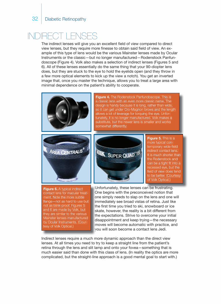
INDIRECT LENSESThe indirect lenses will give you an excellent fi eld of view compared to direct view lenses, but they require more fi nesse to obtain said fi eld of view. An ex-ample of this type of lens would be the various Mainster lenses made by Ocular Instruments or the classic—but no longer manufactured— Rodenstock Panfun-doscope (Figure 4). Volk also makes a selection of indirect lenses (Figures 5 and 6). All of these lenses essentially do the same thing that your 90-diopter lens does, but they are stuck to the eye to hold the eyelids open (and they throw in a few more optical elements to kick up the view a notch). You get an inverted image that, once you master the technique, allows you to treat a large area with minimal dependence on the patient’s ability to cooperate.
Figure 4. The Rodenstock Panfundoscope. This isa classic lens with an even more classic name. Thedesign is handy because it is long, rather than wide,so it can get under Cro-Magnon brows and the length allows a lot of leverage for torquing the eye. Unfor-tunately, it is no longer manufactured. Volk makes asubstitute, but the newer lens is smaller and workssomewhat differently.
an be frustrating
Figure 5. This is amore typical con-temporary wide-fi eld indirect contact lens.It is much shorter thanthe Rodenstock andcan be a tight fi t into a recessed eye, but the fi eld of view does tend to be better. (Courtesy of Volk Optical.)
UOoitstdmy
Figure 6. A typical indirectcontact lens for macular treat-ment. Note the more subtle fl ange—not as hard to use butnot as blink-proof. Figures 5and 6 are made by Volk, but they are similar to the variousMainster lenses manufactured by Ocular Instruments. (Cour-tesy of Volk Optical.)

ch. 4 / Contact Lenses and the Wrangling Thereof 33
Even if you can get the patient lined up to obtain the best view, you need to continually coordinate movements between the slit lamp, the lens and the eye in subtle ways. This can be very frustrating at fi rst because it feels like you will never succeed. It helps to break this process down into a few separate moves until it becomes automatic...
You have to take full advantage of all the different ways you can shift the lens around the eye. A common problem is not being able to get a good view of what you want to see with both of your eyes—a classic sign that you are not as lined up as you think you are. Try moving the lens in a circle or a cone—this is a handy way of scanning for the best line of sight (Figure 7). Remember, you have to be ready to follow any move you make with the lens by shifting the position of the slit lamp. This last point is really important. An understandable novice move is to concentrate on moving just the lens, because it is diffi cult enough to manipulate the lens without letting it slide off the eye. However, if you move only the lens without following that inverted image with the slit lamp, you will never get a good view. It helps to affi x your lens hand to the patient by spreading your fourth and fi fth fi ngers onto the side of the patient’s face or by putting the back of those fi ngers on the patient’s cheek—this gives you more stability and you don’t have to exert as much brain power worrying about keeping the lens on the eye. This way you can better concentrate exclusively on moving the lens and the slit lamp as needed in order to get the best view.
Figure 7. This fi gure shows slightly different ways of moving the lens around the eye in order to fi nd the best view. These are an exaggeration of the movements required—thereal moves are much more subtle. The top image shows a perfectly lined up lens, but this almost never happens spontaneously. The quickest way to fi nd the best view is to rotate the lens around the presumed visual axis. The lower left image shows that you can rotatethe axis of the lens so that it creates a cone. In this case, the part of the lens on the eyemoves just a little, and the part of the lens facing the slit lamp circles around a lot. Thelower right image shows that you can move the lens such that its axis creates a cylinder, with the entire lens moving in the same circular pattern. In reality, you will rapidly learn tocombine both moves to scan for the best view—but it helps to realize that each one of these motions will give a slightly different effect, which may come in handy in different cir-cumstances. Remember to follow any moves you make with the lens with correspondingmovement of the slit lamp; otherwise, you are wasting your time. Note that if you still can’t get a great view, you need to recheck where the patient is looking—they may be rotating their eye out of reach.

Diabetic Retinopathy 34
Figure 8. These are exaggerated images of moves you can make to fi ne-tune your view with an indirect contact lens. On the left, you are moving the entire lens in one direction and keeping the axis of the lens parallel as you move it. At thesame time, you are “pulling” the eye with you to try to line up the axis of the lenswith whatever you need to see. The image on the right shows a slightly different way of doing the same thing; here, you are torquing the lens through its center asyou also rotate the eye, so the axis of the lens is tilting. Both moves accomplishthe same goal, but they will give you slightly different views depending on theorientation of the patient’s eye. You will rapidly learn to use a combination of bothmoves to get the best view, but it helps to consciously try them out at fi rst—with practice, your brain will create a macro that automatically moves the lens into thebest position using whatever move is necessary.
Don’t worry if you aren’t getting a perfect view at fi rst—the goal here is to just see what you want to see with both eyes at the same time. Once you can do this reliably, it is time for some real fi nesse: the “Five Point Palm Exploding Heart Technique” of lens wrassling, if you will. It is similar to the technique mentioned in the section on direct lenses, wherein you use the lens to torque the eye in different directions. Sometimes, you will do this by translating the entire lens in one direction, and sometimes you will do this in a more subtle way: by slightly rotating the lens around its midpoint (kind of like adjusting the pitch of an air-plane). Both moves will slightly change the visual axis of the lens and will slightly rotate the eye at the same time, with the goal being a much more favorable view. (If this is an obtuse paragraph, look at Figure 8, where the proverbial thousand words await). By the way, if you are having a lot of trouble getting a view, even with all these moves, go back and double check where the patient’s fi xation is. There is no way, at least at fi rst, that you can get a good view if the patient is rolling their eye up in their head—you will never line things up. (There will be more discussion about enlisting the patient’s cooperation in Chapter 7.)

ch. 4 / Contact Lenses and the Wrangling Thereof 35
How do you know which way to tilt the lens to get the best view? At fi rst, you may need to use very gross and even random movements to get a rough view of what you want to see. As things get fi ne-tuned, you can sometimes let Dr. Sturm and his conoid guide you. Look at your aiming beam. Unless you are a natural, it is likely the aiming beam will be some sort of smeared oval, suggesting that you are out of alignment to a tiny degree. The long axis of the oval tells you which way to rock the lens. Say the long axis is vertical—then tilt the lens up and down around its center and watch the beam—it will shift around and tighten up into a circle as you become more perfectly aligned. Sometimes you have to be more aggressive and move the whole lens up or down to follow the long axis of the aiming beam and then fi ne-tune with a little gentle tilting. Sometimes you can rotate the eye with the lens as you tilt it and get to your goal even faster. Don’t forget to follow your lens moves with the slit lamp, although as you get closer and closer to a good view, you usually do not need to move the microscope as much.
Sadly, there will still be a lot of hit and miss, and you will fi nd that you have to keep iterating and reiterating all of these moves to get in focus and stay in focus. First, get lined up grossly by circling the visual axis. Then try some tilting to offset the long axis of the aiming beam—then do a little more circling and tilting, and ultimately you will be rewarded with a lovely panoramic view that makes treatment very easy. Eventually, all these actions will be internalized and, as with indirect ophthalmoscopy, you will begin to automatically make the right moves without thinking. Just don’t give up and do keep trying.
You will need different indirect lenses depending on whether you want to treat the macula or the periphery. Macular lenses don’t get out very far, but they give you a nice stereoscopic view of the posterior pole. Wide-fi eld lenses, on the other hand, will let you see much more of the periphery (you will have to ignore the impressive marketing names such as Ultra-Quadro-Magnoview, etc., and ask about the fi eld of view as measured in degrees in order to fi nd out exactly how much of the periphery you are supposed to see). The wide-fi eld lenses will allow you to see the posterior pole, but the view is nowhere near as stereoscop-ic as that obtained with a lens designed to view the macula. Furthermore, there tend to be a lot of light refl exes when viewing the central retina with a wide-fi eld lens—which make viewing the macula problematic. (Sometimes you can mini-mize the refl exes by moving your slit beam a bit off axis and/or by decreasing the size of the slit lamp beam.)
Finally, the wider the fi eld of view, the more the lens will magnify the size of your spot, which means that it is hard to safely place small focal burns in the macula with such a lens. (See the next blue box.) All this is why there is not one indirect lens that can be used to treat both the periphery and the macula. Predictably, manufacturers have created a host of different indirect lenses that cover the whole spectrum from very wide-fi eld to very focal macular viewing. As men-tioned in the beginning of the chapter, you may want to get comfortable with just one macular and one wide-fi eld lens, and then you can decide whether you want all the more nuanced lenses that are available.

Diabetic Retinopathy 36
There are two situations in which the wider-fi eld indirect lenses can be useful beyond their ability to allow easy panretinal photocoagulation (PRP). First, if the patient has media opacities, such as a central posterior subcapsular cataract, you may be able to use these lenses to treat the posterior pole. They tend to “reach around” the opacity better than indirect lenses designed for the macula, and certainly better than direct lenses. As mentioned above, you need to do this carefully because you will be creating a large diffused spot, but it may be better than nothing in diffi cult situations.
The other situation in which the wide-fi eld indirect lenses are extremely useful is when you are looking for retinal breaks in pseudophakic patients. The prismatic effect of the edge of the intraocular lens will often keep you from seeing the far periphery with a direct view lens. A wide-fi eld lens allows better visualization, and such a lens can allow you to fi nd and treat a peripheral tear without the need for a binocular indirect laser or cryotherapy. This last point is a bit off-topic for a book on diabetic retinopathy, but it is good to know.
Indirect lenses and spot size
With the direct view lenses, the spot you set on your laser is pretty much the size you will get on the retina, give or take a few microns. The indirect lenses, however, will change the spot size in a manner proportional to the fi eld of view. For instance, a really wide-fi eld lens, like the Mainster 165, can almost double the diameter of your spot setting (a 100-micron spot on the laser turns into a 200-micron spot on the retina). Indirect lenses designed for the macula tend to deliver spot sizes that are closer to the actual laser setting, but they will still vary depending on the width of the fi eld. Some “high-power” macular lenses can actually make the delivered spot smaller relative to the laser setting.
This becomes very important if you are trying to deliver a specifi c dose of laser energy to a given area (such as with photodynamic therapy for macular degeneration). It is less important in the setting of diabetic lasers, because you will be titrating the energy dose yourself, based on the uptake you see as you do the laser.
Nevertheless, you need to be aware of the effect your chosen lens has on the delivered spot size—lens manufacturers always include this information with each lens and publish it on their websites. If you are trying to, say, follow the Diabetic Retinopathy Study guidelines in terms of spot size and number, you need to realize that if you are using a wide-fi eld lens and you set your laser to 500 microns, you will be placing much bigger burns than you want. Alterna-tively, if you switch from a wide-fi eld lens to a macular lens, you will make your spot size much smaller. If you do not adjust your power accordingly, you can burn a hole in the retina.

ch. 4 / Contact Lenses and the Wrangling Thereof 37
What specifi c lenses should be in your toolbox for retinal lasers? It’s a matter of personal taste. There are two main companies that make lenses: Volk and Ocular Instruments. They both make great laser lenses and their product lines tend to be roughly equivalent (although the manufacturers themselves might disagree). They both have return policies, so you can try out differ-ent lenses and decide what works for you. Here are some suggestions for a basic lineup:
1. A Goldmann three mirror can do everything reasonably well, but it may take longer to do a treatment and it is not as versatile as sepa-rate lenses. Nevertheless, if you can get only one lens, this is the one to have. If you have been good and the Retina Bunny is going to bring you more than one lens, then please read on...
2. A general-purpose indirect macular lens such as the Volk Area Centralis or the Ocular Instruments Standard Mainster will likely become your main lens for focal and grid laser. There are plenty of variations, so you can pretty much choose exactly how much fi eld of view you want—but don’t get a fi eld of view that is too big, because the wider the fi eld of view, the harder it is to treat the macula accu-rately.
3. A pancake lens for an extra-crisp view of the macula (i.e., for diagnosis or very accurate focal treatment of microaneurysms in cooperative patients). Examples would be the Yanuzzi lens by Ocular Instruments or the Fundus 20 by Volk.
4. A wide-fi eld indirect lens for PRPs such as the Mainster 165 by Ocular Instruments or the Super Quad 160 by Volk.
One other weird thing about wide-fi eld indirect contact lenses: The nature of their optics is such that the irradiance can be higher through the patient’s lens and cornea than at the retina. This effect is usually insignifi cant, but it can become a problem with huge (greater than 500-micron) spot sizes—although one would usually not use such large spots. The problem is that if there are a lot of lens opacities the high irradiance through the anterior segment can cause lenticular burns. Corneal burns can also occur with even smaller spots if there is pigment on the surface of the cornea or if you trap an eyelash or mascara under the con-tact lens. Furthermore, it is likely you won’t realize that these burns are occur-ring because you are not able to easily visualize the anterior segment through an indirect-view contact lens. This is why you need to stop and take the contact lens off and look at the front of the eye if you fi nd that your view of the retina is cloud-ing up. These problems are relatively unusual with modern treatment techniques, but it is good to remember that Murphy’s Law can extend far beyond the retina that you are working on. (There will be more on this in Chapter 16, discussing complications.)

Diabetic Retinopathy 38
References and Suggested Reading
Mainster MA, Crossman JL, Erickson PJ, Heacock GL. Retinal laser lenses: magni-fi cation, spot size, and fi eld of view. Br J Ophthalmol 1990;74:177-9.
Folk JC, Pulido JS. Laser photocoagulation of the retina and choroid. San Fran-cisco: American Academy of Ophthalmology, 1997.
L’Esperance FA. Ophthalmic lasers, 3rd ed. St. Louis: Mosby, 1989.

ch. 5 / Diabetic Macular Edema—the Basics 39
Diabetic Macular Edema—the Basicsch 5

Diabetic Retinopathy40
DOING THE EXAM in 2-D
This chapter discusses macular edema resulting from microvascular leakage around the posterior pole. Basically, diabetes turns the retinal capillaries into the vascular equivalent of leaky old garden hoses. The result is that patients develop microaneurysms, hard exudates, and hemorrhages in varying amounts. If there is a lot of leakage from the damaged vessels, then the retina will swell up like a sponge. If this swelling builds up in and around the center of vision, then per-manent damage can occur, and the goal is to identify swelling and treat it well before this happens.
Besides causing leaky blood vessels, diabetes can also just kill off blood ves-sels. Most of the time, there is a combination of both problems—vascular leak-age and capillary death. In some patients, the destruction of blood vessels is the predominant problem, and this is referred to as capillary dropout or macular ischemia. This is always bad and it can cause marked vision loss; so far there is no treatment other than prevention with good systemic control and by trying to address any treatable leakage. Although capillary dropout can cause retinal edema at fi rst as a result of ischemia, the end result is a thinned-out retina. It usually requires a fl uorescein angiogram to identify this problem, although it can be inferred if patients have marked vision loss and an atrophic-appearing fovea. (Figure 7 is an example of capillary dropout around the fovea.)
Unless the patient is truly unlucky, ischemia is usually not a predominant feature in the early stages of diabetic macular edema. Instead, vascular leakage tends to be the initial fi nding. Because this leakage is very treatable, it is crucial to be able to identify the clinical signs that indicate the beginnings of damage. At the very start of your career it is exciting to simply be able to see these fi ndings—your fi rst direct glimpse of a disease hard at work. However, once you master the mechanics of examining the fundus it is easy to become jaded about spot-ting the signs of retinopathy. Try not to let this happen. You can get a lot of clues about a patient’s situation just by looking carefully at each of the various mani-festations.
For instance, you are no doubt aware that most intraretinal hemorrhages in diabetic retinopathy are blot-shaped because they stem from broken capillaries in the outer retinal layers, where the neurons are all jumbled together. As a result, the hemorrhage seeps out radially like a drop of food coloring on a paper towel. Flame-shaped hemorrhages occur when capillaries break in the more superfi cial nerve fi ber layer, where the linear arrangement of the axons spreads the blood lengthwise rather than in all directions.
You may think that you are too cool to care about this second-year medical student stuff. You aren’t. If a patient has an excessive number of fl ame-shaped hemorrhages and/or dot-blot hemorrhages, you should worry about the pres-ence of additional vascular risk factors that are not well controlled. The most likely culprit would be superimposed hypertension, but you might also see this with progressive renal failure or anemia. Many such patients also have poor compliance—usually due to a combination of lack of motivation, lack of insur-ance or lack of a motivated primary-care doctor. Based on a few red smears it is

ch. 5 / Diabetic Macular Edema—the Basics 41
possible to make massive inferences about everything from a patient’s creatinine to their socioeconomic status—and your deductions and consequent actions can have a dramatic impact on how the patient responds to your ministrations.
Figure 1. A patientwith macular edemaand multiple hemor-rhages. This patienthad severe hyper-tension, early renal failure with second-ary anemia and hadbeen uninsured and unable to afford an eye exam untilhe could no longerfunction. You caninfer a lot from a retina.
Another hemorrhagic nuance occurs in patients who are taking Coumadin. These patients will often have many more hemorrhages in the retina relative to their overall degree of retinopathy (in other words, they have more hemorrhages than you would expect, given the number of microaneurysms and non-hemor-rhagic vascular changes that you see). All these hemorrhages may have vary-ing sizes and unusual shapes. Make sure that your patients that are on this rat poison are really getting their levels checked; you will fi nd occasional patients that are not being monitored properly. A more complete discussion of this drug in terms of diabetic retinopathy is found in Chapter 25.
In Chapter 1, it was pointed out that we all went into ophthalmology be-cause taking care of an entire patient is not our bag, man. If we could get the eye mailed to us—without the attached patient—that would be fi ne. How-ever, if you see worrisome hemorrhages, it is defi nitely time to dust off those atrophied clinical skills and check a blood pressure. Right there in the lane. While you are at it, you should also order a CBC, hemoglobin A1c and renal studies if no one has done them lately. These tests will identify signifi cant problems much faster than a referral letter will, and the results will jumpstart the patient’s care. Oh yeah, you might also help save their life (which is a nice break from a day full of “better one, better two”). The point is to try to take advantage of all the information the fundus is willing to give you. If you look, but do not see, you will be failing to treat the patient’s eye properly (and also be very un-Zen).

Diabetic Retinopathy42
Cotton wool spots are another fundus fi nding that can tell you a lot about the patient. These used to be considered very important in terms of predicting future proliferative disease, but this has been disproven (which makes one wonder what other “facts” will be disproved in the future, which, in turn, makes one glad this book is produced with software and not woodcuts). A few scattered cotton wool spots are to be expected, and individual spots may last for several months. However, if there are a lot of cotton wool spots or if crops of new lesions appear rather quickly, it may signal problems with hypertension, renal failure, or hemato-logic abnormalities. Never forget that patients are also allowed to get completely unrelated problems, and it is always possible that a patient with lots of cotton wool spots may have an additional disease such as AIDS, retinal vasculitis or radiation retinopathy. Given the overall sturm and drang of diabetic retinopathy, it may be diffi cult to dissect out the presence of these other diseases unless you remember to think of them in the fi rst place. (Check Chapter 26 for the full scoop on this.)
When actively studying hemorrhages and cotton wool spots, the Renaissance Retina Observer also inspects the hard exudate situation. Hard exudates begin to appear as more and more leakage occurs. You can think of them as high-wa-ter marks—the serum bathtub rings that outline where the retina is desperately trying to suck the abnormal fl uid back into the capillaries and the leftover protein and lipid congeal into little yellow lumps. These lumps may be all over, but often they show up on the border between the healthy and damaged retina. Large amounts of hard exudates should always suggest the possibility of hyperlipi-demia, so be sure to inform their medical doctor. Patients should be trained to consider their lipid profi le to be as important as their blood pressure and hemo-globin A1c. Tight lipid control is a little-recognized aspect of total diabetic care, at least in the ophthalmic community, and pointing out to the patient that you can see “all those little fatty deposits” in their retina may be more of a motivator for healthy living than weeks of diabetic education classes.
There is always something a bit mysterious, even to sophisticated pa-tients, about having someone look into one’s eye and being told that dam-age is visible. Sometimes, this can be a very effective tool for encouraging patients to take better care of themselves. Sometimes, however, it can be very depressing for patients to hear this—and you need to be sensitive to this as well. This is a good reason why it really is better that we don’t get the eyes mailed to us. Chapter 20 will elaborate on issues like this a bit more.
ch. 5 / Diabetic Macular Edema—the Basics 43
Figure 2. This kind of extensivehard exudate formation, especial-ly along peripheral vessels, is verysuggestive of hyperlipidemia.
DOING THE EXAM in 3-D
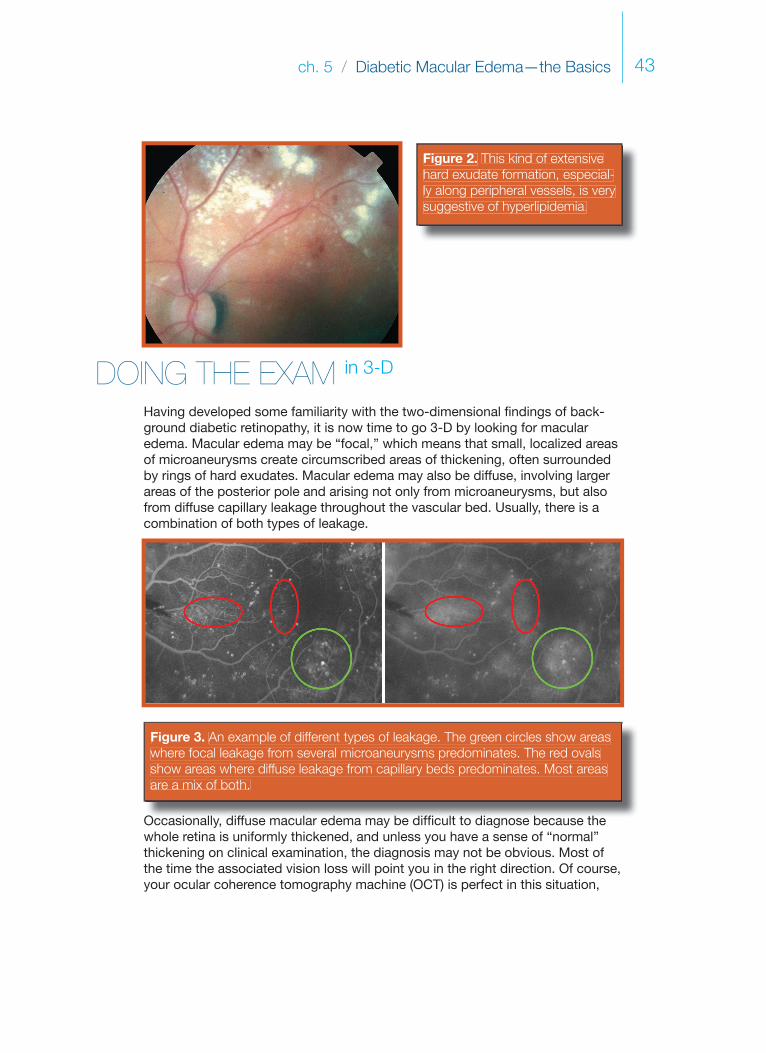
Having developed some familiarity with the two-dimensional fi ndings of back-ground diabetic retinopathy, it is now time to go 3-D by looking for macular edema. Macular edema may be “focal,” which means that small, localized areas of microaneurysms create circumscribed areas of thickening, often surrounded by rings of hard exudates. Macular edema may also be diffuse, involving larger areas of the posterior pole and arising not only from microaneurysms, but also from diffuse capillary leakage throughout the vascular bed. Usually, there is a combination of both types of leakage.
Figure 3. An example of different types of leakage. The green circles show areaswhere focal leakage from several microaneurysms predominates. The red ovalsshow areas where diffuse leakage from capillary beds predominates. Most areas are a mix of both.
Occasionally, diffuse macular edema may be diffi cult to diagnose because the whole retina is uniformly thickened, and unless you have a sense of “normal” thickening on clinical examination, the diagnosis may not be obvious. Most of the time the associated vision loss will point you in the right direction. Of course, your ocular coherence tomography machine ( OCT) is perfect in this situation,

Diabetic Retinopathy44
but not everyone will have access to such high-tech stuff, so do try to perfect your clinical exam. (There is more on OCT at the end of the chapter.)
Perfecting Your Clinical Exam
It is absolutely crucial that you take the time to put a diagnostic contact lens on these patients, especially in the early stages of your diabetic retinopathy treat-ment career. There is simply not enough axial magnifi cation with the indirect slit lamp lenses (such as a 90- or 78-diopter lens) to really allow you to get a sense of how succulent the retina can become.* It helps to visualize individual microaneurysms, hemorrhages and exudates and to note their height above the pigment epithelium at various places in the posterior pole. You can get a very defi nite feel for how thick the retina is by going back and forth between fl atter, more peripheral areas and more swollen central areas. A thin off-axis slit beam helps somewhat in bringing out the three-dimensional structure, but nothing helps as much as just getting the contact lens on the patient and doing the exam multiple times.
It is also very instructive to look at a recent fl uorescein angiogram as you are examining the patient (and defi nitely when you are performing laser treatment). Carefully study each little lesion in the fundus and compare it to the angiograph-ic appearance. You will note that many of the tiny red dots that you assume to be microaneurysms on clinical examination actually do not light up at all on the angiogram. These are simply dot hemorrhages and you may be wasting laser spots (and the patient’s non-expendable RPE) if you shoot at them. Worse, these dot hemorrhages may readily take up laser energy and change color very easily—one of the criteria for successful treatment—and you may feel like you have done a great job, but you may simply have toasted valuable portions of the patient’s nerve fi ber layer. On the other hand, there will be many real microaneu-rysms that are invisible on your initial clinical exam but will then become appar-ent when you trace their location on the angiogram and track them down in the patient’s fundus. These represent your true targets. Chapter 8 will cover all of this at length, but you have to know what you are looking for by doing the drills discussed here.
After tracing all of these lesions out on the angiogram and then fi nding them in the fundus on several patients, you will fi nd that you are better and better at identifying these tiny lesions without an angiogram. Even if you try this only two or three times, you will be amazed at how your clinical skills will improve (and
*Remember, a 60-diopter lens does have more axial magnifi cation, but it is hard for even an experienced user to stuff both visual axes through this lens and the patient’s pupil and have both images be in-focus enough to get a good view. Best to work with a contact lens to build up experience, and then you may want to try a lens of this power—although as you improve your exam you prob-ably won’t need the extra axial magnifi cation, anyway.
ch. 5 / Diabetic Macular Edema—the Basics 45
Stereo Photographs
In the days when retinal photographers used fi lm, it was common to obtain stereo photos of fundus pathology. You should ask around, because even in the most digital photography departments, they may have some old stereo slides of diabetic retinopathy in the fi les. Another good source of stereo photos can be found in some of the older, beat-up textbooks in the back of your departmental library—especially the ones that have the little discs and 3-D viewer in the back. For instance, Gass’s Stereoscopic Atlas of Macular Diseases is so fantastic it can induce LSD fl ashbacks.
Stereo photos and fl uoresceins are a beautiful way to get a sense of what is meant by the term “retinal thickening”—if you are having a hard time fi gur-ing out just what you are supposed to see, it will just take a few seconds of browsing stereo pics and you will understand. Note that stereo imaging with the fundus camera is more exaggerated than on clinical examination, and do not expect your patients to have the kind dramatic elevation that is seen on good stereo photographs. It will, however, give you a very good idea of what to train your eye to look for.
BUT ENOUGH ON THE EXAM on to the disease
All of the above is about being able to identify macular edema in general, but the real enemy is known as “ clinically signifi cant diabetic macular edema” (CSDME). This term is reserved for fi ndings that indicate a very high risk of progressive vi-sual loss. The exact criteria for CSDME should be burned into your brain at this point in your career, but here it is for reference:
Criteria for Clinically Signifi cant Diabetic Macular Edema
1. Retinal thickening within 500 microns of the center of the fovea.
2. Hard exudates within 500 microns of the center of the fovea that are associated with some degree of surrounding retinal thickening. (You should take a moment to ponder this second criterion. Both hard exudates and microaneurysms may be present without retinal thickening and, if there isn’t any thickening, there defi nitely isn’t any CSDME. )
how your lasers will be more effective and less damaging). The result is better patient care and more effi cient use of your valuable time—everybody wins!
The point is that you really need to get a feel for how “unobvious” diabetic retin-opathy can be in order to fi ne-tune your ability to treat it properly, and the only way to develop your skills is to take the time to compare the fl uorescein to the patient on a microscopic level before you take on the responsibility of treating the disease with laser.

Diabetic Retinopathy46
The whole reason for defi ning CSDME this way is because the Early Treatment of Diabetic Retinopa-thy Study (ETDRS)1 showed that unless a patient actually has CSDME, the rate of vision loss was so low that there was hardly any treatment effect. (It is a tribute to the genius of the pioneers of diabetic treatment that they could defi ne the dis-ease in absolutely the most useful way—and do it before they even started the study!)
On the other hand, if patients do have CSDME, then treatment results in about a 50% decrease in the incidence of moderate visual loss at the three-year mark (Figure 4). Moderate vision loss was defi ned as doubling at the visual angle (i.e., 20/30 going to 20/60). Preventing this much vision loss is truly a Good Thing, and should rev you up for reading the chapters that follow.
I stand amid the roarOf a surf-tormented shore,And I hold within my handGrains of the golden sand—How few! yet how they creepThrough my fi ngers to the deep,While I weep—while I weep!O God! can I not graspThem with a tighter clasp?O God! can I not saveOne from the pitiless wave?Is all that we see or seemBut a dream within a dream?
(This is just the second stanza.That guy could write…)
Figure 4. The classicgraph from the ETDRS showing the effect of treatment on the rate of moderate vision lossfrom macular edema.(Photocoagulation fordiabetic macular edema.Early Treatment DiabeticRetinopathy Study report number 1. Arch Ophthal-mol 1985;103:1796-806. Copyright © AmericanMedical Association, 1985.All rights reserved.)
Of course, like everything else in life, the decision to treat a patient is usually more complex than decid-ing that someone has “crossed the line” into CSDME and fi ring away. Here are some factors to consider:
1. What is the patient’s systemic status? A well controlled patient, with disease that is far away from the fovea (i.e., at the outer limits of the disc within a disc rule), may do very well with just observation. Such patients may actually heal themselves and end up not needing treatment. Follow them closely, though, to be sure they don’t progress.
2. Conversely, treat a poorly controlled patient more aggressively to try to keep them out of trouble. Be aware that these patients tend to go downhill even if the treatment works well, and this creates nuances that need to be addressed with the informed consent. The next chapter elaborates on this.
3. One disc area of thickening, part of which is within one disc diameter of the center (or, to paraphrase Edgar Allen Poe, a disc within a disc).

ch. 5 / Diabetic Macular Edema—the Basics 47
DIPLOMACY FOR DUMMIESSomething to Not Do as You Begin to Study Diabetic Fundi
A lot of the above issues, as well as others, will be discussed in the chapters that cover what to do when you actually decide to treat someone. For now, just concentrate on doing the best exam you can and becoming familiar with how to call CSDME. Oh, and one other thing…
If you are examining a diabetic that has already had macular laser—especially old-school treatment that tended to be heavy—you may be surprised about the amount of laser spots that are visible. You have to be very careful about how you refer to these previous laser spots.
For instance, early in your career you may be excited that you have managed to identify spots in the fi rst place and you may gleefully carry on about all the scars that you can see with your whiz-bang 90-diopter skills. Or you may develop the tendency we all have: to try to make oneself look good by pointing out how others have done poorly by commenting about “all those spots back there”—vaguely implying that you are way too chill to drop that many hits into some-one’s macula.
Avoid doing these things.
First of all, if you are seeing a patient years after a treatment, it is very hard to comment wisely because you did not see what the fundus looked like at the time and you don’t have any idea how bad the patient might have been without
3. Be careful with disease close to the fovea. There is a balance to be struck in these situations—if you have to risk torching a patient’s peri-foveal vision with a laser, it may be better to do nothing. Mild disease in this location can be very indolent, and the patient may actually be better off in the long run without intervention. An additional factor to consider in this situation is whether there is a role for intravitreal therapy in order to avoid treatment at the edge of the fovea. Chapter 11 tries to discuss this, but because there are no clear-cut guidelines at present, you will need to get a sense of how your local retinal community wants you to deal with such patients.
4. Is there a reversible factor that is contributing to the edema? For in-stance, some patients with renal failure and fl uid retention will lose their edema once they are on dialysis. Or, edema present in pregnant patients may resolve without treatment after they deliver.

Diabetic Retinopathy48
the laser. Also, remember that if you refer uncharitably to previous laser treat-ment, it may just be a matter of time before what goes around comes around and your laser spots are being disrespected.
More importantly, patients can be very frightened to learn about “laser scars” in the back of their eye because at some level they may imagine crazed doc-tors trying to carve up their vision in order to pay for Hummers. They also will assume that any vision problems they have are due to the scars; they usually do not entirely understand that the lasers have, in fact, managed to save what vision they have.
The problem is that when patients draw incorrect conclusions about the effect of laser treatment on their vision, they can become very reluctant to undergo laser treatment by anyone, including your own bad self, when they desperately need it. As subsequent chapters will discuss, it can be hard enough to get a patient to return for follow up, and you don’t want to contribute to the problem by carrying on about oodles and scads of spots.
This warning also applies to any primary-eye-care practitioners who may be reading this. It is not uncommon for patients to return from their optometrist somewhat upset because, with the best of intentions, the OD has referred to ‘all those scars back there’. Even a casual remark like that can potentially interfere with appropriate follow up. If you are going to talk about laser scars, be sure to remind the patient why they are there in the fi rst place—and where their vision would have ended up without intervention. Perhaps it is better to use the term “treatment” rather than “laser scars” in order to describe the fi ndings.
In fact, carefully placed laser spots usually have nothing to do with a patient’s symptoms. For instance, many diabetic patients will complain of microscotomas around the center of their vision as they age—often manifesting as missing parts of words or letters. It is very easy for them (and you) to assume that these scoto-mas are from laser scars. However, most of the time laser spots are well outside the area where they could interfere with reading. Instead, the “spots” they are seeing are actually caused by capillary dropout around the fovea (Figure 5). If you casually blame the symptoms on the laser, you will have unjustly maligned one of your colleagues and you will be risking the patient’s compliance forever. You are managing to do two really bad things at once without even trying.
Of course, there is no question that previous scars can enlarge over time, and you will see patients that were treated years ago with very heavy treatment and who have undergone scar expansion that can look rather frightening.2 Although many times these patients are surprisingly asymptomatic for such scars, some patients will clearly have vision loss due to this process. If you feel that this is indeed the case, then you have to call it as you see it, but it still helps to remind the patient that without treatment, their vision would likely be far worse.
As an aside, here is something else to be aware of. When hard exudates build
ch. 5 / Diabetic Macular Edema—the Basics 49
Figure 5. A patient complaining of dif-fi culty seeing parts of words when read-ing. The laser scars are far away from areas involved in reading. The para-central scotomas are from the capillarydropout and are not iatrogenic.
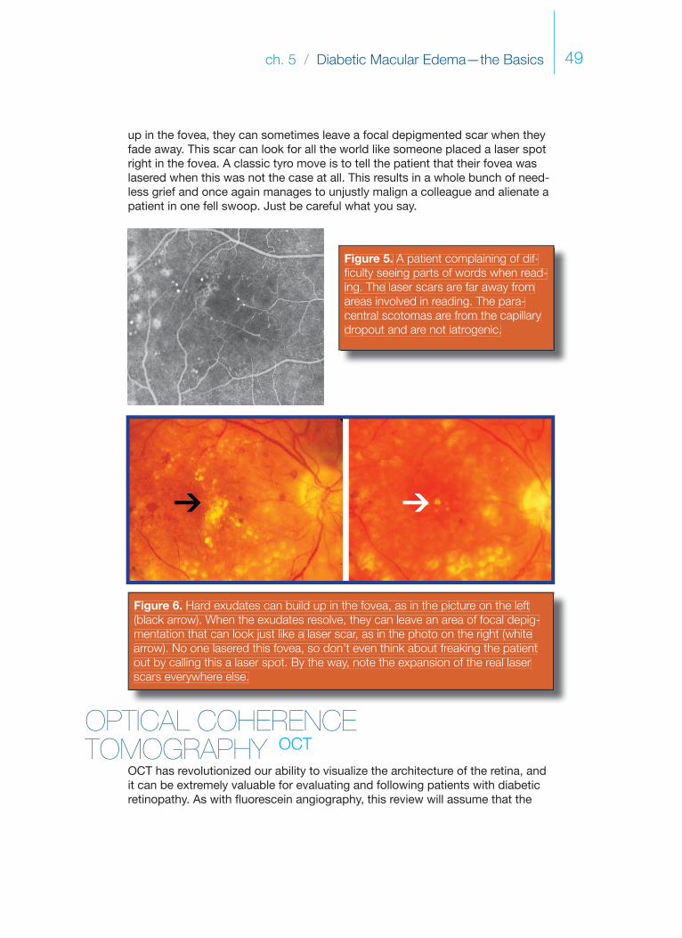
Figure 6. Hard exudates can build up in the fovea, as in the picture on the left(black arrow). When the exudates resolve, they can leave an area of focal depig-mentation that can look just like a laser scar, as in the photo on the right (whitearrow). No one lasered this fovea, so don’t even think about freaking the patientout by calling this a laser spot. By the way, note the expansion of the real laser scars everywhere else.
up in the fovea, they can sometimes leave a focal depigmented scar when they fade away. This scar can look for all the world like someone placed a laser spot right in the fovea. A classic tyro move is to tell the patient that their fovea was lasered when this was not the case at all. This results in a whole bunch of need-less grief and once again manages to unjustly malign a colleague and alienate a patient in one fell swoop. Just be careful what you say.
OPTICAL COHERENCE TOMOGRAPHY OCT
OCT has revolutionized our ability to visualize the architecture of the retina, and it can be extremely valuable for evaluating and following patients with diabetic retinopathy. As with fl uorescein angiography, this review will assume that the
Diabetic Retinopathy50
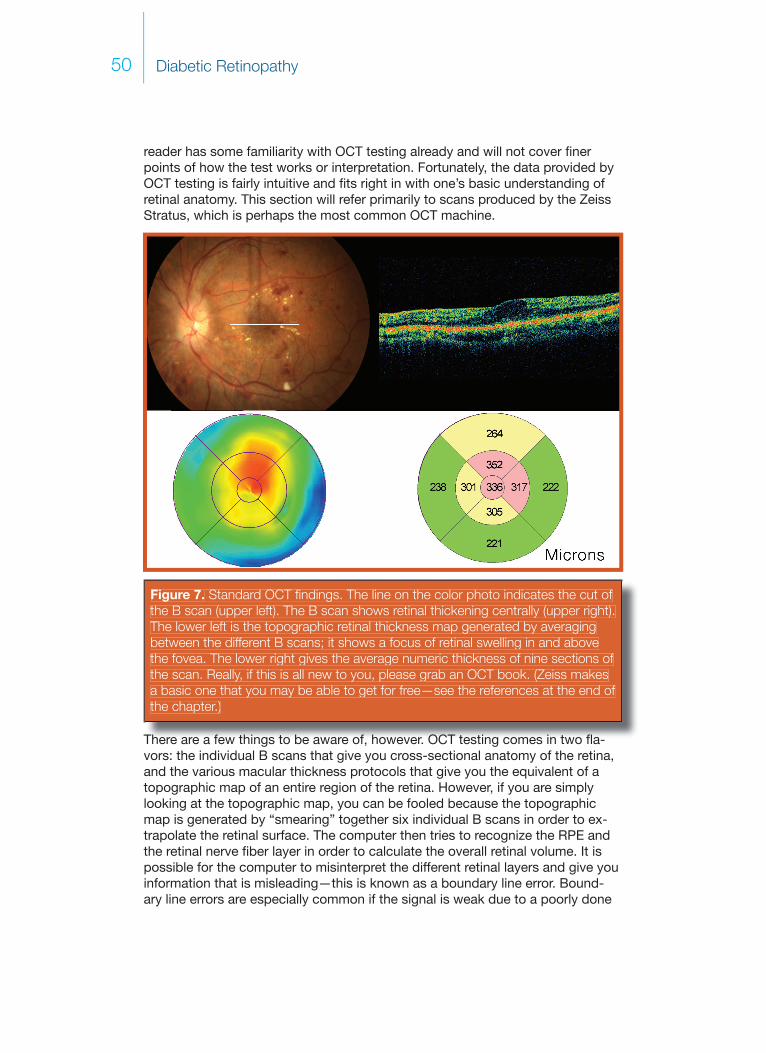
Figure 7. Standard OCT fi ndings. The line on the color photo indicates the cut of the B scan (upper left). The B scan shows retinal thickening centrally (upper right).The lower left is the topographic retinal thickness map generated by averagingbetween the different B scans; it shows a focus of retinal swelling in and abovethe fovea. The lower right gives the average numeric thickness of nine sections of the scan. Really, if this is all new to you, please grab an OCT book. (Zeiss makesa basic one that you may be able to get for free—see the references at the end of the chapter.)
reader has some familiarity with OCT testing already and will not cover fi ner points of how the test works or interpretation. Fortunately, the data provided by OCT testing is fairly intuitive and fi ts right in with one’s basic understanding of retinal anatomy. This section will refer primarily to scans produced by the Zeiss Stratus, which is perhaps the most common OCT machine.
There are a few things to be aware of, however. OCT testing comes in two fl a-vors: the individual B scans that give you cross-sectional anatomy of the retina, and the various macular thickness protocols that give you the equivalent of a topographic map of an entire region of the retina. However, if you are simply looking at the topographic map, you can be fooled because the topographic map is generated by “smearing” together six individual B scans in order to ex-trapolate the retinal surface. The computer then tries to recognize the RPE and the retinal nerve fi ber layer in order to calculate the overall retinal volume. It is possible for the computer to misinterpret the different retinal layers and give you information that is misleading—this is known as a boundary line error. Bound-ary line errors are especially common if the signal is weak due to a poorly done
ch. 5 / Diabetic Macular Edema—the Basics 51
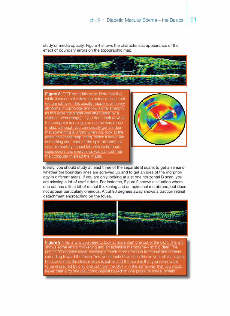
study or media opacity. Figure 4 shows the characteristic appearance of the effect of boundary errors on the topographic map.
Figure 8. OCT boundary error. Note that thewhite lines do not follow the actual retinal archi-tecture (above). This usually happens with veryabnormal morphology and low signal strength (in this case the signal was attenuated by avitreous hemorrhage). If you don’t look at what the computer is doing, you can be very muchmisled, although you can usually get an ideathat something is wrong when you look at theretinal thickness map (right). When it looks like something you made at the spin-art booth atyour elementary school fair, with weird hour-glass colors and everything, you can bet thatthe computer misread the image.
Ideally, you should study at least three of the separate B scans to get a sense of whether the boundary lines are screwed up and to get an idea of the morphol-ogy in different areas. If you are only looking at just one horizontal B scan, you are missing a lot of useful data. For instance, Figure 9 shows a situation where one cut has a little bit of retinal thickening and an epiretinal membrane, but does not appear particularly ominous. A cut 90 degrees away shows a traction retinal detachment encroaching on the fovea.
Figure 9. This is why you need to look at more than one cut of the OCT. The leftshows some retinal thickening and an epiretinal membrane—no big deal. Theright is 90 degrees away, showing a much more ominous tractional detachmentextending toward the fovea. Yes, you should have seen this on your clinical exam,but sometimes the clinical exam is subtle and the point is that you never wantto be reassured by only one cut from the OCT—in the same way that you wouldnever treat a routine glaucoma patient based on one pressure measurement.
Diabetic Retinopathy52
You should also take a look at some of the options available when it comes to using the OCT machine. You should understand what the technicians are doing and be aware of the different fi xation parameters, line cuts, image processing techniques and levels of resolution available. You can even—gasp—do the scan yourself if you have a patient who has an area of concern and you are not seeing what you need to see on the OCT cuts provided.
Although traditional OCT machines acquire data by performing one slice at a time (time domain OCT), newer machines can examine an entire region of the retina with one scan (spectral domain or Fourier domain OCT). Subtle areas of pathology can be missed if they fall in between the scans on a time domain ma-chine, and newer machines are much better able to identify abnormal anatomy, such as early vitreous traction. However, the technology is expensive and tends to Hoover up vast amounts of computer memory, so such machines are usually found in specialist’s offi ces. They can be helpful in diffi cult cases, so if you have a time domain machine like the Stratus remember that some patients may ben-efi t from the next generation of machines. He who dies with the most toys...
OCT & DME That You Don’t See
There is no question that one can sooner identify retinal thickening with OCT testing than with clinical examination. What is not clear is whether this has therapeutic implications. You have to remember that all of the studies looking at the treatment of diabetic macular edema were based on how the retina appears clinically. If the OCT shows thickening that one cannot see on clinical exam, it is not known for sure whether such patients benefi t from treatment.
If you do not see any obvious thickening, then it has been suggested that about two-thirds of the time the edema will not progress to clinically signifi cant dis-ease.3 This can become hard to call, though, because once the OCT brings something to your attention, you can start to hallucinate some thickening. You have to be intellectually honest and promise not to manufacture clinically signifi -cant disease in your head just because you want to treat a retina.
Ultimately, deciding whether to treat such patients is an art-of-medicine thing—there are a lot of variables that may play into the decision. If the process appears to be progressive, then it may be reasonable to get in some early treatment to head off trouble. This is especially true if the thickening appears to be due to a few small microaneurysms that are away from the fovea and can be safely treated. On the other hand, if the patient does not have any symptoms and is systemically well-controlled, very early edema will often resolve without the need for treatment (i.e., without the need for putting permanent spots in and around the patient’s fovea).
It is also important to remember that, in general, the treatment of diabetic macu-lar edema that is not fovea-threatening is never an emergency. It is reasonable

ch. 5 / Diabetic Macular Edema—the Basics 53
to simply re-examine the patient in six to eight weeks and monitor the changes (this interval may vary depending on their disease and your level of concern). If nothing else, being able to use the pretty OCT colors to show asymptomatic pa-tients how they are developing evidence of their diabetes can sometimes serve to motivate patients better than a ton of handouts.
Another extremely important use of OCT is the ability to identify problems with the vitreoretinal interface that can contribute to persistent edema. Although a careful clinical exam can usually suggest the presence of a subtle epiretinal membrane or taut cortical vitreous, abnormalities like this tend to become im-mediately obvious with OCT testing. This is useful when you have a patient with persistent edema and decreasing vision in spite of laser. If you identify subtle traction, then you know that such a patient should be referred to your friendly neighborhood retina specialist, rather than given 300 more laser spots in a fruit-less attempt to seal leaks in a retina that is slowly being pulled apart by the vitre-ous. There will be much more on this in subsequent chapters.
But I Don’t Have an OCT…
Are you going to go to Retina Hell if you try to manage diabetics without an OCT? There are plenty of places around the world where it is simply not possible to generate the capital to obtain an OCT machine. Fortunately, the majority of studies about treating diabetic retinopathy are totally based on clinical examination. Moreover, a careful observer can usually identify—or at least suspect—the kinds of problems that an OCT machine can fi nd. The only difference is that the OCT makes detecting such fi ndings effortless.
On the other hand, most retinal specialists would say that having an OCT is the standard of care when it comes to managing complex diabetic patients. As a result, if you think you can get an OCT it is a good idea to obtain one. The machine will likely keep you from doing the wrong thing to diabetics, and there are lots of other good things it can do. (Perhaps the most useful is the ability to identify subtle macular problems prior to cataract surgery, such as a diaphanous epiretinal membrane. You do not want a patient that is paying you the big bucks for a multifocal implant to be surprised by post-op pucker.)
If you can’t get an OCT, do not worry. Just keep on improving your exam and watching for the kinds of things discussed in this book that can mess you up—almost always, a high index of suspicion and a careful contact lens exam will keep you out of the land of permanent-vision-loss-because-you-missed-something. That particular location is not a happy place to be.
Of course, things start to get complicated if you might be able to obtain an OCT machine but you really need something more important, like a faster car. Only you and the Great Ophthalmic Court in the Sky can decide the answer to that one...

Diabetic Retinopathy54
References and Suggested Reading
1. Photocoagulation for diabetic macular edema. Early Treatment Diabetic Retinopathy Study report number 1. Early Treatment Diabetic Retinopathy Study research group. Arch Ophthalmol 1985;103:1796-806.
2. Maeshima K, Utsugi-Sutoh N, Otani T, Kishi S. Progressive enlargement of scattered photocoagulation scars in diabetic retinopathy. Retina 2004;24:507-11.
3. Browning DJ, Fraser CM. The predictive value of patient and eye charac-teristics on the course of subclinical diabetic macular edema. Am J Ophthalmol 2008;145:149-154.
Schatz H, Madeira D, McDonald HR, Johnson RN. Progressive enlargement of laser scars following grid laser photocoagulation for diffuse diabetic macular edema. Arch Ophthalmol 1991;109:1549-51.
Treatment techniques and clinical guidelines for photocoagulation of diabetic macular edema. Early Treatment Diabetic Retinopathy Study Report Number 2. Early Treatment Diabetic Retinopathy Study Research Group. Ophthalmology 1987;94:761-74.
Chew EY, Ferris FL III. Nonproliferative Diabetic Retinopathy. In: Ryan SJ. Retina, 4th ed. Philadelphia: Elsevier Mosby, 2006:v.2, pp 1271-1284.
Folk JC, Pulido JS. Laser photocoagulation of the retina and choroid. San Fran-cisco: American Academy of Ophthalmology, 1997.
Basic and Clinical Science Course Section 12: Retina and Vitreous. San Francisco: American Academy of Ophthalmology, 2008: pp 109-132
Bressler NM, Ahmed IIK. Essential OCT. Dublin: Carl Zeiss Meditech, 2006.

ch. 6 / Trust Me, I’m a Doctor: Part 1 55
Trust Me, I’m a Doctorch 6
PART ONE: The Informed Con-sent for Treating Diabetic Macular Edema.

Diabetic Retinopathy 56
Patient communication is extremely important when one uses lasers to treat dia-betic retinopathy. You must remember that in spite of your best efforts to relate the concepts involved, there is a strong tendency for the patient’s expectations to be very different from reality. It is certainly reasonable to provide the patient with ancillary information, such as discussions with offi ce staff, video tapes, and handouts. But don’t depend on such things to replace you. (Besides, when was the last time you read that handout your dentist gave you about proper fl oss-ing?) Ultimately, it is the relationship that you foster with the patient that will—hopefully—keep them motivated to persist with the generally distasteful and often-lifelong pursuit of having their retinas lasered.
You should, fi rst of all, take a deep breath and try not to display the overwhelm-ing sense of near-drowning that one feels on a busy clinic day. You don’t neces-sarily need to slow down so much that you can light your corncob pipe and wax nostalgic about doing lasers with Laura Ingalls Wilder at the Little House on the Prairie. You should, however, remember that your patient will not be impressed by how many exams and lasers you can cram into an hour, but rather, by wheth-er you take the time to carefully explain things, answer questions and skillfully anticipate unvoiced concerns. Make sure there is a family member or friend in the room, too. Having another person in the room will give the patient someone to share the experience with, and the second set of ears will be more functional than those of a stressed-out patient. It is simply a given that the average pa-tient will be unlikely to remember much of what you say. Priluck, et al., wrote a fascinating paper on the ability of patients to recall an informed consent discus-sion concerning retinal detachment surgery.1 On average, patients could only remember about 57% of what they had been told, and only 23% remembered the discussion of surgical risks.* Furthermore, patients would commonly state that anything that they did not remember had not been discussed. This is why you have to hyper-document anything you say—because in the polemic world of legal medicine, the paperwork becomes the reality, which is kind of absurd.
What all this really means, though, is that if you truly care more about your patients than about how your paperwork might look to a trial lawyer, you should realize that the data you provide may not be as important as the way in which you deliver it. A machine gun burst of risks will get the job done fast and will meet the “letter of the law” in your chart, but it is unlikely that the patient will remember much of it. However, a slow, careful discussion, with attention to the patient’s concerns, will create a far better memory of the mood of the process in the patient’s mind, even if the actual facts can’t be remembered. Simply know-ing that the doctor is actually interested in trying to transmit the information may be as important as how much is retained. In other words, you can probably deliver an informed consent in a completely unintelligible language, like maybe Klingon, but if you do it in a way that conveys that you will take all the time in
THE INFORMED CONSENT for Treating DME
*And only 3% remembered that they could have a hemorrhage or infection that could destroy the eye. Think about that the next time you give some wired boomer attorney your best clear-lens-extraction spiel.

ch. 6 / Trust Me, I’m a Doctor: Part 1 57
On a darker note, remember that although you know you are a good person, people are constantly reading articles about maniac doctors cutting off the wrong leg or defrauding Medicare. In addition to having trouble understand-ing the nature of diabetic retinopathy in general, patients may also have an imperceptible lack of trust that can blossom into something really bad if a complication occurs. You have to anticipate this and recognize that careful communication from the start is the best way to avoid trouble.
the world to be sure the patient understands the situation, you will have accom-plished a lot more than if you list the complications and then have them sign on the dotted line. (That was perhaps a bit hyperbolic, but hopefully the point is made.)
THE DISEASEYou have to make sure the patient has at least a rudimentary understanding of the pathophysiology involved by using your favorite analogy. For diabetic macular edema, this usually involves something like, “the diabetes has changed the blood vessels in your eye from nice new pipes into old rusty pipes, and they are leaking the clear fl uid that is in blood. This makes the retina swell up like a tiny sponge in the same way the old veins in people’s legs can leak and let their ankles swell up.” Or anything similar to that—you can adjust it to the patient’s level of interest and sophistication.
It is important to point out that, with macular edema, the vessels are not hemor-rhaging actively; many times patients will have been told that they have “burst blood vessels” or “hemorrhages in their eyes,” and they visualize some hor-rible Niagara Falls of blood exploding out of their head. Terms like this generate unnecessary stress, and patients will wonder why you aren’t treating the whole thing as a dire emergency and immediately lasering their gushing blood ves-sels into submission. You really want to dwell on the fact that you are dealing with interstitial fl uid leakage and that any microscopic blood spots are really just old bruising and not any sort of active hemorrhage. Incidentally, using the word “bruising” to refer to intraretinal hemorrhages of any sort seems to be a much less infl ammatory term than “blood” or “hemorrhage.” It tends to avoid the whole Quentin-Tarantino-Kill-Bill connotation and gives you a fi ghting chance that the patient’s mind will not seize up and will, instead, continue to follow your discussion.
It is extremely useful to have the patient’s photographs, fl uorescein angiogram and/or OCT available to show them during this discussion. If you can demon-strate a normal-looking fundus and then show them their own hard exudates and blot hemorrhages moving into the fovea, it is a lot easier for them to under-stand the gravity of this situation, especially if they do not have a lot of symp-
In any event, here are the concepts to convey, regardless of how you choose to convey them (preferably not in Klingon)…

Diabetic Retinopathy 58
toms. This also allows you to point out the fact that you are treating well away from the center of the vision. You would be surprised at how many patients have an unvoiced concern that your main goal is to simply chop away at their vision like a Civil War barber-surgeon and that, just maybe, they might be better off go-ing blind slowly without treatment, rather than letting you hurry things along with your foolish laser.
THE GOALS OF TREATMENTThe patient must also understand the goals of the treatment. They strongly as-sume that your laser will help improve things. Partly, this is because any time they have gone to a doctor in the past, the doctor usually does something that makes their life better, such as fi x a sore throat or stitch a cut. They also know lots of people who have had lasers (YAG and LASIK) and who saw much better immediately after the laser; the distinction between your laser and those lasers can be quite, uh, blurry.
Welcome to the world of retina—a place where patients tend to get worse no matter what you do, and where you will spend a ton of time trying to convince your patients (and perhaps yourself) that going bad slowly is the greatest thing on the planet.
You need to clearly point out that without treatment there is a very good chance the patient will be losing vision over the next one to two years, and that the goal of treatment is to slow down the rate of decay so that instead of ending up terrible, they end up only a little bit worse. This concept is remarkably hard to convey to even a sophisticated patient. Many doctors use terms that suggest to the patient that their vision will stabilize, but even in the best of circumstances, most diabetics don’t remain the same.
Here is why you can’t promise them stability: Even if your laser works superbly, diabetics can still have gradual deterioration of their visual quality—the fi ne print is harder to read, going from light to dark is trickier, it is harder to see traffi c signs, etc. Although you can very effectively overcome large-scale damage like macular edema, you cannot as easily overcome the gradual deterioration of retinal function that occurs at the cellular level with diabetes. And, as with many retinal treatments, you may be very happy with the results but the patients usu-ally aren’t, and they will be particularly unhappy if you have not made sure that they have appropriate expectations.
You may also fi nd that this discussion needs to be repeated at every visit, which becomes tedious, but the perception that diabetics have of the laser can change over time. Initially, there is a strong tendency for patients to assume that a given treatment will make them better, and you have to address such expectations as discussed above. Later on, there is a tendency to assume that any visual problems they have must be from the laser and not from progression of their disease, and you often have to constantly address this as well. Don’t forget that

ch. 6 / Trust Me, I’m a Doctor: Part 1 59
you devoted a large chunk of your life to understanding the statistics that make your treatment logical, but these concepts are very new and counterintuitive to your patients.
The best way to understand this is to go back to the graph in Chapter 5 that shows the ETDRS results for treating macular edema. From the standpoint of a treating physician, the graph is great, but Figure 1 shows what it looks like if it is fl ipped around so you see it from the standpoint of a patient. Although everyone can agree that the control group did horribly, it is clear that, even with treatment, there is a downward trend—especially if the patient lives for any length of time. You might even consider giving patients a crash course in Cartesian coordinates by using your hand to display a rapid downhill course without treatment and then a gentle downhill course with treatment in order to help them understand what they may expect.
Figure 1. A “patient’s eye” view of the graph from Chapter 5 showing the ETDRS results for treating macular edema. This graph makes doctors happy, but this view shows the need to constantly remind the patient about having realisticexpectations; diabetic eye damage can still drag the patient downhill, even withperfect treatment. Note the elegant symbolism suggested by the fl ipped labels: The doctor has to totally wrap his or her head around the patient’s point of view to really be able to relate. This was not done because it is easier to fl ip the originalimage without changing the labels—it was done on purpose for art’s sake.
Of course, patients can improve,or at least stabilize, especially with good control and careful laser (and judicious use of intraocular medications if needed—see Chapter 11). They are no longer condemned to follow the dotted line in the graph above. The problem is that if you dwell too much on the possibility of stability or improvement, it may be all the patient remembers of your discussion. They can then become very frustrated with the reality of treatment and end up not returning for follow up. This is by far the worst possible outcome, because they usually return only when they have severe symptoms and awful disease that may be impossible to control. You must anticipate and address anything that may interfere with compliance from the beginning. (See the section at the end of the chapter about treating retinopathy in developing countries for an expanded explanation of this.)
Diabetic Retinopathy 60
Have you ever had a patient tell you that some doctor told them that they were going blind when they weren’t even close to going blind? You may get a sense of superiority from such a comment—you can reassure the patient (and yourself) that there is no way you would ever be so stupid as to say such a thing. Well, it is hard to imagine that any doctor would be so stupid as to say such a thing, but now you can see how a patient could come to such a conclusion after a thorough informed consent.
If you think you are a great communicator and that no patient would ever think that you would say that they are going to go blind, just try this simple experiment: After you explain the potential downhill trend inherent in even successfully treated diabetic retinopathy, ask the patient if this means that they will inevitably go blind. You will be surprised at the answers you get. And get ready to take a deep breath and start over—patience is your most valu-able surgical tool.
Don’t forget that there is another big reason why a patient may want to blame your laser for vision problems, even if the treatment is working per-fectly: It is reassuring for some patients to be able to blame the treatment because it is a lot easier to do that than to accept the responsibility for years of poor control. The only way you can work around this is with continual education—and sometimes you have to accept the fact that you will always be the bad guy, even if you have snatched such a patient from the jaws of blindness.
There is one big problem, though, with trying to convey the reality of treating diabetic retinopathy. It turns out that if you communicate this information ef-fectively, and if patients seem to understand the possible “slower rate of decay” concept, they may then draw an additional conclusion that is very erroneous (and you may not hear about it). They may well assume that you mean that they will go downhill forever and that, at best, you will only slow down their inevitable descent into total blindness.
Anticipate this type of conclusion as well, and try to head it off. Remind them that although you can never guarantee anything, it is very unlikely that they will go totally blind from diabetic macular edema, especially if they are good about their control and follow up. Explain that, over time, they may be irritated by their vision, but they are unlikely to ever become helpless, which is what they really fear. Again, throw in plenty of “no guarantees” so their conclusion pendulum doesn’t swing to the other side and have them thinking they are off scot-free—but by giving them this reasonable assurance you may save them lots of unnec-essary anxiety.
ch. 6 / Trust Me, I’m a Doctor: Part 1 61
COMPLICATIONSThis is tricky. Never underestimate a patient’s ability to mentally assign a huge signifi cance to a tiny risk of complications while ignoring the vastly greater risk of vision loss if they do not get treated. This is especially important to remember in the setting of diabetic retinopathy where—once again—even perfect treat-ment can’t always stop the disease. Patients are very likely to remember your discussion of complications as their vision gets a bit blurrier over time and then they can assume the laser is the problem, even if the blurriness has nothing to do with any laser complication. This puts them on the path to the Dark Side of Noncompliance—something to be avoided if at all possible. The point is that you have to cover the bad things but think carefully about how you do it.
You will be a sucky doctor if you do not make sure patients hear the words “There is a small chance that the laser can make you worse.” Even if you be-come a laser sensei, it is a sad medical fact that bad things can happen, and you never know when both you and the patient will get an unpleasant surprise. You do not want any patient coming back at you claiming that you never said such a thing—no matter how bullet-proof your malpractice carrier says your written informed consent is.
However, you should not leave this (or any other negative concept) hanging at the end of a conversational paragraph. You need to remind them in the following sentence that the risk of treatment is very tiny, but the risk of permanent loss of vision is extremely large without treatment.
Another important warning should be mentioned, especially for Astute Patients (i.e., engineering types): Sometimes, a good observer may forever-after identify the laser spots that you placed in their paramacular vision. This is surprisingly unlikely—most patients are really oblivious of careful, gentle laser for macular edema. However, some patients will notice this, and you get lots of style points for mentioning it before they come to you to complain about it. The key “next-sentence” point to make, though, is that this problem is rare, and even if they do notice something, it is a heck of a lot better to have a few spots on one’s periph-eral vision than to lose chunks of central vision from untreated disease.
Okay. Once you have gotten the part about the laser making things worse out of the way, it is now even more important to dwell on the biggest problem with the laser: that it may not completely control the disease. Some of this has to do with the general tendency of diabetics to have slowly degraded vision just from being diabetic, as was discussed in the preceding paragraphs. Patients also need to understand that areas of leakage may develop rapidly in any location the dia-betes chooses, and even if the treatment works well in one area, the vision may deteriorate if new leaks continue to show up. Patients must be prepared for the possibility of multiple treatments and rapid worsening of vision from progressive disease.
Then you can add that, unfortunately, there are times when the treatment simply

Diabetic Retinopathy 62
does not work at all, and some patients go downhill even with treatment. You can then allude to more aggressive and risky treatments, such as intravitreal steroids, that may help them if the laser can’t. Don’t let them feel that these more aggressive treatments are Good Things, though. It is easy for patients to think, “Oh well, if the laser doesn’t work I’ll just have an injection or two and be fi ne.” They need to know that it is much better if they can be controlled with safer, simpler things like a gentle focal laser—in other words, that there is not necessarily a pot of gold at the end of your coherent light rainbow. This is also a good time to emphasize the importance of good systemic control so the patient understands that the laser alone is not going to solve their problem—the patient needs to be an active participant in this process. (Much more on this in The Chapter with a Tantalizing Name.)
As you read this, it may seem like a lot of information to convey when time is limited. However, it really just takes about fi ve minutes to go through this—including the time it takes to be sure patients clearly understand the goals of the treatment. Indeed, making sure the patient understands is perhaps the most important part. The best way to do this is simply to ask patients in a non-judg-mental way to repeat the gist of what you have said and then review any areas that aren’t clear. Again, the actual treatment will soon become the least of your worries; making sure that patients understand what you are doing is a job that never stops.
Special Problems with Informed Consent in Developing Countries (and uniquely American institutions such as free clinics, county hospitals, and other examples of how the American healthcare system can bring third-world medicine to your backyard)
The bulk of this chapter refers to dealing with patients who have a fi ghting chance of taking good care of themselves. It is also hoped this book will be helpful in situations where patients have less of an opportunity to obtain state-of-the-art healthcare—and the extended section title will hopefully remind read-ers in a certain developed country without universal coverage to keep on read-ing, because there are special problems that arise when resources are limited, no matter what the gross domestic product happens to be.
Developing regions are cursed with a combination of limited healthcare re-sources and a limited desire on the part of the diabetic population to access healthcare until their disease is far advanced. The reasons for the latter are nu-merous and include the cost, the diffi culty of obtaining an exam, and the lack of symptoms until things are really bad. One big problem is the perennial dilemma inherent in treating diabetic retinopathy: the perception, on the part of patients, that laser treatment causes blindness.
This dilemma deserves further elaboration. Cataract surgery in developing countries (or any country, for that matter) is met with enthusiasm because the
ch. 6 / Trust Me, I’m a Doctor: Part 1 63
patients experience immediate benefi t, and once a cataract is popped out, the problem tends to be over. This sets a level of expectation that is never met by treating diabetic retinopathy in the same setting. Treating diabetic retinopathy usually involves recurring, unpleasant treatments, and there is usually no benefi t that the patient can perceive.
This is because diabetics in this situation tend to seek evaluation only when they are forced to by progressive symptoms, at which point their disease is very ad-vanced. Laser treatment is then attempted, and although the treatment usually helps somewhat, the patients still develop severe vision loss. Although one can explain this course of events to patients and their families, there is an inevitable tendency for patients to assume that the laser was the cause of their disease progression rather than to understand that their disease was so hopelessly advanced that the laser could only partially slow the process. The problem is exacerbated by the fact that the affected population tends to be unsophisticated and to have a hard time understanding the nuances discussed in this chapter. The problem is even further exacerbated by the fact that the healthcare workers have limited time and resources to convey such nuances—let alone have time or equipment to perform the treatments.
A vicious circle is then created as patients receive treatment and then go out and tell their friends and family that the laser is “bad.” The word spreads, and then more diabetics become afraid to come in for early screening and treatment, and then these diabetics show up with advanced disease that the laser can’t stop, and then they think the laser is “bad” because they became worse, and then they tell their friends, and so on and so forth. The problem is compounded by extremely poor systemic control, which makes even timely treatment much less effective.
Overcoming these obstacles by improving the education, monitoring and treat-ment of diabetics is way outside the scope of this book (although screening with cheap, portable retinal cameras using non-ophthalmic personnel is one way to start). What is well within the scope of this book is how this type of situation can change how the informed consent is presented to the patient. When these patients come crashing into the system with horrible disease, you have to be a bit more blunt and you have to try to offset the whole “laser is bad” thing right from the start. You have to lay out the above facts so the patient understands that your goal is to try to hold on to any vision whatsoever and that they cannot expect to get better or even remain stable. Emphasize that you are going to do everything you can to help them, and it will be a heck of a lot better than do-ing nothing, but things would have been a lot better if you had seen them well before this point. You can also beat on them about control, but there may not be much they can do about it in their circumstances. What is really important is that you beat on them to be sure their relatives get checked for diabetes regularly, and if they are found to have diabetes, then they have to get an eye exam once a year. Also, if the patient knows anyone with diabetes, they should tell that person to get in for
Diabetic Retinopathy 64
regular eye exams—no matter what—because if that person doesn’t, they may end up in the same mess that the patient is facing.
All this sounds harsh, but you really have to go after the tendency for folks to expect that diabetic lasers will solve their visual problems in this situation and you have to try to break the vicious cycle of having patients being unwilling or afraid to get an exam and then showing up too late. Finally, you have to try to do all this in a setting where your time and resources are likely very limited.
If you are in such a situation—ask for help. There are international organizations that may be able to lend a hand. The American Academy of Ophthalmology maintains a list of such organizations on their web site (http://www.aao.org/inter-national/links.cfm). There may also be local religious or service-group organiza-tions that can help you—ask around and contact as many people as you can. You may be very surprised at what a difference you can make for your practice and your patients if you are persistent. You are also welcome to write to the ad-dress in the introduction—just try not to get frustrated and give up.
Finally, there will be sections in the upcoming chapters that address how one needs to alter the treatment approach in locations where resources are limited and patients with awful disease are plentiful. It turns out that the focus of the in-formed consent is not the only thing that may be very different these situations.
References and Suggested Reading
1. Priluck IA, Robertson DM, Buettner H. What patients recall of the preopera-tive discussion after retinal detachment surgery. Am J Ophthalmol 1979;87:620-3.
Morgan LW, Schwab IR. Informed consent in senile cataract extraction. Arch Oph-thalmol 1986;104:42-5.
Herz DA, Looman JE, Lewis SK. Informed consent: is it a myth? Neurosurgery 1992;30:453-8.
VanNewkirk, M. ed. International Ophthalmology. San Francisco: American Acad-emy of Ophthalmology, 2005.
Schwab, L. Eye Care in Developing Nations, 4th Ed. Manson Publishing, 2007.