bone marrow-derived mesenchymal stem cells become...
TRANSCRIPT

Original Article
Bone Marrow-Derived Mesenchymal Stem CellsBecome Antiangiogenic When Chondrogenically
or Osteogenically Differentiated:Implications for Bone and Cartilage Tissue EngineeringAU1 c
AU2 c Jennifer J. Bara, PhD,1 Helen E. McCarthy, PhD,2 Emma Humphrey, PhD,3
William E.B. Johnson, PhD,4 and Sally Roberts, PhD5
Osteochondral tissue repair requires formation of vascularised bone and avascular cartilage. Mesenchymal stemcells stimulate angiogenesis both in vitro and in vivo but it is not known if these proangiogenic properties changeas a result of chondrogenic or osteogenic differentiation. We investigated the angiogenic/antiangiogenicproperties of equine bone marrow-derived mesenchymal stem cells (eBMSCs) before and after differentiationin vitro. Conditioned media from chondrogenic and osteogenic cell pellets and undifferentiated cells was appliedto endothelial tube formation assays using Matrigel�. Additionally, the cell secretome was analysed usingLC-MS/MS mass spectrometry and screened for angiogenesis and neurogenesis-related factors using proteinarrays. Endothelial tube-like formation was supported by conditioned media from undifferentiated eBMSCs.Conversely, chondrogenic and osteogenic conditioned media was antiangiogenic as shown by significantlydecreased length of endothelial tube-like structures and degree of branching compared to controls. Un-differentiated cells produced higher levels of angiogenesis-related proteins compared to chondrogenic andosteogenic pellets. In summary, eBMSCs produce an array of angiogenesis-related proteins and support an-giogenesis in vitro via a paracrine mechanism. However, when these cells are differentiated chondrogenically orosteogenically, they produce a soluble factor(s) that inhibits angiogenesis. With respect to osteochondral tissueengineering, this may be beneficial for avascular articular cartilage formation but unfavourable for bone for-mation where a vascularised tissue is desired.
Introduction
The clinical success of osteochondral tissue engineer-ing relies upon the generation of a functional tissue of
physiologically relevant size that is capable of integration atthe repair site. Bone has an extensive blood supply that iscritical to the tissue’s viability and function. In the clinic,disruption or lack of blood supply to bone causes osteone-crosis and is implicated in the pathogenesis of nonunionfractures.1 Conversely, the presence of blood vessels in ar-ticular cartilage is associated with various pathologies, in-cluding osteoarthritis2,3 rheumatoid arthritis,4 and inferiorrepair tissue in autologous chondrocyte implantation.5 Oneapproach to osteochondral tissue engineering is to create abiphasic construct in vitro whereby constituent cells havereached a suitable level of chondrogenic and osteogenic
differentiation before implantation.6 However, nutritionallimitations posed by diffusion gradients currently limit thesize of viable tissues that can be engineered.7–9 Moreover, theviability and successful integration of such constructs intoosteochondral defects relies upon vascular ingrowth afterin vivo implantation. Therefore, much attention is now fo-cused on tissue engineering vascularised constructs. Currentstrategies include incorporating vascular cell types,10,11 orthe addition of slow release angiogenic growth factors boundto scaffolds.12,13 Vascularised bone constructs have also beenengineered in vivo by endocultivation, whereby a construct isimplanted intramuscularly to allow vascular ingrowth beforereimplantation into the defect site.14
Bone marrow-derived mesenchymal stem cells (BMSCs)represent a potential cell source for osteochondral tissueengineering. In the clinic, implantation of in vitro expanded
1Musculoskeletal Regeneration Group, AO Research Institute, Platz, Switzerland.2Connective Tissue Biology Research Group, Cardiff School of Biosciences, Cardiff, United Kingdom.3RJAH Orthopaedic Hospital, Keele University Mass Spectrometry Facility, Oswestry, United Kingdom.4Life and Health Sciences, Aston University, Birmingham, United Kingdom.5Spinal Studies & Institute for Science and Technology in Medicine, RJAH Orthopaedic Hospital, Keele University, Oswestry,
United Kingdom.
TISSUE ENGINEERING: Part AVolume 00, Number 00, 2013ª Mary Ann Liebert, Inc.DOI: 10.1089/ten.tea.2013.0196
1
TEA-2013-0196-ver9-Bara_1P
Type: research-article
TEA-2013-0196-ver9-Bara_1P.3d 08/21/13 1:39pm Page 1

BMSCs has been used to repair long bone15,16 and cartilagedefects.17–19 However, it is not clear if, or how, in vitro dif-ferentiated BMSCs may affect vascular ingrowth once re-implanted into osteochondral defects. UndifferentiatedBMSCs elicit potent angiogenic effects which appear to bedue to a combination of direct cellular interactions and bythe release of soluble factors. In vitro, BMSCs secreteAU3 c VEGF,FGF-2, interleukin-6,AU4 c PIGF, angiopoeitin-1,AU3 c PDGF, plasmin-ogen activator,AU3 c MMP-9 and monocyte chemotactic protein-1(MCP-1).20,21 BMSC conditioned media stimulates endo-thelial cell proliferation and migration20 and tube-likeformation.21 Neutralising antibody experiments have dem-onstrated that the mitogenic effects of BMSC secretome werenot solely due to the presence of one particular growth fac-tor, but a combination of factors.20 In vivo, improved vascularingrowth and osteogenesis at the fracture site was observedafter implantation of gelatin sponges containing MSC con-ditioned media in rats.22 Similarly, injection of BMSC con-ditioned media improved blood flow in a mouse model ofhindlimb ischaemia.20 BMSCs have also been reported tostimulate vascularisation in animal models of myocardialinfarction.23,24 Conversely, there is also evidence to suggestthat BMSCs may inhibit angiogenesis. Otsu et al. reportedthat direct cell–cell contact between rat BMSCs and endo-thelial cells caused apoptosis and destruction of endothelialtube-like structures on Matrigel�. Furthermore, in theirmouse melanoma model, BMSCs appeared to abrogate tu-mour growth by inhibiting tumour angiogenesis.25
It is currently unknown whether these predominantlyproangiogenic properties of undifferentiated BMSCs areconserved after in vitro chondrogenic or osteogenic differ-entiation and the accompanying change from a 2D to 3Denvironment. In the present study, equine BMSCs (eBMSCs)were differentiated along chondrogenic and osteogenic lin-eages in 3D pellet culture as previously described.26 Angio-genic properties of undifferentiated and differentiated cellswere analyzed by collecting serum-free conditioned mediaand (1) screening it for angiogenesis-related proteins and (2)applying it to an in vitro angiogenesis assay, which measuresthe degree of endothelial tube-like formation on Matrigel.Our results show that conditioned media from undifferenti-ated eBMSCs supports endothelial tube-like formationin vitro and contains an array of angiogenic, antiangiogenicand neurotrophic factors. We present novel data showingthat when differentiated along chondrogenic and osteogeniclineages, eBMSCs reduce production of angiogenesis andneurogenesis-related proteins and produce a soluble factor(s)that inhibits endothelial tube-like formation in vitro.
Materials and Methods
Cell culture
eBMSCs were isolated from the bone marrow of the thirdmetacarpal of distal forelimbs from four horses (mean age 7years) as previously described.26 Four days after isolation,flasks were washed to ensure removal of haematopoieticcells. eBMSCs were incubated at 37�C at 5% CO2 and re-ceived media changes with eBMSC medium (DMEM +GlutaMAXTM-1, 100mg mL - 1 gentamicin, 10% foetal calf se-rum [FCS] [PAA]) three times a week. At each passage,population doublings were calculated using the followingformula: LOG10 (final cell number/initial cell number))*3.33.
Subsequent differentiation assays and generation of condi-tioned media were performed using eBMSCs that had un-dergone approximately 25 population doublings (p2–3).
Monolayer adipogenic and osteogenic differentiation
eBMSCs were seeded into six-well plates at 3 · 103 cells/cm2 and cultured in eBMSC medium until 80% confluent. Foradipogenesis, cells were incubated with adipogenic media(DMEM + GlutaMAXTM-1, 100 mg mL - 1 gentamicin, 10mgmL - 1 insulin [Sigma], 1 mM dexamethasone [Sigma], 100mMindomethacin [Sigma], 500mM 3-isobutyl-1-methyl xanthine[Sigma] and 15% normal rabbit serum [Sigma]) for 6 days.Cells were fixed in 10% formalin and treated with 0.5% Oilred O (Sigma) in 60% IPA for 1 h.
For osteogenesis, eBMSCs were incubated for 21 days withDMEM + GlutaMAXTM-1, 0.1 mM L-ascorbic-acid-2-phos-phate (Sigma), 100mg mL - 1 gentamicin, 10 nM dexametha-sone, 10 mM b-glycerophosphate (Sigma), and 10% FCS.Cells were fixed with 10% formalin and treated with thefollowing solution for 1 h in to detect alkaline phosphataseactivity: 10% naphthol solution (50 mg mL - 1 napthol AS-BIphosphate [Sigma] in dimethyl formamide [BDH] pH 8) in0.2 M Tris–HCl (Sigma) buffer pH 9 + 1 mg mL - 1 Fast RedTR (Gurr). eBMSCs treated with standard eBMSC mediaserved as controls.
Chondrogenic and osteogenic differentiationin 3D pellet culture
eBMSCs were chondrogenically and osteogenically dif-ferentiated in defined media using a previously describedpellet culture system.26 To make cell pellets, eBMSCs, sus-pended in either chondrogenic or osteogenic differentiationmedia, were transferred into 1.5 mL Eppendorf tubes(0.5 · 106 cells per Eppendorf) and centrifuged at 500 g for5 min. Chondrogenic differentiation medium consisted of:DMEM + GlutaMAXTM-1, 2% FCS, 100 mg mL - 1 gentamicin,10 mg mL - 1 Insulin-Transferrin-Selenium-X (Gibco), 0.1 mML-ascorbic-acid-2-phosphate, 10 nM dexamethasone and10 ng mL - 1 TGF-b1 (Peprotech). Osteogenic differentiationmedium was as described for monolayer differentiation.Pellets were incubated at 37�C and 5% CO2 for 21 days withmedia changes three times a week.
Generation of conditioned media
Serum-free conditioned media was generated from un-differentiated cells, chondrogenic and osteogenic pellet cul-tures of eBMSCs.27 Undifferentiated eBMSCs were cultureduntil 70% confluent, washed twice with phosphate-bufferedsaline (PBS) and incubated with 20 mL of conditioning media(serum free DMEM + GlutaMAXTM-1, 100mg mL - 1 gentami-cin supplemented with 10mg mL - 1 Insulin-Transferrin-Selenium-X and 1% MEM nonessential amino acids [Gibco])per T75 flask. Conditioning media was incubated with cellsfor 4 days, filtered through a 0.45 mm filter (Sarstedt) andstored at - 20�C. Upon collection of conditioned media, cellswere passaged and a cell count performed. For chondro-genic/osteogenic pellets, at day 21, pellets were washedtwice in PBS and incubated with 1 mL of conditioning mediaper pellet. Conditioned media was collected after 4 days andprocessed as described above. Pellets were snap frozen inliquid nitrogen cooled hexane and stored at - 80�C.
2 BARA ET AL.
TEA-2013-0196-ver9-Bara_1P.3d 08/21/13 1:39pm Page 2

Histological and immunohistochemical analysisof pellet cultures
Histology and immunohistochemistry was performed on7 mm cryosections from a minimum of four pellets per horse(16 chondrogenic & 16 osteogenic). Toluidine blue was ap-plied to cryosections for 1 min, washed and air dried over-night before being mounted in Pertex (Histolab�). For VonKossa staining, 5% silver nitrate (VWR) in distilled waterwas applied to cryosections and placed under a UV lightuntil brown colour development (*2 min). Sections werewashed with 2% sodium thiosulphate (BDH) in distilledwater, dehydrated then mounted with Pertex.
Immunohistochemistry was carried out at room temper-ature unless otherwise stated. For type II collagen, cryosec-tions were pretreated with 4800 U/mL ovine testicularhyaluronidase (Sigma) and 0.25 U/mL chondroitinase ABC(MP Biomedicals) in Tris acetate buffer pH 8 for 2 h. Sectionswere fixed in 10% formalin, blocked with 5% goat serum(Vector), then incubated with mouse anticollagen type II(CIIC1, DSHB) (1:10 in PBS) overnight at 4�C. Negativecontrols were incubated with mouse IgG1a (Dako). Thesecondary antibody goat anti-mouse Alex Fluor 488 (In-vitrogen) (1:200 in PBS) was applied to sections for 1 h. Afterwashing in PBS, sections were mounted with Vectashieldmounting medium for fluorescence containing DAPI (Vec-tor) and observed using a Leitz Diaplan light microscope. Fortype X collagen, cryosections were fixed in a solution ofmethanol and acetone for 10 min, washed in PBS, thentreated with 1 mg/mL protease in PBS (Sigma; P-6911) for30 min. Sections were blocked with 5% bovine serum albumin(BSA) in PBS for 30 min, then incubated with mouse antic-ollagen type X antibody (a kind gift from Gary J. Gibson,Henry Ford Hospital, Detroit, MI) (1:300 in PBS containing 1%BSA) overnight at 4�C. An isotype matched mouse immuno-globulin was used as a negative control. Sections were incu-bated with goat anti-mouse Alexa Fluor 594 (Invitrogen)(1:200 in PBS containing 1% BSA) for 45 min. Slides werewashed in PBS then mounted with Vectashield mountingmedium for fluorescence containing DAPI (Vector) and ob-served using Leica TCS SP2 AOBS confocal microscope.
DNA quantification assay
DNA content of undifferentiated, chondrogenically andosteogenically differentiated eBMSCs that generated a stan-dard volume of conditioned media was calculated using theQuanti-iT� PicoGreen� assay (Invitrogen). Pellets/undif-ferentiated eBMSCs were digested in 1.25 mg/mL ProteinaseK (Sigma) in 100 mM ammonium acetate (Sigma) containing7% EDTA (BDH) in distilled water, at pH 7 at 60�C over-night. Briefly, 200mL of pellet/cell digest were diluted inTris–EDTA buffer and pipetted in triplicate into a 96-wellplate. PicoGreen reagent was incubated with samples for5 min at room temperature. Fluorescence was measured us-ing a fluorescent microplate reader (FLx800 microplatefluorescence reader; Bio-Tek Instruments) at (excitationwavelength 480 nm, emission wavelength 520 nm).
Angiogenesis array
A sample of conditioned media from undifferentiated,chondrogenically, and osteogenically differentiated eBMSCs
from each horse was screened for angiogenesis-related pro-teins using a Human Angiogenesis Proteome Profiler� an-tibody array (R&D Systems) according to the manufacturer’sinstructions. Positive signals were detected by chemilumi-nescence (UptiLight US WBlot HRP chemiluminescent de-tection reagent; Interchim) and visualised with ChemiDOc�EQ (Bio-Rad). Array data was quantified by measuring thesum of the intensities of the pixels within the spot bound-ary · pixel area with image analysis software (Quantity One�
version 4.6.3; Bio-Rad). Array data was normalized tobackground then DNA content as calculated for each culturefrom each respective horse.
Endothelial tube-like formation assay
Endothelial tube-like formation assays were performedusing Matrigel (growth factor reduced, phenol red-free [BDBiosciences]). Twenty-four-well tissue culture plates werecoated with 230 mL Matrigel per well and allowed to solidifyat 37�C for 30 min. The human dermal microvascular cellline, HMEC-1, was seeded onto Matrigel (1 · 105 per well) inMCDB 131 medium, 0.05% penicillin and streptomycin (bothGibco) and 10% FCS. After 4 h plates were washed gentlythrice before application of either undifferentiated, chon-drogenically or osteogenically differentiated eBMSC condi-tioned media. Conditioning media that had not been incontact with cells was used for controls. Time lapse imageswere captured over a 24 h period using a video camera set upto an Olympus CK2 microscope. After 24 h, wells were wa-shed and viewed in phase using a Nikon TS100 fluorescentmicroscope.
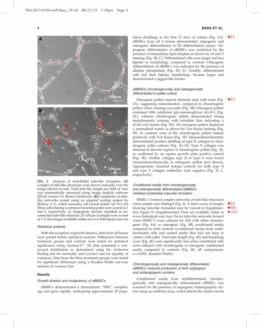
Total tube length in five fields of view per well wasquantified by drawing over and measuring tube-like struc-tures using image analysis software (IPLab version 3.6;Becton Dickinson) ( b F1Fig. 1A). Complexity of tube-like net-works was scored using an adapted scoring system byMcilroy et al. (Fig. 1B).28 Two cells sharing a commonbranching point were scored as 1 (‘‘a’’ in Fig. 1B); three cellssharing a common branching point were scored as 2 (‘‘b’’ inFig. 1B) and so on. Angiogenic sprouts, classified as an un-connected tube-like structure 25–100mm in length werescored as 1. Data for total tube length and branching scorewere normalized to the DNA content calculated for eachculture from each respective horse.
Mass spectrometry
Undifferentiated, chondrogenically, and osteogenicallydifferentiated eBMSC conditioned media from three horseswas analysed by LC-MS/MS mass spectrometry. Condi-tioned media was concentrated 10 · using Amicon Ultra-2centrifugal filter devices (10 kDa molecular weight cut off)according to the manufacturer’s instructions (Millipore), di-gested with trypsin at 37�C then separated by liquid chro-matography. MS/MS analysis was performed onfractionated peptides using a 4800 MALDI TOF/TOF massspectrometer (Applied Biosystems). The search engine‘‘MASCOT’’ was used to identify peptides from the NCBIdatabase. Peptides with a total ion score > 95% confidenceinterval or with a peptide count of > 2 were excluded. Un-amed or hypothetical proteins were identified using an on-line bioinformatics database (DAVID).
ANTIANGIOGENIC PROPERTIES OF DIFFERENTIATED MSCS 3
TEA-2013-0196-ver9-Bara_1P.3d 08/21/13 1:39pm Page 3
Statistical analysis
With the exception of growth kinetics, data from all horseswere pooled before statistical analysis. Differences betweentreatment groups and controls were tested for statisticalsignificance using Analyse-it�. All data presented a non-normal distribution as determined using the AndersonDarling test for normality and Levene’s test for equality ofvariances. Data from the three treatment groups were testedfor significant differences using a Kruskal–Wallis one-wayanalysis of variance test.
Results
Growth kinetics and mutipotency of eBMSCs
eBMSCs demonstrated a characteristic ‘‘MSC’’ morphol-ogy and grew rapidly, undergoing approximately 20 popu-
lation doublings in the first 12 days of culture ( b F2Fig. 2A).eBMSCs from all 4 horses demonstrated adipogenic andosteogenic differentiation in 2D differentiation assays. Ad-piogenic differentation of eBMSCs was confirmed by thepresence of intracellular lipid droplets as shown by oil red Ostaining (Fig. 2B, C). Differentiated cells were larger and lessbipolar in morphology compared to controls. Osteogenicdifferentiation of eBMSCs was indicated by the presence ofalkaline phosphatase (Fig. 2D, E). Notably, differentiatedcells lost their bipolar morphology, became larger anddemonstrated a jagged-like border.
eBMSCs chondrogenically and osteogeniciallydifferentiated in pellet culture
Osteogenic pellets stained intensely pink with eosin ( b F3Fig.3A), suggesting mineralisation, compared to chondrogenicpellets where staining was paler (Fig. 3B). Osteogenic pelletscontained little sulphated glycosaminoglycan (sGAG) (Fig.3C), whereas chondrogenic pellets demonstrated strongmetachromatic staining with toluidine blue indicating asGAG-rich matrix (Fig. 3D). All osteogenic pellets displayeda mineralised matrix as shown by Von Kossa staining (Fig.3E). In contrast, none of the chondrogenic pellets stainedpositively with Von Kossa (Fig. 3F). Immunohistochemistrydemonstrated positive labelling of type II collagen in chon-drogenic pellet cultures (Fig. 3G–H). Type X collagen wasdetected in discrete regions of chondrogenic pellets (Fig. 3J),as confirmed by an equine growth plate positive control(Fig. 3K). Neither collagen type II or type X were foundimmunohistochemically in osteogenic pellets (not shown).Appropriately matched isotype controls for both type IIand type X collagen antibodies were negative (Fig. 3I, L,respectively).
Conditioned media from chondrogenicallyand osteogenically differentiated eBMSCsinhibited endothelial tube-like formation
HMEC-1 formed complex networks of tube-like structureswhen seeded onto Matrigel ( b F4Fig. 4). A time course of imagesshowing tube-like formation may be viewed in b SF1Supplemen-tary Figure S1 b AU5(Supplementary Data are available online atwww.liebertpub.com/tea). Fewer tube-like networks formedwhen HMEC-1 were cultured for 24 h with either chondro-genic (Fig. 4A) or osteogenic (Fig. 4B) conditioned mediacompared to both controls (conditioned media from undif-ferentiated cells and control media that had not been incontact with cells). Total tube length (Fig. 4E) and branchingscore (Fig. 4F) were significantly less when endothelial cellswere cultured with chondrogenic or osteogenic conditionedmedia compared to controls (Fig. 4E, all comparisons:p < 0.0001, Kruskal–Wallis).
Chondrogenically and osteogenically differentiatedeBMSCs reduced production of both angiogenicand antiangiogenic proteins
Conditioned media from undifferentiated, chondro-genically and osteogenically differentiated eBMSCs wasscreened for the presence of angiogenic/antiangiogenic fac-tors using an antibody array, which detects the relative levels
FIG. 1. Analysis of endothelial tube-like formation. (A)Lengths of tube-like structures were drawn manually over theimage (shown in red). Total tube-like length per field of viewwas automatically measured using image analysis software(IPLab version 3.6, Becton Dickinson). (B) Complexity of tube-like networks scored using an adapted scoring system byMcilroy et al., which measures cell branch points. (a) Two (b)Three cells sharing a common branching point were scored as 1and 2, respectively. (c) Angiogenic sprouts, classified as un-connected tube-like structure 25–100mm in length were scoredas 1. Color images available online at www.liebertpub.com/tea
4 BARA ET AL.
TEA-2013-0196-ver9-Bara_1P.3d 08/21/13 1:39pm Page 4

of 55 different angiogenesis related proteins. Six angiogenic(DPPIV, MMP-9, endothelin-1, PDGF-AA,AU4 c UPA, and VEGF)and four antiangiogenic proteins (TIMP-1, IGFBP-2, PF4 andangiopoietin-2) were detected at higher levels compared toall other proteins (F5 c Fig. 5A, E, respectively). UndifferentiatedeBMSC conditioned media contained significantly higherlevels of angiogenesis-related proteins compared to chon-drogenic and osteogenic conditioned media, with the ex-ception of DPPIV, VEGF, and PDGF-AA, which were notsignificantly different between undifferentiated and chon-drogenic conditioned media (Fig. 5A–G). Interestingly,chondrogenic conditioned media contained higher levels ofangiogenic factors, PDGF-AA ( p = 0.0079), VEGF ( p = 0.0497),FGF-2 ( p = 0.0363), FGF-4 ( p = 0.0126), FGF-7 ( p = 0.0225),MCP-1 ( p = 0.0363) and activin A ( p = 0.0230), and the anti-
angiogenic factor, IGFBP-3 ( p = 0.0188), compared to osteo-genic conditioned media.
Chondrogenically and osteogenically differentiatedeBMSCs reduced production of neurotrophic proteins
Undifferentiated eBMSC conditioned media also con-tained higher levels of neurogenic factors compared tochondrogenic and osteogenic secretome (Fig. 5H). Levels ofartemin were significantly higher in undifferentiated com-pared to both chondrogenic ( p = 0.0209) and osteogenic( p = 0.0079) conditioned media. Undifferentiated eBMSC se-cretome contained significantly more persephin than osteo-genic conditoned media ( p = 0.0044). Both chondrogenic( p = 0.0126) and osteogenic ( p = 0.0002) conditoned media
FIG. 2. Multipotent phenotypeof equine bone marrow-derivedmesenchymal stem cells(eBMSCs). (A) Population dou-blings against time in culture.Each data series represents a cellpopulation isolated from a dif-ferent horse. Oil red O stainingof lipid droplets in (B) adipo-genically differentiated and (C)undifferentiated controleBMSCs. Alkaline phosphatasestaining of (D) osteogenicallydifferentiated and (E) undiffer-entiated control eBMSCs. Scalebars 100 mm. Color imagesavailable online at www.liebertpub.com/tea
ANTIANGIOGENIC PROPERTIES OF DIFFERENTIATED MSCS 5
TEA-2013-0196-ver9-Bara_1P.3d 08/21/13 1:39pm Page 5

contained less glial cell-derived neurotrophic factor (GDNF)compared to undifferentiated conditioned media, with chon-drogenic conditioned media containing higher levels of theneurotrophin compared to osteogenic secretome ( p = 0.0363).
Proteomic analysis of eBMSC conditioned media
After applying a stringent set of exclusion criteria, almostall peptides detected in eBMSC conditioned media by massspectrometry were identified as ECM proteins. Type I col-lagen peptides were identified in all samples (T1 c Table 1).Chondrogenic conditioned media contained multiple fibro-nectin peptides. Fibronectin was also detected in the secre-tome of undifferentiated and osteogenically differentiatedeBMSCs (two of the three BMSC populations tested). Pep-tides of cartilage-associated matrix proteins biglycan anddecorin were identified in chondrogenic conditioned media.Similary, type II collagen and lumican were identified in twoout of the three samples of chondrogenic secretome. Thepresence of these peptides are indicative of a cartilaginousECM; thus, further confirming the chondrogenic phenotypeof differentiated eBMSCs. Peptides of the antiangiogenic
molecule thrombospondin were detected in two out of threesamples of chondrogenic and in one sample of both undif-ferentiated and osteogenic conditioned media. Bone sialo-protein-2 was also present in the chondrogenic conditionedmedia from two out of the three BMSC populations testedand at 94.6% confidence interval in the third sample.
Discussion
In the present study, we show for the first time, that wheneither chondrogenically or osteogenically differentiated in a3D culture system, eBMSCs reduce production of angio-genesis and neurogenesis-related proteins and inhibit an-giogenesis in vitro. With respect to osteochondral tissueengineering, this supports the use of BMSCs for avascularcartilage formation; however, maybe of concern for thegeneration of vascularised bone.
Initially, eBMSCs proliferated rapidly in culture, which isconcominant with other studies examining the growth ki-netics of eBMSCs in vitro.26,29 We have previously shownthat these cells express the putative stem cell markers CD90,CD166 and STRO-1.26 eBMSCs demonstrated multipotency
FIG. 3.AU7 c Haematoxylin andeosin staining of (A) osteo-genic and (B) chondrogenicpellets. Metachromasia of (C)osteogenic and (D) chondro-genic pellets using toluidineblue indicates a sulphatedglycosaminoglycan rich ma-trix in chondrogenic pellets.(E) Osteogenic pellets stainedstrongly with Von Kossa in-dicating a calcium rich mi-neralised matrix, whereaschondrogenic pellets ap-peared unmineralised (F). (G–L) Immunohistochemicalanalysis of type II and X col-lagen inAU7 c chondrogenic pelletcultures. Chondrogenic pelletslabelled positively but vari-ably for type II (G–H) andtype X ( J) collagen. Equinegrowth plate type X collagenpositive control (K). IgG andIgM controls were negativefor type II (I) and X (L) colla-gen, respectively. Cell nucleicounterstained blue withDAPI, scale bars are 100mmunless stated otherwise. Colorimages available online atwww.liebertpub.com/tea
6 BARA ET AL.
TEA-2013-0196-ver9-Bara_1P.3d 08/21/13 1:39pm Page 6

as shown by their ability to differentiate along adipogenic,osteogenic and chondrogenic lineages. In addition to stan-dard differentiation in monolayer, osteogenic differentiationwas performed in a 3D pellet culture system as previouslydescribed.30 Mineralization of osteogenic pellets shown byVon Kossa and haematoxylin and eosin suggest that eBMSCsdifferentiated osteogenically. Previous 3D osteogenic differ-entiation of these cells has also identified the presence ofosteocalcin immunohistochemically.26
Chondrogenic differentiation was demonstrated by thepresence of sGAG and type II collagen; however, discreteregions of type X collagen immunostaining indicate areas ofhypertrophy. This, together with the identification of bonesialoprotein in chondrogenic conditioned media, suggestdifferentiation of eBMSCs toward a hypertrophic phenotype.
Hypertrophy of chondrogenically differentiated BMSCs inpellet culture is widely reported in the literature.26,31–34 Inendochondral ossification, hypertrophic chondrocytes areproangiogenic, producing VEGF, which leads to vascularinvasion and ossification of the cartilage anlagen.35 Althoughwe observed a degree of hypertrophy, chondrogenic eBMSCsecretome was antiangiogenic. Our findings highlight im-portant differences between the phenotypes of nascent hy-pertrophic chondrocytes compared to MSC that have beenmanipulated to differentiate chondrogenically in vitro.
Conditioned media from undifferentiated eBMSCs sup-ported the formation of endothelial tube-like structures,although this was comparable to controls. This suggestsangiogenic factors detected in undifferentiated eBMSC con-ditioned media did not further enhance tube-like formation.
FIG. 4. Matrigel assays 24 h after treatment with eBMSC conditioned media. Representative images showing endothelialtube-like formation after treatment with (A) chondrogenic, (B) osteogenic, (C) undifferentiated, and (D) control conditionedmedia. (E) Tube-like length and (F) branching score were significantly greater when endothelial cells were cultured withcontrol media or undifferentiated eBMSC conditioned media compared to both chondrogenic and osteogenic conditionedmedia. Data shown are medians, error bars represent minimal and maximal data points (***p < 0.0001, Kruskal–Wallis, n = 12).
ANTIANGIOGENIC PROPERTIES OF DIFFERENTIATED MSCS 7
TEA-2013-0196-ver9-Bara_1P.3d 08/21/13 1:40pm Page 7

FIG. 5. Angiogenic, antiangiogenic, and neurotrophic factors identified in eBMSC conditioned media, normalised to DNAcontent. Undifferentiated eBMSC secretome generally contained higher levels of angiogenic, antiangiogenic, and neuro-trophic factors compared to both chondrogenic and osteogenic conditioned media. (A) Six angiogenic (Dipeptidyl PeptidaseIV [DPPIV], b AU3MMP-9, endothelin-1,
b AU8b AU3PDGF-AA, b AU3UPA, and b AU3VEGF) and (E) four antiangiogenic (TIMP-1, insulin-like growth
factor binding protein-2 [IGFBP-2], platelet factor-4 [PF4], angiopoietin-2) were detected at high levels compared to all otherproteins. Conditioned media from chondrogenically differentiated eBMSCs contained higher levels of angiogenic factorsPDGF-AA ( p = 0.0079), VEGF ( p = 0.0497), b AU3FGF-2 ( p = 0.0363), FGF-4 ( p = 0.0126), FGF-7 ( p = 0.0225), monocyte chemotacticprotein-1 (MCP-1) ( p = 0.0363), activin A ( p = 0.0230), the antiangiogenic factor IGFBP-3 ( p = 0.0188) and glial derived neu-rotrophic factor (GDNF) ( p = 0.0363) compared to osteogenic conditioned media. Data shown are means – SEM (*p < 0.05,**p < 0.01, Kruskal–Wallis, conditioned media from four horses [n = 4]).
TEA-2013-0196-ver9-Bara_1P.3d 08/21/13 1:40pm Page 8
8

This is contrary to a study by Hung et al., who reportedstimulation of endothelial tube formation by human BMSCsecretome.21 Hung et al. used a different cell line and methodfor the generation of conditioned media and performed theMatrigel assay under hypoxic conditions, which could ac-
count for such discrepancies. Our data show significant in-hibition of endothelial tube-like formation when HMEC-1were cultured with conditioned media from chondro-genically or osteogenically differentiated eBMSCs. eBMSCsproduced lower levels of antiangiogenic factors following
Table 1. Summary of Proteins Identified from Peptides Detected in Equine Bone
Marrow-Derived Stem Cell Conditioned Media by LC-MS/MS Mass Spectrometry
Peptides detected in eBMSC conditioned media by LC-MS/MS mass spectrometry
Peptide count
Peptide Accession number Horse 1 Horse 2 Horse 3
Undifferentiatedgij50978774 28 22 17gij22328092 27 21 17
Collagen type I (multiple) gij27806257 14 10 20gij50978940 17 10 17gij32451581 10 8 15gij111120329 8 7 10gij2894106 14 11 —gij16758080 7 — 13gij470674 19 — 10
Collagen type V gij32822777 4 — 17Procollagen alpha 2 (V) gij2370202 5 — 15Type V preprocollagen alpha 2 chain gij16197600 5 — 17Fibronectin gij31874109 — 7 5Thrombospondin-1 gij37138 — 2 —
Chondrogenically differentiatedgij50978774 20 11 14gij22328092 19 11 15gij27806257 7 2 7
Collagen type I (multiple) gij50978940 9 — 6gij16758080 4 — 6gij32451581 5 — 9gij2894106 — 5 9gij179631 3 — 3gij2497972 5 4 6
Fibronectin (multiple) gij1675365 2 3 3gij31874109 8 7 —
Bone sialoprotein 2 gij160358833 2 2 1 (94.%CI)Biglycan gij20137008 5 — 7Collagen type II gij30410850 5 — 7Clusterin gij126352584 — 3 3Decorin (multiple) gij126352546 — 4 6
gij160333372 — 2 2Lumican (multiple) gij57097203 — 4 5
gij21542114 — 3 3Thrombospondin-1 (multiple) gij37138 — 2 —
gij899229 — — 2
Osteogenically differentiatedgij50978774 17 7 18gij27806257 6 7 11gij50978940 8 5 12gij111120329 3 — 5
Collagen type I (multiple) gij2894106 — 7 11gij22328092 — 7 18gij470674 12 7 13
Clusterin gij126352584 4 — 4Collagen type III gij56711254 2 — 4Fibronectin (multiple) gij31874109 3 — 3
gij1675365 1 — 0gij2497972 3 — 0
Thrombospondin-1 gij37465 1 — —
eBMSCs, equine bone marrow-derived mesenchymal stem cell; —, peptide not detected.
ANTIANGIOGENIC PROPERTIES OF DIFFERENTIATED MSCS 9
TEA-2013-0196-ver9-Bara_1P.3d 08/21/13 1:40pm Page 9

differentiation; therefore, this antiangiogenic activity cannotbe accounted for by the presence of these inhibitory mole-cules. Therefore, we propose, that differentiated cells pro-duced one or more soluble factor(s) not present on ourprotein-antibody arrays that inhibited endothelial cells byeither an angiogenic specific or nonspecific mechanism. It ispossible that antiangiogenic proteins previously reported incartilaginous tissues, such as chondromodulin36,37 may havebeen responsible. Endothelial tube formation assays usingMatrigel are highly sensitive, reproducible assays, used tomodel angiogenesis in vitro (reviewed by Staton et al.).38 Weused growth factor-reduced Matrigel, to avoid any interfer-ence from endogenous factors. However, being composed ofmurine tumor eCM proteins, Matrigel does not preciselymodel the vascular microenvironment associated with os-teochondral tissues. Species difference is also a noteworthyconsideration of our study (equine-human). Whether theobserved antiangiogenic effects of eBMSCs would be reca-pitulated by human BMSCs in vivo is unclear and requiresfurther investigation.
With the aim of identifying this soluble antiangiogenicfactor(s), conditioned media was analysed by mass spec-trometry. The only known antiangiogenic peptide identifiedwas thrombospondin in two samples of chondrogenic and inone sample of osteogenic and undifferentiated conditionedmedia, respectively. However, thrombospondin was de-tected by protein-antibody array at higher levels in undif-ferentiated compared to differentiated eBMSC secretome,making it unlikely to be responsible for the inhibition ofendothelial tube-like formation. Mass spectrometry did notidentify any other angiogenesis-related proteins, which islikely due to masking by more abundant ECM proteins, suchas the collagens.
Angiogenic factors have been previously identified inundifferentiated BMSC conditioned media.20,21 The resultspresented here demonstrate, for the first time, reduction ofangiogenesis-related proteins when BMSCs are differentiatedin a 3D culture system toward chondrogenic and osteogeniclineages. This finding does not appear to have been reportedpreviously in the literature with any species of BMSC. Itcould be argued that the change in culture environment ra-ther than the differentiation status of the cells reduced se-creted levels of angiogenesis-related proteins. However, thisis not supported by a previous study, which reported anincrease in production of angiogenic factors by BMSCs upontransition from a 2D to 3D culture system.39 Moreover, ourdata reveal significant differences in levels of angiogenesis-related proteins between chondrogenic and osteogenic cul-tures. Thus, it is clear that the state of cellular differentiationaffects the angiogenesis-related protein secretome profile ofeBMSCs. The effect of 2D versus 3D culture could be furtherinvestigated by culturing eBMSC pellets in standard mediaas a control. However, this is complicated by the fact thatBMSCs may spontaneously differentiate chondrogenically inpellet culture in the absence of chondrogenic growth factors,such as TGF-b.40,41
Angiogenesis and osteogenesis are coupled in vivo, whichis reflected by cross-talk between vascular and osteogeniccell types in vitro (reviewed by Grellier et al.).42 Osteoblastsproduce VEGF,43 an important angiogenic growth factor,which may also influence osteogenesis as it can function torecruit osteoprogenitor cells and stimulate their differentiation
into osteoblasts.44,45 Recently, endothelial cells have beenshown to promote osteogenic differentiation of BMSCs in a 3Dspheroid coculture model.46 Thus, cellular communicationbetween bone/cartilage forming and vascular cells may notonly influence the vascularisation of osteochondral tissues butmay also affect repair tissue composition. Considering theabove, we were surprised to observe antiangiogenic propertiesof osteogenically differentiated eBMSCs. However, our find-ings are supported by a recent study in which BMSC secre-tome reduced endothelial tube-like formation and angiogeneicfactor production after osteogenic differentiation in monolay-er.47 Importantly, we and the aforementioned author exam-ined the angiogenic properties of in vitro differentiated BMSCsat one stage of cellular differentiation. It is possible that theangiogenic properties of differentiating BMSCs change in atemporal manner as the cells become more committed.
Our findings have important implications for the devel-opment of vascularised osteochondral constructs, utilizingthe regenerative capacity of BMSCs. Antiangiogenic para-crine activity of chondrogenically and osteogenically differ-entiated BMSCs may prevent subchondral vascular ingrowthupon in vivo transplantation into the defect site. This wouldreduce viability, function, and integration of the osseousportion of the construct and potentially compromise thenutritional supply to the overlying articular cartilage. Thereare several strategies that may negate this problem by pro-moting vascularisation: (1) refine/manipulate osteogenicdifferentiation of BMSCs to promote their angiogenic ca-pacity (2) prevascularise the osseous portion of the constructby either incorporating vascular cells10,11 or by intramuscularendocultivation techniques14,48 or (3) angiogenic priming ofthe osseous portion of the construct by slow release angio-genic factor delivery systems.12,13,49
Undifferentiated human BMSCs elicit neurogenic effects,which are attributed, in part, to production of neuro-trophins.27,50–52 Here eBMSCs produced neurogenic proteins,GDNF, NRG1-b1, persephin and artemin, when chon-drogenically/osteogenically differentiated. The fact thatchondrogenic and osteogenic differentiation resulted in re-duced angiogenic and neurogenic properties of eBMSCs isnoteworthy, since during development angiogenesis andneurogenesis often occur simultaneously and are regulatedby similar molecular mechanisms.53,54 For future work itwould be interesting to investigate the effect of conditionedmedia from chondrogenically and osteogenically differenti-ated BMSCs on in vitro models of nerve growth and to ex-amine potential interplay between soluble angiogenesis andneurogenesis-related factors.
In conclusion, when either chondrogenically or osteogenicallydifferentiated, eBMSCs exert antiangiogenic effects by paracrineactivity. With respect to in vitro osteochondral tissue engineeringthis would be beneficial for maintaining an avascular articularcartilage but undesirable for bone formation. Future work inthis field is required to differentially regulate vascularisationin tissue engineered bone and cartilage to improve os-teochondral tissue engineering therapies for clinical use.
Acknowledgments
The monoclonal antibody, CIIC1, was obtained fromthe Developmental Studies Hybridoma Bank developedunder the auspices of the NICHD and maintained by The
10 BARA ET AL.
TEA-2013-0196-ver9-Bara_1P.3d 08/21/13 1:40pm Page 10

University of Iowa, Department of Biology, Iowa City, IA52242. The authors would like to thank the European Union(MyJoint Project FP-6 NEST 028861), Keele University andthe Institute of Orthopaedics, RJAH Orthopaedic Hospitalfor funding.
Disclosure Statement
No competing financial interests exist.
References
1. Reed, A.A., Joyner, C.J., Isefuku, S., Brownlow, H.C., andSimpson, A.H. Vascularity in a new model of atrophicnonunion. J Bone Joint Surg Br 85-B, 604, 2003.
2. Brown, R.A., and Weiss, J.B. Neovascularisation and its rolein the osteoarthritic process. Ann Rheum Dis 47, 881, 1988.
3. Walsh, D.A. Angiogenesis in osteoarthritis and spondylosis:successful repair with undesirable outcomes. Curr OpinRheumatol 16, 609, 2004.
4. Bonnet, C.S., and Walsh, D.A. Osteoarthritis, angiogenesisand inflammation. Rheumatology 44, 7, 2005.
5. Roberts, S., McCall, I.W., Darby, A.J., Menage, J., Evans, H.,Harrision, P.E., and Richardson, J.B. Autologous chon-drocyte implantation for cartilage repair: monitoring itssuccess by magnetic resonance imaging and histology. Ar-thritis Res and Ther 5, R60, 2003.
6. O’Shea, T.M., and Miao, X. Bilayered scaffolds for os-teochondral tissue engineering. Tissue Eng Part B 14, 4, 2008.
7. Ishaug-Riley, S.L., Crane-Kruger, G.M., Yaszemski, M.J., andMikos, A.G. Three-dimensional culture of rat calvarial os-teoblasts in porous biodegradable polymers. Biomaterials19, 15, 1998.
8. Altmann, B., Steinberg, T., Giselbrecht, S., Gottwald, E.,Tomakidi, P., Bachle-Haas, M., and Kohal, R.J. Promotion ofosteoblast differentiation in 3D biomaterial micro-chip ar-rays comprising fibronectin-coated poly(methyl methacry-late) polycarbonate. Biomaterials 32, 34, 2011.
9. Zhao, J., Han, W., Chen, H., Tu, M., Huan, S., Miao, G.,Zeng, R., Wu, H., Cha, Z., and Zhou, C. Fabrication andin vivo osteogenesis of biomimetic poly(propylene carbon-ate) scaffold with nanofibrous chitosan network in macro-pores for bone tissue engineering. J Mater Sci Mater Med 23,
2, 2012.10. Yu, H., VandeVord, P.J., Gong, W., Wu, B., Song, Z., Mat-
thew, H.W., Wooley, P.H., and Yang, S.Y. Promotion ofosteogenesis in tissue-engineered bone by pre-seeding en-dothelial progenitor cells-derived endothelial cells. J OrthopRes 26, 8, 2008.
11. Koob, S., Torio-Padron, N., Stark, B.G., Hannig, C., Stanko-vic, Z., and Finkenzeller, G. Bone formation and neovascu-larisation mediated by mesenchymal stem cells andendothelial cells in critical-sized calvarial defects. Tissue EngPart A 17, 3, 2011.
12. Murphy, W.L., Simmons, C.A., Kaigler, D., and Mooney,D.J. Bone regeneration via a mineral substrate and inducedangiogenesis. J Dent Res 83, 3, 2004.
13. Kaigler, D., Wang, Z., Horger, K., Mooney, D., and Krebs-bach, P.H. VEGF scaffolds enhance angiogenesis and boneregeneration in irradiated osseous defects. J Bone Miner Res21, 5, 2006.
14. Warnke, P.H., Springer, I.N., Wiltfang, J., Acil, Y., and Eu-finger, H. Growth and transplantation of a custom vascu-larised bone graft in man. Lancet 364, 9436, 2004.
15. Quarto, R., Mastrogiacomo, M., Cancedda, R., Kutepov,S.M., Mukhachev, V., Lavroukov, A., Kon, E., and Marcacci,M. Repair of large bone defects with the use of autologousbone marrow stromal cells. N Engl J Med. 344, 5, 2001.
16. Marcacci, M., Kon, E., Moukhachev, V., Lavroukov, A.,Kutepov, S., Quarto, R., Mastrogiacomo, M., and Cancedda,R. Stem cells associated with macroporous bioceramics forlong bone repair: 6- to 7-year outcome of a pilot clinicalstudy. Tissue Eng 13, 5, 2007.
17. Kuroda, R., Ishida, K., Matsumoto, T., Akisue, T., Fujioka,H., Mizuno, K., Ohgushi, H., Wakitani, S., and Kurosaka, M.Treatment of a full-thickness articular cartilage defect in thefemoral condyle of an athlete with autologous bone-marrowstromal cells. Osteoarthritis Cartilage 15, 2, 2007.
18. Nejadnik, H., Hui, J.H., Feng Choong, E.P., Tai, B.C., andLee, E.H. Autologous bone marrow-derived mesenchymalstem cells versus autologous chondrocyte implantation: anobservational cohort study. Am J Sports Med 38, 6, 2010.
19. Wakitani, S., Okabe, T., Horibe, S., Mitsuoka, T., Saito, M.,Koyama, T., Nawata, M., Tensho, K., Kato, H., Uematsu, K.,Kuroda, R., Kurosaka, M., Yoshiya, S., Hattori, K., and Oh-gushi, H. Safety of autologous bone marrow-derived mes-enchymal stem cell transplantation for cartialge repair in 41patients with 45 joints followed up to 11 years and 5 months.J Tissue Eng Regen Med 5, 2, 2011.
20. Kinnaird, T., Stabile, E., Burnett, M.S., Lee, C.W., Barr, S.,Fuchs, S., and Epstein, S.E. Marrow-derived stromal cellsexpress genes encoding a broad spectrum of arteriogeniccytokines and promote in vitro and in vivo arteriogenesisthrough paracrine mechanisms. Circ Res 94, 678, 2004.
21. Hung, S.C., Pochampally, R.R., Chen, S.C., Hsu, S.C., andProckop, D.J. Angiogenic effects of human multipotentstromal cell conditioned medium activate the PI3K-Aktpathway in hypoxic endothelial cells to inhibit apoptosis,increase survival, and stimulate angiogenesis. Stem Cells 25,
2363, 2007.22. Wang, C.Y., Yang, H.B., Hsu, H.S., Chen, L.L., Tsai, C.C.,
Tsai, K.S., Yew, T.L., Kao, Y.H., and Hung, S.C. Mesenchy-mal stem cell-conditioned medium facilitates angiogenesisand fracture healing in diabetic rats. J Tissue Eng Regen Med6, 7, 2012.
23. Nagaya, N., Fujii, T., Iwase, T., Ohgushi, H., Itoh, T., Ue-matsu, M., Yamagishi, M., Mori, H., Kangawa, K., and Ki-tamura, S. Intravenous administration of mesenchymal stemcells improves cardiac function in rats with acute myocardialinfarction through angiogenesis and myogenesis. Am JPhysiol Heart Circ Physiol 287, H2670, 2004.
24. Huang, N.F., Lam, A., Fang, Q., Sievers, R.E., Li, S., and Lee,R.J. Bone marrow-derived mesenchymal stem cells in fibrinaugment angiogenesis in the chronically infarcted myocar-dium. Regen Med 4, 527, 2009.
25. Otsu, K., Das, S., Houser, S.D., Quadri, S.K., Bhattacharya, S.,and Bhattacharya, J. Concentration-dependant inhibition ofangiogenesis by mesenchymal stem cells. Blood 113, 18, 2009.
26. McCarthy, H.E., Bara, J.J., Brakspear, K., Singhrao, S.K., andArcher, C.W. The comparison of equine articular cartilageprogenitor cells and bone marrow-derived stromal cells aspotential cell sources for cartilage repair in the horse. Vet J192, 345, 2012.
27. Wright, K.T., El Masri, W., Osman, A., Roberts, S., Cham-berlain, G., Ashton, B.A., and Johnson, W.E. Bone marrowstromal cells stimulate neurite outgrowth over neural pro-teoglycans (CSPG), myelin associated glycoprotein andNogo-A. Biochem Biophys Res Commun 354, 559, 2007.
ANTIANGIOGENIC PROPERTIES OF DIFFERENTIATED MSCS 11
TEA-2013-0196-ver9-Bara_1P.3d 08/21/13 1:40pm Page 11

28. Mcilroy, M., O’Rourke, M., McKeown, S. R., Hirst, D.G., andRobson, T. Pericytes influence endothelial cell growth char-acterisics: Role of plasminogen activator inhibitor type 1(PAI-1). Cardiovasc Res 69, 207, 2006.
29. Koerner, J., Nesic, D., Romero, J.D., Brehm, W., Mainil-Varlet, P., and Grogan, S.P. Equine peripheral blood-derivedprogenitors in comparison to bone marrow-derived mesen-chymal stem cells. Stem Cells 24, 1613, 2006.
30. Williams, R., Khan, I.M., Richardson, K., Nelson, L.,McCarthy, H.E., Analbelsi, T., Singhrao, S.K., Dowthwaite,G.P., Jones, R.E., Baird, D.M., Lewis, H., Roberts, S., Shaw,H. M., Dudhia, J., Fairclough, J., Briggs, T., and Archer, C.W.Identification and clonal characterisation of a progenitor cellsub-population in normal human articular cartilage. PLoSOne 5, e13246, 2010.
31. Johnstone, B., Hering, T.M., Caplan, A.I., Goldberg, A.M.,and Yoo, J.U. In vitro chondrogenesis of bone marrow-derived mesenchymal progenitor cells. Exp Cell Res 238,
265, 1998.32. Barry, F., Boynton, R.E., Liu, B., and Murphy, J.M. Chon-
drogenic differentiation of mesenchymal stem cells frombone marrow: differentiation-dependent gene expression ofmatrix components. Exp Cell Res 268, 189, 2001.
33. Murdoch, A.D., Grady, L.M., Ablett, M.P., Katopodi, T.,Meadows, R.S., and Hardingham, T.E. Chondrogenic dif-ferentiation of human bone marrow stem cells in transwellcultures: generation of scaffold-free cartilage. Stem Cells 25,
2786, 2007.34. Kim, Y.-J., Kim, H.-J., and Im, G.-I. PTHrP promotes chon-
drogenesis and suppresses hypertrophy from both bonemarrow-derived and adipose tissue-derived MSCs. BiochemBiophys Res Commun 373, 104, 2008.
35. Gerber, H.P., Vu, T.H., Ryan, A.M., Kowalski, J., Werb, Z.,and Ferrara, N. VEGF couples hypertrophic cartilage re-modeling, ossification and angiogenesis during endochon-dral bone formation. Nat Med 5, 6, 1999.
36. Hiraki, Y., Inoue, H., Iyama, K., Kamizono, A., Ochiai, M.,Shukunami, C., Iijima, S., Suzuki, F., and Kondo, J. Identi-fication of chondromodulin I as a novel endothelial cellgrowth inhibitor. J Biol Chem 272, 32419, 1997.
37. Hayami, T., Shukumani, C., Mitsui, K., Endo, N., Tokunaga,K., Kondo, J., Takahashi, H.E., and Hiraki, Y. Specific loss ofchondromodulin-I gene expression in chondrosarcoma andthe supression of tumor angiogenesis and growth by its re-combinant protein in vivo. FEBS Lett 458, 436, 1999.
38. Staton, C.A., Reed, M.W.R., and Brown, N.J. A criticalanalysis of current in vitro and in vivo angiogenesis assays.Int J Exp Pathol 90, 195, 2009.
39. Potapova, I.A., Gaudette, G.R., Brink, P.R., Robinson, R.B.,Rosen, M.R., Cohen, I.S., and Doronin, S.V. Mesenchymalstromal cells support migration, extracellular matrix inva-sion, proliferation, and survival of endothelial cells in vitro.Stem Cells 25, 1761, 2007.
40. Bosnakovski, D., Mizuno, M., Kim, G., Ishiguro, T., Oku-mura, M., Iwanaga, T., Kadosawa, T., and Fujinaga, T.Chondrogenic differentiation of bovine bone marrow mes-enchymal stem cells in pellet cultural system. Exp Hematol32, 502, 2004.
41. Danisovic, L., Lesny, P., Havlas, V., Teyssler, P., Syrova, Z.,Kopani, M., Fujerikova, G., Trc, T., Sykova, E., and Jende-lova, P. Chondrogenic differentiation of human bone mar-row and adipose tissue-derived mesenchymal stem cells. JAppl Biomed 5, 2007AU6 c .
42. Grellier, M., Bordenave, L., and Amedee, J. Cell-to-cellcommunication between osteogenic and endothelial line-ages: implications for tissue engineering. Trends Biotechnol27, 562, 2009.
43. Harada, S., Nagy, J.A., Sullivan, K.A., Thomas, K.A., Endo,N., Rodan, G.A., and Rodan, S.B. Induction of vascular en-dothelial growth factor expression by prostaglandin E2 andE1 in osteoblasts. J Clin Invest 93, 2490, 1994.
44. Deckers, M.M., Karperien, M., Van der Bent, C., Yamashita,T., Papapoulos, S.E., and Lowik, C.W. Expression of vas-cular endothelial growth factors and their receptors duringosteoblast differentiation. Endocrinology 141, 1667, 2000.
45. Mayr-Wohlfart, U., Waltenberger, J., Hausser, H., Kessler, S.,Gunther, K.P., Dehio, C., Puhl, W., and Brenner, R.E. Vas-cular endothelial growth factor stimulates chemotactic mi-gration of primary human osteoblasts. Bone 30, 472, 2002.
46. Saleh, F.A., Whyte, M., and Genever, P.G. Effects of endo-thelial cells on human mesenchymal stem cell activity in athree-dimensional in vitro model. Eur Cell Mater 22, 242, 2011.
47. Hoch, A.I., Binder, B.Y., Genetos, D.C., and Leach, J.K. Dif-ferentiation-dependent secretion of proangiogenic factors bymesenchymal stem cells. PLos One 7, e35579, 2012.
48. Khouri, R.K., Koudsi, B., and Reddi, H. Tissue transforma-tion into bone in vivo: a potential practical application. J AmMed Assoc 266, 14, 1991.
49. Kanczler, J.M., Ginty, P.J., White, L., Clarke, N.M.P., How-dle, S.M., Shakesheff, K.M., and Oreffo, R.O.C. The effect ofthe delivery of vascular endothelial growth factor and bonemorphogenic protein-2 to osteoprogenitor cell populationson bone formation. Biomaterials 31, 6, 2010.
50. Li, Y., Chen, J., Chen, X.G., Wang, L., Gautam, S.C., Xu, Y.X.,Katakowski, M., Zhang, L.J., Lu, M., Janakiraman, N., andChopp, M. Human marrow stromal cell therapy for stroke inrat: neurotrophins and functional recovery. Neurology 59,
514, 2002.51. Neuhuber, B., Himes, B.T., Shumsky, J.S., Gallo, G., and
Fischer, I. Axon growth and recovery of function supportedby human bone marrow stromal cells in the injured spinalcord exhibit donor variations. Brain Res 1035, 73, 2005.
52. Crigler, L., Robey, R.C., Asawachaicharn, A., Gaupp, D.,and Phinney, D.G. Human mesenchymal stem cell subpop-ulations express a variety of neuro-regulatory molecules andpromote neuronal cell survival and neuritogenesis. ExpNeurol 198, 54, 2006.
53. Carmeliet, P. Blood vessels and nerves: common signals,pathways and diseases. Nat Rev Genet 4, 710, 2003.
54. Eichmann, A., Le Noble, F., Autiero, M., and Carmeliet, P.Guidance of vascular and neural network formation. CurrOpin Neurobiol 15, 108, 2005.
Address correspondence to:Jennifer J. Bara, PhD
Musculoskeletal Regeneration GroupAO Research Institute
Clavadelerstrasse 8Davos 7270Switzerland
E-mail: [email protected]
Received: March 27, 2013Accepted: July 17, 2013
Online Publication Date:
12 BARA ET AL.
TEA-2013-0196-ver9-Bara_1P.3d 08/21/13 1:40pm Page 12

Supplementary Data
SUPPLEMENTARY FIG. S1. Time lapse images of Matrigel assays taken over a 24 h period during treatment with equinebone marrow-derived mesenchymal stem cells conditioned media. Representative images showing endothelial tube-likeformation after application of undifferentiated, osteogenic, and chondrogenic conditioned media. Scale bars 100mm.
TEA-2013-0196-ver9-Bara-Suppl_1P.3d 08/21/13 1:41pm Page 1

AUTHOR QUERY FOR TEA-2013-0196-VER9-BARA_1P
AU1: Please note that gene symbols in any article should be formatted per the gene nomenclature. Thus, pleasemake sure that gene symbols, if any in this article, are italicized.
AU2: Please review all authors’ surnames for accurate indexing citations.AU3: Please define VEGF, FGF, PDGF, MMP-9, and UPA.AU4: Please expand PIGF.AU5: Supplementary Figure S4b has been changed to Supplementary Figure S1. Please confirm.AU6: In Ref. 61, please mention the page number.AU7: Please mention the significance of insets in Figure 3B.AU8: Labels are cited in figure but not explained in the legend please check.
TEA-2013-0196-ver9-Bara_1P.3d 08/21/13 1:40pm Page 13