biopsy of the trabecular meshwork*
TRANSCRIPT

BIOPSY OF T H E TRABECULAR MESHWORK*
I N 50 CASES OF CHRONIC PRIMARY GLAUCOMA
H.-H. UNGER, M.D., AND J. ROHEN, M.D. Freiburg im Breisgau, Germany
Tissue disks obtained by Elliot's trephining operation may be used as a biopsy material (Unger, 1956, 1957). By this method we examined the trabecular meshwork of more than 50 glaucomatous eyes.
With this paper we continue our studies on the morphology and pathology of the trabecular meshwork in the human eye (Rohen and Unger), and on the structure of the inner wall of Schlemm's canal (Unger and Rohen). Fundamental results about the cause of increased resistance to aqueous outflow are still lacking.
We have recently demonstrated that, in cases of absolute glaucoma,1- there are two places within the meshwork where morphologic alterations may originate:
1. On the ground substances (glass membrane) enveloping the central core of the trabecular beams (enormous thickening of these substances, loss of the usual staining affinity, decrease of reactivity to peroidate, and so forth).
2. On the inner wall of Schlemm's canal (alterations of a degenerative nature, cir-cumscript cellular proliferations).
Biopsy material now gives us the opportunity to examine early and less progressive glaucomas in tissues which are unchanged by autolysis. We used the same methods for histologie preparation as described in our previous papers. From 150 to 200 sections of each Elliot trephination (corneoscleral disk) were examined, a total of over 10,000
*From the Department of Ophthalmology, University of Freiburg im Breisgau, Director: Prof. Dr. W. Wegner, and the Institute of Anatomy, University of Mainz, Director: Prof. Dr. A. Dabe-low.
t Rohen, J., and Unger, H.-H. : Zur Morphologie und Pathologie der Kammerbucht des Auges. Verl. d. Akad. d. Wissensch. u. d. Lit, Mainz, Verlag Franz Steiner, Wiesbaden, 1959.
sections. Table l shows the clinical data and the histologie findings.
So far we have obtained Elliot's trephina-tions from 140 eyes; 58 disks contained a small part of Schlemm's canal and the adjacent meshwork; 82 showed only Descemet's membrane and the top of the trabeculae joining the deep corneal lamellae. The latter were unfit for our investigations.
Our report refers to 50 patients (24 eyes with chronic congestive glaucoma, 34 eyes with simple glaucoma; eight patients have had operations on both eyes).
Out of 20 patients with chronic congestive glaucoma only 10 had glaucomatous atrophy but these 10 were not quite identical with those who had an altered meshwork. Seven had an extensive loss of visual functions. The average age was less than in the cases with simple glaucoma.
In contrast to that, 21 out of 34 eyes with simple glaucoma showed glaucomatous atrophy but 25 eyes had an acuity which was better than 5/10. In only one case were heavy histologie alterations combined with nearly normal clinical data. On the other hand, two eyes with progressive glaucoma had an unchanged inner wall of Schlemm's canal and unchanged trabeculae within the trephined region of the filtration angle. Of all those with simple glaucoma only six patients (44, 51, 56, 64, 78 and 81 years of age) had an unaltered meshwork.
We got the impression that our biopsy results may be divided into different phases but we can only give some descriptive examples drawn from our research. Each example represents a group of exactly the same or nearly similar alterations:
1. Thickening of the trabecular glass membranes with or without intertrabecular
37

TABLE 1 CLINICAL DATA AND HISTOLOGIC FINDINGS
u <u
s 3
ttl
ni U 93
8 34 40 98 89 13
137 50 83 55 58 54 62
107 59 27 30 91
123 136 68
108 94
10 32
135 28 47
129 7
38 87
126 81
128 106 104 118 17
105 141 121
9 36 44
127 113 66
120 122 124 116 117 101
2 84
102
e te
O.H. A.K. L.B. O.R. W.K. E.G. K.T. A.L. J.O. F.K. H.K. H.K. M.T. M.T. T.S. M.B. W.S. w.s. E.F. J.K. J.K. A.N. M.M. A.Z.
E.M. E.M. S.G. K.S. M.B. H.R. B.H. K.L. M.S. E.S. F.S. L.W. A.R. M.R. E.K. B.S. E.A. E.A. E.M. E.M. H.G. H.G. F.S. L.K. L.L. F.S. J-T. S.H. M.K. M.K. K.B. O.S. F.O. A.V.
c? 9 9 cf c? 9 & 9 9 o" 9 9 9 9 9 9 c? & & c? <? 9 9 9
9 9 9 9 9 9 9 <? 9 9 9 9 9 9 o" 9 9 9 o* o" 9 9 c? cf 9 c?
V 9 9 9 9 cf 9 o*
tu
48 39 54 58 62 61 72 63 71 76 41 41 65 65 46 53 59 59 43 55 55 52 47 60
44 44 69 64 56 55 61 51 69 63 51 59 64 57 47 59 72 72 54 64 79 79 68 69 61 68 70 77 73 73 81 78 70 76
w
r 1 r 1 1 r r 1 r r 1 r 1 r 1 r r 1 1 1 r 1 r r
r 1 r 1 1 1 r 1 r r r r r r 1 r 1 r 1 1 1 r r 1 r 1 r r 1 r r 1 r 1
c _g o a tu
Pi B V
< a. CHRONIC E E H E H H M H H M E E E E E E M E E M M E E E
E E H M M M H E E H E H E H E H E E M H M M M M H M H M E E E M E ?
5/3 5/4p 5/5 5/5 5/4p 5/5p S/7p 5/4 5/5 5/7p 5/4p 5/4 5/5p 5/5 5/5 5/7p 5/10 5/3p 5/15 5/15 5/4 5/20 5/50 1/50
b. SIM 5/4 5/5 5/4 5/5p 5/5p 5/4 5/4p 5/4p 5/5 5/5p 5/4p 5/4p 5/7p 5/4 5/4p 5/Sp 5/5p 5/5 5/5p 5/7 5/7 5/7 5/7 5/7p 5/10p 5/10p 5/15 5/15 5/7.« 5/20 3/50 1/20 1/50' FC.
V
my V» a O
ω c
'S. a 3
u
1 .y +3
ê "S
<
2
E #
> O tfl tri O
►4
:ONGESTIVE GLAUCOMA
+ + + + + + + + + + + + . + + + + + + +
PLE GLt
+ + + + + + + + + + + + + + + + + + + + + + + + + + + + + + +
+ + + + + + + + + + +
IUCOMA
+ + + + + + + + + + + + + + + + +
+ + + +
+
+ + +
+ + + + + + + + + + + + + + + + + +
+ + + + + + + + +
+ + + +
+ + + + + + + + + + + + + +
+ + + + + + + + + + + + + + + + + + + + + + + + + + + + + + +
1
o '£ H tu a 2 E
W _ ni o)
SS
+
+
+
+ +
+ +
4-
+ + + + + + + + + + +
+ + + + + + + + + +
+ +
1
U
J3 u t/3 o <u
, C co ^ 4
c _ o g t—i C
+
+ + +
+
+ +
+
+
+ + + + + + + + + +
+ + + + + + +
+
.2 cd tu
"o u.
1-
'S U
+ +
+
+ +
+
+
+
+
+ +
+ + +
+
1
B .2 Ì5 O &
xt -Γ tri
■Ss
a . o ni 'ΐ->,!« X M
+
+ = blind spot enlarged, Bjerrum skotoma; small narrowing of nasal field. + + =loss of nasal field; Bjerrum scotoma; center spared.
4-++=extensive field loss.

Fig. 1 (Unger and Rohen). Medium-power view of 10 selected sections through Elliot trephinations, showing different phases of glass-membrane changes with or without intertrabecular adhesions. (Stieve, hematoxylin-eosin. )

C. Sdii.
Φ&ΤΪΪΑ
Fig. 2 (Unger and Rohen). Medium-power view of 10 selected sections through Elliot trephinations, showing changes at the inner wall of Schlemm's canal. Slight multiplication of endothelial cells, excessive tumorlike proliferation and degenerated tissue in compact form. (Stieve, hematoxylin-eosin.)

BIOPSY OF TRABECULAR MESHWORK 41
E.A.
Fig. 3A (Unger and Rohen). Visual fields in Case 105, E. A., 9, aged 72 years, simple glaucoma.
adhesions and conglutination of lamellae. If synechias have set in, they are first seen in the vicinity of Schlemm's canal. Then the outermost interstices are narrowed or obstructed (figs. 1, 3B, and 3C).
2. Circumscript proliferations of endo-thelial cells in the inner wall area of the canal of Schlemm (fig. 2). At least two types of cells can be noted there:
a. Cells with plump, pale nuclei which appear to be enlarged. These cells are to be found more often with proliferative altera
tions of the inner wall of Schlemm's canal. b. Cells with dark nuclei of various
shapes. There were four disks from Elliot tre-
phinings in our material with excessive circumscript proliferations (figs. 4B and 4C).
3. Gradual disintegration, hyalinization and loss of the meshwork's normal architecture, especially in old proliferative zones. Most of the trabeculàe are replaced by homogeneous tissues sparse in nuclei (fig. 2,biopsy 120). These changes are well known to
bi 105
weshwork Fig. 3B (Unger and Rohen). Case 10S. Sagittal section of Elliot disk (left eye).
42 H.-H. UNGER AND J. ROHEN
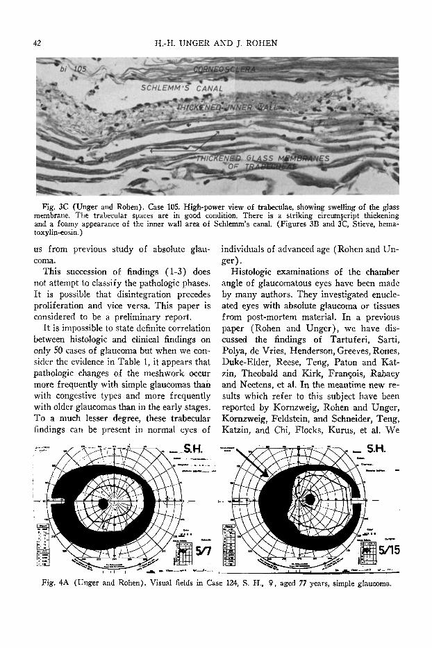
Fig. 3C (Unger and Rohen). Case 105. High-power view of trabeculae, showing swelling of the glass membrane. The trabecular spaces are in good condition. There is a striking circumscript thickening and a foamy appearance of the inner wall area of Schlemm's canal. (Figures 3B and 3C, Stieve, hema-toxylin-eosin.)
us from previous study of absolute glaucoma.
This succession of findings (1-3) does not attempt to classify the pathologic phases. It is possible that disintegration precedes proliferation and vice versa. This paper is considered to be a preliminary report.
It is impossible to state definite correlation between histologie and clinical findings on only 50 cases of glaucoma but when we consider the evidence in Table 1, it appears that pathologic changes of the meshwork occur more frequently with simple glaucomas than with congestive types and more frequently with older glaucomas than in the early stages. To a much lesser degree, these trabecular findings can be present in normal eyes of
individuals of advanced age (Rohen and Unger).
Histologie examinations of the chamber angle of glaucoma tous eyes have been made by many authors. They investigated enucleated eyes with absolute glaucoma or tissues from post-mortem material. In a previous paper (Rohen and Unger), we have discussed the findings of Tartuferi, Sarti, Polya, de Vries, Henderson, Greeves, Rones, Duke-Elder, Reese, Teng, Paton and Kat-zin, Theobald and Kirk, François, Rabaey and Neetens, et al. In the meantime new results which refer to this subject have been reported by Kornzweig, Rohen and Unger, Kornzweig, Feldstein, and Schneider, Teng, Katzin, and Chi, Flocks, Kurus, et al. We
5/15
Fig. 4A (Unger and Rohen). Visual fields in Case 124, S. H., 9, aged 77 years, simple glaucoma.

BIOPSY OF TRABECULAR MESHWORK 43
bi t24
Fig. 4B (Unger and Rohen). Case 124. Sagittal section of Elliot disk (right eye).
think that our findings in a certain degree are similar to those of Flocks (one case of early glaucoma), Kornzweig, et al. (seven cases of early glaucoma), as well as the very important findings of Teng et al. (four cases of early glaucoma) as far as we can compare with the excellent figures given in his last paper. We think it is too early to speak about histologie findings of the meshwork as etio-
logic factors of glaucoma. Further careful studies must be made.
It is most probable that within the meshwork, the main impediment to aqueous outflow is represented by the inner wall of Schlemm's canal. Therefore, we have a special interest in the pathology of this region.
We agree with Vail that the pathologic findings of the trabecular meshwork in cases
J3M8ffCUZ.4*\ Fig. 4C (Unger and Rohen). Case 124. High-power view of Schlemm's canal. Trabecular glass mem
branes are slightly thickened. There is a tumorlike proliferation of endothelium in the region of the inner wall. (Figures 4B and 4C, Stieve, hematoxylin-eosin.)

44 H.-H. UNGER AND J. ROHEN
of simple glaucoma are of a degenerative nature. They may contribute to the increase of resistance to the aqueous circulation, thus making glaucoma worse.
CONCLUSION
Fifty-eight corneoscleral disks of Elliot trephinings (from SO patients) have been examined for histologie alterations of the trabecular meshwork with early and less progressive glaucoma (chronical congestive glaucoma: 24 eyes; simple glaucoma: 34 eyes).
Especially in progressive cases of simple glaucoma two types of biopsy alterations can be observed:
1. Changes of the glass membrane of the trabeculae, with or without intertrabecular adhesions.
2. Changes of the inner wall of Schlemm's canal with or without circumscript proliferation of endothelial cells.
Four cases showed an excessive, tumorlike increase in the number of cells. In one case the meshwork was replaced by amorphous tissues.
These alterations are of a degenerative nature. It is probable that they contribute to the obliteration of the intertrabecular aqueous passages.
Schumannstrasse 14.
REFERENCES
Duke-Elder, S. : Textbook of Ophthalmology. St. Louis, Mosby, 1941, v. 3, p. 3341. Flocks, M. : Tr. Am. Acad. Ophth., 1958, p. 556. François, J., Rabaey, M., and Neetens, A. : Arch. Ophth., 55 :488, 1956. Garron, L. K., Fenney, M. L , Hogan, M. J., and McEwen, W. K.: Am. J. Ophth., 46:27,1958. Greeves, R. A. : Proc. Roy. Soc. Med. London, Sect. Ophth., 7 :112, 1913-14. Henderson, T. : Ophth. Ree, 17 :534, 1908.
: Glaucoma, London, Arnold, 1910, pp. 34-38. Kornzweig, A. L. : Ophth. ibero-am., 18 :12, 1956. Kornzweig, A. L., Feldstein, M., and Schneider, J. : Am. J. Ophth., 46:311, 1958. Kurus, E. : Klin. Monatsbl. f. Augenh., 132:201, 1958. Polya: Ungar. Beitr. 2:319, 1899. Reese, A. B. : Am. J. Ophth., 27 :1193, 1944. Rohen, J., and Unger, H.-H. : Am. J. Ophth., 46:802, 1958.
— : Zur Morphologie und Pathologie der Kammerbucht des Auges. Mainz, Verl. Akad. Wiss. Lit., Wiesbaden, Franz Steiner, 1959.
Rones, B.: Am. J. Ophth., 21:239, 1938; 45:189, 1958. Sarti, V. : Boll. se. med. Bologna, 4:147, 1893. Tartuf eri, F. : Giorn. Acad. med. Torino, 30:624, 1882. Teng, C. C, Paton, R. T., and Katzin, H. M. : Am. J. Ophth., 40 :619, 1955. Teng, C. C, Katzin, H. M., and Chi, H. H.: Am. J. Ophth., 43:193, 1957. Theobald, G., and Kirk, H. Q. : Am. J. Ophth, 41:11, 1956. Unger, H.-H. : D. O. G. Heidelberg, 1956, München, Bergmann, 1957, p. 59.
— : Arch. Ophth, 158:509, 1957. Unger, H.-H, and Rohen, J.: Am. J. Ophth, 48:204, 1959. Vail, D. : Year-Book of Ophthalmology, Chicago, Year-Book, 1958, p. 288. de Vries, W. M. : Nederl. Tijdschr. v. Geneesk, 43 :1688, 1907.