biology 2401 orientation, the microscope, and the language of...
TRANSCRIPT

WCJC Biology 2401 Rev 082010 PAGE 1
Biology 2401 Orientation, the Microscope, and the Language of Anatomy
Purpose: This lab will 1) introduce policies and procedures of the laboratory, 2) introduce anatomical and directional terms used to describe the human body, and 3) identify major organs in the dorsal and ventral body cavities, and 4) describe use of the microscope. I. Orientation
A. Discuss lab syllabus and schedule B. Discuss expectations and testing procedures
II. The Microscope
A. Describe the parts of the compound microscope B. Demonstrate proper use of the microscope
C. Determine total magnification of a viewed specimen
III. The Language of Anatomy
A. Describe anatomical position B. Describe directional terms, anatomical terms/regions
(models), and body planes (models) C. Identify major body cavities (models) D. List and identify locations for serous membranes of the
thoracic and abdominal cavities (models) E. Identify abdominal quadrants and regions (models)
In addition to the general goals above, specific anatomical structures that the student must be able to identify are listed below. A few select structures may be added to the list at the discretion of the instructor.

The Microscope and the Language of Anatomy
Description Page
I. Microscopeeye piece or ocularheadarmbaselight sourcestagemechanical stagerevolving nosepieceobjective lenses - scanning lens (4X) - low power lens (10X) - high power lens (40X)coarse adjustmentfine adjustmentiris diaphragm
II. Calculation of total magnification:total magnification =eye piece x objective lens
III. Anatomical Position and Surface Anatomy 3
IV. Directional Terms 4anterior/ventralposterior/dorsalsuperior/cephaladinferior/caudalsuperficialdeepproximaldistalmediallateral
V. Body positionsupineprone
VI. Body Planes and Sections 5sagittalmidsagittaltransversefrontal/coronal
WCJC Biology 2401 Rev 082010 PAGE 2

The Microscope and the Language of Anatomy
Description PageVII. Body Cavities and Subdivisions 6A. dorsal body cavity 1. cranial cavity 2. spinal/vertebral cavityB. ventral body cavity 1. thoracic cavity a. pericardial cavity b. pleural cavities 2. abdominopelvicIdentify the diaphragm
VIII. Abdominopelvic Quadrants (4) and Regions (9) 7right upper quadrant (RUQ)right lower quadrant (RLQ)left upper quadrant (LUQ)left lower quadrant (LLQ)right/left hypochondriac regionright/left lumbar regionright/left iliac (inguinal) regionepigastric regionumbilical regionhypogastric (pubic) regionIX. Serous Membranes 8A. surrounding the lungs 1. parietal pleura 2. visceral pleuraB. surrounding the heart 1. parietal pericardium 2. visceral pericardiumC. surrounding the abdominopelvic cavity 1. parietal peritoneum 2. visceral peritoneum
WCJC Biology 2401 Rev 082110 PAGE 3

WCJC Biology 2401 Rev 082010 PAGE 4
Biology 2401 The Cell, Tissues, and Integumentary System
Purpose: This lab will describe the structures of a generalized cell, the four main types of tissues (and selected subtypes), and the regions and structures in human skin. I. The Cell A. Identify the plasma membrane (model) B. Identify the organelles found in the cytosol (model)
C. Identify the nucleus and its components (model) II. Tissues A. List the four main types of tissues
B. Classify epithelial tissue based on the number of layers and shape (slides)
C. Observe and identify the main types of tissues and selected
subtypes (models and slides) III. The Integumentary System A. Identify the layers of the epidermis (models and slide)
B. Identify the dermis and accessory structures (models and slide) C. Describe the composition of the hypodermis
D. Observe a slide of human skin and identify the dermis and the layers of the epidermis
In addition to the general goals above, specific anatomical structures that the student must be able to identify are listed below. A few select structures may be added to the list at the discretion of the instructor.

The Cell, Tissues, and Integumentary System
Structure Description Page
I. Cell 11A. plasma membraneB. cytoplasm 1. cytosol 2. organelles a. mitochondria b. smooth endoplasmic reticulum c. rough endoplasmic reticulum d. ribosomes e. golgi apparatus f. centrioles g. centrosome h. lysosome i. nucleus i. nuclear membrane/envelope ii nuclear pores iii. nucleolus iii. chromatin
II. Tissues 12-23A. epithelial 1. simple squamous epithelium 2. stratified squamous epithelium 3. simple cuboidal epithelium 4. simple columnar epitheliumB. connective 1. loose connective tissue 2. cartilage 3. bone 4. bloodC. muscle 1. skeletal muscle 2. cardiac muscle 3. smooth muscleD. nervous
WCJC Biology 2401 Rev 082110 PAGE 5

The Cell, Tissues, and Integumentary System
Structure Description Page
III. Integumentary System 24-25Skin (cutaneous membrane)A. epidermis 1. stratum basale 2. stratum spinosum 3. stratum granulosum 4. stratum lucidum (in thick skin) 5. stratum corneumB. dermis 1. dermal papilla 2. Meissner's corpuscle 3. Pacinian corpuscle 4. sudoriferous glands 5. sebaceous glands 6. hair follicle 7. arrector pili muscle 8. blood vessels
Hypodermis-subcutaneous tissue
WCJC Biology 2401 Rev 082110 PAGE 6

WCJC Biology 2401 Rev 082010 PAGE 7
Biology 2401 The Skeletal System
Purpose: The lab will describe the microscopic and gross anatomy of bone, identify bones of the body, and identify important bone markings. I. Overview of the Skeleton
A. Discuss names and descriptions of bone markings B. Identify the four main types of bone (articulated or disarticulated skeleton) C. Identify major anatomical structures of a typical long bone (model and bone)
II. Histology
A. Identify major structures of the osteon (model and slide)
B. Identify the structures of compact bone
(model and slide) III. Axial Skeleton (Skull, Vertebrae, Ribs, Sternum, Hyoid)
A. Identify bones of the axial skeleton (articulated or disarticulated skeleton)
B. Identify selected bone markings (articulated or disarticulated
skeleton) IV. Appendicular Skeleton
A. Identify selected bones of the appendicular skeleton (articulated or disarticulated skeleton)
B. Identify selected bone markings (articulated or disarticulated skeleton) In addition to the general goals above, specific anatomical structures that the student must be able to identify are listed below. A few select structures may be added to the list at the discretion of the instructor.

The Skeletal System
Description Page
GROSS ANATOMY OF A LONG BONE 30proximal epiphysisdistal epiphysisdiaphysismedullary cavity
X-SECTION OF COMPACT BONE 30, 32periosteumperforating/Sharpey's fibersendosteumperforating/Volkmann's canalosteon (Haversian system) - central/Haversian canal (contains blood vessels, nerves) - lamella - lacuna - osteocyte - canaliculus
SKULL - CRANIAL BONES: 33-36Frontal - frontal/coronal sutureParietal - sagittal sutureTemporal - squamous suture - zygomatic process - external acoustic/auditory meatus - mastoid process - styloid process - carotid canalOccipital - lambdoid suture - occipital condyles - foramen magnum - external occipital protuberanceSphenoid - sella turcicaEthmoid - middle nasal concha 36, 127 - superior nasal concha 36, 127 - ethmoid air cells 38 - crista galli - perpendicular plate
AXIAL SKELETON
WCJC Biology 2401 Rev 082110 PAGE 8

The Skeletal System
Description PageSKULL - FACIAL BONES: 33-37NasalMaxilla - palatine process of the maxillae - infraorbital foramenPalatineInferior nasal conchaZygomaticMandible - mental foramen - mandibular foramen - mandibular condyle - coronoid process - angle - ramus - bodyLacrimalVomer
HYOID BONE:VERTEBRAE: 39-41Cervical - (C1-C7) - atlas (C1) - axis (C2) - dens (odontoid process)Thoracic - (T1-T12)Lumbar - (L1-L5)Sacral (sacrum) - 5 fusedCoccyx - 3 to 5 fused
Markings of a typical vertebrae: dorsal spinous process transverse process vertebral foramen (forms cavity) body lamina pedicle
Other vertebral structures: intervertebral foramen intervertebral disc - annulus fibrosus -- - nucleus pulposus --
WCJC Biology 2401 Rev 082110 PAGE 9
The Skeletal System
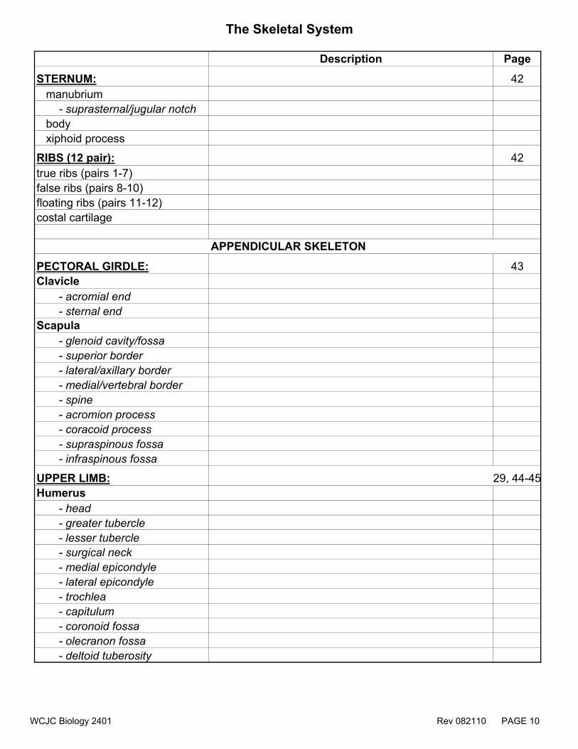
Description Page
STERNUM: 42 manubrium - suprasternal/jugular notch body xiphoid process
RIBS (12 pair): 42true ribs (pairs 1-7)false ribs (pairs 8-10) floating ribs (pairs 11-12)costal cartilage
PECTORAL GIRDLE: 43Clavicle - acromial end - sternal endScapula - glenoid cavity/fossa - superior border - lateral/axillary border - medial/vertebral border - spine - acromion process - coracoid process - supraspinous fossa - infraspinous fossa
UPPER LIMB: 29, 44-45Humerus - head - greater tubercle - lesser tubercle - surgical neck - medial epicondyle - lateral epicondyle - trochlea - capitulum - coronoid fossa - olecranon fossa - deltoid tuberosity
APPENDICULAR SKELETON
WCJC Biology 2401 Rev 082110 PAGE 10

The Skeletal System
Description PageUlna - styloid process - coronoid process - olecranon process - trochlear/semilunar notch - radial notchRadius - head - radial tuberosity - styloid process - neckCarpals (8 per hand)Metacarpals (5 per hand)metacarpophalangeal (MP) jointPhalanges (14 per hand)proximal phalangesmiddle phalangesdistal phalangesinterphalangeal (IP) joint
PELVIC GIRDLE (2 coxal bones): 46 - acetabulumIlium - iliac crest - anterior superior iliac spine - greater sciatic notchIshium - ischial tuberosity - ischial spinePubis - obturator foramen pubic symphysis (cartilage)
WCJC Biology 2401 Rev 082110 PAGE 11
The Skeletal System
Description Page
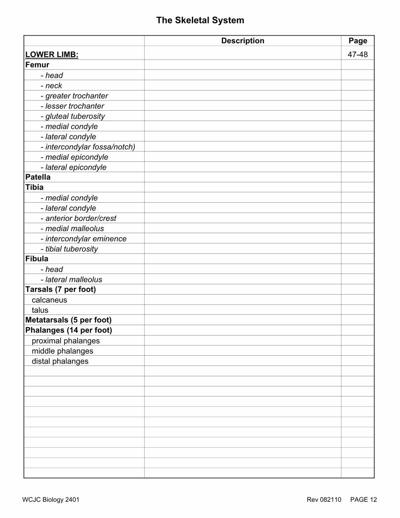
LOWER LIMB: 47-48Femur - head - neck - greater trochanter - lesser trochanter - gluteal tuberosity - medial condyle - lateral condyle - intercondylar fossa/notch) - medial epicondyle - lateral epicondylePatellaTibia - medial condyle - lateral condyle - anterior border/crest - medial malleolus - intercondylar eminence - tibial tuberosityFibula - head - lateral malleolusTarsals (7 per foot) calcaneus talusMetatarsals (5 per foot)Phalanges (14 per foot) proximal phalanges middle phalanges distal phalanges
WCJC Biology 2401 Rev 082110 PAGE 12

WCJC Biology 2401 Rev 082010 PAGE 13
Biology 2401 The Muscular System
Purpose: This lab will discuss the three types of muscle tissue and identify major skeletal muscles. The origins, insertions and actions will also be described for select muscles. I. Muscle Histology
A. Identify the three types of muscle tissue and selected structures that
distinguish each type (model and slides) B. Identify selected structures in skeletal muscle
tissue (model only) C. Identify a neuromuscular junction and identify
selected structures that form the neuromuscular junction (model and slide)
II. Skeletal Muscles
A. Discuss the naming mechanisms of skeletal muscle
B. Identify selected skeletal muscles (muscle models)
III. Origins, Insertions, and Actions A. Discuss the terms: origin, insertion, and action
B. Identify the origin(s), insertion(s), or action(s) of select skeletal muscles with respect to the muscle itself. (muscle models)
C. Identify the origins, insertions, actions or names of select skeletal muscles
with respect to bone markings. (articulated or disarticulated skeleton) In addition to the general goals above, specific anatomical structures that the student must be able to identify are listed below. A few select structures may be added to the list at the discretion of the instructor.

The Muscular System
Description PageMICROSCOPIC ANATOMY OF SKELETAL MUSCLE 51, 68, 71endomysiumsarcolemmasarcoplasmmyofibrilnucleusneuromuscular junction - motor axon - axon teminal - motor end plate (MEP) - myelin
Slides and Muscle Tissue Modelsskeletal muscle -nucleus -striations (model only)cardiac muscle -intercalated discs -nucleus -striations (model only)smooth muscle -nucleus (model only)neuromuscular junction
IDENTIFICATION OF SELECTED SKELETAL MUSCLESOf facial expression 52, 54frontalisorbicularis oculi zygomaticusorbicularis orismentalisbuccinator
Of mastication 52, 54-55masseter *temporalis *digastric *That act the head and neck 52, 55-56sternocleidomastoid *
** structure is not a muscle* also learn insertion, origin, and action of these muscles
WCJC Biology 2401 Rev 082110 PAGE 14

The Muscular System
Description PageOf respiration 57external intercostalsinternal intercostalsdiaphragmOf the abdominal wall 52, 58rectus abdominis *external obliqueinternal obliquetransversus abdominis**linea albaOf the anterior thorax 52. 56pectoralis minorserratus anteriorOf the posterior thorax 53, 60-61trapeziuslevator scapulaerhomboids
That act the arm52-53, 56,
60-62pectoralis major *latissimus dorsi *deltoid *rotator cuff muscles-SITS - supraspinatus - infraspinatus - teres minor - subscapularisteres majortriceps brachii * - long head - medial head - lateral headbiceps brachii *brachialis *brachioradialisThat act the wrist and fingers 52-53, 62flexorsextensors
** structure is not a muscle* also learn insertion, origin, and action of these muscles
WCJC Biology 2401 Rev 082110 PAGE 15

The Muscular System
Description PageThat act the thigh 64iliopsoas *sartorius *adductor magnusadductor longusgracilisgluteus maximus *gluteus mediusgluteus minimus
tensor fasciae latae**iliotibial tractThat act the leg 64quadriceps femoris * - vastus lateralis - vastus medialis - vastus intermedius - rectus femorishamstrings * - biceps femoris
- semitendinosus - semimembranosusThat act the foot and ankle 66tibialis anterior *fibularis longusgastrocnemius * - medial head - lateral headsoleus**Achilles/calcaneal tendon
* also learn insertion, origin, and action of these muscles** structure is not a muscle
WCJC Biology 2401 Rev 082110 PAGE 16

Origin, Insertion and Action of Selected Skeletal Muscles
Muscles Origin Insertion Action
masseter zygomatic bone (zygomatic arch) posterior 1/3 mandible elevates mandible
temporalis temporal bone (temporal fossa)
coronoid process of mandible elevates mandible
digastric inferior mandible, temporal hyoid bone depresses mandible
Nec
k M
uscl
es
sternocleidomastoid manubrium and medial clavicle
mastoid process of temporal bone
single: rotate head to opposite side; both: flex head
Abd
omin
al
Mus
cles
rectus abdominis pubic symphysis xiphoid process, costal cartilages of ribs 5-7
flexes vertebral column, compresses abdomen
trapeziusoccipital bone, dorsal spinous processes (thoracic vertebrae)
clavicle, scapula (acromion process, spine)
extends head; elevates scapula
latissimus dorsi dorsal spine (T7 to sacrum), iliac crest humerus (proximal 1/3) extends arm; adducts and
medially rotates arm
rhomboids (major & minor)
dorsal spinous processes of C7-T5
medial border of scapula adducts scapula
deltoidclavicle, acromion process, spine of scapula
deltoid tuberosity of humerus abducts arm
triceps brachii scapula and humerus olecranon process of ulna extends forearm
pectoralis majorclavicle, sternum, costal cartilages of ribs 1-7
greater tubercle of humerus
flexes arm; adducts, and medially rotates arm
biceps brachiicoracoid process and supraglenoid tubercle of scapula
radial tuberosity flexes forearm
iliopsoas transverse processes of T12-L5; iliac fossa
lesser trochanter of femur major flexor of thigh
sartorius anterior superior iliac spine proximal tibia flexes and laterally rotates
thigh
origin - attachment point of the muscle that does not move during muscular contractioninsertion - attachment that moves during contractionaction - effect the muscle has on a part of the body
Mus
cles
of
Mas
ticat
ion
Post
erio
r Mus
cles
Act
ing
on th
e Sh
ould
er,
Arm
or F
orea
rm
Ant
erio
r Mus
cles
A
ctin
g on
the
Shou
lder
, Arm
or
Fore
arm
Mus
cles
of t
he
Med
ial T
high
WCJC Biology 2401 Rev 082110 PAGE 17

Origin, Insertion and Action of Selected Skeletal Muscles
Muscles Origin Insertion ActionM
uscl
es o
f th
e A
nter
ior
Thig
h quadriceps femoris muscles
ilium (rectus femoris) and femur tibial tuberosity extends leg (rectus femoris
also flexes thigh)
hamstrings ischial tuberosity head of fibula, proximal 1/3 tibia extends thigh, flexes leg
gluteus maximus iliac crest, sacrum, coccyx proximal 1/3 femur extends thigh
Mus
cles
of
the
Ant
erio
r Le
g tibialis anterior lateral condyle and upper 2/3 of tibia metatarsal 1 dorsiflexion
Mus
cles
of
the
Post
erio
r Le
g gastrocnemius condyles of femur calcaneus plantar flexes (extends) foot
Mus
cles
of
the
Post
erio
r Th
igh
WCJC Biology 2401 Rev 082110 PAGE 18

WCJC Biology 2401 Rev 082010 PAGE 19
Biology 2401 The Nervous System
Purpose: This lab will describe the composition of the nervous system including the brain, spinal cord, nerves, and selected special senses. I. Introduction to the Nervous System
A. Identify the structures of a neuron (models) B. List the main divisions of the nervous system II. The Central Nervous System
A. Identify the regions and lobes of the brain (models)
B. Identify the selected structures in the brain
(models and preserved specimen)
C. Identify the ventricles of the brain (models and preserved specimen)
D. Identify major regions in a cross section of a spinal cord
(models and slide) III. The Peripheral Nervous System
A. Identify selected cranial nerves (models and preserved specimen)
B. Identify each major nerve plexus and selected nerves that arise from each plexus (models)
IV. The Special Senses
A. Identify selected structures of the mammalian eye (models and preserved specimen)
B. Identify extrinsic eye muscles (models)
C. Identify selected structures of the external, middle, and inner ear (models)
V. Dissections
A. Dissect the sheep brain (p. 78-81) B. Dissect the cow eye (p. 90) In addition to the general goals above, specific anatomical structures that the student must be able to identify are listed below. A few select structures may be added to the list at the discretion of the instructor.

The Nervous System
Structure Description Page
Model of Neuron 71cell bodyaxon
dendritenucleusaxon hillockmyelin sheathSchwann cell axon terminalNissl bodiesnodes of Ranvier
Slide of multipolar neuron 21, 71
axoncell bodydendrite
Brain CEREBRUM* 72-74
cerebral hemisphereslongitudinal fissure*gyri*sulci*cerebral cortex (gray matter)*cerebral white matter*precentral gyruscentral sulcuspostcentral gyrusfrontal lobeparietal lobesoccipital lobetemporal lobescorpus callosum*cingulate gyrus --lateral ventricles*
* also identify structure in dissected organ
WCJC Biology 2401 Rev 082110 PAGE 20

The Nervous System
Description PageDIENCEPHALON 74,77-78
optic chiasm*thalamus*hypothalamus*pituitary gland*infundibulumthird ventriclepineal gland*
CEREBELLUM* 74-75, 81vermisarbor vitae*
BRAIN STEM 73-74, 80midbrainpons*medulla oblongata*corpora quadrigemina* - superior colliculus* - inferior colliculus*cerebral aqueduct*fourth ventricle*
Spinal Cord 82-83
white mattergray matter - gray commissure - ventral/anterior horn - dorsal/posterior horn - central canaldorsal root gangliondorsal rootventral rootspinal nerve
* also identify structure in dissected organ
WCJC Biology 2401 Rev 082110 PAGE 21

The Nervous System
Description Page
Meninges and Spaces 76, 82
dura mater*arachnoid materpia materepidural spacesubarachnoid space
Cranial Nerves 78-79
olfactory bulb and tract * (site where olfactory nerve fibers forming CN I will synapse)optic nerves (CN II)trigeminal nerve (CN V)facial nerve (CN VII)vagus nerve (CN X)vestibulocochlear nerve (CN VIII)
Nerve Plexuses & Spinal Nerves 83-87
cervical plexusbrachial plexus -radial nerve -ulnar nerve -median nervelumbar plexussacral plexus -sciatic nerve -tibial nerve -common fibular nervesympathetic chain ganglion
* also identify structure in dissected organ
WCJC Biology 2401 Rev 082110 PAGE 22

The Nervous System
Description Page
Anatomy of the Eye 88-90
sclera*choroid*retina*ciliary body*irispupil*
suspensory ligaments of lenslens*optic nerve (CN II)*optic discanterior segment/cavity - aqueous humorposterior segment/cavity - vitreous humor*ora serratacornea*lacrimal glandfovea centralis (fovea)
tapetum lucidum **
Extrinsic Eye Musclessuperior rectusinferior rectus medial rectus lateral rectus superior oblique inferior oblique
* *identify structure only in dissected cow/calf eye* also identify structure in dissected organ
WCJC Biology 2401 Rev 082110 PAGE 23

The Nervous System
Description Page
Anatomy of the Ear 91-92auricle/pinna
external acoustic/auditory meatus (external auditory canal)tympanic membranemiddle ear cavityauditory ossicles - malleus - incus - stapespharyngotympanic/auditory tubeoval windowround windowvestibulesemicircular canals (contain semicircular ducts) - anterior semicircular canal - posterior semicircular canal - lateral semicircular canalvestibulocochlear nerve (CN VIII)internal carotid artery --tensor tympani muscle --cochlea -scala vestibuli
-scala tympani -cochlear duct (scala media) -tectorial membrane -basilar membrane -hair cells
* also identify structure in dissected organ
WCJC Biology 2401 Rev 082110 PAGE 24

WCJC Biology 2401 Rev 082010 PAGE 25
Biology 2401 The Endocrine System
Purpose: This lab will identify the selected endocrine glands (indicated by *). Gland Hormone Function *HYPOTHALAMUS Releasing hormones Control ant. pituitary
*PITUITARY GLAND
Adrenocorticotrophic hormone ACTH
Controls 3 adrenal cortex hormones
Human Growth hormone hGH
Anabolic hormone targets bones, muscles, liver & other tissues
Prolactin
Initiates milk secretion
Thyroid stimulating hormone TSH
Controls the thyroid gland
Follicle stimulating hormone FSH
Stimulates ovarian follicle maturation & estrogen production. Sperm production in males
Adenohypophysis – (6)
Luteinizing hormone LH
Females- triggers ovulation Males- Promotes testosterone production
OT- Oxytocin
uterine contraction, milk ejection
Neurohypophysis (2)
ADH – Antidiuretic hormone
Conserves water thus it, concentrates the urine
*PINEAL GLAND Melatonin
Circadian rhythm & Induces sleep
WCJC Biology 2401 Rev 082010 PAGE 26
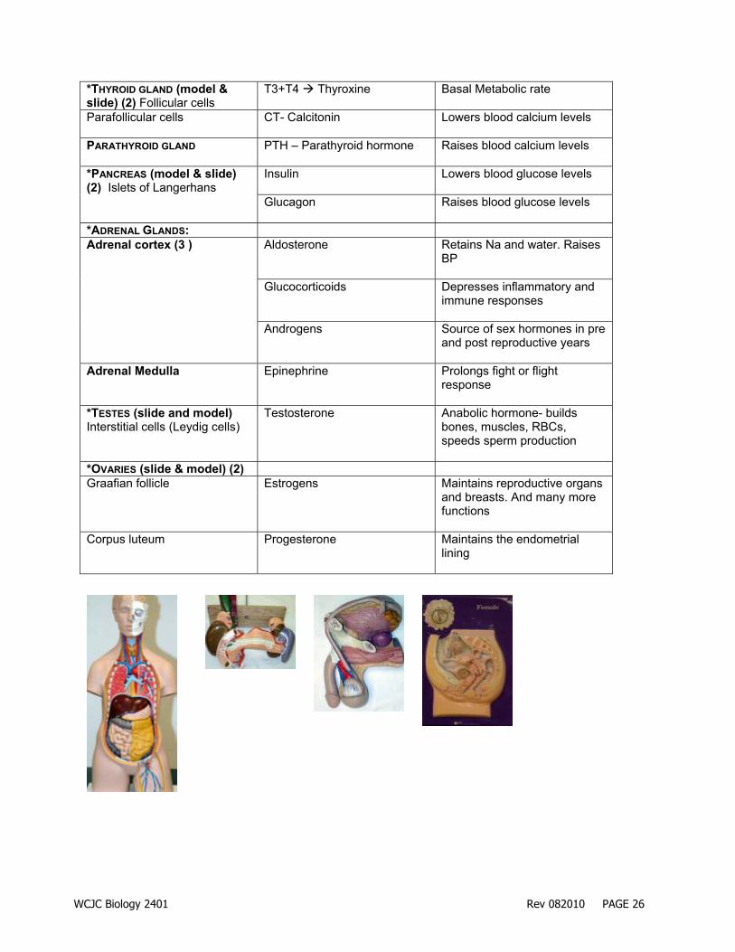
*THYROID GLAND (model & slide) (2) Follicular cells
T3+T4 Thyroxine
Basal Metabolic rate
Parafollicular cells CT- Calcitonin
Lowers blood calcium levels
PARATHYROID GLAND PTH – Parathyroid hormone
Raises blood calcium levels
Insulin
Lowers blood glucose levels
*PANCREAS (model & slide) (2) Islets of Langerhans
Glucagon
Raises blood glucose levels
*ADRENAL GLANDS: Aldosterone
Retains Na and water. Raises BP
Glucocorticoids
Depresses inflammatory and immune responses
Adrenal cortex (3 )
Androgens
Source of sex hormones in pre and post reproductive years
Adrenal Medulla Epinephrine Prolongs fight or flight response
*TESTES (slide and model) Interstitial cells (Leydig cells)
Testosterone
Anabolic hormone- builds bones, muscles, RBCs, speeds sperm production
*OVARIES (slide & model) (2) Graafian follicle Estrogens
Maintains reproductive organs and breasts. And many more functions
Corpus luteum Progesterone
Maintains the endometrial lining