biological responses of brushite-forming zn- and znsr
TRANSCRIPT
162 www.ecmjournal.org
S Pina et al. Brushite-forming bone cementsEuropean Cells and Materials Vol. 20 2010 (pages 162-177) DOI: 10.22203/eCM.v020a14 ISSN 1473-2262
Abstract
The core aim of this study was to investigate zinc (Zn)-and zinc and strontium (ZnSr)-containing brushite-formingβ-tricalcium phosphate (TCP) cements for their effects onproliferation and differentiation of osteoblastic-like cells(MC3T3-E1 cell line) as well as for their in vivo behaviourin trabecular bone cylindrical defects in a pilot study. Invitro proliferation and maturation responses of MC3T3-E1 osteoblastic-like cells to bone cements were studied atthe cellular and molecular levels. The Zn- and Sr-containingbrushite cements were found to stimulate pre-osteoblasticproliferation and osteoblastic maturation. Indeed, MC3T3-E1 cells exposed to the powdered cements had increasedproliferative rates and higher adhesiveness capacity, incomparison to control cells. Furthermore, they exhibitedhigher alkaline phosphatase (ALP) activity and increasedType-I collagen secretion and fibre deposition into theextracellular matrix. Proliferative and collagen depositionproperties were more evident for cells grown in cementsdoped with Sr. The in vivo osteoconductive properties ofthe ZnCPC and ZnSrCPC cements were also pursued.Histological and histomorphometric analyses wereperformed at 1 and 2 months after implantation, usingcarbonated apatite cement (Norian SRS®) as control. Therewas no evidence of cement-induced adverse foreign bodyreactions, and furthermore ZnCPC and ZnSrCPC cementsrevealed better in vivo performance in comparison to thecontrol apatite cement. Additionally, the presence of bothzinc and strontium resulted in the highest rate of new boneformation. These novel results indicate that the investigatedZnCPC and ZnSrCPC cements are both biocompatible andosteoconductive, being good candidate materials to use asbone substitutes.
Key words: Zinc, strontium, brushite cement, osteoblastproliferation, alkaline phosphatase, MC3T3-E1 celladhesion, Type-I collagen, trabecular bone regeneration.
*Address for correspondence:S. PinaDept. of Ceramics and Glass Engineering,University of Aveiro, CICECO,3810-193 Aveiro, Portugal
E-mail: [email protected]
Introduction
Calcium phosphate cements (CPC) have uniquecharacteristics for bone substitution compared with otherbiomaterials. Their excellence is due to goodbiocompatibility, excellent bioactivity, self-settingcharacteristics, low setting temperature, adequate stiffness,and easy shaping for any complicated geometry (Brownand Chow, 1983; Yuan et al., 2000; Baroud et al., 2005;Bohner and Baroud, 2005; Gauthier et al., 2005; Boeseland Reis, 2006; Wang et al., 2006; Alves et al., 2008;Burguera et al., 2008). In addition, CPC have suchcompositional resemblance to bone mineral that theyinduce a biological response similar to the one generatedduring bone remodelling.
Among the different biocompatible calcium phosphate(CaP) phases present in human bone, brushite (dicalciumphosphate dihydrate, DCPD) has a higher solubility thanhydroxyapatite (HA) at physiological pH, and an ideal invivo resorption rate (Constantz et al., 1998; Bohner et al.,2003; Gisep et al., 2003). Brushite-based bone cementsare generally well tolerated by the bone and soft tissueenvironment in vivo, such that cement resorption is closelyfollowed by new bone formation. Furthermore, brushitecements are known to be biocompatible, osteoconductiveand bioresorbable, having a potential interest for boneregeneration procedures.
From a cellular and molecular perspective, it has beenshown that some biomaterials are able to directly modifythe osteoblastic proliferation rate and some of theirfunctions, such as the synthesis of alkaline phosphatase(ALP) (Naji and Harmand, 1991; Malik et al., 1992; Puleoet al., 1993). The ionic composition of such biomaterialsis a key factor in their bioactivity (Ito et al., 2002; Li etal., 2008; Pina et al., 2010a). Zn and Sr incorporated intotricalcium phosphate (TCP) have a direct specificproliferative effect on osteoblastic cells in vitro and aselective inhibitory effect on osteoclastic bone resorptionin vivo (Ito et al., 2000; Otsuka et al., 2000; Ito et al.,2002; Li et al., 2008). Moreover, Zn is involved in manymetallo-enzymes and proteins, including ALP, whereasSr enhances collagen synthesis and has beneficial effectsin the treatment of osteoporosis, due to the prevention ofbone loss by a mechanism that depresses bone resorption(McComb et al., 1979; Rokita et al., 1993; Dahl et al.,2001; Marie et al., 2001).
Among various calcium phosphate phases β-TCP isgreatly biocompatible and resorbable. Furthermore, β-
BIOLOGICAL RESPONSES OF BRUSHITE-FORMING Zn- AND ZnSr-SUBSTITUTED βββββ-TRICALCIUM PHOSPHATE BONE CEMENTS
S. Pina1a*, S.I. Vieira2a, P. Rego3, P.M.C. Torres1, O.A.B. da Cruz e Silva2, E.F. da Cruz e Silva4, and J.M.F. Ferreira1
1University of Aveiro, Dept. of Ceramics and Glass Engineering, CICECO, 3810-193 Aveiro, Portugal.2University of Aveiro, Health Sciences Dept., Centro de Biologia Celular, 3810-193 Aveiro, Portugal.
3University of Lisbon, Orthopaedic Clinic, Medicine Faculty, 1600-190 Lisbon, Portugal.4University of Aveiro, Dept. of Biology, Centro de Biologia Celular, 3810-193 Aveiro, Portugal
aBoth authors contributed equally to this work
163 www.ecmjournal.org
S Pina et al. Brushite-forming bone cements
TCP is an adequate carrier for doping ions such as Sr andZn that might be released at low rates. Ito et al. (Ito et al.,2000) found that a slow Zn2+ release from a biphasicZnTCP/AP ceramics containing Zn doped β-TCPimplanted in rabbit femora stimulated bone formation.Based on a histological and histomorphometrical studyusing femora of rabbits, Kawamura et al. (Kawamura etal., 2000) reported that 51% of new bone has grown aroundZnTCP/AP implants.
Osteoblasts are cells found on bone surfaces, and aredirectly responsible for bone formation. As osteoblastsdifferentiate from their precursors and alter theirmorphology and expression and/or activity of functionalmarkers (maturation process), they begin to secrete bonematrix proteins such as Type-I collagen that representsabout 90% of the organic matrix. The network of Type-Icollagen fibres provides the structure on which bonemineral is deposited. Type-I collagen and endogenousbrushite particles are both predominant in fracture callus(Lenart et al., 1972; Kurdy et al., 1998), with the mainstructural framework of a human fracture callus consistingof disordered, mineralized collagen fibrils containing CaPcrystals. Brushite particles are found in the noncollagenousorganic matter around nonmineralized, ordered collagenfibrils, and it is believed that these particles serve as thereservoir of calcium and phosphate ions for subsequentmineralization (Wen et al., 1995).
The in vivo osteoconductive relevance of brushitecements has been well documented but the knowledgeconcerning the in vivo behaviour of brushite-formingcements with Zn and Sr co-substituted in β-TCP is stilllimited. The aim of the present study was to examine theosteoconductive properties of two new brushite-formingZn and ZnSr-containing CPCs, previously studied by us(Pina et al., 2010b). The in vitro proliferation anddifferentiation responses of MC3T3-E1 osteoblast-likecells to the bone cements were initially studied. Variouscellular and molecular responses to the biomaterials wereassayed, including cell viability determinations by theresazurin assay, photometric evaluation of ALP activityafter enzymatic cleavage of p-nitrophenyl phosphate, andtime-dependent Type-I collagen production. The in vivoosteoconductive properties of these bone cements werefurther assayed after implantation in trabecular bone inpigs, and compared to a commercial apatite CPC.
Materials and Methods
Cement preparationBrushite cements were prepared as described in theprevious study (Pina et al., 2010b). Briefly, the cementpastes were prepared by mixing 55 wt.% of Zn- or ZnSr-substituted β-tricalcium phosphate (β-TCP) powder with45 wt.% of monocalcium phosphate monohydrate(MCPM, Sigma-Aldrich, Munich, Germany) using liquid-to-powder ratio (LPR) of 0.34 mL g-1. The aqueous solutionused was 10 wt.% poly(ethylene glycol) (PEG) (200,Sigma-Aldrich) + 15 wt.% citric acid solution.
Zn- and ZnSr-substituted β-TCP powders weresynthesized by aqueous precipitation as described in detail
in previous reports (Kannan et al., 2006; Kannan et al.,2008; Kannan et al., 2009). The precipitates were vacuumfiltrated, dried at 110ºC, heat treated for 2 h at 1100ºC andgrounded under dry conditions in a planetary mill, andfinally passed through a sieve with a mesh size of 36 μm.The starting powders were constituted by: 2.4 ± 0.1 mol.%Zn2+ for ZnCPC, and 2.3 ± 0.2 mol.% Zn2+ and 2.1 ± 0.1mol.% Sr2+ for ZnSrCPC, determined by ICP spectroscopyin a previous work by the same authors (Pina et al., 2010b).The Zn- and ZnSr-substituted β-TCP cements weredesignated as ZnCPC and ZnSrCPC, respectively.
MC3T3-E1 cell cultureThe osteoblastic cell line MC3T3-E1 (ATCC, Barcelona,Spain; CRL-2593) was established from C57BL/6 mousecalvaria and selected on the basis of high ALP activity inthe resting state. MC3T3-E1 cells were maintained at 37ºCin a humidified atmosphere of 5% CO2 in air, in 2mMGlutamine-containing Minimum Essential α-Medium inEBSS (Eagle’s Balanced Salt Solution) supplemented with10% (v/v) foetal bovine serum (FBS), 1% (v/v) of a 100U mL-1 penicillin and 100 mg mL-1 streptomycin solution(Gibco BRL, Invitrogen, Life Technologies, Carlsbad, CA,USA) and 3.7 g L-1 NaHCO3. Sub-confluent cultures (80-90% confluency) were split 1:5 using a 0.25% trypsin/EDTA (Gibco BRL, Invitrogen) solution at 5% CO2, 37ºC.
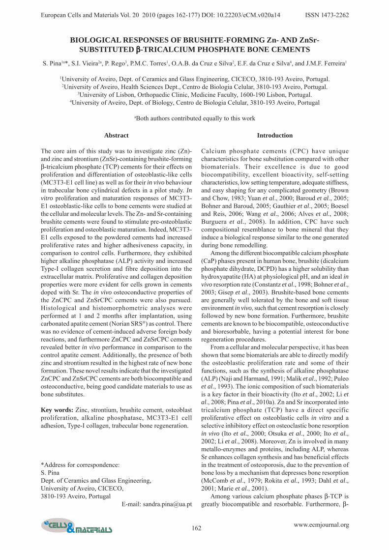
Cytotoxicity and cell proliferation assays in cementsThe resazurin metabolic assay (Ueno et al., 2006; Pina etal., 2010b) was used to determine the cements’cytotoxicity/biocompatibility to MC3T3-E1 cells. Thesensitivity and the range of linearity between resazurinreduction to resorufin and the density of MC3T3-E1 viablecells were first determined in a cell curve assay, asdescribed previously (Pina et al., 2010b) (Fig. 1). Briefly,cells were seeded in 35 mm plates at 1 x 10³ cells cm-2
and, at the indicated time points, cells were incubated for4 h with fresh medium containing 10% of a resazurin(Sigma-Aldrich) solution (0.1 mg mL-1 resazurin inphosphate buffer saline (PBS) [Pierce, Perbio, ThermoScientific, Bonn, Germany]). Resazurin reduction was
Fig. 1. Growth curve of MC3T3-E1, carried out usingthe resazurin assay, was carried out in order to determinethe zone of linearity between resazurin assay O.D. andcell density.

164 www.ecmjournal.org
S Pina et al. Brushite-forming bone cements
thereafter measured spectrophotometrically (Cary 50 BIO,Varian, Palo Alto, CA, USA) at 570 and 600 nm. Thenumber of viable cells was further counted using a 0.4%solution of the Trypan blue dye (Sigma-Aldrich) and ahaemocytometer. For each day, a final resazurin value(O.D.F) was calculated as the ratio O.D. 570/O.D. 600 nmminus the O.D. 570/O.D. 600 nm ratio of a negative control(resazurin media incubated for 4 h in the absence of cells),and the O.D. F was plotted against cell density.
To determine dose-dependent cell viability andproliferation when exposed to the powdered cements milledup to an average particle size of about 15 μm. 1 x 10⁵ cellswere seeded in 35 mm wells (density of 1 x 10⁴ cells cm-2),and cultured in cell media supplemented with 0, 0.01, 0.1or 1 mg mL-1 of ZnCPC or ZnSrCPC cement powders.This range of cement concentrations was chosen as 1 mgmL-1 already resulted in saturated solutions. Of note, priorto cell experiments, cement samples were sterilized by γ-radiation. Following 24, 48 and 72 h of incubation at 37°Cin a humidified 5% CO2 and 95% air atmosphere, growthmedium was removed by aspiration and replaced with freshmedium containing 10% of the 0.1 mg mL-1 resazurinsolution. After 4 h of incubation, 1 mL of the medium wascollected and cell viability determined by measuring O.D.at 570 and 600 nm, as described above. For each time point,the O.D.F levels of cells incubated with 0 mg mL-1 cementpowder were taken as 100% and cell viability calculatedas a percentage of these control values. All experimentswere carried out in triplicate and expressed as the mean ±standard error of the mean. Upon 72 h, cells morphologywas evaluated by phase contrast (PhC) microscopy in aninverted Olympus (Hamburg, Germany) IX81epifluorescence microscope, and microphotographs weretaken at 18ºC with a Digital CCD monochrome camera F-View II (Olympus Soft Imaging System).
Determination of alkaline phosphatase activityMC3T3-E1 cells were seeded at 1 x 10⁴ cells cm-2 in 35mm wells and cultured on 1 mg mL-1 ZnCPC and ZnSrCPCpowders. At day 3 in culture, achievement of cellconfluency was visually confirmed. Alkaline phosphatase(ALP) activity of exposed cells was evaluated at 0, 1, 3, 6,14 and 21 days post-confluency (DPC). After removal ofthe culture medium, cells were washed with PBS andharvested in 1 mL universal ALP buffer (100 mM citricacid, 100 mM KH2PO4, 100 mM sodiumtetraborate.10H2O, 100 mM Tris, 100 mM KCl; pH 11)with a disposable cell scraper. Cells with or without cementwere sonicated twice for 20 sec and centrifuged for 5 minat 2000 rpm at room temperature. ALP activity in thesupernatant was determined following addition of the p-nitrophenyl phosphate substrate (Fluka, Buchs,Switzerland). The production of p-nitrophenol wasdetermined spectrophotometrically at 420 nm, after 2 and5 min of reaction at 37ºC. The ALP activity was expressedas the increase in p-nitrophenol aborbance per min permicrogram of protein (ΔOD.min-1.mg-1), upon subtractionof the blank O.D. (1 mL universal buffer plus substrate).To determine protein mass, parallel samples were harvestedwith 1% SDS, and total protein content measured using aBCA kit (Pierce).
ZnCPC and ZnSrCPC time-dependent cell adhesionassaysThe capacity of cells to adhere to a plastic support wasmeasured upon cell exposure to the cement powders forincreasing time periods. Post-exposure adhesion assayswere performed at 0, 3, 14 and 21 days post-confluency(DPC), upon cell plating as described above for the ALPassay (day 0 = 3 days post-seeding). Cell media waschanged every other ~3-5 days in culture. At eachexperimental day, cells were washed with PBS, incubatedwith Trypsin-EDTA solution for 5 min at 37ºC, andresuspended in 3 mL fresh media. Cells were counted usingthe Trypan blue dye; 1 x 10⁵ cells were seeded into 24-well plates with 1 mL fresh media (final volume) and leftto adhere for 1 h in an atmosphere of 5% CO2 at 37°C.Quantification of adherent cells was performed indirectlyby scoring the number of non-adherent resuspended cellsin the media. Thus, cell media were collected and an aliquotapplied to a haemocytometer. Resuspended cells werecounted using the Trypan blue dye and the number of viablenon-adherent cells determined. Consequently, thepercentage of adherent cells was calculated, taking as100%the 1 x 10⁵ cells seeded. Three independentexperiments were performed.
Type-I collagen time-dependent protein expressionand cellular localizationMC3T3-E1 cells were treated as described for the ALPassays and subjected to immunoblot analyses to determinethe profile of Type-I procollagen and collagen proteinexpression, and secretion into the medium, with time ofexposure to 1 mg/ml cement powders for various DPC(confluency achieved at 3 days in vitro). Type-I collagencellular distribution was further analyzed byimmunocytochemistry procedures at 3 DPC.
AntibodiesThe primary antibody used was anti-Collagen Type-I(Novus Biologicals, Cambridge, UK), which recognizesType-I procollagen (monomeric α1(I) and α2(I) chains, βchains corresponding to α/α dimmers, and trimeric γchains), and various collagen forms (tropocollagen, themature trimeric γ chains, microfibrils, fibrils and fibresF(I)). Secondary antibodies used were FITC-conjugatedanti-rabbit IgGs (Calbiochem, Merck4Biosciences,Nottingham, UK) for immunocytochemistry analyses, andhorseradish peroxidase-linked anti-rabbit IgGs forenhanced chemiluminescence (ECL) detection (GEHealthcare, Chalfont St. Giles, UK).
Immunoblot analyses of Type-I collagen proteinexpression and media secretionAt the indicated days in post-confluency cultures, cellsconditioned media (1 mL) were collected into 10% SDS-containing microtubes, cells were washed with PBS andharvested with a 1% SDS boiling solution. Samples wereboiled for 10 min, subjected to a routine sonication periodin ice (3x10 sec) (S1), and their total mass contentdetermined using the BCA kit (Pierce). Given that thedifficulty of the insoluble fibres present in the samples,these were subjected to an additional extensive sonication

165 www.ecmjournal.org
S Pina et al. Brushite-forming bone cements
period (S2). Mass-normalized cell lysates (S1 and S2) andmedia samples (60 μg) were subjected to reducing 6.5%SDS-PAGE in Tris-Glycine buffer, and electrophoreticallytransferred onto nitrocellulose membranes. Precision PlusDual Color (Bio-Rad, Amadora, Portugal) were used asprotein standards. Immunoblotting of the transferredproteins was performed by incubating membranes O/Nwith the anti-Collagen Type-I primary antibody, afterblocking non-specific binding sites with non-fat dry milkin TBS-T (10 mM Tris-HCl at pH 8.0, 150 mM NaCl,0.5% Tween). Detection was achieved using a horseradishperoxidase-linked secondary antibody and an ECL kit (GEHealthcare).
Confocal microcopy analyses of Type-I collagenprotein distributionCells treated as above (section 2.6.2) were grown on 35mm plates-containing pre-treated coverslips and fixed witha 4% paraformaldehyde PBS solution at 3 DPC. Cells weremethanol-permeabilized, non-specific sites blocked by 1h incubation with a 3% bovine serum albumin (BSA)-PBSsolution, and coverslips submitted to immuno-cytochemistry procedures using the anti-Collagen Type-Iantibody diluted in 3% BSA-PBS. Upon three washes withPBS, cells were incubated with the fluoresceinisothiocyanate (FITC)-conjugated secondary antibody.Coverslips were mounted on microscope slides with 4',6-diamidino-2-phenylindole (DAPI)-containing Vectashieldantifading reagent (Vector, Burlingame, CA, USA). Imageswere acquired in a LSM 510 META confocal microscope(Zeiss, Göttingen, Germany) using an Argon laser line of488 nm (FITC channel), and a Diode 405-430 laser (DAPIchannel).
Animal model and implantation procedureAnimal studies were performed according to Europeanregulations and following permission granted by theEthical Committee. Owing to ethical and economicreasons, only two animals were used for implantationprocedures. Two mature male pigs weighting between 40and 50 kg were used as experimental animals, forobservation periods of 1 month (one pig) and 2 months(the other pig). Injection sites were shaved and cleanedwith Betadine (10% povidone-iodine). The animals wereoperated under general anaesthesia performed withintravenous injection of ketamine (Ketalar, Pfizer, NewYork City, NY, USA) and xylazine hydrochloride (Rompun2%, Bayer, Leverkusen, Germany) under asepticconditions. To implant the cement pastes, a longitudinalincision on the outer surface of the foot was made untilexpose the tarsal bone and three circular holes (∅ 3.5x10mm3) drilled perpendicular to the long axis of the bonewith a drill bit (Synthes, West Chester, PA, USA) in eachanimal. The CPCs to be implanted were prepared by handmixing for 1 min and then injected in the cavities with a 5mL syringe. The holes were filled, respectively, withZnSrCPC, ZnCPC and reference apatite cement (NorianSRS®, Norian Corp., Synthes). In each hole, one marker(skin stapler) was placed into the cement paste, duringsetting, for localization of the implant position at the endof the implantation period. Setting time of 11 ± 2 min after
filling the holes was granted for the cements, ZnSrCPCand ZnCPC, before closing. Norian SRS® was mixed andapplied according to the specifications of the producer.Subcutaneous tissues were closed using resorbable Dafilon2/0 sutures (Braun, Melsungen, Germany), whereas skinwas closed with a skin stapler. Both animals recoveredfrom the operative procedure without incident.Postoperative pain killer, Tramadol (Medinfar, Amadora,Portugal) and antibiotic cefazolina (Labesfal, Fresenius,Campo de Besteiros, Portugal) were administered for 3days. By the second postoperative day, the pigs began towalk freely with access to food and water. At the end ofthe predetermined period of implantation (1 and 2 months),the animals where operated (not sacrificed) according tothe same protocol. Removal of the implants and somesurrounding bone was performed using a hollow drill bitof ∅ 6.5 mm (Synthes).
Histological and histomorphometric analysesThe samples containing the defects with the cements werefixed in 4% formaldehyde, decalcified in nitric acid andembedded in paraffin. For histological andhistomorphometric analysis, cross-sections of 5 μmthicknesses were cut (Leica RM 2145 saw microtome;Leica, Wetzlar, Germany) from the middle of the sampletowards the periphery and stained with haematoxylin/eosin(H&E). Haematoxylin stains cells’ nuclei and eosin is afluorescent red acid dye commonly used to stain cytoplasmand collagen, since it binds to proteins in general.Histological and histomorphometric analyses wereperformed using a laser scanning confocal fluorescencemicroscope (Zeiss LSM 510 META), equipped with a 561nm DPSS laser. To quantitatively determine the amountof newly formed bone, the histological sections wereanalyzed after both implantation time periods (1 and 2months). At least 12 H&E stained histological sectionswere randomly chosen for every condition, and eachsection was observed under a confocal laser scanningmicroscope at Plan-Neofluar 10x/0.3 magnification(histological and histomorphometrical analyses) and Plan-Apochromat 100x/1.4 Oil (histological analyses). Theremoved cylindrical sample (∅ 6.5 mm) contained oldtrabecular bone at the periphery (~1.5 mm at each side).Hence, images of newly formed matrix and bone weretaken at the middle and periphery of each section, here oncalled ‘implant area’. A total of 25 microphotographs weretaken and analyzed for every condition. The Zeiss LSM510 4.0 image analysis software was used to determinethe areas occupied by the total organic material positivefor the protein-staining eosin dye and, within this, the areasspecifically occupied by newly formed bone or organicmatrix. Data is presented as mean ± standard error of themean for each implant. The intensity of the fluorescentorganic material was also recorded as a measure of density,as microphotographs were taken with the same laser andgain settings.
Statistical analysisStatistical significance analysis was conducted using theSPSS (Chicago, IL, USA) 16.0 software, by one wayanalysis of variance ANOVA, followed by the Tukey-
166 www.ecmjournal.org
S Pina et al. Brushite-forming bone cements
Kramer post hoc test, with the level of statisticalsignificance set at p<0.05. In vitro data are expressed asmeans ± standard error of the mean of at least threeindependent experiments.
Results
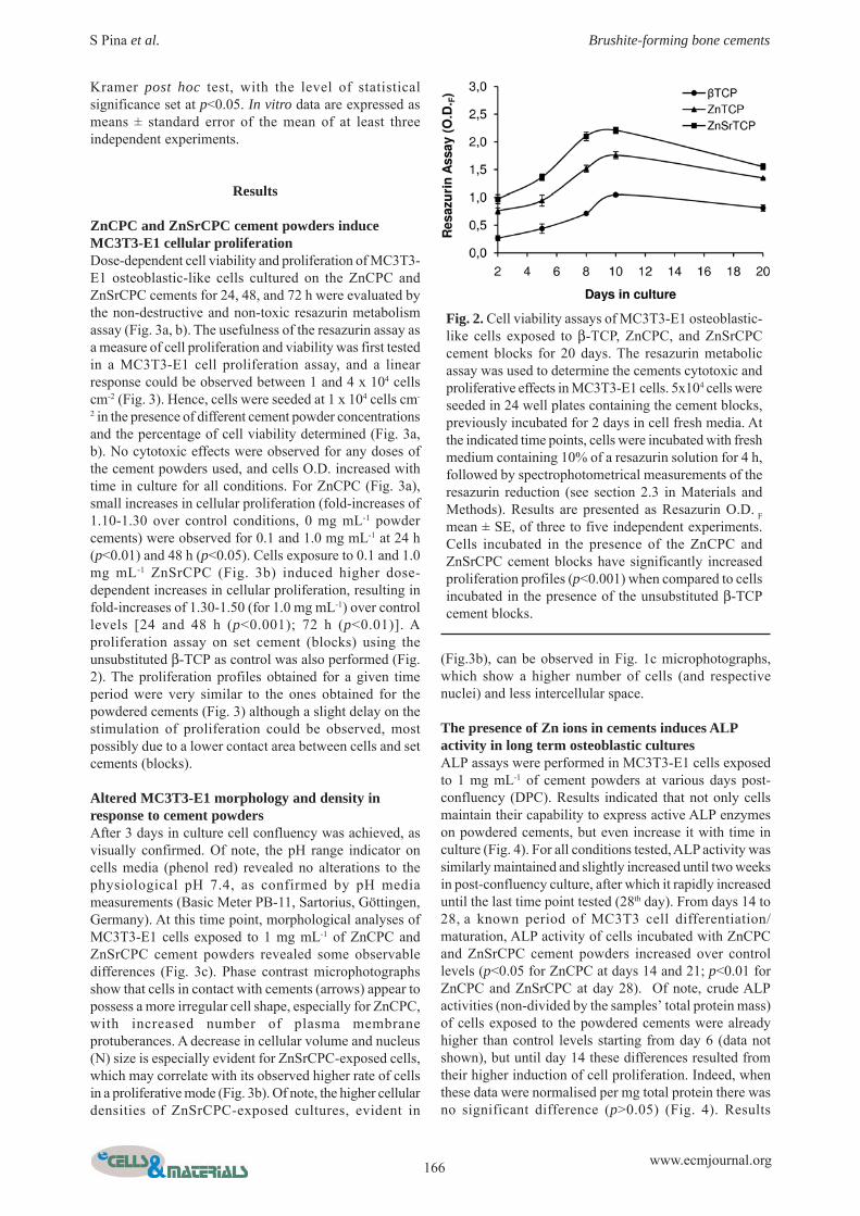
ZnCPC and ZnSrCPC cement powders induceMC3T3-E1 cellular proliferationDose-dependent cell viability and proliferation of MC3T3-E1 osteoblastic-like cells cultured on the ZnCPC andZnSrCPC cements for 24, 48, and 72 h were evaluated bythe non-destructive and non-toxic resazurin metabolismassay (Fig. 3a, b). The usefulness of the resazurin assay asa measure of cell proliferation and viability was first testedin a MC3T3-E1 cell proliferation assay, and a linearresponse could be observed between 1 and 4 x 10⁴ cellscm-2 (Fig. 3). Hence, cells were seeded at 1 x 10⁴ cells cm-
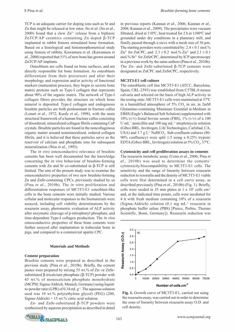
2 in the presence of different cement powder concentrationsand the percentage of cell viability determined (Fig. 3a,b). No cytotoxic effects were observed for any doses ofthe cement powders used, and cells O.D. increased withtime in culture for all conditions. For ZnCPC (Fig. 3a),small increases in cellular proliferation (fold-increases of1.10-1.30 over control conditions, 0 mg mL-1 powdercements) were observed for 0.1 and 1.0 mg mL-1 at 24 h(p<0.01) and 48 h (p<0.05). Cells exposure to 0.1 and 1.0mg mL-1 ZnSrCPC (Fig. 3b) induced higher dose-dependent increases in cellular proliferation, resulting infold-increases of 1.30-1.50 (for 1.0 mg mL-1) over controllevels [24 and 48 h (p<0.001); 72 h (p<0.01)]. Aproliferation assay on set cement (blocks) using theunsubstituted β-TCP as control was also performed (Fig.2). The proliferation profiles obtained for a given timeperiod were very similar to the ones obtained for thepowdered cements (Fig. 3) although a slight delay on thestimulation of proliferation could be observed, mostpossibly due to a lower contact area between cells and setcements (blocks).
Altered MC3T3-E1 morphology and density inresponse to cement powdersAfter 3 days in culture cell confluency was achieved, asvisually confirmed. Of note, the pH range indicator oncells media (phenol red) revealed no alterations to thephysiological pH 7.4, as confirmed by pH mediameasurements (Basic Meter PB-11, Sartorius, Göttingen,Germany). At this time point, morphological analyses ofMC3T3-E1 cells exposed to 1 mg mL-1 of ZnCPC andZnSrCPC cement powders revealed some observabledifferences (Fig. 3c). Phase contrast microphotographsshow that cells in contact with cements (arrows) appear topossess a more irregular cell shape, especially for ZnCPC,with increased number of plasma membraneprotuberances. A decrease in cellular volume and nucleus(N) size is especially evident for ZnSrCPC-exposed cells,which may correlate with its observed higher rate of cellsin a proliferative mode (Fig. 3b). Of note, the higher cellulardensities of ZnSrCPC-exposed cultures, evident in
Fig. 2. Cell viability assays of MC3T3-E1 osteoblastic-like cells exposed to β-TCP, ZnCPC, and ZnSrCPCcement blocks for 20 days. The resazurin metabolicassay was used to determine the cements cytotoxic andproliferative effects in MC3T3-E1 cells. 5x10⁴ cells wereseeded in 24 well plates containing the cement blocks,previously incubated for 2 days in cell fresh media. Atthe indicated time points, cells were incubated with freshmedium containing 10% of a resazurin solution for 4 h,followed by spectrophotometrical measurements of theresazurin reduction (see section 2.3 in Materials andMethods). Results are presented as Resazurin O.D. Fmean ± SE, of three to five independent experiments.Cells incubated in the presence of the ZnCPC andZnSrCPC cement blocks have significantly increasedproliferation profiles (p<0.001) when compared to cellsincubated in the presence of the unsubstituted β-TCPcement blocks.
(Fig.3b), can be observed in Fig. 1c microphotographs,which show a higher number of cells (and respectivenuclei) and less intercellular space.
The presence of Zn ions in cements induces ALPactivity in long term osteoblastic culturesALP assays were performed in MC3T3-E1 cells exposedto 1 mg mL-1 of cement powders at various days post-confluency (DPC). Results indicated that not only cellsmaintain their capability to express active ALP enzymeson powdered cements, but even increase it with time inculture (Fig. 4). For all conditions tested, ALP activity wassimilarly maintained and slightly increased until two weeksin post-confluency culture, after which it rapidly increaseduntil the last time point tested (28th day). From days 14 to28, a known period of MC3T3 cell differentiation/maturation, ALP activity of cells incubated with ZnCPCand ZnSrCPC cement powders increased over controllevels (p<0.05 for ZnCPC at days 14 and 21; p<0.01 forZnCPC and ZnSrCPC at day 28). Of note, crude ALPactivities (non-divided by the samples’ total protein mass)of cells exposed to the powdered cements were alreadyhigher than control levels starting from day 6 (data notshown), but until day 14 these differences resulted fromtheir higher induction of cell proliferation. Indeed, whenthese data were normalised per mg total protein there wasno significant difference (p>0.05) (Fig. 4). Results

167 www.ecmjournal.org
S Pina et al. Brushite-forming bone cements
therefore reflect ZnCPC and ZnSrCPC-induced variationsin the state of cell maturation leading to increased ALPactivities.
ZnCPC and ZnSrCPC enhance MC3T3-E1osteoblasts adhesion capacitiesWhen collecting cell lysates for analysis of their ALPactivity, it was noted that non-exposed control cells weremore easily detached from the plate’s plastic bottom thancells exposed to ZnCPC and ZnSrCPC, suggesting thatthey may exhibit alterations in their adhesiveness. Hence,further studies assayed differential adhesion capacities ofMC3T3-E1 cells exposed to the cement powders. Upon 0,3, and 14 DPC cell culture, a time-dependent increase inthe capacity of MC3T3-E1 cells to adhere to a plasticsupport was observed (Fig. 5), but decreasing until day21. Both ZnCPC and ZnSrCPC cement powders were ableto increase cell adhesion, in comparison with non-exposedcontrol cells (p<0.05 at 0, 3 and 21 DPC and p<0.001 at14 DPC for ZnCPC; p<0.001 for ZnSrCPC at 14 DPC).Hence, ZnCPC was even slightly more effective while theZnSrCPC cement appeared to have an intermediatebehaviour between control and ZnCPC exposed cells at
days 0 and 3, possibly reflecting a higher number ofZnSrCPC dividing cells, known to be less adhesive.
ZnSrCPC alters SDS-PAGE Type-I procollagenmigration profiles and enhances formation ofextracellular fibrilsThe effect of the cements on collagen protein levels,secretion and fibrillar deposition was studied byimmunoblot and immunocytochemistry analyses. In termsof immunoblot bands profile, collagen is somewhatcomplex (Fig. 6). Monomeric Type-I procollagen consistsof α1 (~140 kDa) and α2 (~130 kDa) chains, which areintracellular processed (signal peptide cleavage,hydroxylation of proline and lysine residues, glycosylationof lysine residues). Alpha chains can intracellularlyassociate into dimers (~270 kDa) and trimers (~400 kDa;triple helix consisting of two α1 and one α2 chains). Thereis some covalent cross-linking already within these forms,which are not breakable by SDS-PAGE denaturingconditions (Lareu et al., 2006). All these cell-associatedType-I pro-collagen forms were present in all samples anda time-dependent profile for the various cellularprocollagen forms could be observed (Fig. 6a). Between
Fig. 3. Cell viability assays of MC3T3-E1 osteoblastic-like cells exposed to ZnCPC and ZnSrCPC cement powders:(a) cell viability of cells cultured with increasing concentrations of ZnCPC and (b) ZnSrCPC cement powders for 24,48 and 72 h; results presented are the mean ± SE of at least three independent experiments; (c) phase contrastmicrophotographs of MC3T3-E1 cells exposed to 1 mg mL-1 of cement powders (ZnCPC and ZnSrCPC) for 72 h. N:cells’ nuclei. Arrows: visible aggregates of cement powders. Bar 10 μm.

168 www.ecmjournal.org
S Pina et al. Brushite-forming bone cements
0, 3 (proliferation period) and 14, 21 (maturation period)DPC, a high increase (3-4 fold-increase) in the levels ofcellular pro-collagen is noted, especially for the dimericβ(I) and trimeric γ(I) forms . In panel (a), levels of cell-associated Type-I pro-collagen were in the following order:Control > ZnSrCPC > ZnCPC, except at 3 DPC.
Remarkably, the cements collagen levels at 14 and 21DPC strongly increased following extensive samplessonication (panel b), which permitted the visualization ofcell layer-associated fibrils F(I) and γ(I)) forms, and theirbreakdown products (Fig. 6b). Further, this alloweddetecting an increase in the formation of collagen fibrilsfor ZnSrCPC at 3 DPC, observable in all experimentaldeterminations. Surprisingly, ZnSrCPC-induced alterationsin the electrophoretic migration of all procollagenmonomer and dimeric bands could also be denoted (Fig.6a,b “*”), revealing higher levels of post-translationalprocollagen modifications. This also occurred to a lowerextent for β chains in ZnCPC samples.
Intracellular procollagen is exported to the trans-Golginetwork (TGN) to be medium secreted. Extracellularly, γchains are enzymatically processed into tropocollagen,promoting their aggregation into cell layer-associatedcollagen microfibrils and fibrils (Sweeney et al., 2008).Hence, the levels of medium soluble procollagen were alsoanalysed (Fig. 6c). It could be observed that procollagensecretion (peaking at 3 DPC) precedes, as expected,trimeric procollagen, tropocollagen and fibril formation(abundant from 14 DPC onward). Fig. 6c shows higherprocollagen secretion for ZnCPC and ZnSrCPC-exposedcells at 3 DPC (p<0.05), and lower for 14 and 21 DPC,suggesting higher depletion of intracellular collagen at 3DPC.
In order to confirm the immunoblot observations at 3DPC, and as collagen antibodies exhibit a higher degreeof specificity under non-denaturing native conditions, cellswere subjected to microscopy analysis of Type-I collagencellular distribution and fibril deposition (Fig. 5).
Consistent with the immunoblot results of Fig. 6b, theoverall levels of intracellular collagen at 3 DPC were inthe following order: ZnSrCPC > Control ≅ ZnCPC, butcollagen was differently distributed. While for control cells,procollagen mainly accumulated inside intracellulargranule-like densities (Fig. 5, arrowheads), for ZnCPC andZnSrCPC-exposed cells intracellular procollagen wasfound in smaller but more numerous cytoplasmic vesicles.These results suggest CPC-induced alterations in themechanisms of collagen secretion. Further and moreremarkable, the extracellular cell-associated collagen fibres(Fig. 7, ‘F(I)’) were generally longer and denser for cellsexposed to cements, especially for ZnSrCPC, confirmingan earlier/enhanced aggregation of tropocollagen intofibrils and fibres.
ZnCPC and ZnSrCPC improve osteoconductivity inbone defectsThe excellent in vitro properties observed for the cementsled to the analysis of their in vivo osteoconductive andbiocompatible properties following implantation intrabecular pig bone defects. Transversal sections of 1 and2 month implants were stained with H&E dyes, takingadvantage of the fluorescent properties of eosin (Fua etal., 2006), histological and histomorphometric analyseswere conducted using a confocal laser scanningmicroscope. Eosin is an acidic coloured and fluorescentdye that stains proteins but not nuclei or lipids. Hence, influorescent microscopy, the organic protein materialappears red and the nuclei and lipids appear black.Microphotographs of implant cross-sections revealed thatnewly formed bone (NB) has grown from the peripheryinwards (Fig. 8). Organic protein-containing matrix (Mx)was also observed, although at higher amounts at 1 monthof implantation and less after 2 months. Of note, no
Fig. 4. Alkaline phosphatase (ALP) activity of MC3T3-E1 cells cultured on 1 mg mL-1 ZnCPC and ZnSrCPCpowdered cements for several days (0, 1, 3, 6, 14 and21) post-confluency. The ALP activity was expressedas the increase in p-nitrophenol absorbance per min permicrogram of protein (ΔOD.min-1.mg-1). Resultspresented are the mean of two independent experiments.
Fig. 5. ZnCPC and ZnSrCPC-dependent MC3T3-E1cells’ adhesion capacities. The capacity of cells to adhereto a plastic support for 1 h was measured following cellexposure to the cement powders for 0, 3, 14 and 21days post-confluency. At each experimental day, 1 x10⁵ cells were seeded into 24-well plates and left toadhere for 1 h at 37°C. Media was further collected andthe number of non-adherent ressuspended cells counted(x). The percentage of adherent cells was calculated(1x10⁵ - x), taking the initial 1x10⁵ cells as 100%.Results presented are the mean ± SE of threeindependent experiments.

169 www.ecmjournal.org
S Pina et al. Brushite-forming bone cements
inflammation or other complications associated with theimplanted materials were observed throughout theexperimental periods.
Three different osteoconductive profiles were denotedfor Norian (control), ZnCPC, and ZnSrCPC cements. ForNorian and ZnCPC, at 1 month new bone was mainly foundat the implant periphery (Fig. 6a,b), while the centrepresented black areas and areas filled with cells-enrichedunorganized matrix, probably corresponding to activeremodelling areas of osteoid secretion and cement and bonecallus resorption (Fig. 6a and b). Of note, osteoclast-likecells occur within these areas, while being only sparselyfound in ZnSrCPC sections (Fig. 9). The fact that theseareas were almost absent for ZnSrCPC (Fig. 8c) and the
presence of high amounts of NB suggest higher rates ofZnSr cement resorption and subsequent bone formationduring the first month of implantation. Of note, especiallyfor ZnSrCPC, osteoblast-like cells (OB) could be observedin organized matrix that appears to lay foundations for thenew bone being formed, that is found in very close relationwith it (Fig. 9b). Further, the new bone being depositedafter 1 month increases in organization in the followingorder: Norian < ZnCPC < ZnSrCPC (Fig. 8a-c), in termsof trabeculae number, shape and distribution throughoutthe implant area. In the ZnSrCPC (Fig. 8c), NB was mainlyfound in trabeculae-like concentric structures and crossingthe section. For ZnCPC (Fig. 8b) similar structures couldalso be found but in less concentric structures and mainly
Fig. 6. Immunoblot analysis of cell-associated and medium secreted Type-I collagen with time in post-confluencyculture. MC3T3 cells lysates (a, b) and conditioned media (c) were collected at days 0, 3, 14 and 21 of post-confluencyculture (DPC). Samples were resolved under reducing conditions and Type-I collagen was immunodetected. Panel(a) corresponds to lysates submitted to S1 (routine sonication), and panel (b) to the same lysates submitted to S2(extensive sonication, for partial fibres breakdown, which allow for their gel entry and visualization by immunoblotprocedures). Note that collagen protein profiles and levels are equal in pre-plated (preP, cells initially plated) andcontrol 0 DPC cells, as expected. α1(I) and α2(I), procollagen monomeric chains, unprocessed (arrowhead ) andprocessed (arrow ); β(I), procollagen dimeric forms; γ(I), procollagen trimeric forms; F(I), collagen fibres. Migrationof molecular weight markers is indicated to the right.

170 www.ecmjournal.org
S Pina et al. Brushite-forming bone cements
Fig. 7. Confocal microscopy analysis of intra and extracellular Type-I collagen at 3 days post-confluency (3 DPC).Cells in the absence of (Control) or exposed to 1 mg mL-1 ZnCPC and ZnSrCPC for 3 DPC (a total of 6 days ofexposition) were subjected to immunocytochemistry procedures in order to detect Type-I collagen distribution (greenfluorescence) by confocal microscopy. Arrowheads, intracellular granule-like densities. F/arrows, extracellular cell-associated collagen fibres. DAPI (blue fluorescence), nuclear marker. Bar, 10 μm.
Fig. 8: Confocal fluorescent micrographs of histological H&E stained sections, showing the tissue osteogenicresponse to Norian SRS® (a), ZnCPC (b,d,f) and ZnSrCPC (c,e,g) cements after 1 (a-c) and 2 (d-g) months ofimplantation. NB: new bone; Mx: protein matrix; arrows/OT in g,h: osteocytes.
1 month
2 months

171 www.ecmjournal.org
S Pina et al. Brushite-forming bone cements
at the periphery of the implant, while for Norian, the newbone was mainly deposited in less circular and lengthystructures (Fig. 8a).
Following 2 months of implantation (Fig. 8d,e), theconcentric lamellae deposition into trabecular bonebecomes obvious and closed circular trabeculae are themain structures found, especially for ZnSrCPC implants.For this time period, it was not possible to distinguish newbone from host old trabecular bone at the periphery of theZnSrCPC implants (Fig. 10). The continuity between newand old bone and within the newly formed trabeculae wasnotorious for the ZnSrCPC and less for ZnCPC (Fig. 10).Fig. 8f and 8g show magnified structures of Fig. 8d and8e, where arrows indicate osteocytes (OT) black nuclei.These are mature osteoblasts that are trapped inside bone,occupying small cavities known as lacunae at the junctionsof the lamellae, visible in all photos of newly formed bone.The layers of deposited lamellae are also visible (Fig. 9b3).Of note, accordingly with the ZnSrCPC high induction ofin vitro osteoblastic-cells proliferation and differentiation,after 1 month of in vivo implantation a high number ofosteoblasts (OB) could be observed adjacent to newlyformed bone (NB) (Fig. 11). Note the highly definedcanaliculi of the OB at the left, appearing to bedifferentiating into an osteocyte (OT).
ZnSrCPC has excellent osteoconductive propertiesThe deposition of more organized new bone in ZnSrCPCimplants, as early as 1 month, suggests that this cementpresented higher osteoconductive properties in comparisonwith ZnCPC, possibly due to a higher osteoblast induction.The excellent ZnSrCPC osteoconductive properties wereconfirmed by histomorphometric analyses of the in vivocross-sections. The percentage of area occupied by neworganic proteinaceous material was determined (Fig. 12).At 1 month of cement implantation, total proteinaceousarea increased as follows: Norian < ZnSrCPC < ZnCPC.Detailed analysis clarified that while for ZnSrCPC thisproteinaceous material was almost exclusively new bone,for ZnCPC this equally corresponded to new bone andmatrix. From 1 to 2 months of implantation, the percentageof matrix in the implant decreased as osteogenesiscontinued, and the area of ZnSrCPC ‘black space’ alsodecreased as new bone formation increased (data notshown) reinforcing that, with time of implantation, cementwas being resorbed as new-bone formed. Accordingly, newbone fluorescence intensity increased between 1 and 2months of implantation while matrix average intensitydecreased, indicating an increase in bone average densityand resorption of initially unorganized protein matrix andbone callus (data not shown). Osteoconductivity was more
Fig. 9. Confocal fluorescence micrographs of H&E stained sections, showing histological details of the tissueosteogenic response to Norian SRS®(a1), ZnCPC (a2) and ZnSrCPC (a3,b1-3) cements after 1 and 2 months (mth)of implantation. OC: osteoclast-like cells, indentified by their multiple nuclei and high protein-dense cytoplasm(arrows); NB: new bone; OB: osteoblast-like cells; OT: osteocytes; L: lamellae. Images from a to b1 were takenwith a 10x objective and zoomed 1.5-2x, and b2 was zoomed 4x; b3 represents an image projection of a z-stack,taken with a 100x objective. (S3)

172 www.ecmjournal.org
S Pina et al. Brushite-forming bone cements
evident for ZnSrCPC, which showed a notorious increasein the area occupied by new bone, reaching 50.0 ± 5.1%of the implant area (compared to 34.7 ± 8.7% for ZnCPC).
Discussion
The present study examined and compared the in vitro andin vivo response of an osteoblast-like cellular line (MC3T3-E1 cells) and two brushite-forming Zn and ZnSr-containingCPCs and their capacity to conduct osteoregeneration afterbeing implanted in pig trabecular bone. Osteoblasts play acentral inhibitory role in the pathophysiology ofosteoporosis, where reduced bone mass and deteriorationin bone microarchitecture lead to enhanced skeletalfragility. Osteoporosis results from a negative balancebetween the bone-forming activities of osteoblasts and theresorptive activities of osteoclasts. Inadequate osteoblastactivity may result from a relative deficiency inproliferation or differentiation of osteoprogenitors, or fromtheir excessive apoptosis. Hence, it is important to developbiomaterials with the capacity to stimulate osteoblasticproliferation and differentiation, and able to positivelyassist osteoregeneration.
The MC3T3-E1 cell line has the capacity todifferentiate into osteoblasts and osteocytes, and has beendemonstrated to have high ALP activity, to secrete collagenand to form calcified bone tissue in vitro, thus providingan excellent model to study the cellular and molecularresponses to the biomaterials tested. Dose-dependentproliferation of MC3T3-E1 osteoblastic-like cells culturedon either ZnCPC or ZnSrCPC cements confirmed theirbiocompatibility and capability of induction of cellularproliferation (Fig. 1). These results are consistent withprevious studies, where another zinc-releasing CPC witha zinc content of 1.20 wt % was also able to significantlypromote MC3T3-E1 proliferation in vitro (Ito et al., 2000;Kawamura et al., 2000). Our data further revealed that theincorporation of Sr into brushite cement greatly enhancesosteoblastic proliferation in a dose-dependent manner (Fig.1b), visually confirmed by the higher cellular density ofZnSrCPC cultures (Fig. 1c). Thus, the ZnCPC andZnSrCPC cements behaved in accordance with resultsobtained by other authors, where Sr-containing ioniccements were more osteoconductive than Zn-containingionic cements (Johal et al., 2002). Additionally, ALPactivity and collagen secretion and fibre deposition alsoimplicated ZnCPC and ZnSrCPC in osteoblasticdifferentiation. The cements were able to increase MC3T3-E1 cells ALP activity over control levels at later periods inculture (Fig. 2), which indicates their higher maturationstate at this period. Otsuka et al. (Otsuka et al., 2004) haveobserved that MC3T3-E1 cells’ ALP activity is lower forthe first 2 weeks in culture (proliferative state), whileincreasing from days 15 to 35, and related this increase tothe cells’ maturation state. MC3T3-E1 maturation isreached following ~14 days in culture (Owen et al., 1990;Stein et al., 1989), with ALP activity being used as anindicator of cell differentiation (Lecoeur et al., 1997). Theresults here obtained indicate that MC3T3-E1 cell
Fig. 10. Contiguous fluorescent microphotographs weretaken of histological sections of ZnCPC (a) andZnSrCPC (b) cements upon 2 months of implantation,using an Olympus IX81 epifluorescence microscope.(S4)

173 www.ecmjournal.org
S Pina et al. Brushite-forming bone cements
Fig. 11. Microphotographs of a ZnSrCPC 1 month (mth) implant cross-section. Phase contrast (PhC) and correspondentfluorescence (Fl) microphotographs were taken using an Olympus IX81 epifluorescence microscope. Zoomed areadenotes osteoblasts (OB) adjacent to the new formed bone (NB) and bone embedded osteocytes (OT).
PhC
Fluorescence
Fig. 12. Histomorphometric analysis of the implantareas of H&E stained sections of ZnCPC, ZnSrCPCand Norian SRS® cements for 1 and 2 months ofimplantation. The percentage (%) of the implant areaoccupied by: new bone formed, protein matrix and both(‘Total proteic’). For each condition, twelve cross-sections were analysed and error bars representvariation within each implant cylinder.

174 www.ecmjournal.org
S Pina et al. Brushite-forming bone cements
maturation mainly occurs at the 14th day in culture, whenALP activity increases (Fig. 2). This is in accordance withthe results on protein content determined for ALP assays(data not shown) that revealed an increase in total proteincontent until 14 days in culture that further decreased andstabilized until the last day tested (28th DPC). In summary,results showed that both CPCs can increase osteoblasticcells maturation in terms of ALP activity. Further, asZnCPC and ZnSrCPC results are virtually equal, Zn ionsappear to be the relevant inducers of ALP activity, asexpected since this is a Zn-dependent enzyme (McCombet al., 1979).
Subsequent data demonstrated that ZnCPC andZnSrCPC were also able to increase osteoblasts intrinsiccell-to-matrix adhesion capacities (Fig. 3), what can berelevant in the osteoconductive process, e.g. for osteoblastsadhesion to the protein matrix and to new bone. This wasfirst observed visually when collecting cells lysates forALP determination, where cells grew in the presence ofthe cements biofilms, attached more tightly to the plasticsupport. Indeed, cells previously exposed to the cementpowders exhibited higher adhesive capacities to a newplastic support (Fig. 3). Of note, during the proliferativeperiod (e.g. 3DPC), the adhesiveness of cells exposed toZnSrCPC were slightly lower than for ZnCPC exposedcells. This may again correlate to its higher induction ofcellular division and with the fact that dividing cells aremore loosely connected to the support than interphase cells.Of note, the higher number of cell membranarprotuberances observed for ZnCPC cells (Fig. 1c) suggestsaltered cytoskeleton dynamics, potentially related to itsaltered adhesiveness. Interestingly, all cells exhibited adecrease in their intrinsic adhesive capacities in thedifferentiation period (Fig. 3, 14-21st DPC) which mightbe correlated with their increased dependence on anextracellular matrix. Indeed, this corresponds to the periodof higher deposition of extracellular collagenous matrix(ECM), as observed in Fig. 4b. Immunoblot andimmunocytochemistry methods revealed that CPCsinduced alterations in MC3T3-E1 collagen production,secretion and extracellular fibres formation. In essence,cements enhanced collagen secretion in the pre-differentiation period (3 DPC, Fig. 4c) correlate well withtheir subsequent higher collagen fibre formation in thedifferentiation period (14 and 21 DPC, Fig. 4b).Additionally, ZnSrCPC induces a higher rate of collagenaggregation into fibres, as early as at 3 DPC, as confirmedby both immunoblot and immunocytochemistry analyses(Fig. 4b and 5). Indeed, in ZnSrCPC exposed cells, Type-I collagen fibres were observed to be longer and denser,suggesting an earlier/enhanced aggregation oftropocollagen into fibrils and fibres. This may be relatedto the observed altered SDS-PAGE migration profiles ofprocollagen in ZnSrCPC cell lysates (Fig. 4a and b ‘*’),which reflect a higher level of protein post-translationalmodification, potentially proline hydroxylation andsubsequent hydroxylysine glycosylation. Indeed,hydroxyproline and hydroxylysine, which derive fromproline and lysine hydroxylation at the endoplasmicreticulum, are necessary for the formation and stabilization
of collagen, playing key roles in collagen stability (Nelsonet al., 2005). Hence, ZnSrCPC may induceoverhydroxylation of proline and/or lysine amino acids,potentially accelerating collagen cross-linking and fibreformation, explaining collagen results at 3 DPC.
All results obtained in in vitro assays proved thatZnCPC and ZnSrCPC cements are able to inducephysiologically relevant properties, which may result inenhanced in vivo osteoconductive capacities. Indeed, thefirst stage for bone formation by osteoblasts is collagensecretion. Type-I collagen is the most abundant type ofcollagen in bone, being the most abundant proteincomponent in the ECM. Given that CPCs induce Type-Icollagen deposition into the ECM and increase the numberof the collagen-secreting osteoblastic cells and theirdifferentiation, especially for ZnSrCPC, it was attractiveto speculate their good in vivo performance. This wasconfirmed to be correct in pilot in vivo assays. Cementimplants were performed in trabecular bone due to itshigher metabolic rate per unit of volume, when comparedto cortical bone. Further, rates of change in bone densityare likely to be greater at sites that are predominantlytrabecular (Dempster et al., 1992) and trabecular bonedensity decreases faster than that of compact bone asosteoporosis progresses (Seeman et al., 1993). Histologicaland histomorphometrical analyses of the implants revealedthat, with time, new bone regenerated and graduallypenetrated into the implant. The pattern of trabecular bonethat was being formed in the implanted area wasqualitatively similar to the adjacent old trabecular bone inthe sections’ periphery. New bone grew towards theimplant centre, replacing fluorescent-black spaces andsuggesting that cement was being first resorbed in theimplantation periphery. This is expected, as it is the site ofcontact between the implant and old bone, mesenchymalcells, and macrophages that ultimately differentiate intoosteoclasts. Reduction of the protein-containing organicmatrix from 1 to 2 months, along with increased new boneformation to relative high levels (Fig. 6 and 8) also reflectedthat ZnCPC and ZnSrCPC had good osteoconductivities.The cements are potentially good inductors ofosteoprogenitor cell proliferation and differentiation, asupon 1 month intensive bone remodelling was undergoingin the implant. Further, results indicated that the presenceof Sr ions, rather than the presence of Zn ions, greatlyinhibits osteoclastic activity, with much fewer osteoclast-like cells being found in ZnSrCPC implants (Fig. 9a).Partial substitution of Zn ions by Sr ions in ZnSrCPCrevealed to be further beneficial, as organized boneformation and most probably cement resorption were fasterand higher for this cement (Fig. 8, 11, 12). In contrast, thepresence of higher areas of unorganised matrix for ZnCPCand more so for Norian, (Figs. 2 and 8) indicated thatcement and primary matrix were still being resorbed atthose zones at 1 month. Of note, the larger solubility ofbrushite compared to apatite is one essential reason forthe faster resorption of the brushite matrix. While apatitecements, such as Norian, remain almost unchangedthroughout study periods of 6 months, brushite cementsare resorbed at a rate between 60% and 90% amid 2-6

175 www.ecmjournal.org
S Pina et al. Brushite-forming bone cements
months of study periods (Apelt et al., 2004). In osteogenicterms, for the ZnCPC cement new bone was mainly foundat the periphery of the implant but not at the centre (1month), while for ZnSrCPC, NB was found evenlydistributed throughout the implant, with several trabeculae-like structures already crossing the entire histologicalsections. Upon 2 months, higher levels of boneregeneration could be noted for ZnCPC and especially forZnSrCPC, where contiguous trabeculae crossing thehistological sections could be found, suggesting thecompletion of the osteoregenerative process mediated bybrushite conversion into apatite.
Conclusions
High biocompatibility and in vivo performance of brushite-forming Zn- and ZnSr-substituted β-TCP cements injectedinto pig trabecular bone cylindrical defects were observedin a pilot study. Mechanistically, the in vivo performancemay rely on the in vitro properties observed for thesecements: induction of osteoblastic proliferation,differentiation, adhesiveness, ALP activity, Type-I collagensecretion and deposition into extracellular fibres. All theseproperties are of crucial relevance in the process ofosteoregeneration. In addition, ZnSrCPC was generally ahigher inductor of these osteoblastic properties and,correspondently, presented the best osteoconductiveproperties. Both ZnCPC and ZnSrCPC presented better invivo performance in comparison to the control carbonatedapatite cement (Norian SRS®), but the presence of Srenhanced the rate of cement resorption and new boneformation. In conclusion, the biological propertiesobserved for the investigated ZnCPC and ZnSrCPCcements strongly suggest them as good candidate materialsto be used as bone substitutes.
Acknowledgments
To CICECO and to CBC for the support and to thePortuguese Foundation for Science and Technology forthe project REEQ/1023/BIO/2005 and for the fellowshipgrants of S.P. (SFRH/BPD/64119/2009), S.I.V. (SFRH/BPD/19515/2004) and P.M.C.T. (SFRH/BD/62021/2009).The authors are also very grateful to Prof. Doutor HenriqueBicha Castelo, Director of the Service of Medicine andExperimental Surgery, Hospital of Santa Maria, Lisbon,for the facilities supply in experimental animal studies,according to European regulations and followingpermission granted by the Ethical Committee. In addition,the authors are thankful to A. Pinto, Institute of MolecularMedicine, Lisbon, for the preparation of the sections forhistological and histomorphometric analysis.
References
Alves HLR, dos Santos LA, Bergmann CP (2008)Injectability evaluation of tricalcium phosphate bonecement. J Mater Sci - Mater Med 19: 2241-2246.
Apelt D, Theiss F, El-Warrak AO, Zlinszky K,Bettschart-Wolfisberger R, Bohner M, Matter S, Auer JA,Von Rechenberg B (2004) In vivo behavior of threedifferent injectable hydraulic calcium phosphate cements.Biomaterials 25: 1439-1451.
Baroud G, Cayer E, Bohner M (2005) Rheologicalcharacterization of concentrated aqueous á-tricalciumphosphate suspensions: The effect of liquid-to-powderratio, milling time, and additives. Acta Biomater 1: 357-363.
Boesel L, Reis RL (2006) The effect of water uptakeon the behaviour of hydrophilic cements in confinedenvironments. Biomaterials 27: 5627-5633.
Bohner M, Baroud G (2005) Injectability of calciumphosphate pastes. Biomaterials 26: 1553-1563.
Bohner M, Theiss F, Apelt D, Hirsiger W, Houriet R,Rizzoli G (2003) Compositional changes of a dicalciumphosphate dihydrate cement after implantation in sheep.Biomaterials 24: 3463-3474.
Brown WE, Chow LC (1983) A new calcium-phosphate setting cement. J Dent Res 62: 672-672.
Burguera EF, Xu HHK, Sun LM (2008) Injectablecalcium phosphate cement: Effects of powder-to-liquidratio and needle size. J Biomed Mater Res B 84: 493-502.
Constantz BR BB, Ison IC, Fulmer MT, Baker J,McKinney LA, Goodman SB, Gunasekaren S, DelaneyDC, Ross J, Poser R (1998) Histological, chemical, andcrystallographic analysis of four calcium phosphatecements in different rabbit osseous sites. J Biomed MaterRes Part B 43: 451-461.
Dahl S, Allain P, Marie P, Mauras Y, Boivin G, AmmannP (2001) Incorporation and distribution of strontium inbone. Bone 28: 446-453.
Dempster DW (1992) Bone remodeling. Disorders ofbone and mineral metabolism. Raven Press, New York,pp. 355-380.
Fua CY, Dinishb US, Nga BK, Murukeshanb VM,Seahb LK, Lim-Tanc SK (2006) Fluorescence lifetimeimaging of haematoxylin and eosin-stained cervical tissue.Proc. Int. Conf. Biomed. Pharm. Eng., Singapore.
Gauthier O, Muller R, von Stechow D, Lamy B, WeissP, Bouler JM (2005) In vivo bone regeneration withinjectable calcium phosphate biomaterial: A three-dimensional micro-computed tomographic, biomechanicaland SEM study. Biomaterials 26: 5444-5453.
Gisep A, Wieling R, Bohner M, Matter S, SchneiderE, Rahn B (2003) Resorption patterns of calcium-phosphate cements in bone. J Biomed Mater Res Part A66: 532-540.
Ito A, Ojima K, Naito H, Ichinose N, Tateishi T (2000)Preparation, solubility, and cytocompatibility of zinc-releasing calcium phosphate ceramics. J Biomed MaterRes 50: 178-183.
Ito A, Kaeamura H, Otsuka M, Ikeuchi M, Ohgushi H,Ishikawa K, Onuma K, Kanzaki N, Sogo Y, Ichinose N(2002) Zinc-releasing calcium phosphate for stimulatingbone formation. Mater Sci Eng C 22: 21-25.
Johal KK, Hill RG, Brook IM (2002) In vivo responseof strontium and zinc based ionomeric cement implants inbone. J Mater Sci - Mater Med 13: 543-552.

176 www.ecmjournal.org
S Pina et al. Brushite-forming bone cements
Kannan S, Pina S, Ferreira JMF (2006) Formation ofstrontium-stabilized α-tricalcium phosphate from calcium-deficient apatite. J Amer Ceram Soc 89: 3277-3280.
Kannan S, Goetz-Neunhoeffer F, Neubauer J, FerreiraJMF (2008) Ionic substitutions in biphasic hydroxyapatiteand beta-tricalcium phosphate mixtures: Structural analysisby rietveld refinement. J Amer Ceram Soc 91: 1-12.
Kannan S, Goetz-Neunhoeffer F, Neubauer J, FerreiraJMF (2009) Synthesis and structure refinement of zinc-doped beta-tricalcium phosphate powders. J Amer CeramSoc 92: 1592-1595.
Kawamura H, Ito A, Miyakawa S, Layrolle P, OjimaK, Ichinose N (2000) Stimulatory effect of zinc-releasingcalcium phosphate implant on bone formation in rabbitfemora. J Biomed Mater Res 50: 184-190.
Kurdy NM, Bowles S, Marsh DR, Davies A, FranceM (1998) Serology of collagen types I and III in normalhealing of tibial shaft fractures. J Orthop Trauma 12: 122-126.
Lareu R R, Arsianti I, Harve KS, Peng Y, RaghunathM (2006) In vitro augmentation of collagen matrixformation – Applications in tissue engineering. 3rd KualaLumpur Intern Conf Biomed Eng, 15. (DOI 10.1007/978-3-540-68017-8).
Lecoeur L, Ouhayoun JP (1997) In vitro induction ofosteogenic differentiation from non-osteogenicmesenchymal cells. Biomaterials 18: 989-993.
Lenart G, Bidlo G, Pinter J (1972) Some basic problemsin examination of calcium hydrogen phosphates of bone.Clinical Orthop Rel Res 83: 263-272.
Li X, Sogo Y, Ito A, Mutsuzaki H, Ochiai N, KobayashiT (2009) The optimum zinc content in set calciumphosphate cement for promoting bone formation in vivo.Mater Sci and Eng C 29: 969-975.
Malik M, Puleo D, Bizios R, Doremus R (1992)Osteoblasts on hydroxyapatite, alumina and bone surfacesin vitro: morphology during the first 2 hours of attachment.Biomaterials 13: 123-128.
Marie P, Ammann P, Boivin G, Rey C (2001)Mechanisms of action and therapeutic potential ofstrontium in bone. Calcif Tissue Int 69: 121-129.
McComb R, Bowers G, Posen S. Alkaline Phosphatase.Plenum, New York, 1979.
Naji A, Harmand M (1991) Cytocompatibility of twocoating materials, amorphous alumina and silicon carbide,using human differentiated cell cultures. Biomaterials 12:690-694.
Nelson DL, Cox MM (2005) Lehninger’s Principlesof Biochemistry. W.H. Freeman, New York, NY.
Otsuka M, Marunaka S, Matsuda Y, Ito A, Layrolle P,Naito H (2000) Calcium level-responsive in vitro zincrelease from zinc containing tricalcium phosphate(ZnTCP). J Biomed Mater Res 52: 819-824.
Otsuka M, Ohshita Y, Marunaka S, Matsuda Y, Ito A,Ichinose N, Otsuka K, Higuchi WI (2004) Effect ofcontrolled zinc release on bone mineral density frominjectable Zn-containing beta-tricalcium phosphatesuspension in zinc-deficient diseased rats. J Biomed MaterRes A 69: 552-560.
Owen TA, Aronow M, Shalhoub V, Barone LM,Wilming L, Tassinari MS (1990) Progressive development
of the rat osteoblast phenotype in vitro: reciprocalrelationships in expression of genes associated withosteoblast proliferation and differentiation duringformation of the bone extracellular matrix. J Cell Phys143: 420-430.
Pina S, Torres PMC, Goetz-Neunhoffer F, NeubauerJ, Ferreira JMF (2010a) Newly developed Sr-substituteda-TCP bone cements. Acta Biomaterialia 6: 928-935 (doi:10.1016/j.actbio.2009.09.001).
Pina S, Vieira SI, Torres PMC, Goetz-Neunhoeffer F,Neubauer J, da Cruz e Silva OAB, da Cruz e Silva EF,Ferreira JMF (2010b) In vitro performance assessment ofnew brushite-forming Zn- and ZnSr-substituted b-TCPbone cements. J Biomed Mater Res B, in press.
Puleo D, Preston K, Shaffer J, Bizios R (1993)Examination of osteoblast-orthopaedic biomaterialinteractions using molecular techniques. Biomaterials 14:111-114.
Rokita E, Hermes C, Nolting HF, Ryczek J (1993)Substitution of calcium by strontium within selectedcalcium phosphates. J Crystal Growth 130: 543-552.
Seeman E, Young N, Szmukler G, Tsalamandris C,Hopper JL (1993) Risk-factors for osteoporosis.Osteoporosis Int 3: S40-S43.
Stein GS, Lian JB, Gerstenfeld LG, Shalhoub V,Aronow M, Owen TA (1989) The onset and progressionof osteoblast differentiation is functionally related tocellular proliferation. Connect Tissue Res 20: 3-13.
Sweeney S, Orgel J, Fertala A, McAuliffe JD, TurnerKR, Di Lullo GA, Chen S, Perumal S, Ala-Kokko L,Forlino A, Cabral WA, Barnes AM, Marini JC, San AntonioJD (2008) Candidate cell and matrix interaction domainson the collagen fibril, the predominant protein ofvertebrates. J Biol Chem 283: 21187-21197.
Ueno A, Miwa Y, Miyoshi K, Horiguchi T, Inoue H,Ruspita I (2006) Constitutive expression ofthrombospondin 1 in MC3T3-E1 osteoblastic cells inhibitsmineralization. J Cell Phys 209: 322-332.
Wang XP, Ye JD, Wang H (2006) Effects of additiveson the rheological properties and injectability of a calciumphosphate bone substitute material. J Biomed Mater ResB 78: 259-264.
Wen HB, Cui FZ, Feng QL, Li HD, Zhu XD (1995)Microstructural investigation of the early external callusafter diaphyseal fractures of human long-bone. J StructBiol 114: 115-122.
Yuan HP, Li YB, de Bruijn JD, de Groot K, Zhang XD(2000) Tissue responses of calcium phosphate cement: astudy in dogs. Biomaterials 21: 1283-1290.
Discussion with Reviewers
Reviewer I: Is the positive effect of the incorporation ofZn and Sr ions into brushite cements due to the release ofZn and Sr ions or due to a different reason such as a changeof surface roughness or a change of Ca or phosphate ionrelease?Authors: Based on our previous study (Pina et al., 2010b,text reference), ZnCPC and ZnSrCPC cements were ableto release, into the simulated body fluid (SBF) solution,

177 www.ecmjournal.org
S Pina et al. Brushite-forming bone cements
the relevant (from the physiological view point) Zn andSr ions and to transform into apatite under that conditions.Therefore we think that the positive effect of theincorporation of Zn and Sr ions into brushite cements isrelated with the release of Zn and Sr ions into thephysiological fluid.
Reviewer II: What was the rationale for using theparticular substitution levels?Authors: The available literature reports on the effect ofionic substitution levels on in vitro and in vivo tests arescarce. In our previous works dealing with substitution ofZn and Sr ions in the structure of calcium phosphates wehave covered a broad range of compositions. The levelsselected in the present work are intermediate values andthe aim was to evaluate their effects on the response ofbone cements in vitro and in vivo.
Reviewer III: Regarding the in vitro experiments, pleasediscuss why you chose to test the behaviour of cells byapplying cement-conditioned medium, instead of cementitself. Also, it is mentioned that for cytotoxicityexperiments, cement powder was added to the well plateswith cells. This needs clarification. Was the cement itself(i.e., cement set upon mixing of powder and liquid phase)used, or only the powder fraction of the cement. If theformer is the case, please explain how the particles weremade, and what their size was. If the latter is the case, thenthe rationale behind this choice needs to be explained.Authors: Concerning the in vitro studies, we chose to testthe behaviour of cells by applying powdered set cementsto conditioned medium mainly due to the higher surfacearea of contact between the biomaterials and cells andconditioned medium when using powder formulation,when comparing with cement in blocks. Additionally, wehave previously observed in preliminary experiments thatpre-osteoblastic MC3T3 cells adhere better to powders thanto cements in blocks. The cytotoxicity tests were performedwith powdered cement, i.e.,cement set upon mixing ofpowder and liquid, followed by milling. The powder hadan average particle size of about 15 μm.
Reviewer III: A similar question goes for the ALP activity.Why were the experiments performed on powders and noton set cement?
Authors: The same rationale as for the proliferation studieswas applied here, and the experimental design wasmaintained throughout the in vitro analyses for the sake ofconsistency.
Reviewer III: Regarding the cell adhesion test, what isthe meaning of the adhesiveness of cells to plastic in thepresence or absence of cement? Adhesiveness to cementsseems a more relevant analysis. Please comment!Authors: Observations of altered cell adhesiveness to theplastic in the ALP assays prompted us to analyse this issue.Hence, we tested if cells gained more capacity to adhereto an extracellular support when previously cultivated inthe presence of cements. Cell adherence to an extracellularsupport (as the plastic plate) or matrix requires a series oftransmembrane proteins (cell adhesion molecules such asintegrins and glycoproteins) generally involved in cell-to-cell and cell-to-matrix adhesion. The data obtained arerelevant for the ability of cells to adhere to organicstructures such as other cells, extracellular protein matrix,and new bone, which can accelerate the remodellingprocess. It would indeed also be relevant to analyse celladhesiveness to cements, although it is more difficult todirectly quantify cells adherent to the powders (e.g., cellcollection and counting) accurately.
Reviewer III: In the Discussion it is stated that the twocements induced cellular proliferation. How was theinduction proven?Authors: The cellular toxicity/proliferation assay provesthat for certain cements concentrations the number of cellsincreases with time over control cells. This was denotedfrom the Resazurin assay (Fig. 3) and also when countingcells for the set up of the adhesion assay (data not shown).This is a strong indicator of increased cell proliferation,and a higher number of cells was indeed observed in thecorresponding phase contrast microphotographs. It couldalso result from increased cellular survival, but typicallythis does not render the fold-increases obtained e.g., for1.0 mg/mL ZnSrCPC (Fig. 3b). Indeed, the percentage ofnon-viable (compromised) cells in the MC3T3 controlpopulation is typically no more than 5% (as we previouslyobserved when counting cells for a growth curve (Pina etal., 2010b) and when counting cells for the adhesion assayof Fig. 5).