author(s): matthew velkey, 2009 unless otherwise noted ... · pdf fileb. lymphatic...
TRANSCRIPT
Author(s): Matthew Velkey, 2009 License: Unless otherwise noted, this material is made available under the terms of the Creative Commons Attribution–Non-commercial–Share Alike 3.0 License: http://creativecommons.org/licenses/by-nc-sa/3.0/
We have reviewed this material in accordance with U.S. Copyright Law and have tried to maximize your ability to use, share, and adapt it. The citation key on the following slide provides information about how you may share and adapt this material. Copyright holders of content included in this material should contact [email protected] with any questions, corrections, or clarification regarding the use of content. For more information about how to cite these materials visit http://open.umich.edu/education/about/terms-of-use. Any medical information in this material is intended to inform and educate and is not a tool for self-diagnosis or a replacement for medical evaluation, advice, diagnosis or treatment by a healthcare professional. Please speak to your physician if you have questions about your medical condition. Viewer discretion is advised: Some medical content is graphic and may not be suitable for all viewers.
Citation Key for more information see: http://open.umich.edu/wiki/CitationPolicy
Use + Share + Adapt
Make Your Own Assessment
Creative Commons – Attribution License
Creative Commons – Attribution Share Alike License
Creative Commons – Attribution Noncommercial License
Creative Commons – Attribution Noncommercial Share Alike License
GNU – Free Documentation License
Creative Commons – Zero Waiver
Public Domain – Ineligible: Works that are ineligible for copyright protection in the U.S. (USC 17 § 102(b)) *laws in your jurisdiction may differ
Public Domain – Expired: Works that are no longer protected due to an expired copyright term.
Public Domain – Government: Works that are produced by the U.S. Government. (USC 17 § 105)
Public Domain – Self Dedicated: Works that a copyright holder has dedicated to the public domain.
Fair Use: Use of works that is determined to be Fair consistent with the U.S. Copyright Act. (USC 17 § 107) *laws in your jurisdiction may differ
Our determination DOES NOT mean that all uses of this 3rd-party content are Fair Uses and we DO NOT guarantee that your use of the content is Fair.
To use this content you should do your own independent analysis to determine whether or not your use will be Fair.
{ Content the copyright holder, author, or law permits you to use, share and adapt. }
{ Content Open.Michigan believes can be used, shared, and adapted because it is ineligible for copyright. }
{ Content Open.Michigan has used under a Fair Use determination. }

Lymphatic Histology
M1 – Immunology Sequence J. Matthew Velkey, Ph.D.
Winter 2009

Learning Objectives Text: Ross, 5th ed., pp. 396-441
Atlas: Wheater’s, 5th ed., pp. 215-233 1. Understand the distinction between PRIMARY and
SECONDARY lymphoid organs 2. Be able to describe the organization and function of:
• Mucosa-associated lymphoid tissue • Diffuse and nodular lymphoid tissue, also including regions of extensive
lymphoid infiltration such as Peyer’s patches, appendix, and tonsils. • lymph nodes • Spleen • Thymus
3. Be able to identify the regions rich in B and T lymphocytes in each organ and explain the cellular processes, relevant to immune functions, that are taking place in these regions.
4. Know the homing patterns of B & T lymphocytes.

Functions of the Lymphatic System 1. Monitor body surfaces and fluid compartments (e.g. epidermis, mucosae*, interstitium) 2. React to the presence of potentially harmful antigens recognized as “non-self” 3. Autoimmune diseases (rheumatoid arthritis, type I diabetes, etc.)
Lymphatic System consists of: A. Cells
1. Lymphocytes (B,T, natural killer) 2. Antigen-presenting cells (dendritic cells, Langerhans’ cells & macrophages)
B. Lymphatic “tissue” –diffuse and nodular C. Lymphatic “organs” (lymph nodes, spleen, thymus) D. Lymphatic vessels that carry the cells and fluid *Mucosae refers to lining tissue of the body cavities, e.g. GI tract,
respiratory tract, genitourinary tract
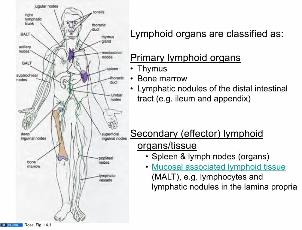
Lymphoid organs are classified as: Primary lymphoid organs • Thymus • Bone marrow • Lymphatic nodules of the distal intestinal
tract (e.g. ileum and appendix) Secondary (effector) lymphoid
organs/tissue • Spleen & lymph nodes (organs) • Mucosal associated lymphoid tissue
(MALT), e.g. lymphocytes and lymphatic nodules in the lamina propria
Ross, Fig. 14.1

Primary Lymphoid Organs: The bone marrow and the thymus and the Gut-Associated Lymphoid Tissue (e.g. appendix, terminal ileum) are the initial “education centers” of the immune system In these organs, lymphocytes (T cells in the thymus, B cells in bone marrow and gut) differentiate into immunocompetent cells (i.e. they can recognize “self” vs. “nonself”) This differentiation is said to be antigen-independent The lymphocytes then enter the blood and lymph to populate:
• epidermis and mucosae • connective tissue • secondary lymphoid organs

Secondary Lymphoid Organs: The lymph nodes, lymphatic nodules, tonsils, spleen are the secondary “education centers” of the immune system In these organs, immunocompetent lymphocytes differentiate into immune effector and memory cells that undergo antigen-dependent activation and proliferation in these organs. These lymphocytes then carry out their functions in the:
• connective tissue • secondary lymphoid organs • mucosal surfaces lining epithelia
They participate in: • Cell mediated immunity (mostly “cytotoxic” T cells) • Humoral responses (production of antibody) (B cells,
also requires “helper” T cells.

Lymphocytes in peripheral blood smear
These are B and T-cells that have undergone antigen-INDEPENDENT differentiation and are trafficking through the bloodstream on their way to lymphoid organs/tissue.
lymphocyte
Mizobuti histology slide set

Source Undetermined

cytokines
Diapedesis: it’s not just for the Normans and the Saxons…
Roll Tether Migrate Arrest
blood flow
APCs and other cells
chemokines
Cytokines and chemokines (along with selectins and integrins) mediate EXTRAvasation of lymphocytes into tissues.
L. Stoolman

MALT: intraepithelial lymphocytes: γδT-cells (neither helper nor cytotoxic): first to see antigens
U-M Histology Collection

Intraepithelial lymphocytes
Shown here in resp. epith. Homing mediated by “addressins” (a sort of lymphocyte “GPS”)
U-M Histology Collection

Diffuse lymphoid tissue Lamina propria (LP) of gut shown here, but can be found associated with mucosae anywhere in the gut, respiratory, and genitourinary tracts.
Primary lymphatic nodule/follicle (LN) Aggregation of lymphocytes in lamina propria or submucosa
LYMPHOCYTES IN CONNECTIVE TISSUE: MALT = mucosa-associated lymphoid tissue
LN
Ross and Pawlina, Histology: A Text and Atlas U-M Histology Collection

Secondary follicles/nodules
• Contain germinal centers • Arise when B-lymphocytes are
presented with appropriate antigen, receive T-cell help, and then begin proliferating as lymphoblasts
• Lymphoblasts differentiate into
plasma cells or memory cells; aberrant lymphoblasts undergo apoptosis.
Ross and Pawlina, Histology: A Text and Atlas
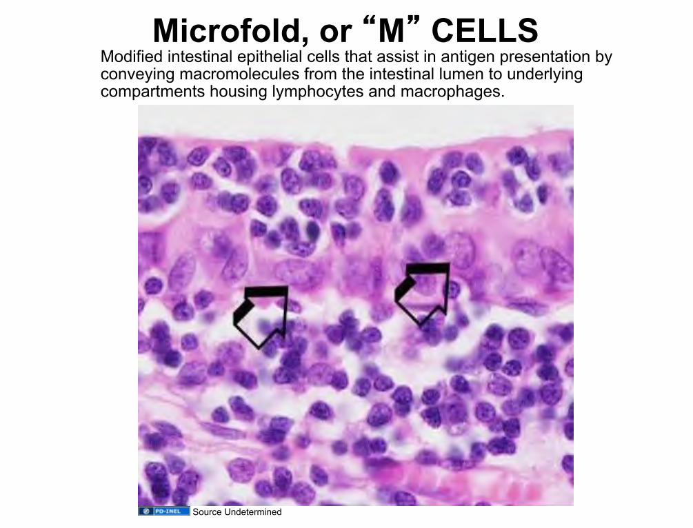
Microfold, or “M” CELLS Modified intestinal epithelial cells that assist in antigen presentation by conveying macromolecules from the intestinal lumen to underlying compartments housing lymphocytes and macrophages.
Source Undetermined
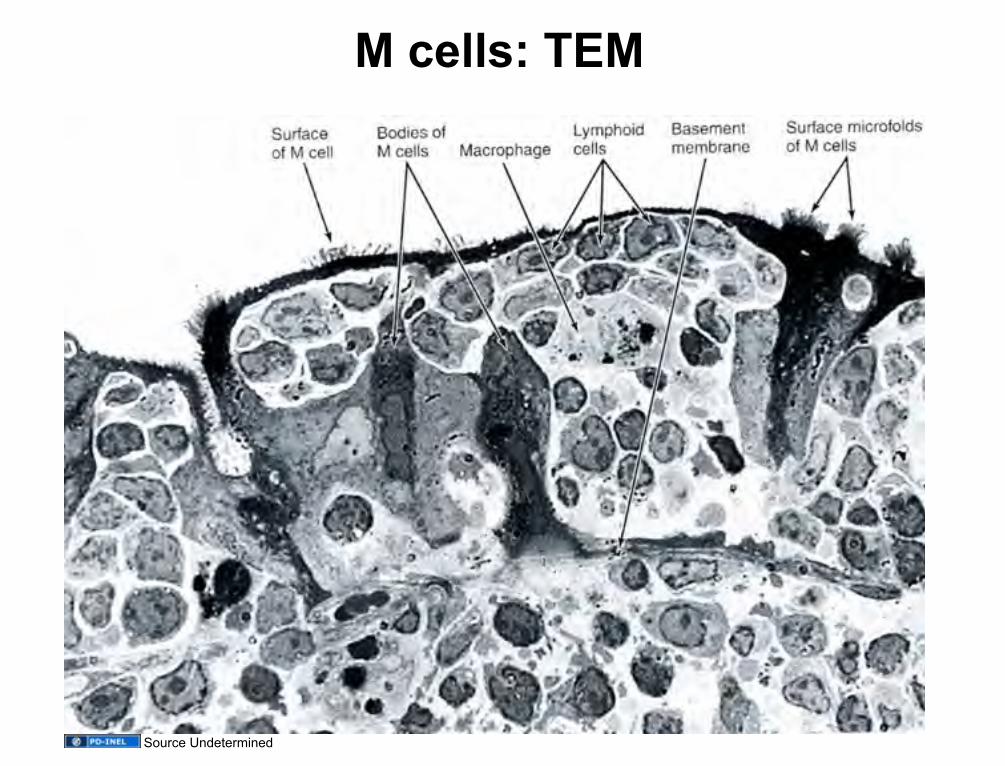
M cells: TEM
Source Undetermined

After antigen presentation and T-cell help, activated B-cells set up germinal centers in secondary follicles
Secondary follicle germinal centers • Arise when B-lymphocytes are
presented with appropriate antigen, receive T-cell help, and then begin proliferating as lymphoblasts
• Lymphoblasts differentiate into
plasma cells or memory cells; aberrant lymphoblasts undergo apoptosis.
Ross and Pawlina, Histology: A Text and Atlas

Germinal center: high magnification
U-M Histology Collection. Slide 175.

Lymphoblast viewed by transmission electron microscopy
Source Undetermined

Source Undetermined

Plasma Cells are mature B lymphocytes
White arrows = Golgi regions Black arrows indicate several plasma cells U-M Histology Collection Junquiera and Carneiro. Basic Histology. Tenth
Ed. 2003

EM of Plasma
Cells
Source Undetermined Source Undetermined
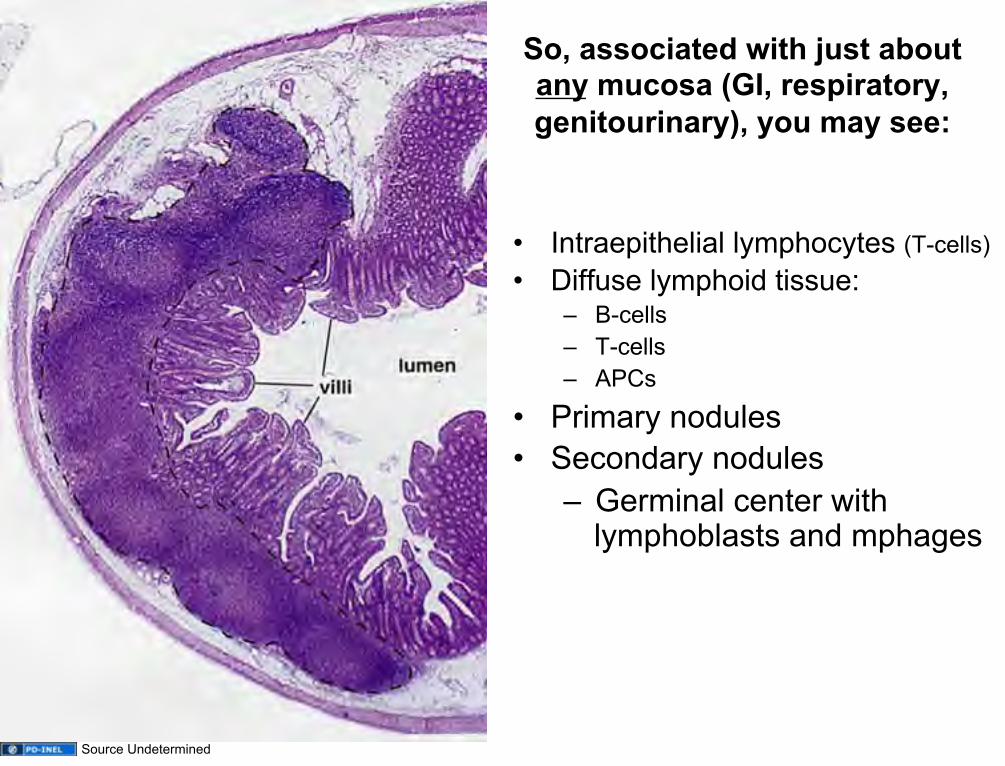
So, associated with just about any mucosa (GI, respiratory, genitourinary), you may see:
• Intraepithelial lymphocytes (T-cells) • Diffuse lymphoid tissue:
– B-cells – T-cells – APCs
• Primary nodules • Secondary nodules
– Germinal center with lymphoblasts and mphages
Source Undetermined

Aggregates of lymphoid follicles in the ileum.
Regions of extensive lymphoid infiltration: Peyer’s patches
Source Undetermined
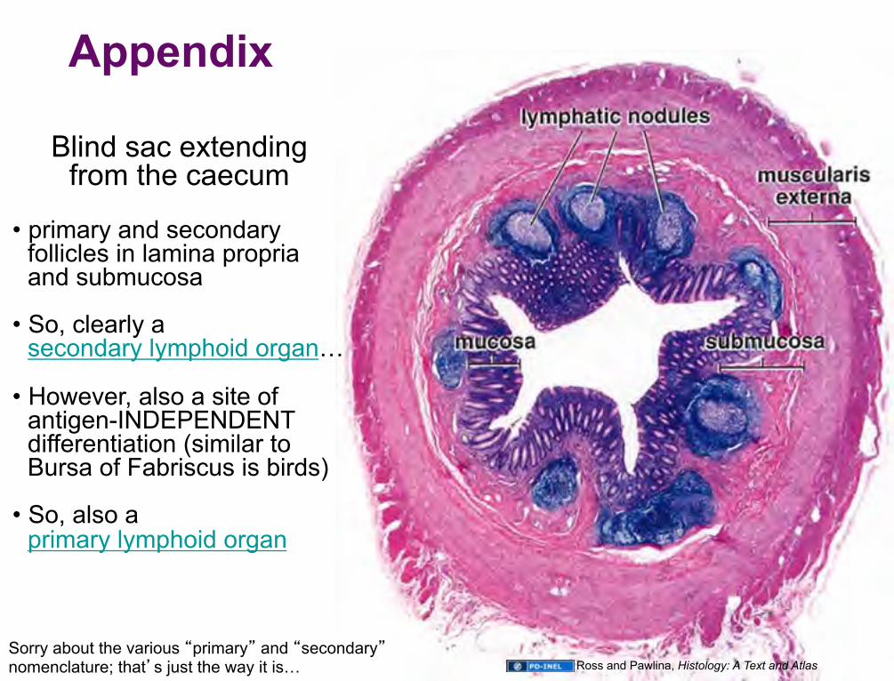
Appendix
Blind sac extending from the caecum
• primary and secondary
follicles in lamina propria and submucosa
• So, clearly a secondary lymphoid organ…
• However, also a site of antigen-INDEPENDENT differentiation (similar to Bursa of Fabriscus is birds)
• So, also a primary lymphoid organ
Sorry about the various “primary” and “secondary” nomenclature; that’s just the way it is… Ross and Pawlina, Histology: A Text and Atlas
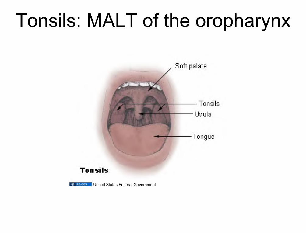
Tonsils: MALT of the oropharynx
United States Federal Government

TONSILS
The palatine tonsils are paired structures made of dense accumulations of lymphatic tissue located in the mucous membrane of the junction of the oropharynx and oral cavity. The tonsils dip down into the underlying CT, forming crypts. There are also lingual tonsils and pharyngeal tonsils (under the roof of the nasopharynx and around the opening of the Eustachian tubes). Key features: crypts, abundant nodules, stratified squamous epithelium
Ross and Pawlina, Histology: A Text and Atlas
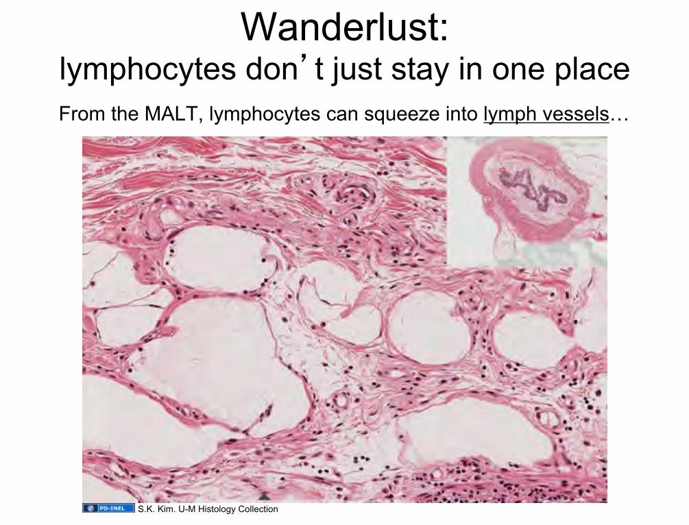
Wanderlust: lymphocytes don’t just stay in one place From the MALT, lymphocytes can squeeze into lymph vessels…
S.K. Kim. U-M Histology Collection
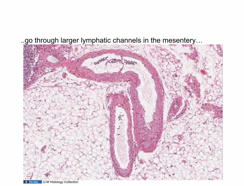
..go through larger lymphatic channels in the mesentery…
U-M Histology Collection

..and end up at a LYMPH NODE.
U-M Histology Collection
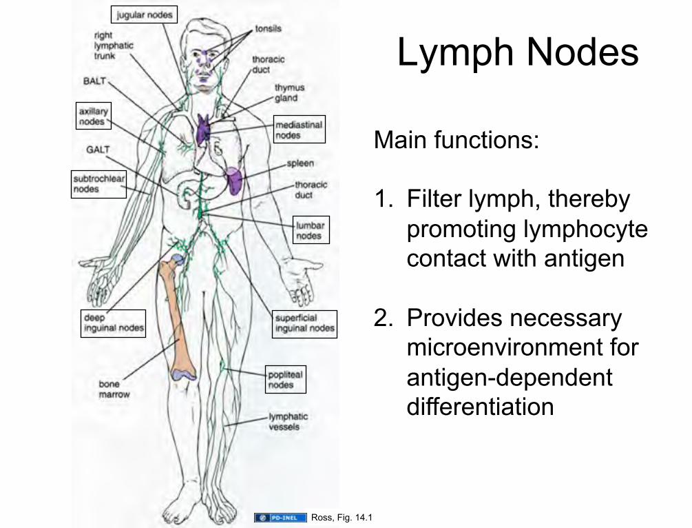
Lymph Nodes
Main functions: 1. Filter lymph, thereby
promoting lymphocyte contact with antigen
2. Provides necessary microenvironment for antigen-dependent differentiation
Ross, Fig. 14.1
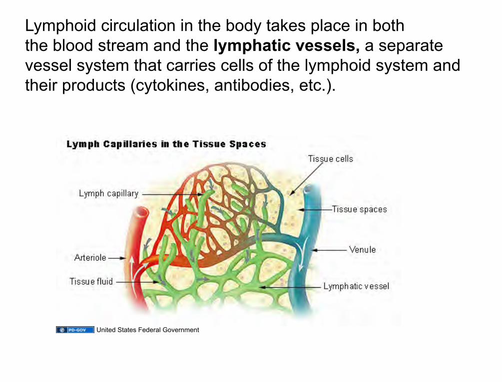
Lymphoid circulation in the body takes place in both the blood stream and the lymphatic vessels, a separate vessel system that carries cells of the lymphoid system and their products (cytokines, antibodies, etc.).
United States Federal Government

Lymphatic drainage: anatomy
Image of lymphatic drainage anatomy
removed
Original Image: http://health-tune-ups.com/wp-content/uploads/2009/04/cdr533339-750.jpg
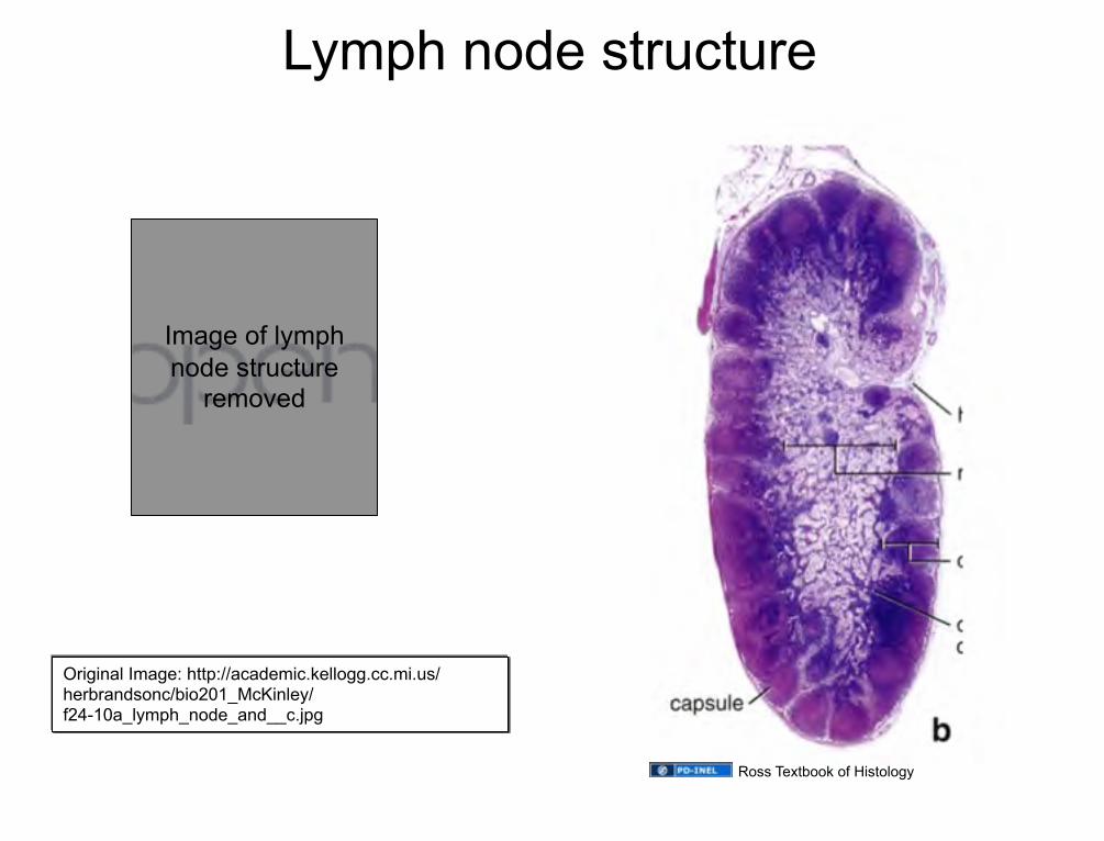
Lymph node structure
Ross Textbook of Histology
Original Image: http://academic.kellogg.cc.mi.us/herbrandsonc/bio201_McKinley/f24-10a_lymph_node_and__c.jpg
Image of lymph node structure
removed

Lymph nodes filter lymph
1. Afferent lymphatic vessels drain lymph into the Subcapsular Sinus
2. Lymph then passes to the Trabecular sinuses
3. From there, the lymph goes to the Medullary sinuses.
4. Lymphocytes and macrophages pass easily between these sinuses and the tissue of the lymph node.
5. Macrophages in sinuses monitor the fluids. Macs phagocytose the antigenic material and present it to T- and B-cells
Lymphatic Circulation Through a Lymph Node
Original Image: http://human.freescience.org/images/Illu_lymph_node_structure.png
Image of lymph node circulation
removed

Lymph Node Structure
- Capsule & subcapsular sinus - Trabeculae & trabecular sinuses
sinuses contain lymph, macrophages, and reticular cells
- Cortex:
• superficial cortex (B-cells) -primary follicles/nodules -secondary follicles/nodules (i.e. with germinal centers)
• “deep” cortex (T-cells, dendritic cells)
- Medulla: • medullary cords (B-cells, plasma cells) • medullary sinuses (lymph, more
macrophages, plasma cells, and reticular cells)
U-M Histology Collection
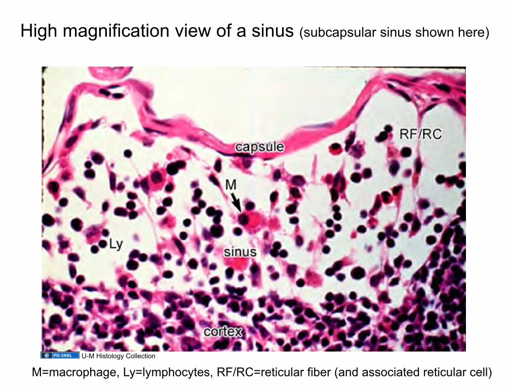
High magnification view of a sinus (subcapsular sinus shown here)
M=macrophage, Ly=lymphocytes, RF/RC=reticular fiber (and associated reticular cell) U-M Histology Collection

From the sub-capsular sinus, lymph percolates through trabecular sinuses, and finally into medullary sinuses
U-M Histology Collection

Reticular (Reticulin) Fibers • Form a delicate supporting framework for highly cellular tissues (endocrine glands, lymph nodes, liver, bone marrow, spleen, smooth muscle).
• Composed mainly of Type III collagen, with a carbohydrate moiety that reduces Ag+ to metallic sliver = argyrophilic.
• Special stain: silver impregnation to visualize.
• Thinner than type I collagen (Type III fibrils are 30-40 nm diameter; type I fibrils are ~200 nm diameter)
Source Undetermined
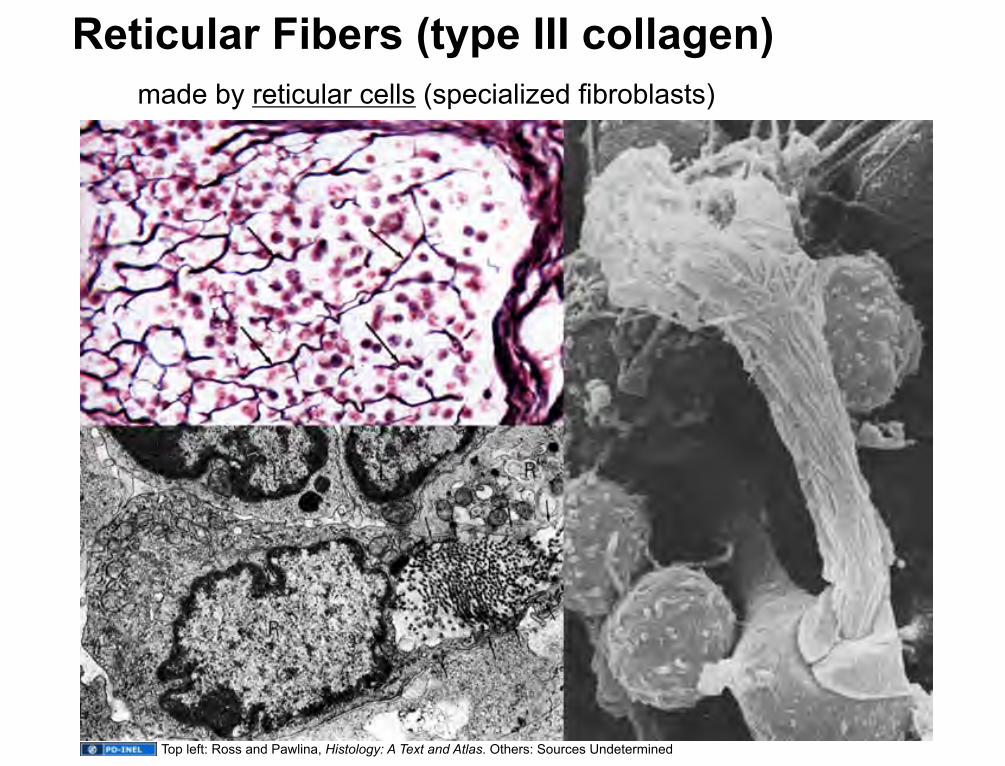
Reticular Fibers (type III collagen) made by reticular cells (specialized fibroblasts)
Top left: Ross and Pawlina, Histology: A Text and Atlas. Others: Sources Undetermined

Medullary sinuses drain into EFFERENT lymphatics that exit from the hilum of the lymph node
U-M Histology Collection

1. Blood enters through an artery at the hilus
2. Arterioles branch from hilar artery to feed into capillary beds
3. Capillary beds are drained by high endothelial venules*
4. HEVs drain into hilar vein
Original Image: Ross, fig. 14.18
Blood Circulation Through a Lymph Node
*HEVs are sites where lymphocytes can leave blood stream to enter the lymph node tissue bed.
Image of lymph node blood circulation removed

capsule
U-M Histology Collection

U-M Histology Collection

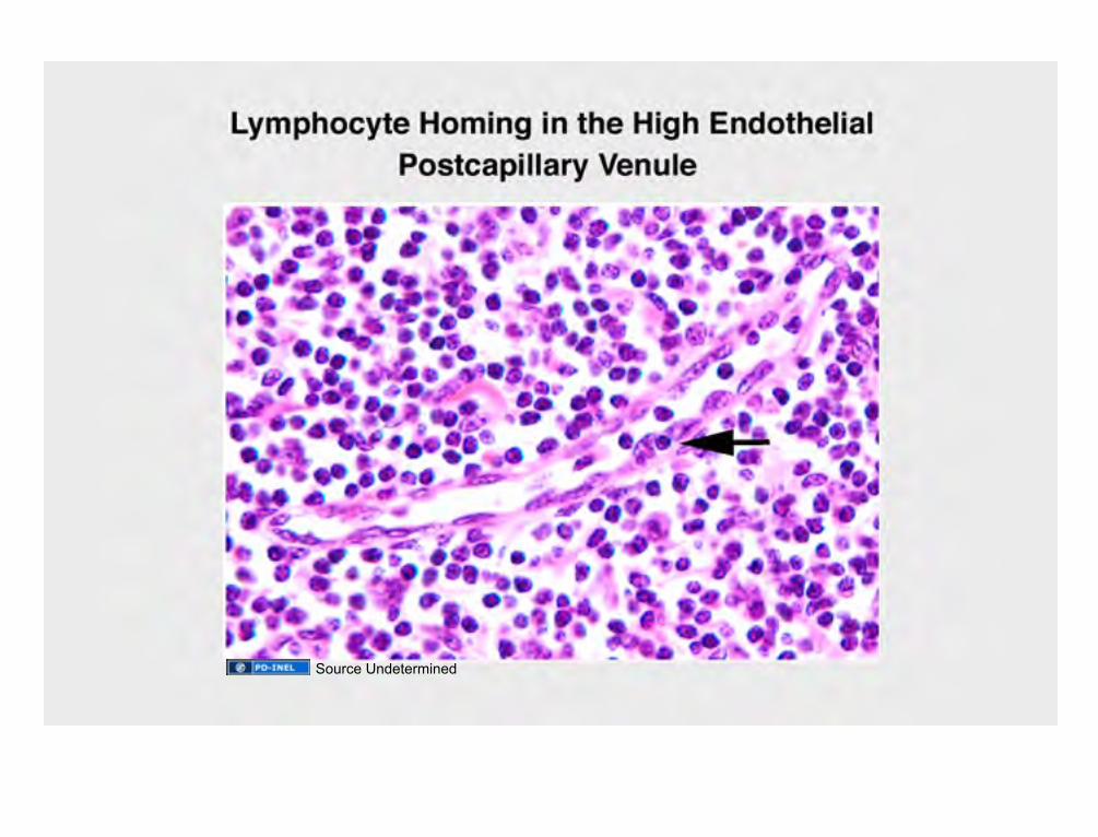
High Endothelial Venules
Site of:
• Fluid absorption (via aquaporin-1 channels), which causes lymph flow
• EXIT of lymphocytes from bloodstream via diapedesis
Source Undetermined

Source Undetermined

Source Undetermined
Source Undetermined
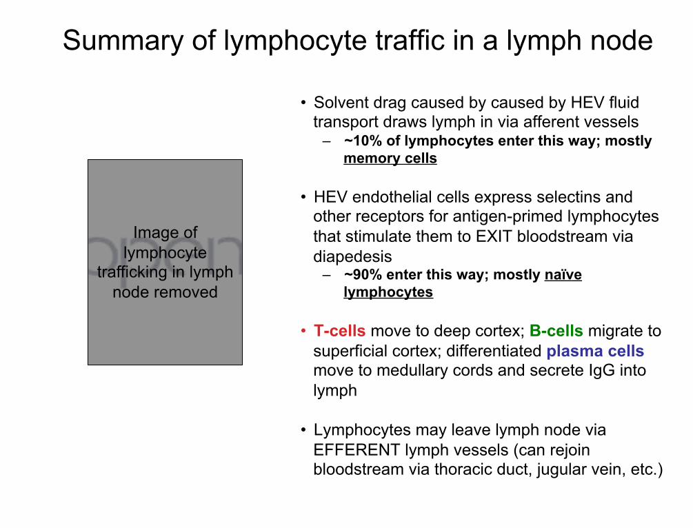
• Solvent drag caused by caused by HEV fluid transport draws lymph in via afferent vessels
– ~10% of lymphocytes enter this way; mostly memory cells
• HEV endothelial cells express selectins and other receptors for antigen-primed lymphocytes that stimulate them to EXIT bloodstream via diapedesis
– ~90% enter this way; mostly naïve lymphocytes
• T-cells move to deep cortex; B-cells migrate to superficial cortex; differentiated plasma cells move to medullary cords and secrete IgG into lymph
• Lymphocytes may leave lymph node via EFFERENT lymph vessels (can rejoin bloodstream via thoracic duct, jugular vein, etc.)
Summary of lymphocyte traffic in a lymph node
Image of lymphocyte
trafficking in lymph node removed

The Spleen
Filters the blood Destroys old red blood cells Serves as an immune organ Divided into Red Pulp (RBC/ hemoglobin recycling) White Pulp (responsible for immune functions)
Ross, Fig. 14.1

• Monitoring antigens in blood • Proliferation of lymphocytes • Production of humoral antibodies
• Formation of blood cells in fetal life • Removal and destruction of RBCs & platelets • Retrieval of iron from RBC hemoglobin • Storage of RBCs and platelets (more so in
non-human species)
Immune Functions Of the Spleen
Hematopoietic Functions Of the Spleen

Spleen: anatomy
Gray’s Anatomy

Spleen: anatomy
Cancer.gov, Wikipedia, http://commons.wikimedia.org/wiki/File:Illu_spleen.jpg

ORGANIZATION OF THE SPLEEN
Ross, 14.29
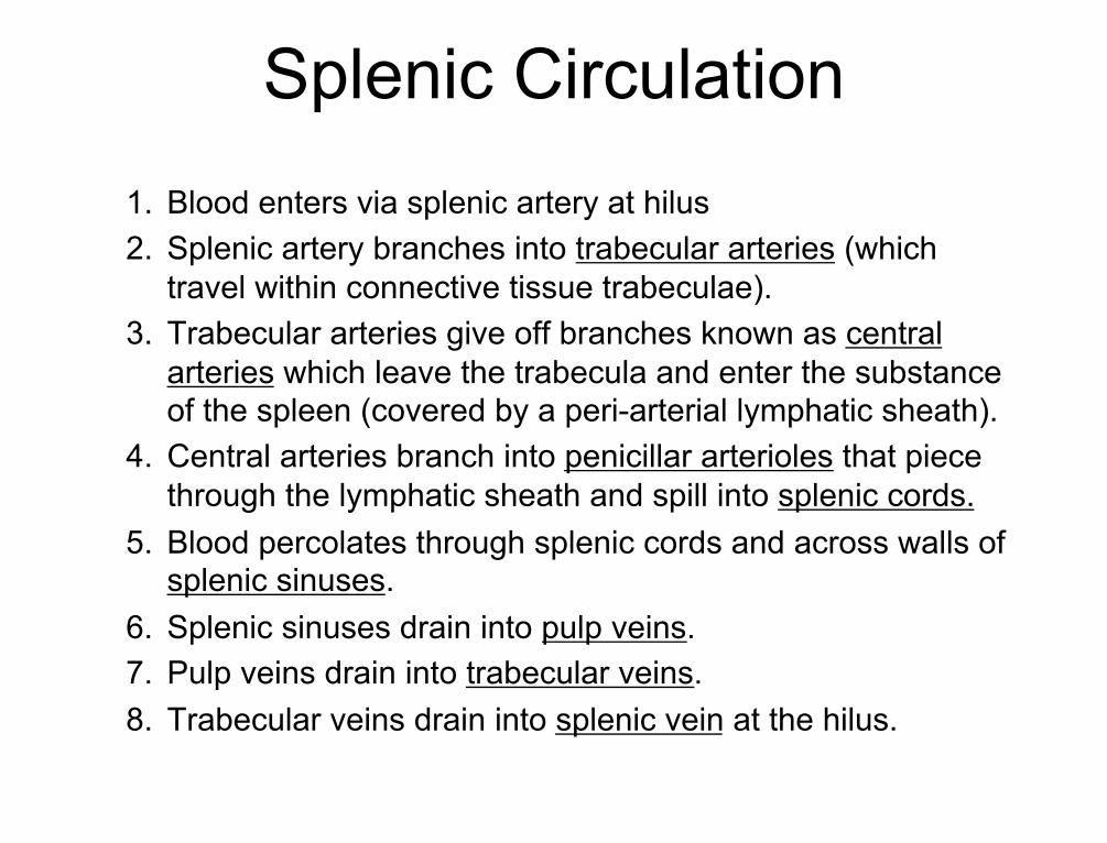
Splenic Circulation 1. Blood enters via splenic artery at hilus 2. Splenic artery branches into trabecular arteries (which
travel within connective tissue trabeculae). 3. Trabecular arteries give off branches known as central
arteries which leave the trabecula and enter the substance of the spleen (covered by a peri-arterial lymphatic sheath).
4. Central arteries branch into penicillar arterioles that piece through the lymphatic sheath and spill into splenic cords.
5. Blood percolates through splenic cords and across walls of splenic sinuses.
6. Splenic sinuses drain into pulp veins. 7. Pulp veins drain into trabecular veins. 8. Trabecular veins drain into splenic vein at the hilus.

Circulation in the human spleen is primarily “OPEN:” blood pours into the red pulp, percolates through red pulp cords, and re-enters the bloodstream at splenic sinuses
NOTE: NO afferent lymph vessels –not necessary because lymphocytes can easily enter splenic parenchyma via “open” circulation pattern.
Original Image: http://www.mc.vanderbilt.edu/histology/images/histology/lymph/display/lymph20015.jpg
Image of splenic circulation removed
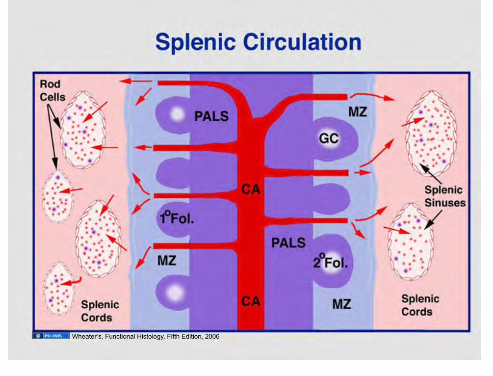
Wheater’s, Functional Histology, Fifth Edition, 2006
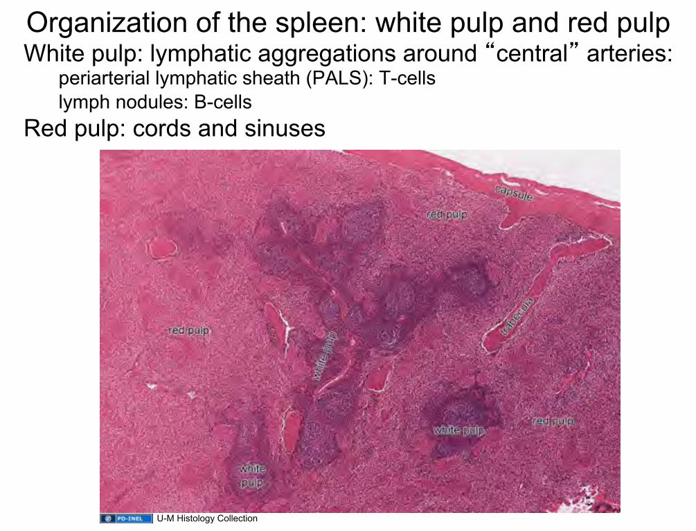
Organization of the spleen: white pulp and red pulp White pulp: lymphatic aggregations around “central” arteries:
periarterial lymphatic sheath (PALS): T-cells lymph nodules: B-cells
Red pulp: cords and sinuses
U-M Histology Collection

White pulp function Blood and antigens pour into red pulp (more on that later) Antigen presentation takes place in MARGINAL ZONE T-cells (from PALS) provide “help” to activate mphages and B-cells
• activated mphages stimulated to destroy ingested material (e.g. bacteria) • activated B-cells set up proliferative germinal centers
U-M Histology Collection
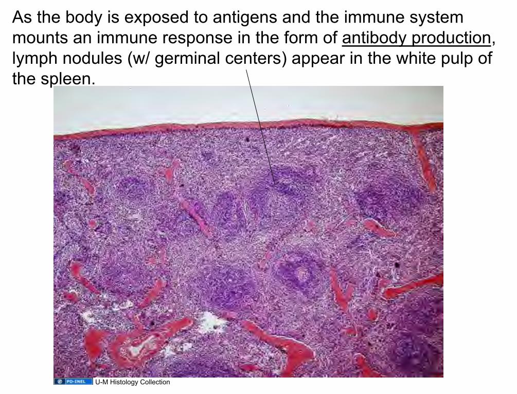
As the body is exposed to antigens and the immune system mounts an immune response in the form of antibody production, lymph nodules (w/ germinal centers) appear in the white pulp of the spleen.
U-M Histology Collection
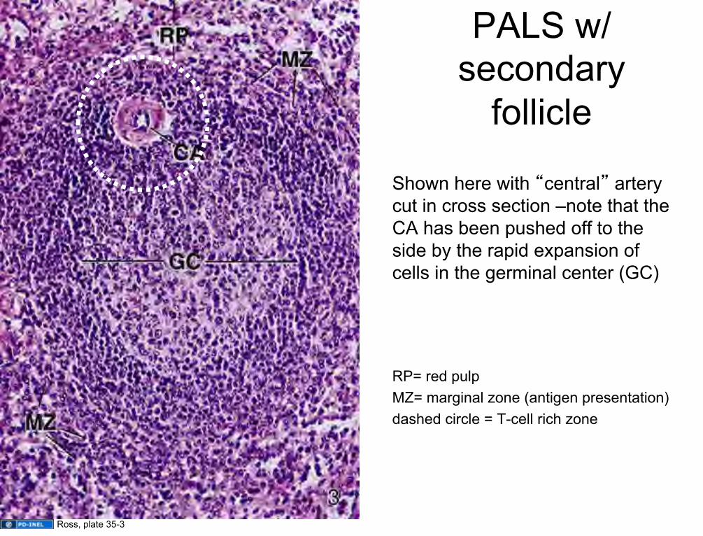
PALS w/ secondary
follicle
Shown here with “central” artery cut in cross section –note that the CA has been pushed off to the side by the rapid expansion of cells in the germinal center (GC) RP= red pulp MZ= marginal zone (antigen presentation) dashed circle = T-cell rich zone
Ross, plate 35-3

Scanning EM of a Splenic Sinus (SS) and Cord of Billroth
The cords contain, RBCs, neutrophils (N), macrophages (M), blood platelets (P) A reticular cell framework (RC) supports the cord. The sinus is bounded by the epithelial cells that form the basket-like structure of the sinus (VS)
Ross 14.30a

Spleen (red pulp) at high power (40x)
sinus
cord sinus
cord
U-M Histology Collection
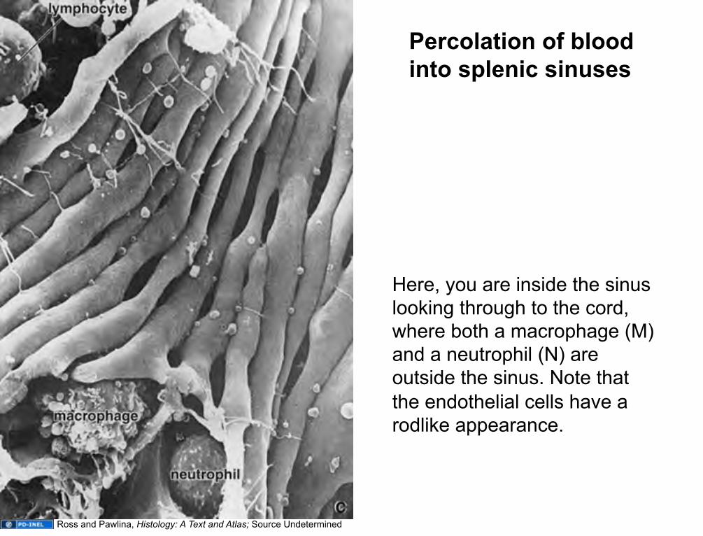
Percolation of blood into splenic sinuses
Here, you are inside the sinus looking through to the cord, where both a macrophage (M) and a neutrophil (N) are outside the sinus. Note that the endothelial cells have a rodlike appearance.
Ross and Pawlina, Histology: A Text and Atlas; Source Undetermined
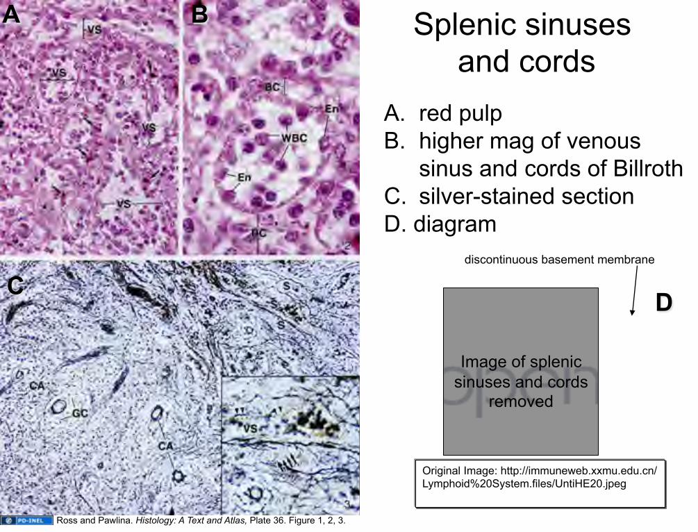
A. red pulp B. higher mag of venous
sinus and cords of Billroth C. silver-stained section D. diagram
Splenic sinuses and cords
A B
C D
discontinuous basement membrane
Ross and Pawlina. Histology: A Text and Atlas, Plate 36. Figure 1, 2, 3.
Original Image: http://immuneweb.xxmu.edu.cn/Lymphoid%20System.files/UntiHE20.jpeg
Image of splenic sinuses and cords
removed

SPLEEN: venous sinus showing rodlike endothelial cells
Source Undetermined

SPLENIC CIRCULATION Sinuses drain into splenic pulp veins, which, in turn, drain into trabecular veins. Trabecular veins travel within trabeculae and drain into splenic vein at the hilus.
red pulp white
pulp
U-M Histology Collection

The Thymus
T-cell education Self vs. nonself distinctions Cell-mediated immune functions Populates effector organs
Lymph nodes Lymphatic nodules Spleen Tonsils
Ross, Fig. 14.1

The Thymus is a Primary Lymphoid (Immune) Organ Responsible For the Education of T-Cells Located over the great vessels of the heart in the area of the body called the mediastinum Develops from an invagination of EPITHELIUM of the 3rd pharyngeal pouch, so an endodermal organ. Specialized epithelial cells (called epithioreticular cells) that are joined to one another by long processes with desmosomes on the extremities of the cells (like starfish joined together at the tips) make up the bag-like support for: Lymphocytes that, when the organ is young, fill this “bag”. NOTE: There are generally no B cells in the Thymus.

The Young Thymus Surrounded by a CT capsule; cortex has a lot of lymphocytes, fewer in the medulla THERE ARE NO GERMINAL CENTERS IN THE THYMUS!
Ross and Pawlina, Histology: A Text and Atlas
Gray’s Anatomy

Source Undetermined

The Thymus undergoes a process called THYMIC INVOLUTION, as T cells leave the thymus to populate other lymphoid effector organs, the organ shrinks, leaving only the epithelioretucular cells
The young thymus
Thymus at puberty U-M Histology Collection
U-M Histology Collection
Overview of T-cell “education” 1. Naïve T-cells enter medulla via diapedesis
across venules 2. Pass into cortex to undergo POSITIVE
selection: • Presented with MHC molecules and self or
non-self antigens by ERCs • T-cells that recognize MHCs and self/non-
self antigens “pass” this selection process and survive (those that don’t undergo apoptosis)
3. Move into medulla to undergo NEGATIVE selection:
• T-cells that recognize SELF antigens displayed by self MHCs (i.e. are :autoreactive”) are eliminated
4. Differentiate into helper (CD4+) or cytotoxic (CD8+) T-cells and leave medulla via diapedesis across venules
Original Image: http://www.nature.com/nri/journal/v6/n2/images/nri1781-f4.jpg
Image of T cell education removed

Arterioles & capillaries in the thymic cortex are ensheathed by epithelioreticular cells forming a blood-thymus barrier.
Image of thymic cortex removed
Image of thymic cortex removed
Blood-Thymus Barrier
1. The blood capillary wall • endothelial cells • endothelial cell basal laminae • pericytes
2. Perivascular connective tissue • type III collagen • macrophages
3. Epithelioreticular cell layer • basal lamina of the epithelial reticular cells (type I ERCs) • epithelial reticular cells
Education of T-cells must occur in a very controlled environment such that antigens are ONLY presented by epithelial reticular cells. To ensure that no other cells or free antigens are present, there is a very tight BLOOD-THYMUS BARRIER consisting of:
(NOTE: T-cells can enter thymus ONLY via bloodstream –NO AFFERENT LYMPH VESSELS!)

Macrophage
Source Undetermined
Source Undetermined

Source Undetermined

Source Undetermined

Source Undetermined

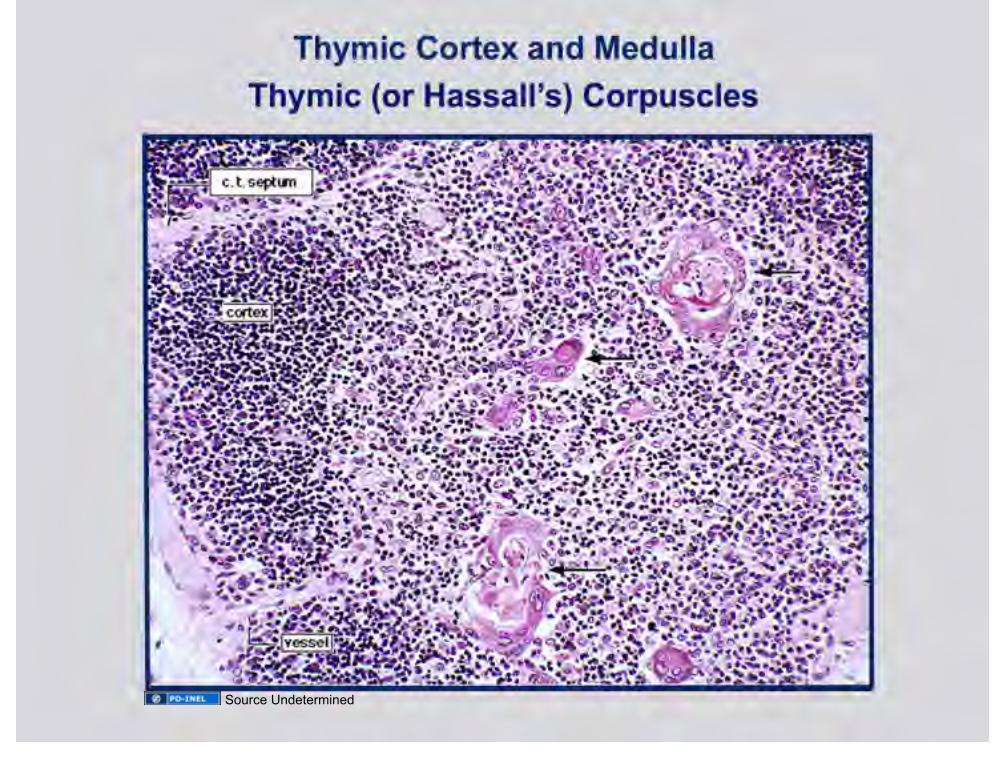
High mag view of medulla
T-cells that survive selection process allowed to cross venule endothelium (INTRAvasation) to enter circulation.
Source Undetermined

Hassall’s corpuscles Type VI ERCs; function not very well known, but produce interleukins (such as IL-4 and IL-7) and so likely influence T-cell differentiation
Source Undetermined

In the medulla, epithelioreticular cells form onionized structures called Hassall’s corpuscles –quite prevalent in older thymus
LM view EM view
Ross and Pawlina, Histology: A Text and Atlas Ross and Pawlina, Histology: A Text and Atlas

Source Undetermined

Slide 6: Ross, Fig. 14.1 Slide 9: Mizobuti histology slide set Slide 10: Source Undetermined Slide 11: Dr. Lloyd Stoolman Slide 12: U-M Histology Collection Slide 13:U-M Histology Collection Slide 14: Ross and Pawlina, Histology: A Text and Atlas; U-M Histology Collection Slide 15: Ross and Pawlina, Histology: A Text and Atlas Slide 16: Source Undetermined Slide 17: Source Undetermined Slide 18: Ross and Pawlina, Histology: A Text and Atlas Slide 19: U-M Histology Collection. Slide 175. Slide 20: Source Undetermined Slide 21: Source Undetermined Slide 22: U-M Histology Collection; Junquiera and Carneiro. Basic Histology. Tenth Ed. 2003 Slide 23: Source Undetermined; Source Undetermined Slide 24: Source Undetermined Slide 25: Ross and Pawlina, Histology: A Text and Atlas Slide 26: Source Undetermined Slide 27: United States Federal Government Slide 28: Ross and Pawlina, Histology: A Text and Atlas Slide 29: Dr. S.K. Kim Slide 30: U-M Histology Collection Slide 31: U-M Histology Collection Slide 32: Ross, Fig. 14.1 Slide 33: United States Federal Government Slide 34: Original Image from http://health-tune-ups.com/wp-content/uploads/2009/04/cdr533339-750.jpg Slide 35: Original Image: http://academic.kellogg.cc.mi.us/herbrandsonc/bio201_McKinley/f24-10a_lymph_node_and__c.jpg; Ross Textbook
of Histology Slide 36: Original Image: http://human.freescience.org/images/Illu_lymph_node_structure.png
Additional Source Information for more information see: http://open.umich.edu/wiki/CitationPolicy

Slide 37: U-M Histology Collection Slide 38: U-M Histology Collection Slide 39: U-M Histology Collection Slide 40: Source Undetermined Slide 41: Ross and Pawlina, Histology: A Text and Atlas; Source Undetermined (Rest of Images) Slide 42: U-M Histology Collection Slide 43: Original Image: Ross, fig. 14.18 Slide 44: U-M Histology Collection Slide 45: U-M Histology Collection Slide 46: Source Undetermined Slide 47: Source undetermined Slide 48: Source Undetermined Slide 49: Source Undetermined Slide 51: Ross, fig. 14.18 Slide 53: Gray’s Anatomy Slide 54: Cancer.gov, Wikipedia, http://commons.wikimedia.org/wiki/File:Illu_spleen.jpg Slide 55: Ross 14.29 Slide 57: Original Image: http://www.mc.vanderbilt.edu/histology/images/histology/lymph/display/lymph20015.jpg Slide 58: Wheater’s, Functional Histology, Fifth Edition, 2006 Slide 59: U-M Histology Collection Slide 60: U-M Histology Collection Slide 61: U-M Histology Collection Slide 62: Ross. Plate 35-3 Slide 63: Ross 14.30a Slide 64: U-M Histology Collection Slide 65: Ross and Pawlina, Histology: A Text and Atlas; Source Undetermined Slide 66: Ross and Pawlina. Plate 36. Figure 1, 2, 3.; Original Image
http://immuneweb.xxmu.edu.cn/Lymphoid%20System.files/UntiHE20.jpeg Slide 67: Source Undetermined Slide 68: U-M Histology Collection Slide 69: Ross Fig. 14.1 Slide 71: Ross and Pawlina, Histology: A Text and Atlas; Gray’s Anatomy Slide 72: Source Undetermined Slide 73: U-M Histology Collection; U-M Histology Collection Slide 74: Original Image from http://www.nature.com/nri/journal/v6/n2/images/nri1781-f4.jpg Slide 77: Source Undetermined Slide 78: Source Undetermined Slide 79: Source Undetermined Slide 80: Source Undetermined Slide 81: Source Undetermined Slide 82: Source Undetermined

Slide 83: Source Undetermined Slide 84: Source Undetermined Slide 84: Ross and Pawlina, Histology: A Text and Atlas; Ross and Pawlina, Histology: A Text and Atlas Slide 85: Source Undetermined